probiotics and berry-associated polyphenols - lucris
TRANSCRIPT
LUND UNIVERSITY
PO Box 117221 00 Lund+46 46-222 00 00
Probiotics and berry-associated polyphenols: catabolism and antioxidative effects
Jakesevic, Maja
2011
Link to publication
Citation for published version (APA):Jakesevic, M. (2011). Probiotics and berry-associated polyphenols: catabolism and antioxidative effects.
Total number of authors:1
General rightsUnless other specific re-use rights are stated the following general rights apply:Copyright and moral rights for the publications made accessible in the public portal are retained by the authorsand/or other copyright owners and it is a condition of accessing publications that users recognise and abide by thelegal requirements associated with these rights. • Users may download and print one copy of any publication from the public portal for the purpose of private studyor research. • You may not further distribute the material or use it for any profit-making activity or commercial gain • You may freely distribute the URL identifying the publication in the public portal
Read more about Creative commons licenses: https://creativecommons.org/licenses/Take down policyIf you believe that this document breaches copyright please contact us providing details, and we will removeaccess to the work immediately and investigate your claim.
Download date: 09. Jan. 2022
1
Probiotics and berry-associated polyphenols:
catabolism and antioxidative effects
Maja Jakešević
2011
Food Hygiene
Division of Applied Nutrition and Food Chemistry
Department of Food Technology, Engineering and Nutrition
Lund Institute of Technology, Lund University
2
Copyright © 2011 Maja Jakešević
Doctoral Thesis
Division of Applied Nutrition and Food Chemistry
Department of Food Technology, Engineering and Nutrition
Lund University
P.O. Box 124
SE-221 00 Lund
Sweden
Cover: bilberry, chokeberry, rosehips, Lactobacillus and chemical structure of
anthocyanin
ISBN 978-91-7473-091-3
Printed by Media-Tryck, Lund University
Lund, Sweden
3
Abstract
Oxidative stress can cause damage to DNA, proteins and lipids and is associated
with inflammation and various human diseases as cancer, atherosclerosis, and
autoimmune diseases. Polyphenol-rich diet, such as fruits and berries, may act as
antioxidants and prevent oxidative stress and, thereby, associated diseases.
Administration of lactic acid bacteria (LAB) can affect the microflora in the
gastrointestinal (GI) tract and may increase the capability of the bacterial flora to
digest polyphenols. Some strains of Lactobacillus may break down phenolic acids
and hydrolyzable tannins into phenolic metabolites that are more easily absorbed
in the body and may enhance antioxidative effects. The aim of this thesis was to
clarify the protective effects of polyphenol-rich fruits and berries alone or in
combination with different strains of LAB on oxidative stress in mice.
Furthermore, transformation of polyphenols in a bilberry beverage by LAB was
examined.
Supplementation with rosehips of the rose species Rosa pimpinellifolia or an LAB
mixture decreased lipid peroxidation and oxidative stress in colon of mice after
ischemia-reperfusion (I/R) injury. Adding an LAB supplement to the rosehips
increased the concentrations of phenolic compounds, antioxidative capacity and
total phenolic content in cecum. Rosehips of R. piminellifolia are a rich source of
cyanidin-3-O-glucoside and this compound and its degradation product,
protocatechuic acid, were detected in the cecum content.
Administration of bilberry, either alone or together with Lactobacillus plantarum
HEAL19, decreased lipid peroxidation and oxidative stress in colon of mice after
I/R injury. A chokeberry-supplement showed no antioxidative effect. Bilberry was
found to have a more complex anthocyanin profile than chokeberry. Higher
concentrations and a more varied composition of anthocyanins were seen in colon
than in cecum. More phenolic metabolites were found in the intestines of bilberry-
fed mice than in the chokeberry-fed ones. Chokeberry or bilberry alone decreased
the number of LAB on the colonic mucosa but addition of L. plantarum HEAL19
prevented this reduction.
In a more extensive ischemia-reperfusion injury, diet supplemented with bilberry,
but without addition of different LAB strains, reduced lipid peroxidation and
protected the small intestine against oxidative stress. The highest concentration
and recovery of anthocyanins was seen in the ileal content followed by that of
4
colon and finally cecum. Anthocyanin arabinosides, and especially malvidin-3-O-
arabinoside, were accumulated in the colon content. Glucosides and galactosides
of malvidin, peonidin and petunidin seemed to be digested by the microflora in the
cecum. Supplementation of bilberry to the diet influenced the composition of
cecum microflora.
Anthocyanins in bilberry beverages inoculated with different LAB strains, alone or
in combination with wine yeast, decreased during 3 weeks incubation at 30C.
Arabinosides of malvidin and petunidin showed the greatest decrease. Addition of
yeast improved the stability of the anthocyanins. In contrast to anthocyanins,
quercetin, quercetin-3-glucoside and detected phenolic acids were relatively
stable. Antioxidative capacity and total phenolic content decreased in all samples.
In conclusion, dietary supplementation of rosehips from Rosa pimpinellifolia or
bilberry suppressed oxidative stress in colonic tissue of mice. Protective effects
may be due to the high anthocyanin content, presence of phenolic metabolites and
changed microflora. Addition of LAB improved status of the colonic but not the
ileal tissue.
5
List of Papers
This thesis is based on the following papers, referred to in the text by their Roman numerals.
Paper I Jakesevic, M., Håkansson, Å., Adawi D., Jeppsson, B., Rumpunen, K., Ekholm, A., Ahrné, S. and Molin, G. (2009). Antioxidative protection of dietary rosehips and polyphenol active lactobacilli in mice subjected to intestinal oxidative stress by ischemia-reperfusion. Microbial Ecology in Health and Disease 21:193–202.
Paper II Jakesevic, M., Aaby, K., Borge, G-I., Jeppsson, B., Ahrné, S. and Molin, G. (2011). Antioxidative protection of dietary bilberry, chokeberry and Lactobacillus plantarum HEAL19 in mice subjected to intestinal oxidative stress by ischemia-reperfusion. BMC Complementary and Alternative Medicine, 11: (8) http://www.biomedcentral.com/1472-6882/11/8
Paper III Jakesevic, M., Aaby, K., Borge, G-I., Rumpunen, K., Ahrné, S. and Molin, G. (2010). Phenolics in bilberry (Vaccinium myrtillus) beverage incubated with lactic acid bacteria and yeast (in manuscript).
Paper IV Jakesevic, M., Xu, J., Aaby, K., Jeppsson, B., Ahrné, S. and Molin, G. (2010) Effects of bilberry (Vaccinium myrtillus) in combination with lactic acid bacteria on intestinal oxidative stress induced by ischemia-reperfusion in mouse (in manuscript).
6
The author´s contribution to the papers
Paper I The author, Maja Jakešević (MJ), coordinated and performed the
experimental work together with Åsa Håkansson and Diya Adawi.
MJ performed the analysis regarding malondialdehyde, analysis of
intestinal microflora by randomly amplified polymorphic DNA and
viable count, analyses of total phenolic content and antioxidative
capacity. MJ performed the analysis of polyphenols together with
Anders Ekholm. MJ evaluated the results and wrote the paper.
Paper II The author, Maja Jakešević, coordinated and performed the
experimental work together with Åsa Håkansson, and performed
the analysis of malondialdehyde and viable count. Analyses of
anthocyanins and phenolic metabolites were performed by MJ in
collaboration with Kjersti Aaby. MJ evaluated the results and wrote
the paper.
Paper III The author, Maja Jakešević, coordinated and performed the
experimental work, performed the analyses of pH and alcohol
content, analyses regarding total phenolic content and antioxidative
capacity. MJ performed analysis of polyphenols in collaboration
with Kjersti Aaby and Grethe Iren Borge. MJ evaluated the results
and wrote the manuscript.
Paper IV The author, Maja Jakešević, coordinated and performed the
experimental work together with Siv Ahrné, performed the analysis
regarding malondialdehyde, myeloperoxidase, and histology
samples. Analysis of anthocyanins was performed by MJ together
with Kjersti Aaby. MJ evaluated the results and wrote the
manuscript.
7
Abbreviations
I/R Ischemia-reperfusion
ROS Reactive Oxygen Species
RNS Reactive Nitrogen Species
MDA Malondialdehyde
MPO Myeloperoxidase
RAPD Randomly Amplified Polymorphic DNA
T-RFLP Terminal Restriction Fragment Length Polymorphism
H&E Hematoxylin and Eosin
FRAP Ferric Reducing Activity Power
TP Total phenolic content
PCR Polymerase Chain Reaction
CFU Colony-forming units
DAD Diode array absorbance detector
ESI Electrospray ionization
HPLC High-Performance Liquid-Chromatography
MS Mass Spectroscopy
UV-vis Ultraviolet-visible light
GI Gastrointestinal
SOD Superoxide dismutase
GPx Glutathione peroxidase
GRx Glutathione reductase
GSH Glutathione
8
XDH Xanthine dehydrogenase
XO Xanthine oxidase
SMA Superior Mesenteric Artery
SCFA Short-chain fatty acids
LAB Lactic acid bacteria
TBARS Thiobarbituric acid-reacting substances
TFA Trifluoroacetic acids
ICAM-1 Intercellular adhesion molecule
VCAM-1 Vascular cellular adhesion molecule
PCA Principal component analysis
9
Contents
Abstract ............................................................................................. 3
List of papers ............................................................................................... 5
The author´s contribution to the papers ........................................................ 6
Abbreviations .................................................................................... 7
Contents ........................................................................................... 9
Introduction ..................................................................................... 11
Background ..................................................................................... 13
Oxidation and antioxidants ......................................................................... 13
Intestinal ischemia-reperfusion ................................................................... 15
Polyphenols ............................................................................................... 17
Flavonoids and phenolic acids ......................................................... 19
Absorption and metabolism.............................................................. 22
Berries ....................................................................................................... 24
Chokeberry ...................................................................................... 24
Bilberry ............................................................................................ 25
Rosehip ........................................................................................... 26
Intestinal microflora .................................................................................... 27
Development of microflora ............................................................... 28
Functions of the microflora ............................................................... 29
Probiotics ........................................................................................ 30
Aims ................................................................................................ 33
Methodology ................................................................................... 34
Experimental design................................................................................... 34
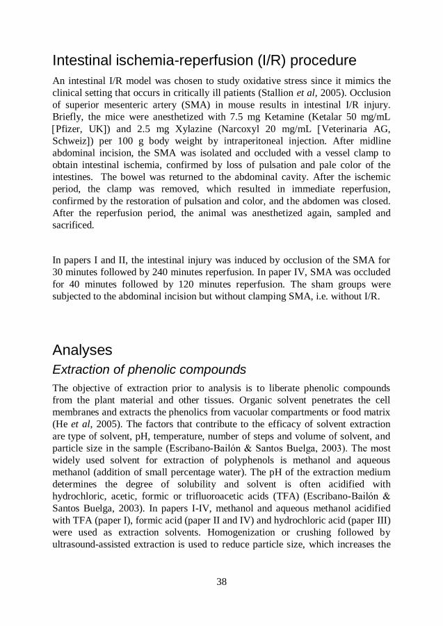
Intestinal ischemia-reperfusion (I/R) procedure........................................... 38
10
Analyses .................................................................................................... 38
Extraction of phenolic comppounds .................................................. 38
Analysis of phenolic compounds ...................................................... 39
Ferric reducing activity power (FRAP) .............................................. 40
Total phenolic content (TP) .............................................................. 40
Malondialdehyde (MDA) .................................................................. 41
Myeloperoxidase (MPO) .................................................................. 41
Histopathology ................................................................................. 41
Randomly amplified polymorphic DNA (RAPD) ................................ 42
Terminal restriction fragment length polymorphism (T-RFLP) ........... 42
Statistics .......................................................................................... 42
Results and discussion ................................................................... 44
Polyphenols ............................................................................................... 44
Anthocyanins ................................................................................... 44
Phenolic compounds other than anthocyanins ................................. 49
Antioxidative capacity and total phenolic content .............................. 52
Inflammatory markers ................................................................................ 53
Ischemia-reperfusion ....................................................................... 53
Malondialdehyde (MDA) .................................................................. 54
Myeloperoxidase (MPO) .................................................................. 60
Histopathology ................................................................................. 61
Bacteriology ............................................................................................... 61
Lactobacilli and Enterobacteriaceae ................................................. 61
The composition of the bacterial microflora ...................................... 62
Conclusions .................................................................................... 64
Populärvetenskaplig sammanfattning ............................................. 66
Acknowledgements ......................................................................... 69
References ..................................................................................... 71
11
Introduction
Oxidative stress occurs when oxygen free radicals are overproduced in the cells. It
can cause damage to DNA, proteins and lipids and is associated with inflammation
and various human diseases such as cancer, type-2 diabetes and cardiovascular
diseases. Furthermore, oxidative stress and inflammation increase intestinal
permeability, allowing translocation of pathogenic bacteria and endotoxins to
extraintestinal sites which may lead to systemic inflammation, sepsis and
eventually multiple-organ failure. Antioxidant therapy has been suggested to
prevent and attenuate oxidative stress and thereby associated diseases.
Polyphenols are secondary metabolites in plants. They are proposed to act as
antioxidative and anti-inflammatory agents. Fruits and berries are important
dietary sources of phenolic compounds. Antimicrobial properties of polyphenols
may influence the composition and function of the indigenous intestinal microflora
by inhibiting certain bacterial groups (those sensitive to the antimicrobial effect of
polyphenols) and promote others (those more resistant to poyphenols). Hopefully
the sensitive ones are also those with negative effects on the health and the
resistant ones are bacterial groups with health-promoting effects. The bacterial
flora of the gut can influence the nutritional status and the health of the host via
modulation of metabolic functions. Polyphenols that are not absorbed in the small
intestine can be degraded by colonic microflora to simpler and more easily
absorbable bioactive compounds with potential physiological effects.
Dietary supplements probiotics (microorganisms with health-promoting properties
after ingestion) may, at least theoretically, change the intestinal microflora and
increase the number of polyphenol-degrading groups in the intestines, at least if a
probiotic bacterium capable of degrading polyphenols has been chosen.
Furthermore, probiotics may mitigate the inflammation by promoting the normalization of intestinal microflora and exclusion of pathogens, decreasing intestinal permeability, improving the intestine´s immunological barrier functions and alleviating the intestinal inflammatory response.
A polyphenol-rich diet and supplementation with probiotics may improve the
intestinal status and possibly attenuate oxidative stress and suppress inflammation.
The work described in this thesis is devoted to the study of antioxidative and anti-
inflammatory effects of the diet when supplemented with polyphenol-rich fruits
and berries and potential probiotic strains in an oxidative stress model in mice. All
12
bacterial supplements involved lactic acid bacteria, mostly Lactobacillus strains
with tannase activity. The influence of potential probiotic strains on transformation
of polyphenols is also investigated.
13
Background
Oxidation and antioxidants
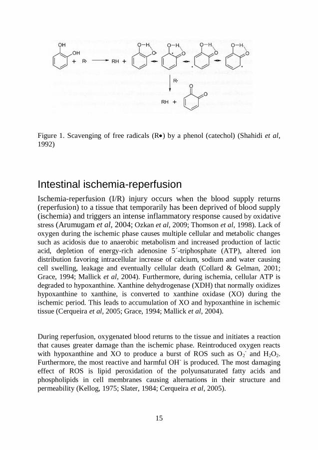
A free radical is any atom or molecule that contains one or more unpaired
electrons, which makes it highly reactive with other atoms and molecules
(Halliwell, 1994a). Oxidation is a free radical chain reaction comprising initiation,
propagation and termination. Reactive oxygen species (ROS) include not only
oxygen-centered free radicals such as superoxide (O2-), peroxyl (ROO
-), alkoxyl
(RO-) and hydroxyl (OH
-) but also some nonradical derivates, e.g. hydrogen
peroxide (H2O2) and hypochlorous acid (HOCl). Nitric oxide (NO-) is one of the
more important free radicals known as reactive nitrogen species (RNS) (Pietta,
2000; Shahidi et al, 2010; Halliwell, 1994b). The chemical reactivity of free
radicals varies greatly, with OH- being the most reactive one.
In biological systems, ROS are involved in energy production, synthesis of
biologically important compounds, regulation of cell growth and intracellular
signaling (Pietta, 2000). They are also an important part of immune defense since
phagocytes (neutrophils, monocytes, macrophages, eosinophils) produce large
amounts of O2- and H2O2 to kill bacteria and fungi and to inactivate virus
(Halliwell, 1997; Pietta, 2000). Some physiopathological situations such as
cigarette smoke, air pollutants, UV radiation, high polyunsaturated fatty acid diet,
drugs, inflammation and ischemia-reperfusion lead to overproduction of ROS,
known as oxidative stress (Halliwell, 1997; Pietta, 2000). Oxidative stress can
cause damage to DNA, proteins in tissues and lipids in cell membranes and is
associated with different human diseases such as cancer, atherosclerosis, chronic
inflammation, neurodegenerative diseases (Parkinson´s Disease and Alzheimer´s
Disease), rheumatoid arthritis and tissue injury (Halliwell, 1997; Halliwell, 1994b;
Pietta, 2000).
As a protection against ROS, the human body has evolved an antioxidant system
in the form of endogenous antioxidative enzymes, e.g. superoxide dismutase
(SOD), catalase, glutathione peroxidase (GPx) and glutathione reductase (GRx)
and nonenzymatic antioxidant defenses, e.g. glutathione (GSH), -tocopherol,
14
ascorbic acid, iron-binding proteins transferrin and ferritin, histidine-peptides,
urate and plasma-protein thiols (Halliwell, 1994a; Pietta, 2000; Shahidi et al,
2010). SOD enzymes convert O2- to H2O2. Generated H2O2 is then removed by
GPx enzymes that contain selenium at their active site. Superoxide and hydrogen
peroxide are not so reactive chemically but if brought into contact with iron and/or
copper ions they generate the highly reactive and harmful OH- that attack and
damage almost all molecules in living cells (Halliwell, 1994a; Halliwell, 1997;
Pietta, 2000). The human body does not synthesize an overwhelming excess in its
antioxidant defense system but seems to aim at a balance between production of
ROS and levels of antioxidant protection.
Since the body´s endogenous antioxidant defenses are not always sufficient to
prevent oxidative stress, dietary antioxidants can be needed for diminishing the
cumulative effects of oxidative damage. Antioxidants are substances that, when
present at low concentrations compared to that of an oxidizable substrate,
markedly delay or prevent its oxidation (Shahidi et al, 2010). In addition to
providing vitamins C, E, A and carotenoids, plant polyphenols are important
antioxidants derived from the diet (Pulido R, 2000; Pietta P-G, 2000). Different
mechanisms by which antioxidants may exert their inhibitory effects against
oxidation include free radical scavenging, chelation of metal ions, inactivation of
peroxides and other ROS and inhibition of pro-oxidative enzymes (Pulido et al,
2000; Shahidi et al, 2010). Based on their mode of action, antioxidants are broadly
divided into primary and secondary antioxidants. Most polyphenols are primary
antioxidants, so they break the chain reaction of oxidation and neutralize free
radicals by donating a hydrogen atom. The resulting antioxidant radicals are
stabilized by delocalization of the unpaired electron around the phenol ring to
form stable resonance hybrids. These radicals have low reactivity and generally do
not initiate creation of new radicals (Shahidi et al, 2010).
15
Figure 1. Scavenging of free radicals (R) by a phenol (catechol) (Shahidi et al,
1992)
Intestinal ischemia-reperfusion
Ischemia-reperfusion (I/R) injury occurs when the blood supply returns (reperfusion) to a tissue that temporarily has been deprived of blood supply (ischemia) and triggers an intense inflammatory response caused by oxidative
stress (Arumugam et al, 2004; Ozkan et al, 2009; Thomson et al, 1998). Lack of
oxygen during the ischemic phase causes multiple cellular and metabolic changes
such as acidosis due to anaerobic metabolism and increased production of lactic
acid, depletion of energy-rich adenosine 5´-triphosphate (ATP), altered ion
distribution favoring intracellular increase of calcium, sodium and water causing
cell swelling, leakage and eventually cellular death (Collard & Gelman, 2001;
Grace, 1994; Mallick et al, 2004). Furthermore, during ischemia, cellular ATP is
degraded to hypoxanthine. Xanthine dehydrogenase (XDH) that normally oxidizes
hypoxanthine to xanthine, is converted to xanthine oxidase (XO) during the
ischemic period. This leads to accumulation of XO and hypoxanthine in ischemic
tissue (Cerqueira et al, 2005; Grace, 1994; Mallick et al, 2004).
During reperfusion, oxygenated blood returns to the tissue and initiates a reaction
that causes greater damage than the ischemic phase. Reintroduced oxygen reacts
with hypoxanthine and XO to produce a burst of ROS such as O2- and H2O2.
Furthermore, the most reactive and harmful OH- is produced. The most damaging
effect of ROS is lipid peroxidation of the polyunsaturated fatty acids and
phospholipids in cell membranes causing alternations in their structure and
permeability (Kellog, 1975; Slater, 1984; Cerqueira et al, 2005).
16
Besides participating in lipid peroxidation, the ROS are involved in attraction and
activation of leukocytes at the sites of injury (Santen, 2008; Riaz, 2002; Cerqueira
et al, 2005). Leukocyte recruitment is a multiple-step process that includes
bordering the vessel wall, rolling along endothelium, firm adherence and finally
transmigration through endothelium (Kubes & Kerfoot 2001; Mölne & Wold,
2007). Adhesion molecules P- and E-selectins, expressed on the endothelial
surface, are up-regulated and weakly interact with L-selectin expressed by
leukocytes resulting in rolling of leukocytes along the endothelium (Mölne &
Wold, 2007; Granger, 1988). As the rolling step proceeds, the expression and
binding activity of 2-integrins (LFA-1 and MAC-1) on leukocytes is increased,
resulting in firm adhesion to ICAM-1 (intercellular adhesion molecule) and
VCAM-1 (vascular cellular adhesion molecule) expressed on endothelial cells.
The firm adhesion is followed by leukocyte migration out of the circulation to the
site of injury (Mölne & Wold, 2007; Panes et al, 1999). After migration through
endothelium, the active leukocytes release toxic ROS, proteases and elastases
causing local damage and destruction (Panes et al, 1999).
I/R is encountered in strangulated bowel, hemorrhagic shock and organ
transplantation, and is associated with high morbidity and mortality in surgical and
trauma patients (Mallick et al, 2004; Collard & Gelman, 2001). Among the
internal organs, intestinal mucosa is highly sensitive to ischemia-reperfusion
injury (Mallick et al, 2004; Ozer et al, 2005; Ozkan et al, 2009). Colonic mucosa
is less sensitive to I/R than that of the small intestine, but once damaged it
recovers more slowly (Stallion et al, 2005; Robinson et al, 1981; Leung et al,
1992). The superior mesenteric artery (SMA) has been shown to maintain
intestinal perfusion and mucosal integrity in rodents (Leung et al, 1992).
Occlusion of SMA results in mucosal damage of small intestine, cecum and colon.
Loss of mucosal barrier-function due to the I/R leads to increased translocation of
enteric bacteria and local production of cytokines. If sufficiently severe, intestinal
I/R injury can lead to multiple organ failure and death (Leung et al, 1992; Stallion
et al, 2005; Riaz, 2002). The clinical setting that occurs in critically ill patients has
been mimicked by using a murine model, in which occlusion of SMA results in
intestinal I/R injury.
17
Polyphenols
Polyphenols are produced as secondary metabolites of plants, and are involved in
plant growth and reproduction and provide protection against ultraviolet radiation,
oxidative stress and pathogens (Ross & Kasum, 2002). Phenolic compounds are
regular constituents of human diet and are found in fruits, beverages such as tea,
coffee, wine and fruit juices, chocolate and, to lesser extent, in vegetables, cereals
and legume seeds (Scalbert et al, 2002). The astringency and bitterness of foods
and beverages depend on the content of polyphenolic compounds which are
partially responsible for the sensory and nutritional qualities of plant foods (Bravo,
1998). Polyphenols possess antioxidative, metal-chelating and free radical-
scavenging abilities, and they may prevent various diseases associated with
oxidative stress, such as cardiovascular diseases, cancer, chronic inflammation,
atherosclerosis and type-2 diabetes (Ross & Kasum, 2002; Scalbert et al, 2000).
Chemically, phenolics can be defined as molecules possessing an aromatic ring
bearing one or more hydroxyl groups. More than 8,000 phenolic structures are
currently known (Ross & Kasum, 2002; Bravo, 1998). According to the nature of
their carbon skeleton polyphenols can be classified into four major groups:
phenolic acids, flavonoids, stilbenes and lignans (Scalbert et al, 2000).
18
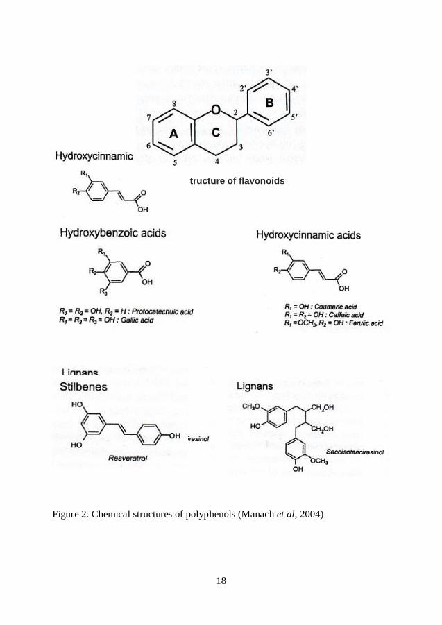
Basic structure of flavonoids
Figure 2. Chemical structures of polyphenols (Manach et al, 2004)
19
Flavonoids and phenolic acids
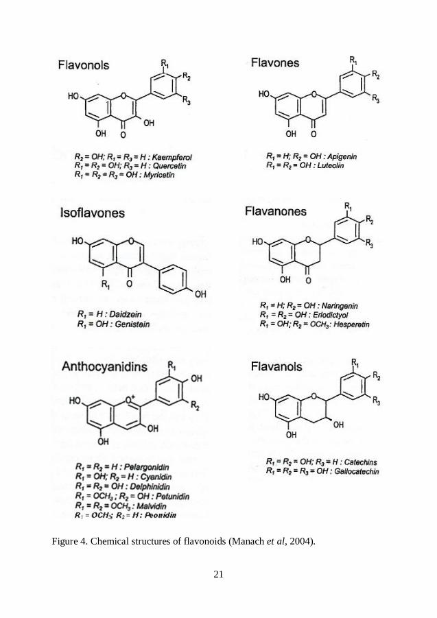
Flavonoids are the most abundant polyphenols in the diet. All flavonoid phenolics
share a basic skeleton consisting of two aromatic rings (A and B) linked through
three carbons that form an oxygenated heterocycle (C ring) (Bravo, 1998; Manach
et al, 2004). According to the oxidation degree of the C ring, flavonoids can be
divided into several classes: flavones, flavonols, isoflavones, anthocyanins,
flavanols, flavanones and proanthocyanidins (Scalbert et al, 2000). Flavonoids
occasionally occur in plants as aglycones, but are most commonly found attached
to sugars (glycosides) (Ross & Kasum, 2002; Bravo, 1998).
Flavonols are the most ubiquitous flavonoids in foods with quercetin and
kaempferol as the main representatives (Manach et al, 2004). These compounds
are present in glycosylated forms. Glucose and rhamnose are the most common
sugar residues, although other sugars such as galactose, xylose and arabinose are
also found (Bravo, 1998). Blueberries are rich source of flavonols.
Catechin and epicatechin are the main flavanols in fruits. In contrast to the other
flavonoid groups, flavanols are not glycosylated in food (Manach et al, 2004).
Proanthocyanidins, also known as condensed tannins, are found in large amounts
in fruits, berries, nuts, cocoa and wine (Rasmussen et al, 2005). In
proanthocyanidins, flavan-3-ol (catechin or epicatechin) units are linked mainly
through a C4C8 bond, but also through a C4C6 bond, to form dimers,
oligomers and high-molecular-weight polymers. These linkages are both called B-
type linkages. An additional ether bond between C2C7, resulting in double
linked catechin units is called A-type linkage (Gu et al, 2004). B-type
proanthocyanidins are the most common, but A-type linkages were also found in
some foods (Rasmussen et al, 2005). Proanthocyanidins consisting exclusively of
epicatechin units are called procyanidins (Gu et al, 2004). Through the formation
of the complexes with salivary proteins, proanthocyanidins are responsible for the
astringent character of some fruits and beverages. When fruit becomes ripe, this
astringency often disappears (Santos-Buelga et al, 2000).
As a major sub-group of flavonoids, anthocyanins are water-soluble plant
pigments responsible for pink, red, blue and purple colors of plants. Most
anthocyanins occur as glycosides of their respective anthocyanidin (aglycone),
with the sugar moiety mainly bound to the 3-position on C-ring or the 5, 7-
position on the A-ring (Prior & Wu, 2006). The most common sugars are glucose,
20
galactose, arabinose, rhamnose and xylose while most common aglycones are
pelargonidin, cyanidin, delphinidin, peonidin, petunidin and malvidin (Bravo,
1998; Prior & Wu, 2006). In addition to glycosylation, esterification with organic
acids and phenolic acids also occurs. Anthocyanins are reactive compounds and
readily degrade in the presence of oxygen, various enzymes, light and at high
temperatures (Kalt et al, 2000). Variations in pH also affect the anthocyanin
stability. The basic anthocyanidin structure, red flavylium cation, is the
predominant molecular form at low pH (<2). Increase in pH leads to the loss of a
proton and generates the blue quinonoidal structure. At the same time, a much
slower hydration of the flavylium cation occurs, yielding small portions of the
colorless chalcone forms (McGhie et al, 2007; Rivas-Gonzalo, 2003).
Phenolic acids can be classified as hydroxybenzoic acids and hydroxycinammic
acids. They are present in fruits, vegetables, beverages and cereals. The
hydroxycinammic acids are more commonly found in foods than hydroxybenzoic
acids and consist mainly of p-coumaric, caffeic, ferulic and sinapic acids.
Hydroxycinnamic acids occur in foods as simple esters with quinic acid or
glucose. Chlorogenic acid, the most abundant hydroxycinnamic acid, is composed
of caffeic and quinic acids. Hydroxybenzoic acids are generally present in foods in
the form of glucosides and are often the components of complex structures like
lignins and hudrolyzable tannins. Gallic acid, protocatechuic acid, vanillic acid
and syringic acid are the most common hydroxybenzoic acids (Manach et al,
2004; Mattila et al, 2006). Hydroxycinnamic acids are more effective antioxidants
than hydroxybenzoic acids. The presence of the –CH=CH-COOH group in
hydroxycinammic acids ensures greater H-donating ability and radical
stabilization by chemical resonance than the –COOH group in hydroxybenzoic
acids (Rice-Evans et al, 1996).
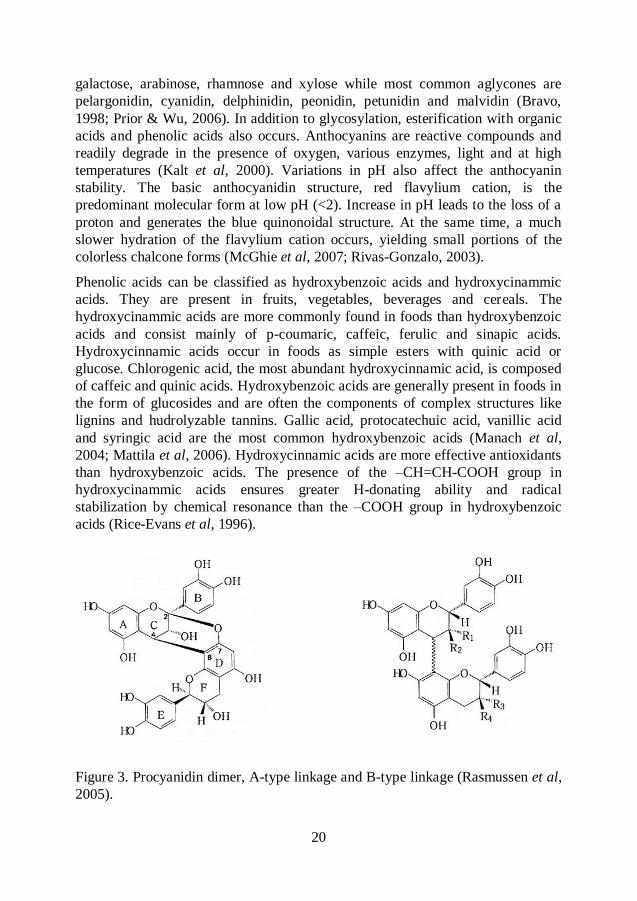
Figure 3. Procyanidin dimer, A-type linkage and B-type linkage (Rasmussen et al,
2005).
21
Figure 4. Chemical structures of flavonoids (Manach et al, 2004).
22
Absorption and metabolism
The absorption and metabolism of polyphenols are determined by their chemical
structure, which includes the degree of glycosylation, acylation and
polymerization, their basic structure, conjugation with other phenolics, molecular
size and solubility. Aglycones and phenolic acids can be directly absorbed through
the small intestinal mucosa (Lafay et al, 2006). Most polyphenols exist in food in
the form of esters, glycosides or polymers that cannot be absorbed in their native
form. These compounds probably resist acid hydrolysis in the stomach and arrive
intact in the small intestine where they are hydrolyzed by intestinal enzymes to
their aglycones. Free aglycones are conjugated by methylation, sulfation or
glucuronidation, first in the small intestine and later in the liver (Scalbert et al,
2000). This is a metabolic detoxication process that facilitates biliary and urinary
elimination of xenobiotics by increasing their hydrophilicity (Manach et al, 2004).
Once they are absorbed through the gut barrier, polyphenols are able to penetrate
tissues where they are metabolized. Polyphenols that are not absorbed in the small
intestine and re-excreted in the bile reach the colon where they are metabolized by
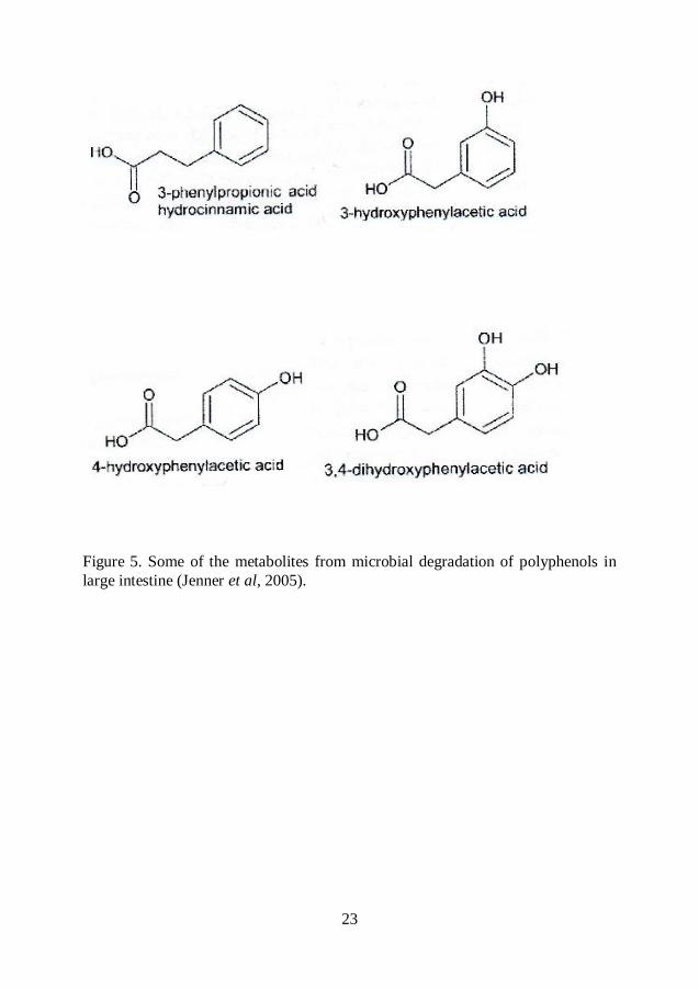
the microflora (Scalbert et al, 2002). Glycosides are hydrolyzed by the microflora
and the resulting aglycones are further metabolized into various low molecular
aromatic acids such as phenylvaleric, phenylpropionic, phenylacetic and benzoic
acids that can be absorbed (Rechner et al, 2004; Lee et al, 2006; Gonthier et al,
2003a).
Proanthocyanidins differ from other polyphenols because of their polymeric nature
and high molecular weight which limit their absorption. Only dimers and trimers
are absorbed in the small intestine and the large non-absorbed fraction of polymers
is degraded by colonic microflora to phenylvaleric, phenylpropionic, phenylacetic
and benzoic acids (Déprez et al, 2000; Gonthier et al, 2003b; Rios et al, 2003).
The absorption of anthocyanins also differs from the other flavonoids because they
are mainly absorbed as intact glycosides from the upper part of the small intestine
but also from the stomach (McGhie et al, 2007; Matuschek et al, 2006; Passamonti
et al, 2003). Since absorption of anthocyanins is very low most of the
anthocyanins are transformed to phenolic acids by microbial populations in the
colon (Fleschhut et al, 2005; Aura et al, 2005; Keppler & Humpf, 2005).
23
Figure 5. Some of the metabolites from microbial degradation of polyphenols in
large intestine (Jenner et al, 2005).
24
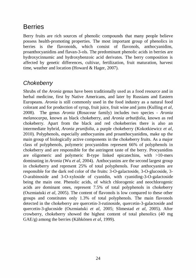
Berries
Berry fruits are rich sources of phenolic compounds that many people believe
possess health-promoting properties. The most important group of phenolics in
berries is the flavonoids, which consist of flavonols, anthocyanidins,
proanthocyanidins and flavan-3-ols. The predominant phenolic acids in berries are
hydroxycinnamic and hydroxybenzoic acid derivates. The berry composition is
affected by genetic differences, cultivar, fertilization, fruit maturation, harvest
time, weather and location (Howard & Hager, 2007).
Chokeberry
Shrubs of the Aronia genus have been traditionally used as a food resource and in
herbal medicine, first by Native Americans, and later by Russians and Eastern
Europeans. Aronia is still commonly used in the food industry as a natural food
colorant and for production of syrup, fruit juice, fruit wine and jams (Kulling et al,
2008). The genus Aronia (Rosaceae family) includes two species - Aronia
melanocarpa, known as black chokeberry, and Aronia arbutifolia, known as red
chokeberry. Apart from the black and red chokeberries there is also an
intermediate hybrid, Aronia prunifolia, a purple chokeberry (Kokotkiewicz et al,
2010). Polyphenols, especially anthocyanins and proanthocyanidins, make up the
main group of biologically active components in the chokeberry fruits. As a major
class of polyphenols, polymeric procyanidins represent 66% of polyphenols in
chokeberry and are responsible for the astringent taste of the berry. Procyanidins
are oligomeric and polymeric B-type linked epicatechins, with >10-mers
dominating in Aronia (Wu et al, 2004). Anthocyanins are the second largest group
in chokeberry and represent 25% of total polyphenols. Four anthocyanins are
responsible for the dark red color of the fruits: 3-O-galactoside, 3-O-glucoside, 3-
O-arabinoside and 3-O-xyloside of cyanidin, with cyaniding-3-O-galactoside
being the main one. Phenolic acids, of which chlorogenic and neochlorogenic
acids are dominant ones, represent 7.5% of total polyphenols in chokeberry
(Oszmiański et al, 2005). The content of flavonols is low compared to these other
groups and constitutes only 1.3% of total polyphenols. The main flavonols
detected in the chokeberry are quercetin-3-rutinoside, quercetin-3-galactoside and
quercetin-3-glucoside (Oszmiański et al, 2005; Slimestad et al, 2005). After
crowberry, chokeberry showed the highest content of total phenolics (40 mg
GAE/g) among the berries (Kähkönen et al, 1999).
25
Aronia has been claimed to alleviate oxidative stress, exhibits anti-inflammatory,
antimicrobial and antiviral activities due to the high content of polyphenols, and
has antioxidative properties. It has been reported that chokeberry juice and
chokeberry extract reduced lipid peroxidation (measured by MDA or TBARS),
decreased tissue damage and improved antioxidative status in different organs
exposed to oxidative stress induced by chemical treatment such as carbon
tetrachloride, indomethacin and N-nitrosodiethylamine (Valcheva-Kuzmanova et
al, 2004; Valcheva-Kuzmanova et al, 2005; Kujawska et al, 2010), high-fructose
diet in combination with low-dose streptozotocin injection (Jurgoński et al, 2008)
or exercise (Pilaczynska-Szczesniak et al, 2005). Administration of chokeberry
extract showed a dose-dependent anti-inflammatory effect on endotoxin-induced
uveitis in rats (Ohgami et al, 2005). Aronia juice exhibited a bacteriostatic activity
in vitro against Staphylococcus aureus and E. coli and an antiviral activity against
type A influenza virus (Valcheva-Kuzmanova et al, 2006).
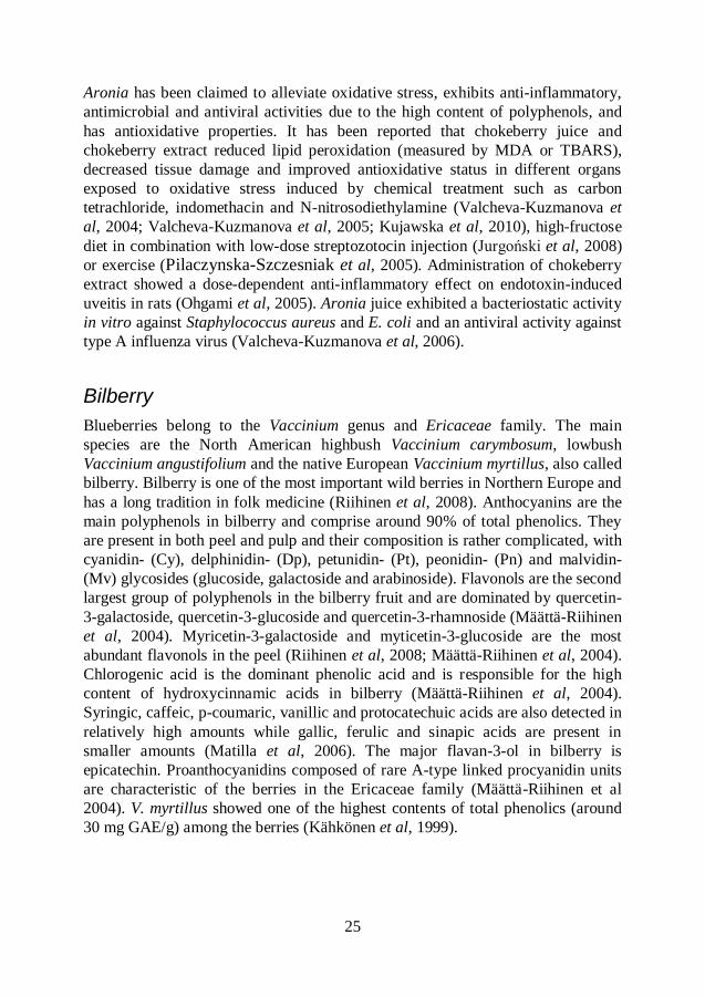
Bilberry
Blueberries belong to the Vaccinium genus and Ericaceae family. The main
species are the North American highbush Vaccinium carymbosum, lowbush
Vaccinium angustifolium and the native European Vaccinium myrtillus, also called
bilberry. Bilberry is one of the most important wild berries in Northern Europe and
has a long tradition in folk medicine (Riihinen et al, 2008). Anthocyanins are the
main polyphenols in bilberry and comprise around 90% of total phenolics. They
are present in both peel and pulp and their composition is rather complicated, with
cyanidin- (Cy), delphinidin- (Dp), petunidin- (Pt), peonidin- (Pn) and malvidin-
(Mv) glycosides (glucoside, galactoside and arabinoside). Flavonols are the second
largest group of polyphenols in the bilberry fruit and are dominated by quercetin-
3-galactoside, quercetin-3-glucoside and quercetin-3-rhamnoside (Määttä-Riihinen
et al, 2004). Myricetin-3-galactoside and myticetin-3-glucoside are the most
abundant flavonols in the peel (Riihinen et al, 2008; Määttä-Riihinen et al, 2004).
Chlorogenic acid is the dominant phenolic acid and is responsible for the high
content of hydroxycinnamic acids in bilberry (Määttä-Riihinen et al, 2004).
Syringic, caffeic, p-coumaric, vanillic and protocatechuic acids are also detected in
relatively high amounts while gallic, ferulic and sinapic acids are present in
smaller amounts (Matilla et al, 2006). The major flavan-3-ol in bilberry is
epicatechin. Proanthocyanidins composed of rare A-type linked procyanidin units
are characteristic of the berries in the Ericaceae family (Määttä-Riihinen et al
2004). V. myrtillus showed one of the highest contents of total phenolics (around
30 mg GAE/g) among the berries (Kähkönen et al, 1999).
26
High polyphenol content has been responsible for antioxidative, anti-inflammatory
and antimicrobial activities of bilberries. V. myrtillus has been used in different
studies to prevent oxidative stress injury in liver, kidney, skin and eyes (Bao et al,
2008a; Bao et al, 2008b; Svobodová et al, 2008; Yao et al, 2010). Bilberry
prevented oxidative stress by increasing the levels of endogenous antioxidative
enzymes (SOD, GPx), by improving the antioxidative status, and by decreasing
lipid peroxidation (measured by MDA) and ROS production. Oral administration
of V. myrtillus decreased the number of adhering leucocytes to venular vessels,
preserved the capillary perfusion and reduced microvascular permeability during
ischemia-reperfusion in hamster cheek pouch microcirculation (Bertuglia et al,
1995).
Supplementation with bilberry was suggested to modulate the inflammation
process and prevent atherosclerosis and cardiovascular disease by decreasing the
plasmatic total cholesterol, reducing the release of pro-inflammatory mediators in
the liver and through reduction of several NF-B regulated inflammatory
mediators in plasma (Mauray et al, 2010; Karlsen et al, 2010). Salmonella enterica
sv. Typhimurium, Salmonella enterica sv. Infantis and Staphylococcus aureus
were inhibited by bilberry extract (Puupponen-Pimiä et al, 2005a). Bacteriostatic
effects due to anti-adhesion have been observed in Vaccinium berries.
Procyanidins with A-type linkages in Vaccinium berries have been associated with
inhibition of Escherichia coli and Helicobacter pylori adhesion in uroepithelium
and gastric epithelial cells, respectively (Puupponen-Pimiä et al, 2005b).
Rosehip
Rosa canina and Rosa pimpinellifolia are fruits, or rosehips, of Rosa genus in the
Rosaceae family. R. pimpinellifolia has the black fruits while fruits of R. canina
are red-orange. Both species show strong resistance to harsh environmental
conditions. Rosehips are used in many European countries in food products such
as tea, jam, marmalade, soup and for medical purposes (Demir et al, 2001).
Rosehips are a rich source of minerals (K and P), vitamins C and E, carotenoids
(lycopene), folate and phenolic compounds (Demir et al, 2001; Böhm et al, 2003;
Strålsjö et al, 2003; Hvattum, 2002). Analysis of phenolics in rosehips revealed
the presence of 15 individual proanthocyanidin aglycones and 19
proanthocyanidin glycosides as the major phenolics (Salminen et al, 2005).
Several glycosides of quercetin and quercetin aglycone are identified flavonols in
rosehips. The major flavan-3-ol is catechin, which is also the building unit for
proanthocyanidins (Hvattum, 2002). Cyanidin-3-glucoside is the only anthocyanin
present in the rosehips (Hvattum, 2002).
27
Rosehips are believed to possess antioxidative and anti-inflammatory properties.
Administration of rosehips, especially together with probiotics, reduced lipid
peroxidation (measured by MDA) in cecum and colon and decreased the count of
pro-inflammatory Enterobacteriaceae in cecum of mice subjected to oxidative
stress injury caused by intestinal I/R (Håkansson et al, 2006; Jakesevic et al,
2009). R. canina extract composed of proanthocyanidins and flavonoids was able
to scavenge ROS released by activated neutrofils as inflammatory response
(Daels-Rakotoarison et al, 2002). An extract made from dry powder of R. canina
inhibited chemotaxis and generation of ROS by polymorphonuclear leucocytes (in
vitro and in vivo) and showed anti-inflammatory effects comparable to the non-
steroid anti-inflammatory drugs ibuprofen and acetylsalicylic acid (aspirin) in
arthritis (Kharazmi & Winther, 1999; Winther et al, 1999).
Intestinal microflora
The human gastrointestinal tract is inhabited by a complex and dynamic
population of different microbial species that exist in a complex equilibrium
(Collado et al, 2009). The GI tract of an adult is estimated to harbor about 1014
viable bacteria which is 10 times more than the total number of eukaryotic cells in
the human body (Holzapfel et al, 1998). This complex microbial community
differs in composition and population levels in specific regions along the GI tract
(Berg, 1996). Due to the low pH and relatively short transit times, the stomach
contains approximately 103 bacteria/mL and is dominated by Gram-positive
species such as streptococci and lactobacilli, and by Helicobacter pylori and yeasts
(Holzapfel et al, 1998; Berg, 1996; Dunne, 2001). In the distal part of small
intestine (ileum) the number of microbes (108 bacteria/mL) as well as the diversity
increases. In addition to streptococci and lactobacilli, high concentrations of
bifidobacteria, Bacteroides, Fusobacteria and Enterobacteriaceae are found in the
ileum (Holzapfel et al, 1998). Slow intestinal motility and hence slow transit time
and the very low oxidation-reduction potentials make the large intestine (colon)
the primary site of microbial colonization with 1010
-1011
bacteria/g intestinal
content (Holzapfel et al, 1998; Berg, 1996; Dunne, 2001). Strict anaerobes such as
Bacteroides, Eubacterium, Bifidobacterium and Peptostreptococcus dominate the
colonic microflora and are 100 to 1,000-fold more numerous than facultative
anaerobes constituting Enterobacteriaceae, streptococci and lactobacilli (Berg,
1996; Holzapfel et al, 1998).
28
Development of microflora
The fetus is sterile in utero and is subjected to microbial contamination first during
the delivery process. The type of birth delivery has a significant effect on the
development of the intestinal microflora. With vaginal or cesarean delivery
newborn infants are contaminated by the mother´s fecal matter and vaginal flora
and/or the surrounding environment such as equipment, air, nursing staff and other
infants (Mackie et al, 1999; Dunne, 2001). Escherichia coli and streptococci are
most commonly isolated immediately after birth (Mackie et al, 1999; Fanaro et al,
2003; Holzapfel et al, 1998).
After the initial colonization, the composition of the microflora is greatly
influenced by the diet of the infant. In the infants fed solely with human breast
milk, the microflora is dominated by bifidobacteria, whereas similar numbers of
bifidobacteria and Bacteroides are found in formula-fed infants (Harmsen et al,
2000). As the minor components of the microflora, streptococci and lactobacilli
are found in breast-fed infants whereas formula-fed infants possess more
staphylococci and clostridia and are generally colonized by more diverse
microflora (Harmsen et al, 2000; Stark & Lee, 1982). On the introduction of solid
food and weaning, the microbial differences between breast-fed and formula-fed
infants disappear. After the second year of life the infant flora become more
complex and resembles that of adults (Mackie et al, 1999; Stark & Lee, 1982).
Every individual has its own dominant microbial composition that is unique and
stable over time during adulthood (Collado et al, 2009; Guarner & Malagelada,
2003).
In comparison to humans, mice are dominated by Lactobacillus spp.,
Streptococcus spp., E. coli and flavobacteria during the first few days after birth
(Inoue et al, 2005; Schaedler et al, 1965). Lactobacilli and streptococci are
isolated from the stomach, small intestine and large intestine and remain at a high
and approximately constant level throughout life. Strict anaerobic bacteria
including Bacteroidaceae, eubacteria, clostridia and fusiform-shaped bacteria
become established after weaning and are mainly isolated from the large intestine
in high numbers (Inoue et al, 2005; Hirayama et al, 1995). Stabilized intestinal
microflora of mice is established within 3 to 5 weeks after the birth (Hirayama et
al, 1995).
29
Functions of the microflora
Since potentially pathogenic microorganisms can be members of normal, resident
microflora, a balance among the bacterial groups present in the gut can be crucial
for maintaining health. Microbial imbalance has been associated with enhanced
risk of different diseases, such as antibiotic-associated diarrhea (Marteau et al,
2001), inflammatory bowel diseases (Marteau et al, 2001), allergy (Wang et al,
2008), obesity (Ley, 2010) and type-2 diabetes (Larsen et al, 2010). Aging, stress,
diet and use of medicines are the major factors influencing changes in the
composition of the gut microflora.
The major functions of the gut microflora include metabolic activities, protection
against pathogens and interaction with immune system. The essential metabolic
function of colonic microflora is to supply the colon with energy by fermenting
non-digestible carbohydrates such as resistant starches, cellulose, hemicelluloses,
pectins and gums (Guarner & Malagelada, 2003; Sekirov et al, 2010). The
principal end products of carbohydrate fermentation are short-chain fatty acids
(SCFA), especially acetate, propionate and butyrate. All three SCFA have trophic
effects on the intestinal epithelium but butyrate seems to be the most effective.
Butyrate provides intestinal epithelium with around 70% of all energy and
regulates cell growth and differentiation. It is important for maintaining mucosal
health in the colon. Acetate and propionate are taken up by the epithelium, appear
in portal blood and eventually pass through the liver to peripheral tissues where
they are metabolized by muscle. Acetate inhibits fatty acid oxidation, while
propionate may lower cholesterol and may also have a role as modulator of
glucose metabolism (Cummings et al, 1987a; Cummings & Englyst, 1987b;
Salminen et al, 1998). Several members of intestinal microflora can also
synthesize vitamin K and some vitamins B (Berg, 1996).
Gut microflora provides its host with a physical barrier to incoming pathogens by
competitive exclusion, such as occupation of attachment sites, exhaustion of or
competition for the same nutrient sources, and production of antimicrobial
substances (bacteriocins) that inhibit the growth of pathogens. An additional
mechanism involved in the barrier effects is the creation of physiologically
restrictive environments in terms of pH, redox potential, hydrogen sulfide
production or production of metabolites (ammonia, phenol compounds, amines
etc) toxic to invading bacteria (Bourlioux et al, 2003; Sekirov et al, 2010; Collado
et al, 2009).
30
The intestine is the largest immune organ of the body. Gut-associated lymphoid
tissue (GALT) contains 80% of all antibody-producing cells (Ouwehand et al,
2002). Intestinal microflora seems to be crucial for the development of the host
immune system by increasing the number of Peyer´s patches and immunoglobulin
(Ig) A-producing cells. Microflora also plays an important role in the regulation of
immune responses at local and systemic levels (Salminen et al, 1998; Isolauri et
al, 2004).
Probiotics
Probiotics are defined as live microorganisms that, when administered in adequate
amounts, confer a health benefit on the host (FAO/WHO, 2001). A potential
probiotic strain should be of human origin and non-pathogenic, tolerate low pH
and bile in order to survive passage through the stomach and upper intestinal tract
and be able to adhere to the epithelial cells, resist technological processes and
withstand incorporation into a foodstuff at high cell counts, and remain viable
throughout the shelf-life of the product (Dunne, 2001; Collado et al, 2009; Rolfe,
2000; Parvez et al, 2006). The most commonly used probiotics are strains of
Bifidobacterium or lactic acid bacteria (LAB), especially of the genus
Lactobacillus. A probiotic mixture may contain one or several different strains of
microorganisms (Timmerman et al, 2004).
Beneficial effects of probiotic consumption include stabilizing of intestinal
mucosal barrier function, normalizing of indigenous microflora, stimulating of the
immune system, synthesizing and enhancing the bioavailability of nutrients and
reducing risk of certain diseases (Collado et al, 2009; Rolfe, 2000).
Probiotics have been successfully used in prevention and treatment of rotavirus
diarrhea, traveller´s diarrhea and antiobiotic-associated diarrhea (Isolauri et al,
1991; Oksanen et al, 1990; Black et al, 1989; Siitonen et al, 1990; Vanderhoof &
Young, 2002; Marteau et al, 2001). Certain probiotic strains of Lactobacillus have
shown an improvement in bacterial flora and maintained the remission of pouchitis
(Gionchetti et al, 2000), reduction in abdobinal pain, bloating, flatulence and
constipation in inflammatory bowel disease and irritable bowel syndrome (Nobaek
et al, 2000; Niedzielin et al, 2001), down-regulation of inflammation (Parvez et al,
2006), atopic eczema (Isolauri et al, 2000) and food allergy (Kirjavainen et al,
1999), decrease in intestinal permeability and bacterial translocation (Mangell et
al, 2006).
31
Several mechanisms of action have been proposed to explain those beneficial
effects of probiotics, including: i) inhibition of pathogens by production of
antimicrobial substances e.g. organic acids, hydrogen peroxide and bacteriocins,
ii) competition with pathogens for nutrients and adhesion sites on the intestinal
epithelial surfaces, iii) modulation of pH in the gut, and iv) stimulation of
immunomodulatory cells (Collado et al, 2009; Parvez et al, 2006; Rolfe, 2000).
Lactobacillus plantarum possesses PAD (phenolic acid decarboxylase) enzymes
that transform phenolic acids (p-coumaric, m-coumaric, caffeic and ferulic acid)
into their vinyl derivates and phenylpropionic acids (Barthelmebs et al, 2000,
Rodríguez et al, 2009). Furthermore, L. plantarum strains possess enzyme tannase
(tannin acylhydrolase) and are able to degrade hydrolyzable tannins and tannic
acid, thereby producing gallic acid and the antioxidant pyrogallol (Osawa et al,
2000). Hydrolyzable tannins are composed of esters of gallic acid (gallotannins) or
ellagic acid (ellagitannins) with a sugar moiety, usually glucose, and may be
hydrolysed into monomeric products.
Additionally, some strains of LAB possess antioxidative ability, scavenge reactive
oxygen species and chelate metal ions providing protection against oxidative stress
and lipid peroxidation (Lin & Yen, 1999a; Lin & Yen, 1999b; Kullisaar et al,
2002; Kaizu et al, 1993; Songisepp et al, 2005). Consumption of foods containing
LAB may also contribute to the health effects associated with dietary antioxidants.

32
33
AIMS
The general aims of this thesis were to evaluate the protective effects of different
polyphenol-rich berries and probiotics in oxidative stress injury induced by
intestinal ischemia-reperfusion (I/R) model in mouse and to study the interaction
between probiotic strains and polyphenols in berries. This was done in four papers
with the following specific aims:
Paper I: to evaluate the potential of rosehips from Rosa canina or Rosa
pimpinellifolia, alone or together with a mixture of eight probiotic Lactobacillus
strains, to suppress oxidative stress in the large intestine of mice. Focus was put on
antioxidative effects and phenolic profile of proanthocyanidins and anthocyanins
in the cecum and colon.
Paper II: to clarify the antioxidative effects of bilberry and chokeberry fruits
alone or in combination with Lactobacillus plantarum HEAL19 on oxidative
stress in the large intestine of mice and to study the phenolic profile in the cecum
and colon.
Paper III: to map the phenolic profile and evaluate the total phenolic content and antioxidative capacity of bilberry beverage inoculated with three different strains of L. plantarum and one strain of Pediococcus acidilactici, either alone or in combination with wine yeast.
Paper IV: to clarify the anti-inflammatory and antioxidative effects of bilberry
fruit alone or in combination with either L. plantarum RESO56, L. plantarum
HEAL19 or Pediococcus acidilactici on oxidative stress in the small and large
intestine of mice and to study the anthocyanin-profile in different parts of the gut.
34
METHODOLOGY
Experimental design
The influence of polyphenol-rich diet and lactic acid bacteria (LAB) on oxidative
stress caused by intestinal ischemia-reperfusion (I/R) was studied in vivo in papers
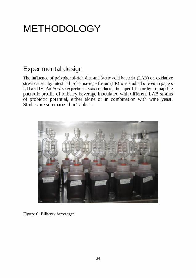
I, II and IV. An in vitro experiment was conducted in paper III in order to map the phenolic profile of bilberry beverage inoculated with different LAB strains of probiotic potential, either alone or in combination with wine yeast. Studies are summarized in Table 1.
Figure 6. Bilberry beverages.
35
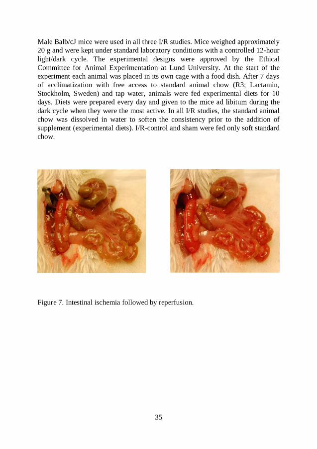
Male Balb/cJ mice were used in all three I/R studies. Mice weighed approximately
20 g and were kept under standard laboratory conditions with a controlled 12-hour
light/dark cycle. The experimental designs were approved by the Ethical
Committee for Animal Experimentation at Lund University. At the start of the
experiment each animal was placed in its own cage with a food dish. After 7 days
of acclimatization with free access to standard animal chow (R3; Lactamin,
Stockholm, Sweden) and tap water, animals were fed experimental diets for 10
days. Diets were prepared every day and given to the mice ad libitum during the
dark cycle when they were the most active. In all I/R studies, the standard animal
chow was dissolved in water to soften the consistency prior to the addition of
supplement (experimental diets). I/R-control and sham were fed only soft standard
chow.
Figure 7. Intestinal ischemia followed by reperfusion.
36
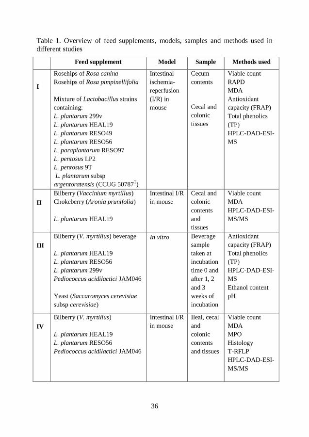
Table 1. Overview of feed supplements, models, samples and methods used in
different studies
Feed supplement Model Sample Methods used
I
Rosehips of Rosa canina
Rosehips of Rosa pimpinellifolia
Mixture of Lactobacillus strains
containing:
L. plantarum 299v
L. plantarum HEAL19
L. plantarum RESO49
L. plantarum RESO56
L. paraplantarum RESO97
L. pentosus LP2
L. pentosus 9T
L. plantarum subsp
argentoratensis (CCUG 50787T)
Intestinal
ischemia-
reperfusion
(I/R) in
mouse
Cecum
contents
Cecal and
colonic
tissues
Viable count
RAPD
MDA
Antioxidant
capacity (FRAP)
Total phenolics
(TP)
HPLC-DAD-ESI-
MS
II
Bilberry (Vaccinium myrtillus)
Chokeberry (Aronia prunifolia)
L. plantarum HEAL19
Intestinal I/R
in mouse
Cecal and
colonic
contents
and
tissues
Viable count
MDA
HPLC-DAD-ESI-
MS/MS
III
Bilberry (V. myrtillus) beverage
L. plantarum HEAL19
L. plantarum RESO56
L. plantarum 299v
Pediococcus acidilactici JAM046
Yeast (Saccaromyces cerevisiae
subsp cerevisiae)
In vitro Beverage
sample
taken at
incubation
time 0 and
after 1, 2
and 3
weeks of
incubation
Antioxidant
capacity (FRAP)
Total phenolics
(TP)
HPLC-DAD-ESI-
MS
Ethanol content
pH
IV
Bilberry (V. myrtillus)
L. plantarum HEAL19
L. plantarum RESO56
Pediococcus acidilactici JAM046
Intestinal I/R
in mouse
Ileal, cecal
and
colonic
contents
and tissues
Viable count
MDA
MPO
Histology
T-RFLP
HPLC-DAD-ESI-
MS/MS

37
In paper I, the anti-oxidative effects of two different species of rosehips, Rosa
canina and Rosa pimpinellifolia, were compared. Freeze-dried and grounded
rosehips were administered, either alone or together with a mixture of eight
different Lactobacillus strains, to examine whether a combination may enhance
the antioxidative properties of the rosehips. The bacterial mixture contained the
following strains: Lactobacillus plantarum 299v, L. plantarum HEAL19, L.
plantarum RESO49, L. plantarum RESO56, L. paraplantarum RESO97, L.
pentosus LP2, L. pentosus 9T, and L. plantarum subsp. argentoratensis (CCUG
50787T). The daily dose of probiotic mixture was 8*10
8 cfu.
In paper II, freeze-dried and ground bilberry (Vaccinium myrtillus) and chokeberry
(Aronia x prunifolia) were compared in their efficacy in preventing oxidative
stress. In this study only one probiotic strain, L. plantarum HEAL19, was
administered, either alone or together with the berries. The daily dose of L.
plantarum HEAL19 was 1*108 cfu.
In paper IV, freeze-dried and ground bilberry (V. myrtillus) was administered
alone or in combination with either Lactobacillus plantarum RESO56, L.
plantarum HEAL19 or Pediococcus acidilactici JAM046 to examine if
supplementation with different strain may influence the I/R-injury in different
ways. The daily dose of each probiotic strain was 1*109 cfu.
In paper III, a bilberry beverage (V. myrtillus) was prepared by blending whole fruits with an equivalent amount of distilled water. Plastic bottles (500 mL) were filled with the beverage and sterilized at 121C for 15 minutes followed by rapid cooling to room temperature before inoculation. The beverage was inoculated with one of the following four strains of LAB: L.
plantarum HEAL19, L. plantarum RESO56, L. plantarum 299v and P. acidilactici
JAM046, alone or together with a commercial starter culture of wine yeast
(Saccaromyces cerevisiae subspecies cerevisiae), and incubated at 30C for 3
weeks. In one series, the bilberry beverage was only inoculated with the wine
yeast. Sterile, non-inoculated bilberry beverage was used as control. The yeast and
bacteria were added in equivalent quantity 50:50, according to the weight of the
pellet.
38
Intestinal ischemia-reperfusion (I/R) procedure
An intestinal I/R model was chosen to study oxidative stress since it mimics the
clinical setting that occurs in critically ill patients (Stallion et al, 2005). Occlusion
of superior mesenteric artery (SMA) in mouse results in intestinal I/R injury.
Briefly, the mice were anesthetized with 7.5 mg Ketamine (Ketalar 50 mg/mL
Pfizer, UK) and 2.5 mg Xylazine (Narcoxyl 20 mg/mL Veterinaria AG,
Schweiz) per 100 g body weight by intraperitoneal injection. After midline
abdominal incision, the SMA was isolated and occluded with a vessel clamp to
obtain intestinal ischemia, confirmed by loss of pulsation and pale color of the
intestines. The bowel was returned to the abdominal cavity. After the ischemic
period, the clamp was removed, which resulted in immediate reperfusion,
confirmed by the restoration of pulsation and color, and the abdomen was closed.
After the reperfusion period, the animal was anesthetized again, sampled and
sacrificed.
In papers I and II, the intestinal injury was induced by occlusion of the SMA for
30 minutes followed by 240 minutes reperfusion. In paper IV, SMA was occluded
for 40 minutes followed by 120 minutes reperfusion. The sham groups were
subjected to the abdominal incision but without clamping SMA, i.e. without I/R.
Analyses
Extraction of phenolic compounds
The objective of extraction prior to analysis is to liberate phenolic compounds
from the plant material and other tissues. Organic solvent penetrates the cell
membranes and extracts the phenolics from vacuolar compartments or food matrix
(He et al, 2005). The factors that contribute to the efficacy of solvent extraction
are type of solvent, pH, temperature, number of steps and volume of solvent, and
particle size in the sample (Escribano-Bailόn & Santos Buelga, 2003). The most
widely used solvent for extraction of polyphenols is methanol and aqueous
methanol (addition of small percentage water). The pH of the extraction medium
determines the degree of solubility and solvent is often acidified with
hydrochloric, acetic, formic or trifluoroacetic acids (TFA) (Escribano-Bailόn &
Santos Buelga, 2003). In papers I-IV, methanol and aqueous methanol acidified
with TFA (paper I), formic acid (paper II and IV) and hydrochloric acid (paper III)
were used as extraction solvents. Homogenization or crushing followed by
ultrasound-assisted extraction is used to reduce particle size, which increases the
39
extractability (Escribano-Bailόn & Santos Buelga, 2003). These extraction steps
were applied in papers I-IV and were performed at low temperature (4C) in order
to prevent enzymatic degradation. During centrifugation, proteins (enzymes) were
precipitated, which also reduces enzyme activity.
Analysis of phenolic compounds
High-performance liquid chromatography (HPLC) coupled to mass spectrometry
(MS) detection is a very powerful and widely used technique for separation,
screening, identification and quantitative determination of polyphenols. In papers
I-IV, different phenolic groups were detected and identified by using HPLC-DAD-
ESI-MS/MS. The basic principle of HPLC is that a small volume of a liquid
sample is injected into a moving stream of mobile phase (liquid) that passes
through a column packed with particles of stationary phase. In the column, the
sample is separated into its components that have different affinities towards
different phases and so exit the column at different times (retention time, Rt). As
compounds leave the column, they pass through a detector where the data about
each eluted compound is acquired and processed. The outcome from HPLC is
presented as a chromatogram where different components of the sample are
represented by peaks, and as a table showing the retention time and the height/area
of each peak (Snyder et al, 2010).
The mobile phases usually consist of an acidified aqueous solvent (solvent A) and
methanol or acetonitrile (solvent B). Acidification provides better retention and
separation on the C8- and C18-RP (reversed-phase) columns, which are the most
commonly used for separation of polyphenols. Acetic, formic or trifluoroacetic
acids are the most commonly used acid modifiers (Merken & Beecher, 2000).
Thermostatically controlled columns are normally kept at ambient temperature or
slightly above (30-35C) to improve separations and keep retention times stable.
Polyphenols absorb in the ultraviolet (UV) region and are usually analyzed by
UV-vis with diode array detection (DAD). The UV-vis spectrum obtained
indicates which class the phenolic compound belongs to, e.g. flavonols have max
at 280 nm, hydroxycinnamic acid derivates at 300-320 nm and anthocyanins at
500-520 nm. The wavelength and intensity of absorption maximum may also
provide information about substitution (glycosylation and acylation) patterns of
the phenolic compound (Santos-Buelga et al, 2003).
However, UV-vis spectrum cannot distinguish between the compounds with
similar spectral characteristics and gives no information about the identity of the
conjugate (sugars or acids) attached to the aglycone. Mass spectrum obtained
40
under electrospray ionization (ESI-MS) is used for more detailed and accurate
structural information and identification of polyphenols. Sample in a liquid phase
is sprayed into a chamber with high voltage where a dry gas flows in the opposite
direction to the mist causing disintegration of the drops into charged droplets. As
the solvent evaporates the droplets become smaller. Eventually, solvent-free ions
are produced that are passed through the mass analyzer. In the mass analyzers the
ions are separated according to their mass-to-charge ratio (m/z) prior to detection
(Downard, 2004). Ionization in both positive and negative mode is used, but the
highest sensitivity is obtained using ESI in negative mode (Cuyckens et al, 2004).
Because of the positive charge in their structure, anthocyanidins are more suitable
for mass spectrometry analysis in positive mode.
Ferric reducing activity power (FRAP)
Ferric reducing activity power (FRAP) is an assay based on single electron
transfer (ET). ET-based assays measure capacity of an antioxidant to reduce an
oxidant, thereby causing the color change. The degree of color change is
proportional to the concentration of the antioxidant in the sample. The reaction
reaches the end point when color change stops (Huang et al, 2005). Antioxidative
capacity determined by FRAP was performed in paper I and paper III according to
the description by Benzie & Strain (1996). Briefly, colorless ferric
tripyridiyltriazine (Fe3+
-TPTZ) is reduced to blue-colored ferrous form (Fe2+
-
TPTZ) at low pH. The color shift is measured spectrophotometrically at 593 nm.
Any half-reaction that has a redox potential lower than that of Fe3+
-TPTZ (0.77
V) will drive the ferric (Fe3+
) to ferrous (Fe2+
) reaction. The antioxidative capacity
is expressed as mmol Fe. The FRAP assay is quick, simple and inexpensive and
the reaction is highly reproducible over a wide concentration range. It directly
measures antioxidants in the sample. A disadvantage with the method is that
absorption slowly increases after the end point of the reaction for some
polyphenols.
Total phenolic content (TP)
In paper I and paper III, total phenolic content (TP) was measured by the Folin-
Ciocalteu reagent. It is an ET-based assay that measures the reducing properties of
phenolic compounds. In the assay, the Folin-Ciocalteu reagent (a mixture of
phosphomolybdate and phosphotungstate) is reduced and the blue-colored
product is measured spectrophotometrically at 765 nm (Singleton et al, 1999). TP
is most often expressed as gallic acid equivalents in milligrams. The Folin-
Ciocalteu reagent is commercially available and the procedure is rather
standardized. It is a commonly accepted assay and is routinely used in dietary
41
antioxidant research throughout the world with a large amount of comparable data
produced.
Malondialdehyde (MDA)
Oxidation of polyunsaturated fatty acids in lipoproteins leads to formation of
hydro- and endo-peroxides, which are unstable and decompose to yield a broad
range of reactive intermediates, including alkanals, alkenals, hydroxyalkenals and
most abundantly, malondialdehyde (MDA). MDA reacts readily with amino
groups in proteins, resulting in chemical modification of the protein. Mutagenic
and carcinogenic adducts are formed when MDA reacts with DNA bases.
Measurement of MDA is widely used as an indicator of lipid peroxidation and
oxidative stress in cells and tissues (Del Rio et al, 2005). MDA-586TM
(Oxis
International Inc. Portland, Oregon, USA), a colorimetric assay, was used in
papers I, II and IV to determine MDA levels in cecal, colonic and ileal tissues. The
MDA-586TM
assay measures free MDA and protein-bound MDA (total MDA) and
minimizes the interference from other lipid peroxidation products such as alkanals,
alkenals and hydroxyalkenals. The method is based on the reaction of a
chromogenic reagent, N-methyl-2-phenylindole (NMPI), with MDA at 45C to
form intensely colored carbocyanine, which is spectrophotometrically measured at
586 nm.
Myeloperoxidase (MPO)
Myeloperoxidase (MPO) is a membrane-bound, green heme protein found almost
exclusively in neutrophils and to lesser extent in circulating monocytes. The MPO
adequately reflects the numbers of neutrophils in the healthy and inflamed tissue
(Grisham et al, 1988). In paper IV, MPO was spectrophotometrically measured as
a marker of neutrophil accumulation and tissue inflammation.
Histopathology
The degree of injury caused by intestinal ischemia-reperfusion is assessed by
histological evaluation. The procedure for tissue preparation is formalin fixation,
dehydration, paraffin embedding and cutting followed by staining with
haematoxylin and eosin (H&E). In paper IV, the grading system by Chiu was used
to score mucosal injury in the small intestine according to the following criteria: 0
= normal mucosa; 1 = development of subepithelial Gruenhagen´s space at the tip
of the villus; 2 = extension of the subepithelial space with moderate lifting of the
epithelial layer from the lamina propria; 3= massive epithelial lifting with a few
42
denuded villi, 4 = denuded villi with exposed dilated capillaries; 5 = digestion and
disintegration of lamina propria, hemorrhage, and ulceration (Chiu et al, 1970).
Randomly amplified polymorphic DNA (RAPD)
Randomly amplified polymorphic DNA (RAPD) is a PCR-based method where
arbitrary, short primers (around 8-10 bases in length) are used to amplify the DNA
at low stringency achieved by low annealing temperature. The DNA fragments of
different sizes are separated by agarose gel electrophoresis, to give a characteristic
genomic fingerprint of the organism (Power, 1996; Hadrys et al, 1992). The
method is informative, simple, reliable and suitable for studying a large number of
isolates in a short time. Different parameters such as choice and concentration of
the primer, annealing temperature and concentration of the chemicals included in
the PCR reaction must be standardized to avoid non-reproducible results (Power,
1996; Hadrys et al, 1992). RAPD was used in paper I for grouping and typing of
Lactobacillus isolates. A 9-mer with sequence 5'-ACG CGC CCT-3' was used as
primer for DNA amplification.
Terminal Restriction Fragment Length Polymorphism (T-RFLP) analysis
T-RFLP is a method used for fingerprinting and comparing bacterial community
structures. In the first step, bacterial DNA is extracted from the sample. The genes
of interest are PCR amplified with primers in which one is fluorescently labeled.
Purified PCR products are digested with a restriction enzyme and fragments are
separated by electrophoresis. The outcome from T-RFLP is presented as an
electopherogram where the profile of the bacterial community is represented by
colored peaks and as a table showing the size, base pairs and the height/area of
each peak. The T-RFLP profiles of the bacterial community between the samples
are most commonly compared by using principle component analysis (PCA). The
method is rapid, reproducible and gives high resolution (Wang, 2004). T-RFLP
was used in paper IV to analyze bacterial community profile in the cecum of mice
fed different diets, and comparison of the cecal microflora between the groups was
obtained by PCA.
Statistics
The differences between all groups (papers I-IV) were evaluated by Kruskal-
Wallis one way ANOVA on ranks. The Mann-Whitney rank sum test was used
when only two experimental groups were compared. These statistical analyses
were conducted in SigmaStat version 3.1 (SPSS Inc., Chicago, Ill, USA). One-way
43
ANNOVA was applied to the data on the number of T-RFs (paper IV) as the
distribution was normal. This evaluation was performed using Minitab statistical
software (release 16; State College, PA, USA). Values were considered
statistically significant when p<0.05.
Multivariate data analysis with principal component analysis (PCA) was used to
reveal the internal structure and possible correlations of the data with a large
number of variables. PCA was applied in paper III to reveal the correlation pattern
between different anthocyanins in bilberry beverages, and in paper IV to reveal the
possible differences in cecal microbial population between the experimental
groups. Multivariate data analyses were performed in SIMCA-P+12.0.1 (Umetrics,
Sweden).
Modde 9.0 software (Umetrics, Sweden) was used for evaluation of experimental
design, and for analysis and interpretation of all data and variables obtained in
paper III.
44
Results and discussion
Polyphenols
As a general rule, the strongly colored fruits and berries contain high
concentrations of anthocyanins as pigments and also flavonol glycosides and/or
conjugated hydroxycinnamic acids (Määttä-Riihinen et al, 2004; Wu et al, 2004).
Anthocyanins are suggested to possess antioxidative (Tsuda et al, 1999), anti-
inflammatory (Wang et al, 1999), and antimicrobial (Puupponen-Pimiä et al, 2005)
properties. The bioavailability of anthocyanins is reported to be low (Murkovic et
al, 2001; McGhie et al, 2003). They are mostly absorbed and excreted
unmetabolized, i.e. in the glycosylated form in which they were administered
(McGhie et al, 2003). Polyphenols are usually characterized and quantified by
HPLC-DAD-ESI-MSn, which was also used in papers I-IV.
Anthocyanins
No anthocyanins were found in animals not fed berry-supplemented diets.
In papers I, II and IV, anthocyanins were analyzed in the intestinal content of mice
fed berries, while in paper III they were analyzed in bilberry beverages. Bilberry
(Vaccinium myrtillus) contained 14 anthocyanins (papers II-IV; Table 2; Lätti et
al, 2010; Laaksonen et al, 2010), chokeberry (Aronia x prunifolia) contained four
anthocyanins (paper II; Oszmiański & Wojdylo, 2005) and one anthocyanin was
found in rosehips (paper I, Hvattum, 2002). Anthocyanins Dp-3-arab and Cy-3-gal
could not be separated in paper II and IV, and Pt-3-arab and Pn-3-gal were co-
eluted in paper IV.
In paper I, anthocyanins were analyzed in the cecal content. Cyanidin-3-O-glucoside, the only anthocyanin found in rosehips (Hvattum, 2002), was present in significantly higher concentrations (p<0.001) in Rosa pimpinellifolia-fed groups compared to Rosa canina-fed groups. There was a trend that LAB supplementation to the rosehips increased the concentrations of anthocyanins in cecum. The resident microflora of mice
45
seemed to have higher activity towards anthocyanin-degradation than supplementation with LAB mixture.
In papers II and IV, bilberry (B) was administered, either alone or together with different strains of lactic acid bacteria (LAB). In paper II, chokeberry (Ar) was also administered, either alone or together with a LAB strain. Strains used in different papers are listed in Table 1. In paper II, cecal and
colonic contents were collected for analysis of anthocyanins, and ileum contents were also analyzed in paper IV.
Chokeberry powder consisted of four anthocyanins: Cy-3-glu, Cy-3-gal, Cy-3-arab and Cy-3-xyl (xyloside). Of these, Cy-3-glu was not detected in cecal and colonic contents of chokeberry-fed mice (groups Ar and Ar+LplH19) (Paper II). In paper IV, all 14 anthocyanins from the bilberry
powder were detected in ileum of the bilberry-treated animals (B, B+Lpl56, B+LplH19 and B+Ped) (Table 2). Recovery of Cy-3-glu and also Dp-3-glu was
extremely low in the ileum. The low concentrations of these anthocyanins in ileum
and cecum may be due to several factors. One possibility is that they were
absorbed from the upper GI tract or metabolized by bacteria and enzymes in the
small intestine. It has been shown that Cy-3-glu was mainly absorbed from the
stomach (Passamonti et al, 2003) and jejunum (Matuschek et al, 2006; He et al,
2009). Furthermore, it has been suggested (Del Bό et al, 2010; Felgines et al,
2002) that Cy and Dp glycosides are subjected to methylation in the small
intestine, resulting in the increase of Pn, Pt and Mv glycosides under physiological
conditions. This trend was also observed in the ileum in paper IV. It has also been
proposed that Cy-3-glu may be degraded to protocatechuic acid (PCA) and Cy
aglycone both in vitro and in vivo (animals and humans) (Aura et al, 2005;
Keppler & Humpf, 2005; Fleschhut et al, 2006; Tsuda et al, 1999a; Wu et al,
2009; Vitaglione et al, 2007). In paper I, Cy-3-glu was the only anthocyanin found
in the rosehips, with significantly higher concentration detected in R.
pimpinellifolia than R. canina. This could be expected on account of dark blue colour of R. pimpinellifolia. PCA was detected in the cecal content of mice fed RP and seemed to be negatively correlated with Cy-3-glu, i.e. when concentration of Cy-3-glu was high the concentration of PCA was low, and vice versa. The results suggested that PCA was a metabolite from microbial degradation of Cy-3-glu. Ingestion of anthocyanin-rich black raspberry powder in pigs resulted in high concentration of PCA in the cecum followed by ileum and colon (Wu et al, 2009).
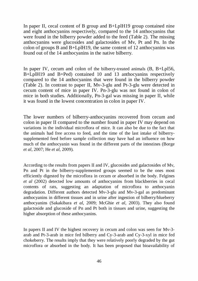
46
In paper II, cecal content of B group and B+LplH19 group contained nine and eight anthocyanins respectively, compared to the 14 anthocyanins that were found in the bilberry powder added to the feed (Table 2). The missing anthocyanins were glucosides and galactosides of Mv, Pt and Pn. In the colon of groups B and B+LplH19, the same content of 12 anthocyanins was found out of the 14 anthocyanins in the native bilberry.
In paper IV, cecum and colon of the bilberry-treated animals (B, B+Lpl56, B+LplH19 and B+Ped) contained 10 and 13 anthocyanins respectively compared to the 14 anthocyanins that were found in the bilberry powder (Table 2). In contrast to paper II, Mv-3-glu and Pt-3-glu were detected in cecum content of mice in paper IV. Pn-3-glu was not found in colon of mice in both studies. Additionally, Pn-3-gal was missing in paper II, while it was found in the lowest concentration in colon in paper IV.
The lower numbers of bilberry-anthocyanins recovered from cecum and colon in paper II compared to the number found in paper IV may depend on variations in the individual microflora of mice. It can also be due to the fact that
the animals had free access to feed, and the time of the last intake of bilberry-
supplemented feed before sample collection may have had an influence on how
much of the anthocyanin was found in the different parts of the intestines (Borge
et al, 2007; He et al, 2009).
According to the results from papers II and IV, glucosides and galactosides of Mv,
Pn and Pt in the bilberry-supplemented groups seemed to be the ones most
efficiently digested by the microflora in cecum or absorbed in the body. Felgines
et al (2002) detected low amounts of anthocyanins from blackberries in cecal
contents of rats, suggesting an adaptation of microflora to anthocyanin
degradation. Different authors detected Mv-3-glu and Mv-3-gal as predominant
anthocyanins in different tissues and in urine after ingestion of bilberry/blueberry
anthocyanins (Sakakibara et al, 2009; McGhie et al, 2003). They also found
galactoside and glucoside of Pn and Pt both in tissues and urine, suggesting the
higher absorption of these anthocyanins.
In papers II and IV the highest recovery in cecum and colon was seen for Mv-3-
arab and Pt-3-arab in mice fed bilberry and Cy-3-arab and Cy-3-xyl in mice fed
chokeberry. The results imply that they were relatively poorly degraded by the gut
microflora or absorbed in the body. It has been proposed that bioavailability of
47
anthocyanins is modulated by both the nature of the sugar conjugate and the
phenolic aglycone (Sakakibara et al, 2009; McGhie et al, 2003; Wu et al, 2005).
Pentosides (arabinoside and xyloside) seem to be quite stable in the gut and
resistant to the microbial degradation in the large intestine (He et al, 2005;
Ichiyanagi et al, 2006).
The opposite seemed to be true for in vitro studies. In paper III, bilberry beverages
were incubated with one of the three LAB strains (Table 1), either alone or in
combination with wine yeast, over 3 weeks at 30C. All 14 anthocyanins present
in the native bilberry were detected in the beverages. In addition, Pt aglycone was
found in the samples. All anthocyanins decreased during incubation, but Pt
aglycone, Mv-3-arab and co-eluted Pt-3-arab and Pn-3-gal were strongly
correlated and showed the highest reduction in beverages, especially those
inoculated with only LAB. The degradation of anthocyanins seemed to be due to
the exogenic factors and not due to the bacterial activity, since non-inoculated,
sterile bilberry-control and LAB-inoculated beverages showed the same reduction
of anthocyanins. Beverages were autoclaved prior to inoculation in order to
inactivate both bilberry enzymes and contaminating microorganisms. They were
prepared and incubated in plastic bottles sealed with screw cap with a water seal.
Bottles were not completely filled and had a certain headspace. Heat, pH, oxygen,
light and various storage conditions have marked effects on anthocyanin stability
(Kalt et al, 2000). Heat applied during autoclavation, or oxygen present in the
headspace or diffusing through the packaging material or during sampling, may
have influenced the stability of anthocyanins and especially Pt aglycone, Mv-3-
arab and co-eluted Pt-3-arab and Pn-3-gal. Anthocyanins conjugated with sugar
pentoses (arabinosides) are less stable during heating and processing than
anthocyanin with galactoside or glucoside (Yue et al, 2008; Howard et al, 2010).
It has been shown that Mv-3-arab, but also Pt-3-arab, were very sensitive to
oxygen during the storage in oxygen-permeable packaging materials (Trošt et al,
2008; Giovanelli et al, 2007). Plastic packages allow a certain permeability of
oxygen (Trošt et al, 2008; Giovanelli et al, 2007).
Addition of wine yeast to the beverages resulted in much higher recovery of
anthocyanins (Paper III). The protective effect may have been due to the high
yield of CO2 during alcoholic fermentation, which decreased oxygen levels in the
bottles and preserved anthocyanins from oxidation. Furthermore, anthocyanins
may have been retained by the yeast cell walls during the incubation period and
then released during extraction. Results conflict on the influence of anthocyanin´s
polarity on its adsorbtion by yeast. Some authors (Lubbers et al, 1994; Morata et
al, 2003) suggested that more hydrophobic anthocyanins (Mv, Pn and even Pt) are
48
adsorbed by yeast to a greater extent while others (Mazauric et al, 2006) showed
that anthocyanins may not be adsorbed in relation to their sole polarity. However,
malvidin derivates were the most extracted anthocyanins from yeast lees (Lubbers
et al, 1994; Morata et al, 2003; Mazauric et al, 2006).
Table 2. Anthocyanins found in native bilberry and in contents of different parts of
the intestines.
Bilberry
(powder/beverage)
MS (MS2)
ions (m/z) Paper II
detection site
Paper IV
detection site
Delphidin-3-O-galactoside
(Dp-3-gal)
465 (303) Cecum, colon Ileum, cecum,
colon
Delphidin-3-O-glucoside
(Dp-3-glu) 465 (303) Cecum, colon Ileum, cecum,
colon
Delphidin-3-O-arabinoside
(Dp-3-arab)
435 (303) Cecum, colon Ileum, cecum,
colon
Cyanidin-3-O-galactoside
(Cy-3-gal)
449 (287) Cecum, colon Ileum, cecum,
colon
Cyanidin-3-O-glucoside
(Cy-3-glu)
449 (287) Cecum, colon Ileum, cecum,
colon
Petunidin-3-O-galactoside
(Pt-3-gal)
479 (317) Colon Ileum, colon
Cyanidin-3-O-arabinoside
(Cy-3-arab)
419 (287) Cecum, colon Ileum, cecum,
colon
Petunidin-3-O-glucoside
(Pt-3-glu)
479 (317) Colon + cecum
in B-group
Ileum, cecum,
colon
Petunidin-3-O-arabinoside
(Pt-3-arab)
449 (317) Cecum, colon Ileum, cecum,
colon
Peonidin-3-O-galactoside
(Pn-3-gal)
463 (301) ND Ileum
Peonidin-3-O-glucoside
(Pn-3-glu)
463 (301) ND Ileum, colon
Malvidin-3-O-galactoside
(Mv-3-gal)
493 (331) Colon Ileum, colon
Malvidin-3-O-glucoside
(Mv-3-glu)
493 (331) Colon Ileum, cecum,
colon
Malvidin-3-O-arabinoside
(Mv-3-arab)
463 (331) Cecum, colon Ileum, cecum,
colon
ND = not detected
49
Phenolic compounds other than anthocyanins
In papers I-III, other polyphenolic groups than anthocyanins were analyzed, i.e.
flavonols, flavanols, proanthocyanins, phenolic acids and phenolic metabolites. In
animals not fed berry-supplemented diets, no phenolic compounds were found.
In paper I, phenolic compounds analyzed were catechin (m/z 289), metabolite I (m/z 291), proanthocyanidin-monomer-glycoside (m/z 451), proanthocyanidin-dimer-glycoside (m/z 739), proanthocyanidin-dimer-diglycoside (m/z 901). These compounds were previously detected in rosehips (Salminen et al, 2005). Significantly higher concentrations of these compounds were detected in cecal content of mice fed Rosa canina than Rosa pimpinellifolia. Furthermore, there was a trend towards higher concentration of phenolic compounds when LAB supplemented the rosehip-diet. The LAB mixture may have modified the composition of colonic microflora, probably increasing the metabolic activity towards higher degradation of proanthocyanidins into monomers and dimers, or certain strains in the mixture themselves may have contributed to the degradation of polyphenols.
In paper II, five aromatic acids were detected in cecum of bilberry-fed groups (B
and B+LplH19) (Table 3). Two isomers of 3,4-dihydroxyphenylacetic acid
(diHPA) and 3-hydroxyphenylpropionic acid (3-HPP) were identified while two
compounds could not be identified. Additionally, in colon of bilberry-
supplemented animals, three compounds were detected but could not be identified.
The detected aromatic acids were probably produced during microbial degradation
of different polyphenols present in the berries (Rusell et al, 2007). Identified
isomers of diHPA are suggested to be metabolites from the microbial degradation
of quercetin or proanthocyanidin-dimer (Aura et al, 2002; Wu et al, 2009;
Gonthier et al, 2003b). Catechin, epicatechin, proanthocyanidins/procyanidins, as
well as chlorogenic acid, have been proposed as precursors of 3-HPP (Rios et al,
2003; Gonthier et al, 2003b; Gonthier et al, 2003c). Procyanidins (epicatechin-
monomers linked with A-type and B-type bonds), quercetin glycosides and
chlorogenic acid have been identified in the bilberry (Prior et al, 2001; Määttä-
Riihinen et al, 2004).
In cecum and colon of chokeberry-supplemented groups (Ar and Ar+LplH19), 4-
hydroxyphenylacetic acid (4-HPA) and 3-hydroxyphenylacetic acid (3-HPA)
respectively were identified (Paper IV). Dehydroxylation of diHPA at meta and
para positions results in formation of 3-HPA and 4-HPA (Rios et al, 2003). diHPA
50
has been described as an intermediate between flavanols and the more
dehydroxylated 3-HPA and 4-HPA. Since procyanidins have been identified as the
major class of polyphenols in chokeberries (Kulling & Rawel, 2008), they were
probably degraded by microflora, firstly into diHPA and then into 3-HPA or 4-
HPA.
More than 80% of procyanidins in chokeberries have a higher polymerization
degree than 10-mers and only around 4% are monomers, dimers and trimers
(Kulling & Rawel, 2008). As the degree of procyanidin polymerization increases,
the yield of microbial metabolites decreases due to the reduced accessibility of the
substrate or interaction of procyanidin with proteins in the gut lumen (Gonthier et
al, 2003b). The higher content of procyanidin polymers in chokeberry compared to
bilberry may have been the reason that fewer metabolites were detected in the
cecum and colon of chokeberry-treated groups.
In general, large individual differences in concentration and detectable levels of
different phenolic compounds between different individuals were observed in
papers I and II.
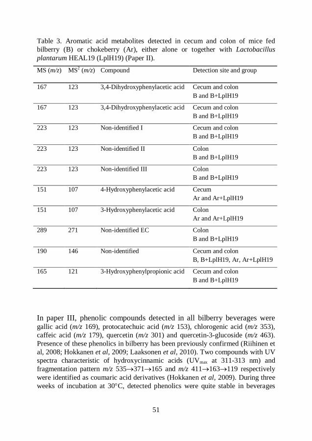
51
Table 3. Aromatic acid metabolites detected in cecum and colon of mice fed
bilberry (B) or chokeberry (Ar), either alone or together with Lactobacillus
plantarum HEAL19 (LplH19) (Paper II).
MS (m/z) MS2 (m/z) Compound Detection site and group
167 123 3,4-Dihydroxyphenylacetic acid Cecum and colon
B and B+LplH19
167 123 3,4-Dihydroxyphenylacetic acid Cecum and colon
B and B+LplH19
223 123 Non-identified I Cecum and colon
B and B+LplH19
223 123 Non-identified II Colon
B and B+LplH19
223 123 Non-identified III Colon
B and B+LplH19
151 107 4-Hydroxyphenylacetic acid Cecum
Ar and Ar+LplH19
151 107 3-Hydroxyphenylacetic acid Colon
Ar and Ar+LplH19
289 271 Non-identified EC Colon
B and B+LplH19
190 146 Non-identified Cecum and colon
B, B+LplH19, Ar, Ar+LplH19
165 121 3-Hydroxyphenylpropionic acid Cecum and colon
B and B+LplH19
In paper III, phenolic compounds detected in all bilberry beverages were gallic acid (m/z 169), protocatechuic acid (m/z 153), chlorogenic acid (m/z 353),
caffeic acid (m/z 179), quercetin (m/z 301) and quercetin-3-glucoside (m/z 463).
Presence of these phenolics in bilberry has been previously confirmed (Riihinen et
al, 2008; Hokkanen et al, 2009; Laaksonen et al, 2010). Two compounds with UV
spectra characteristic of hydroxycinnamic acids (UVmax at 311-313 nm) and
fragmentation pattern m/z 535371165 and m/z 411163119 respectively
were identified as coumaric acid derivatives (Hokkanen et al, 2009). During three
weeks of incubation at 30C, detected phenolics were quite stable in beverages
52
inoculated with different LAB (Table 1), either alone or in combination with yeast.
Chlorogenic acid and also quercetin aglycone and glycosides in apple juice were
not much influenced by storage conditions, oxygen pressure or thermal processing
(Van der Sluis et al, 2005). Furthermore, LAB strains added to the bilberry
beverage seemed to lack the metabolic activity towards phenolic acids in the tested
conditions. Metabolic activity was recorded in studies where LAB strains were
cultivated in different growth media containing a certain concentration of pure
phenolic acid such as caffeic, p-coumaric, ferulic, m-coumaric, gallic or
protocatechuic acid (Calvin et al, 1993; Rodríguez et al, 2008).
Antioxidative capacity and total phenolic content
In papers I and III, antioxidative capacity was measured using the ferric reducing
activity power (FRAP) assay, while total phenolic content (TP) was determined
with Folin-Ciocalteau reagent. Both are electron transfer (ET) based assays that
measure the reducing properties of phenolic compounds.
In paper I, antioxidative capacity and total phenolic content were measured in
cecum contents of mice fed either of two different Rose species alone or in
combination with a mixture of Lactobacillus strains (Table 1). In paper III,
antioxidative capacity and total phenolic content were determined in a bilberry
beverage inoculated with either of three different LAB strains alone or in
combination with yeast.
LAB supplementation to the rosehips tended to increase the concentrations of total
phenolic contents and antioxidative capacity in cecum in paper I. Antioxidative
capacity was 6.21 mmol Fe2+/100g in the Rosa canina (RC) fed group and 6.95
mmol Fe2+/100g in combination with Lactobacillus mixture (RC+LAB).
Corresponding values were 5.47 mmol Fe2+/100g in Rosa pimpinellifolia (RP) fed
group and 6.45 mmol Fe2+/100g in combination with Lactobacillus mixture
(RP+LAB). Total phenolic content values in groups RC and RC+LAB were 373
and 428 GAE/100 g respectively. Corresponding values in groups RP and
RP+LAB were 395 and 423 mg GAE/100 g respectively.
In paper III, antioxidative capacity and TP decreased during 3 weeks of incubation
in all bilberry beverages, i.e. both with different LAB strains alone and together
with yeast. After 3 weeks of incubation, the average FRAP values were 3.0 mmol
Fe2+/100g in beverage supplemented with LAB and 3.3 mmol Fe2+/100g in
beverage supplemented with LAB and yeast. After the same incubation period, the
53
average TP value of beverages incubated with LAB was 284 mg GAE/100 g and
beverages incubated with LAB plus yeast was 306 mg GAE/100 g.
The results obtained in paper I differed from the results in paper III. Results in
paper I indicate that supplementation with the LAB mixture has a different influence on the degradation of polyphenols than the indigenous microflora. Lactobacillus strains in the mixture demonstrated tannase activity. The higher concentrations of phenolic compounds were also found in groups where rosehips were supplemented with LAB mixture.
In paper III, decreases in antioxidative capacity and TP in all bilberry beverages were probably due to the decrease of phenolic compounds and especially anthocyanins. Anthocyanins are suggested to be strong antioxidants, but are quite unstable during processing and storage (Wang et al, 1999; Kader
et al, 1997; Buckow et al, 2010; Skrede et al, 2000).
Polyphenols differ in their behavior and stability between in vitro and in vivo systems. Many factors, such as interactions with metal ions, proteins and other antioxidants, as well as distribution between the lipid and water phase and exposure to the complex microflora in the GI tract, may affect the action of an
antioxidant in in vivo models compared to in vitro models. The concentration of phenolics and pH of the environment may also be crucial factors.
Inflammatory markers
Ischemia-reperfusion
In rodents, the blood supply to the intestines is regulated by the superior
mesenteric artery (SMA) (Leung et al, 1992). Occlusion of the SMA results in
ischemia and upon removal of the clamp reperfusion is observed. In papers I and
II, the time-interval for ischemia was 30 min and 240 min for reperfusion. This
time-interval was chosen according to Riaz et al (2002) and Håkansson et al
(2006) since it gave the highest lipid peroxidation (MDA) in colon of mice. In
paper IV, time-intervals for I/R were changed to 40 min ischemia and 120 min
reperfusion to obtain more severe intestinal damage. Increases in the ischemic
time were associated with comparable decreases in the tickness of the mucosal
layer and severity of the intestinal damage (Boros et al, 1995). After 40-50 min of
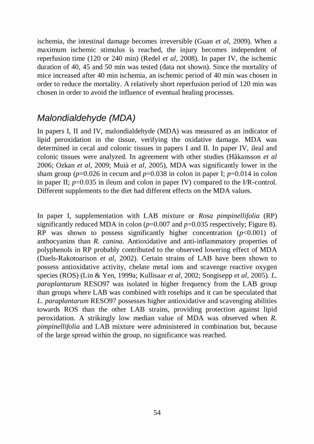
54
ischemia, the intestinal damage becomes irreversible (Guan et al, 2009). When a
maximum ischemic stimulus is reached, the injury becomes independent of
reperfusion time (120 or 240 min) (Redel et al, 2008). In paper IV, the ischemic
duration of 40, 45 and 50 min was tested (data not shown). Since the mortality of
mice increased after 40 min ischemia, an ischemic period of 40 min was chosen in
order to reduce the mortality. A relatively short reperfusion period of 120 min was
chosen in order to avoid the influence of eventual healing processes.
Malondialdehyde (MDA)
In papers I, II and IV, malondialdehyde (MDA) was measured as an indicator of
lipid peroxidation in the tissue, verifying the oxidative damage. MDA was
determined in cecal and colonic tissues in papers I and II. In paper IV, ileal and
colonic tissues were analyzed. In agreement with other studies (Håkansson et al
2006; Ozkan et al, 2009; Muià et al, 2005), MDA was significantly lower in the
sham group (p=0.026 in cecum and p=0.038 in colon in paper I; p=0.014 in colon
in paper II; p=0.035 in ileum and colon in paper IV) compared to the I/R-control.
Different supplements to the diet had different effects on the MDA values.
In paper I, supplementation with LAB mixture or Rosa pimpinellifolia (RP)
significantly reduced MDA in colon (p=0.007 and p=0.035 respectively; Figure 8).
RP was shown to possess significantly higher concentration (p<0.001) of
anthocyanins than R. canina. Antioxidative and anti-inflammatory properties of
polyphenols in RP probably contributed to the observed lowering effect of MDA
(Daels-Rakotoarison et al, 2002). Certain strains of LAB have been shown to
possess antioxidative activity, chelate metal ions and scavenge reactive oxygen
species (ROS) (Lin & Yen, 1999a; Kullisaar et al, 2002; Songisepp et al, 2005). L.
paraplantarum RESO97 was isolated in higher frequency from the LAB group
than groups where LAB was combined with rosehips and it can be speculated that
L. paraplantarum RESO97 possesses higher antioxidative and scavenging abilities
towards ROS than the other LAB strains, providing protection against lipid
peroxidation. A strikingly low median value of MDA was observed when R.
pimpinellifolia and LAB mixture were administered in combination but, because
of the large spread within the group, no significance was reached.
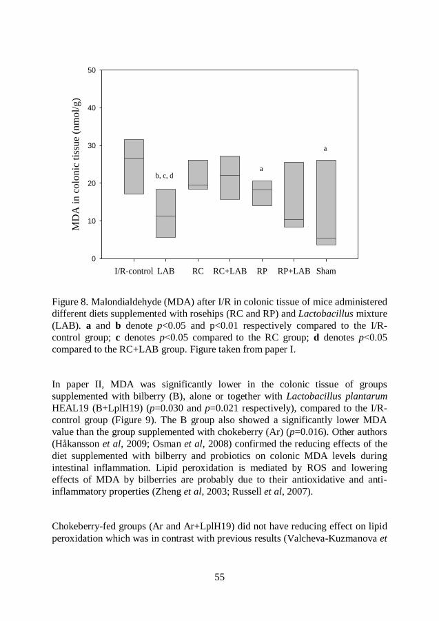
55
Figure 8. Malondialdehyde (MDA) after I/R in colonic tissue of mice administered
different diets supplemented with rosehips (RC and RP) and Lactobacillus mixture
(LAB). a and b denote p<0.05 and p<0.01 respectively compared to the I/R-
control group; c denotes p<0.05 compared to the RC group; d denotes p<0.05
compared to the RC+LAB group. Figure taken from paper I.
In paper II, MDA was significantly lower in the colonic tissue of groups
supplemented with bilberry (B), alone or together with Lactobacillus plantarum
HEAL19 (B+LplH19) (p=0.030 and p=0.021 respectively), compared to the I/R-
control group (Figure 9). The B group also showed a significantly lower MDA
value than the group supplemented with chokeberry (Ar) (p=0.016). Other authors
(Håkansson et al, 2009; Osman et al, 2008) confirmed the reducing effects of the
diet supplemented with bilberry and probiotics on colonic MDA levels during
intestinal inflammation. Lipid peroxidation is mediated by ROS and lowering
effects of MDA by bilberries are probably due to their antioxidative and anti-
inflammatory properties (Zheng et al, 2003; Russell et al, 2007).
Chokeberry-fed groups (Ar and Ar+LplH19) did not have reducing effect on lipid
peroxidation which was in contrast with previous results (Valcheva-Kuzmanova et
I/R-control LAB RC RC+LAB RP RP+LAB Sham
MD
A i
n c
olo
nic
tis
sue (
nm
ol/
g)
0
10
20
30
40
50
a
b, c, da
56
al, 2004; Valcheva-Kuzmanova et al, 2005). Different chokeberry species (Aronia
x prunifolia powder versus A. melanocarpa juice) as well as different
administration methods (ad libitum versus stomach intubation) were used in the
studies, which may have influenced the results. Factors such as cultivar,
fertilization, maturation of the berries, harvest date and habitat may also contribute
to the differences in quantity and profile of phenolics in chokeberry fruits and
hence antioxidant properties (Kulling & Rawel, 2008).
Although chokeberry was shown to possess higher total phenolic content
(Kähkönen et al, 1999) and higher antioxidative capacity in vitro (Zheng et al,
2003) than bilberry, it did not have antioxidative effects in colon of mice subjected
to intestinal I/R (Paper II). As previously mentioned, in vivo systems are more
complex than in vitro systems and it may affect the action of an antioxidant. Polymeric procyanidins are the major polyphenols found in chokeberries. More
than 80% of these procyanidins have a polymerization degree higher than 10-mers
(Wu et al, 2004; Kulling & Rawel, 2008), which may decrease their absorption,
cause interaction with proteins in the gut lumen and also make them less
accessible for the microbial degradation (Gonthier et al, 2003b). It could be noted
in paper II that fewer metabolites were found in the cecum and colon of mice fed
chokeberry than in bilberry-fed mice. Aromatic acid metabolites may be more
efficient antioxidants than the parent phenolic compounds (Gao et al, 2006; Kim et
al, 1999; Monagas et al, 2009). They are proposed to be more easily absorbed
from the large intestine into the circulation and may contribute not only to the
local but also to the systemic protection against oxidative stress injury.
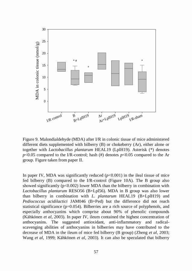
57
Figure 9. Malondialdehyde (MDA) after I/R in colonic tissue of mice administered
different diets supplemented with bilberry (B) or chokeberry (Ar), either alone or
together with Lactobacillus plantarum HEAL19 (LplH19). Asterisk (*) denotes
p<0.05 compared to the I/R-control; hash (#) denotes p<0.05 compared to the Ar
group. Figure taken from paper II.
In paper IV, MDA was significantly reduced (p<0.001) in the ileal tissue of mice
fed bilberry (B) compared to the I/R-control (Figure 10A). The B group also
showed significantly (p<0.002) lower MDA than the bilberry in combination with
Lactobacillus plantarum RESO56 (B+Lpl56). MDA in B group was also lower
than bilberry in combination with L. plantarum HEAL19 (B+LplH19) and
Pediococcus acidilactici JAM046 (B+Ped) but the difference did not reach
statistical significance (p=0.054). Bilberries are a rich source of polyphenols, and
especially anthocyanins which comprise about 90% of phenolic compounds
(Kähkönen et al, 2003). In paper IV, ileum contained the highest concentration of
anthocyanins. The suggested antioxidant, anti-inflammatory and radical-
scavenging abilities of anthocyanins in bilberries may have contributed to the
decrease of MDA in the ileum of mice fed bilberry (B group) (Zheng et al, 2003;
Wang et al, 1999; Kähkönen et al, 2003). It can also be speculated that bilberry
I/R control B
B+LplH19 Ar
Ar+LplH19LplH19
I/R-sham
MD
A i
n c
olo
nic
tis
sue (
nm
ol/
g)
0
5
10
15
20
25
30
* #
*
*
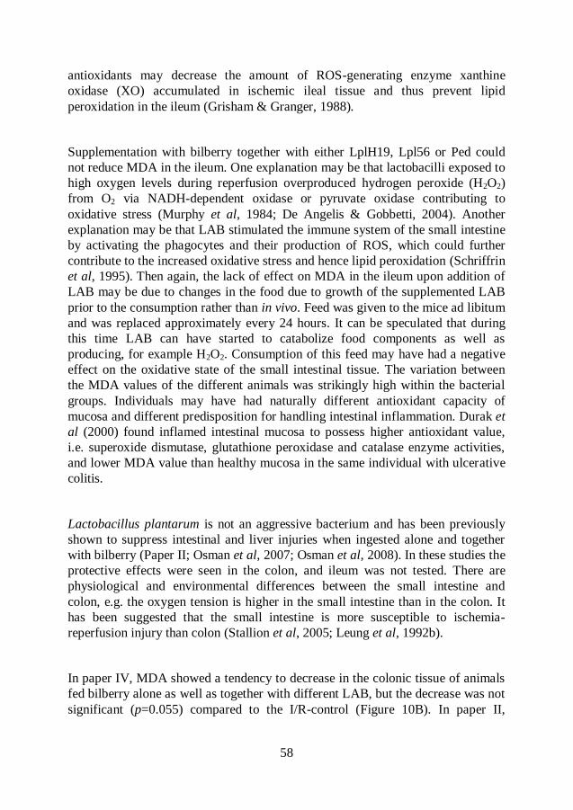
58
antioxidants may decrease the amount of ROS-generating enzyme xanthine
oxidase (XO) accumulated in ischemic ileal tissue and thus prevent lipid
peroxidation in the ileum (Grisham & Granger, 1988).
Supplementation with bilberry together with either LplH19, Lpl56 or Ped could
not reduce MDA in the ileum. One explanation may be that lactobacilli exposed to
high oxygen levels during reperfusion overproduced hydrogen peroxide (H2O2)
from O2 via NADH-dependent oxidase or pyruvate oxidase contributing to
oxidative stress (Murphy et al, 1984; De Angelis & Gobbetti, 2004). Another
explanation may be that LAB stimulated the immune system of the small intestine
by activating the phagocytes and their production of ROS, which could further
contribute to the increased oxidative stress and hence lipid peroxidation (Schriffrin
et al, 1995). Then again, the lack of effect on MDA in the ileum upon addition of
LAB may be due to changes in the food due to growth of the supplemented LAB
prior to the consumption rather than in vivo. Feed was given to the mice ad libitum
and was replaced approximately every 24 hours. It can be speculated that during
this time LAB can have started to catabolize food components as well as
producing, for example H2O2. Consumption of this feed may have had a negative
effect on the oxidative state of the small intestinal tissue. The variation between
the MDA values of the different animals was strikingly high within the bacterial
groups. Individuals may have had naturally different antioxidant capacity of
mucosa and different predisposition for handling intestinal inflammation. Durak et
al (2000) found inflamed intestinal mucosa to possess higher antioxidant value,
i.e. superoxide dismutase, glutathione peroxidase and catalase enzyme activities,
and lower MDA value than healthy mucosa in the same individual with ulcerative
colitis.
Lactobacillus plantarum is not an aggressive bacterium and has been previously
shown to suppress intestinal and liver injuries when ingested alone and together
with bilberry (Paper II; Osman et al, 2007; Osman et al, 2008). In these studies the
protective effects were seen in the colon, and ileum was not tested. There are
physiological and environmental differences between the small intestine and
colon, e.g. the oxygen tension is higher in the small intestine than in the colon. It
has been suggested that the small intestine is more susceptible to ischemia-
reperfusion injury than colon (Stallion et al, 2005; Leung et al, 1992b).
In paper IV, MDA showed a tendency to decrease in the colonic tissue of animals
fed bilberry alone as well as together with different LAB, but the decrease was not
significant (p=0.055) compared to the I/R-control (Figure 10B). In paper II,

59
administration of bilberry, either alone or in combination with L. plantarum
HEAL19 (LplH19), significantly decreased MDA in the colonic tissue after I/R
injury. The difference between these studies was the duration of ischemia and
reperfusion. In paper IV, ischemia lasted for 40 min followed by 120 min
reperfusion, compared to the 30 min ischemia and 240 min reperfusion in paper II.
After an ischemic period of 40-50 min (long ischemia) irreversible damage occurs
and reperfusion can not restore normal epithelial cell structure and function (Guan
et al, 2009). When a maximum ischemic stimulus (long ischemia) is reached, the
time of reperfusion (120 or 240 min) has no influence on the severity of the
damage (Redel et al, 2008).
A)
B)
I/R-controlB-group
B+Lpl56
B+LplH19B+Ped
I/R-sham
MD
A i
n i
leal
tiss
ue (
nm
ol/
g)
0
10
20
30
40
50
*** ##*
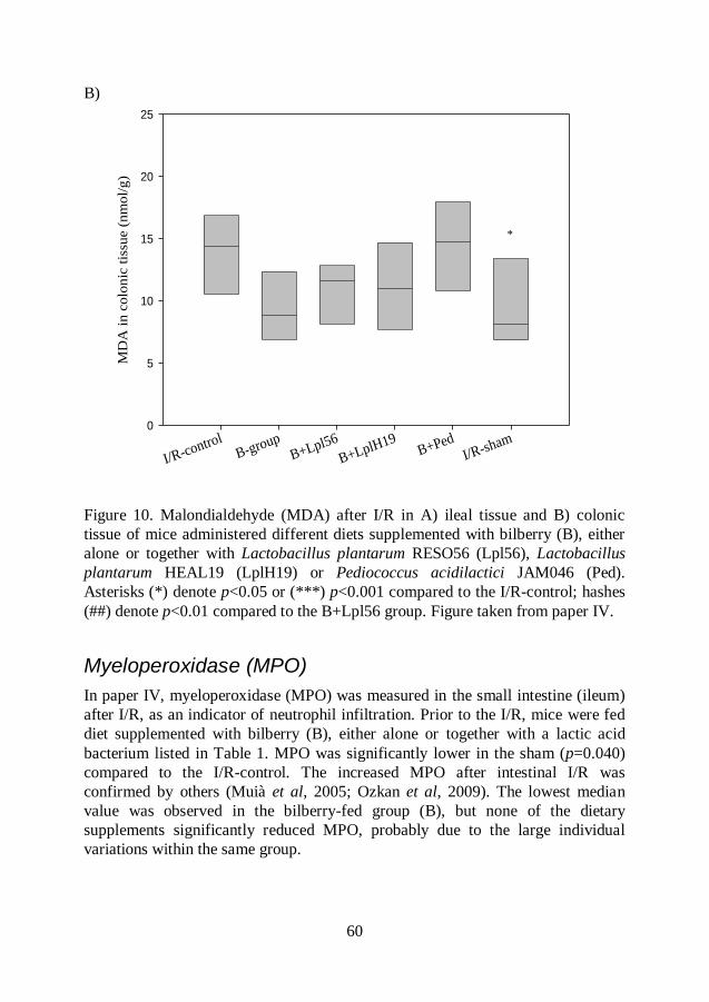
60
B)
Figure 10. Malondialdehyde (MDA) after I/R in A) ileal tissue and B) colonic
tissue of mice administered different diets supplemented with bilberry (B), either
alone or together with Lactobacillus plantarum RESO56 (Lpl56), Lactobacillus
plantarum HEAL19 (LplH19) or Pediococcus acidilactici JAM046 (Ped).
Asterisks (*) denote p<0.05 or (***) p<0.001 compared to the I/R-control; hashes
(##) denote p<0.01 compared to the B+Lpl56 group. Figure taken from paper IV.
Myeloperoxidase (MPO)
In paper IV, myeloperoxidase (MPO) was measured in the small intestine (ileum)
after I/R, as an indicator of neutrophil infiltration. Prior to the I/R, mice were fed
diet supplemented with bilberry (B), either alone or together with a lactic acid
bacterium listed in Table 1. MPO was significantly lower in the sham (p=0.040)
compared to the I/R-control. The increased MPO after intestinal I/R was
confirmed by others (Muià et al, 2005; Ozkan et al, 2009). The lowest median
value was observed in the bilberry-fed group (B), but none of the dietary
supplements significantly reduced MPO, probably due to the large individual
variations within the same group.
I/R-controlB-group
B+Lpl56
B+LplH19B+Ped
I/R-sham
MD
A i
n c
olo
nic
tis
sue (
nm
ol/
g)
0
5
10
15
20
25
*

61
Histopathology
Histological damage that occurs after intestinal I/R is characterized by shortening
and loss of intestinal villi, necrosis and neutrophil infiltration (Chiu et al, 1970). In
paper IV, the most severe mucosal damage was observed in the I/R-control, with
complete loss of villi and marked congestion. The median value of the mucosal
injury score of the I/R-control was 4.5, and this was significantly (p<0.001)
higher than that of the sham (0.25). No significant differences were recorded in the
diet-supplemented groups. The median values of the injury scores were 2.5, 3.5, 4.0 and 3.3 for the groups B, B+Lpl56, B+LplH19 and B+Ped respectively.
Pretreatment with bilberry alone (B group) showed the lowest median valuefor the
mucosal integrity and morphology even if the difference in injury score was not
significant. This coincides with the observations that the B group also showed a
lower MDA (significant) and MPO values (non-significant) in ileum than the LAB
groups. As previously suggested, the protective effects of the bilberry may be due
to the high content of polyphenols with antioxidative and anti-inflammatory
properties. In histopathological analysis large individual variations within the
same group were also observed, suggesting that different individuals may have
differed naturally in terms of antioxidant capacity of the mucosa and were not
equally sensitive to the I/R damage (Durak et al, 2000).
Bacteriology
Lactobacilli and Enterobacteriaceae
In papers I, II and IV, the lactobacilli and Enterobacteriaceae counts were
analyzed in colonic tissue, and in paper IV also in cecal tissue.
In paper I, the lactobacilli count of colonic tissue varied between 105 and 10
6 cfu/g.
Diets supplemented with either Rosa canina (RC) or Rosa pimpinellifolia (RP)
alone (median values 5.0 log cfu/g colonic tissue) reduced the number of viable
lactobacilli compared to the diets supplemented with rosehips in combination with
a mixture of Lactobacillus strains (median values 6.22 and 6.41 log cfu/g colonic
tissue for RC+LAB and RP+LAB respectively), but no significant differences
were found between the groups. All eight Lactobacillus strains in the mixture
(Table 1) could be re-isolated from the colonic tissue of groups administered LAB
(RC+LAB, RP+LAB and LAB). The most frequently occurring strain was L.
plantarum 299v followed by L. plantarum RESO56, L. paraplantarum RESO97
and L. plantarum HEAL19. L. paraplantarum RESO97 was re-isolated to a higher
62
degree in the LAB group compared to groups fed a combination of rosehips and
LAB.
In paper II, the lactobacilli count of colonic tissue varied between 104 and 10
7
cfu/g. Groups supplemented with either bilberry (B) or chokeberry (Ar) alone
showed significantly (p=0.002 and p=0.008 respectively) lower count of
lactobacilli compared to the group supplemented with L. plantarum HEAL19
(LplH19) alone. Daily administration of LplH19 together with berries prevented
the decrease of lactobacilli.
In paper IV, the lactobacilli count of colonic tissue varied between 105 and 10
6
cfu/g. All groups fed bilberry showed a non-significant decrease in lactobacilli
compared to the IR-control and sham groups. In the cecal tissue, lactobacilli count
varied between 105 and 10
7 cfu/g. Bilberry supplementation significantly (p=0.030
and p=0.003 respectively) reduced the count of lactobacilli compared to the
supplementation with bilberry in combination with either L. plantarum RESO56
(B+Lpl56) or P. acidilactici JAM046 (B+Ped).
In agreement with the results obtained in papers I, II and IV, Håkansson et al
(2009) reported that supplementation with bilberry decreased the total lactobacilli
count in the cecum of rat while combination of bilberry and probiotic mixture
prevented the decrease. There are contradictory results on the influence of
polyphenols on lactobacilli. Some authors (Lee et al, 2006; Tzounis et al, 2008;
Puupponen-Pimiä et al, 2005a) claim that unabsorbed polyphenols and their
metabolites can stimulate the growth of lactobacilli while others (Rodríguez et al,
2009; Parkar et al, 2008) suggest that several polyphenols, of which some are
present in bilberry, have concentration-dependent inhibitory effects on the growth
of L. plantarum and reduce adhesion ability of probiotic L. rhamnosus to human
gut cell line. Generally, it appears that some species or strains of lactobacilli may
be more susceptible to polyphenols than others.
In papers I, II and IV, the Enterobacteriaceae count in colonic and cecal tissues of
all animals was generally low, and varied between <102 cfu/g and 10
4 cfu/g. There
were no significant differences in Enterobacteriaceae count between the groups.
The composition of the bacterial flora
In paper IV, the microbiota of the cecal content of each animal was analyzed by T-
RFLP. A total of 40 T-RFs were recorded for the whole cohort of animals. There
63
were no significant differences in the number of T-RFs between the groups.
Furthermore, when bacterial diversity of the microbiota was calculated with the
Shannon-Wiener diversity index and compared between the groups, no significant
differences were obtained. However, supplementing the diet with bilberry seemed
to have changed the composition of the cecal microbiota. It has been shown by
others that a diet rich in proanthocyanins, catechin or anthocyanins can inhibit the
growth of gram-positive Clostridium leptum cluster, Clostridium histolyticum,
Staphylococcus spp., Salmonella spp., Helicobacter pylori, and Bacillus cereus
while it can increase the proportions of Enterobacteriaceae and Bacteroides, C.
cocoides-Eubacterium rectale, Lactobacillus, Bifidobacterium and Escherichia
coli (Smith et al, 2004; Tzounis et al, 2008; Hara et al, 1995; Dolara et al, 2005;
Puupponen-Pimiä et al, 2005).
64
Conclusions
The main conclusions drawn from this work are presented below.
Diet supplementation with bilberry (Vaccinium myrtillus), Rosa pimpinellifolia
rosehips, or a mixture of eight different Lactobacillus strains can decrease the lipid
peroxidation in the colon of mice. A corresponding supplementation with
chokeberry (Aronia x prunifolia) or Rosa canina rosehips did not have this effect.
Furthermore, bilberry supplemented to the diet can reduce the lipid peroxidation in
the ileum of mice subjected to a more severe ischemia-reperfusion injury.
Anthocyanins can be detected in the GI tract of mice fed bilberry, chokeberry and
rosehips. Anthocyanins, especially malvidin and petunidin, with arabinoside sugar
seem to be more resistant to the absorption and microbial degradation than
glucosides and galactosides. Cyanidin-3-O-glucoside and delphinidin-3-O-
glucoside were mostly absorbed in the upper part of the small intestine, while
malvidin-3-O-galactoside, peonidin-3-O-glucoside, peonidin-3-O-galactoside and
petunidin-3-O-galactoside were among the ones most efficiently digested by the
microbiota, or absorbed in the cecum. Malvidin and petunidin arabinosides were
the least stable anthocyanins during storage of billberry products. Addition of wine
yeast may improve their stability.
Aromatic acids such as 3,4-dihydroxyphenylacetic acid, 3-hydroxyphenylacetic
acid, 4-hydroxyphenylacetic acid and 3-hydroxyphenylpropionic acid were
detected as products of microbial degradation in the large intestine of mice fed
bilberry or chokeberry. Protocatechuic acid was found to be the main product of
cyanidin-3-O-glucoside degradation by cecal microflora after administration of R.
pimpinellifolia rosehips.
Bilberry, chokeberry and rosehips supplemented to the diet reduced the count of
lactobacilli in the large intestine. Daily administration of the lactic acid bacteria
together with berries prevented the decrease of lactobacilli. The bilberry
65
supplementation also strongly influenced the cecum microbiota, while no clear
differences could be seen by the additional supplementation with different strains
of lactic acid bacteria.
66
Populärvetenskaplig sammanfattning
Yttre faktorerna i omgivningen som UV-strålning från solen, rökning, läkemedel,
maten och påfrestande livssituationer kan leda till att kroppens celler börjar
överproducera molekyler som kallas syrefria radikaler. Dessa molekyler reagerar
kemist mycket lätt med omgivande ämnen. När syrefria radikaler bildas i kroppen
i normala mängder ingår de som en del av immunförsvaret och medverkar i att
döda mikroorganismer som virus och bakterier, men också cancerceller. Överskott
av syrefria radikaler i kroppen leder till en så kallad oxidativ stress vilket innebär
att de reagerar med DNA, proteiner och lipider i cellen och orsakar skada. Detta
sätter igång inflammation, som om den blir långvarig kan leda till sjukdomar som
t.ex. cancer, hjärt- och kärlsjukdomar, och typ 2 diabetes. För att skydda cellerna
producerar kroppen ämnen som reagerar med de syrefria radikalerna och
förhindrar de att reagera med DNA, lipid eller protein i cellen. Sådana
skyddsämnen kallas antioxidanter. Även om kroppen själv producerar
antioxidanter så räcker de inte alltid till för att skydda cellerna från skador.
Däremot så finns det mycket antioxidativa ämnen i vissa livsmedel, t.ex. frukt och
bär och tanken är att man genom att äta antioxidanter även kan skydda kroppen
från skador orsakade av oxidativ stress. Till antioxidanter räknas bland annat
vitaminerna C och E, karotenoider och polyfenoler. Speciellt intressanta är
polyfenoler eftersom de utöver antioxidativa effekter även kan ha anti-
inflammatoriska och antimikrobiella egenskaper. Det senare gör att de kan påverka
bakterieflora i tarmen. En ofördelaktig sammansättning av tarmfloran där andelen
med sjukdomsframkallande bakterier är hög (t.ex. bakterier som hör till familjen
Enterobacteriaceae) och där andelen inflammationsdämpande bakterier är låg
(t.ex. laktobaciller och bifidobakterrier) kan leda till inflammation och andra
kroppsliga störningar. Tarmfloran är förmodligen viktig för att vi på rätt sätt skall
kunna tillgodogöra oss många polyfenoler då det är svårt för kroppen att ta upp
dessa innan de brutits ner till mindre molekyler. Bakterierna i tarmarna måste
oftast hjälpa till i denna process.
Mjölksyrabakterier, oftast laktobaciller, är viktiga för tarmhälsan och har länge
använts i livsmedel för att skapa balans i mag-tarmkanalen. Sådana bakterier som
äts i levande form och har positiva hälsoeffekter på konsumenten har fått namnet
67
probiotika. Probiotiska bakterier normaliserar bakteriefloran i tarmen genom att
konkurrera ut sjukdomsframkallande bakterier, stimulerar immunförsvaret, stärker
tarmens barriärfunktion och höjer biotillgänglighet av näringsämnen i kroppen.
Vissa laktobaciller har antioxidativa egenskaper och kan skydda mot oxidativ
stress. Vidare kan en del laktobaciller omvandla och bryta ner antioxidativa
polyfenoler i frukter och bär till mindre metaboliter, hanterbara för kroppen, och
möjligen härigenom förstärka bärens antioxidativa effekter i tarmen.
Oxidativ stress och tarminflammation har studerats i en så kallad ischemia-
reperfusion (I/R) modell i mus. I/R innebär att tarmen har varit utsatt för syrebrist
p.g.a. avstängt blodflöde (ischemia) under en viss period och när blodflödet
återställs (reperfusion) tillförs inflammatoriska mediatorer som t.ex. neutrofiler
(vita blodkroppar) och syrefria radikaler. Detta leder till oxidativ stress och s.k.
lipid peroxidation (vilket innebär en cellskada).
Målsättning med detta avhandlingsarbete har varit att studera om olika frukter och
bär med högt innehåll av polyfenoler i kombination med potentiellt probiotiska
mjölksyrabakterier kan dämpa och motverka oxidativ stress och tarminflammation
i I/R-modellen. Mjölksyrabakteriernas förmåga att konvertera polyfenoler i en
blåbärsdryck har studerats. Dessutom undersöktes om närvaro av jäst kunde
påverka konverteringen av polyfenoler.
Tillsats av blåsvarta nypon av pimpinellrosa (Rosa pimpinellifolia) eller en
blandning av olika laktobaciller till musfoder har minskat cellskada och oxidativ
stress i musens tjocktarm. Ett kosttillskott av stenros-nypon (Rosa canina) gav inte
samma skyddande effekt. Det var en allmän trend att nyponen i kombination med
den probiotiska bakterieblandningen ökade koncentration av polyfenoler och deras
antioxidativ kapacitet i tjocktarmen. Den grupp av polyfenoler som ger frukter och
bär deras blå, röd och lila pigment benäms i kemiska termer anthocyaniner. Ett
högt innehåll av anthocyanin och en nedbrytningsprodukt av dessa återfanns i
tarmar hos möss som hade ätit pimpinell-nypon.
Cellskada och oxidativ stress i I/R-modellen i mus har också visats kunna dämpas
när musfodret blandades med blåbärspulver eller blåbärspulver i kombination med
en potentiell probiotisk laktobacill. Ett kosttillskott av aronia skyddade emellertid
inte tarmcellerna. Blåbär visade sig ha en mer komplex sammansättning av
polyfenoler, framförallt av olika anthocyaniner (pigmentämnen), än aronia. I
tjocktarmen hos möss som hade ätit blåbär spårades ett större antal
nedbrytningsprodukter av anthocyaniner än hos möss som ätit aronia. Dessa
nedbrytningsprodukter är ett resultat av att tarmens bakterier har modifierat och
delvis brutit ner de ursprungliga polyfenolerna i bäret.
Vi ändrade sedan I/R-modellen lite för att försöka öka skadan av den oxidativa
stressen och studerade då även skadan i tunntarmen. Blåbärspulver blandad med
68
musfodret skyddade även tunntarmen mot cellskada. När blåbär gavs i
kombination med var och en av tre olika mjölksyrabakterier, uteblev emellertid
den skyddande effekten i tunntarmen. Tunntarmens innehåll hade högre
koncentration av anthocyaniner jämfört med innehållet i tjocktarmen eftersom en
del anthocyaniner bryts ner och absorberas på vägen ner. Den kemiska strukturen
hos olika anthocyaniner påverkade deras absorption och nedbrytning i tarmen. Ett
kosttillskott av blåbär förändrade bakterieflorans sammansättning i tjocktarmen.
När blåbärsdryck, baserad på krossade bär blandade med vatten, fick stå med olika
mjölksyrabakterier under 3 veckor i 30C, minskade koncentrationen av olika
anthocyaniner i drycken. Nedgången var beroende av anthocyaninernas kemiska
struktur och vissa minskade mer än andra. Tillsats av jäst ihop med
mjölksyrabakterier ökade anthocyaninernas stabilitet så att negången blev mindre
påtaglig.
Sammanfattningsvis, kan sägas att en tillsats av piminellrosens nypon eller av
blåbär till musfoderet dämpade cellskadan och den oxidativa stressen i
tjocktarmen. Skyddseffekterna var tydliga även när bären gavs tillsammans med
laktobaciller. När modellen utformades för att öka den oxidativa stressen och
tydliggöra cellskada visade sig blåbär fortfarande ha en skyddseffekt. Den
tarmskyddande effekten av blåbär och pimpinellrosens nypon beror sannolikt på
det höga innehållet av antioxidativa polyfenoler, framförallt pigmentämnena
(anthocyaniner), och deras förmåga att inaktivera syrefria radikaler men kanske
också på anthocyaninernas påverkan på tarmens bakterieflora i en positiv riktning.
69
Acknowledgements
I would like to express my enormous appreciation and gratitude to all of you very
fine people that have been of great importance to me and my project throughout
these years:
My supervisors Göran Molin and Siv Ahrné for your supervision,
encouragement and all the helpful suggestions and feedback. I really enjoyed all
our stimulating conversations about both science and life and the relaxed
atmosphere that you create in our little group. Thank you!
Annika Nilsson (Kiviks Musteri), Kimmo Rumpunen (SLU Balsgård), Kenneth
Andersson (Skånemejerier), Jan Alenfall (Probi), Niklas Larsson (Probi) and
Bengt Jeppsson (Malmö University Hospital) for participating in the VINNOVA
project with such great enthusiasm and for contributing with all your great ideas
and expertise. I learned so much about different processes in the industry and
about medicine, agriculture and all the phenol-rich berries. Thank you for all the
enjoyable and fruitful meetings!
Anders Ekholm and Dorota Piwowar-Zail for all your help in the lab, pleased
chats and the comfortable working atmosphere at Balsgård.
Anna Berggren, Gunilla Önning and Pär Ekelöw for helping me with different
analyses, deliveries and packages, and for always being so kind and positive. Also
many thanks to the helpful lab staff at Probi AB and Maria Kala.
Martin Antonsson for being an excellent LiFT mentor.
Pia Knuthsen at National Food Institute in Denmark for having me in your group
and for being so kind.
Margareta Nyman, Kerstin Lundström and Ulf Svanberg for organizing the
LiFT-program and allowing us to meet other food PhD-students from Lund,
Göteborg and Uppsala as well as people from the food-industry. The courses and
meetings were always so much fun!
Two great ladies at Nofima Mat in Ås (Norway), Kjersti Aaby and Grethe-Iren
Borge, for accepting me into your group and for making me feel so very welcome!
You have been like extra supervisors to me. You generously shared your great
experience and knowledge about polyphenols and analytical methods and I learned
70
so much from you during my stay in Ås. I will never forget all your help and
without you this project wouldn´t be the same! Thank you!
All the kind people at Nofima Mat for creating such a great working
environment. I really enjoyed my stay thanks to you, although I know that I was a
bit preoccupied with my work…Special thanks to Helle and Linda for lovely girl-
chats and dinners. Helle, I will never forget that beautiful “hytte-tur” to the
Norwegian mountains with you and Marianne!
All the nice people at the Surgery Department at Malmö University Hospital for
sharing your knowledge about mouse anatomy, different analysis techniques and
histology and for all your kind help in the lab. I enjoyed working with you.
My previous and present working colleagues at Food Technology, Engineering
and Nutrition for all your help, kindness, lunches, fika, pleasant chats and laughs,
and of course parties and excursions through the years.
My wonderful amigo, Federico, for your beautiful friendship. You have always
been such a great support to me! Thank you for lunches, movies, chats and for
listening to my complaints and trying to help in all possible ways. There are no
words to express my gratitude, amigo…
My uncles, aunts, cousins and their families for all the support and everything
that we together went through in life…
My dear friend Amir (and his family) for your love, friendship and for believing
in me and supporting me through the years. Thank you for the chats, trips, concerts
and for letting me stay at your place during my work in Norway.
My sweet and cool friend Ivona for your friendship, love, support, rebellious
spirit and all the fun that we have had together during the past years.
Hvala familiji Zec za podršku, pomoć, ugodne razgovore, druženja i hranu koju
ste slali dok sam imala najviše obaveza oko ove teze.
Mojoj najdražoj sestri Ivoni, zetu Vedranu i tetkinoj ljubavi Ines hvala za svu
vašu ljubav, podršku i vjeru u mene. Hvala za sva druženja i pomoć na sve
moguće načine, od hrane do kompjuterskih konsultacija i instalacija…
Mojim najdražim i najboljim roditeljima, Mladenu i Slavici, najveće hvala za
vašu neiscrpnu ljubav, ogromnu podršku, vjeru u mene u svakom djeliću mog
života i za sve poučne savjete koje sam dobila od vas tokom života. Bez vas
definitivno ne bi bila ono što sam i ko sam danas! Riječi nisu dovoljne da opišem
sve ono što mi značite…Hvala i mom dragom Pablitu koji uvijek uspije da me
nasmije i razveseli!
71
References
Arumugam T.V., Shiels A.A., Woodruff T.M., Granger D.N., Taylor A.M: The role of the complement system in ischemia-reperfusion injury. Shock 2004, 21:401-409
Aura A-M., O´Leary K.A., Williamson G., Ojala M., Bailey M., Puupponen-Pimiä R., Nuutila A.M., Oksman-Caldentey K-M., Poutanen K: Quercetin derivates are deconjugated and converted to hydroxyphenylacetic acids but not methylated by human fecal flora in vitro. J Agric Food Chem 2002, 50:1725-1730
Aura A-M, Martin-Lopez P., O'Leary K.A., Williamson G., Oksman-Caldentey K-M.., Poutanen K., Santos-Buelga C: In vitro metabolism of anthocyanins by human gut microflora. Eur J Nutr 2005, 44:133-142
Bao L.,Yao X.S., Yau C.C., Tsi D., Chia C.S., Nagai H., Kurihara H: Protective effects of bilberry (Vaccinium myrtillus L.) extract on restraint stress-induced liver damage in mice. J Agric Food Chem 2008a, 56:7802-7807
Bao L., Yao X.S., Tsi D., Yau C.C., Chia C.S., Nagai H., Kurihara H: Protective effects of bilberry (Vaccinium myrtillus L.) extract on KBrO3-induced kidney damage in mice. J Agric Food Chem 2008b, 56:420-425
Barthelmebs L., Divies C., Cavin J-F: Knockout of the p-coumarate dexarboxylase gene from Lactobacillus plantarum reveals the existence of two other inducible enzymatic activities involved in phenolic acid metabolism. App Environ Microbiol 2000, 66:3368-3375
Benzie I.F.F., Strain J.J: The Ferric Reducing Ability of Plasma (FRAP) as a measure of “antioxidant power”: the FRAP assay. Anal Biochem 1996, 239:70-76
Berg R.D: The indigenous gastrointestinal microflora. Trends Microbiol 1996, 4:430-435
Bertuglia S., Malandrino S., Colantuoni A: Effect of Vaccinium myrtillus anthocyanosides on ischemia reperfusion injury in hamster cheek pouch microcirculation. Pharmacol Res 1995, 31:183-187
Black F.T., Anderson P.L., Orskov J., Gaarslev K., Laulund S: Prophylactic efficacy of lactobacilli on traveller´s diarrhea. J Travel Med 1989, 7:333-335
72
Boros M., Takaichi S., Hatanaka K: Ischemic time-dependent microvascular changes and reperfusion injury in the rat small intestine. J Surg Res 1995, 59:311-320
Bourlioux P., Koletzko B., Guarner F., Braesco V: The intestine and its microflora are partners for the protection of the host: report on the Danone Symposium “The Intelligent Intestine” held in Paris, June 14, 2002. Am J Clin Nutr 2003, 78:675-83
Bravo L: Polyphenols: chemistry, dietary sources, metabolism, and nutritional significance. Nutr Rev 1998, 56:317-333
Buckow R., Kastell A., Terefe N.S., Versteeg C: Pressure and temperature effects on degradation kinetics and storage stability of total anthocyanins in blueberry juice. J Agric Food Chem 2010, 58:10076-10084
Böhm V., Fröhlich K., Bitsch R: Rosehip-a new source of lycopene? Mol Aspects Med 2003, 24:385-389
Calvin J.F., Andioc V., Etievant P.X., Divies C: Ability of wine lactic acid bacteria to metabolize phenol carboxylic acids. Am J Enol Vitic 1993, 44:76-80
Cerqueira N.F., Hussni C.A., Yoshida W.B: Pathophysiology of mesenteric ischemia/reperfusion: a review
1. Acta Cir Bras 2005, 20:336-343
Chiu C.J., McArdle A.H., Brown R: Intestinal mucosal lesions in low-flow states. A morphological, hemodynamic, and metabolic reappraisal. Arch Surg 1970, 101:478-483
Collado M.C., Isolauri E., Salminen S., Sanz Y: The impact of probiotic on gut health. Curr Drug Metab 2009, 10:68-78
Collard C.D., Gelman S: Pathophysiology, clinical manifestations, and prevention of ischemia-reperfusion injury. Anesthesiology 2001, 94:1133-38
Cummings J.H., Pomare E.W., Branch W.J., Naylor C.P., Macfarlane G.T: Short chain fatty acids in human large intestine, portal, hepatic and venous blood. Gut 1987a, 28:1221-1227
Cummings J.H., Englyst H.N: Fermentation in the human large intestine and the available substrates. Am J Clin Nutr 1987b, 45 (suppl):1243-1255
Cuyckens F., Claeys M: Mass spectrometry in the structural analysis of flavonoids. J Mass Spectrom 2004, 39:1-15
Daels-Rakotoarison D.A., Gressier B., Trotin F., Brunet C., Luyckx M., Dine T., Bailleul F., Cazin M., Cazin J-C: Effects of Rosa canina fruit extract on neutrophil respiratory burst. Phytother Res 2002, 16:157-161
De Angelis M., Gobbetti M: Environmental stress responses in Lactobacillus: A review. Proteomics 2004, 4:106-122
73
Del Rio D., Stewart A.J., Pellegrini N: A review of recent studies on malondialdehyde as toxic molecule and biological marker of oxidative stress. Nutr Metab Cardiovasc Dis 2005, 15:316-328
Demir F., Özcan M: Chemical and technological properties of rose (Rosa canina L.) fruits grown wild in Turkey. Journal of Food Engineering 2001, 47:333-336
Déprez S., Brezillon C., Rabot S., Philippe C., Mila I., Lapierre C., Scalbert A: Polymeric proanthocyanidins are catabolized by human colonic microflora into low-molecular-weight phenolic acids. J Nutr 2000, 130:2733-2738
Dolara P., Luceri C., De F.C., Femia A.P., Giovannelli L., Caderni G., Cecchini C., Silvi S., Orpianesi C: Red wine polyphenols influence carcinogenesis, intestinal microflora, oxidative damage and gene expression profiles of colonic mucosa in F344 rats. Mutat Res 2005, 591:237-246
Downard K: The mass spectrometer. In Mass Spectrometry: A Foundation Course. Published by Royal Society of Chemistry, Cambridge, UK, 2004, pp 22-66
Dunne C: Adaption of bacteria to the intestinal niche: probiotics and gut disorder. Inflamm Bowel Dis 2001, 7:136-145
Durak I., Yasa M.H., Bektas A., Kaҫmaz M., Cimen M.Y., Oztürk H.S: Mucosal antioxidant defense is not impaired in ulcerative colitis. Hepatogastroenterology 2000, 47:1015-1017
Escribano-Bailόn M.T., Santos-Buelga C: Polyphenol extraction from foods. In Methods in Polyphenols Analysis; edited by Santos-Buelga C., Williamson G.; Published by Royal Society of Chemistry, Cambridge, UK, 2003, pp 1-16
Fanaro S., Chierici R., Guerrini P., Vigi V: Intestinal microflora in early infancy: composition and development. Acta Pædiatr Suppl 2003, 441:48-55
FAO/WHO. Evaluation of health and nutritional properties of probiotics in food including powder milk with live lactic acid bacteria. Expert consultation report: Cόrdoba, Argentina: Food and Agricultural Organization oft he United Nations and World Health Organization. 1-4 October 2001
Felgines C., Texier O., Besson C., Fraisse D., Lamaison J-L., Rémésy C: Blackberry anthocyanins are slightly bioavailable in rats. J Nutr 2002, 132:1249-1253
Fleschhut J., Kratzer F., Rechkemmer G., Kulling E.S: Stability and biotransformation of various dietary anthocyanins in vitro. Eur J Nutr 2005, 45:7-18
74
Gionchetti P., Rizzello F., Venturi A., Brigidi P., Matteuzzi D., Bazzocchi G., Poggioli G., Miglioli M., Campieri M: Oral bacteriotherapy as maintenance treatment in patients with chronic pouchitis: a double-blind, placebo-controlled trial. Gastroenterology 2000, 119:305-309
Giovanelli G., Brenna O.V: Oxidative stability of red wine stored in packages with different oxygen permeability. Eur Food Res Technol 2007, 226:169-179
Gonthier M-P, Cheynier V, Donovan LJ, Manach C, Morand C, Mila I, Lapierre C, Rémésy C, Scalbert A: Microbial aromatic acid metabolities formed in the gut account for a major fraction of the polyphenols excreted in urine of rats fed red wine polyphenols. J. Nutr 2003a, 133:461-467.
Gonthier M-P., Donovan L.J., Texier O., Felgines C., Rémésy C., Scalbert A: Metabolism of dietary procyanidins in rats. Free Radic Biol Med 2003b, 35:837-844
Gonthier M-P., Verny M-A., Besson C., Rémésy C., Scalbert A: Chlorogenic acid bioavailability largely depends on its metabolism by the microflora in rats. J Nutr 2003c, 133:1853-1859
Grace P.A: Ischemia-reperfusion injury. Br J Surg 1994, 81:637-647
Granger D.N: Role of xanthine oxidase and granulocytes in ischemia-reperfusion injury. Am J Physiol 1988, 255:H1269-H1275
Grisham M.B., Granger D.N: Neutrophil-mediated mucosal injury. Role of reactive oxygen metabolites. Dig Dis Sci 1988, 33:6S-15S
Gu L., Kelm M.A., Hammerstone J.F., Beecher G., Holden J., Haytowitz D., Gebhardt S., Prior R.L: Concentrations of proanthocyanidins in common foods and estimations of normal consumption. J Nutr 2004, 134:613-617
Guan Y., Worrell R.T., Protts T.A., Montrose M.H: Intestinal ischemia-reperfusion injury: reversible and irreversible damage in vivo. Am J Physiol Gastrointest Liver Physiol 2009, 297:G187-G196
Guarner F., Malagelada J-R: Gut flora in health and disease. Lancet 2003, 361:512-519
Hadrys H., Balick M., Schierwater B: Applications of random amplified polymorphic DNA (RAPD) in molecular ecology. Mol Ecol 1992, 1:55-63
Halliwell B: Free radicals, antioxidants, and human disease: curiosity, cause, or consequence? Lancet 1994a, 344:721-724
Halliwell B: Free radicals and antioxidants: a personal view. Nutr Rev 1994b, 52:253-265
Halliwell B: Antioxidants and human disease: a general introduction. Nutr Rev 1997, 55:S44-S52
75
Hara H., Orita N., Hatano S., Ichikawa H., Hara Y., Matsumoto N., Kimura Y., Terada A., Mitsuoka T: Effect of tea polyphenols on fecal flora and fecal metabolic products of pigs. J Vet Med Sci 1995, 57:45-49
Harmsen H.J.M., Wildeboer-Veloo A.C.M., Raangs G.C., Wagendorp A.A., Klijn N., Bindels J.G., Welling G.W: Analysis of intestinal flora development in breast-fed and formula-fed infants by using molecular identification and detection methods. J Pediatr Gastroenterol Nutr 2000, 30:61-67
He J., Wallace T.C., Keatley K.E., Failla M.L., Giusti M.M: Stability of black raspberry anthocyanins in the digestive tract lumen and transport efficiency into gastric and small intestinal tissue in the rat. J Agric Food Chem 2009, 57:3141-3148
He J., Magnuson B.A., Giusti M.M: Analysis of anthocyanins in rat intestinal contents-impact of anthocyanin chemical structure on fecal excretion. J Agric Food Chem 2005, 53:2859-2866
Hirayama K., Miyaji K., Kawamura S., Itoh K., Takahashi E., Mitsuoka T: Development of intestinal flora of human-flora-associated (HFA) mice in the intestine of their offspring. Exp Anim 1995, 44:219-222
Hokkanen J., Mattila S., Jaakola L., Pirttilä A.M., Tolonen A: Identification of phenolic compounds from lingonberry (Vaccinium vitis-idaea L.), bilberry (Vaccinium myrtillus L.) and hybrid bilberry (Vaccinium x intermedium Ruthe L.) leaves. J Agric Food Chem 2009, 57:9437-9447
Holzapfel W.H., Haberer P., Snel J., Schillinger U., Huis in´t Veld J.H.J: Overview of gut flora and probiotics. Int J Food Microbiol 1998, 41:85-101
Howard L.R., Hager T.J: Berry fruit phytochemicals. In Berry Fruit; edited by Yanyun Zhao; Published by CRC Press, Taylor & Francis Group, USA, 2007; pp 74-104
Howard L.R., Castrodale C., Brownmiller C., Mauromoustakos A: Jam processing and storage effects on blueberry polyphenolics and antioxidant capacity. J Agric Food Chem 2010, 58:4022-4029
Huang D., Ou B., Prior R.L: The chemistry behind antioxidant capacity assays. J Agric Food Chem 2005, 53:1841-1856
Hvattum E: Determination of phenolic compounds in rosehip (Rosa canina) using liquid chromatography coupled to electrospray ionisation tandem mass spectrometry and diode-array detection. Rapid Commun Mass Spectrom 2002, 16:655-662
Håkansson Å., Stene C., Mihaescu A., Molin G., Ahrné S., Thorlacius H., Jeppsson B: Rosehip and Lactobacillus plantarum DSM 9843 reduce ischemia-reperfusion injury in the mouse colon. Dig Dis Sci 2006, 51:2094-2101
76
Håkansson Å., Bränning C., Adawi D., Molin G., Nyman M., Jeppsson B., Ahrné S: Blueberry husks, rye bran and multi-strain probiotics affect the severity of colitis induced by dextran sulphate sodium. Scand J Gastroenterol 2009, 44:1213-1225
Ichiyanagi T., Shida Y., Rahman M.M., Hatano Y., Konishi T: Bioavailability and tissue distribution of anthocyanins in bilberry (Vaccinium Myrtillus L.) extract in rats. J Agric Food Chem 2006, 54:6578-6587
Inoue R., Otsuka M., Ushida K: Development of intestinal microflora in mice and its possible interaction with the evolution of luminal IgA in the intestine. Exp Anim 2005, 54:437-445
Isolauri E., Juntunen M., Rautanen T., Sillanaukee P., Koivula T: A human Lactobacillus strain (Lactobacillus casei sp strain GG) promotes recovery from acute diarrhea in children. Pediatrics 1991, 88:90-97
Isolauri E., Arvola T., Sütas Y., Moilanen E., Salminen S: Probiotics in management of atopic eczema. Clin Exp Allergy 2000, 30:1604-1610
Isolauri E., Salminen S., Ouwehand A.C: Probiotics. Best Pract Res Clin Gastroenterol 2004, 18:299-313
Jakesevic M., Håkansson Å., Adawi D., Jeppsson B., Rumpunen K., Ekholm A., Ahrné S, Molin G: Antioxidative protection of dietary rosehips and polyphenol active lactobacilli in mice subjected to intestinal oxidative stress by ischemia-reperfusion. Microb Ecol Health Dis 2009, 21:193-202
Jenner A.M., Rafter J., Halliwell B: Human fecal water content of phenolics: the extent of colonic exposure to aromatic compounds. Free Radic Biol Med 2005, 38:763-772
Jurgoński A., Juśkiewicz J., Zduńczyk Z: Ingestion of black chokeberry fruit extract leads to intestinal and systemic changes in a rat model of prediabetes and hyperlipidemia. Plant Foods Hum Nutr 2008, 63:176–182
Kader F., Rovel B., Girardin M., Metche M: Mechanism of browning in fresh highbush blueberry fruit (Vaccinium corymbosum L). Role of blueberry polyphenol oxidase, chlorogenic acid and anthocyanins. J Sci Food Agric 1997, 74:31-34
Kaizu H., Sasaki M., Nakajima H., Suzuki Y: Effect of antioxidative lactic acid bacteria on rats fed diet deficient in vitamin E. J Dairy Sci 1993, 76:2493-2499
Kalt W., McDonald J.E., Donner H: Anthocyanins, phenolics, and antioxidant capacity of processed lowbush blueberry products. J Food Sci 2000, 65:390-393
Karlsen A., Paur A., Bøhn S.K., Sakhi A.K., Borge G.I., Serafini M., Erlund I., Laake P., Tonstad S., Blomhoff R: Bilberry juice modulates plasma
77
concentration of NF-B related inflammatory markers in subjects at increased risk of CVD. Eur J Nutr 2010, 49:345–355
Kellog E.W: Superoxide, hydrogen peroxide and singlet oxygen in lipid peroxidation by a xanthine oxidase system. J Biol Chem 1975, 250:8812-8817
Keppler K., Humpf H-U: Metabolism of anthocyanins and their phenolic degradation products by the intestinal microflora. Bioorg Med Chem 2005, 13:5195-5205
Kharazmi A., Winther K: Rose hip inhibits chemotaxis and chemiluminescence of human peripheral blood neutrophils in vitro and reduces certain inflammatory parameters in vivo. Inflammopharmacology 1999, 7:377-386
Kirjavainen P.V., Apostolou E., Salminen S.J., Isolauri E: New aspects of probiotics – a novel approach in the management of food allergy. Allergy 1999, 54:909-915
Kokotkiewicz A., Jaremicz Z., Luczkiewicz M: Aronia plants: a review of traditional use, biological activities, and perspectives for modern medicine. J Med Food 2010, 13:255-269
Kubes P., Kerfoot S.M: Leukocyte recruitment in the microcirculation: the rolling paradigm revisited. News Physiol Sci 2001, 16:76-80
Kulling S.E., Rawel H.M: Chokeberry (Aronia melanocarpa) – a review on the characteristic components and potential health effects. Planta Med 2008, 74:1625-1634
Kullisaar T., Zilmer M., Mikelsaar M., Vihalemm T., Annuk H., Kairane C., Kilk A: Two antioxidative lactobacilli strains as promising probiotics. Int J Food Microbiol 2002, 72:215-224
Kujawska M., Ignatowicz E., Ewertowska M., Oszmiański J., Jodynis-Liebert J: Protective effect of chokeberry on chemical-induced oxidative stress in rat. Hum Exp Toxicol 2010, May 20 (Epub ahead of print)
Kähkönen M.P., Hopia A.I., Vuorela H.J., Rauha J.P., Pihlaja K., Kujala T.S., Heinonen M: Antioxidant activity of plant extracts containing phenolic compounds. J Agric Food Chem 1999, 47:3954-62
Lafay S., Morand C., Manach C., Besson C., Scalbert A: Absorption and metabolism of caffeic acid and chlorogenic acid in the small intestine of rats. Br J Nutr 2006 96:39-46
Laaksonen O., Sandell M., Kallio H: Chemical factors contributing to orosensory profiles of bilberry (Vaccinium myrtillus) fractions. Eur Food Res Technol 2010, 231:271-285
78
Larsen N., Vogensen F.K., van den Berg F.W.J., Nielsen D.S., Andreasen A.S., Pedersen B.K., Al-Soud W.A., Sørensen S.J., Hansen L.H., Jakobsen M: Gut microflora in human adults with type 2 diabetes differs from non-diabetic adults. PLoS One 2010, 5:e9085
Lee H.C., Jenner A.M., Low C.S., Lee Y.K: Effect of tea phenolics and their aromatic fecal bacterial metabolites on intestinal microflora. Res Microbiol 2006, 157:876-884
Leung F.W., Su K.C., Pique J.M., Thiefin G., Passaro E., Guth P.H: Superior mesenteric artery is more important than inferior mesenteric artery in maintaining colonic mucosal perfusion and integrity in rats. Dig Dis Sci 1992, 37:1329-1335
Leung F.W., Su K.C., Passaro E., Guth P.H: Regional differences in gut blood flow and mucosal damage in response to ischemia-reperfusion. Am J Physiol 1992b, 263:G301-305
Ley R.E: Obesity and the human microbiome. Curr Opin Gastroenterol 2010, 26:5-11
Lin M-Y., Yen C-L: Antioxidative ability of lactic acid bacteria. J Agric Food Chem 1999a, 47:1460-1466
Lin M-Y., Yen C-L: Inhibition of lipid peroxidation by Lactobacillus acidophilus and Bifidobacterium longum. J Agric Food Chem 1999b, 47:3661-3664
Lubbers S., Charpentier C., Feuillat M., Voilley A: Influence of yeast walls on the behavior of aroma compounds in a model wine. Am J Enol Vitic 1994, 45:29-33
Lätti A.K., Jaakola L., Riihinen K.R., Kainulainen P.S: Anthocyanin and Flavonol variation in bog bilberries (Vaccinium uliginosum L.) in Finland. J Agric Food Chem 2010, 58:427-433
Mackie R.I., Sghir A., Gaskins H.R: Developmental microbial ecology of the neonatal gastrointestinal tract
1,2. Am J Clin Nutr 1999, 69 (suppl):1035S-
1045S
Mallick I.H., Yang W., Winslet M.C., Seifalian A.M: Ischemia-reperfusion injury of the intestine and protective strategies against injury. Dig Dis Sci 2004, 49:1359-1377
Manach C., Scalbert A., Morand C., Rémésy C., Jiménez J: Polyphenols: food sources and bioavailability. Am J Clin Nutr 2004, 79:727-747
Mangell P., Lennernäs P., Wang M., Olsson C., Ahrné S., Molin G., Thorlacius H., Jeppsson B: Adhesive capability of Lactobacillus plantarum 299v is important for preventing bacterial translocation in endotoxaemic rats. APMIS 2006, 114:611-618
79
Marteau P.R., de Vrese M., Cellier C.J., Schrezenmeir J: Protection from gastrointestinal diseases with the use of probiotics
1–3. Am J Clin Nutr
2001, 73(suppl):430S–436S
Mattila P., Hellström J., Törrönen R: Phenolic acids in berries, fruits, and beverages. J Agric Food Chem 2006, 54:7193-7199
Matuschek M.C., Hendriks W.H., McGhie T.K.;,Reynolds G.W: The jejunum is the main site of absorption for anthocyanins in mice. J Nutr Biochem 2006, 17:31-36
Mauray A., Felgines C., Morand C., Mazur A., Scalbert A., Milenkovic D: Nutrigenomic analysis of the protective effects of bilberry anthocyanin-rich extract in apo E-deficient mice. Genes Nutr 2010, 5:343–353
Mazauric J-P., Salmon J-M: Interactions between yeast lees and wine polyphenols during simulation of wine aging: II. Analysis of desorbed polyphenol compounds from yeast lees. J Agric Food Chem 2006, 55:3876-3881
McGhie T.K., Ainge G.D., Barnett L.E., Cooney J.M., Jensen D.J: Anthocyanin glycosides from berry fruit are absorbed and excreted unmetabolized by both humans and rats. J Agric Food Chem 2003, 51:4539-4548
McGhie T.K., Walton M.C: The bioavailability and absorption of anthocyanins: towards a better understanding. Mol Nutr Food Res 2007, 51:702-713
Merken H.M., Beecher G.R: Measurement of food flavonoids by high-performance liquid chromatography: a review. J Agric Food Chem 2000, 48:577-599
Morata A., Gόmez-Cordovés M.C., Suberviola J., Bartolomé B., Colomo B., Suárez J.A: Adsorption of anthocyanins by yeast cell walls during the fermentation of red wines. J Agric Food Chem 2003, 51:4084-4088
Muiá C., Mazzon E., Di Paola R., Genovese T., Menegazzi M., Caputi A.P., Suzuki H., Cuzzocrea S: Green tea polyphenol extract attenuates ischemia/reperfusion injury of the gut. Naunyn-Schmiedeberg´s Arch Pharmacol 2005, 371:364-374
Murkovic M., Mulleder U., Adam U., Pfannhauser W: Detection of anthocyanins from elderberry juice in human urine. J Sci Food Agric 2001, 81:934-937
Murphy M.G., Condon S: Correlation of oxygen utilization and hydrogen peroxide accumulation with oxygen induced enzymes in Lactobacillus plantarum cultures. Arch Microbiol 1984, 138:44-48
Määttä-Riihinen K.R., Kamal-Eldin A., Mattila P.H., González-Paramás A.M., Törrönen A-R: Distribution and contents of phenolic compounds in eighteen Scandinavian berry species. J Agric Food Chem 2004, 52:4477-4486
80
Mölne J., Wold A: Akut inflammation. In Inflammation. 1st edition. Published by Liber AB, Stockholm, Sweden, 2007, pp 129-146
Niedzielin K., Kordecki H., Birkenfeld B: A controlled, double-blind, randomized study on the efficacy of Lactobacillus plantarum 299V in patients with irritable bowel syndrome. Eur J Gastroenterol Hepatol 2001, 13:1143-1147
Nobaek S., Johansson M-L., Molin G: Alteration of intestinal microflora is associated with reduction in abdominal bloating and pain in patients with irritable bowel syndrome. Am J Gastroenterol 2000, 95:1231-1238
Ohgami K., Ilieva I., Shiratori K., Koyama Y., Jin X-H., Yoshida K., Kase S., Kitaichi N., Suzuki Y., Tanaka T., Ohno S: Anti-inflammatory effects of Aronia extract on rat endotoxin-induced uveitis. Invest Ophthalmol Vis Sci 2005, 46:275–281
Oksanen P.J., Salminen S., Saxelin M., Hämäläinen P., Ihantola-Vormisto A., Muurasniemi-Isoviita L., Nikkari S., Oksanen T., Pörsti I., Salminen E: Prevention of travellers' diarrhoea by Lactobacillus GG. Ann Med 1990, 22:53-56
Osawa R., Kuroiso K., Goto S., Shimizu A: Isolation of tannin-degrading lactobacilli from humans and fermented foods. Appl Environ Microbiol 2000, 66:3093-3097
Osman N., Adawi D., Ahrné S., Jeppsson B., Molin G: Probiotics and blueberry attenuate the severity of dextran sulfate sodium (DSS)-induced colitis. Dig Dis Sci 2008, 53:2464-2473
Oszmiański J., Wojdylo A: Aronia melanocarpa phenolics and their antioxidant activity. Eur Food Res Techol 2005, 221:809-813
Ouwehand A., Isolauri E., Salminen S: The role of the intestinal microflora for the development of the immune system in early childhood. Eur J Nutr 2002, 41 (suppl):I/32-I/37
Ozer M.K., Parlakpinar H., Cigremis Y., Ucar M., Vardi N., Acet A: Ischemia-reperfusion leads to depletion of glutathione content and augmentation of malondialdehyde production in the rat heart from overproduction of oxidants: Can caffeic acid phenethyl ester (CAPE) protect the heart? Mol Cell Biochem 2005, 273:169-175
Ozkan O.V., Yuzbasioglu M.F., Ciralik H., Kurutas E.B., Yonden Z., Aydin M., Bulbuloglu E., Semerci E., Goksu M., Atli Y., Bakan V., Duran N: Resveratrol, a natural antioxidant, attenuates intestinal ischemia/reperfusion injury in rats. Tahoku J Exp Med 2009, 218:251-258
Panes J., Perry M., Granger D.N: Leukocyte-endothelial cell adhesion: avenues for therapeutic intervention. Br J Pharmacol 1999, 126:537-550
81
Parkar S.G., Stevenson D.E., Skinner M.A: The potential influence of fruit polyphenols on colonic microflora and human gut health. Int J Food Microbiol 2008, 124:295-298
Parvez S., Malik K.A., Ah Kang S., Kim H-Y: Probiotic and their fermented food products are beneficial for health. J Appl Microbiol 2006, 100:1171-1185
Passamonti S., Vrhovsek U., Vanzo A., Mattivi F: The stomach as a site for anthocyanins from food. FEBS Lett. 2003, 544:210-213
Pietta P-G: Flavonoids as antioxidants. J Nat Prod 2000, 63:1035-1042
Pilaczynska-Szczesniak L., Skarpanska-Steinborn A., Deskur E., Basta P., Horoszkiewicz-Hassan M: The influence of chokeberry juice supplementation on the reduction of oxidative stress resulting from an incremental rowing ergometer exercise. Int J Sport Nutr Exerc Metab 2005, 15:48-58.
Power E.G.M: RAPD typing in microbiology – a technical review. J Hosp Infect 1996, 34:247-265
Prior R.L., Lazarus S.A., Cao G., Muccitelli H., Hammerstone J.F: Identification of procyanidins and anthocyanidins in blueberries (Vaccinium Spp.) using high-performance liquid chromatography/mass spectrometry. J Agric Food Chem 2001, 49:1270-1276
Prior R.L., Wu X: Anthocyanins: structural characteristics that result in unique metabolic patterns and biological activities. Free Radic Res 2006, 40:1014-1028
Pulido R., Bravo L., Saura-Calixto F: Antioxidant activity of dietary polyphenols as determined by a modified ferric reducing/antioxidant power assay. J Agric Food Chem 2000, 48:3396-3402
Puupponen-Pimiä R., Nohynek L., Hartmann-Schmidlin S., Kähönen M., Heinonen M., Määttä-Riihinen K., Oksman-Caldentey K-M: Berry phenolics selectively inhibit the growth of intestinal pathogens. J Appl Microbiol 2005a, 98:991-1000
Puupponen-Pimiä R., Nohynek L., Alakomi H-L., Oksman-Caldentey K-M: The action of berry phenolics against human intestinal pathogens. BioFactors 2005b, 23:243-251
Rasmussen S.E., Frederiksen H., Krogholm K.S., Poulsen L: Dietary proanthocyanidins: occurrence, dietary intake, bioavailability, and protection against cardiovascular disease. Mol Nutr Food Res 2005, 49:159-174
Rechner A.R., Smith M.A., Kuhnle G., Gibson G.R., Debnam E.S., Srai S.K.S., Moore K.P., Rice-Evans C.A: Colonic metabolism of dietary polyphenol:
82
influence of structure on microbial fermentation products. Free Radical Biol Med 2004, 36:212-225
Redel A., Jazbutyte V., Smul T.M., Lange M., Eckle T., Eltzschig H., Roewer N., Kehl F: Impact of ischemia and reperfusion times on myocardial infarct size in mice in vivo. Exp Biol Med 2008, 233:84-93
Riaz A.A: Ischemia/reperfusion-induced leukocyte-endothelial cell interactions in the colon. PhD Thesis. Faculty of Medicine, Lund University, Sweden; 2002
Riaz A.A., Wan M.X., Schäfer T., Dawson P.M., Menger M.D., Jeppsson B., Thorlacius H: Allopurinol and superoxide dismutase protects against leucocyte-endothelium interactions in a novel model of colonic ischemia-reperfusion. Br J Surg 2002, 89:1572-1580
Rice-Evans C.A., Miller N.J., Paganga G: Structure-antioxidant activity relationships of flavonoids and phenolic acids. Free Radic Biol Med 1996, 20:933-956
Riihinen K., Jaakola L., Kärenlampi S., Hohtola A. Organ-specific distribution of phenolic compounds in bilberry (Vaccinium myrtillus) and `northblue´ blueberry (Vaccinium corymbosum x V. angustifolium). Food Chem 2008, 110:156-160
Rios L.Y., Gonthier M-P., Rémésy C., Mila I., Lapierre C., Lazarus S.A., Williamson G., Scalbert A: Chocolate intake increases urinary excretion of polyphenol-derived phenolic acids in healthy human subjects. Am J Clin Nutr 2003, 77:912-918
Rivas-Gonzalo J.C: Analysis of anthocyanins. In Methods in Polyphenol Analysis; edited by Santos-Buelga C., Williamson G.; Published by Royal Society of Chemistry, Cambridge, UK, 2003, pp338-358
Robinson J.W.L., Mirkovitch V., Winistörfer B., Saegesser F: Response of the intestinal mucosa to ischaemia. Gut 1981, 22:512-527
Rodríguez H., Landete J.M., De Las Rivas B., Muñoz R: Metabolism of food phenolic acids by Lactobacillus plantarum CECT 748
T. Food Chem 2008,
107:1393-1398
Rodríguez H., Curiel J.A., Landete J.M., De Las Rivas B., Lόpez de Felipe F., Gόmez-Cordovés C., Mancheño J.M., Muñoz R: Food phenolics and lactic acid bacteria. Int J Food Microbiol 2009, 132:79-90
Rolfe R.D: The role of probiotic cultures in the control of gastrointestinal health. J Nutr 2000, 130:396S-402S
Ross J.A., Kasum C.M: Dietary flavonoids: bioavailability, metabolic effects, and safety. Annu Rev Nutr 2002, 22:19-34
83
Russell W.R., Labat A., Scobbie L., Duncan S: Availability of blueberry phenolics for microbial metabolism in the colon and the potential inflammatory implications. Mol Nutr Food Res 2007, 51:726-731.
Sakakibara H., Ogawa T., Koyanagi A., Kobayashi S., Goda T., Kumazawa S., Kobayashi H., Shimoi K: Distribution and excretion of bilberry anthocyanins in mice. J Agric Food Chem 2009, 57:7681-7686
Salminen J-P., Karonen M., Lempa K., Liimatainen J., Sinkkonen J., Lukkarinen M., Pihlaja K: Characterisation of proanthocyanidin aglycones and glycosides from rosehips by high-performance liquid chromatography-mass spectrometry, and their rapid quanification together with Vitamin C. J Chromatogr A 2005, 1077:170-180
Salminen S., Bouley C., Boutron-Ruault M-C., Cummings J.H., Franck A., Gibson G.R., Isolauri E., Mores M-C., Roberfroid M., Rowland I: Functional food science and gastrointestinal physiology and function. Br J Nutr 1998, 80 (suppl):S147-S171
Santén S: On leukocyte recruitment in colonic ischemia-reperfusion. PhD Thesis. Faculty of Medicine, Lund University, Sweden; 2008
Santos-Buelga C., García-Viguera C., Tomás-Barberán F.A: On-line identification of flavonoids by HPLC coupled to diode array detection. In Methods in Polyphenol Analysis; edited by Santos-Buelga C., Williamson G.; Published by Royal Society of Chemistry, Cambridge, UK, 2003, pp 92-127
Santos-Buelga C., Scalbert A: Proanthocyanidins and tannin-like compounds: nature, occurrence, dietary intake and effects on nutrition and health. J Sci Food Agric 2000, 80:1094-1117
Scalbert A., Williamson G: Dietary intake and bioavailability of polyphenols. J Nutr 2000, 130:2073S-2085S
Scalbert A., Morand C., Manach C., Rémésy C: Absorption and metabolism of polyphenols in the gut and impact on health. Biomed Pharmacother 2002, 56:276-282
Schaedler R.W., Dubos R., Costello R: The development of the bacterial flora in the gastrointestinal tract of mice. J Exp Med 1965, 122:59-66
Schriffrin E.J., Rochat F., Link-Amster H., Aeschlimann J.M., Donnet-Hughes A: Immunomodulation of human blood cells following ingestion of lactic acid bacteria. J Dairy Sci 1995, 78:491-497
Sekirov I., Russell S.L., Caetano L., Antunes M., Brett Finlay B: Gut microflora in health and disease. Physiol Rev 2010, 90:859-904
Shahidi F., Zhong Y: Lipid oxidation and improving the oxidative stability. Chem Soc Rev 2010, 39:4067-4079
84
Shahidi F., Janitha P.K., Wanasundara P.D: Phenolic antioxidants. Crit Rev Food Sci Nutr 1992, 32:67-103
Siitonen S., Vapaatalo H., Salminen S., Gordin A., Saxelin M., Wikberg R., Kirkkola A.L: Effect of Lactobacillus GG yoghurt in prevention of antibiotic associated diarrhoea. Ann Med 1990, 22:57-59
Singleton V.L, Orthofer R., Lamuela-Raventós R.M: Analysis of total phenols and other oxidation substrates and antioxidants by means of Folin-Ciocalteu reagent. Methods Enzymol 1999, 299:152-178
Skrede G., Wrolstad R.E., Durst R.W: Changes in anthocyanidins and polyphenolics during juice processing of highbush blueberries (Vaccinium corymbosum L.). J Food Sci 2000, 65:357-364
Slater T.F: Free radical mechanisms in tissue injury. Biochemistry 1984, 222:1-15
Slimestad R., Torskangerpoll K., Nateland H.S., Johannessen T, Giske N.H: Flavonoids from black chokeberries, Aronia melanocarpa. J Food Compost Anal 2005, 18:61-68
Smith A.H., Mackie R.I: Effect of condensed tannins on bacterial diversity and metabolic activity in rat gastrointestinal tract. Appl Environ Microbiol 2004, 70:1104-1115
Snyder L.R., Kirkland J.J, Dolan J.W: Basic concepts and the control of separation. In Introduction to Modern Liquid Chromatography. 3rd Edition. Published by John Wiley & Sons,Inc, New Jersey, USA, 2010, pp 19-81
Songisepp E., Kals J., Kullisaar T., Mändar R., Hütt P., Zilmer M., Mikelsaar M: Evaluation of the functional efficacy of an antioxidative probiotic in healthy volunteers. Nutr J 2005, 4:22
Stallion A., Kou T.D., Latifi S.Q., Miller K.A., Dahms B.B., Dudgeon D.L., Levine A.D: Ischemia/reperfusion: a clinically relevant model of intestinal injury yielding systemic inflammation. J Pediatr Surg 2005, 40:470-477
Stark P.L., Lee A: The microbial ecology of the large bowel of breast-fed and formula-fed infants during the first year of life. J Med Microbiol 1982, 15:189-203
Strålsjö L., Alklint C., Olsson E.M., Sjöholm I: Total folate content and retention in rosehips (Rosa spp.) after drying. J Agric Food Chem 2003, 51:4291-4295
Svobodová A., Rambousková J., Walterová D., Vostalová J: Bilberry extract reduces UVA-induced oxidative stress in HaCaT keratinocytes: a pilot study. Biofactors 2008, 33:249-266
Thomson A., Hemphill D., Jeejeebhoy K.N: Oxidative stress and antioxidants in intestinal disease. Dig Dis 1998, 16:152-158
85
Timmerman H.M., Koning C.J., Mulder L., Rombouts F.M., Beynen A.C: Monostrain, multistrain and multispecies probiotics – A comparison of functionality and efficacy. Int J Food Microbiol 2004, 96:219-233
Trošt K., Golc-Wondra A., Prošek M., Milivojevič L: Anthocyanin degradation of blueberry-aronia nectar in glass compared with carton during storage. J Food Sci 2008, 73:S405-S411
Tsuda T., Horio F., Kitoh J., Osawa T: Protective effects of dietary cyanidin-3-O--D-glucoside on liver ischemia-reperfusion injury in rats. Arch Biochem Biophys 1999, 368:361-366
Tsuda T., Horio F, Osawa T: Absorption and metabolism of cyanidin-3-O--D-glucoside in rats. FEBS Letters1999a, 449:179-182
Tzounis X., Vulevic J., Kuhnle G.G.C., George T., Leonczak J., Gibson G.R., Kwik-Uribe C., Spencer J.P.E: Flavonol monomer-induced changes to the human faecal microflora. Br J Nutr 2008, 99:782-792
Valcheva-Kuzmanova S., Borisova P., Galunska P., Krasnaliev I., Belcheva A: Hepatoproctective effect of the natural fruit juice from Aronia melanocarpa on carbon tetra-chloride-induced acute liver damage in rats. Exp Toxicol Pathol 2004, 56:195-201
Valcheva-Kuzmanova S., Marazova K., Krasnaliev I., Galunska P., Borisova P., Belcheva A: Effect of Aronia melanocarpa fruit juice on indomethacin-induced gastritic mucosal damage and oxidative stress in rats. Exp Toxicol Pathol 2005, 56:385-392
Valcheva-Kuzmanova S.V., Belcheva A: Current knowledge of Aronia melanocarpa as a medicinal plant. Folia Med 2006, 48:11-17
Vanderhoof J.A., Young R.J: Probiotics in pediatrics. Pediatrics 2002, 109:956-958
Van der Sluis A.A., Dekker M., Van Boekel M: Activity and concentration of polyphenolic antioxidants in apple juice. 3. Stability during storage. J Agric Food Chem 2005, 53:1073-1080
Vitaglione P., Donnarumma G., Napolitano A., Galvano F., Gallo A., Scalfi L., Fogliano V: Protocatechuic acid is the major human metabolite of cyaniding-glucosides. J Nutr 2007, 137:2043-2048
Wang M: Characterization of the intestinal microflora by DNA-based methods. PhD Thesis. Division of Food Technology, Lund University, Sweden; 2004
Wang M., Karlsson C., Olsson C., Adlerberth I., Wold A.E., Strachan D.P., Martricardi P.M., Åberg N., Perkin M.R., Tripodi S., Coates A.R., Hesselmar B., Saalman R., Molin G., Ahrné S: Reduced diversity in the
86
early fecal microflora of infants with atopic eczema. J Allergy Clin Immunol 2008, 121:129-134
Wang H., Nair M.G., Strasburg G.M., Chang Y-C., Booren A.M., Gray J.I., DeWitt D.L: Antioxidant and anti-inflammatory activities of anthocyanins and their aglycon, cyanidin, from tart cherries. J Nat Prod 1999, 62:294-296
Winther K., Rein E., Kharazmi A: The anti-inflammatory properties of rose-hip. Inflammopharmacology 1999, 7:63-68
Wu X., Gu L., Prior R.L., McKay S: Characterization of anthocyanins and proanthocyanidins in some cultivars of Ribes, Aronia, and Sambucus and their antioxidant capacity. J Agric Food Chem 2004, 52:7846-7856
Wu X., Pittman III H.E., McKay S., Prior R.L: Aglycones and sugar moieties alter anthocyanin absorption and metabolism after berry consumption in weanling pigs. J Nutr 2005, 135:2417-2424
Wu X., Pittman III H.E., Hager T., Hager A., Howard L., Prior R.L: Phenolic acids in black raspberry and in the gastrointestinal tract of pigs following ingestion of black raspberry. Mol Nutr Food Res 2009, 53:S76-S84
Yao N., Lan F., He R-R., Kurihara H: Protective effects of bilberry (Vaccinium myrtillus L.) extract against endotoxin-induced uveitis in mice. J Agric Food Chem 2010, 58:4731-4736
Yue X., Xu Z: Changes of anthocyanins, anthocyanidins, and antioxidant activity in bilberry extract during dry heating. J Food Sci 2008, 73:C494-C499
Zheng W., Wang S.Y: Oxygen radical absorbing capacity of phenolics in blueberries, cranberries, chokeberries and lingonberries. J Agric Food Chem 2003, 51:502-509