nicotine-mediated activation of dopaminergic neurons in
TRANSCRIPT
Nicotine-Mediated Activation of Dopaminergic Neuronsin Distinct Regions of the Ventral Tegmental Area
Rubing Zhao-Shea1, Liwang Liu1, Lindsey G Soll1, Ma Reina Improgo1, Erin E Meyers2, J Michael McIntosh3,Sharon R Grady2, Michael J Marks2, Paul D Gardner1 and Andrew R Tapper*,1
1Department of Psychiatry, Brudnick Neuropsychiatric Research Institute, University of Massachusetts Medical School, Worcester, MA, USA;2Institute for Behavioral Genetics, University of Colorado, Boulder, CO, USA; 3Departments of Psychiatry and Biology, University of Utah, Salt Lake
City, UT, USA
Nicotine activation of nicotinic acetylcholine receptors (nAChRs) within the dopaminergic (DAergic) neuron-rich ventral tegmental area
(VTA) is necessary and sufficient for nicotine reinforcement. In this study, we show that rewarding doses of nicotine activated
VTA DAergic neurons in a region-selective manner, preferentially activating neurons in the posterior VTA (pVTA) but not in the anterior
VTA (aVTA) or in the tail VTA (tVTA). Nicotine (1 mM) directly activated pVTA DAergic neurons in adult mouse midbrain slices, but had
little effect on DAergic neurons within the aVTA. Quantification of nAChR subunit gene expression revealed that pVTA DAergic
neurons expressed higher levels of a4, a6, and b3 transcripts than did aVTA DAergic neurons. Activation of nAChRs containing the a4subunit (a4* nAChRs) was necessary and sufficient for activation of pVTA DAergic neurons: nicotine failed to activate pVTA DAergic
neurons in a4 knockout animals; in contrast, pVTA a4* nAChRs were selectively activated by nicotine in mutant mice expressing agonist-
hypersensitive a4* nAChRs (Leu90Ala mice). In addition, whole-cell currents induced by nicotine in DAergic neurons were mediated by
a4* nAChRs and were significantly larger in pVTA neurons than in aVTA neurons. Infusion of an a6* nAChR antagonist into the VTA
blocked activation of pVTA DAergic neurons in WT mice and in Leu90Ala mice at nicotine doses, which only activate the mutant
receptor indicating that a4 and a6 subunits coassemble to form functional receptors in these neurons. Thus, nicotine selectively activates
DAergic neurons within the pVTA through a4a6* nAChRs. These receptors represent novel targets for smoking-cessation therapies.
Neuropsychopharmacology (2011) 36, 1021–1032; doi:10.1038/npp.2010.240; published online 2 February 2011
Keywords: dopamine; acetylcholine; nicotine; smoking; ventral tegmental area; mice
��������������������������������������������������
INTRODUCTION
Smoking is the primary cause of preventable mortalityin the world (CDC, 2010). When volatilized, nicotine,the addictive component of tobacco smoke, is absorbedinto the blood stream through the lungs and rapidly crossesthe blood–brain barrier on the order of seconds (Tapperet al, 2006). In the CNS, nicotine targets and activatesneuronal nicotinic acetylcholine (ACh) receptors (nAChRs),ligand-gated cation channels that, under normal conditions,are activated by the endogenous neurotransmitter, ACh(Albuquerque et al, 2009). A total of 12 mammalian genesencoding nAChR subunits have been identified (a2–a10 andb2–b4), and 5 subunits coassemble to form a functional
receptor (Albuquerque et al, 2009). The majority of nAChRswith high affinity for agonists are heteromeric consistingof two or three a-subunits coassembled with two or threeb-subunits, whereas a subset of low-affinity receptorsare homomeric, consisting of predominantly a7 subunits(Albuquerque et al, 2009). The subunit composition of thereceptor determines the biophysical and pharmacologicalproperties of each receptor subtype. Thus, a vast array ofnAChR subtypes exists.Although nAChRs are expressed throughout the CNS,
nicotine-induced activation of the mesocorticolimbic re-ward circuitry likely initiates addiction (Laviolette and vander Kooy, 2004). Within this pathway, nicotine ultimatelydrives the activity of dopaminergic (DAergic) neuronsoriginating in the ventral tegmental area (VTA), resulting inincreased DA release in the nucleus accumbens (NAc) andthe prefrontal cortex, a phenomenon widely associated withdrug reward or reinforcement (Dani, 2003). Variousnicotinic receptor subtypes are widely expressed in theVTA in both DAergic projection neurons and localGABAergic interneurons (Champtiaux et al, 2002, 2003;
Received 10 November 2010; revised 7 December 2010; accepted 11December 2010
*Correspondence: Dr AR Tapper, Department of Psychiatry, BrudnickNeuropsychiatric Research Institute, University of MassachusettsMedical School, 303 Belmont Street, Worcester, MA 01604, USA, Tel:+ 1 508 856 2674, Fax: + 1 508 856 2627, E-mail: [email protected]
Neuropsychopharmacology (2011) 36, 1021–1032
& 2011 American College of Neuropsychopharmacology. All rights reserved 0893-133X/11 $32.00
www.neuropsychopharmacology.org

Klink et al, 2001; Wooltorton et al, 2003). Several specificsubunits have been identified that are critical for nicotinedependence-associated behaviors. Mice that do not expressthe b2 subunit fail to maintain nicotine self-administration,indicating that nAChRs containing b2 subunits (denoted byb2*) are necessary for nicotine reinforcement (Picciottoet al, 1998). In addition, selective activation of a4* nAChRsis sufficient for reward, tolerance, and sensitization (Tapperet al, 2004). Knockout (KO) mice that do not express b2, a4,or a6* nAChRs fail to self-administer nicotine but nicotineintake can be rescued by re-expressing these subunitsinto the VTA, thus highlighting the importance of thisbrain structure in nicotine reinforcement (Maskos et al,2005; Pons et al, 2008).Although the VTA consists of two predominant neuronal
subtypes as discussed above, there is mounting evidencethat suggests that this brain structure is not homogeneousbut can be divided into discrete subregions, includinganterior, posterior, and tail (Ikemoto et al, 1989; Ikemoto,2007; Kaufling et al, 2009; Perrotti et al, 2005). Recent datahave suggested that the anterior and posterior VTA (aVTAand pVTA, respectively) project to distinct regions of thestriatum and are differentially responsive to various drugsof abuse, suggesting functional heterogeneity (Boehm et al,2002; Ericson et al, 2008; Ikemoto et al, 2006; Shabat-Simonet al, 2008; Zangen et al, 2006). Importantly, rats will self-administer nicotine directly in the pVTA but not in theaVTA, although the mechanistic basis of this regionalselectivity is unknown (Ikemoto et al, 2006).The goal of this study was to determine whether nicotine,
at rewarding doses, activates DAergic neurons within theVTA in a region-selective manner and to identify amechanism that may account for this selectivity. Insightsinto nicotine-mediated activation of this important brainregion will significantly contribute to understanding themolecular basis of nicotine addiction. In addition, identi-fication of nAChR subtypes activated by physiologicallyrelevant concentrations of nicotine will provide novelmolecular targets for smoking-cessation therapeutics.
MATERIALS AND METHODS
Animals
Adult (aged 8–10 weeks), male C57BL/6J mice (JacksonLaboratory, West Grove, PA, USA), were used in experi-ments, in addition to Leu90Ala heterozygous and a4 KOhomozygous mice as indicated. Leu90Ala and a4 KO lineshave been back-crossed to the C57BL/6J strain for at leastnine generations. The genetic engineering of the Leu90Alaand a4 KO line has been described previously (Ross et al,2000; Tapper et al, 2004). Animals were housed four percage up until the start of each experiment. They were kepton a standard 12-h light–dark cycle with lights on at 0700hours and off at 1900 hours. Mice had access to foodand water ad libitum. All experiments were conductedin accordance with the guidelines for care and use oflaboratory animals provided by the National ResearchCouncil (1996), as well as with an approved animal protocolfrom the Institutional Animal Care and Use Committee ofthe University of Massachusetts Medical School or theUniversity of Colorado.
Drugs
Nicotine hydrogen tartrate, mecamylamine hydrochloride,CNQX, atropine, and bicuculline methbromide (Sigma-Aldrich, St Louis, MO, USA) were dissolved in sterile salineor artificial cerebrospinal fluid (ACSF) as indicated.ACSF solution contained (in mM): 125 NaCl, 2.5 KCl, 1.2NaH2PO4 .H2O, 1.2 MgCl2 6H2O, 2.4 CaCl2 . 2H2O, 26NaHCO3, and 11 D-glucose. Nicotine doses are reported asnicotine-free base. Both a-conotoxin MII and a-conotoxinMII (E11A) were synthesized as described previously(McIntosh et al, 2004).
Immunofluorescence
Before the start of double-labeling experiments, all micewere administered an intraperitoneal (i.p.) injection ofsaline for 3 days to habituate them to handling and toexclude neuron activation due to stress. C57BL/6J mice(n¼ 9) were divided into three groups and received either asaline injection followed by a second saline injection, or asaline injection followed by a 0.5mg/kg nicotine injection,or an injection of 3.0mg/kg mecamylamine followed by a0.5mg/kg nicotine injection as indicated. The time betweenthe first and second injection was 15min. Ninety minutesafter the second injection, all mice were deeply anesthetizedwith sodium pentobarbital (200mg/kg, i.p.) and perfusedtranscardially with 10ml ice-cold 0.1M phosphate-bufferedsaline (PBS), followed by 10ml ice-cold 4% (W/V)paraformaldehyde dissolved in 0.1M PBS (pH 7.4). Thebrains were removed and processed for double immunola-beling as described previously (Hendrickson et al, 2009).For details, see Supplementary Materials and Methods.
Laser Capture Microdissection and Real-Time PCRfor Measurement of nAChR Subunit Gene Expression
The brains were removed, snap frozen in dry ice-cooled2-methylbutane (�601C), and stored at �801C. Coronalserial sections (10 mm) of the VTA were cut on a cryostat(Leica CM 3050s, Meyer Instrument, Houston, TX, USA)and affixed to uncoated, precleaned glass slides. Slides wererinsed with RNase Away (Invitrogen, Carlsbad, CA, USA)before usage to prevent RNA degradation. To furtherpreserve RNA integrity, no tissue block was sectioned morethan once and tissue was processed for laser capturemicrodissection (LCM) within 1 week of sectioning. A quickimmunofluorescence staining protocol for TH was used toidentify DA neurons. First, frozen sections were allowed tothaw for 30 s. Slides were immediately fixed in ice-coldacetone for 4min. Slides were then washed in PBS,incubated with primary antibody mouse anti-TH (SantaCruz Biotechnology, Santa Cruz, CA, 1 : 50 dilution for10min), washed in PBS once, followed by incubation insecondary fluorescent-labeled antibodies (MolecularProbes; goat anti-mouse Alex Fluor 594, 1 : 100) for10min. The slides were washed in PBS once, and thensubsequently dehydrated in graded ethanol solution (30 seach in 70% ethanol, 95% ethanol, 100% ethanol, and oncefor 5min in xylene). All antibodies were diluted in PBS,including 2% BSA and 0.2% Triton X-100. All PBS anddH2O used were treated with diethylpyrocarbonate (DEPC)
Nicotine-mediated activation of VTA DAergic neuronsR Zhao-Shea et al
1022
Neuropsychopharmacology
for RNA preservation. The Veritas Microdissection SystemModel 704 (Arcturus Bioscience, Mountain View, CA, USA)was used for LCM. Spot sizes were set at 7.5–8.5mm, powerat 45mW, and duration at 650 ms. Approximately 800–1400TH-immunoreactive (TH-ir) neurons were cut in the regionof the VTA in each animal (n¼ 5–7). Neurons werecaptured on CapSure Macro LCM caps (MDS AnalyticalTechnologies, Sunnyvale, CA, USA). Total RNA wasextracted from individual samples using RNAqueousFMicro (Micro Scale RNA Isolation kit, Ambion, Austin, TX,USA). Reverse transcription was performed using TaqManGene Expression Cells-to-CT kit (Ambion). PCR reactionswere set up in 10 ml reaction volumes using TaqMan GeneExpression Master Mix and the TaqMan Gene ExpressionAssays for nAChR subunits. Glyceraldehyde-3-phosphatedehydrogenase (GAPDH) was used as an internal controlgene. PCR was performed on neurons that were pooled foreach animal in each brain region using an ABI PRISM 7500Sequence Detection System, Version 1.4. Both no-templateand no-reverse-transcriptase negative controls were per-formed. All reactions were performed in triplicate. Relativeamplicon quantification was calculated as the differencebetween Ct values of GAPDH and that of the gene of interest(�DCt). Relative gene expression differences betweengroups of neurons were calculated using the 2�DDCt method.
Slice Preparation and Electrophysiological Recordings
Mice were deeply anesthetized with sodium pentobarbital(200mg/kg, i.p.) and then decapitated. Their brains werequickly removed and placed in an oxygenated ice-cold high-sucrose ACSF (SACSF) containing kynurenic acid (1mM,Sigma-Aldrich). Brain slices (180–200mm) were cut using aLeica VT1200 vibratome (Leica Microsystem). The brainslices were incubated in oxygenated Earl’s balanced saltsolution supplemented with glutathione (1.5mg/ml, Sigma-Aldrich), N-o-nitro-L-arginine methyl ester hydrochloride(2.2mg/ml, Sigma-Aldrich), pyruvic acid (11mg/ml, Sigma-Aldrich), and kynurenic acid (1mM) for 45min at 341C.Slices were transferred into oxygenated ACSF at roomtemperature for recording. SACSF solution contained (inmM): 250 sucrose, 2.5 KCl, 1.2 NaH2PO4 .H2O, 1.2MgCl2 . 6H2O, 2.4 CaCl2 . 2H2O, 26 NaHCO3, and 11 D-glucose. Single slices were transferred into a recordingchamber that was continually superfused with oxygenatedACSF. The junction potential between the patch pipette andbath ACSF was nullified just before obtaining a seal on theneuronal membrane. Action potentials and currents wererecorded at room temperature (22–241C) using the whole-cell configuration of a Multiclamp 700B patch-clampamplifier (Axon Instruments, Foster City, CA). Actionpotentials measured in a cell-attached mode were recordedat 31–321C, and were obtained using a gap-free acquisitionmode and Clampex software (Axon Instruments). Ihcurrents were elicited every 5 s by stepping from �60mVto a test potential of �120mV for 1 s using Clampex. Inputresistances were calculated using steady-state currentselicited by 5-mV hyperpolarizing pulses. Signals werefiltered at 1 kHz using the amplifier’s four-pole, low-passBessel filter, digitized at 10 kHz with an Axon Digidata1440A interface, and stored on a personal computer.Potential DAergic neurons were selected for recording,
initially based on neuroanatomical region and soma shape.Action potential frequency (1–5Hz) and Ih expression werealso used for identification of neuronal identity. In addition,at the end of recording, the neuron cytoplasm was aspiratedinto the recording pipette and the contents were expelledinto a microcentrifuge tube containing 75% ice-cold ethanoland stored at �201C for at least 2 h before single-cell RT-PCR experiments to verify expression of TH. Overall, 91.3%of recorded neurons expressed TH (73/80) using the above-described criteria. Non-TH-expressing neurons (n¼ 7) wereexcluded from analysis. Pipette solution contained (in mM):121 KCl, 4 MgCl2 . 6H2O, 11 EGTA, 1 CaCl2 . 2H2O, 10HEPES, 0.2 GTP, and 4mM ATP. The pipette solution wasprepared using sterile-filtered DEPC-treated distilled water.Nicotine hydrogen tartrate (Sigma-Aldrich) was dissolvedin distilled water. Drugs were applied to slices by gravitysuperfusion.
VTA Infusions
C57BL/6J animals were anesthetized with ketamine/xylazine(0.1ml/10 g body weight, 10mg/ml ketamine, 1mg/mlxylazine). The surgical area was shaved and disinfected.Mice were placed in a stereotaxic frame (Stoelting, WoodDale, IL, USA) using a mouse adaptor, and then a smallincision was cut in the scalp to expose the skull. Usingbregma and lambda as landmarks, the skull was leveled inthe coronal and sagittal planes. For drug infusion, holeswere drilled in the skull at the anteroposterior (AP, inreference to bregma) and the mediolateral (ML) coordinatesthat correspond to the pVTA (�3.4mm AP, ±0.5mm ML)based on ‘The Mouse Brain in Stereotaxic Coordinates’(Paxinos and Franklin, 2000). An injection syringe (Hamil-ton, Reno, NV) connected to a syringe pump (StoeltingQuintessential Stereotaxic Injector, Stoelting) was slowlylowered just dorsal to the pVTA, �3.9mm from the skulland ACSF, or 100 nM a-conotoxin MII (E11A) was deliveredat a constant volume of 0.133 ml/min. The injection needlewas left in place for 5min after each injection and thenremoved at a rate of 1mm/min. Immediately after needlewithdrawal, mice were injected i.p. with nicotine or saline.Mice were culled and their brains isolated for immuno-fluorescence 90min after injection.
Data Analysis
Data were analyzed using two-way ANOVA with drug treat-ment and brain region as variables as indicated, followed byBonferroni’s post hoc tests. Data were analyzed usingGraphpad Prism 5 software (Graphpad Software, La Jolla,CA, USA). Paired T-tests were used to analyze fold expres-sion of qRT-PCR data. Results were considered significantat po0.05. All data are expressed as means±SEM.
RESULTS
DAergic Neuron Size and Cell Density in the VTA
The VTA is not a homogeneous brain structure and can bedivided into at least three distinct subregions, namely theaVTA, pVTA, and tail VTA (tVTA) according to theirfunctional differentiation and projection targets (Kaufling
Nicotine-mediated activation of VTA DAergic neuronsR Zhao-Shea et al
1023
Neuropsychopharmacology

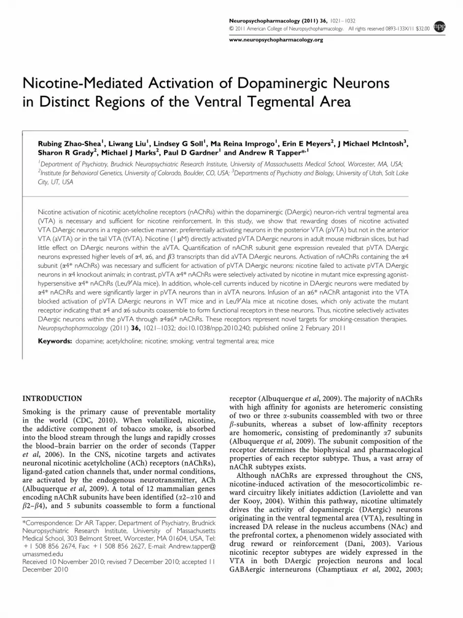
et al, 2009; Perrotti et al, 2005; Rodd et al, 2004; Shabat-Simon et al, 2008). Figure 1 illustrates representativetyrosine hydoxylase-immunolabeled midbrain slices fromeach VTA subregion along with their precise description.To determine whether DAergic neurons within the aVTA,pVTA, and tVTA are morphologically distinct, we analyzedsoma diameters in midbrain coronal sections fromC57BL/6J mice. Somas were visualized by TH immuno-labeling. The pVTA was anatomically recognized from theaVTA and tVTA by known landmarks (Shabat-Simon et al,2008) and stereotaxic coordinates based on the mouse brainatlas of Paxinos and Franklin (2000) (Figure 1a–c). A totalof 3301, 3877, and 832 TH-immunoreactive (TH-ir) neuronswithin the aVTA, pVTA, and tVTA, respectively, weremeasured from 3 mice. Neurons were classified into sixgroups based on diameter size (Figure 1d). Two-wayANOVA revealed that there was a significant main effectof brain region (F2, 36¼ 615.3, po0.0001), neuron size(F5, 36¼ 725.3.27, po0.001), and a significant brain re-gion� neuron size interaction (F10, 36¼ 193.3, po0.001).The pVTA contained significantly more small DAergicneurons than did the aVTA or tVTA (ie, neurons smallerthan 10–15 mm, Figure 1d). In contrast, there was asignificantly greater percentage of large-size DAergicneurons located in the aVTA compared with the pVTA ortVTA (ie, neurons with soma diameter 20–30 mm,Figure 1d). In addition, analysis of the number of DAergic
neurons per unit area (420 mm� 320 mm) indicated thatthere was a small but significantly greater density of TH-irneurons in the pVTA than in the aVTA (po0.01), whereasthere were much fewer TH-ir neurons in the tVTA than inthe aVTA and pVTA (po0.001, Figure 1e).
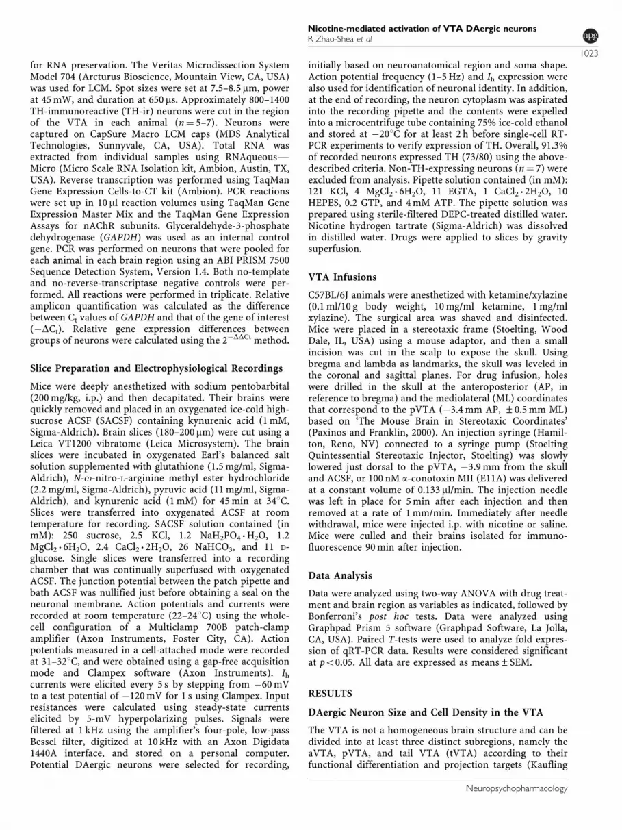
Acute Nicotine Preferentially Activates DAergicNeurons within the pVTA
Previous studies have indicated that VTA subregions havedifferent roles in modulating the rewarding properties ofseveral drugs of abuse (Ikemoto, 2007; Ikemoto and Wise,2002; Olson et al, 2005; Shabat-Simon et al, 2008). Todetermine whether nicotine preferentially activates the VTAin a subregion-selective manner, we challenged C57BL/6Jmice with nicotine and counted the number of activatedDAergic neurons in the aVTA, pVTA, and tVTA. Expressionof the immediate early gene, c-Fos, was used as a measure ofneuronal activation (Cole et al, 1989) and was analyzed inTH-ir neurons within the VTA by double-labeling immuno-fluorescence. Two-way ANOVA indicated that therewas a significant main effect of brain region (F8, 54¼ 99.4,po0.0001), treatment (F2, 54¼ 444.4, po0.001), and a signi-ficant brain region� treatment interaction (F16, 54¼ 70.7,po0.001). Mice challenged with 0.5mg/kg (i.p.) nicotine, adose that conditions a place preference (ie, a ‘rewarding’dose (Tapper et al, 2004)), exhibited a significant increase in
Figure 1 Definition, size, and density of DAergic neurons within the aVTA, the pVTA, and the tVTA. (a–c) Definitions of VTA subregions (Ikemoto, 2007;Kaufling et al, 2009; Paxinos and Franklin, 2000; Perrotti et al, 2005; Shabat-Simon et al, 2008). Representative midbrain slices containing the aVTA, pVTA, ortVTA. TH-ir neurons are labeled red. Brain region borders are outlined in white. (Panel a) aVTA (bregma from �2.92 to �3.28mm): defined as the regiondorsal to the medial mammillary nucleus and medial to the substantia nigra pars compacta (SNC). It contains two subregions: the ventral tegmental arearostral (VTAR) and the parabrachial pigmental area (PBP). The aVTA does not include midline nuclei such as the interfascicular nucleus (IF) and the rostrallinear nucleus (RLi). A10 DAergic neurons located in the supramammillary nucleus described by Hokfelt et al (1984a, b) also are not included in the aVTA.(Panel b) pVTA (bregma from �3.28 to �3.80mm): defined as the region dorsal to the interpeduncular nucleus (IPR), medial to the SNC and ventral to thered nucleus (RPC and/or RMC). It contains three subregions: the PBP, ventral tegmental area caudal (VTAC), and the paranigral nucleus (PN). The pVTAdoes not include the midline nuclei, such as the IF and the RLi and/or the caudal linear nucleus (CLi). A10 DAergic neurons located in the dorsal raphenucleus described by Hokfelt et al (1984a, b) are not included in the pVTA. (Panel c) tVTA (bregma from �3.80 to �4.04mm): defined as the most caudalextent of the VTA. Rostrally, the tVTA is limited to a subregion posterior to the PN and dorsolateral to the IP. More caudally, the position of the tVTA shiftsdorsally and slightly laterally to become embedded within the superior cerebellar peduncle decussation (SCP). The tVTA has a low density of DAergicneurons and a high density of GABAergic neurons. The tVTA does not include the midline nuclei RLi or CLi. (Panel d) Quantification of DAergic neuronsoma size in the aVTA (open bar), the pVTA (filled bar), and the tVTA (gray bar). Each bar represents the percentage of DAergic neurons with the indicatedsoma diameter (x axis in mm). (Panel e) Bar graph representation of DAergic neuron density in the aVTA, the pVTA, and the tVTA. In all, 2508–2864DAergic neurons were measured per mouse (n¼ 3). **po0.01, ***po0.001 compared with that of aVTA. #po0.001 compared with that of aVTA and pVTA.
Nicotine-mediated activation of VTA DAergic neuronsR Zhao-Shea et al
1024
Neuropsychopharmacology
the number of neurons that were both TH-ir and c-Fos-ir(TH-ir, c-Fos-ir) compared with control animals. TH-ir,c-Fos-ir neurons were restricted to the pVTA (�3.28 to�3.64mm from the bregma, Figure 2a and b). Preinjectionof 3mg/kg mecamylamine, a nonselective nAChR antago-nist, 15min before nicotine treatment, significantly reducedthe number of TH-ir, c-Fos-ir neurons compared with asaline preinjection. There were no significant effects oftreatment on the number of TH-ir, c-Fos-ir neurons withinthe aVTA or tVTA when compared with saline injection(Figure 2a and b). Nicotine also increased the numberof TH-non-ir, c-Fos-ir neurons selectively in the pVTA,but the difference in the number of activated neuronscompared with a saline injection was smaller than that ofTH-ir neurons activated by nicotine (Supplementary Figure 1).Owing to the relatively small number of DAergic neuronspresent in the tVTA, we excluded this VTA subregion from
further analysis. Acute nicotine could significantly activateaVTA DAergic neurons, but only at much higher doses.Thus, mice challenged with 2mg/kg nicotine exhibited asignificant increase in the number of TH-ir, c-Fos-ir aVTAneurons than did saline-injected animals (7.88±1.38 perslice compared with 0.34±0.09 per slice, respectively,po0.01, Supplementary Figure 2). Taken together, thesedata indicate that the rewarding, lower dose of nicotineselectively activates DAergic neurons in the pVTA.
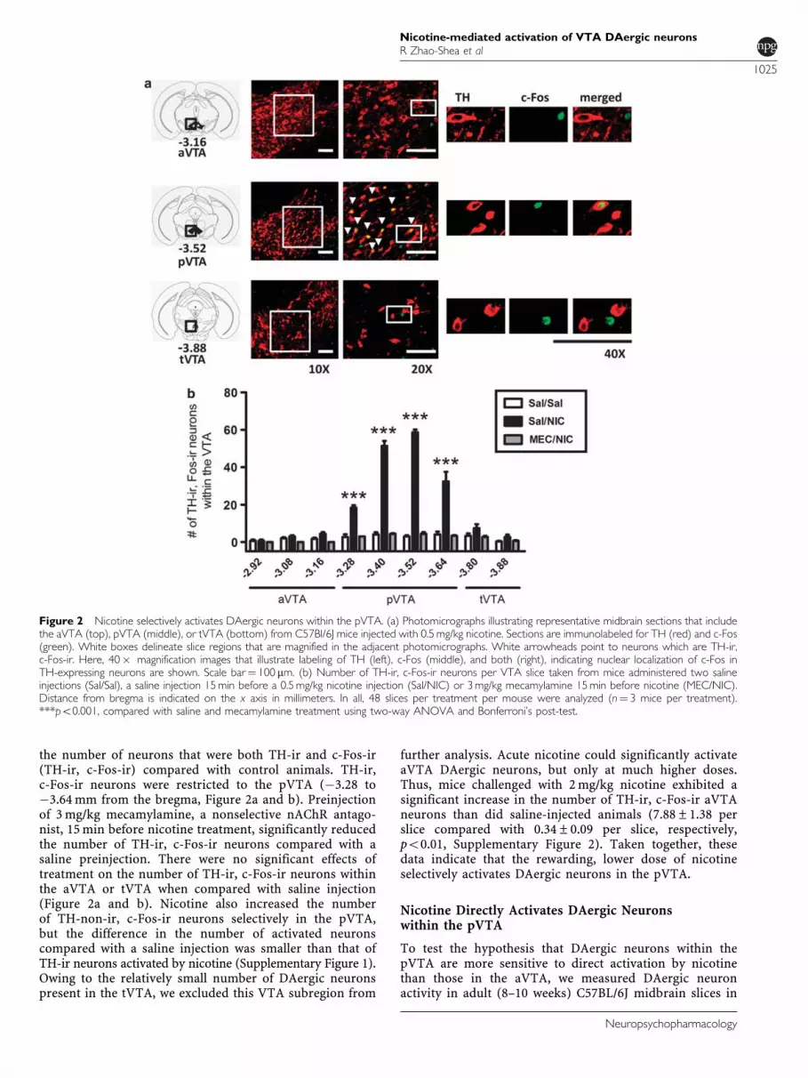
Nicotine Directly Activates DAergic Neuronswithin the pVTA
To test the hypothesis that DAergic neurons within thepVTA are more sensitive to direct activation by nicotinethan those in the aVTA, we measured DAergic neuronactivity in adult (8–10 weeks) C57BL/6J midbrain slices in
Figure 2 Nicotine selectively activates DAergic neurons within the pVTA. (a) Photomicrographs illustrating representative midbrain sections that includethe aVTA (top), pVTA (middle), or tVTA (bottom) from C57Bl/6J mice injected with 0.5mg/kg nicotine. Sections are immunolabeled for TH (red) and c-Fos(green). White boxes delineate slice regions that are magnified in the adjacent photomicrographs. White arrowheads point to neurons which are TH-ir,c-Fos-ir. Here, 40� magnification images that illustrate labeling of TH (left), c-Fos (middle), and both (right), indicating nuclear localization of c-Fos inTH-expressing neurons are shown. Scale bar¼ 100 mm. (b) Number of TH-ir, c-Fos-ir neurons per VTA slice taken from mice administered two salineinjections (Sal/Sal), a saline injection 15min before a 0.5mg/kg nicotine injection (Sal/NIC) or 3mg/kg mecamylamine 15min before nicotine (MEC/NIC).Distance from bregma is indicated on the x axis in millimeters. In all, 48 slices per treatment per mouse were analyzed (n¼ 3 mice per treatment).***po0.001, compared with saline and mecamylamine treatment using two-way ANOVA and Bonferroni’s post-test.
Nicotine-mediated activation of VTA DAergic neuronsR Zhao-Shea et al
1025
Neuropsychopharmacology
response to nicotine using whole-cell patch-clamp electro-physiology. DAergic neurons were identified based onexpression of the hyperpolarization-activated cation cur-rent, Ih (Figure 3c), and baseline firing frequency (1–5Hz,Figure 3b). In addition, after each recording, the content ofthe cell was aspirated into the patch pipette, and single-neuronal RT-PCR was performed to verify TH expression.DAergic neurons within the aVTA and the pVTA didnot differ in resting membrane potential (�48.4±2.7mVand �52.7±1.7mV, respectively) input resistance (289.0±43.0MO and 317.9±122.0MO, respectively), or firing freq-uency (3.75±1.5Hz and 4.24±0.52Hz, respectively). Tomeasure the direct activation by nicotine, we applied 1mMof the drug, a concentration within the range of peaknicotine blood levels found in smokers (Henningfield et al,1993; Russell et al, 1980). Nicotine was bath applied inthe presence of a cocktail of inhibitors including CNQX,bicuculline, and atropine to block NMDA, GABAa, andmuscarinic receptors, respectively. The firing frequencyof DAergic neurons within the aVTA exhibited a smallincrease compared with baseline in response to nicotine(B1.2-fold, n¼ 8/8 neurons, Figure 3d and f). In contrast,
43.8% (n¼ 7/16) of DAergic neurons within the pVTAexhibited a robust increase in firing (B4-fold comparedwith baseline, Figure 3e and f) in response to nicotine,which was reversible upon washout and could be blockedby the noncompetitive nicotinic receptor antagonist,mecamylamine (10 mM). The remaining 9 out of 16 pVTADAergic neurons had very small changes in firing frequencyin response to nicotine (1.18±0.08-fold compared withbaseline). To ensure that DAergic neuron responses tonicotine were not artificially influenced by temperature orthe whole-cell mode of recording, we also measured cell-attached nicotine-induced responses in VTA DAergicneurons in midbrain slices incubated at 31–321C. Similarto room temperature whole-cell recordings, our cell-attached data revealed that nicotine had little effect onaVTA DAergic neuron activity, whereas nicotine increasedDAergic neuron activity in the pVTA in all neurons tested(Figure 3g, 5/5 neurons). In addition, these responses werecompletely blocked by mecamylamine. Taken together,these data indicate that nicotine, at physiologically relevantconcentrations, activated a subset of DAergic neuronswithin the pVTA, but not the aVTA.
Figure 3 Direct activation of VTA DAergic neurons by nicotine. (a) Diagram of a mesocortical slice that includes the pVTA (red circle). Recordings weremade from both pVTA and aVTA neurons. (b) Current-clamp (I¼ 0) trace illustrating the baseline firing frequency of a VTA DAergic neuron. (c) Whole-cellvoltage clamp recording from a VTA neuron that expresses the hyperpolarization-activated cation current, Ih. The neuron was voltage clamped at �60mVand hyperpolarized to �120mV for 1.0 s before returning to the holding potential (Top). (d) Representative current-clamp (I¼ 0) recording from an aVTADAergic neuron at baseline (ACSF, left), during application of 1 mM nicotine (middle, 3min application), and after washout of nicotine (right).(e) Representative current-clamp (I¼ 0) recording from a pVTA DAergic neuron at baseline (ACSF, left), during application of 1 mM nicotine (middle, 3minapplication), and after washout of nicotine (right). Burst firing upon washout (3–7 spikes per burst) was seen in 5 of 7 neurons with robust nicotineresponses. (f) Summary of responses to nicotine in DAergic neurons from either the aVTA (white bar, n¼ 8/8) or the pVTA (black bar, n¼ 7/16). Each bargraph represents the fold change in action potential number produced by nicotine normalized to the baseline firing frequency.The response to nicotine in pVTA DAergic neurons was blocked by pre-exposure to 10 mM mecamylamine (gray bar). (g) Average response tonicotine as described in panel f (n¼ 5 neurons per region). Responses were recorded in cell-attached mode at 30–321C. **po0.01.
Nicotine-mediated activation of VTA DAergic neuronsR Zhao-Shea et al
1026
Neuropsychopharmacology
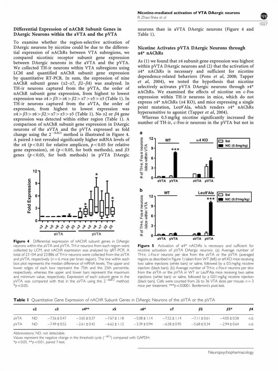
Differential Expression of nAChR Subunit Genes inDAergic Neurons within the aVTA and the pVTA
To examine whether the region-selective activation ofDAergic neurons by nicotine could be due to the differen-tial expression of nAChRs between VTA subregions, wecompared nicotinic receptor subunit gene expressionbetween DAergic neurons in the aVTA and the pVTA.We collected TH-ir neurons within VTA subregions usingLCM and quantified nAChR subunit gene expressionby quantitative RT-PCR. In sum, the expression of ninenAChR subunit genes (a2–a7, b2–b4) was analyzed. InTH-ir neurons captured from the pVTA, the order ofnAChR subunit gene expression, from highest to lowestexpression was a44b34a64b24a74a54a3 (Table 1). InTH-ir neurons captured from the aVTA, the order ofexpression, from highest to lowest expression wasa44b34a64b24a74a34a5 (Table 1). No a2 or b4 geneexpression was detected within either region (Table 1). Acomparison of nAChR subunit gene expression in DAergicneurons of the aVTA and the pVTA expressed as foldchange using the 2�DDCt method is illustrated in Figure 4.A paired t-test revealed significantly higher mRNA levels ofthe a4 (po0.01 for relative amplicon, po0.05 for relativegene expression), a6 (po0.05, for both methods), and b3genes (po0.05, for both methods) in pVTA DAergic
neurons than in aVTA DAergic neurons (Figure 4 andTable 1).
Nicotine Activates pVTA DAergic Neurons througha4* nAChRs
As (1) we found that a4 subunit gene expression was highestwithin pVTA DAergic neurons and (2) that the activation ofa4* nAChRs is necessary and sufficient for nicotinedependence-related behaviors (Pons et al, 2008; Tapperet al, 2004), we tested the hypothesis that nicotineselectively activates pVTA DAergic neurons through a4*nAChRs. We examined the effects of nicotine on c-Fosexpression within TH-ir neurons in mice, which do notexpress a4* nAChRs (a4 KO), and mice expressing a singlepoint mutation, Leu90Ala, which renders a4* nAChRshypersensitive to agonist (Tapper et al, 2004).Whereas 0.5mg/kg nicotine significantly increased the
number of TH-ir, c-Fos-ir neurons in the pVTA but not in
Table 1 Quantitative Gene Expression of nAChR Subunit Genes in DAergic Neurons of the aVTA or the pVTA
a2 a3 a4** a5 a6* a7 b2 b3* b4
aVTA ND �7.56±0.47 �3.65±0.37 �7.67±1.18 �5.08±1.14 �7.52±1.14 �7.11±0.61 �4.05±0.58 n.d.
pVTA ND �7.49±0.52 �2.61±0.42 �6.62±1.12 �3.39±0.94 �6.58±0.95 �5.68±0.34 �2.94±0.64 n.d.
Abbreviations: ND, not detectable.Values represent the negative change in the threshold cycle (�DCt) compared with GAPDH.*po0.05, **po0.01, paired T-test.
Figure 4 Differential expression of nAChR subunit genes in DAergicneurons within the aVTA and pVTA. TH-ir neurons from each region werecollected by LCM, and nAChR expression was analyzed by qRT-PCR. Atotal of 23 104 and 23 886 of TH-ir neurons were collected from the aVTAand pVTA, respectively (n¼ 6 mice per brain region). The line within eachbox plot represents the median difference of mRNA levels. The upper andlower edges of each box represent the 75th and the 25th percentile,respectively, whereas the upper and lower bars represent the maximumand minimum value, respectively. Expression of each subunit gene in thepVTA was compared with that in the aVTA using the 2�DDCt method.*po0.05.
Figure 5 Activation of a4* nAChRs is necessary and sufficient fornicotine activation of pVTA DAergic neurons. (a) Average number ofTH-ir, c-Fos-ir neurons per slice from the aVTA or the pVTA (averagedregions as described in Figure 1) taken fromWT (left) or a4 KO mice receivingtwo saline injections (white bars) or saline, followed by a 0.5mg/kg nicotineinjection (black bars). (b) Average number of TH-ir, c-Fos-ir neurons per slicefrom the aVTA or the pVTA in WT or Leu90Ala mice receiving two salineinjections (white bars) or saline, followed by a 0.01mg/kg nicotine injection(black bars). Cells were counted from 26 to 36 VTA slices per mouse. n¼ 3mice per treatment. ***po0.0001. Bonferroni’s post-test.
Nicotine-mediated activation of VTA DAergic neuronsR Zhao-Shea et al
1027
Neuropsychopharmacology

the aVTA of WT mice (Figure 5a), there was less of an effectof the drug on a4 KO animals. We did observe elevatedbasal c-Fos expression in a4 KO mice likely reflectingthe disinhibition of DAergic neurons in these animals asreported previously (Marubio et al, 2003). In KOs, two-wayANOVA revealed that there was a significant main effect ofbrain region (F1, 10¼ 26.39, po0.001), but no significantmain effect of treatment (0.5mg/kg nicotine, F1, 10¼ 2.539,p¼ 0.142) or brain region� treatment interaction (F1, 10¼3.848, p¼ 0.078). Thus, no significant differences wereobserved in the number of TH-ir, c-Fos-ir neurons betweensaline- and nicotine-injected animals either within theaVTA or within the pVTA (Figure 5a), indicating that theexpression of a4* nAChRs is required for nicotine activa-tion of pVTA DAergic neurons.In response to 0.01mg/kg nicotine, both WT aVTA and
pVTA DAergic neurons were not activated by nicotine(Figure 5b). However, in Leu90Ala mice, two-way ANOVArevealed that there was a significant main effect of brainregion (F1, 9¼ 173.1, po0.001), treatment (F1, 9¼ 189.6,po0.001), and a significant brain region� treatmentinteraction (F1, 9¼ 148.4, po0.001). Thus, in contrast to a4KO mice, Leu90Ala mice challenged with a single injection of0.01mg/kg nicotine, the rewarding dose of nicotine in theseanimals (Tapper et al, 2004) and one that is 50-fold lowerthan the rewarding dose in WT mice, exhibited a significantand dramatic increase in the number of TH-ir, c-Fos-irneurons within the pVTA (po0.001; Figure 5b) comparedwith saline-injected animals, whereas the aVTA was notaffected (Figure 5b). Importantly, this low dose of nicotinedid not activate DAergic neurons in WT animals, indicating
that activation in the pVTA of Leu90Ala mice by 0.01mg/kgnicotine is due solely to activation of a4* nAChRs. Takentogether, these data suggest that activation of a4* nAChRs isnecessary and sufficient for nicotine-induced activation ofDAergic neurons within the pVTA.
a4a6* nAChR-Binding Sites in the pVTA
On the basis of functional assays using striatal synapto-somes, a4a6* nAChRs have been shown to exhibit thehighest sensitivity to nicotine of any nAChR to date(Salminen et al, 2007). To determine whether a4a6*nAChRs are expressed in the pVTA, we quantified a6*nAChR-binding sites in WT and a4 KO mice using theb2* nAChR radioligand, 125I-A85380, in the presenceand absence of the cold a6*-nAChR selective ligand,a-conotoxin MII. WT and a4 KO coronal sections spanningposterior regions of the VTA (n¼ 6 mice) were used for thisanalysis. In the absence of a-conotoxin MII, total bindingwas 8.82±0.68 fmol/mg wet weight and 2.42±0.20 fmol/mgwet weight in WT and a4 KO mice, respectively. In thepresence of a-conotoxin MII, 125I-A85380 binding wasreduced to 7.43±0.45 fmol/mg wet weight in WT miceand to 1.66±0.76 fmol/mg wet weight in a4 KO mice. Thus,a-conotoxin MII-sensitive binding was reduced by 46% ina4 KO mice compared with WT mice (0.76±0.23 fmol/mgwet weight compared with 1.39±0.37 fmol/mg wet weight,respectively, Supplementary Figure 3). These data suggestthat a portion of pVTA a6* nAChR-binding sites alsocontain the a4 subunit.
Figure 6 Response to nicotine in DAergic neurons under voltage clamp. Representative whole-cell responses to nicotine in (a) aVTA or (b) pVTADAergic neurons from a4 KO (left), WT (middle), and Leu90Ala mice. Neurons were held at �60mV, and 1 mM nicotine was bath applied for 4min.(c) Average peak responses to 1 mM nicotine in the aVTA (white bar) or pVTA (black bar) DAergic neurons from each mouse line. Peak responses typicallyoccurred 2–3min after initial nicotine application. The gray bar illustrates the average peak response to nicotine in pVTA DAergic neurons after 10minapplication of 100 nM a-conotoxin MII (E11A). (n¼ 7 neurons per region per genotype). *po0.05.
Nicotine-mediated activation of VTA DAergic neuronsR Zhao-Shea et al
1028
Neuropsychopharmacology
Whole-Cell Currents Induced by Nicotine in aVTA andpVTA DAergic Neurons
To test the hypothesis that selective activation of pVTADAergic neurons by nicotine was directly mediated byincreased functional a4* nAChR expression, we measuredwhole-cell currents induced by bath application of 1mMnicotine in midbrain slices containing aVTA or pVTADAergic neurons from a4 KO, WT, and Leu90Ala mice. InWT mice, 1 mM nicotine induced whole-cell currents with asignificantly larger peak response in the pVTA comparedwith aVTA DAergic neurons (Figure 6a–c, middle panels).In slices obtained from a4 KO mice, 1mM nicotine did notelicit detectable currents in DAergic neurons from eitherVTA subregion (Figure 6a–c, left panels). Conversely, inslices obtained from Leu90Ala mice, 1 mM nicotine elicitedsignificant whole-cell currents in both aVTA and pVTADAergic neurons (Figure 6a–c, right panels). However, themagnitude of current was significantly larger in pVTADAergic neurons than in aVTA DAergic neurons. To testthe hypothesis that the nicotine-induced currents weobserved in pVTA DAergic neurons were mediated by a6*nAChRs, we tested the sensitivity of these currents to the a6selective antagonist, a-conotoxin MII (E11A). Pretreatmentwith 100 nM a-conotoxin MII (E11A) significantly blockedwhole-cell current in response to nicotine in pVTA DAergicneurons from WT and Leu90Ala mice (Figure 6c, middle andright panels, respectively). These data indicate that thefunctional expression of a4* nAChRs is greater in pVTADAergic neurons than in aVTA neurons and that thesereceptors also contain the a6 subunit.
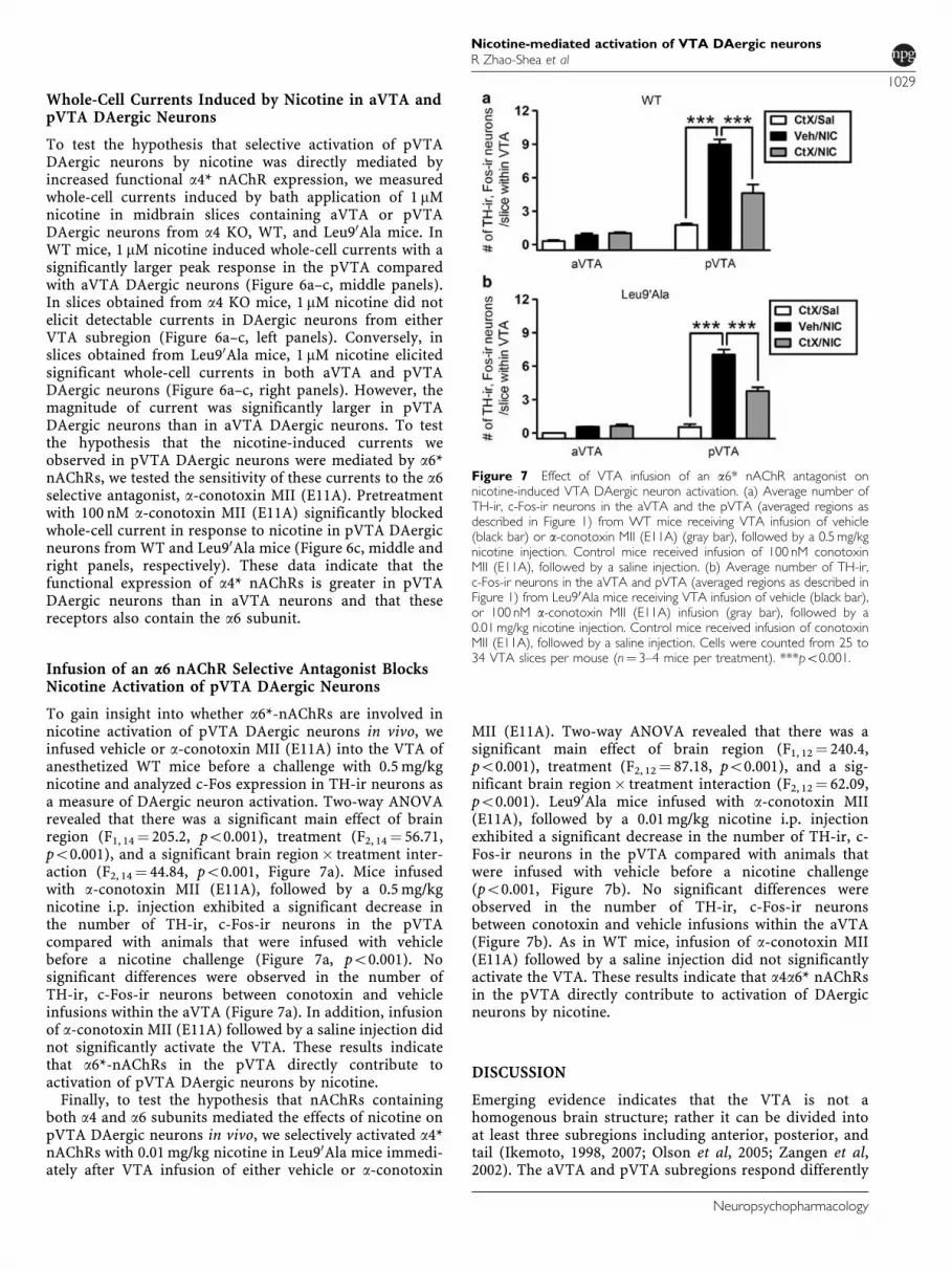
Infusion of an a6 nAChR Selective Antagonist BlocksNicotine Activation of pVTA DAergic Neurons
To gain insight into whether a6*-nAChRs are involved innicotine activation of pVTA DAergic neurons in vivo, weinfused vehicle or a-conotoxin MII (E11A) into the VTA ofanesthetized WT mice before a challenge with 0.5mg/kgnicotine and analyzed c-Fos expression in TH-ir neurons asa measure of DAergic neuron activation. Two-way ANOVArevealed that there was a significant main effect of brainregion (F1, 14¼ 205.2, po0.001), treatment (F2, 14¼ 56.71,po0.001), and a significant brain region� treatment inter-action (F2, 14¼ 44.84, po0.001, Figure 7a). Mice infusedwith a-conotoxin MII (E11A), followed by a 0.5mg/kgnicotine i.p. injection exhibited a significant decrease inthe number of TH-ir, c-Fos-ir neurons in the pVTAcompared with animals that were infused with vehiclebefore a nicotine challenge (Figure 7a, po0.001). Nosignificant differences were observed in the number ofTH-ir, c-Fos-ir neurons between conotoxin and vehicleinfusions within the aVTA (Figure 7a). In addition, infusionof a-conotoxin MII (E11A) followed by a saline injection didnot significantly activate the VTA. These results indicatethat a6*-nAChRs in the pVTA directly contribute toactivation of pVTA DAergic neurons by nicotine.Finally, to test the hypothesis that nAChRs containing
both a4 and a6 subunits mediated the effects of nicotine onpVTA DAergic neurons in vivo, we selectively activated a4*nAChRs with 0.01mg/kg nicotine in Leu90Ala mice immedi-ately after VTA infusion of either vehicle or a-conotoxin
MII (E11A). Two-way ANOVA revealed that there was asignificant main effect of brain region (F1, 12¼ 240.4,po0.001), treatment (F2, 12¼ 87.18, po0.001), and a sig-nificant brain region� treatment interaction (F2, 12¼ 62.09,po0.001). Leu90Ala mice infused with a-conotoxin MII(E11A), followed by a 0.01mg/kg nicotine i.p. injectionexhibited a significant decrease in the number of TH-ir, c-Fos-ir neurons in the pVTA compared with animals thatwere infused with vehicle before a nicotine challenge(po0.001, Figure 7b). No significant differences wereobserved in the number of TH-ir, c-Fos-ir neuronsbetween conotoxin and vehicle infusions within the aVTA(Figure 7b). As in WT mice, infusion of a-conotoxin MII(E11A) followed by a saline injection did not significantlyactivate the VTA. These results indicate that a4a6* nAChRsin the pVTA directly contribute to activation of DAergicneurons by nicotine.
DISCUSSION
Emerging evidence indicates that the VTA is not ahomogenous brain structure; rather it can be divided intoat least three subregions including anterior, posterior, andtail (Ikemoto, 1998, 2007; Olson et al, 2005; Zangen et al,2002). The aVTA and pVTA subregions respond differently
Figure 7 Effect of VTA infusion of an a6* nAChR antagonist onnicotine-induced VTA DAergic neuron activation. (a) Average number ofTH-ir, c-Fos-ir neurons in the aVTA and the pVTA (averaged regions asdescribed in Figure 1) from WT mice receiving VTA infusion of vehicle(black bar) or a-conotoxin MII (E11A) (gray bar), followed by a 0.5mg/kgnicotine injection. Control mice received infusion of 100 nM conotoxinMII (E11A), followed by a saline injection. (b) Average number of TH-ir,c-Fos-ir neurons in the aVTA and pVTA (averaged regions as described inFigure 1) from Leu90Ala mice receiving VTA infusion of vehicle (black bar),or 100 nM a-conotoxin MII (E11A) infusion (gray bar), followed by a0.01mg/kg nicotine injection. Control mice received infusion of conotoxinMII (E11A), followed by a saline injection. Cells were counted from 25 to34 VTA slices per mouse (n¼ 3–4 mice per treatment). ***po0.001.
Nicotine-mediated activation of VTA DAergic neuronsR Zhao-Shea et al
1029
Neuropsychopharmacology
to drugs of abuse, including opiates and alcohol (Boehmet al, 2002; Ericson et al, 2008; Rodd et al, 2004, 2005;Shabat-Simon et al, 2008). Supporting this hypothesis,Figure 1 illustrates that DAergic neurons within the aVTA,pVTA, and tVTA differ in size distribution and density,suggesting that distinct populations of DAergic neuronsmay exist. This is consistent with recent studies indicatingthat, in primates, individual DAergic neurons within theVTA differentially respond to rewarding or aversive stimuli(Matsumoto and Hikosaka, 2009). Furthermore, nicotine, atrewarding doses, selectively activates DAergic neuronswithin the pVTA and does not activate the aVTA or thetVTA. Activation of pVTA DAergic neurons is dependenton nAChR activation because preinjection of the non-competitive nAChR antagonist, mecamylamine, completelyblocked activation. Thus, DAergic neurons specificallywithin the pVTA are likely critical for nicotine reinforce-ment. These data support previous studies, indicating thatrats will self-administer nicotine directly into the posterior,but not into the aVTA (Ikemoto et al, 2006). Nicotine canactivate DAergic neurons within the aVTA but only at ahigh, extraphysiological dose near the seizure-inducingthreshold for C57BL/6J mice (Fonck et al, 2005).Our data from midbrain slices indicate that pVTA
DAergic neurons are more sensitive to direct activation by1 mM nicotine compared with those in aVTA, suggestingthat nAChR sensitivity differs between regions. Thisconcentration of nicotine is within the range found insmokers’ blood (Henningfield et al, 1993; Russell et al,1980) and was bath applied to slices. As nicotine-induceddesensitization of nAChRs occurs rapidly (Mansvelder et al,2002a; Mansvelder and McGehee, 2002b), we cannot ruleout the possibility that smaller responses to nicotine inDAergic neurons are caused by nAChR desensitization.However, bath application of nicotine more accuratelymimics exposure from cigarette smoke, and our dataindicate that DAergic neurons only within the pVTA arerobustly activated by nicotine. This result was independentof temperature or mode of recording, although the responseto nicotine in the pVTA was more consistent in a cell-attached mode in which the integrity of the recordedneurons was retained. Previous studies have examinednicotine activation of DAergic neurons in midbrain slices,and our data are consistent with these studies in that (1)bath application of nicotine can directly activate theseneurons and (2) responses to nicotine in DAergic neuronsare variable likely reflecting multiple DAergic populationsthat express distinct nAChR subtypes (Dani et al, 2000;Fisher et al, 1998; Klink et al, 2001; Pidoplichko et al, 1997;Wooltorton et al, 2003). It is important to point out that themajority of studies using electrophysiology to analyzenicotine effects on VTA DAergic neurons in midbrain slicehave used young (ie, mostly 12–25-day-old) animals and didnot analyze VTA subregions in detail.Taken together, our immunohistochemical and biophysi-
cal analyses suggest that DAergic neurons within the aVTAand pVTA may express distinct nAChR subtypes, whichcould explain the differential response to nicotine. This alsosupports a recent study that identified three distinct whole-cell current responses to nicotine in acutely dissociatedVTA DAergic neurons (Yang et al, 2009). Indeed, DAergicneurons within the pVTA exhibited a significantly higher
expression of a4, a6, and b3 subunit genes than did DAergicneurons in the aVTA. Several studies have analyzed nAChRexpression in the VTA and substantia nigra pars compacta,and our data are in general agreement that expressions ofa4, a6, b2, and b3 nAChR subunit genes are abundant inDAergic neurons (Klink et al, 2001; Wooltorton et al, 2003).Our results indicate that activation of a4* nAChRs is
necessary and sufficient for activation of pVTA DAergicneurons. Analysis of c-Fos induction in DAergic neuronsindicated that nicotine failed to activate pVTA DAergicneurons in a4 KO mice, whereas selective activation of a4*nAChRs with low doses of nicotine in Leu90Ala mice wassufficient to activate pVTA DAergic neurons. In midbrainslices, DAergic neurons within the pVTA exhibited largernicotine-induced whole-cell current than did aVTA DAergicneurons, indicating that the observed increase in subunitgene transcripts was also reflected in greater functionalexpression of nAChRs. Indeed, functional nAChRs activatedby 1 mM nicotine in pVTA DAergic neurons containedthe a4 subunit because nicotine-induced current wasnot observed in a4 KO mice. In addition, pVTA DAergicneurons exhibited larger nicotine-induced whole-cell cur-rent than did aVTA DAergic neurons recorded fromLeu90Ala mice supporting the contribution of a4* nAChRsto the nicotine response in pVTA neurons.In WT mice, a6* nAChRs also contributed to nicotine
activation of pVTA DAergic neurons as infusion of a-conotoxin MII (E11A) into the VTA significantly blockedactivation. These data are in agreement with a recent studyindicating that VTA infusion of a-conotoxin MII blocksnicotine-induced DA release into the NAc and also blocksnicotine self-administration in rats (Gotti et al, 2010).Although conotoxin infusion significantly blocked nicotineactivation of the pVTA, it was not complete. Thus, wecannot rule out the possibility that other mechanisms, suchas nicotine-mediated glutamate release, may also contributeto activation of this brain region (Kenny et al, 2009;Mansvelder et al, 2002a). However, consistent with ourimmunohistochemical and gene expression data, nicotine-induced currents in WT pVTA DAergic neurons were alsoblocked by a-conotoxin MII (E11A) indicating a directeffect of nicotine by activation of a6* nAChRs expressed inDAergic neurons.Importantly, at least two subtypes of a6* nAChRs have
been identified, a6 (non-a4) b2* and a6a4b2* nAChRs, thelatter subtype being the most nicotine-sensitive receptoridentified to date (Grady et al, 2007; Salminen et al, 2007).The identity and functional role of these nAChR subtypeshave been elucidated predominantly using striatal synapto-somes, and our studies extend these observations toDAergic neurons within the pVTA. Our radioligand-bindingdata indicate that a portion of a6* nAChR-binding sites alsocontain the a4 subunit. In addition, nicotine-inducedcurrents in DAergic neurons were dependent on a4* nAChRexpression and blocked by a-conotoxin MII (E11A) in bothWT and Leu90Ala midbrain slices, indicating functionalexpression of a4a6* nAChRs in the VTA. Finally, selectiveactivation of pVTA DAergic neurons in Leu90Ala mice usinga small dose of nicotine that has no effect in WT mice, wasalso blocked by VTA infusion of a-conotoxin MII (E11A).These data provide the first direct evidence that functionala4a6* nAChRs contribute to nicotine-mediated activation of
Nicotine-mediated activation of VTA DAergic neuronsR Zhao-Shea et al
1030
Neuropsychopharmacology
pVTA DAergic neurons. Although a4* nAChRs are apredominant subtype expressed throughout the VTA,functional a4a6* nAChRs are enriched in pVTA DAergicneurons, such that these neurons are activated by lowerconcentrations of nicotine compared with DAergic neuronsin other subregions of the VTA. Thus, our data, incombination with the recent study by Gotti et al (2010)indicate that a6a4b2* nAChRs expressed in DAergicneurons of the pVTA mediate the reinforcing propertiesof nicotine.Taken together, our data indicate that, at rewarding
doses, nicotine selectively activates DAergic neurons withinthe posterior subregion of the VTA through a4a6* nAChRs.These receptors could provide a novel molecular target forsmoking-cessation therapeutics.
ACKNOWLEDGEMENTS
This study was supported by the National Institute On AlcoholAbuse and Alcoholism award numbers R01AA017656 andR21AA018042 (ART), the National Institute of Mental HealthR01MH53631 (JMM), the National Institute on Drug Abuseaward numbers DA003194 (MJM) and DA012242 (MJM andJMM), and the National Institute on Neurological Disordersand Stroke award number R01NS030243 (PDG). The contentis solely the responsibility of the authors and does notnecessarily represent the official views of the National Instituteon Alcohol Abuse and Alcoholism, the National Institute onDrug Abuse, the National Institute on Neurological Disordersand Stroke, or the National Institutes of Health.
DISCLOSURE
The authors declare no conflict of interest.
REFERENCES
Albuquerque EX, Pereira EF, Alkondon M, Rogers SW (2009).Mammalian nicotinic acetylcholine receptors: from structure tofunction. Physiol Rev 89: 73–120.
Boehm II SL, Piercy MM, Bergstrom HC, Phillips TJ (2002).Ventral tegmental area region governs GABA(B) receptormodulation of ethanol-stimulated activity in mice. Neuroscience115: 185–200.
CDC (2010). Vital signs: current cigarette smoking among adultsaged X18 yearsFUnited States, 2009. MMWR Morb MortalWkly Rep 59: 1135–1140.
Champtiaux N, Gotti C, Cordero-Erausquin M, David DJ,Przybylski C, Lena C et al (2003). Subunit composition offunctional nicotinic receptors in dopaminergic neurons inves-tigated with knock-out mice. J Neurosci 23: 7820–7829.
Champtiaux N, Han ZY, Bessis A, Rossi FM, Zoli M, Marubio Let al (2002). Distribution and pharmacology of alpha 6-contain-ing nicotinic acetylcholine receptors analyzed with mutant mice.J Neurosci 22: 1208–1217.
Cole AJ, Saffen DW, Baraban JM, Worley PF (1989). Rapid increaseof an immediate early gene messenger RNA in hippocampalneurons by synaptic NMDA receptor activation. Nature 340:474–476.
Dani JA (2003). Roles of dopamine signaling in nicotine addiction.Mol Psychiatry 8: 255–256.
Dani JA, Radcliffe KA, Pidoplichko VI (2000). Variations indesensitization of nicotinic acetylcholine receptors from
hippocampus and midbrain dopamine areas. Eur J Pharmacol393: 31–38.
Ericson M, Lof E, Stomberg R, Chau P, Soderpalm B (2008).Nicotinic acetylcholine receptors in the anterior, but notposterior, ventral tegmental area mediate ethanol-inducedelevation of accumbal dopamine levels. J Pharmacol Exp Ther326: 76–82.
Fisher JL, Pidoplichko VI, Dani JA (1998). Nicotine modifies theactivity of ventral tegmental area dopaminergic neurons andhippocampal GABAergic neurons. J Physiol Paris 92: 209–213.
Fonck C, Cohen BN, Nashmi R, Whiteaker P, Wagenaar DA,Rodrigues-Pinguet N et al (2005). Novel seizure phenotype andsleep disruptions in knock-in mice with hypersensitive alpha 4*nicotinic receptors. J Neurosci 25: 11396–11411.
Gotti C, Guiducci S, Tedesco V, Corbioli S, Zanetti L, Moretti M et al(2010). Nicotinic acetylcholine receptors in the mesolimbic path-way: primary role of ventral tegmental area alpha6beta2* receptorsin mediating systemic nicotine effects on dopamine release,locomotion, and reinforcement. J Neurosci 30: 5311–5325.
Grady SR, Salminen O, Laverty DC, Whiteaker P, McIntosh JM,Collins AC et al (2007). The subtypes of nicotinic acetylcholinereceptors on dopaminergic terminals of mouse striatum.Biochem Pharmacol 74: 1235–1246.
Hendrickson LM, Zhao-Shea R, Tapper AR (2009). Modulation ofethanol drinking-in-the-dark by mecamylamine and nicotinicacetylcholine receptor agonists in C57BL/6J mice. Psychophar-macology (Berl) 204: 563–572.
Henningfield JE, Stapleton JM, Benowitz NL, Grayson RF, LondonED (1993). Higher levels of nicotine in arterial than in venousblood after cigarette smoking. Drug Alcohol Depend 33: 23–29.
Hokfelt T, Everitt BJ, Theodorsson-Norheim E, Goldstein M(1984a). Occurrence of neurotensinlike immunoreactivity insubpopulations of hypothalamic, mesencephalic, and medullarycatecholamine neurons. J Comp Neurol 222: 543–559.
Hokfelt T, Johansson O, Goldstein M (1984b). Chemical anatomyof the brain. Science 225: 1326–1334.
Ikemoto S (2007). Dopamine reward circuitry: two projectionsystems from the ventral midbrain to the nucleus accumbens-olfactory tubercle complex. Brain Res Rev 56: 27–78.
Ikemoto S, Murphy JM, McBride WJ (1998). Regional differenceswithin the rat ventral tegmental area for muscimol self-infusions.Pharmacol Biochem Behav 61: 87–92.
Ikemoto S, Qin M, Liu ZH (2006). Primary reinforcing effects ofnicotine are triggered from multiple regions both inside andoutside the ventral tegmental area. J Neurosci 26: 723–730.
Ikemoto S, Wise RA (2002). Rewarding effects of the cholinergicagents carbachol and neostigmine in the posterior ventraltegmental area. J Neurosci 22: 9895–9904.
Kaufling J, Veinante P, Pawlowski SA, Freund-Mercier MJ,Barrot M (2009). Afferents to the GABAergic tail of the ventraltegmental area in the rat. J Comp Neurol 513: 597–621.
Kenny PJ, Chartoff E, Roberto M, Carlezon Jr WA, Markou A(2009). NMDA receptors regulate nicotine-enhanced brainreward function and intravenous nicotine self-administration:role of the ventral tegmental area and central nucleus of theamygdala. Neuropsychopharmacology 34: 266–281.
Klink R, de Kerchove d’Exaerde A, Zoli M, Changeux JP (2001).Molecular and physiological diversity of nicotinic acetylcholinereceptors in the midbrain dopaminergic nuclei. J Neurosci 21:1452–1463.
Laviolette SR, van der Kooy D (2004). The neurobiology of nicotineaddiction: bridging the gap from molecules to behaviour. NatRev Neurosci 5: 55–65.
Mansvelder HD, Keath JR, McGehee DS (2002a). Synapticmechanisms underlie nicotine-induced excitability of brainreward areas. Neuron 33: 905–919.
Mansvelder HD, McGehee DS (2002b). Cellular and synapticmechanisms of nicotine addiction. J Neurobiol 53: 606–617.
Nicotine-mediated activation of VTA DAergic neuronsR Zhao-Shea et al
1031
Neuropsychopharmacology
Marubio LM, Gardier AM, Durier S, David D, Klink R, Arroyo-Jimenez MM et al (2003). Effects of nicotine in the dopaminergicsystem of mice lacking the alpha4 subunit of neuronal nicotinicacetylcholine receptors. Eur J Neurosci 17: 1329–1337.
Maskos U, Molles BE, Pons S, Besson M, Guiard BP, Guilloux JPet al (2005). Nicotine reinforcement and cognition restored bytargeted expression of nicotinic receptors. Nature 436: 103–107.
Matsumoto M, Hikosaka O (2009). Two types of dopamine neurondistinctly convey positive and negative motivational signals.Nature 459: 837–841.
McIntosh JM, Azam L, Staheli S, Dowell C, Lindstrom JM,Kuryatov A et al (2004). Analogs of alpha-conotoxin MII areselective for alpha6-containing nicotinic acetylcholine receptors.Mol Pharmacol 65: 944–952.
National Research Council (1996). Guide for the Care and use ofLaboratory Animals. National Academy Press: Washington, DC.
Olson VG, Zabetian CP, Bolanos CA, Edwards S, Barrot M,Eisch AJ et al (2005). Regulation of drug reward by cAMPresponse element-binding protein: evidence for two functionallydistinct subregions of the ventral tegmental area. J Neurosci 25:5553–5562.
Paxinos G, Franklin K (2000). The Mouse Brain in StereotaxicCoordinates 2nd edn. Academic Press: San Diego, CA. p 296.
Perrotti LI, Bolanos CA, Choi KH, Russo SJ, Edwards S,Ulery PG et al (2005). DeltaFosB accumulates in a GABAergiccell population in the posterior tail of the ventral tegmental areaafter psychostimulant treatment. Eur J Neurosci 21: 2817–2824.
Picciotto MR, Zoli M, Rimondini R, Lena C, Marubio LM, Pich EMet al (1998). Acetylcholine receptors containing the beta2subunit are involved in the reinforcing properties of nicotine.Nature 391: 173–177.
Pidoplichko VI, DeBiasi M, Williams JT, Dani JA (1997). Nicotineactivates and desensitizes midbrain dopamine neurons.Nature 390: 401–404.
Pons S, Fattore L, Cossu G, Tolu S, Porcu E, McIntosh JM et al(2008). Crucial role of alpha4 and alpha6 nicotinic acetylcholinereceptor subunits from ventral tegmental area in systemicnicotine self-administration. J Neurosci 28: 12318–12327.
Rodd ZA, Bell RL, Zhang Y, Murphy JM, Goldstein A, Zaffaroni Aet al (2005). Regional heterogeneity for the intracranial self-administration of ethanol and acetaldehyde within theventral tegmental area of alcohol-preferring (P) rats: involve-ment of dopamine and serotonin. Neuropsychopharmacology 30:330–338.
Rodd ZA, Melendez RI, Bell RL, Kuc KA, Zhang Y, Murphy JM et al(2004). Intracranial self-administration of ethanol within theventral tegmental area of male Wistar rats: evidence forinvolvement of dopamine neurons. J Neurosci 24: 1050–1057.
Ross SA, Wong JY, Clifford JJ, Kinsella A, Massalas JS, Horne MKet al (2000). Phenotypic characterization of an alpha 4 neuronalnicotinic acetylcholine receptor subunit knock-out mouse.J Neurosci 20: 6431–6441.
Russell MA, Jarvis M, Iyer R, Feyerabend C (1980). Relation ofnicotine yield of cigarettes to blood nicotine concentrations insmokers. Br Med J 280: 972–976.
Salminen O, Drapeau JA, McIntosh JM, Collins AC, Marks MJ,Grady SR (2007). Pharmacology of alpha-conotoxin MII-sensitive subtypes of nicotinic acetylcholine receptors isolatedby breeding of null mutant mice. Mol Pharmacol 71: 1563–1571.
Shabat-Simon M, Levy D, Amir A, Rehavi M, Zangen A (2008).Dissociation between rewarding and psychomotor effects ofopiates: differential roles for glutamate receptors within anteriorand posterior portions of the ventral tegmental area. J Neurosci28: 8406–8416.
Tapper AR, McKinney SL, Nashmi R, Schwarz J, Deshpande P,Labarca C et al (2004). Nicotine activation of alpha4* receptors:sufficient for reward, tolerance, and sensitization. Science 306:1029–1032.
Tapper AR, Nashmi R, Lester HA (2006). Neuronal nicotinicacetylcholine receptors and nicotine dependence. Cell Biology ofAddiction. Cold Spring Harbor Laboratory Press: Cold Spring,Harbor, NY. pp 179–191.
Wooltorton JR, Pidoplichko VI, Broide RS, Dani JA (2003).Differential desensitization and distribution of nicotinic acet-ylcholine receptor subtypes in midbrain dopamine areas.J Neurosci 23: 3176–3185.
Yang K, Hu J, Lucero L, Liu Q, Zheng C, Zhen X et al (2009).Distinctive nicotinic acetylcholine receptor functional pheno-types of rat ventral tegmental area dopaminergic neurons.J Physiol 587(Part 2): 345–361.
Zangen A, Ikemoto S, Zadina JE, Wise RA (2002). Rewarding andpsychomotor stimulant effects of endomorphin-1: anteroposter-ior differences within the ventral tegmental area and lack ofeffect in nucleus accumbens. J Neurosci 22: 7225–7233.
Zangen A, Solinas M, Ikemoto S, Goldberg SR, Wise RA(2006). Two brain sites for cannabinoid reward. J Neurosci 26:4901–4907.
Supplementary Information accompanies the paper on the Neuropsychopharmacology website (http://www.nature.com/npp)
Nicotine-mediated activation of VTA DAergic neuronsR Zhao-Shea et al
1032
Neuropsychopharmacology