laminin deficits induce alterations in the development of dopaminergic neurons in the mouse retina
TRANSCRIPT
Laminin deficits induce alterations in the development ofdopaminergic neurons in the mouse retina
Viktória Dénes1,2, Paul Witkovsky3, Manuel Koch4, Dale D. Hunter2, Germán Pinzón-Duarte1,2, and William J. Brunken1,21Department of Anatomy and Cellular Biology, Tufts University School of Medicine, Boston,Massachusetts2Tufts Center for Vision Research, Boston, Massachusetts3Department of Ophthalmology, New York University School of Medicine, New York, New York4Center for Biochemistry and Department of Dermatology, University of Köln, Köln, Germany
AbstractGenetically modified mice lacking the β2 laminin chain (β2null), the γ3 laminin chain (γ3 null), orboth β2/γ3 chains (compound null) were produced. The development of tyrosine hydroxylase (TH)immunoreactive neurons in these mouse lines was studied between birth and postnatal day (P) 20.Compared to wild type mice, no alterations were seen in γ3 null mice. In β2 null mice, however, thelarge, type I TH neurons appeared later in development, were at a lower density and had reduced THimmunoreactivity, although TH process number and size were not altered. In the compound nullmouse, the same changes were observed together with reduced TH process outgrowth. Surprisingly,in the smaller, type II TH neurons, TH immunoreactivity was increased in laminin-deficientcompared to wild type mice. Other retinal defects we observed were a patchy disruption of the innerlimiting retinal basement membrane and a disoriented growth of Müller glial cells. Starburst and AIItype amacrine cells were not apparently altered in laminin-deficient relative to wild type mice. Wepostulate that laminin-dependent developmental signals are conveyed to TH amacrine neuronsthrough intermediate cell types, perhaps the Müller glial cell and/or the retinal ganglion cell.
KeywordsExtracellular matrix; Inner limiting; Membrane; Amacrine cell; Muller cell
IntroductionLaminins are heterotrimeric glycoproteins found in basement membranes and the extracellularmatrix of nervous tissue (Colognato & Yurchenco, 2000). The component molecules (chains)fall into three families, each of which has multiple members: 5α, 3β, and 3γ chains have beenidentified. Of the approximately 45 possible combinations of these chains, only 16 have beenidentified (Yurchenco & Wadsworth, 2004; Yan & Cheng, 2006). Cell surface receptormolecules such as integrins (DeCurtis & Reichardt, 1993), dystroglycans (Barresi & Campbell,2006), and syndecans (Suzuki et al., 2005) bind to laminins, and use this interaction to activatea variety of intracellular pathways involved in cell survival, growth and differentiation (Ivins
Copyright © 2007 Cambridge University PressAddress correspondence and reprint requests to: William J. Brunken, Department of Anatomy and Cell Biology, Box 5, State Universityof New York–Downstate Medical Center, 450 Clarkson Ave, Brooklyn, NY 11203. [email protected].
NIH Public AccessAuthor ManuscriptVis Neurosci. Author manuscript; available in PMC 2010 September 8.
Published in final edited form as:Vis Neurosci. 2007 ; 24(4): 549–562. doi:10.1017/S0952523807070514.
NIH
-PA Author Manuscript
NIH
-PA Author Manuscript
NIH
-PA Author Manuscript
et al., 2000; Clegg et al., 2000). Laminin defects are implicated in several developmentaldisorders in humans and mice, including muscular dystrophy in both the peripheral, and centralnervous systems (Colognato & Yurchenco, 2000; Miner & Yurchenco, 2004; Olson & Walsh,2002). Mutations in the human β2 gene result in Pierson’s syndrome, and other ocular disorders(Zenker et al., 2004, 2005).
In the mammalian retina, the focus of the present study, several laminin isoforms have beenidentified (Hunter et al., 1992; Libby et al., 2000; Aisenbrey et al., 2006). Laminins aredistributed widely within the retina, either in basement membranes (Bruch’s membrane, innerlimiting membrane, vascular basement membrane) or in the extracellular matrix, e.g., the outerplexiform layer (OPL) in which photoreceptors synapse with second-order retinal neurons andin the interphotoreceptor matrix. These studies demonstrated that the β2 chain is located in theOPL and in basement membranes of the retina, whereas the γ3 chain is deposited in retinalbasement membranes. A prior study (Libby et al., 1999) found that disruption of the lamininβ2 chain altered photoreceptor development in several ways, including synaptic disruption andalterations of photoreceptor outer segment morphology. On the other hand, the inner plexiformlayer (IPL), in which bipolar cells and inner retinal neurons make synaptic contacts, appearedunaltered. This prior study, however, did not exhaustively report the possible effects of laminindeletion on the inner retina. Because inner retinal neurons are found in multiple subtypes(MacNeil et al., 1999; Rockhill et al., 2002), here we explore the possibility that only certainof them might be affected by laminin deficits, leaving the overall appearance of the inner retinaunchanged. Specifically, we examine possible effects of particular laminin chain deletions onthe development of identifiable subtypes of inner retinal neuron: cholinergic, AII and tyrosinehydroxylase (TH)-containing amacrine cells.
Emphasis was placed on the TH amacrine cells because in preliminary screens it was the cellclass in which clear developmental deficits related to laminin deletions were noted. RetinalTH-containing neurons are of two subtypes, each having a characteristic size, shape and domainof process arborization in the IPL. The larger, type I TH immunoreactive (IR) amacrine(Mariani & Hokoc, 1988) is known to be dopaminergic (Ehinger & Floren, 1978), whereas theneurotransmitter released by the smaller, type II TH neuron is not yet definitely established.
We compared neuronal development in wild type mouse retinas and in mice of the same strainin which the genes encoding either laminin β2 or laminin γ3 were deleted by homologousrecombination techniques; we produced a mouse line with deletion of both β2 and γ3 chainsby crossing these knockout animals. Our results indicate that both the β2 laminin chain deletionand the double β2/γ3 laminin deletions were associated with substantial alterations in THneuronal development, including increased apoptosis, delayed process outgrowth and alteredTH levels; in contrast, the γ3 laminin deletion was not linked to evident modifications of THneuronal development. In none of the mutant mice we examined were changes noted in thedevelopment either of cholinergic or AII amacrine cells.
The same laminin chain deletions also resulted in disruption of the inner limiting membrane,a basement membrane that lines the vitreal surface of the retina. Moreover, the radial glial cellsof the retina, the Müller cells, showed a marked disarray of vitreal end feet and inner retinalexpansions, consistent with the finding that Müller cells express laminin receptor complexesand interact with laminins (Claudepierre et al., 2005, 2000; Noël et al., 2005; Méhes et al.,2002; Moukhles et al., 2000). Our data provide additional support for the idea that radial glialcells play crucial, if still poorly defined, roles in retinal development (Willbold & Layer,1998; Blackshaw et al., 2004).
Dénes et al. Page 2
Vis Neurosci. Author manuscript; available in PMC 2010 September 8.
NIH
-PA Author Manuscript
NIH
-PA Author Manuscript
NIH
-PA Author Manuscript
Methods and materialsAnimals
The lines of mice we used were bred and maintained and in the animal facility of TuftsUniversity. All procedures involving animals were approved by the Tufts University AnimalCare Committee and were in accordance with the standards established by the NationalInstitutes of Health and the Association for Research in Vision and Ophthalmology.
The standard recombinant procedures used for generating the null mutations in β2 have beenreported elsewhere (Noakes et al., 1995). Lamc3 −/− mice were generated using standardrecombination methods; a complete description is given elsewhere (Li, Y., French, M.,Burgeson, R.E., Koch, M., & Brunken, W.J., unpublished observation). In brief, a targetingvector for homologous recombination was created in which a 2.2 kb spanning exon 1 and partof intron 1 of the Lamc3 gene was deleted and replaced with promoterless IRES β-Geo cassette.The targeting construct was linearized and electroporated into 129/SvJ ES cells. Neomycin-resistant ES cell clones were selected, expanded, and injected into C57Bl/6 blastocysts for theproduction of chimeric mice. Mice from both lines (β2+/− and γ3+/−) were backcrossed towild type C57Bl/6J mice. β2null mice have renal and neuromuscular deficits that are lethaltypically during the fourth postnatal week; therefore they were maintained as heterozygotelines. γ3 null mice show only very subtle differences from wild type and are fertile; wemaintained them as heterozygote lines. To generate the compound null (β2−/−, γ3−/−) mice,we mated β2 +/− mice with γ3 −/− mice and using breeding pairs of β2+/−, γ3 −/− to generateall the progeny examined in this study. While these mice are on a mixed genetic backgroundof C57bl/6J and 129Sv/J, the founder mice of this line had been backcrossed at least five timesto C57bl/6J. In all cases, littermates were used as controls. β2 +/+, γ3 +/+ animals werelittermates of the β2null mice and the genotypes of the progeny were determined by PCR fromgenomic tail DNA.
Dissection and tissue processingMice were euthanized by a brief exposure to CO2, and then decapitated. The eyes wereenucleated, the cornea and the lens removed and the eyecups fixed by immersion in 4%paraformaldehyde in 0.1M phosphate buffer, pH 7.4 for 1 h at room temperature. Thereafterthe eyecups were washed 3 × 20 min in phosphate-buffered saline (PBS, 7.4 pH). Aftercryoprotection in sucrose solutions of increasing concentration (10, 20, 30% in PBS), frozensections (10 µm) were cut in a cryostat. The sections were mounted on Superfrost slides (FisherScientific), air-dried and processed for immunocytochemistry as described below. For wholemount preparations, eyecups were fixed, washed, and the retinas freed and processed forimmunocytochemistry, using either 3,3 diaminobenzidine tetrahydrochloride (DAB) orfluorescent secondary antibodies to visualize the immunostained cells.
Immunocytochemistry1. DAB staining—Whole mounts were pretreated with DMSO for 30 min followed by 3 ×10 min washes with 0.3% Triton-X 100 in PBS to increase the penetration of antibodymolecules. After pre-incubation in blocking solution (5% donkey serum, 1% bovine serumalbumin, 0.1% Triton-X 100 in PBS) whole mounts were incubated in anti-TH antibody (Table1) overnight at 4°C, washed in PBS (6 × 10 min) and placed into solution containing biotin-conjugated anti-rabbit IgG antibody (1:250) (Table 2) for 6 h at room temperature. After 6 ×10 min washes in PBS, preparations were incubated in extravidin-peroxidase complex (1:100)(Sigma) for 4 h. The retinas were rinsed in PBS prior to their incubation in 0.05% DAB in PBSfor 15 min, followed by 0.05% DAB and 0.01% hydrogen peroxide dissolved in PBS. Thereaction was stopped by a rinse in PBS.
Dénes et al. Page 3
Vis Neurosci. Author manuscript; available in PMC 2010 September 8.
NIH
-PA Author Manuscript
NIH
-PA Author Manuscript
NIH
-PA Author Manuscript
2. Double label immunofluorescent staining—Retinal sections were treated with 0.1%Triton-X 100 in PBS and incubated in blocking solution as described above. Mixtures of theprimary antibodies (Table 1) were left on the sections for 2–4 h then the sections were washed6 × 5 min in PBS. Immunoreactivity was detected by incubation with immunofluorescentsecondary antibodies conjugated to appropriate fluorochromes (Table 2) for 2–3 h. Afterrinsing in PBS (6 × 5 min), sections were mounted with Vectashield (Vector Laboratories).Two controls were performed for immunocytochemical labeling. Omission of the primaryantibodies in both the single- and double-labeling experiments resulted in no staining. Cross-reactivity of the non-corresponding primary and secondary antibodies was not detected.Reactions were carried out on tissues derived at least from three different animals.
Indolyl phosphate stainingFor demonstration of human placental alkaline phosphatase (PLAP) activity in 2 and 3 daysold transgenic mouse retinas, eyecups were fixed in 4% paraformaldehyde for 2 h at roomtemperature. Whole mount preparations were warmed to 65°C for 30 min to inactivate otherphosphatases. They were incubated in the dark for 3 h in 0.1% 5-bromo-4-chloro-3-indolylphosphate and 1% nitroblue tetrazolium solved in 0.1 M Tris-HCL buffer (pH 9.5) followedby rinsing in 0.02 EDTA solution. As a result, purple-blue reaction product appeared at thesurface of the neurons.
Western blottingTo obtain sufficient protein, four P20 retinas were processed for Western blotting from eachgenotype (wild type (wt); β2−/−:γ3+/+; β2+/+ :γ3−/−; β2−/−: γ3−/−). The tissue washomogenized by sonication and the protein extracted in lysis buffer containing 50 mM TRIS-HCl, 150 mM sodium chloride, 1% Nonidet P-40, 1% SDS, 0.5% sodium deoxycholate, 1%PMSF, 1% α2-macroglobulin. After establishing the protein concentration by BCA ProteinAssay Kit (Pierce), the samples were boiled for 5 min in sample buffer (62.5 mM Tris-HCl,10% glycerol, 2% SDS, 5% mercaptoethanol, 0.05% bromophenol blue, pH 6.8). Ten µgproteins from each sample was loaded and separated by electrophoresis through a 10 %polyacrylamide gel. The proteins were transferred to PVDF membrane (BioRad) by semi-dryimmunoblotting. Membranes were pre-incubated in a blocking buffer (5% milk protein, 0.1%Tween-20 in TBS) for 1 h at room temperature prior to the incubation with anti-TH antibody(1:5000) overnight at 4°C. The membranes then were rinsed in Tris-buffered saline, 0.1%Tween-20 (TBS-Tween) 3 × 10 min, and incubated with horse-radish peroxidase-conjugatedanti-rabbit IgG (1:10.000) (Molecular Probes) for 2 h at room temperature. Finally, themembranes were thoroughly washed in TBS-Tween solution, soaked in chemiluminescentsubstrate (PerkinElmer LAS Inc) for 1 min then exposed to film for signal detection.
StatisticsData populations are given as means ± standard errors. Data groups were evaluated forsignificant differences (p < 0.05) by a Student t-test.
Reverse transcription and real time PCRTotal RNA was extracted from wt, β2null, γ3null and compound null retinas aged P20 and P4using RNeasy Kit (Qiagen) according to the manufacturer’s instructions; cDNAs weresynthesized from 60 to 120 ng of total RNA. Primers designed for real time PCR were asfollows: for mouse TH, the forward primer was GCCGTCT CAGAGCAGGATAC, the reverseprimer was AGCATTTCCATC CCTCTCCT; for the mouse β actin, which served as aninternal control, the forward primer was GGCTATGCTCTCCCTCACG, the reverse primerwas CTTCTCTTTGATGTCACGCACG. For the mouse RPL13A control, the forward primerwas GAGGT CGGGTGGAAGTACCA and the reverse primer was TGCATCT
Dénes et al. Page 4
Vis Neurosci. Author manuscript; available in PMC 2010 September 8.
NIH
-PA Author Manuscript
NIH
-PA Author Manuscript
NIH
-PA Author Manuscript
TGGCCTTTTCCTT. Reactions were carried out in a 25 µl volume using Quantitect SYBRGreen PCR Kit (Qiagen). Each experiment was repeated twice in triplicate. The results wereanalyzed by the ΔCt method that reflects the difference in cycle threshold for the target generelative to that for β actin in each sample.
Data processingMicrographs were taken with an OrcaER digital camera (Hamamatsu Ltd, Tokyo, Japan) anddigital images recorded with the help of an image analyzer program (Openlab ImprovisionLtd., Lexington, MA or Auto Quant Imaging, Watervliet, NY). The digital images wereadjusted for intensity and contrast in Adobe Photoshop 7.0. Cell density, cell lengths andnearest neighbor analyses were performed on retinal whole mounts, using a semiautomaticcomputer-assisted image analysis system (Openlab). The microscopic field was examined witha 20x objective on a video screen. The distance of TH-IR neurons to their nearest neighborwas measured and the data were grouped and plotted in 10 µm increments. A sample size ofapproximately 2000 neurons was used to determine cell dimensions. These data were obtainedfrom four retinas derived from four different animals.
ResultsComparison of wild-type and laminin-deficient retinas at P20
As a first step in identifying laminin-dependent alterations in neuronal development, we studiedmouse retinas after 20 days of post-natal development (P20). Mice open their eyes around P14and by P20 retinal dopaminergic neurons have achieved their adult dimensions (Wulle &Schnitzer, 1989). We examined dopaminergic, cholinergic, and AII amacrine cells, usingantibodies specific for each of these subtypes of amacrine cell. In addition we used an anti-calretinin antibody, which labels several types of inner retinal neuron.
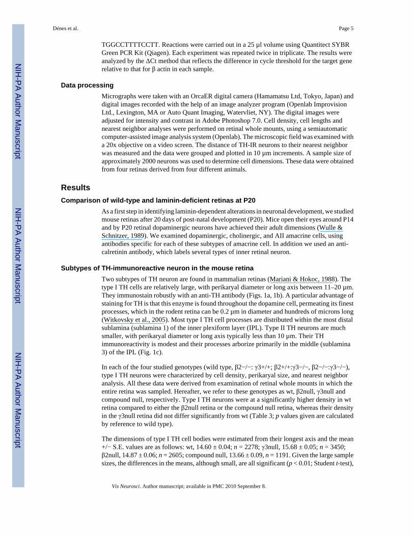
Subtypes of TH-immunoreactive neuron in the mouse retinaTwo subtypes of TH neuron are found in mammalian retinas (Mariani & Hokoc, 1988). Thetype I TH cells are relatively large, with perikaryal diameter or long axis between 11–20 µm.They immunostain robustly with an anti-TH antibody (Figs. 1a, 1b). A particular advantage ofstaining for TH is that this enzyme is found throughout the dopamine cell, permeating its finestprocesses, which in the rodent retina can be 0.2 µm in diameter and hundreds of microns long(Witkovsky et al., 2005). Most type I TH cell processes are distributed within the most distalsublamina (sublamina 1) of the inner plexiform layer (IPL). Type II TH neurons are muchsmaller, with perikaryal diameter or long axis typically less than 10 µm. Their THimmunoreactivity is modest and their processes arborize primarily in the middle (sublamina3) of the IPL (Fig. 1c).
In each of the four studied genotypes (wild type, β2−/−: γ3+/+; β2+/+:γ3−/−, β2−/−:γ3−/−),type I TH neurons were characterized by cell density, perikaryal size, and nearest neighboranalysis. All these data were derived from examination of retinal whole mounts in which theentire retina was sampled. Hereafter, we refer to these genotypes as wt, β2null, γ3null andcompound null, respectively. Type I TH neurons were at a significantly higher density in wtretina compared to either the β2null retina or the compound null retina, whereas their densityin the γ3null retina did not differ significantly from wt (Table 3; p values given are calculatedby reference to wild type).
The dimensions of type I TH cell bodies were estimated from their longest axis and the mean+/− S.E. values are as follows: wt, 14.60 ± 0.04; n = 2278; γ3null, 15.68 ± 0.05; n = 3450;β2null, 14.87 ± 0.06; n = 2605; compound null, 13.66 ± 0.09, n = 1191. Given the large samplesizes, the differences in the means, although small, are all significant (p < 0.01; Student t-test),
Dénes et al. Page 5
Vis Neurosci. Author manuscript; available in PMC 2010 September 8.
NIH
-PA Author Manuscript
NIH
-PA Author Manuscript
NIH
-PA Author Manuscript

indicating that in the compound null retinas, the perikarya of type I TH neurons are somewhatsmaller than the comparable cells in wt, γ3 null or β2 null mice.
Fig. 2 illustrates the nearest neighbor distribution of type I TH neurons in wt and each type oflaminin-deficient retina we examined. Wild-type and γ3null nearest neighbor distributionswere essentially identical, whereas the distributions of type I cells in the β2 null and compoundnull retinas were skewed, with an evident shift of the nearest neighbor distributions to greaterdistances, as would be expected from the lower densities of type I TH neurons in these laminin-deficient retinas compared to wt (cf., Table 3).
Type 2 TH neuronsAlthough type II DA neurons are present in wt and γ3null retinas, their TH-IR in these twogroups was very weak, preventing a reliable survey of their numbers in whole mountpreparations. We were unable, therefore, to calculate a nearest-neighbor distribution for thesecells. They were detectable, however, in vertical sections of the retina (Fig. 1a). In contrast,the immunoreactivity of type II TH neuronal perikarya in β2null and compound null retinaswas, surprisingly, stronger (Fig. 1b) than in littermate γ3null littermates or in wt retinas of thesame age (Fig. 1a), suggesting that the TH content of type II TH neurons is increased whenthe β2 laminin chain is genetically ablated.
Thus, to summarize the findings for TH neurons at P20, the γ3null retina was not different inany measure from the wt. The β2null and compound null retinas showed similar alterationsfrom the wt in the following ways: (1) type I DA cells are at a lower density; (2) type II DAcells show a greater TH immunoreactivity (3) in the compound null retina, type I TH perikaryaldimensions are slightly smaller than in the wt retina.
Given that (1) and (2) tend to produce opposite changes in total retinal TH, the possibledifferences in retinal TH content between wt and β2null retinas could not be predicted. Toobtain further information on this point, we performed immunoblots on extracts from P20pooled retinas from each genetic group, using β-tubulin as a standard. Wild type retinas werecompared either to γ3null (n = 1), β2null (n = 3), or compound null retinas (n = 1); an example,comparing wt and β2null retinas, is illustrated in Fig. 3. The bands were scanned and theiroptical densities measured. After correction for differences in β-tubulin content, the ratios ofoptical densities are: γ3null/wt = 1.16; β2null/wt, 0.32 +/− .06; compound null/wt = 0.17.
Because the sample sizes were small, and we did not linearize our immunoblot detectionmethods, these data provide only a semi-quantitative indication of differences in TH content.Nevertheless they are consistent with the anatomical measures, in that in both β2null andcompound null retinas, the type I TH neurons are at a lower density compared to wt. The ratiosof TH content, however, indicate a greater difference between wt and β2null or compound nullretinas than would be predicted solely by the measured differences in type I TH cell densities.Thus they may indicate additional differences in TH dendritic/axonal dimensions between wtand laminin-deficient mice. In fact, given the very long dendritic and axonal processes of THneurons, and the observation that all such processes are filled with TH enzyme, most of theTH is estimated to be within them rather than in the perikaryon. A working calculation, basedon the geometry of type I TH neurons in primate (Dacey, 1990) and rat retinas (Witkovsky etal., 2005) supposes that the perikaryon is a sphere, 15 µm in diameter containing a nucleus 7µm in diameter, that each type I TH cell has four dendrites 300 µm long and 1 µm in diameterand four axons, 500 µm long and 0.2 µm in diameter. Assuming further that TH is uniformlydistributed; these dimensions suggest that less than 20% total TH enzyme is in the perikaryon,the balance being spread among the dendritic and axonal processes. Thus, even smalldifferences in TH process dimensions between wt and laminin-deficient mice would stronglyinfluence the ratios of total TH. This possibility is considered in greater detail below in relation
Dénes et al. Page 6
Vis Neurosci. Author manuscript; available in PMC 2010 September 8.
NIH
-PA Author Manuscript
NIH
-PA Author Manuscript
NIH
-PA Author Manuscript
to the developmental component of our study of TH neurons in wt versus laminin-deficientretinas. Type II TH neurons also contribute to total retinal TH, but based on dimensionalconsiderations and their relatively low TH-IR compared to that of type I TH neurons, weestimate that their contribution to total retinal TH content is small (<5%).
Effect of laminin deficiency on basement membrane and glial Müller cell structure in the P20mouse
Prior studies of the distribution of β2 containing-laminins in vertebrate retinas (Hunter et al.,1992; Libby et al., 1996, 1999, 2000) showed that they are concentrated in the basementmembranes and in the photoreceptor layer. They are sparse or absent, however, in the innernuclear and plexiform layers, where the amacrine cell bodies and dendrites are located. Thissuggests an indirect mechanism whereby laminin deficits might affect amacrine celldevelopment. In this regard, the Müller glial cell of the retina is an obvious point of focus.Müller cells attach to the retinal basement membrane (inner limiting membrane) and span theentire width of the retina. Recent studies suggest that Müller cells have multiple roles in retinalneuronal development (Reh & Levine, 1998; Blackshaw et al., 2004), including as a scaffoldfor neurite extension.
We began by examining the basement membrane of the retina, using antibodies directed againstperlecan, an intrinsic molecule (heparin sulfate proteoglycan) of the basement membrane. Fig.4 illustrates that in P20 wt retinas the basement membrane is a continuous sheet, but in bothβ2null and compound null retinas, the basement membrane showed discontinuous staining,indicating local disruptions of its structure. A more detailed and complete study of the effectof laminin deletion on basement membrane integrity is the subject of another study (Daly etal., 2006).
We similarly compared Müller cell structure in wt versus laminin-deficient retinas using eitheran antibody against glutamine synthetase (GS), a Müller cell specific marker (Vardimon et al.,1986), or an antibody directed against vimentin (VIM), an abundant intermediate filamentprotein found in Müller cells (McGillem et al., 1998), particularly in the proximal portion ofthe cell, i.e., that part which lies close to the retinal inner surface. A striking difference wasrevealed in Müller cell organization. In wt retinas, each Müller cell has a basal terminalexpansion that attaches to the basement membrane; the alignment of these end feet creates anorderly array of glial cell terminal expansions (Fig. 5a). In either β2null or compound nullretinas, however, the basal membrane was discontinuous, showing numerous small breaks atmany points over the retinal surface. At these breaks, Müller cells did not attach to the basementmembrane; instead their basal terminals formed a disorganized array (Fig. 5b) from the innerplexiform layer (IPL) through the ganglion cell layer. At other points, the Müller cell terminalsaggregated at blood vessels (Fig. 5c). These observations suggest that attachment of the Müllercell end foot to the inner limiting membrane is critical in maintaining the polarity andorganization of the Müller cell. Removal of all β2-containing laminins appears to disruptsignificantly this attachment mechanism. In the compound null retina, in these regions ofbasement membrane disruption, the Müller cell had a reduced overall length and a disarrayeddisposition of its fine lateral branches within IPL, relative to wt retina, as described andillustrated below in the section on development. No differences in basement membranes orMüller cell organization were noted in between WT and γ3 null mice.
Effect of laminin deficiency on cholinergic, AII amacrine and calretinin-IR in the P20 mouseCholinergic amacrine cells were revealed by immunostaining with an antibody against cholineacetyltransferase (ChAT; Zhang et al., 2005), whereas AII amacrines were labeled with anantibody against disabled-1 (Dab-1), a developmental marker (Rice & Curran, 2000). The anti-calretinin antibody immunostains numerous cells among the ganglion and amacrine cell layers
Dénes et al. Page 7
Vis Neurosci. Author manuscript; available in PMC 2010 September 8.
NIH
-PA Author Manuscript
NIH
-PA Author Manuscript
NIH
-PA Author Manuscript
of the mouse retina, and also reveals three prominent bands of staining in the IPL. In relationto their cell density, process distribution and robustness of immunostaining, cholinergic andAII amacrine cells did not differ between wt and laminin-deficient retinas (not illustrated).With regard to calretinin immunostaining, in most regions of the retina, we noted no disruptionsof the lamination pattern in the IPL in β2null and compound null mice relative to wt, althoughthe IPL dimensions were somewhat reduced in the compound null mouse retina relative to wt.A comparison of calretinin-IR in wt, β2null and compound null retinas is illustrated in Fig. 6.It is important to note that the middle lamina of calretinin reactive processes arise fromprocesses of type II TH neurons (Zhang, 2004); the normal appearance of this layerdemonstrates that these cells are not disrupted by the mutations.
Laminin deficit-induced alterations in TH neuron developmentThe constellation of differences described above between P20 wt and laminin-deficient retinassuggests that the combined absence of β2- and γ3-containing laminins induces alterations inTH neuron, basement membrane and Müller cell development. These alterations are similarbut less severe in the β2null retina. In order to study the onset of these defects, we extendedour observations to the first two postnatal weeks when Müller cells and TH neurons becomepost-mitotic and undergo differentiation (Rapoport et al., 2004). In younger animals, there isless overlap of the dendritic or axonal arbors of neighboring TH cells making it possible toexamine the arbors of individual TH neurons.
Developmental dependence of type I TH neuron cell density in wt and laminin-deficientretinas
We characterized the laminin-induced deficits in TH neuronal development by reference to thesequence of TH cell post-mitotic maturation in wt retinas. Because of the difficulty of seeingthe smaller type II TH neurons in wt retinas our observations are confined to type I TH neurons.The development of type I TH neurons in mouse retina was studied in detail by Wulle andSchnitzer (1989); nevertheless we found substantial differences between our respective databases in relation to the age at which TH-IR first appears and the distribution of TH neuronswithin the retina (see Discussion).
The density of type I TH neurons as a function of post-natal age is compared in Fig. 7 for wt,β2null, and compound null retinas. We made maps of the retinal distribution of TH cells, andcomputed cell densities from them. At postnatal day 3 (P3), type I TH cell density in wt retinaswas low and variable (10–32 cells/mm2; n = 3 retinas from three different animals). BetweenP3–5, however, cell density increased rapidly to about 50 type I TH cells/ mm2. In the β2nullretinas, the same growth pattern emerged but delayed by about 1 day with respect to wt retinas.The compound null retinas revealed an even greater delay; between P3–5, type I TH neuronswere either completely absent, or present at a very low density. In the insert to Fig. 7, regressionlines are fitted to the temporal sequence of Type I TH cell generation in wt and laminin-deficientretinas. Data for wt and β2null retinas are fitted by hyperbolas, whereas the data for compoundnull retinas are better matched by a linear function.
Fig. 7 thus illustrates two points about β2null and compound null retinas relative to their wtcounterpart: a delayed appearance and a lower density. Cell density differences between wtand either β2null or compound null retinas are statistically significant (p ≪ 0.01; cf. Table 3).Additionally Fig. 7 documents that for both wt and laminin-deficient retinas, there is a rathersharp reduction of approximately one-third in type I TH cell density between ages P10 andP12, with cell density remaining constant thereafter to P20. This loss is not attributable to anincrease in retinal area, because the dimensions of the eye do not change appreciably betweenP10 and P12.
Dénes et al. Page 8
Vis Neurosci. Author manuscript; available in PMC 2010 September 8.
NIH
-PA Author Manuscript
NIH
-PA Author Manuscript
NIH
-PA Author Manuscript
Growth pattern of type I TH neurons in wt and laminin-deficient retinasIn wt retinas, between P3-P6, type I TH cells have an inhomogeneous rate of development, inthat in some local areas, cells have long processes with robust TH-IR, whereas in adjacent areasthe development of the type I TH cell processes is retarded, and even the TH-IR of the perikaryais reduced. This patchy growth pattern gradually disappears between P6 and P15.
A comparison of type I TH neurons in wt, β2null and compound null retinas at differentpostnatal ages is depicted in Fig. 8. Panels a, b, c, illustrate the appearance of type I TH cellsin wt, β2null and compound null retinas, respectively, at P7. The morphology, dimensions andTH-IR of type I TH neurons is similar between wt and β2null retinas, although the cell densityis lower in the β2null than in the wt retina. In the compound null retina, however, the type ITH neurons at P7 show a markedly reduced TH-IR and restricted process outgrowth relativeto their wt counterparts. It is apparent that at P10 (Figs. 8d–8f) and P12 (Figs. 8g–8i) the wtretinas develop an increasingly overlapping pattern of dendrites and axons (Figs. 8d, 8g), whichis densest in the most distal (sublamina 1) portion of the IPL (the focal plane of all the imagesof Fig. 8). This network is reduced in the β2null retina (Figs. 8e, 8h) and is still sparser in thecompound null retina (Figs. 8f, 8i). Parenthetically, at any age examined we found nodistinctions between wt and the γ3null retinas.
During the third postnatal week the most prominent developmental change in type I TH neuronsis the elaboration of axon terminal rings containing the presynaptic apparatus for the releaseof dopamine (Witkovsky et al., 2004). Between P12 and P20, the TH-IR network of axonterminals in the β2null retina approaches the density seen in the wt (Figs. 8j, 8k). Since, at P20,type I TH cells remain at a lower density than in wt retina (Table 1), the implication is that inthe β2null retina, each type I TH cell generates extra axon terminals over what is seen in thewt retina. With regard to the compound null retina, its process development is poorer than thatof wt retina at any developmental stage, including P20 (Fig. 8l). The density of axon terminalsis reduced and the dendrites of the type I TH cell are thinner than those seen in wt retinas.
Thus, the data of Fig. 8 indicate that in compound null retinas, the growth of dendrites in typeI TH retinas is reduced compared to wt. That is, even when the type I TH perikarya of wt andcompound null have comparable TH-IR, the dendrites of those cells in wt retinas are longer,of greater diameter and more abundant. An alternative possibility, however, is that type I THneurons in compound null retinas elaborate a full complement of dendritic and axonalprocesses, but that TH enzyme is somehow distributed only into a fraction of the existingdendrites, leaving others devoid of enzyme. Because in both wt and β2null retinas TH isdistributed uniformly to all processes, including those of the finest caliber, we think thispossibility is unlikely.
One feature of type I TH neuron development in laminin-deficient retinas is not revealed inFig. 8. It is that the patchy development, i.e., adjacent retinal regions showing quite disparatedegrees of TH-IR and process outgrowth, is still observed at P12 in compound null retinas(Fig. 9), although at this age it is no longer seen in wt retinas (not illustrated).
Activation of the TH gene in wt and laminin-deficient retinasTo gain some insight into the onset of TH protein production, we looked at the activation ofthe TH gene. To examine this point in wt mice we took advantage of a genetically modifiedmouse, produced in the Raviola laboratory (Gustincich et al., 1997), in which the TH promoteris linked to alkaline phosphatase. The background strain of these mice is the same, C57BL/6J,as the mutant mice employed in the present study. Positive staining for alkaline phosphataseis an indicator that the TH gene has been activated. We found that in P2 mice, perikarya at thesame retinal position and the same low density as type I TH cells stained for alkaline
Dénes et al. Page 9
Vis Neurosci. Author manuscript; available in PMC 2010 September 8.
NIH
-PA Author Manuscript
NIH
-PA Author Manuscript
NIH
-PA Author Manuscript
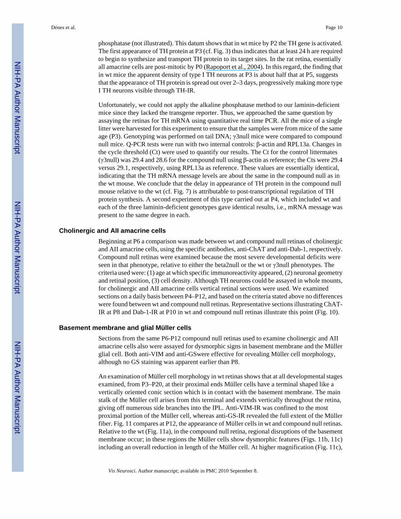
phosphatase (not illustrated). This datum shows that in wt mice by P2 the TH gene is activated.The first appearance of TH protein at P3 (cf. Fig. 3) thus indicates that at least 24 h are requiredto begin to synthesize and transport TH protein to its target sites. In the rat retina, essentiallyall amacrine cells are post-mitotic by P0 (Rapoport et al., 2004). In this regard, the finding thatin wt mice the apparent density of type I TH neurons at P3 is about half that at P5, suggeststhat the appearance of TH protein is spread out over 2–3 days, progressively making more typeI TH neurons visible through TH-IR.
Unfortunately, we could not apply the alkaline phosphatase method to our laminin-deficientmice since they lacked the transgene reporter. Thus, we approached the same question byassaying the retinas for TH mRNA using quantitative real time PCR. All the mice of a singlelitter were harvested for this experiment to ensure that the samples were from mice of the sameage (P3). Genotyping was performed on tail DNA; γ3null mice were compared to compoundnull mice. Q-PCR tests were run with two internal controls: β-actin and RPL13a. Changes inthe cycle threshold (Ct) were used to quantify our results. The Ct for the control littermates(γ3null) was 29.4 and 28.6 for the compound null using β-actin as reference; the Cts were 29.4versus 29.1, respectively, using RPL13a as reference. These values are essentially identical,indicating that the TH mRNA message levels are about the same in the compound null as inthe wt mouse. We conclude that the delay in appearance of TH protein in the compound nullmouse relative to the wt (cf. Fig. 7) is attributable to post-transcriptional regulation of THprotein synthesis. A second experiment of this type carried out at P4, which included wt andeach of the three laminin-deficient genotypes gave identical results, i.e., mRNA message waspresent to the same degree in each.
Cholinergic and AII amacrine cellsBeginning at P6 a comparison was made between wt and compound null retinas of cholinergicand AII amacrine cells, using the specific antibodies, anti-ChAT and anti-Dab-1, respectively.Compound null retinas were examined because the most severe developmental deficits wereseen in that phenotype, relative to either the beta2null or the wt or γ3null phenotypes. Thecriteria used were: (1) age at which specific immunoreactivity appeared, (2) neuronal geometryand retinal position, (3) cell density. Although TH neurons could be assayed in whole mounts,for cholinergic and AII amacrine cells vertical retinal sections were used. We examinedsections on a daily basis between P4–P12, and based on the criteria stated above no differenceswere found between wt and compound null retinas. Representative sections illustrating ChAT-IR at P8 and Dab-1-IR at P10 in wt and compound null retinas illustrate this point (Fig. 10).
Basement membrane and glial Müller cellsSections from the same P6-P12 compound null retinas used to examine cholinergic and AIIamacrine cells also were assayed for dysmorphic signs in basement membrane and the Müllerglial cell. Both anti-VIM and anti-GSwere effective for revealing Müller cell morphology,although no GS staining was apparent earlier than P8.
An examination of Müller cell morphology in wt retinas shows that at all developmental stagesexamined, from P3–P20, at their proximal ends Müller cells have a terminal shaped like avertically oriented conic section which is in contact with the basement membrane. The mainstalk of the Müller cell arises from this terminal and extends vertically throughout the retina,giving off numerous side branches into the IPL. Anti-VIM-IR was confined to the mostproximal portion of the Müller cell, whereas anti-GS-IR revealed the full extent of the Müllerfiber. Fig. 11 compares at P12, the appearance of Müller cells in wt and compound null retinas.Relative to the wt (Fig. 11a), in the compound null retina, regional disruptions of the basementmembrane occur; in these regions the Müller cells show dysmorphic features (Figs. 11b, 11c)including an overall reduction in length of the Müller cell. At higher magnification (Fig. 11c),
Dénes et al. Page 10
Vis Neurosci. Author manuscript; available in PMC 2010 September 8.
NIH
-PA Author Manuscript
NIH
-PA Author Manuscript
NIH
-PA Author Manuscript
it can be seen that the vertical shafts of the Müller cells were thinner in the region of the breakand had reduced side branches in the IPL, compared to glial cells on either side of the break.
Our data show that, in the compound null retinas, Müller cell and basal membraneabnormalities were evident throughout the period in which post-mitotic TH neurons began toextend their dendritic and axonal arbors and to acquire TH-IR. On the other hand, at earlydevelopmental stages (P3–P10), the Müller fibers in β2 null retinas were not dysmorphic. Theywere of the same length as their counterparts in wt retinas, maintained the same strict verticalorientation and showed the same age-dependent sequence of process arborization within theIPL as noted in wt retinas. By P20, however, Müller glial cells in β2 null retinas also weredysmorphic in some respects (cf. Fig. 5) as reported earlier (Libby et al., 1999).
DiscussionPrincipal findings
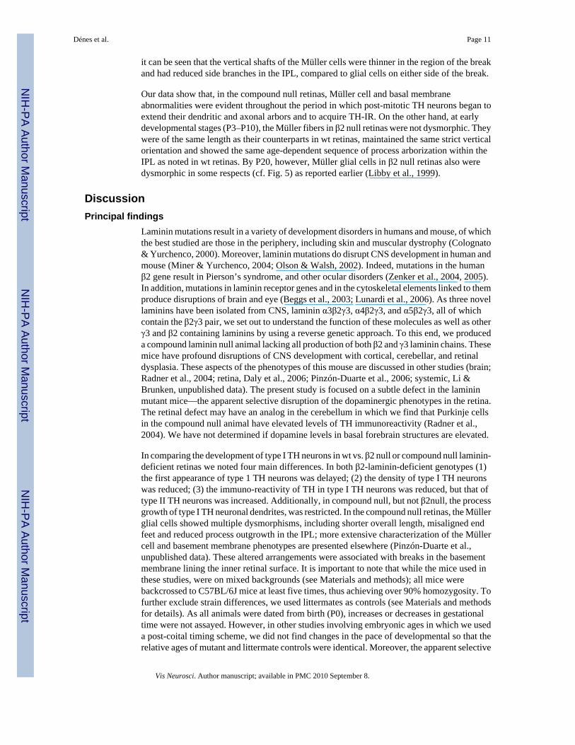
Laminin mutations result in a variety of development disorders in humans and mouse, of whichthe best studied are those in the periphery, including skin and muscular dystrophy (Colognato& Yurchenco, 2000). Moreover, laminin mutations do disrupt CNS development in human andmouse (Miner & Yurchenco, 2004; Olson & Walsh, 2002). Indeed, mutations in the humanβ2 gene result in Pierson’s syndrome, and other ocular disorders (Zenker et al., 2004, 2005).In addition, mutations in laminin receptor genes and in the cytoskeletal elements linked to themproduce disruptions of brain and eye (Beggs et al., 2003; Lunardi et al., 2006). As three novellaminins have been isolated from CNS, laminin α3β2γ3, α4β2γ3, and α5β2γ3, all of whichcontain the β2γ3 pair, we set out to understand the function of these molecules as well as otherγ3 and β2 containing laminins by using a reverse genetic approach. To this end, we produceda compound laminin null animal lacking all production of both β2 and γ3 laminin chains. Thesemice have profound disruptions of CNS development with cortical, cerebellar, and retinaldysplasia. These aspects of the phenotypes of this mouse are discussed in other studies (brain;Radner et al., 2004; retina, Daly et al., 2006; Pinzón-Duarte et al., 2006; systemic, Li &Brunken, unpublished data). The present study is focused on a subtle defect in the lamininmutant mice—the apparent selective disruption of the dopaminergic phenotypes in the retina.The retinal defect may have an analog in the cerebellum in which we find that Purkinje cellsin the compound null animal have elevated levels of TH immunoreactivity (Radner et al.,2004). We have not determined if dopamine levels in basal forebrain structures are elevated.
In comparing the development of type I TH neurons in wt vs. β2 null or compound null laminin-deficient retinas we noted four main differences. In both β2-laminin-deficient genotypes (1)the first appearance of type 1 TH neurons was delayed; (2) the density of type I TH neuronswas reduced; (3) the immuno-reactivity of TH in type I TH neurons was reduced, but that oftype II TH neurons was increased. Additionally, in compound null, but not β2null, the processgrowth of type I TH neuronal dendrites, was restricted. In the compound null retinas, the Müllerglial cells showed multiple dysmorphisms, including shorter overall length, misaligned endfeet and reduced process outgrowth in the IPL; more extensive characterization of the Müllercell and basement membrane phenotypes are presented elsewhere (Pinzón-Duarte et al.,unpublished data). These altered arrangements were associated with breaks in the basementmembrane lining the inner retinal surface. It is important to note that while the mice used inthese studies, were on mixed backgrounds (see Materials and methods); all mice werebackcrossed to C57BL/6J mice at least five times, thus achieving over 90% homozygosity. Tofurther exclude strain differences, we used littermates as controls (see Materials and methodsfor details). As all animals were dated from birth (P0), increases or decreases in gestationaltime were not assayed. However, in other studies involving embryonic ages in which we useda post-coital timing scheme, we did not find changes in the pace of developmental so that therelative ages of mutant and littermate controls were identical. Moreover, the apparent selective
Dénes et al. Page 11
Vis Neurosci. Author manuscript; available in PMC 2010 September 8.
NIH
-PA Author Manuscript
NIH
-PA Author Manuscript
NIH
-PA Author Manuscript
effects on the TH system noted here appear to rule out a simple developmental delay as themechanism for the laminin-dependent alterations in development.
Possible mechanisms linking laminin deficiency to altered neuronal developmentWe found that the development of TH neurons in retinas of γ3null mice was indistinguishablefrom those of wt mice. On the other hand, the compound null mouse, in which both β2 andγ3 laminin components were deleted, showed more severe deficits in TH development than inthe β2 deletion alone. The spatial and temporal patterning of laminin component chains isregulated developmentally and laminin assembly and component regulation is complex.During the course of normal development, numerous “substitutions” of laminins chains areobserved (Miner & Yurchenco, 2004). Moreover, the apparent promiscuity of laminin chainpartnering and the homology of laminin domain structure of some of laminin chains supporttwo substitutions—laminin β1 is likely to substitute for laminin β2 and laminin γ1 for lamininγ3. Both β1 and γ1 are expressed early in development, before either β2 or γ3, and have broaderdistribution, indeed they account for most of the known isoforms (16 ca.) of laminin. However,the observation that the phenotype of the β2−/−:γ3−/− mouse has a more profound disruptionthan either the β2−/−:γ3+/+ or the β2+/+ :γ3−/− mouse suggests that these substitutions are notperfect. Specifically, our observations suggest that while β2γ1 laminins may be very goodsubstitutes for β2γ3 (based on the apparent lack of a retinal phenotype), β1γ3 laminins are apoorer substitute for the β2γ3 (based on the disruptions seen), and finally, β1γ1 is a very poorsubstitute for β2γ3. This argument has focused on those molecules containing both β2 and γ3,but we recognize that laminins contain either one or the other chains are also found, furthercomplicating the genetic analysis. Thus, we cannot ascribe the defects we observe in thesemutant animals, to any specific laminin molecule.
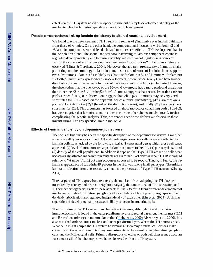
Effects of laminin deficiency on dopaminergic neuronsThe focus of this study has been the specific disruption of the dopaminergic system. Two otheramacrine cell types we examined, AII and cholinergic amacrine cells, were not affected bylaminin deficits as judged by the following criteria: (1) post-natal age at which these cell typesappeared; (2) level of immunoreactivity; (3) laminin pattern in the IPL; (4) perikaryal size; and(5) density of the cell populations. In addition it appears that Type II TH amacrine cells werenot adversely affected in the laminin mutants we examined. Not only was their TH IR increasedrelative to Wt mice (Fig. 1) but their processes appeared to be robust. That is, in Fig. 6, the tri-laminar appearance of calretinin-IR process in the IPL was strong in all genotypes. The middlelamina of calretinin immuno-reactivity contains the processes of Type II TH neurons (Zhang,2004).
Three aspects of TH-expression are altered: the number of cell adopting the TH-fate (asmeasured by density and nearest-neighbor analysis), the time course of TH expression, andTH cell dendritogenesis. Each of these aspects is likely to result from different developmentalmechanisms. Indeed, for retinal ganglion cells, cell fate, cell body positioning (spacing) anddendritic arborization are regulated independently of each other (Lin et al., 2004). A similarseparation of developmental processes is likely to occur in amacrine cells.
The disruption of the TH system must be indirect because, although β2 and γ3 chainsimmunoreactivity is found in the outer plexiform layer and retinal basement membranes (ILMand Bruch’s membrane) in mammalian retina (Libby et al., 2000; Aisenbrey et al., 2006), it isabsent at the border of inner nuclear and inner plexiform layers where the TH neurons reside.What cells might couple the TH system to laminins? Two major retinal cell classes makecontact with these laminin-containing compartments in the neural retina, the retinal ganglioncells and the Müller glial cells. Primary disruptions of either or both cell classes may accountfor some or all of the phenotypes we have observed within the TH system.
Dénes et al. Page 12
Vis Neurosci. Author manuscript; available in PMC 2010 September 8.
NIH
-PA Author Manuscript
NIH
-PA Author Manuscript
NIH
-PA Author Manuscript

Possible involvement of Müller cells in laminin-dependent developmental changesOne possibility is that the laminin deficiencies we report here lead to altered TH neurondevelopment by a pathway involving the following sequence: (1) loss of β2- and/or γ3-containing laminins results in a defective basement membrane characterized by many localtears and disruptions in structure (cf. Fig. 4); (2) disrupted basement membrane prevents Müllerglial cells from attaching to it (cf. Fig. 5); (3) failure of Müller cell attachment results indysmorphic growth and defective Müller cell process layering in the IPL (cf. Fig. 11c); (4)during the period of maximum TH neuronal process growth and synapse formation—P3 toP12—Müller cells promote the dendrite expansion of TH cells.
Several lines of indirect evidence support the Müller cell hypothesis outlined above. Müllercells have the appropriate molecules. They are at least one source of β2 laminin (Libby et al.,1997) and they express laminin receptor molecules including, integrins (Sherry & Proske,2001), dystroglycan-associated complex (Moukhles et al., 2000; Claudepierre et al., 2000),and a transmembrane collagen (Claudepierre et al., 2005). Functional data indicating laminin-dependent interactions come from Méhes et al. (2002), who report that laminin-1 increasesMüller cell motility and process outgrowth and from Halfter (1998), who found that disruptionof the basement membrane led to Müller cell end foot retraction. It is worth noting in thiscontext that retinal detachments produce many of the same disruptions of retinal architecturethat we have observed in laminin mutants including sprouting of Müller cells (Fisher et al.,2005) and remodeling of inner retinal neurons. Finally, on a more speculative note, glial cells,perhaps including Müller cells, are important regulators of synapse formation in the CNS; thusthe Müller cell may play a role in promoting synapse formation and concomitantly dendriteelongation (Steinmetz et al., 2006). Thus, it may be the defects in TH dendritogenesis are theresult of a failure of Müller cell-aided synapse formation and dendrite elongation.
It is unlikely, however, that defects in the Müller cells can account for all of the developmentalchanges we noted in TH cells—specifically with respect to cell fate adoption. We found alowered density of type I TH neurons in the laminin-deficient mice compared to wt animals atevery age studied from P6 to P20. Studies of cell birthdays in mouse (Young, 1985) and in ratretinas (Rapoport et al., 2004) demonstrate, on average, that retinal amacrine cells are bornbefore Müller glial cells and complementary studies, in mouse, show that programmed celldeath of retinal amacrine cell occurs before cell death of Müller cells (Young, 1984). However,the range of times over which these two processes (birth and death) occur in the two classesof retinal cell (amacrine and Müller) overlap considerably. The decrease in TH cell densitymay indicate more apoptotic death of TH cells in laminin-deficient mice prior to theirdifferentiation. We lack any experimental evidence for the notion that laminin-deficienciessomehow induce increased apopotosis; indeed the original report on at least one of thesemutants, the β2 null, ruled out an increase in cell death (Libby et al., 1999). In non-neuralsystems, integrins have been described as contributing to cell survival (Meredith et al., 1993;Bates et al., 1995); in neural systems laminins promote neural and glial survival (Chen &Strickland, 1997; Yu et al., 2005). Recent reviews of apoptosis in the retina implicate a widevariety of molecules that influence cell growth and death (Linden et al., 1999, 2005); it ispossible that laminins are among these but further experiments are needed to define a role forthem in retinal apoptosis.
Potential molecular components of laminin-induced alterationsAnother component of altered TH cell development we noted in the laminin-deficient mice ischanged levels of TH enzyme production: a decrease in type I TH cells and an increase in typeII TH cells. The TH gene is well known to be affected by activity-dependent gene promoterfactors such as AP-1 and CREB (Ghee et al., 1998; Lewis-Tuffin et al., 2004). Borba et al.(2005) showed that blockage of cAMP production by pituitary adenylyl cyclase-activating
Dénes et al. Page 13
Vis Neurosci. Author manuscript; available in PMC 2010 September 8.
NIH
-PA Author Manuscript
NIH
-PA Author Manuscript
NIH
-PA Author Manuscript
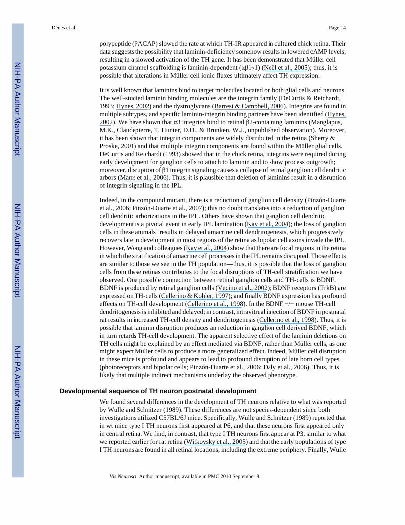
polypeptide (PACAP) slowed the rate at which TH-IR appeared in cultured chick retina. Theirdata suggests the possibility that laminin-deficiency somehow results in lowered cAMP levels,resulting in a slowed activation of the TH gene. It has been demonstrated that Müller cellpotassium channel scaffolding is laminin-dependent (αβ1γ1) (Noël et al., 2005); thus, it ispossible that alterations in Müller cell ionic fluxes ultimately affect TH expression.
It is well known that laminins bind to target molecules located on both glial cells and neurons.The well-studied laminin binding molecules are the integrin family (DeCurtis & Reichardt,1993; Hynes, 2002) and the dystroglycans (Barresi & Campbell, 2006). Integrins are found inmultiple subtypes, and specific laminin-integrin binding partners have been identified (Hynes,2002). We have shown that α3 integrins bind to retinal β2-containing laminins (Manglapus,M.K., Claudepierre, T, Hunter, D.D., & Brunken, W.J., unpublished observation). Moreover,it has been shown that integrin components are widely distributed in the retina (Sherry &Proske, 2001) and that multiple integrin components are found within the Müller glial cells.DeCurtis and Reichardt (1993) showed that in the chick retina, integrins were required duringearly development for ganglion cells to attach to laminin and to show process outgrowth;moreover, disruption of β1 integrin signaling causes a collapse of retinal ganglion cell dendriticarbors (Marrs et al., 2006). Thus, it is plausible that deletion of laminins result in a disruptionof integrin signaling in the IPL.
Indeed, in the compound mutant, there is a reduction of ganglion cell density (Pinzón-Duarteet al., 2006; Pinzón-Duarte et al., 2007); this no doubt translates into a reduction of ganglioncell dendritic arborizations in the IPL. Others have shown that ganglion cell dendriticdevelopment is a pivotal event in early IPL lamination (Kay et al., 2004); the loss of ganglioncells in these animals’ results in delayed amacrine cell dendritogenesis, which progressivelyrecovers late in development in most regions of the retina as bipolar cell axons invade the IPL.However, Wong and colleagues (Kay et al., 2004) show that there are focal regions in the retinain which the stratification of amacrine cell processes in the IPL remains disrupted. Those effectsare similar to those we see in the TH population—thus, it is possible that the loss of ganglioncells from these retinas contributes to the focal disruptions of TH-cell stratification we haveobserved. One possible connection between retinal ganglion cells and TH-cells is BDNF.BDNF is produced by retinal ganglion cells (Vecino et al., 2002); BDNF receptors (TrkB) areexpressed on TH-cells (Cellerino & Kohler, 1997); and finally BDNF expression has profoundeffects on TH-cell development (Cellerino et al., 1998). In the BDNF −/− mouse TH-celldendritogenesis is inhibited and delayed; in contrast, intravitreal injection of BDNF in postnatalrat results in increased TH-cell density and dendritogenesis (Cellerino et al., 1998). Thus, it ispossible that laminin disruption produces an reduction in ganglion cell derived BDNF, whichin turn retards TH-cell development. The apparent selective effect of the laminin deletions onTH cells might be explained by an effect mediated via BDNF, rather than Müller cells, as onemight expect Müller cells to produce a more generalized effect. Indeed, Müller cell disruptionin these mice is profound and appears to lead to profound disruption of late born cell types(photoreceptors and bipolar cells; Pinzón-Duarte et al., 2006; Daly et al., 2006). Thus, it islikely that multiple indirect mechanisms underlay the observed phenotype.
Developmental sequence of TH neuron postnatal developmentWe found several differences in the development of TH neurons relative to what was reportedby Wulle and Schnitzer (1989). These differences are not species-dependent since bothinvestigations utilized C57BL/6J mice. Specifically, Wulle and Schnitzer (1989) reported thatin wt mice type I TH neurons first appeared at P6, and that these neurons first appeared onlyin central retina. We find, in contrast, that type I TH neurons first appear at P3, similar to whatwe reported earlier for rat retina (Witkovsky et al., 2005) and that the early populations of typeI TH neurons are found in all retinal locations, including the extreme periphery. Finally, Wulle
Dénes et al. Page 14
Vis Neurosci. Author manuscript; available in PMC 2010 September 8.
NIH
-PA Author Manuscript
NIH
-PA Author Manuscript
NIH
-PA Author Manuscript
and Schnitzer (1989) indicate a steady increase in the density of type I TH neurons from P5 toP25, whereas we noted a rise in density from P3 to P10, followed by a fall to P15, remainingconstant thereafter.
The reasons for these discrepancies are not entirely clear. In our hands, the rabbit anti-THantibody from Chemicon is more sensitive than its counterpart from Eugene Tech. Thefluorescent secondary we used for the developmental sequence reveals more fine TH processesthan does the DAB method. Possibly a more sensitive primary antibody reveals TH-IR at P3-P5, when TH levels are far from peak. Still, at age P6 and older, TH perikarya are readilydetected with either set of antibodies, so this methodological difference does not explain whywe obtained different cell densities and retinal distributions of type I TH neurons.
In summary, we provide evidence that genetic deletion of particular laminin chains leads todysmorphic changes in TH neurons. This effect is likely mediated indirectly by an alterationof the Müller cell or the ganglion cell population or both. Disruptions of these latter cells(Müller and ganglion cells) are likely to be secondary effects of the disruption of the basementmembrane in these animals. Earlier it was shown that the same laminin chain deletionsadversely affect photoreceptor development (Libby et al., 1999). Our study thus contributes toa growing body of data showing that components of the extracellular matrix surrounding nervecells exert a profound influence on the maturation and function of neural circuits.
AcknowledgmentsSupported by: R01 EY 12676; NS 39508 (WJB), Richard H. Chartrand Fdn. (PW). We dedicate this study to our latefriend and colleague, Ray Dacheux. We remember him not only as an outstanding investigator of retinal structure andfunction, but as a man of balanced judgment and personal warmth. We thank Dr Josuha Sanes for the gift of ourfounding Lamb2 null mice; Dr Elio Raviola for the gift of transgenic reporter mice. We also thank Dr Kathleen Yeeand Yong Li for helpful comments on aspects of the experimental design and the manuscript.
ReferencesAisenbrey S, Zhang M, Bacher D, Yee J, Brunken WJ, Hunter DD. Retinal pigment epithelial cells
synthesize laminins including laminin 5 and adhere to them using α3- and α6-containing integrins.Investigative Ophthalmology & Visual Science 2006;47:5537–5544. [PubMed: 17122146]
Barresi R, Campbell KP. Dystroglycan: Form biosynthesis to pathogenesis of human disease. Journal ofCell Science 2006;119:199–207. [PubMed: 16410545]
Bates RC, Lincz LF, Burns GF. Involvement of integrins in cell survival. Cancer Metastasis Review1995;14:191–203.
Beggs HE, Schahin-Reed D, Zang K, Goebbels S, Nave KA, Gorski J, Jones KR, Sretavan D, ReichardtLF. FAK deficiency in cells contributing to the basal lamina results in cortical abnormalitiesresembling congenital muscular dystrophies. Neuron 2003;40:501–514. [PubMed: 14642275]
Blackshaw S, Harpavat S, Trimarchi J, Cai L, Huang H, Kuo WP, Weber G, Lee K, Fraioli RE, Cho SH,Yung R, Asch E, Ohno-Machado L, Wong WH, Cepko CL. Genomic analysis of mouse retinaldevelopment. Public Library of Science Biology 2004;2:1411–1431.
Borba JC, Henze IP, Silvera MS, Kubrusly RCC, Gardino PF, de Mello MCF, Hokoc JN, de Mello FG.Pituitary adenylate cyclase-activating polypeptide (PACAP) can act as determinant of the tyrosinehydroxylase phenotype of dopaminergic cells during retina development. Developmental BrainResearch 2005;156:193–201. [PubMed: 16099306]
Cellerino A, Kohler K. Brain-derived neurotrophic factor/ neurotrophin-4 receptor TrkB is localized onganglion cells and dopaminergic amacrine cells in the vertebrate retina. Journal of ComparativeNeurology 1997;386:149–160. [PubMed: 9303531]
Cellerino A, Pinzón-Duarte G, Carroll P, Kohler K. Brain-derived neurotrophic factor modulates thedevelopment of the dopaminergic network in the rodent retina. Journal of Neuroscience1998;189:3351–3362. [PubMed: 9547243]
Dénes et al. Page 15
Vis Neurosci. Author manuscript; available in PMC 2010 September 8.
NIH
-PA Author Manuscript
NIH
-PA Author Manuscript
NIH
-PA Author Manuscript
Chen ZL, Strickland S. Neuronal death in the hippocampus is promoted by plasmin-catalyzed degradationof laminin. Cell 1997;91:917–925. [PubMed: 9428515]
Claudepierre T, Dalloz C, Mornet D, Matsumura K, Sahel J, Rendon A. Characterization of theintermolecular associations of the dystrophin-associated glycoprotein complex in retinal Muller glialcells. Journal of Cell Science 2000;113:3409–4317. [PubMed: 10984432]
Claudepierre T, Manglapus MK, Marengi N, Radner S, Champliaud M-F, Tasanen K, Bruckner-Tuderman L, Hunter DD, Brunken WJ. Collagen XVII and BPAG1 expression in the retina: Evidencefor an anchoring complex in the central nervous system. Journal of Comparative Neurology2005;487:190–203. [PubMed: 15880472]
Clegg DO, Mullick LH, Wingerd KL, Lin H, Atienza JW, Bradshaw AD, Gervin DB, Cann GM. Adhesiveevents in retinal development and function: The role of integrin receptors. Results and Problems inCell Differentiation 2000;31:141–156. [PubMed: 10929405]
Colognato H, Yurchenco PD. Form and function: The laminin family of heterotrimers. DevelopmentalDynamics 2000;218:213–234. [PubMed: 10842354]
Dacey DM. The dopaminergic amacrine cell. Journal of Comparative Neurology 1990;301:461–489.[PubMed: 1979792]
Daly G, Pinzón-Duarte G, Dénes V, Koch M, Hunter DD, Brunken WJ. Genetic deletion of lamininβ2γ3 chains results in a retinal dysplasia. Investigative Ophthalmology & Visual Science2006;47:2794.
DeCurtis I, Reichardt LF. Function and spatial distribution in developing chick retina of the lamininreceptor α6β1 and its isoforms. Development 1993;118:377–388. [PubMed: 8223267]
Ehinger B, Floren I. Quantitation of the uptake of indolemaines and dopamine in the rabbit retina.Experimental Eye Research 1978;26:1–11. [PubMed: 624320]
Fisher SK, Lewis GP, Linberg KA, Verardo MR. Cellular remodeling in mammalian retina: Results fromstudies of experimental retinal detachment. Progress in Retinal and Eye Research 2005;24:395–431.[PubMed: 15708835]
Ghee M, Baker H, Miller JC, Ziff EB. AP-1, CREB and CBP transcription factors differentially regulatethe tyrosine hydroxylase gene. Brain Research Molecular Brain Research 1998;55:101–114.[PubMed: 9645965]
Gustincich S, Feigenspan A, Wu DK, Koopman LJ, Raviola E. Control of dopamine release in the retina:A transgenic approach to neural networks. Neuron 1997;18:723–736. [PubMed: 9182798]
Halfter W. Disruption of the retinal basal lamina during early embryonic development leads to a retractionof vitreal end feet, an increased number of ganglion cells, and aberrant axonal outgrowth. Journal ofComparative Neurology 1998;397:89–104. [PubMed: 9671281]
Hunter DD, Murphy MD, Olsson CV, Brunken WJ. S-laminin expression in adult and developing retinae:A potential cue for photoreceptor morphogenesis. Neuron 1992;8:399–413. [PubMed: 1550669]
Hynes RO. Integrins: Bidirectional, allosteric signaling machines. Cell 2002;110:673–687. [PubMed:12297042]
Ivins JK, Yurchenco PD, Lander AD. Regulation of neurite outgrowth by integrin activation. Journal ofNeuroscience 2000;20:6551–6560. [PubMed: 10964960]
Kay JN, Roeser T, Mumm JS, Godinho L, Mrejeru A, Wong RO, Baier H. Transient requirement forganglion cells during assembly of retinal synaptic layers. Development 2004;131:1331–1342.[PubMed: 14973290]
Lewis-Tuffin LJ, Quinn PG, Chikaraishi DM. Tyrosine hydroxylase transcription depends primarily oncAMP response element activity, regardless of the type of inducing stimulus. Molecular and CellularNeuroscience 2004;25:536–547. [PubMed: 15033181]
Libby RT, Hunter DD, Brunken WJ. Developmental expression of laminin β2 in the rat retina.Investigative Ophthalmology and Visual Science 1996;37:1651–1661. [PubMed: 8675409]
Libby RT, Xu Y, Selfors LM, Brunken WJ, Hunter DD. Identification of the cellular source of lamininβ2 in adult and developing vertebrate retinae. Journal of Comparative Neurology 1997;389:655–667.[PubMed: 9421145]
Libby RT, Lavallee CR, Balkema GW, Brunken WJ, Hunter DD. Disruption of laminin β2 chainproduction causes alterations in morphology and function in the CNS. Journal of Neuroscience1999;19:9399–9411. [PubMed: 10531444]
Dénes et al. Page 16
Vis Neurosci. Author manuscript; available in PMC 2010 September 8.
NIH
-PA Author Manuscript
NIH
-PA Author Manuscript
NIH
-PA Author Manuscript

Libby RT, Champliaud M-F, Claudepierre T, Xu Y, Gibbons EP, Koch M, Burgeson RE, Hunter DD,Brunken WJ. Laminin expression in adult and developing retinae: Evidence of two novel CNSlaminins. Journal of Neuroscience 2000;20:6517–6528. [PubMed: 10964957]
Lin B, Wang SW, Masland RH. Retinal ganglion cell type, size and spacing can be specified independentof homotypic dendritic contacts. Neuron 2004;43:475–485. [PubMed: 15312647]
Linden R, Rehen SK, Chiarini LB. Apoptosis in developing retinal tissue. Progress in Retinal and EyeResearch 1999;18:133–165. [PubMed: 9932281]
Linden R, Martins RAP, Silveira MS. Control of programmed cell death by neurotransmitters andneuropeptides in the developing mammalian retina. Progress in Retinal and Eye Research2005;24:457–491. [PubMed: 15845345]
Lunardi A, Cremisi F, Dente L. Dystroglycan is required for proper retinal layering. DevelopmentalBiology 2006;290:411–420. [PubMed: 16406325]
MacNeil MA, Heussy JK, Dacheux R, Raviola E, Masland RH. The shapes and numbers of amacrinecells: Matching of photo-filled with Golgi-stained cells in the rabbit retina and comparison with othermammalian species. Journal of Comparative Neurology 1999;423:305–326. [PubMed: 10524341]
Mariani AP, Hokoc JN. Two types of tyrosine hydroxylase-immunoreactive amacrine cells in the rhesusmonkey retina. Journal of Comparative Neurology 1988;276:81–91. [PubMed: 2903868]
Marrs GS, Honda T, Fuller L, Thangavel R, Balsamo J, Lilien J, Dailey ME, Arregui C. Dendritic arborsof developing retinal ganglion cells are stabilized by β1-integrins. Molecular and CellularNeuroscience 2006;32:230–241. [PubMed: 16757177]
McGillem GS, Guidry C, Dacheux RF. Antigenic changes of rabbit retinal Muller cells in culture.Investigative Ophthalmology & Visual Science 1998;39:1453–1461. [PubMed: 9660494]
Méhes E, Czirók A, Hegedüs B, Vicsek T, Jancsik V. Laminin-1 increases motility, path-searching, andprocess dynamism of rat and mouse Muller glial cells in vitro: Implication of relationship betweencell behavior and formation of retinal morphology. Cell Motility Cytoskeleton 2002;53:203–213.
Meredith JE, Fazeli B, Schwartz MA. The extracellular matrix as a cell survival factor. Molecular Biologyof the Cell 1993;4:953–961. [PubMed: 8257797]
Miner JH, Yurchenco PD. Laminin functions in tissue morphogenesis. Annual Review of Cell Biology2004;20:255–284.
Moukhles H, Roque R, Carbonetto S. Alpha-dystroglycan isoforms are differentially distributed in adultrat retina. Journal of Comparative Neurology 2000;420:182–194. [PubMed: 10753306]
Noakes PG, Gautam M, Mudd J, Sanes JR, Merlie JP. Aberrant differentiation of neuromuscular junctionsin mice lacking s-laminin/laminin β2. Nature 1995;374:258–262. [PubMed: 7885444]
Noël G, Belda M, Guadagno E, Micoud J, Klöcker N, Moukhles H. Dystroglycan and Kir4.1 coclusteringin retinal Müller glia is regulated by laminin-1 and requires the PDZ-ligand domain of Kir4.1. Journalof Neurochemistry 2005;94:691–702. [PubMed: 16033419]
Olson EC, Walsh CA. Smooth, rough, and upside-down neorcortical development. Current Opinion inGenetics and Development 2002;12:320–327. [PubMed: 12076676]
Pinzón-Duarte G, Dénes V, Daly G, Koch M, Hunter DD, Brunken WJ. β2γ3 laminin deletion causesdefective development of Müller cells and retinal vasculature. Investigative Ophthalmology & VisualScience 2006;47:2796.
Pinzón-Duarte G, Daly G, Hunter DD, Brunken WJ. Defective formation of the inner limiting membranein the β2γ3 laminin null retina alters number and spatial organization of retinal ganglion cells.Investigative Ophthalmology & Visual Science 2007;48:5686.
Radner S, Lefkowitz JJ, Koch M, Hunter DD, Brunken WJ. Aberrant CNS development in β2γ3 lamininknockout mice. Society for Neuroscience 2004;609.4 Abstract Viewer on-line.
Rapoport DH, Wong LL, Wood ED, Yasumura D, LaVail MM. Timing and topography of cell genesisin the rat retina. Journal of Comparative Neurology 2004;474:304–324. [PubMed: 15164429]
Reh TA, Levine EM. Multipotential stem cells and progenitors in the vertebrate retina. Journal ofNeurobiology 1998;36:206–220. [PubMed: 9712305]
Rice DS, Curran T. Disabled-1 is expressed in type AII amacrine cells in the mouse retina. Journal ofComparative Neurology 2000;424:327–338. [PubMed: 10906706]
Dénes et al. Page 17
Vis Neurosci. Author manuscript; available in PMC 2010 September 8.
NIH
-PA Author Manuscript
NIH
-PA Author Manuscript
NIH
-PA Author Manuscript
Rockhill RL, Daly FJ, MacNeil MA, Brown SP, Masland RH. The diversity of ganglion cells in amammalian retina. Journal of Neuroscience 2002;22:3831–3843. [PubMed: 11978858]
Sherry DM, Proske PA. Localization of alpha integrin subunits in the neural retina of the tiger salamander.Graefe’s Arch. Clinical and Experimental Ophthalmology 2001;239:278–287.
Steinmetz CC, Buard I, Claudepierre T, Nagler K, Pfrieger FW. Regional variations in the glial influenceon synapse development in the mouse CNS. Journal of Physiology 2006;577:249–261. [PubMed:16959855]
Suzuki N, Yokoyama F, Nomizu M. Functional sites in the laminin alpha chains. Connective TissueResearch 2005;46:142–152. [PubMed: 16147852]
Vardimon L, Fox LE, Moscona AA. Developmental regulation of glutamine synthetase and carbonicanhydrase II in neural retina. Proceedings of the National Academy of Sciences of the United Statesof America 1986;83:9060–9064. [PubMed: 2878430]
Vecino E, Garcia-Grespo D, Garcia M, Martinez-Millan L, Carrascal E. Rat retinal ganglion cells co-express brain derived neurotrophic factor (BDNF) and its receptor TrkB. Vision Research2002;42:151–157. [PubMed: 11809469]
Willbold E, Layer PG. Müller glia cells and their possible roles during retina differentiation in vivo andin vitro. Histology Histopathology 1998;13:531–552.
Witkovsky P, Veisenberger E, Haycock JW, Akopian A, Garcia-Espana A, Meller E. Activity-dependentphosphorylation of tyrosine hydroxylase in dopaminergic neurons of the rat retina. Journal ofNeuroscience 2004;24:4242–4249. [PubMed: 15115820]
Witkovsky P, Arango-Gonzalez B, Haycock JW, Kohler K. Rat retinal dopaminergic neurons:Differential maturation of somatodendritic and axonal compartments. Journal of ComparativeNeurology 2005;481:352–362. [PubMed: 15593337]
Wulle I, Schnitzer J. Distribution and morphology of tyrosine hydroxylase-immunoreactive neurons inthe developing mouse retina. Developmental Brian Research 1989;48:59–72.
Yan HHN, Cheng CY. Laminin α3 forms a complex with/33 and γ3 chains that serves as the ligand forα6β1-integrin at the apical ectoplasmic specialization in adult rat testes. Journal of BiologicalChemistry 2006;281:17286–17309. [PubMed: 16608848]
Young RW. Cell death during differentiation of the retina in the mouse. Journal of ComparativeNeurology 1984;229:362–373. [PubMed: 6501608]
Young RW. Cell differentiation in the retina of the mouse. Anatomical Record 1985;212:199–205.[PubMed: 3842042]
Yu WM, Feltri ML, Wrabetz L, Strickland S, Chen ZL. Schwann cell-specific ablation of laminin gamma1causes apoptosis and prevents proliferation. Journal of Neuroscience 2005;25:4463–4472. [PubMed:15872093]
Yurchenco PD, Wadsworth WG. Assembly and tissue functions of early embryonic laminins and netrins.Current Opinion in Cell Biology 2004;16:572–579. [PubMed: 15363809]
Zenker M, Aigner T, Wendler O, Tralau T, Müntefering H, Fenski R, Pitz S, Schumacher V, Royer-Pokora B, Wühl E, Cochat P, Bouveier R, Kraus C, Mark K, Madlon H, Dötsch J, Rascher W,Maruniak-Chudek I, Lennert T, Neumann LM, Reis A. Human laminin β2 deficiency causescongenital nephrosis with mesangial sclerosis and distinct eye abnormalities. Human MolecularGenetics 2004;13:2625–2632. [PubMed: 15367484]
Zenker M, Pierson M, Jonveaux P, Reis A. Demonstration of two novel LAMB2 mutations in the originalPierson syndrome family reported 42 years ago. American Journal of Medical Genetics 2005;13873–44.
Zhang J, Yang Z, Wu SM. Development of cholinergic amacrine cells is visual activity-dependent in thepostnatal mouse retina. Journal of Comparative Neurology 2005;484:331–343. [PubMed: 15739235]
Zhang D-Q. Type 2 catecholaminergic amacrine cells express calcium-binding proteins in the mouseretina. Investigative Ophthalmology & Visual Science 2004;45:4253.
Dénes et al. Page 18
Vis Neurosci. Author manuscript; available in PMC 2010 September 8.
NIH
-PA Author Manuscript
NIH
-PA Author Manuscript
NIH
-PA Author Manuscript
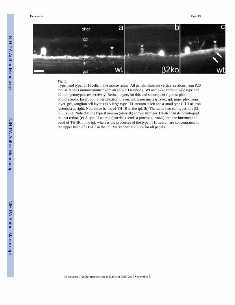
Fig. 1.Type I and type II TH cells in the mouse retina. All panels illustrate vertical sections from P20mouse retinas immunostained with an anti-TH antibody. Wt and b2ko refer to wild type andβ2 null genotypes, respectively. Retinal layers for this and subsequent figures: phot,photoreceptor layer; opl, outer plexiform layer; inl, inner nuclear layer; ipl, inner plexiformlayer; gcl, ganglion cell layer. (a) A large type I TH neuron at left and a small type II TH neuron(asterisk) at right. Note three bands of TH-IR in the ipl. (b) The same two cell types in a β2null retina. Note that the type II neuron (asterisk) shows stronger TH-IR than its counterpartin a wt retina. (c) A type II neuron (asterisk) sends a process (arrows) into the intermediateband of TH-IR in the ipl, whereas the processes of the type I TH neuron are concentrated inthe upper band of TH-IR in the ipl. Marker bar = 20 µm for all panels.
Dénes et al. Page 19
Vis Neurosci. Author manuscript; available in PMC 2010 September 8.
NIH
-PA Author Manuscript
NIH
-PA Author Manuscript
NIH
-PA Author Manuscript
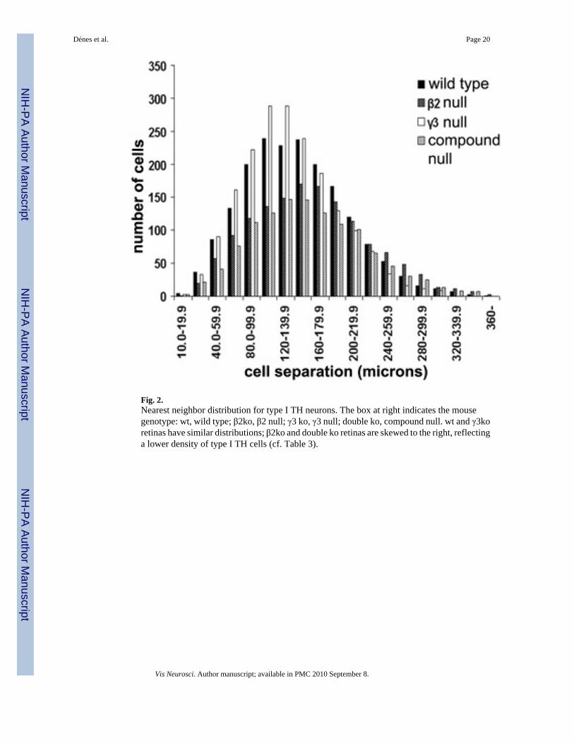
Fig. 2.Nearest neighbor distribution for type I TH neurons. The box at right indicates the mousegenotype: wt, wild type; β2ko, β2 null; γ3 ko, γ3 null; double ko, compound null. wt and γ3koretinas have similar distributions; β2ko and double ko retinas are skewed to the right, reflectinga lower density of type I TH cells (cf. Table 3).
Dénes et al. Page 20
Vis Neurosci. Author manuscript; available in PMC 2010 September 8.
NIH
-PA Author Manuscript
NIH
-PA Author Manuscript
NIH
-PA Author Manuscript
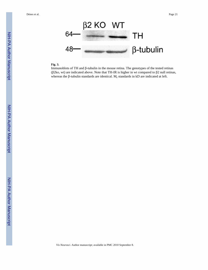
Fig. 3.Immunoblots of TH and β-tubulin in the mouse retina. The genotypes of the tested retinas(β2ko, wt) are indicated above. Note that TH-IR is higher in wt compared to β2 null retinas,whereas the β-tubulin standards are identical. Mr standards in kD are indicated at left.
Dénes et al. Page 21
Vis Neurosci. Author manuscript; available in PMC 2010 September 8.
NIH
-PA Author Manuscript
NIH
-PA Author Manuscript
NIH
-PA Author Manuscript
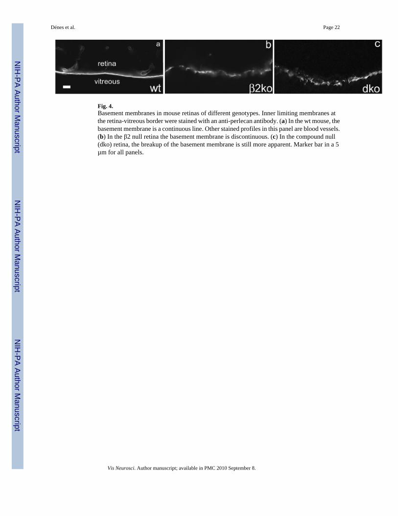
Fig. 4.Basement membranes in mouse retinas of different genotypes. Inner limiting membranes atthe retina-vitreous border were stained with an anti-perlecan antibody. (a) In the wt mouse, thebasement membrane is a continuous line. Other stained profiles in this panel are blood vessels.(b) In the β2 null retina the basement membrane is discontinuous. (c) In the compound null(dko) retina, the breakup of the basement membrane is still more apparent. Marker bar in a 5µm for all panels.
Dénes et al. Page 22
Vis Neurosci. Author manuscript; available in PMC 2010 September 8.
NIH
-PA Author Manuscript
NIH
-PA Author Manuscript
NIH
-PA Author Manuscript
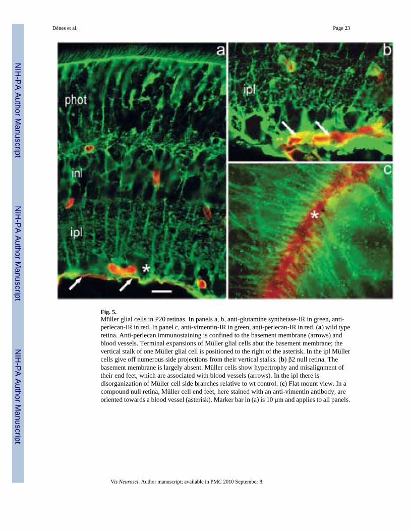
Fig. 5.Müller glial cells in P20 retinas. In panels a, b, anti-glutamine synthetase-IR in green, anti-perlecan-IR in red. In panel c, anti-vimentin-IR in green, anti-perlecan-IR in red. (a) wild typeretina. Anti-perlecan immunostaining is confined to the basement membrane (arrows) andblood vessels. Terminal expansions of Müller glial cells abut the basement membrane; thevertical stalk of one Müller glial cell is positioned to the right of the asterisk. In the ipl Müllercells give off numerous side projections from their vertical stalks. (b) β2 null retina. Thebasement membrane is largely absent. Müller cells show hypertrophy and misalignment oftheir end feet, which are associated with blood vessels (arrows). In the ipl there isdisorganization of Müller cell side branches relative to wt control. (c) Flat mount view. In acompound null retina, Müller cell end feet, here stained with an anti-vimentin antibody, areoriented towards a blood vessel (asterisk). Marker bar in (a) is 10 µm and applies to all panels.
Dénes et al. Page 23
Vis Neurosci. Author manuscript; available in PMC 2010 September 8.
NIH
-PA Author Manuscript
NIH
-PA Author Manuscript
NIH
-PA Author Manuscript
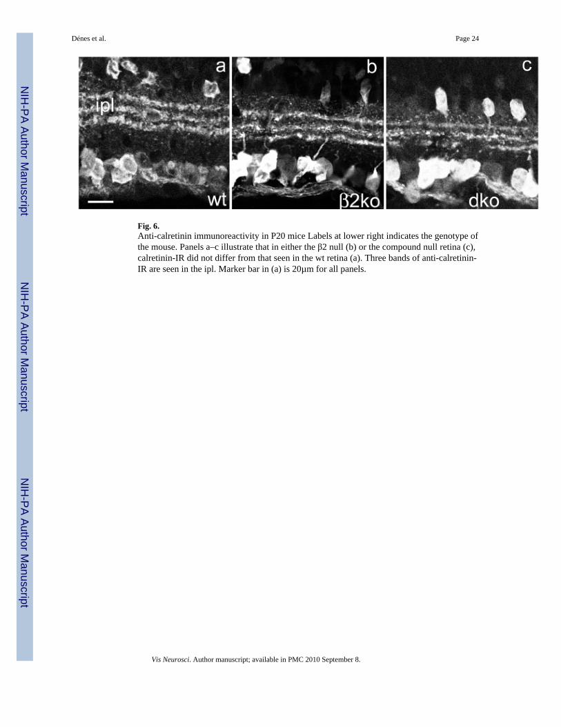
Fig. 6.Anti-calretinin immunoreactivity in P20 mice Labels at lower right indicates the genotype ofthe mouse. Panels a–c illustrate that in either the β2 null (b) or the compound null retina (c),calretinin-IR did not differ from that seen in the wt retina (a). Three bands of anti-calretinin-IR are seen in the ipl. Marker bar in (a) is 20µm for all panels.
Dénes et al. Page 24
Vis Neurosci. Author manuscript; available in PMC 2010 September 8.
NIH
-PA Author Manuscript
NIH
-PA Author Manuscript
NIH
-PA Author Manuscript
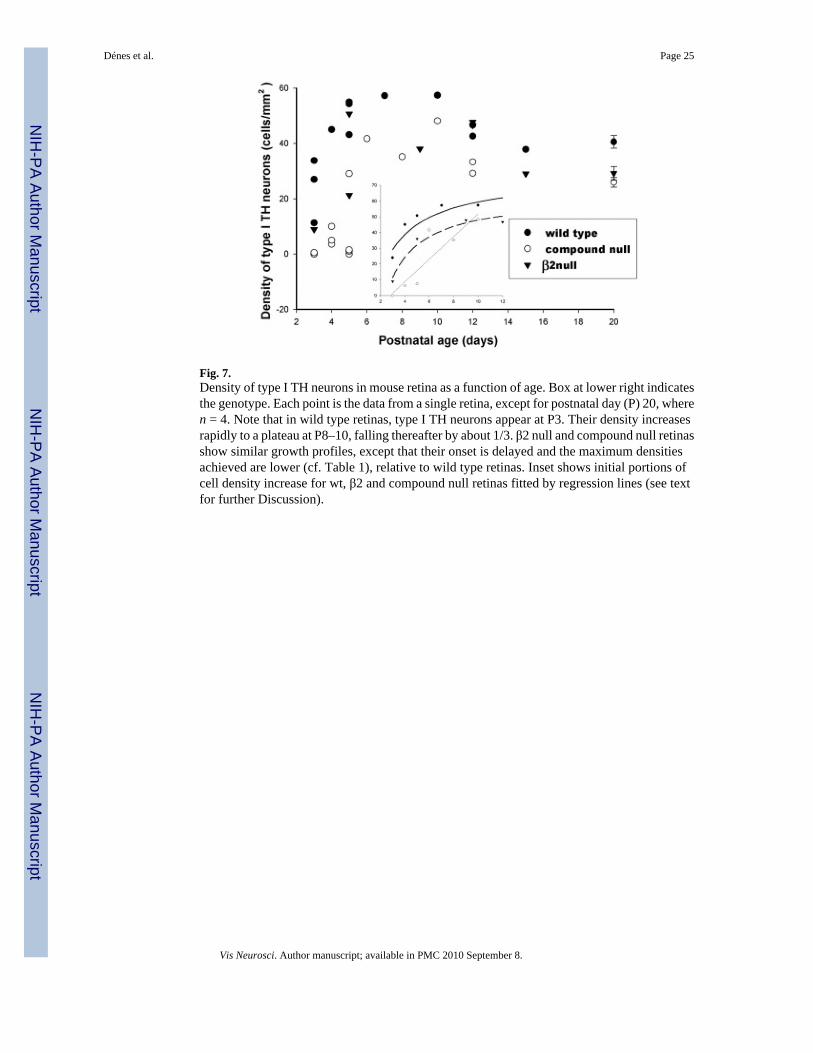
Fig. 7.Density of type I TH neurons in mouse retina as a function of age. Box at lower right indicatesthe genotype. Each point is the data from a single retina, except for postnatal day (P) 20, wheren = 4. Note that in wild type retinas, type I TH neurons appear at P3. Their density increasesrapidly to a plateau at P8–10, falling thereafter by about 1/3. β2 null and compound null retinasshow similar growth profiles, except that their onset is delayed and the maximum densitiesachieved are lower (cf. Table 1), relative to wild type retinas. Inset shows initial portions ofcell density increase for wt, β2 and compound null retinas fitted by regression lines (see textfor further Discussion).
Dénes et al. Page 25
Vis Neurosci. Author manuscript; available in PMC 2010 September 8.
NIH
-PA Author Manuscript
NIH
-PA Author Manuscript
NIH
-PA Author Manuscript
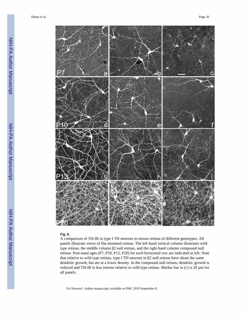
Fig. 8.A comparison of TH-IR in type I TH neurons in mouse retinas of different genotypes. Allpanels illustrate views of flat mounted retinas. The left hand vertical column illustrates wildtype retinas, the middle column β2 null retinas, and the right hand column compound nullretinas. Post-natal ages (P7, P10, P12, P20) for each horizontal row are indicated at left. Notethat relative to wild type retinas, type I TH neurons in β2 null retinas have about the samedendritic growth, but are at a lower density. In the compound null retinas, dendritic growth isreduced and TH-IR is less intense relative to wild type retinas. Marker bar in (c) is 20 µm forall panels.
Dénes et al. Page 26
Vis Neurosci. Author manuscript; available in PMC 2010 September 8.
NIH
-PA Author Manuscript
NIH
-PA Author Manuscript
NIH
-PA Author Manuscript
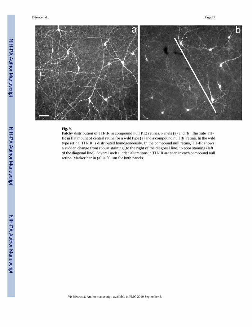
Fig. 9.Patchy distribution of TH-IR in compound null P12 retinas. Panels (a) and (b) illustrate TH-IR in flat mount of central retina for a wild type (a) and a compound null (b) retina. In the wildtype retina, TH-IR is distributed homogeneously. In the compound null retina, TH-IR showsa sudden change from robust staining (to the right of the diagonal line) to poor staining (leftof the diagonal line). Several such sudden alterations in TH-IR are seen in each compound nullretina. Marker bar in (a) is 50 µm for both panels.
Dénes et al. Page 27
Vis Neurosci. Author manuscript; available in PMC 2010 September 8.
NIH
-PA Author Manuscript
NIH
-PA Author Manuscript
NIH
-PA Author Manuscript

Fig. 10.Comparison of ChAT and Dab-1 immunoreactivity in wild type and compound null retinas.Each panel illustrates a vertical section through the retina; a,c, wild type retinas, b,d compoundnull retinas. Anti-choline acetyl transferase (ChAT)-IR is illustrated for P8 retinas; anti-disabled 1 (Dab-1)-IR is shown for P10 retinas. There is no difference in immuno-reactivitybetween wild type and compound null retinas for these markers. Marker bar in (a) is 20µm forall panels.
Dénes et al. Page 28
Vis Neurosci. Author manuscript; available in PMC 2010 September 8.
NIH
-PA Author Manuscript
NIH
-PA Author Manuscript
NIH
-PA Author Manuscript
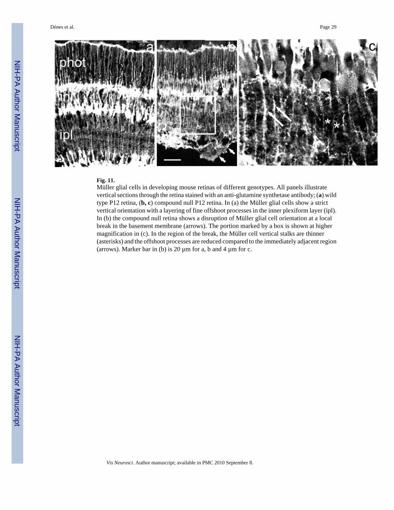
Fig. 11.Müller glial cells in developing mouse retinas of different genotypes. All panels illustratevertical sections through the retina stained with an anti-glutamine synthetase antibody; (a) wildtype P12 retina, (b, c) compound null P12 retina. In (a) the Müller glial cells show a strictvertical orientation with a layering of fine offshoot processes in the inner plexiform layer (ipl).In (b) the compound null retina shows a disruption of Müller glial cell orientation at a localbreak in the basement membrane (arrows). The portion marked by a box is shown at highermagnification in (c). In the region of the break, the Müller cell vertical stalks are thinner(asterisks) and the offshoot processes are reduced compared to the immediately adjacent region(arrows). Marker bar in (b) is 20 µm for a, b and 4 µm for c.
Dénes et al. Page 29
Vis Neurosci. Author manuscript; available in PMC 2010 September 8.
NIH
-PA Author Manuscript
NIH
-PA Author Manuscript
NIH
-PA Author Manuscript

NIH
-PA Author Manuscript
NIH
-PA Author Manuscript
NIH
-PA Author Manuscript
Dénes et al. Page 30
Table 1
Primary antibodies
Name Host Dilution Supplier
anti-Tyrosine hydroxylase rabbit 1:1000 Chemicon
anti-Choline-acetyl-transferase rabbit 1:500 Chemicon
anti-Disabled-1 rabbit 1:1000 Chemicon
anti-Calretinin mouse 1:1000 Chemicon
anti-Vimentin rabbit 1:2000 Chemicon
anti-Glutamine synthetase mouse 1:2000 Sigma
anti-Perlecan rat 1:1000 Chemicon
Vis Neurosci. Author manuscript; available in PMC 2010 September 8.

NIH
-PA Author Manuscript
NIH
-PA Author Manuscript
NIH
-PA Author Manuscript
Dénes et al. Page 31
Table 2
Secondary antibodies
Name Host Dilution Supplier
anti-rabbit IgG-Alexa 488 donkey 1:250 Molecular Probes
anti-rabbit IgG-Alexa 594 donkey 1:250 Molecular Probes
anti-mouse IgG-Alexa 488 donkey 1:250 Molecular Probes
anti-mouse IgG-Alexa 594 donkey 1:250 Molecular Probes
anti-rat IgG-Alexa 594 donkey 1:250 Molecular Probes
anti-rabbit IgG-biotin conjugated donkey 1:250 Molecular Probes
Vis Neurosci. Author manuscript; available in PMC 2010 September 8.

NIH
-PA Author Manuscript
NIH
-PA Author Manuscript
NIH
-PA Author Manuscript
Dénes et al. Page 32
Table 3
Density (cells/mm2) of type I TH neurons in P20 mouse retina
Wild type γ3−/− β2−/− β2−/−:γ3−/−
42.7 ± 2.3 45.3 ± 3.8 29.3 ± 1.7 26.0 ± 2.4
p = 0.29 p ≪ 0.01 p ≪ 0.01
Vis Neurosci. Author manuscript; available in PMC 2010 September 8.