dopaminergic neurodegeneration in
TRANSCRIPT
UNIVERSITÉ DU QUÉBEC
THÈSE PRÉSENTÉE À
L'UNIVERSITÉ DU QUÉBEC À TROIS-RIVIÈRES
COMME EXIGENCE PARTIELLE
DU DOCTORAT EN BIOLOGIE CELLULAIRE ET MOLÉCULAIRE
PAR
JUSTINE RENAUD
LA NEURODÉGÉNÉRESCENCE DOP AMINERGIQUE
EN CONDITIONS D'HYPERGLYCÉMIE
DOPAMINERGIC NEURODEGENERATION IN HYPERGLYCAEMIC CONDITIONS
AOÛT 2018
Université du Québec à Trois-Rivières
Service de la bibliothèque
Avertissement
L’auteur de ce mémoire ou de cette thèse a autorisé l’Université du Québec à Trois-Rivières à diffuser, à des fins non lucratives, une copie de son mémoire ou de sa thèse.
Cette diffusion n’entraîne pas une renonciation de la part de l’auteur à ses droits de propriété intellectuelle, incluant le droit d’auteur, sur ce mémoire ou cette thèse. Notamment, la reproduction ou la publication de la totalité ou d’une partie importante de ce mémoire ou de cette thèse requiert son autorisation.

UNIVERSITÉ DU QUÉBEC À TROIS-RIVIÈRES
Cette thèse a été dirigée par:
Maria-Grazia Martinoli, Ph. D. Université du Québec à Trois-Rivières
Directrice de recherche Institution à laquelle se rattache l'évaluateur
Jury d'évaluation de la thèse:
Maria-Grazia Martinoli, Ph. D. Université du Québec à Trois-Rivières
Prénom et nom, grade Institution à laquelle se rattache l' évaluateur
Michel Cyr, Ph. D. Université du Québec à Trois-Rivières
Prénom et nom, grade Institution à laquelle se rattache l' évaluateur
Gilles Bronchti, Ph. D. Université du Québec à Trois-Rivières
Prénom et nom, grade Institution à laquelle se rattache l' évaluateur
Lynda M. Williams, Ph. D. Université d 'Aberdeen, Royaume-Uni
Prénom et nom, grade Institution à laquelle se rattache l' évaluateur
Thèse soutenue le 21 juin 2018


Per ardua ad astra


ACKNOWLEDGMENTS
DearMom,
1 made it. It cornes as no surprise, 1 suspect, as never was there any space to spare
for doubt in anyone ' s mind. Including my own. 1 propelled myself on this joumey,
poised and tall and voracious. No space for doubt. Utterly unsuspecting of the obstacle
that would lay its livid body on the tracks on my way there. The obstacle was me.
Mom, 1 met many a great demons within myself: marbles rolling to the back of
the mind, dark moths fluttering in the peripheral vision, entire veils obstructing the
panorama of a long-time goal. 1 did not al ways fight the good fight. At times, 1 would let
them flatten me to sheets and redefine my Self. So 1 started buttering the plaster and
brai ding the bricks into a fortress to keep me out of me. A Great Wall if there is one.
With every single door leading me back inside.
On this joumey inside the outside of myself, 1 was lent a sea of hands.
They reached to me and reeled me onto the continent, out of this illusory pit that we're
aIl so familiar with. And when my identity eluded me - and this, it did many times -
they gathered my pieces for when 1 would retum to reclaim them.
Mom, among these hands, there was my research supervisor, the mightiest of
lighthouses, who taught me the art of focus , patience, diligence and perseverance against
the stemest of storms. You know, Maria has al ways reminded me of you, in the
relentless faith she bore in my aptitude to cut through the crests of waves. Across the
ocean that is Research. And during this time in her lab, 1 owe today a great many other
people, in tum, for their patience when 1 had none, for their incessant support when
1 was submerged. As you showed me the golden mIe of solidarity, my labmates shaped
me in its application.

Vlll
On my joumey, 1 sometimes shared walls with other friends who were enduring
the same transformation. l've shared these on more th an one continent, countless times
rescued by the voices on the other side. In Italy, Annesha, kindred spirit with an
insatiable mind and the keenest sense of meta-awareness to whom my heartstrings will
always be tied, however far apart our shores grow. Here, in my hometown, these voices
came from the loves of my life, Simon and Jo, from the canyon of their he arts wherein
whatever l've poured has al ways echoed back louder. And 1 know this thing about life
now: where hands are outstretched, may the y be themselves freezing and reaching to be
rescued, they are the most generous source of warmth and solace.
Looking back now at who the people closest to my heart are, 1 realize that
the y share this one thing: authenticity. The ineluctable, unsaid necessity to aIl my
relationships, in fact. Because 1 was raised to be true, to see truth, to eamestly trail the
footfall of what is, to me, absolute. Because you raised me to embrace authenticity as the
gold standard of virtue. This truth, as found in my friends and harboured in your very
DNA, has salvaged my mind and kept me lucid, time and again. This truth, 1 have found
it in one particular person now, Maude, on whose providential shores 1 washed up.
To her, 1 owe health, 1 owe resolution, 1 owe a bewildering encounter with the idea that
life may weIl just be a spectacular collision of souls. And 1 would be fine with that.
Mom, 1 made it. 1 tore down my walls, emptied the water in the belly of my boat,
opened my sails like the widest eyes, catching ail the colours. 1 think you know why
these words are addressed to you, why there was no doubt that 1 would succeed to begin
with. Because every dimension of my intelligence, of my heart, of my resolve that
allowed me to conquer myself, 1 will fore ver owe them to you. Even in the remotest
moments of this strange, strange drift, Mom, your love, your music, your rightfulness
have never eluded me, like an inbuilt compass. To you, 1 dedicate this thesis,
the thickest, starkest endeavour into my mind and soul that 1 have ever taken on.
To my True North,
from your ever-loving daughter

PREFACE
The work featured in this thesis represents part of the research 1 perfonned
throughout my doctoral studies. These results were harvested from the Laboratory of
Cellular Neurobiology at the University of Québec à Trois-Rivières, Canada, but also
from a collaborative effort with the Laboratory of Neurophannacology at the University
of Cagliari, Italy, where 1 completed six months of research during my doctoral studies.
This thesis is presented in the fonn of scientific articles and manuscripts: as of
August 2018, two have been published and one has been submitted. It also refers to
two reviews and one book chapter pertinent to the core subject and for which 1 am lead
author. These can be found in the appendices.
As per the authorization granted by the Cellular and Molecular Biology Graduate
Program Committee on April 20th, 2017, this thesis is written entirely in English.
A French surnmary of the thesis is provided in the following pages, and French
surnrnaries of each of the articles are supplied at the beginning of the corresponding
chapters. Please take note that the introduction and discussion sections are written in
Canadian English, whereas the articles, manuscripts and reviews are written in American
or British English, according to the language requirements specified by the editors of the
scientific journals in which they were published or submitted. One of the reviews is also
written in French.
AlI abbreviations and acronyrns employed throughout the text or within figures are
listed in the table provided for this purpose. Abbreviations in text are defined upon first
encounter, except for common biochemical tenns or when the fluidity of the text is
hindered. Within figures, abbreviations are described in the legend upon first encounter,
regardless of whether the y were first introduced in the text, but are not defined in
subsequent legends. It is important to note that each chapter that features an article
stands alone within this thesis and may therefore present tenns or abbreviations that are
inconsistent with the remainder of the text.

x
Last, 1 wish to mention that this thesis explores three distinct subject
matters integrated in a single discourse: dopaminergic neurons, hyperglycaemia and the
polyphenol resveratrol. Granted the broadness of the topics discussed herein,
the introduction is accordingly lengthy, as 1 was committed to appropriately ventilate aIl
dimensions impinging on the central hypothesis and on future perspectives. Likewise,
owing to the multiple techniques and models used to achieve the objectives of this
thesis, the methodology is meticulously described in the aim of guiding the reader
through the numerous decisions made upstream of the results presented herein.
RÉSUMÉ
Au deuxième rang des maladies neurodégénératives les plus communes après la maladie d'Alzheimer, la maladie de Parkinson atteint quelque 1 % de la population âgée de plus de 60 ans. Cette pathologie est caractérisée par la perte des neurones dopaminergiques logés dans la substance noire pars compacta du mésencéphale, conduisant à une diminution de la dopamine dans le striaturn dorsal et à l' apparition de symptômes moteurs. Malgré d ' amples efforts dévoués à l'élucidation des ses mécanismes neuropathologiques, la neurodégénérescence sélective des neurones de la voie nigrostriée demeure incomprise. En effet, la voie dopaminergique mésocorticolimbique, provenant du mésencéphale au niveau de l'aire ventrale tegmentale et innervant le striaturn ventral et le cortex préfrontal, ne semble pas dégénérer. Une explication voudrait que les neurones de la voie nigrostriée expriment un phénotype distinct les rendant plus vulnérables au stress oxydant. Selon cette conjecture, les neurones dopaminergiques de la voie nigrostriée devraient exhiber une plus grande susceptibilité à toute source de stress oxydant.
Dans cette optique, nous avons émis l' hypothèse que les neurones de la voie nigrostriée sont plus vulnérables au stress oxydant engendré par l 'hyperglycémie en comparaison, par exemple, à ceux de la voie mésocorticolimbique. En premier lieu, nous avons vérifié que de fortes concentrations de glucose pouvaient causer un stress oxydant menant à la dégénérescence de neurones dopaminergiques en culture. En effet, chez les cellules PCI2 différenciées en neurones dopaminergiques, ces conditions conduisaient à une hausse des niveaux de l'anion superoxyde, une espèce réactive de l' oxygène produite en excès dans les premiers instants suivant une hyperglycémie. En maintenant ces concentrations élevées sur une période de 96 h, les cellules neuronales PCI2 entraient en apoptose, tel que le confirmaient la fragmentation de l' ADN et l' altération des profils d'expression de plusieurs marqueurs apoptotiques. De plus, l'usage d ' un antioxydant éprouvé, le polyphénol resvératrol, abrogeait la hausse des niveaux d'anion superoxyde et la mort des cellules neuronales PCI2.
Suite à la confirmation que de fortes concentrations de glucose pouvaient conduire à la mort de neurones dopaminergiques en culture, nous avons voulu vérifier notre hypothèse centrale dans un modèle rongeur d'hyperglycémie. Nous avons employé un paradigme bien connu qui fait appel à l' administration de streptozotocine, une toxine ciblant les cellules productrices d'insuline, afin de générer un modèle de rat exhibant une hyperglycémie pouvant être maintenue pour une durée de 6 mois. À l'aide de méthodes immunologiques, nous avons démontré qu 'une hyperglycémie chronique induisait la dégénérescence des neurones dopaminergiques de la substance noire pars compacta, mais pas de ceux de l' aire ventrale tegmentale. Conséquemment, nous observions une perte des terminaisons dopaminergiques dans le striatum dorsal qui n' était pas perceptible dans le striatum ventral. De plus, par microdialyse intracérébrale, nous avons confirmé une baisse des niveaux de dopamine explicitement dans le striatum dorsal. Cette neurodégénérescence était accompagnée de l' augmentation du nombre

XlI
d'astrocytes et de la perte de cellules microgliales au niveau de la substance noire pars compacta et du striatum, mais pas dans l'aire ventrale tegmentale.
En complément, nous avons examiné les aspects comportementaux de notre modèle de rat hyperglycémique à l'aide de tests utilisés chez les rongeurs parkinsoniens. Nos résultats démontrent l'existence de déficits moteurs évoquant ceux retrouvés chez les patients parkinsoniens. Il était intéressant de constater que ces troubles moteurs ainsi que la baisse des niveaux de dopamine se manifestaient avant que ne soit perceptible la neurodégénérescence, indiquant un possible dysfonctionnement du système nigrostrié en amont de la mort neuronale. Puisque la dopamine occupe un rôle prépondérant dans la régulation des comportements sociaux, nous avons également procédé à l' étude des interactions entre paires de rats non-familiers, tout en nous attardant à leurs communications ultrasoniques dont les paramètres sont vraisemblablement modulés par la dopamine. Lors de ces rencontres sociales, nous avons découvert que les rats hyperglycémiques exhibaient une hyper-sociabilité et une agressivité accompagnées de l'accroissement du nombre de vocalisations ultrasoniques émises, suggérant l'existence d'un dysfonctionnement dopaminergique. L'intensité de ces témoignages hyper-réactifs était de plus corrélée au degré de perte d'innervation du striatum dorsal.
La somme de ces résultats nous permet de conclure que les neurones de la voie nigrostriée exhibent une susceptibilité manifeste vis-à-vis de conditions hyperglycémiques soutenues. Ces démonstrations viennent en appui aux études épidémiologiques soulignant un risque accru de développer la maladie de Parkinson chez les patients diabétiques.
Mots-clés: comportements sociaux anormaux, déficits moteurs, diabète, hyperglycémie, maladie de Parkinson, neurones dopaminergiques, stress oxydant, voie mésocorticolimbique, voie nigrostriée.
SUMMARY
Parkinson 's disease affects an estimated 1 % of the population over the age of 60 years, making it the second most cornrnon neurodegenerative disorder after Alzheimer's disease. Fundamentally, this pathology features a progressive loss of dopaminergic neurons harboured in the substantia nigra pars compacta of the midbrain, which leads to decreased dopamine release in the dorsal striatum responsible for the emergence of motor symptoms. Despite arduous efforts deployed to improve our understanding of the neuropathological underpinnings of this disease, researchers are still at loss as to why the nigrostriatal dopaminergic pathway undergoes preferential early degeneration compared, for instance, to the neighbouring mesocorticolimbic pathway originating from the ventral tegmental area in the midbrain and projecting to the ventral striatum and prefrontal cortex. Rapidly gaining momentum is a proposition providing that nigrostriatal dopaminergic neurons possess a distinctive phenotypic liability responsible for their relative susceptibility to oxidative stress. If this holds true, nigrostriatal dopaminergic neurons should be preferentially vulnerable to unspecific oxidative insults.
On these bases, we hypothesized that nigrostriatal dopaminergic neurons are more vulnerable to hyperglycaemia-induced oxidative stress compared to other neuronal populations, expressly ones of the mesocorticolimbic pathway. We began by verifying that high glucose conditions are conducive to the death of dopaminergic neurons in culture and that this degeneration is linked to oxidative stress. When cultured in elevated yet physiological concentrations of glucose, differentiated dopaminergic neuronal PC12 cells promptly exhibited high levels of superoxide anion, a key reactive oxygen species who se overproduction constitutes the earliest event in hyperglycaemia-induced oxidative stress. Sustained for 96 h, high glucose conditions led to the apoptotic death of neuronal PC12 cells substantiated by DNA fragmentation and altered expression profiles of various markers of apoptosis. Treating neuronal dopaminergic PC12 cells with a potent antioxidative polyphenol, resveratrol, attenuated the rise in superoxide anion levels and afforded protection against high glucose-induced apoptosis.
After confirming that high glucose conditions elicit a state of oxidative stress leading to the death of dopaminergic neurons in culture, we set out to verify our central hypothesis in a rat model of long-term hyperglycaemia. We employed a well-known paradigm that utilizes streptozotocin, a toxin that targets insulin-producing pancreatic ~ cells, to generate a model presenting a hyperglycaemic phenotype that could be maintained for up to 6 months. Employing irnrnunohistochemical and immunoblotting techniques, we demonstrated that long-term hyperglycaemia in rats causes the degeneration of dopaminergic neurons in the substantia nigra pars compacta, but not in the ventral tegmental area. Accordingly, dopaminergic terminal fibres were less dense in the dorsal than in the ventral striatum of hyperglycaemic rats. Utilizing the intracerebral microdialysis technique, we also showed that dopamine release was diminished in the dorsal striatum, but not in the ventral striatum or in the prefrontal cortex. We further discovered a noticeable increase in astrocytes that was neuroanatomically coincidental to

XIV
a loss of microglial cells in the substantia nigra pars compacta and striatum, but not in the ventral tegrnental area.
Behavioural alterations were assessed in a series of tasks destined to uncover motor deficits in rodent models of Parkinson's disease. Long-term hyperglycaemic rats manifested signs of bradykinesia and gait disturbances reminiscent of parkinsonian motor syrnptoms. Interestingly, motor deficits and dampened dorsostriatal dopamine release were apparent before neurodegeneration could be discerned, suggesting possible functional impairments of the nigrostriatal pathway upstream of neuronal death. Considering dopamine also exerts an important control on social behaviours, we examined interactions between pairs of unacquainted rats and analyzed dopamineregulated ultrasonic vocalizations known to reflect the subjects ' affective state. In particular, hyperglycaemic rats engaged in markedly hyper-sociable and hyperaggressive encounters, while emitting a greater number of ultrasonic vocalizations, evocative of a dopaminergic dysfunction. Remarkably, the intensity of these hyperreactive phenotypes correlated with the degree of dorsostriatal dopaminergic denervation.
Taken together, our data expose the preferential vulnerability of the nigrostriatal pathway to sustained hyperglycaemia, supporting the physiological significance of their phenotypic liability to oxidative stress. Our findings further strengthen the apparent epidemiological link between pre-existing diabetes and an increased risk of developing Parkinson's disease.
Keywords: abnormal social behaviours, diabetes, dopaminergic neurons, hyperglycaemia, mesocorticolimbic pathway, motor deficits, nigrostrial pathway, oxidative stress, Parkinson' s disease.

TABLE OF CONTENTS
ACKNOWLEDGMENTS ......................................................................................... vii
PREFACE................................................................................................................... ix
RÉSUMÉ..................................................................................................................... xi
SUMMARY ................................................................................................................ xiii
LIST OF T ABLES ..................................................................................................... xxiii
LIST OF FIGURES ................................................................................................... xxv
LIST OF ABBREVIATIONS AND ACRONYMS ................................................. xxix
CHAPTERI INTRODUCTION ...................................................................................................... 1
1.1 Dopaminergic neurons .... ............. ................... ........ .... .... ............. ........ ............... 1
1.1.1 Dopamine metabolism and neurotransmission ... .... ........ ...... ........ .. ........ 3
1.1.2 Central dopaminergic systems.......... ...... ........ ............. ... ............ .... ........ 9
1.1.2.1 The nigrostriatal pathway .... ..... ... ..... . .... ....... . ... .... .. .. .... .. .... ...... Il
1.1.2.2 The mesocorticolimbic pathway... ....... ....... .................. .... ...... .. 15
1.1.2.3 Converging and diverging functions ... .. ...... ...... ........ .............. . 16
1.1.3 Parkinson ' s disease ...... .... ......... .......... .... ........ ........ ........ .... .. ...... ... ..... .... 18
1.1.3.1 Clinical symptomatology and treatments .. .... .. .................. ..... .. 19
1.1.3.2 Prodrome and biomarkers.. .................. ............ ........ ............. .... 24
1.1.3.3 Pathophysiology ........ .............. ...... .......................... ........ ........ . 26
1.1.3.4 Vulnerability of the nigrostriatal pathway ......... ...... ........ ......... 31
1.2 Hyperglycaemia in the central nervous system... ........ ........ ......... ..... .. ......... ....... 38
1.2.1 Glucose as a preferential fuel for the brain ......... ..... ...... ..... ....... ... .......... 38
1.2.2 Neuronal glucose transport .................... .... .... ................ ......... ...... .. ...... .. 39
1.2.2.1 Transporters and kinetics ......... ...................... ... .. ... ......... .......... 39
1.2.2.2 Physiological considerations .... ........ .... ... ........ ... ...................... 44
1.2.3 Neuronal glucose metabolism. ..... .. ... ........ .......... .. .. .. ............... .... .... ....... 46
1.2.3.1 Specifie metabolic fates ... .... .... .... ... .................... ........... ......... .. 48
1.2.3.2 CUITent hypotheses in neuroenergetics..... ..... ........ ... .... ... ... .. .... 50
XVI
l.2.4 Neuronal oxidative stress in hyperglycaemia .................... ............ ........ . 54
1.2.4.1 Mitochondrial mechanisms ................................ .............. .... ..... 55
1.2.4.2 Rerouting mechanisms: polyol pathway and macromolecule glycation.. ................... .... ...... ...... .... ............. ................ ... .. .. ....... 60
l.2.5 Vulnerability of the nigrostriatal pathway .................................. ...... .... .. 65
1.2.5.1 Epidemiological basis: Parkinson 's disease in diabetic patients ... ...... ........ ........................ .... ............ .... ......... ...... ......... . 65
1.2.5.2 Molecular and cellular bases.......................................... .. ........ . 68
1.3 The antioxidative polyphenol resveratrol.................................. .......................... 72
1.3.1 Background..... ............................... ..................... ...... .............................. 72
1.3.1.1 Historical context...................................................................... 72
1.3.1.2 Dietary origins ............ .......................... ................................ .... 74
1.3.1.3 Structure, chemistry and antioxidative functions ...... ........ .. ..... 76
1.3.2 Protection of dopaminergic neurons against oxidative stress .. ........ .... ... 78
1.3.3 Direct putative targets ................................ .................... ............ ............. 81
1.3.3.1 Ribosyldihydronicotinamide dehydrogenase (quinone) and oxidative stress.... ... ............................................. .... ..... ...... ....... 82
1.3.3.2 Phosphodiesterases and the energy sensing axis .............. .. ...... 83
1.3.3.3 Mammalian target of rapamycin and autophagy ...................... 85
1.3.3.4 Other targets....... ...... ......... ........ .. .............................................. 87
1.4 Research aims and hypotheses ............ ........ .. .. .. .............................. ............ .... .... 88
1.4.1 Objective 1: Evaluate the degeneration of cultured dopaminergic neuronal cells in high glucose conditions ............ .............. .. ................... 89
1.4.2 Objective 2: Determine the potential of the antioxidative polyphenol resveratrol to hamper the high glucose-induced degeneration of cultured dopaminergic neuronal cells ...... .... .. ........................................ . 90
1.4.3 Objective 3: Characterize dopaminergic neurodegeneration in a rat model of long-term hyperglycaemia .......... .. ...................... .. ................... 90
1.4.4 Objective 4: Assess the behavioural alterations resulting from nigrostriatal neurodegeneration in a rat model of long-term hyperglycaemia ............ ......... .. .... .... .... .... ...... ... ...... ........ .......... ............... 91
1.5 Methodology .. .............. ........ ..... ... .. .... ............ ..... .. .... ...... ...................... .... ... .. .. ... 92
l.5.1 Objective 1: Evaluate the degeneration of cultured dopaminergic neuronal cells in high glucose conditions .... ................ ...... .. .... .. ...... ....... 92
XVll
1.5.1.1 CeU culture. ...... ........................... ... ................ ............ ......... .... .. 92
1.5.1.2 High glucose conditions ..... .... ............................ ..... .. ............... 94
1.5.1.3 Superoxide anion quantification ............................. .................. 96
1.5.1.4 Evaluation of apoptotic death ................... ....... .................... ..... 98
1.5.2 Objective 2: Determine the potential of the antioxidative polyphenol resveratrol to hamper the high glucose-induced degeneration of cultured dopaminergic neuronal ceUs ..... ... ..... ........ .......... ..... ........... ...... 105
1.5.2.1 Resveratrol treatments .......... .................... ........ ... ..... ........ ... ..... 105
1.5.3 Objective 3: Characterize dopaminergic neurodegeneration in a rat model of long-term hyperglycaemia ....... ........ ................ ........ ................ 106
1.5.3.1 Rat model of long-term hyperglycaemia .................................. 106
1.5.3.2 Intracerebral glucose measurements ..... ........ ........ .... .... .... ........ 114
1.5.3.3 Assessment of neurodegeneration ....... ......... ........ ............ ... ..... 116
1.5.3.4 Assessment of glial profiles ....... .... .... ........ .......... .. ... ... ....... ..... . 117
1.5.3.5 Intracerebral dopamine measurements ....... .... ... .......... ... ... .... ... 11 8
1.5.4 Objective 4: Assess the behavioural alterations resulting from nigrostriatal neurodegeneration in a rat model of long-term hyperglycaemia ............ ............. ... ...... ..................... ... ..... ........................ 122
1.5.4.1 Assessment of motor deficits ...... .... .... ..... ... ................ ........ ...... 122
1.5.4.2 Evaluation of social behaviour ... ........ ......................... ........... .. 125
CHAPTERII RESVERA TROL PROTECTS DOP AMINERGIC PC12 CELLS FROM HIGH GLUCOSE-INDUCED OXIDA TIVE STRESS AND APOPTOSIS: EFFECT ON P53 AND GLUCOSE-REGULATED PROTEIN 75 LOCALIZA TION ...................................................................................................... 129
2.1 Author contributions ......... .... ................. ..... ....... ... ........ .......... ... ....... ... ... .... ........ 129
2.2 Résumé .... ...... .. ... ..... .. ......... ..... ............................................................................ 130
2.3 Full article in English: Resveratrol protects dopaminergic PC12 ce Us from high glucose-induced oxidative stress and apoptosis: effect on p53 and glucose-regulated protein 75 localization ........................ .. .... ...... .. .. ..... ..... ......... 131
Abstract...... .......... . ............... ....... ........... ........... .. ................... ................. ... ..... .... 131
Introduction. ....... ........... .... ........... .... .................... ................... ...... ........ .............. 131
Materials and methods. ........................................... ............................................ 134
Drugs and chemicals .................... .......... ................... ....... ...... ... ....... ...... . 134
Cell culture and treatments ...... .. ... ........ ......... .... ....... ....... ........ ......... ...... 135

XVlll
Detection of mitochondrial superoxide radical............... ...... .......... .. ...... 136
Immunofluorescence and terminal deoxynucleotidyl transferase dUTP nick end labeling as say ............................ ............ ........ .... ..... ..... .. . 136
Specific apoptotic DNA denaturation analysis ....... ...................... ......... . 137
Protein extraction............ ........ ............ ........................... ........ ..... .... ....... . 137
Electrophoresis and Western blotting analysis .......... ... .......................... 138
Glucose-regulated prote in 75-p53 colocalization ................................... 139
Statistical analysis .............. ........................ ............................................. 139
Results ........................... ..... .. ..... ... ..... .... .... ................ .... ........ ....................... ....... 140
Resveratrol rescues high glucose-induced production of superoxide ..... 140
Resveratrol reduces high glucose-induced apoptosis ......... ............. ..... ... 141
Resveratrol modulates p53 and glucose-regulated protein 75 subcellular localization and colocalization ........................... ...... ..... ....... 146
Discussion. ........ ........... ........ ..... ...... ............ .............. ............... ......... ...... ... .. ....... 149
Acknowledgments ....... .......... .... ..... ...... .......... .... ...... ........... .......... .... ... ...... ........ 153
References........ ................. .... ............. ............ ........ ...................................... ....... 154
CHAPTERIII DOPAMINERGIC NEURODEGENERA TION IN A RA T MODEL OF LONG-TERM HYPERGL YCEMIA: PREFERENTIAL DEGENERA TION OF THE NIGROSTRIATAL MOTOR PATHWAY ............................................. 163
3.1 Author contributions .................................................................................... ....... 163
3.2 Résumé ......................................................................................................... ....... 164
3.3 Full article in English: Dopaminergic neurodegeneration in a rat model of long-term hyperglycemia: preferential degeneration of the nigrostriatal motor pathway ....... .............. ........... .... .......... ....... ..... . .......... .... ...... .... .... .... .. ..... .. 165
Abstract ..... ................ .... ....... ............... ................... .. ..... ...... ....... .. .... .............. .... . 165
Introduction ...... ...... ........... ........... ..... .. ......... ....................... .......... ..... ................. 165
Research design and methods ......... .... ..... ..... ... ... ..... ..... ............... .... ........ ...... .... . 167
Subjects ............. ...... ......... ....... ........ ............ ............ .... ........ .... .. ........ .... .. 167
Induction of long-term hyperglycemia ... ........................ ....... ... .............. 168
Motor behavior assessments ... ................................... ........ .. ...... ........ ..... 168
Cognitive behavior. .... ..... ... ..... ......................... ...... .... .... ....... .... .............. 169
Sacrifices and tissue harvest .................................. ..... .... ............ ........... . 169
X1X
Immunohistochemistry ....... ...... ... .... .......... ......... ........ ........ ......... ...... .. ... 170
Immunoblotting ....... .... ... .... .......... ....... .. ................. .............. ...... ..... ....... 171
Intracerebral microdialysis in freely moving rats ................................... 171
Brain tissue and microdialysate glucose concentrations......................... 173
Statistical analyses .......... ................ ...... .... .... ........ ... ...................... ......... 173
Results ..... ......................................... ... ..... ....... ...... ... ........ ........ .......................... 173
Glucose concentrations increase in aU brain regions of interest........... .. 173
Long-term hyperglycemia causes preferential degeneration of dopaminergic neurons in the substantia nigra pars compacta................. 174
Long-term hyperglycemia causes preferential degeneration of dopaminergic fiber terminaIs in the dorsal striatum ............... ...... ... ....... 176
Long-term hyperglycemia does not cal}se substantial neurodegeneration in the prefrontal cortex or in the hippocampus ..... .. . 179
Long-term hyperglycemic rats display astrogliosis and loss of microglial cells in degenerated dopaminergic regions .......... ....... ......... 182
Long-term hyperglycemic rats show altered motor behaviour .......... ..... 183
Discussion ............ .......... ...... .... ............ ......... ..... ........ ......... ....... .... ........... .... ..... . 185
Acknowledgments ............... ....... ..... ... ........................................... ....... .. .... ........ 188
References .... .............................. ................. ........................... ............... ...... .... .. .. 189
Supplementary data ..... .... .... .. ...... .......... ........ ............. ........ ............ ...... ......... ..... 195
Metabolic follow-up and disease progression ... .... .... ..... .... ................ .... 195
CHAPTERIV LONG-TERM HYPERGL YCAEMIA MODIFIES SOCIAL BEHA VIOUR AND EMISSION OF ULTRASONIC VOCALISATIONS IN RATS: A POSSIBLE EXPERIMENTAL MODEL OF AL TE RED SOCIABILITY IN DIABETES ............................................................................................................ 203
4.1 Author contributions ........... ......... .... .. .. ............ ... .... .... .......... ......... .... ................. 203
4.2 Résumé .... .... ... ........ .... ... ...... ... ...... ........ ........ .. ..... ........ ........... . ... ....... . ....... ........ .. 203
4.3 Full article in English: Altered social behaviour in long-term hyperglycemic rats displaying dopaminergic striatal denervation: an ultrasonic vocalisation study .......... ............... ..... ... ................ .... ........ .. ..... .... ........... .... ........... ............. .... . 205
Abstract ... ............... ................ ..................... ........ ......... ...... ..... .. ... ..... .... ........ .... .. 205
Introduction. .... ...... ..... .. .. ......... .... ...... . .... ...... ........... ............... ............................ . 205
Results ........ ....... .... ....... ....... .... .... ... ... ........... ............................................... ....... 207
xx
Occurrences of social behaviours and ultrasonic vocalisations ...... .. .... .. 207
Behavioural covariance profile .......................................... .............. ...... . 209
Behaviour-vocalisation covariance profile ........................ .. .................. . 211
Magnitude of social interactivity and ultrasonic vocalisations in relation to the degree of striatal denervation, hypoinsulinaemia and glucose intolerance .. ...... ..... ........ ........ ................ ........ ................... ... ....... 212
Discussion ........................................................................................ .. ................. 217
Methods ... ........ .... ...... ..... ........... ........ ................... ........ ........ .... .... .... .. ..... ...... ..... 220
Subjects ... ........ .......... ..... ...... ..... ............ ........ ........ ...... .. .... ......... ....... ...... 220
Induction ofhyperglycaemia .............. .. .... .. ........ .. .... .... .. .. .. .... .... .... .. ...... 22 1
Oral glucose intolerance test and baseline insulinemia .. .......... .. .... .. .. .... 22 1
Ultrasonic vocalisations and social behaviour ........ ...... .. .......... .. ...... ...... 222
Immunohistochemistry .... ... ............... .. ....... . ..... .......... ..... .. .... ................. 223
Statistical analyses .... .. ........... ... ...... ... ..... ................ ........ ................ ...... .. 224
Acknowledgments ... ......... .... ........ ....... ....... .................... .............. .. .. .. .... ............ 225
References......... .......................... ........ ........ ........ ... .... ......................... .... ..... ....... 226
CHAPTERV DISCUSSION ............................................................................................................. 235
5.1 Objectives 1 and 2: In vitro , high glucose-induced oxidative stress leads to the death of dopaminergic neurons avertible by resveratrol treatments ...... .. .... . 235
5.1 .1 Drawing parallels with Brownlee's theory .. ................................ .. .. .. ..... 235
5.1.1.1 From oxidative stress to apoptosis ........................ .... .. .... .. .. ...... 236
5.1.1.2 Paradoxical poly(adenosine diphosphate-ribose) polyrnerase inactivation ....... .... ...... ........ .... ........ ..... ............... ... ....... ...... .... .. . 238
5.1.1.3 Validating Brownlee ' s model .. .. .. .... ...... ........ .. .............. .. .. .. .... . 239
5.1.2 Resveratrol: partial antioxidative effects, but full neuroprotection .. ...... 241
5.1.3 The relevance of glucose-regulated protein 75 in Parkinson 's di sease .. 243
5.2 Objectives 3 and 4: In vivo, long-term hyperglycaemia causes preferential nigrostriatal dopaminergique neurodegeneration and consequential behavioural alterations .... ............................... ...... .............. ...... ... .... .. .. .. .... .. ........ 243
5.2.1 Intracerebral glucose concentrations .. ................................ ...... .... .. ...... ... 244
5.2.2 Altered glial profiles as an indicator of oxidative stress .... .. ................... 245
5.2.3 Nigrostriatal dopaminergic neuronal death: beyond the validation of our hypothesis ................. ........ ...... ....... .......... ..... .... ..... ....... ... .... ....... ...... 249
XXI
5.2.3.1 Subtle neurodegeneration and motor deficits ........................... 249
5.2.3.2 Time course of neurodegeneration ........................................... 251
5.2.4 Hyper-aggressive and hyper-sociable manifestations .......... ...... ......... .... 252
5.2.4.1 A possible relationship with nigrostriatal dopaminergic neurodegeneration.............. ......... ....... ... ........... ............. .. .... ..... . 253
5.2.4.2 A possible relationship with phasic and tonic dopaminergic neurotransmission ... ......... .... .................... ........ .... .... ............ ..... 254
5.2.5 Effects attributable to hypoinsulinaemia ................................................ 256
5.2.5.1 Insulin in neurodegeneration ............ ................ ... ........ ........... .. 256
5.2.5.2 Insulin in behaviour ...................................... ... ..... .................... 260
5.2.6 Improving the model .... .......... .... .............................. ........ .... .. .... ............. 265
5.3 Therapeutic perspectives ... ........ ....... ........... .... .... ........ ...... ...... ........ ......... ........... 266
5.3.1 Implications for diabetic patients ........ .... .... ........ ........ ..... ... ........... ...... ... 267
5.3 .2 Implications for parkinsonian patients ................... .......... ... ........ ......... ... 268
5.3 .3 Employing resveratrol to therapeutic ends ............. ........ ... .. .. ..... ......... ... 271
5.4 Concluding remarks ..... ...... ......... ...... ............................................. ............... ...... 271
REFERENCES ........................................................................................................... 273
APPENDIXA LA NEURO-INFLAMMA TION : DR JEKYLL OU MR HYDE? ....................... 389
APPENDIXB OLD MOLECULES, NEW INSIGHTS: THERAPEUTIC CONSIDERATIONS FOR THE USE OF POL YPHENOLS IN NEURODEGENERATIVE DISEASES .................................................................. 411
APPENDIXC PREVENTION OF NEUROINFLAMMATION BY RESVERA TROL: FOCUS ON EXPE~ENTAL MO DELS AND MOLECULAR MECHANISMS .......................................................................................................... 461
APPENDIXD THE LINK BETWEEN PARKINSON'S DISEASE AND ATTENTIONDEFICIT HYPERACTIVITY DISORDER: AN ACCOUNT OF THE EVIDENCE ................................................................................................................. 491

LIST OF TABLES
Table Page
1.1 Projected prevalence of select neurological conditions in Canada .... ...... ... ... 24
1.2 Markers of prodromal Parkinson' s disease ......... ........ ... ..... ........ .... ..... ...... .... 25
1.3 List of monogenic Parkinson 's disease and parkinsonism ............................ 28
1.4 Overview of the 26 genetic risk variants showing consistent association with Parkinson's disease in genome-wide association studies .... ......... ......... 33
1.5 Glucose transporter expression sites and substrates . ... ........ ...... ............. ....... 40
1.6 Glucose transport capacities of brain cells..................................................... 42
1.7 Computed compartment glucose levels for the core (primary) model or the astrocyte-neuron lactate shuttle (ANLS)..... ............................................. 43
1.8 Recent studies investigating the association between diabetes and Parkinson 's disease ...... ... ........ .... ....... ....................................... ... .. ................ 68
1.9 Direct putative targets of resveratrol.............................. ... ............ ... ............ .. 82
1.10 Animal models of diabetes ..... ............... ............... .... .... ...... .... ...... ............ ...... 108
1.11 Ingredient lists of the rat diets provided by Harlan........ ................................ 113

-------------------------------------------------------------------------------------------------

LIST OF FIGURES
Figure Page
1.1 The catecholamine biosynthetic pathway ... .. ..... .... .... ............. ............. .... ...... 5
1.2 Tonic and phasic dopaminergic neurotransmission ...... ................................. 6
1.3 Dopamine reuptake and catabolism ...... ............ ..... ........ .. ..... ...... .. .... ..... .. ...... 8
lA Basal ganglia circuitry in decision-making. .... ....... ............ .. .. ..... .... .. .... ........ . 10
1.5 A8- AI0 dopaminergic neuronal clusters in the rat brain .......................... .... 12
1.6 Mesocorticolimbic and nigrostriatal projections .. ....... ......... ........ ..... ... ... .. .... 14
1.7 The various forms ofParkinson's disease .... ........ ........ ........ .......... ....... ........ 20
1.8 CUITent parkinsonian drug therapies ....................... .......... .... .... .... ........ ..... .... 22
1.9 Braak staging of the progression of Parkinson's disease-related Lewy body pathology................. ...... ........... ...... ...... ........... ........ .. ..... ...... ...... ........... 30
1.10 Disease mechanisms implicated in Parkinson ' s disease .............. ...... ....... .... . 34
1.11 Crosstalk between aging and Parkinson' s disease. ...... .. ........ ... .... ... .. .... ..... ... 35
1.12 Glucose and monocarboxylate transporters in the mammalian brain.... ...... .. 41
1.13 Schematic representation of glucose metabolism and pertinent connecting pathways in neurons and astrocytes ........................... ................. 47
1.14 A schematic representation of the astrocyte-neuron lactate shuttle theory... . 53
1.15 Hyperglycaemia-induced generation of superoxide anion at the level of the mitochondrial electron transport chain ... .. ... .............. ...... ........ ... ... . ....... .. 57
1.16 Detrimental activation of P ARP in response to DNA damage..... .. .... .. .. .. .. ... 58
1.17 NAD in glucose metabolism .............. ........... .... ...... ...... ... .... ......... ... .... ......... . 61
1.18 The chemistry of prote in gl ycation ....... .. .. ............. .......... ... ......... ........ .... ...... 63
1.19 Resveratrol in the scientific literature ..... ...... :....... .... .... .. ..... ............ ..... .. .. ..... 74

XXVI
1.20 Frequently encountered polyphenols ....................... ...... ....... ........... ... ... ........ 75
1.21 Resveratrol's putative direct scavenging mechanisms.......... ............. ............ 77
1.22 Molecular mainstays of metabolic homeostasis.................... .... ......... ............ 85
1.23 Metabolic regulation of cell growth and autophagy dynamics ... .. ......... ... .... . 87
1.24 Characterization of dopaminergic neuronal cell cultures ..... ...... ..... ............. . 93
1.25 Time-course and dose-response study of the toxicity of glucose on dopaminergic cells in culture .. .... .... ..... ........ ...... .... ...... ........ ..................... .... . 95
1.26 Structure and fluorescent mechanism ofMitoSOXTM Red... ......................... 97
1.27 Selectivity ofMitoSOXTM Red ............... ... ......... ... ..... ... ............. ........ ..... ...... 98
1.28 The classic intrinsic and extrinsic pathways in apoptosis............ ...... ........... . 99
1.29 Specificity with respect to temperature kinetics of formamide-induced DNA denaturation ........ ....... ........... ............ .... .... ........ ........ .. ........ ..... ......... .... 101
1.30 Mitochondrial translocation of Bax followed by the formation of the mitochondrial permeability transition pore .................... ........... ..... ... ............. 103
1.31 The multiple roles of GRP75 ............................ ... ..... ... ... ..... .......... ..... ... ... ..... 104
1.32 Mechanisms of pancreatic B cel! death in chemical-induced diabetes ... ... .... . 111
1.33 Metabolic follow-up of nicotinamide-streptozotocin or high fat-streptozotocin treated rats ........................ .... ..... ........... .... ............... ..... ... ..... .. 112
1.34 Schematic representation of solute ex changes during microdialysis experiments ......... ................... ................... ... ............................ .... ...... ............ 120
1.35 Experimental setup of social behaviour assessments .... ........... .... ........... ....... 127
2.1 RESV reduces HG-induced superoxide anion production DAergic PC12 cel!s ... ... ......... ...................................... ..................... ............... .... .. ....... 141
2.2 RESV reduces HG-induced apoptosis in DAergic PC12 cel!s ..... ......... ........ 143
2.3 RESV modulates the expression of apoptotic protein markers. ........ ...... ....... 145
2.4 Effect ofRESV on the subcellular localization ofp53 and GRP75 ....... ....... 147
2.5 RESV modulates p53 and GRP75 colocalization ........................ ..... ...... ....... 148
XXVll
3.1 Brain glucose concentrations in HG rats compared to CTRL rats................. 174
3.2 Analyses ofmidbrain markers ofneurodegeneration in HG rats compared to CTRL rats .......................................................... .... ........ .................. .......... 175
3.3 Analyses of striatal markers of neurodegeneration in HG rats compared to CTRL rats........ .................. ..... ...... ...... .... ... .... ........ ................ .................... 178
3.4 Analyses of prefrontal markers of neurodegeneration in HG rats compared to CTRL rats .................. .... .................... ..................... ... ....... .... .... 180
3.5 Analyses of hippocampal markers of neurodegeneration in HG rats compared to CTRL rats .............................................................................. ... 181
3.6 Immunohistochemical analyses of astrocytes and microglial cells in the brains of HG rats compared to CTRL rats ..................................................... 183
3.7 Behavioral assessments of HG rats compared to CTRL rats ...... ........ ....... .... 184
4.1 Occurrences of social behaviours and USV s..... ..................... ...... ............ ..... 208
4.2 Affiliative/exploratory and aggression behavioural covariance matrices ...... 210
4.3 Behaviour-vocalisation covariance matrix ........ ...... ...... ..... .... .......... ......... .... 212
4.4 Classification of long-term HG rats based on degree of striatal denervation, hypoinsulinaemia and glucose Intolerance ........................ . ...... 213
4.5 Occurrences of social behaviours in relation to the degree of striatal denervation, hypoinsulinaemia or glucose Intolerance ....... ........ .... ...... ......... 215
4.6 Numbers of ultrasonic vocalisations emitted in relation to the degree of striatal denervation, hypoinsulinaemia or glucose Intolerance. ................. .... 216
4.7 Examples of sonograms of ultrasonic vocalisations ... .............. .... .... ........ ..... 223
5.1 Totallevels ofp53 and GRP75 in neuronal PC12 cells treated with high glucose concentrations ......................... ............... .... .... .................... .... .... ..... .. 237
5.2 Role ofGRP75 in oxidative stress-induced apoptosis ....... ......................... ... 238
5.3 Dose-response curve depicting the quantitative features of hormesis ........... 242
5.4 Metabolic activities of astrocytes and neurons .................. ........... ...... ........... 247
5.5 Classification of long-term hyperglycaemic rats based on the degree of striatal denervation in multiple regions.............. ........ ...... ................. ........ ..... 253

XXVlll
5.6 Overview of insu lin and insulin-like peptide signalling in the brain .. .. ........ . 257
5.7 Insulin content and binding in the brain of streptozotocin-treated rats...... .... 259
5.8 Nigrostriatal dopaminergic neurodegeneration in relation to the degree of hypoinsulinaemia or glucose intolerance ........................ .. .. .............. .. ........ .. . 260
5.9 lnsulin ' s action on the hypothalamus .................................... .. ............ .. ......... 262

3-MT
3-0MD
4EBPl
6-0HDA
AADC
Ac
ac
Acetyl-CoA
ACMSD
aCSF
AD
ADP
ADPR
Affin Chrom
AGE
AgRP
Akt or AKT-l
AMP
AMPK
AN69
LIST OF ABBREVIA TIONS AND ACRONYMS
3-Methoxytyramine
3-0-Methyldopa
Eukaryotic translation initiation factor 4E (eIF4E)binding protein 1
6-H ydroxydopamine
Aromatic amino acid decarboxylase
Beta-amyloid
Acetyl group
Anterior commissure
Acetyl coenzyme A
Aminocarboxymuconate semialdehyde decarboxylase gene
Artificial cerebrospinal fluid (CSF)
Aldehyde dehydrogenase or autosomal dominant or Alzheimer's disease
Adenosine diphosphate
Adenosine diphosphate ribosyl
Affinity chromatography
Advanced glycation end-product
Agouti-related protein
Protein kinase B
Adenosine monophosphate
Adenosine monophosphate kinase
Acrylonitrile 69 membrane
xxx
ANLS
ANOVA
AP-1
Apaf-1
APE1 /Ref-1
Apo2L1TRAIL
aq
AR
Arc
ARE
Arg
ARN
Atg13
ATM
ATP
AUC
Bad
Bak
Bax
BBB
BCKDK
Bel-2
Bel-XL
BDNF
BHE
Astrocyte-neuron lactate shuttle
Anal ysis of variance
Activator prote in 1
Apoptotic protease activating factor 1
Apurinic/apyramidic endonuelease l /redox factor-1
Apoptosis antigen 2 ligand/tumour necrosis factor (TNF)-related apoptosis-inducing ligand
Cerebral aqueduct
Autosomal recessive
Arcuate nueleus
Antioxidant response element
Arginine
Acide ribonucléique
Autophagy-related protein 13
Ataxia telangiectasia mutated serine/threonine kinase
Adenosine triphosphate or adénosine triphosphate
Area under curve
B celllymphoma 2-associated death promoter
B celllymphoma 2 homologous antagonist/killer
B celllymphoma 2-associated X protein
Blood-brain barrier
Branched chain ketoacid dehydrogenase kinase gene
B celllymphoma 2
B celllymphoma-extra large
Brain-derived neurotrophic factor
Barrière hémato-encéphalique
Bill
Bim
BSA
BSLCR
BSTl
b.w.
c-FLIP
C6orfiO
C
CA
CamKKB
cAMP
CART
CB
CBR1
cc
CCDC62
CCL
CCR6
CD
B celllymphoma 2 homology 3 (BH3) interactingdomain death agoni st
B cell Iymphoma 2-like protein 11
Bovine serum albumin
Barrière sang-liquide céphalo-rachidien
Bone marrow stromal cell antigen 1 gene
Body weight
Cell
XXXI
First apoptosis signal receptor (Fas)-associated protein with death domain (FADD)-like interleukin-1-beta converting enzyme (lCE) (FLICE/caspase 8)-like inhibitory protein
Chromosome 6 open reading frame 10 gene
Cytoplasmic fraction
Radio-carbon dioxide
Cornu ammonis
Calciurn/calmodulin-dependent protein kinase kinase 2 or beta
Cyc1ic adenosine monophosphate
Cocaine and amphetamine regulated transcript
Cytochalasin B
Carbonyl reductase 1
Corpus callosum
Coiled-coil domain containing 62 gene
Chemokine [C-C motif] ligand
Chemokine [C-C motif] receptor type 6
Cluster of differentiation

XXXll
CD200R
CD95L
cGMP
CGRP
Chem Prot
CMH
CN
CNS
CNTF
co
COMT
cox
CR
CSF
CSFIR
CTC
CTLA-4
CTRL or Ctrl
CX3CL
CX3CR
CXCL
Cy3
CYP
Cytc
Dl
Cluster of differentiation 200 receptor
Cluster of differentiation 95 ligand
Cyclic guanosine monophosphate
Calcitonin gene-re lated peptide
Chemical proteomics
Comp/ex majeur d 'histocompatibilité
Caudate nucleus
Central nervous system
Ciliary neurotropic factor
Coeruleus/subcoeruleus complex
Catecho 1-0-meth y Itransferase
Cyclooxygenase or cyclooxygénase
Complement receptor
Cerebrospinal fluid
Colony stimulating factor 1 receptor
Cerebellothalamocortical
Cytotoxic T -1 ymphocyte-associated prote in 4
Control
Chemokine [C-X3-C motif] ligand or fractalkine
Chemokine [C-X3-C motif] receptor
Chemokine [C-X-C motif) ligand
Cyanine 3
Cytochrome P450
Cytochrome c
Dl-like dopamine (DA) receptor-expressing medium spmy neurons

D2
DIID2 DA-R
DA
DAB
DAergic
DAMP
DAPI
DAQ
DAT
dc
DDC
DDRGKI
DG
DRAP
DISC
DLS
dm
DMEM
DMS
DNA
DOPA or L-DOPA
DOPAC
DOPAL
DR4/DR5
XXXlli
D2-like dopamine (DA) receptor-expressing medium spmy neurons
D l-like/D2-like dopamine (DA) receptors
Dopamine
3,3'-Diaminobenzidine
Dopaminergic
Damage-associated molecular pattern
4',6-Diamidino-2-phen yi indole
Dopamine quinones
Dopamine transporter
Dorsal, caudal or dorsocaudal
3,4-Dihydroxyphenylalanine (DOPA) decarboxylase or diethyldithiocarbamate
DDRGK domain containing 1 gene
Dentate gyrus
Dihydroxyacetone phosphate
Death-inducing signalling complex
Dorsolateral striatum
Dorsal IX/X motor nucleus
Dulbecco ' s Modified Eagle medium
Dorsomedial striatum
Deoxyribonucleic acid
L-3 ,4-Dihydroxyphenylalanine
3,4-Dihydroxyphenylacetic acid
3,4-D ihydroxypheny lacetaldehyde
Death receptor 4/5

XXXIV
DS or dStr
dUTP
e-
EGCG
ELKI
Enz Inhib
Epacl or EPAC
ER
ERE
ERK
Ethidium
f
FI-ATPase
F6P
FAD
FADD
FAM47E
Fas
FasL
FBS
fc
Dorsal striatum
Deoxyuridine triphosphate
Electron
Epigallocatechin-3-gallate
E26 transformation-specifie (ETS)-like transcription factor
Enzyme inhibition assay
Exchange factor directly activated by cyc1ic adenosine monophosphate 1
Estrogen receptor
Estrogen response element
Extracellular signal-related kinase
3,8-Diamino-5-ethyl-6-phenylphenanthridinium
Fomix
FI portion of adenosine triphosphatase (ATPase or A TP synthase)
Fructose 6-phosphate
Flavin adenine dinuc1eotide (F AD+ oxidized, F ADH2 reduced)
First apoptosis signal receptor (Fas)-associated death domain
Family with sequence similarity 47 member E gene
First apoptosis signal receptor ligand
First apoptosis signal receptor (Fas) ligand or ligand Fas
Fetal bovine serum
First order sensory association areas and premotor areas and/or primary sensory and motor fields of the neocortex
FcR~
FGF
FIP200
PITC
Fluor
FM
fMRI
FOXO
Fox03a
Freq
Fru-2,6-P2
F.U.
G6P
G6PD
GA3P
GABA
GAK
GAPDH
GBA
GCHl
GDNF
GFAP
GI
Fragment crystallizable region (Fc) receptor beta subunit
Ferrous iron ion (reduced iron 2+)
Ferric iron ion (reduced iron 3+)
Fibroblast growth factor
xxxv
Retinoblastoma 1 (RBl)-inducible coiled-coil protein 1
Fluorescein isothiocyanate
Fluorescence assay
Frequency modulated
Functional magnetic resonance imaging
Forkhead box 0
Forkhead box 03
Frequency
Fructose 2,6-bisphosphate
Fluorescence units
Glucose 6-phosphate
Glucose 6-phosphate dehydrogenase
Glyceraldehyde 3-phosphate
Gamma-aminobutyric acid
Cyclin G associated kinase/auxilin-2
Glyceraldehyde 3-phosphate dehydrogenase
Beta-glucocerebrosidase gene
Guanosine triphosphate (GTP) cyclohydrolase 1 gene
Glial cell line-derived neurotrophic factor
Glial fibrillary acidic prote in
Gastrointestinal

XXXVI
Glc Glucose
Glo Glyoxalase
Glu Glutamate
GLUT Glucose transporter
GMP Guanosine monophosphate
gp9lphox or Nox-2 Cytochrome b-245 heavy chain
GPe External globus pallidus
GPI Glucose 6-phosphate isomerase
GPi Internai globus pallidus
GPNMB Glycoprotein neuromedin B gene
GRP75 or mtHSP70 or mot-2 Glucose-regulated prote in 75 or mortalin or mitochondrial heat shock protein 70
GS· Glutathione (GSH) radical
GSH Gamma-L-glutamyl-L-cysteinylglycine or glutathione
GSHR Growth hormone secretagogue receptor or ghrelin receptor
GSK-3 or GS3K
GSSG or GSSH
GSTPI
GTP
HAD
Hb
HbAlc
HBSS
hc
Glycogen synthase kinase 3
Glutathione disulphide
Glutathione S-transferase P
Guanosine triphosphate
Hydrogen peroxide
Human immunodeficiency virus-associated dementia
Hemoglobin
Glycated hemoglobin subunit alpha 1
Hank ' s balanced salt solution
High order sensory association areas and prefrontal areas of the neocortex
XXXVll
HDAC Histone deacetylase
HDACI Histone deacetylase 1
HDACi Histone deacetylase inhibitor
HF High fat, high-fat diet or high fat-fed
HG Hyperglycaemic or high glucose
hg38 Human genome build 38
HIV-l Human immunodeficiency virus-l
HK Hexokinase
HLA Human leucocyte antigen
HLA-DQBl Major histocompatibility complex, class II, DQ beta 1 gene
HMGBl High mobility group box 1
HMIT H+/myo-inositol transporter
HO-l Herne oxygenase-l
HPC Hippocampus
HPLC High-performance liquid chromatography
HRP Horseradish peroxidase
Hsp or HSP Heat shock protein
HVA Homovanillic acid
IAP Inhibitor of apoptosis proteins
Thal Ionized calcium-binding adapter molecule 1
ICso Half maximal inhibitory concentration
ICAM Intercellular adhesion molecule
IDO Indoleamine 2,3-dioxygenase
IFN Interferon
IGF Insulin-like growth factor

XXXVlll
IGFIR
IGF2R
IGFBP or IGFBF
mc
IL
IL-IR
iNOS
INPP5F
l.p.
IPN
IR or INSR
IRS
l.v.
JAK
JAM
JC polyomavirus
JNK
k cat
Keapl
KIR
KSRP
Insulin-like growth factor receptor I
Insulin-like growth factor receptor 2
Insulin-like growth factor binding proteins
Immunohistochemistry
Infralimbic prefrontal cortex or interleukin or interleukine
lnterleukin-I receptor
Inducible nitric oxide synthase
Inositol polyphosphate-5-phosphatase F gene
Intraperitoneal
Interpeduncular nucleus
Insulin receptor
Insulin receptor substrate
Intravenous
Janus kinase
Junctional adhesion molecules
John Cunningham polyomavirus
c-Jun N-terminal kinases
Maximum turnover rate
Dissociation constant
Kelch-like erythroid cell-derived protein with cap 'n' collar (CNC) homology (ECH)-associated prote in 1
Inhibition constant
Killer-cell immunoglobulin-like receptor
Maximum affinity or Michaelis constant
K homology (KH)-type splicing regulator protein
LC-MS
LCR
LepR
LFA
LHA
LKBl
Lmxla/b
LOPD
LPS
LRPl
Lst8
luc
M
MAC-l
MANN
MAO
MA PK
MAPT
Mb
mc
MC4R
MCCC1
Mcl-l
Liquid chromatography-mass spectrometry
Liquide céphalo-rachidien
Leptin receptor
Lymphocyte function-associated antigen-l
Lateral hypothalamic area
Liver kinase B 1
Linll , Isl-l and Mec-3 do main (LIM) homeobox transcription factor a/b
Late-onset Parkinson's disease
Lipopolysaccharide
Low-density lipoprotein receptor-related protein 1
Target of rapamycin complex subunit lethal with SEC 13 protein 8
Leukotriene A4 hydrolase
Luciferase
Mitochondrial fraction
Macrophage-l antigen
D-Mannitol
Monoamine oxidase
Mitogen-activated protein kinase
Microtubule-associated prote in tau gene
Mega base pair
Transentorhinal region and/or ectorhinal region (anteromedial temporal mesocortex)
Melanocortin 4 receptor
Methylcrotonoyl-coenzyme A carboxylase 1 gene
XXXIX
Induced myeloid leukemia cell differentiation protein
xl
MCP-l
MCT
MDM2
MGor3MG
MGO
MHC
MIR4697HG
ml
MMP
mn-SOD
mPGES-l
MPO
MPP+
MPTP
rnRNA
a-MSH
MT
mt
mtDNA
mTOR
mTORCl
MTT
MVDI
N
NA
Monocyte chemotactic protein-l
Monocarbox ylate transporter
Mouse double minute 2 homologue
3 -0-Methylglucose
Methylglyoxal
Major histocompatibility complex
Microribonuc1eic acid (miRNA) 4697 host gene
Medial lemniscus
Matrix metalloproteinases
Manganese superoxide dismutase
lnducible microsomal prostaglandin E synthase-l
M yeloperoxidase
I-Methyl-4-phenylpyridinium
I-Methyl-4-phenyl-l ,2,3,6-tetrahydropyridine
Messenger ribonuc1eic acid
Alpha-melanocyte-stimulating hormone
Mammillothalamic tract
Mitochondria
Mitochondrial DNA
Mammalian target of rapamycin
Mammalian target of rapamycin complex 1
3-(4,5-Dimethylthiazol-2-yl)-2 ,5-diphenyltetrazolium bromide
Diphosphomevalonate decarboxylase
Nuc1ear fraction
Nicotinamide

NAcc orNAc
NAD
NADP
NCAM
NE
NF-KB
NGA neurons
NGF
NIB
NK
NO
NOEL
NOR
Noxa
NPY
NQ02
Nr4a2 or NUITl
Nrf2
NSERC
NT3
NTS
Nucleus accumbens
Nicotinamide adenine dinucleotide (NAD+ oxidized, NADH reduced)
Nicotinamide adenine dinucleotide phosphate (NADP+ oxidized, NADPH reduced)
Neural cell adhesion molecule
Norepinephrine or noradrenaline
xli
Nuclear factor kappa-light-chain-enhancer of activated B cells
Neuropeptide Y (NPY)/gamma-aminobutyric acid (GABA)/agouti-related protein neurons (AgRP)
Nerve growth factor
National Institute of Health
Natural killer
Nitric oxide
Peroxynitrite
No observed effect level
Novel object recognition
Phorbol-l2-myristate-13-acetate-induced protein 1
Neuropeptide Y
Ribosyldihydronicotinamide dehydrogenase (qui none)
Nuclear receptor 4a2
Nuclear factor erythroid-derived 2 like 2
Natural Sciences and Engineering Research Council of Canada
Neurotrophin-3
Nucleus of the solitary tract

xlii
NUCKSl
OGTT
OH
·OH
OMIM
OR
OS
OT
Ox Phos
P
Porp
p38MAPK or p38 MAPK
p47phox
p66Shc
PAGvl
PAMP
PARK-A TP13A2
PARK-DJl
PARK-DNAJC6
PARK-FBX07
PARK-LRRK2
PARK-parkin or PARK2
Nuclear case in kinase and cyclin dependent kinase substrate 1 gene
Superoxide anion
Oral glucose tolerance test
Hydroxyl radical
Hydroxyl radical
Online Mendelian Inheritance in Man
Odds ratio
Oxidative stress
Olfactory tubercle
Oxidative phosphorylation
Phosphate group
Probability
p38 Mitogen-activated protein kinases
Neutrophil cytosol factor 1
66 kDa Proto-oncogene Src homologous-collagen homologue (Shc) adaptor protein
Ventrolateral periaqueductal grey
Pathogen-associated mo\ecular pattern
Probable cation-transporting adenosine triphosphatase (ATPase) 13A2 gene
Oncogene DJ-l gene
Auxilin gene
F-box only protein 7 gene
Leucine-rich repeat kinase 2 gene
Parkin gene
PARK-PINKl
PARK-SNCA
PARK-SYNJl
PARK-VPS35
PARP or PARP-l
PBS
PC12
PD
PD-l
PD-LI
PDE
PDH
PET
PFA
PFC
PFK
Pfkfb3
PGC-la
PGE2
PGP
PI3K
PIGD
PKA
Phosphatase and tensin homologue (PTEN)-induced putative kinase 1 gene
Alpha-synuclein gene
Synaptojanin 1 gene
Vacuolar protein sorting-associated protein 35 gene
Poly( adenosine diphosphate-ribose) polymerase
Phosphate-buffered saline
Pheochromocytoma cell line 12
Parkinson's disease
Prograrnmed cell death protein 1
Programmed death-ligand 1
Phosphodiesterase
Pyruvate dehydrogenase
Positron emission tomography
Perifomical area
Prefrontal cortex
Phosphofructokinase
xliii
6-Phosphofructo-2-kinase/fructose-2 ,6-bisphosphatase 3
Peroxisome proliferator-activated receptor gamma coactivator l-alpha
Prostaglandin E2
Proline glycine proline peptide
Inorganic phosphate
Phosphatidylinositol 3-kinase or phosphoinositide 3-kinase
Postural instability and gait difficulty
Protein kinase A

xliv
PKC
PKD
PKM
PL
PLC
PMVK
POHEM
POMC
PPAR
PPP
PRR
PSG
PTP
Puma
Put
PVN
Q
RAS
RBC
REM
RESV
RlPA
RIT2
RNA
Protein kinase C
Protein kinase D
Pyruvate kinase isozymes
Prelimbic prefrontal cortex
Phospholipase C
Phosphomevalonate kinase gene
Population health model
Pro-opiomelanocortin
Peroxisome proliferator-activated receptor
Pentose phosphate pathway
Pattern-recognition receptors
Parkinson's Study Group
Penneability transition pore
p53 upregulated modulator of apoptosis
Putamen
Paraventricular nucleus
Quinone
Free radical
Rat sarcoma
Red blood cell
Rapid eye movement
Resveratrol or trans-3,5,4'-trihydroxystilbene
Radioimmunoprecipitation as say
Ras-like without CAAX 2 gene
Ribonuc\eic acid

RNS
ROH
ROOH
ROS
RPMI
RRF
RT
SCI
SDFl
SDS-PAGE
SE
SEM
SIPA1L2
SIRPa
SIRTI
SLC2A
SLC50Al
Smac/DIABLO
SN
sn
SNC
SNc or SNpc
SNI
Reactive nitrogen species
Alcohol
Hydroperoxide compound
Reactive oxygen species
Roswell Park Memorial Institute medium
Retrorubral field
Room temperature
Spinal cord injuries
Stromal cell-derived factor 1
Sodium dodecyl sulphate-polyacrylamide gel electrophoresis
Status epilepticus
Standard error of the me an
Signal induced proliferation associated 1 like 2 gene
Signal-regulatory prote in alpha
Silent mating type information regulation (Sir) 2 homologue 1
Solute carrier family 2 gene
Solute carrier family 50 member 1 gene
Second mitochondria-derived activator of caspases/direct inhibitor of apoptosis (IAP) binding protein with low pl
Substantia nigra
Posterior portion of substantia nigra pars compacta
Système nerveux central
Substantia nigra pars compacta
Substantia nigra pars lateralis
xlv

xlvi
SNP
SNr
SOCS-l
SOD
Spec
SPECT
ssDNA
STAT
STK39
STN
STZ
TIDM
T2DM
TA
TBHQ
TBI
tBID
TCA
TGF-~
TH
Th
TIMP
TK
TLR
TMEM1 75
Single nucleotide polymorphism
Substantia nigra pars reticulata
Suppressor of cytokine signalling-l
Superoxide dismutase
Spectroscopie assay
Single-photon emission computed tomography
Single-stranded DNA
Signal transducer and activator of transcription
Serine/threonine kinase 39 gene
Subthalamic nucleus
Streptozotocin
Type I diabetes mellitus
Type II diabetes mellitus
Transaldolase
Tert-butylhydroquinone
Traumatic brain injury
Truncated B celllymphoma 2 homology 3 (BH3) interacting-domain death agonist (BID)
Tricarboxylic acid
Transforming growth factor-beta
Tyrosine hydroxylase
T helper cell subtype
Tissue inhibitor of metalloproteinase
Transketolase or tyrosine kinase
Toll-like receptor
Transmembrane protein 175 gene

TNF-a
TPI
Treg
TREM2
Trp
TSB
TSC
TUNEL
TyrRS
UCP
ULKI
UPS
USCC
USV
v/v
VDAC
VEGF
VIP
VMAT
Vmax
VPS13C
VS
VTA
WB
WBSSH
xlvii
Tumor necrosis factor-alpha
Triosephosphate isomerase
Regulatory T ceU
Triggering receptor expressed on myeloid ceUs 2
Tryptophane
Thrombospondin
Tuberous sc1erosis complex
Terminal deoxynuc1eotidyl transferase deoxyuridine triphosphate (dUTP) nick end labeling
Tyrosine-transfer RNA ligase
Uncoupling protein
Une-51 like autophagy activating kinase
Ubiquitin-proteasome system
University of California, Santa Cruz
Ultrasonic vocalization
Volume/volume
Voltage-dependent anion channel
Vascular endothelial growth factor
Vasoactive intestinal peptide
Vesicular monoamine transporter
Maximum rate of transport
Vacuolar protein sorting 13 homologue C gene
Ventral striatum
Ventral tegmental area
Western blotting or immunoblotting
White, Bate-Smith, Swain and Haslam

xlviii
Xray
YIR
YOPD
Z-DEVD-FMK
ZDF
ZO
- ----------------------------- ------------
X-ray cocrystal structure
Neuropeptide Y receptor YI
Young-onset Parkinson 's disease
Fluormeth ylketone-con jugated tetrapeptide (Z-Asp[OMe ]-Glu[OMe]-Val-Asp[OMe ]-FMK)
Zucker diabetic fatty
Zona occ/udens protein

CHAPTERI
INTRODUCTION
Of all the intricate systems that make up the hurnan organism, no other has elicited
more debate or has inspired such a vast array of theories regarding its functions than the
central nervous system (CNS). An era's worth of research in neuroscience has amounted
to our CUITent appreciation of the various temporal, spatial and thermodynamic
circumstances that dictate neural outcomes in countless contexts. Nevertheless,
particular modules of the CNS continue to galvanize neuroscientists, in particular the
multifarious roles dopaminergic systems fulfil in health and disease. This thesis aims to
address one specific aspect of dopaminergic neurons: their selective vulnerability in
certain pathological settings, with a keen focus on the nigrostriatal dopaminergic
pathway involved in Parkinson ' s disease.
1.1 Dopaminergic neurons
Possibly the most impressive feature of CNS dopaminergic neurons is their small
population size, totalling approximately 200 000 cells per hemisphere in humans
(Stark and Pakkenburg, 2004), weighed against the astonishing number of pro cesses
the y carry out, for instance motor control, cue-related leaming, arousal, social play,
mood modulation, endocrine regulation and many more. In fact, to sustain these
functions, dopaminergic neurons make synapses with no fewer th an 200 million other
neurons in the striatum, cortex, amygdala and other structures (Stark and Pakkenburg,
2004). Emerging late in prenatal development, it is possible that dopamine holds a
particular position in stabilizing and integrating various other brain circuits (Grace,
2016). Conversely, dopaminergic neurons are phylogenetically ancient, occUITing in all
mammals, birds, reptiles and insects, which allows assurnptions on their crucial role in

2
the adaptation of animal behaviour throughout evolution (Jones and Pilowski, 2002;
Smeets and Gonzalez, 2000; Yamamoto and Vernier, 2011).
Dopaminergic subpopulations were first described in the 1960s upon identification
and classification of groups of catecholaminergic neurons (Dahlstr6m and Fuxe, 1964),
later updated by others (H6kfelt et al., 1984). Of the designated A1-Al7 groups,
it is understood today that only groups A8-All comprise proper dopaminergic neurons
(Bj6rklund and Dunnett, 2007). By definition, dopaminergic neurons utilize the
catecholamine neurotransmitter dopamine (3-hydroxytyramine) to communicate and,
thus, express the rate-limiting enzyme of catecholamine biosynthesis, tyrosine
hydroxylase (TH), as weIl as aromatic amino acid decarboxylase that ultimately
produces dopamine (Bj6rklund and Dunnett, 2007). Conversely, they do not express the
enzymes necessary for the conversion of dopamine to the succeeding catecholamines in
the biosynthetic pathway, namely noradrenaline and adrenaline (Bj6rklund and Dunnett,
2007). Several other ontological hallmarks are also exclusive to dopaminergic neurons,
reviewed in greater detail by Arenas et al. (2015) 1 •
Of equal importance to their role in the CNS, dopaminergic neurons occupy
important functions in the enteric nervous system (Li et al., 2006). They are principally
located in the submucosal plexus, only sparingly so in the myenteric plexus, and are
distributed more densely in the small intestine in comparison to gastric or colonic tissues
(Li et al., 2004). By secreting dopamine onto smooth muscle cells, dopaminergic
neurons inhibit gastrointestinal motility (Li et al., 2006). Although the place he Id by
dopaminergic neurons in the proper operation of the enteric nervous system is
increasingly acknowledged (see for review Mittal et al., 2017), this thesis will focus on
CNS dopaminergic neurons.
1 The presence or absence of developmental factors, such as LIM homeobox transcnptlon factors (Lmx 1 a/b) or the late transcription factor nuclear receptor 4a2 (N r4a2 , better known as NUIT 1), determines neuronal identity by suppression of lateral fates or activation of dopaminergic ones. Today, human dopaminergic neurons can be prepared through direct reprogramming of pluripotent stem cells or somatic cells, for the purpose of disease modeling or regenerative therapies (Arenas et al., 2015).
3
1.1.1 Dopamine metabolism and neurotransmission
Dopamine was first described in the hum an brain in the 1950s by
Kathleen Montagu (Montagu, 1957) and named after its previously achieved chemical
synthe sis utilizing the precursor L-3 ,4-dihydroxyphenylalanine (L-DOPA) (Fahn, 2008).
Arvid Carlsson and Nils-Âke Hillarp confirmed its identity as a neurotransmitter in the
CNS (Carlsson et al., 1958), the former receiving the 2000 Nobel Prize in Physiology
and Medicine for this demonstration (Benes, 2001). A host of researchers later
confirmed dopamine ' s implication in Parkinson's disease2 (Bazelton et al. , 1967;
Ehringer and Homykiewicz, 1960; Homykiewicz, 1966) and behavioural disorders,
such as psychoses and addiction (Baldessarini, 1985). Advances in lesion models and
dopamine visualization techniques further permitted the discovery of its role in a
plethora of brain functions upon which were built entire fields still evolving rapidly
today.
Alongside noradrenaline and adrenaline, dopamine belongs to the catecholamine
subcategory of monoamine neurotransmitters, aIl produced from amino acid metabolism
and collectively important in various aspects of behaviour. The non-essential amino acid
L-tyrosine is the precursor for catecholamines, easily obtained in a balanced diet or
derived from L-phenylalanine hydroxylation in the liver, but not in the brain.
Tyrosine must gain entry to the CNS via the large neutral amino acid transporter by
competing with other amino acids (Duelli et al. , 2000). Once inside neurons, the first
step consists in the hydroxylation of L-tyrosine to L-DOPA by the rate-limiting enzyme
TH. Aromatic amino acid decarboxylase3 then promptly decarboxylates L-DOPA to
dopamine. In adrenergic neurons, dopamine is further hydroxylated into noradrenaline,
which is in tum methylated to yield adrenaline (see for review of catecholamine
biosynthetic pathways Daubner et al., 2011) (Figure 1.1). Whereas dopamine is
produced in the cytosol of neurons, noradrenaline and adrenaline are mainly synthesized
in synaptic vesicles. In order to access these vesicles for transformation into other
2 Parkinson ' s disease is later described in section 1.1.3 .
3 This enzyme is also referred to as DOPA decarboxylase despite its lack of substrate specificity.

4
catecholamines or for storage until its release, dopamine must use vesicular monoamine
transporter 2 (VMAT2) (Cartier et al., 2010; Eiden et al., 2004). To reduce dopamine's
presence in the cytosol, TH and aromatic amino acid decarboxylase are accordingly
complexed to the cytosolic portion of VMAT2, ensuring rapid vesicular uptake and
stabilization of dopamine by the acidic environment (Guillot and Miller, 2009).
This mechanism is important considering that lingering cytosolic dopamine undergoes
deleterious auto-oxidation reactions (see for review Segura-Aguilar et al., 2014) ,
which yield dopamine-derived quinones known to react with nucleophilic components in
cells like cysteine residues of proteins (Belluzzi et al. , 2012). Quinones also participate
in the synthesis of neuromelanin, a pigmented melanin analogue responsible for the dark
coloration of certain catecholamine-rich structures4 (Sulzer et al. , 2000).
Dopaminergic neurotransmission occurs by phasic or tonic release (Grace, 2000)
(Figure 1.2). Phasic firing involves quick spiking activity produced in dopaminergic
neurons in response to a proper stimulus-mediated action potential (Grace and Bunney,
1984a) or to presynaptic receptor activation by neighbouring neurons (Rice et al., 20 I l).
Phasic bursts of dopamine carry proper information to targets and provide them with
spiking currents necessary for long-term potentiation (Wickens et al., 1996).
In opposition, tonic firing represents the slower baseline pacemaker activity of
dopaminergic neurons (Grace and Bunney, 1984b) and rather fulfil a modulatory role
by regulating postsynaptic targets that are innervated by other afferents. Once secreted,
dopamine binds G-protein linked metabotropic receptors that exist in five types
(Missale et al., 1998) and in heterocomplexes with other dopamine or non-dopamine
receptors (Borroto-Escuela et al., 2017), allowing for a great number of outcomes at the
postsynaptic membrane. Dopamine receptors can be conveniently separated into the
stimulatory, low-affinity Dl-like (Dl and D5) and inhibitory, high-affinity D2-like
(D2-D4) families. It is commonly accepted that D2-like receptors are more sensitive to
tonic neurotransmission than D l-like receptors that rather transduce phasic bursts
4 Neuromelanin's role in health and disease is unclear. On the one hand, it chelates transition metals like iron ions and may therefore protect catecholaminergic neurons from Fenton reactions that produce reactive oxygen species (ROS). However, under certain circumstances, neuromelanin also departs itself from these metals, acting thereby as an agent of neurotoxicity (Segura-Aguilar et al. , 20 14).

5
(Dreyer et al. , 2010), but paradigms have been shifting in recent years (Yapo et al.,
2017).
Figure 1.1
HO n ° OH L-Tyrosine
1 # NH2
~ Tetrahydro- ~ biopterin
Tyrosine hydroxylase
~o. Dihydro-biopterin
HoX){~ a OH L-Dihydroxyphenylalanine # NH2 (L-DOPA)
HO
~ DOPA decarboxylase 1 Aromatic L-amino acid decarboxylase
HO~
~ NH2 HO
Dopamine
acid ~Ascorbic ~ Dopamine J3-hydroxylase
~. Dehydro-ascorbic acid
HO~ N ~H2
HO
Norepinephrine
S-adenosyl- ~ methiomne
Homocysteine
Phenylethanolamine N-methyltransferase
Epinephrine
The catecholamine biosynthetic pathway. (From Wikimedia Commons: https://commons.wikimedia.org/wikilFile: Biosynthese _ Catecholamine.svg.)

6
Figure 1.2
Tonie dopamine release
low, irregular firing
~ Dopamine neuron
1 Glutamate
receptor
Dopamine -;,e •
•
Phasie dopamine release
Dopamine release in synapse
Burst firing
WrU 1 •• •
•
Extra-synaptic dopamine release
Escape From synapse curtailed bydopamine transporter
Dopamine tran porter
Tonie and phasie dopaminergic neurotransmission. Top: Constant low levels of extracellular dopamine are ensured by tonic release, dependent on slow irregular firing and modulated by glutamatergic afferents. Release mainly occurs extrasynaptically where catechol-O-methyltransferase ensures dopamine degradation. Bottom: Phasic dopamine neurotransmission is triggered by burst firing of dopaminergic neurons, which release very high levels of dopamine into the synaptic c1eft that stimulate postsynaptic dopamine receptors. Phasic dopamine is rapidly inactivated by removal from the synaptic c1eft via the dopamine transporter. (Adapted from Grace, 2016.)

7
Owing to the relatively slow response of metabotropic dopamine receptors,
which involves voltage-gated ion channels, dopamine earns the title of neuromodulator,
alongside serotonin, noradrenaline and histamine, to name a few. Neuromodulators are
characterized by their capacity to regulate diverse populations of neurons, in contrast to
other neurotransmitters binding fast-acting ionotropic receptors on more explicitly
determined postsynaptic targets. As such, dopamine mainly acts by modulating
the excitability of receptive postsynaptic elements to afferent inputs instead of
directly affecting their conductance (O'Donnell, 2003). In addition, 60-70% of
neurotransmission events occur ectopically, outside the synaptic cleft, implying
significant delays in receptor activation, extension of spatial domains to somatodendritic
and presynaptic targets, and diluted mediation (Descarries et al., 1996; Sesack et al.,
2003; Venton et al., 2003).
Aside from synaptic overflow, negative feedback onto presynaptic D2
autoreceptors and presynaptic influences from other neurotransmitters, dopaminergic
neurotransmission is principally regulated by reuptake or catabolism (Figure 1.3).
In particular, phasic bursts of dopamine are largely cleared via the Na+-CI- - dependent
dopamine transporter (DAT) on presynaptic elements (McElvain and Schenk, 1992;
Schroeter et al., 2000). Tonic dopamine, on the other hand, occurs mainly outside of
synapses and is therefore predominantly metabolized by catechol-O-methyltransferases.
Once in the cytosol, dopamine is rapidly catabolized by monoamine oxidase and
aldehyde dehydrogenase into 3,4-dihydroxyphenylacetic acid that is further excreted and
transformed extracellularly by catechol-O-methyltransferase into homovanillic acid
(Eisenhofer et al. , 2004). Noteworthy, two isoforms of the key degradation enzyme
exist: monoamine oxidase A, expressed in CNS neuronal and glial cells but also in
peripheral adrenergic neurons, predominantly metabolizes serotonin and catecholamines,
whereas monoamine oxidase B, which makes up 80% of this class of enzymes in
dopaminergic loci, metabolizes catecholamines with a preference for dopamines.
5 Preferential midbrain and striatal expression renders monoamine oxidase B favoured targets in the elaboration oftreatments for Parkinson's disease (Youdim and Weinstock, 2004).

8
Both ofthem were found to generate reactive oxygen species (ROS), which contribute to
oxidative stress in dopaminergic neurons6 (Andersen et al., 1994; Graham, 1984).
AD/MAO HVA 4 3-MT
COMI /cOMT 4 DA AD/MAO
DOPAC .4_A_D_ DOP~ .~:~~
MAO DA
Tyrosine ---.~ DOPA ~C \ TH DAQ
+ Dopaminergic
neuron presyna~ Cyt0l?lasmati
l 'pllC ~~~tem targeA
eement '-/
Figure 1.3 Dopamine reuptake and catabolism.
Postsynaptic element
Besides presynaptic reuptake and metabolism described in the text, dopamine can also be absorbed by astrocytes via a transporter whose identity remains unknown. There, it undergoes degradation mediated by catechol-O-methyltransferase before it is transformed by monoamine oxidase and aldehyde dehydrogenase into homovanillic acid (Dahlin et al., 2007; Hansson and Sellstrom, 1983; Takeda et al., 2002). 3-MT, 3-methoxytyramine; AADC, aromatic amino acid decarboxylase; AD, aldehyde dehydrogenase; COMT, catechol-O-methyltransferase; Dl/D2 DA-R, Dl-like/D2-1ike dopamine receptors; DA, dopamine; DAQ, dopamine quinones; DA T, dopamine transporter; DOPA, L-3 ,4-dihydroxyphenylalanine; DOPAC, 3,4-dihydroxyphenylacetic acid; DOPAL, 3,4-dihydroxyphenylacetaldehyde; HV A, homovanillic acid; MAO, monoamine oxidase; TH, tyrosine hydroxylase; VMA T2, vesicular monoamine transporter 2.
6 Oxidative stress is later described in section 1.2.4.

9
1.1.2 Central dopaminergic systems
As previously mentioned, dopaminergic neurons are divided into groups
concurring to cytoarchitectonic and chemoarchitectonic stains, best described in the
mouse (Fu et al., 2012) and rat (Ikemoto, 2007). Most of these neuronal clusters are
found in midbrain structures. Explicitly, groups A8-AlO correspond neuroanatomically
to the retrorubral field, the substantia nigra and the ventral tegmental area, respectively.
On the other hand, group AlI is rather located in the hypothalamic arcuate nucleus and
projects to the median eminence where it releases dopamine in the circulation via the
hypophyseal portal system to influence the secretion of pituitary hormones, notably
prolactin (see for review Grattan, 2015). Neurons of the AlI group that constitute this
dopaminergic system, termed the tuberoinfundibular pathway, will not be discussed here
as its endocrinological role dwells outside of the scope of our present work.
Midbrain dopaminergic neurons receive afferents from the striatum, the brainstem
pedunculopontine nucleus, and the lateral habenula via the globus pallidus, the superior
colliculus and the rostromedial tegmental nucleus. In tum, they extend their projections
to various subcortical and cortical targets, principally the striatum, thalamus, amygdala,
globus pallidus and hippocampus (see for reviews Haber, 2014; Lanciego et al., 2012).
As such, the midbrain participates in basal ganglia circuitry, principally by providing a
dopaminergic input to gamma-aminobutyric acid (GABA)-producing medium spiny
neurons of the striatum (Figure lA). The latter receive incoming goal-directed messages
from various cortical areas, acting as a gateway to basal ganglia that process and manage
the information before it is transmitted to the thalamus for action 7 (Schultz, 2002).
Basal ganglia allow action selection by exerting a modulatory foothold on cortical
information via the stimulatory direct and inhibitory indirect pathways, which oppose
each other to activate or silence the thalamus. Midbrain dopamine serves to modulate
7 There are currently two opposing models to explain action selection by dopaminergic inputs to basal ganglia. One of these is the Go-NoGo model, which stipulates that direct and indirect pathways act in opposition to, respectively, allow actions (Go) or inhibit them (NoGo) (Frank et al., 2004; Shen et al., 2008). Contrarily, the prepare-and-select model grants basal ganglia dopamine more complicated functions , wherein initial direct pathway activation in reaction to stimuli allows the preparation of a set of appropriate responses, and subsequent indirect pathway activity permits the selection of one action among these choices (Keeler et al., 2014).

10
this tight control and to allow more activation of the thalamus by phasically activating
direct pathway medium spiny neurons that harbour stimulatory Dl , and by tonically
inhibiting indirect pathway medium spiny neurons that harbour inhibitory D2 receptors.
r------1 1 1 L ________ _
Figure 1.4
[ ____ ~-------c-e-re-b-~-I-c-o-rt-ex------~-----J
OLS Habituai behavior
Glutamate
NAc
--. GABA
OMS Instrumental learnmg
--. Dopamine
Basal ganglia circuitry in decision-ma king. Top: Basal ganglia comprise the caudate-putamen, and the internaI and externa1 globus pallidus. The subthalamic nucleus, substantia nigra pars compacta and ventral tegmental area are sometimes considered as secondary basal ganglia. Bottom: At rest, the thalamus, central activator
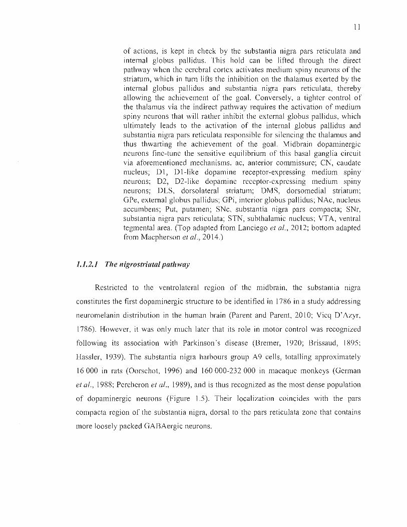
Il
of actions, is kept in check by the substantia nigra pars reticulata and internai globus pallidus. This hold can be lifted through the direct pathway when the cerebral cortex activates medium spiny neurons of the striaturn, which in turn lifts the inhibition on the thalamus exerted by the internai globus pallidus and substantia nigra pars reticulata, thereby allowing the achievement of the goal. Conversely, a tighter control of the thalamus via the indirect pathway requires the activation of medium spiny neurons that will rather inhibit the external globus pallidus, which ultimately leads to the activation of the internai globus pallidus and substantia nigra pars reticulata responsible for silencing the thalamus and thus thwarting the achievement of the goal. Midbrain dopaminergic neurons fine-tune the sensitive equilibrium of this basal ganglia circuit via aforementioned mechanisms. ac, anterior commissure; CN, caudate nucleus; Dl , D1-like dopamine receptor-expressing medium spiny neurons; D2, D2-like dopamine receptor-expressing medium spiny neurons; DLS, dorsolateral striatum; DMS, dorsomedial striatum; GPe, external globus pallidus; GPi, interior globus pallidus; NAc, nucleus accumbens; Put, putamen; SNc, substantia nigra pars compacta; SNr, substantia nigra pars reticulata; STN, subthalamic nucleus; VTA, ventral tegmental area. (Top adapted from Lanciego et al. , 2012; bottom adapted from Macpherson et al. , 2014.)
1.1.2.1 The nigrostriatal pathway
Restricted to the ventrolateral reglOn of the midbrain, the substantia mgra
constitutes the first dopaminergic structure to be identified in 1786 in a study addressing
neuromelanin distribution in the human brain (Parent and Parent, 2010; Vicq D'Azyr,
1786). However, it was only much later that its role in motor control was recognized
following its association with Parkinson ' s disease (Bremer, 1920; Brissaud, 1895;
Hassler, 1939). The substantia nigra harbours group A9 cells, totalling approximately
16000 in rats (Oorschot, 1996) and 160000-232000 in macaque monkeys (German
et al. , 1988; Percheron et al. , 1989), and is thus recognized as the most dense population
of dopaminergic neurons (Figure 1.5). Their localization coincides with the pars
compacta region of the substantia nigra, dorsal to the pars reticulata zone that contains
more loosely packed GABAergic neurons.

12
Figure 1.5 A8-AIO dopaminergic neuronal c1usters in the rat brain. Immunohistochemical staining of dopaminergic neurons in rat brain coronal sections organized rostrocaudally (A-F) depict the neuroanatomical location of each cluster at the level of the midbrain. Particularly important to us, the substantia nigra pars compacta is found laterally to the ventral tegmental area. Scalebars: A-F, 1 mm; inset F, 0.5 mm. aq, cerebral aqueduct; de, dorsocaudal; f, fomix; IPN, interpeduncular nucleus; ml, medial lemniscus; MT, mammillothalamic tract; P AGvl, ventrolateral periaqueductal grey; RRF, retrorubral field ; SNI, substantia nigra pars lateralis. (From Yetnikoff et al., 2014.)

13
The substantia nigra pars compacta provides the greatest dopaminergic input to
the striatum, particularly - but not restricted (Yetnikoff et al. , 2014) - to the dorsal
striatum better known in primates as the caudate nucleus and putamen or simply the
caudate-putamen8 (Figure 1.6). Nigrostriatal dopaminergic neurons project in a fairly
mediolateral topographical manner (Lynd-Balta and Haber, 1994) to an estimated
2.8 million and 31 million striatal neurons in rats and macaques, respectively, each axon
ramifying abundantly and harbouring sorne 500 000 varicosities (Andén et al., 1966).
This affords the dorsal striatum with a near-total exposure to dopamine input, implying a
significant divergence factor of 300-400 between nigrostriatal neurons and striatal
targets. The importance of nigrostriatal arborizations is even better appreciated when
taking into account that, at least in rats, one striatal neuron is innervated by an average
of 100-200 dopaminergic neurons and that one nigrostriatal axon is estimated to interact
with sorne 75 000 striatal neurons (Matsuda et al., 2009). Such a profuse dopaminergic
innervation necessarily in fers critical modulatory effects on the dorsal striatum,
which receives important glutamatergic inputs from the cerebral cortex, thalamus and
amygdala (Donoghue and Herkenham, 1986; Graybiel, 1990; Sadikot et al., 1992).
Striatal modulation by nigrostriatal neurons is best appraised in corticostriatal
motor circuits. As it consists in the most important source of tonic dopamine to
the striatum, the substantia nigra pars compacta especially inactivates indirect
pathway medium spiny neurons expressing D2 receptors, thereby downgrading the
inhibition on the thalamus. In Parkinson's disease, degeneration of nigrostriatal
dopaminergic neurons disrupts tonic firing in the striatum, which allows for the
D2 receptor-modulated indirect pathway to exert its full inhibitory effect on the
thalamus. This pathophysiological feature dwells at the core of parkinsonian motor
symptoms, chiefly bradkyinesia/akinesia and rigidity (Albin et al., 1989). In fact, several
animal models employ nigrostriatal pathway les ions of ail sorts to emulate parkinsonian
symptoms (Deumens et al., 2002; Dut Y and Jenner, 2011; Jackson-Lewis et al. , 2012;
Nagatsu, 1997; Pinna and Morelli, 2014; Ungerstedt, 1968).
8 Whether in rodents, primates or humans, the caudate-putamen will hereafter be termed dorsal striatum.

14
A mesocortical pathway
B
midbrain
mesolimbic pathway
nigrostriatal pathway
#
midbrain
Llmbic
Figure 1.6 Mesocorticolimbic and nigrostriatal projections. Both mesocorticolimbic and nigrostriatal pathways ongmate in the midbrain, illustrated sagittally (A) and coronally (B). The nigrostriatal pathway projects especially onto dorsal regions of the striatum, while the mesocorticolimbic pathway mainly innervates the ventral striatum (mesolimbic) and the prefrontal cortex (mesocortical). Midbrain projections onto the striatum are fairly topographic, depicted by colour gradients (B), and modulate specific corticostriatal circuits (C). Afferents supplied by sensorimotor, associative and limbic cortical are as are also distributed in a particular neuroanatomical manner, which bears implications on the behavioural programs that different midbrain projections can control (C). dStr, dorsal striatum; OT, olfactory tubercle. (Adapted from Chuhma et al., 2017.)

15
1.1.2.2 The mesocorticolimbic pathway
In the first half of the 20th century, neuroscientists bore a rather unyielding vision
of basal ganglia as movement processing centres, still somewhat enduring today.
More complex functions were much later ascribed to these structures upon the
identification of corticostriatal circuit loops that did not involve motor areas (Alexander
et al. , 1990; Haber et al., 1985; Young et al. , 1984), and the parallel discovery of a
particular striatal subdivision located ventrally and sUITounding the anterior commissure
termed the nucleus accumbens (Heimer, 1978). In the meantime, a collection of studies
were showing rats to electrically self-stimulate certain regions of their brain as positive
reinforcement (Olds and Milner, 1954), later found to receive dopaminergic inputs
(Crow, 1972) and to be sensitive to dopamine agonists or antagonists (Stein, 1964;
Wise and Bozarth, 1987). Pioneering microdialysis work from the laboratory of
Prof. Gaetano Di Chiara at the University of Cagliari, Italy, later confirmed the nucleus
accumbens identity of this region, shown to be sensitive to dopamine agonizing drugs9
(Di Chiara and Imperato, 1988).
The nucleus accumbens, found in the ventral striatum, is one of the key dopamine
terminaIs of the mesocorticolimbic pathway. This system is composed of the mesolimbic
and mesocortical pathways, originating from groups A8 and Al 0 neurons (Figure 1.5).
Both lodged in the midbrain, group Al 0 coïncides with the ventral tegmental area
medial to the substantia nigra pars compacta, whereas group A8 is contained in the
retrorubral field 10 located dorsocaudally to the substantia nigra pars compacta (Fu et al. ,
2012; Halliday et al. , 20 12a). Mesolimbic dopaminergic neurons project to the striatum,
preferentially to ventral areas such as the nucleus accumbens and the olfactory tubercle,
whereas the mesocortical pathway connects the ventral tegmental are a and retrorubral
9 Di Chiara' s group also made the groundbreaking di scovery that the nucleus accumbens is in fact di vided into core and shell subregions, which operate very di fferently in reward valuation (Pontieri et al., 1995 ).
10 The retrorubral field does not account for much of the dopaminergic innervation provided by the midbrain and therefore little attention is pa id to it in this thesi s. Likewise, the olfactory tuberc\e is a secondary ventral striatum structure whose functions in the mesolimbic pathway will not be granted much focus .

16
field to the pre frontal cortex, but these connections are mu ch less dense than striatal ones
(Figure 1.6).
Akin to nigrostriatal dopaminergic neurons, the ventral tegmental area
operates via modulation of corticostriatal circuits at the level of the striatum. However,
inasmuch as the nigrostriatal pathway is known for tonically enabling movement,
the mesocorticolimbic pathway stands out as a reward processing system endowed with
limbic and executive dimensions permitted by phasic bursts of dopamine (Haber et al. ,
2000). There exists today a wide array of pharmacological paradigms mimicking
dysfunctional dopaminergic neurotransmission in the ventral striatum and prefrontal
cortex (Espafia and Jones, 2013; Jones et al., 2011). Notably, these models bear a
striking likeness to pathologies such as addiction and other neuropsychiatric disorders
(Bassareo et al., 2011 ; Di Chiara et al., 1993; Grace, 2000, 2016; Tanda et al. , 1996).
1.1.2.3 Converging and diverging functions
While nigrostriatal and mesocorticolimbic pathways appear to operate very
differently considering their fairly different roles in human behaviour and disease,
they actually function in similar ways. In fact, aside from their archetypal tonic-motor
and phasic-reward instructions, the nigrostriatal and mesocorticolimbic pathways are
both endowed with the following characteristics: 1) they comprise dopaminergic
neurons that can fire both phasically and tonically depending on the information
conveyed by dopamine; 2) they project onto striatal medium spiny neuron D 1-like
and D2-1ike receptors thereby modulating corticostriatal circuitry, and; 3) their
neuroanatomical origins receive feedback projections from the targets they innervate
that refine dopaminergic neurotransmission. Despite the widely acknowledged
movement enabling functions of the nigrostriatal pathway, regulation of the striatum by
its afferents also holds a role in many other decision-making processes, leamed
behaviours and habits arising from outcome prediction, especially with regard to
sensorimotor functions. These roles may have to do with time scales of dopamine
impulses, where sensory-evoked phasic firing encodes teaching signaIs for the
17
acquisition of a behaviour, (Chaudhury et al., 2013; Redgrave and Gurney, 2006;
Schultz et al., 2006), and where baseline tonic firing enables the proper selection of
these pre-Iearned behaviours by the basal gang lia (Ellwood et al., 2017; Hikosaka et al.,
2000; Redgrave et al., 1999). Both firing modes are impaired in Parkinson's disease
(Beeler, 2011; Marklund et al., 2009; Redgrave et al., 2010), although the loss of
dopamine tone is more typically acknowledged with respect to the motor symptoms it
produces. The same holds true for the mesocorticolimbic pathway, which is also devoted
to tonic enabling of pre-Ieamed behaviours aimed at seeking pleasure and rewards that
had previously required a learning period provided by phasic firing of dopamine
(Ellwood et al., 2017).
In a larger sense, nigrostriatal and mesocorticolimbic dopamine operates both as
a phasic carrier of predicted outcomes and a tonic enabler of postsynaptic neurons,
from which originates its dual informationallmodulatory role (Saddoris et al., 2013;
Schultz, 1998). Indeed, in both pathways, fast phasic bursts encode reward, slow phasic
changes inform on punishing/ambiguous elements, and tonic maintenance enables
postsynaptic functions (Schultz, 2007). In this respect, an overarching the ory proposes
CNS dopamine to mediate the basic reactivity of animaIs to their environrnent, ensuring
their survival by permitting economic decision-making, reward recognition, monitoring
of uncertainty and detection of punishing elements via the prediction of favourable or
unfavourable outcomes, wherein dopamine encodes prediction errors (Schultz, 2007)
and leaming entails progressive reduction of prediction errors. This is applicable to
motor, social, cognitive, motivational and reward processes regulated by these pathways.
This begs the following questions: Why are mesocorticolimbic dysfunctions
highly associated with addiction, whereas nigrostriatal ones are linked with Parkinson's
disease? And why is the mesocorticolimbic pathway largely spared in Parkinson's
disease? At first glance, behavioural and pathological disparities in these pathways are
the result of rather c1ear neuroanatomical distinctions between their origins, embedded
in different environrnents, and between their targets, innervated by distinct cortical
inputs and expressing differential distribution patterns of D l-like and D2-like

18
receptors (Bertran-Gonzalez et al., 2008; Centonze et al., 2003; Lanciego et al., 2012).
As such, even if both pathways operate with a similar dopamine interface regulating
corticostriatal circuitry, they modulate different cortical programs. Other sources of
dopaminergic dichotomies in health and disease may rather lie in differential exposures
to noise-to-signal (tonic-to-phasic) ratios. Indeed, the dorsal striatum is submitted to a
greater noise-to-signal ratio than the ventral striatum, due to the marked tonicity of
nigrostriatal neurons responsible for very high basal release of dopamine in the dorsal
striatum (Dreyer, 2014; Zhang et al., 2009). This al10ws two implications: first, phasic
firing do es not have the same weight in the dorsal striatum than in the ventral striatum,
which explains the great differences in the information carried by dopamine in these
two areas; second, high basal pacemaking activity may in itself constitute a risk factor in
neurodegeneration, later discussed in the context ofParkinson's disease.
In keeping with the discriminating elements that pick these pathways apart,
the remainder of this thesis will address the apparent preferential degeneration of
nigrostriatal dopaminergic neurons in various conditions, as in Parkinson's disease next
addressed, and the behavioural alterations that may arise from this neurodegeneration.
1.1.3 Parkinson 's disease
The year 2017 marked the 200th anniversary of Dr. James Parkinson's An Essay on
the Shaking Paisy wherein were described 6 subjects suffering from a pathological state
that he termed at the time paralysis agitans (see for historical reviews Obeso et al. ,
2017; Parent and Parent, 2010). In this es say, Dr. Parkinson illustrated with acute
precision several symptomatic aspects exhibited by his subjects that we know today
were effectively attributable to Parkinson's disease. Indeed, he accurately identified
the graduai nature of both onset and progression, as well as the expression of gait
disturbances, forward flexion of the trunk, resting tremors, sleeping troubles and
gastrointestinal issues in these individuals (Parkinson, 1817). Charcot later expanded on
these observations to include bradykinesia and rigidity to the key features of the disease,
and proposed to rename the paralysis agitans disorder Parkinson's disease upon

19
observing that the loss of motor functions was not attributable to weakness (paIsy)
(Charcot, 1877).
Since the publication of this pioneering report, the field of Parkinson ' s disease has
phenomenally progressed. In this age of effervescent neuroscientific developments,
clinical profiles are more accurately drawn, long-term patient management is improving,
and the pursuit of etiological and pathophysiological explanations is starting to bear fruit
(Chaudhuri and Jenner, 2017). The preferential degeneration of the dopaminergic
nigrostriatal pathway is weIl appreciated today and additional pieces of the puzzle are
continuously falling into place, especially with respect to non-dopaminergic affections
(Hall et al., 2014; Qarnhawi et al. , 2015; Remy, 2005). The following section
surnmarizes state-of-the-art knowledge on Parkinson's disease and draws particular
attention to the apparent vulnerability of nigrostriatal dopaminergic neurons in
this pathology. The CUITent state of the field regarding symptomatology, diagnostics,
pathological basis, genetic and idiopathie etiopathogeneses, experimental models,
neuroimaging, biomarkers, treatments and unmet needs is extensively reviewed
elsewhere by preeminent researchers in a special collaborative publication underlining
the second centenary of the shaking paIsy (Obeso et al. , 2017).
1.1.3.1 Clinical symptomatology and treatments
As highlighted by Dr. Parkinson's and others ' early observations, a wide array of
motor and non-motor symptoms characterize Parkinson ' s disease, which both enable and
complicate the process for clinicians to arrive to a diagnosis. Indeed, this disorder is
pronouncedly heterogeneous in terms of inter-individual symptomatology, but also with
respect to disease progression and sensitivity to treatments (Lang and Obeso, 2004)
(Figure 1.7). For the sake of example, the age of onset can range from as early as the
third de cade of life to very old ages (Pagano et al. , 2016); initial motor manifestations
may affect either upper or lower limbs on either side of the body (Roberts et al. , 2017);
deterioration rates may present benign or malignant courses (Jankovic et al., 1990), and;
distinct subpopulations of patients may respond differently to dopamine treatments
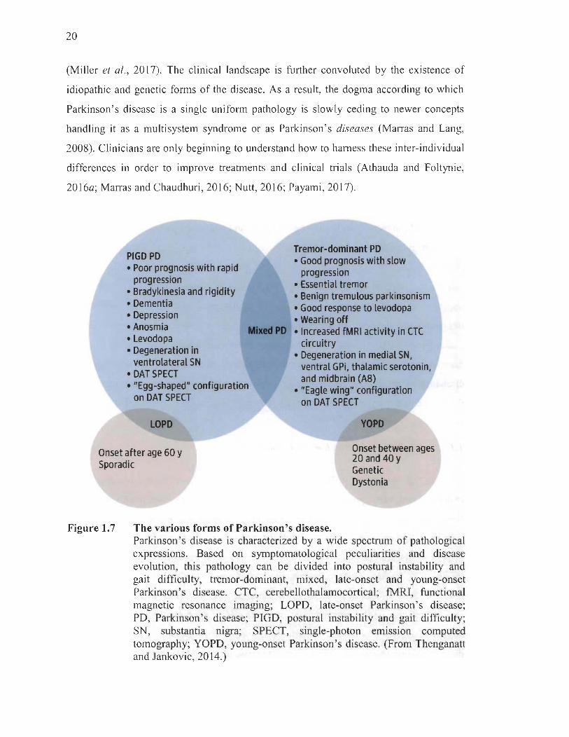
20
(Miller et al., 2017). The clinical landscape is further convoluted by the existence of
idiopathie and genetic forrns of the disease. As a result, the dogma according to which
Parkinson's disease is a single uniforrn pathology is slowly ce ding to newer concepts
handling it as a multisystem syndrome or as Parkinson 's diseases (Marras and Lang,
2008). Clinicians are only beginning to understand how to hamess these inter-individual
differences in order to improve treatments and clinical trials (Athauda and Foltynie,
2016a; Marras and Chaudhuri, 2016; Nutt, 2016; Payami, 2017).
PIGD PD • Poor prognosis with rapid
progression • Bradykinesia and rigidity • Dementia • Depression • Anosmia • Levodopa • Degeneration in
ventrolateral SN
MlxedPO
• DAT SPECT • "Egg-shaped" configuration
on DAT SPECT
LOPD
Onset after age 60 y Sporadic
Tremor-dominant PD • Good prognosis with slow
progression • Essential tremor • Benign tremulous parkinsonism • Good response to levodopa • Wearing off • Increased fMRI activity in CTC
circuitry • Degeneration in medial SN,
ventral GPi, thalamic serotonin, and midbrain (AS)
• "Eagle wing" configuration on DAT SPECT
YOPD
Onset between ages 20 and 40 y Genetic Dystonia
Figure 1.7 The various forms of Parkinson 's disease. Parkinson's disease is characterized by a wide spectrum of pathological expressions. Based on symptomatological peculiarities and disease evolution, this pathology can be divided into postural instability and gait difficulty, tremor-dominant, mixed, late-onset and young-onset Parkinson 's disease. CTC, cerebellothalamocortical; fMRI, functional magne tic resonance imaging; LOPD, late-onset Parkinson 's disease; PD, Parkinson's disease; PIGD, postural instability and gait difficulty; SN, substantia nigra; SPECT, single-photon emission computed tomography; YOPD, young-onset Parkinson's disease. (From Thenganatt and Jankovic, 2014.)

21
The most prominent feature of Parkinson's disease remams the presence of
motor symptoms expressed in a majority of patients as a classical triad of resting
tremors, bradykinesia and rigidity; two of them are required for diagnostic purposes
(Parkinson Québec, 2018). Later, motor disabilities evolve to implicate gait and postural
reflex disturbances, though nowadays they are no longer required to emit a diagnosis.
Non-dopaminergic (extranigral) motor features also arise and consist in falls , freezing
of gait, speech impairment and difficulty swallowing. Accompanying these motor
symptoms are equally disabling non-motor manifestations, such as psychiatric
disturbances, dementia and autonomic failure . Granted Parkinson ' s disease is not a fatal
condition, patients are expected to live almost as long as healthy individuals and the
primary cause of death appears to be pneumonia (lwasaki et al. , 1990; Pennington et al.,
2010), although manifestations of more advanced symptoms, such as falls and
swallowing problems, may lead to premature accidentaI mortalities.
Adequate management of the incapacitating motor symptoms in Parkinson' s
disease was only made possible 150 years after the issuing of the shaking paIsy essay
when George C. Cotzias and his colleagues published a trailblazing paper on L-DOPA
(Cotzias et al., 1967) that remains, with certain modem ad just ment s, the benchmark
therapy (Freitas et al. , 2016, Lotia and Jankovic, 2016). While its function is
unmistakably to restore tonic dopaminergic neurotransmission (Dreyer, 2014), exactly
how L-DOPA operates remains hazy as ofyet. Indeed, L-DOPA may not act as a simple
precursor for dopamine allowing for its tonic replenishment in the striatum: sorne have
advanced its possible role as a neurotransmitter itself (LeWitt and Fahn, 2016).
Nevertheless, a slew of clinical trials, intervention studies and longitudinal observations
ceaselessly validate L-DOPA's efficacy in alleviating motor symptoms (LeWitt and
Fahn, 2016; Olanow, 2014; Yahr, 1969), although several collateral effects are
manifested by acute and chronic users, the most notable being dyskinesias, impulse
control disorders and wearing-off in longstanding administration (Aquino and Fox,
2014; Cilia, 2012; Parkinson Study Group, 2004). In the hop es of delaying the
apparition of these side effects, sorne patients are initially administered dopamine

22
receptor agonists or monoamine oxidase B inhibitors until these milder medicines can no
longer manage the motor symptoms as the disease progresses (Figure 1.8).
Circulation
COMT inhibitors (central)
l Dopamine availability • Amanladlne
• L-DOPA -----+-+--tl~ • • Dopamine
receptors • • • • •
3-0MD
Dopamine Agonists
COMT inhibitors (peripheral)
'Selegillne • Rasagillne
• Bromocrtptlne (ergol) • Pramlpexole (non ... rgol) • Roplnlrole (non ... rgol)
• Tolcapone • Enlacapone
Presynaptic neuron Postsynaptic neuron
Figure 1.8
Blood-brain barrier
Current parkinsonian drug therapies. On the left hand side are depicted adjuvant agents necessary for L-3 ,4-dihydroxyphenylalanine (L-DOPA) to reach the brain considering its propensity to be metabolized in the periphery, thereby dampening its efficacy. Other drugs are illustrated on the right, including more recent dopamine agonists and inhibitors of the reuptake and metabolism of dopamine. It is important to note that no therapy discovered to date achieves curative endpoints in Parkinson's disease. 3-0MD, 3-0-methyldopa; DDC, DOPA decarboxylase. (From http: //step2.medbullets. com/step2-3-neurology/121704/parkinsons-disease-drugs.)
Inter-individual irregularities not only complicate diagnosis, but also shape
treatment efficacy in Parkinson's disease. Sorne patients, usually younger diagnostics,
will develop a benignly progressive form presenting few non-dopaminergic symptoms
and these usually respond weil to L-DOPA treatments. Others may suffer from a
more malignant, rapidly progressing disease with typically early manifestations of
non-dopaminergic motor and non-motor symptoms that resist to L-DOPA interventions
23
(Lang and Obeso, 2004). Regardless of age at onset, older individuals are more likely to
express L-DOPA-resistant motor symptoms in conjunction with cognitive decline and
autonomic failure (Kempster et al., 2007). In patients with severe motor fluctuations,
other strategies may be employed, for instance deep brain stimulation of the subthalamic
nucleus or internaI globus pallidus in basal ganglia circuitry, performed in sorne
150 000 patients (Obeso et al., 2017) since its groundbreaking successful utilization in
the 1990s (Pollak et al. , 1993). Akin to L-DOPA therapy, the mechanistic underpinnings
of deep brain stimulation are ill understood (Lozano and Lipsman, 20 l3) and adverse
effects may supervene in the likes of impaired verbal fluency, depression and ev en
suicidaI tendencies (Benabid et al., 2005; Lang et al., 2006). To these motor symptom
therapies are sometimes layered supplementary treatments to address psychiatric
manifestations arising from the disease or motor symptom treatments themselves,
for instance cholinesterase inhibitors for cognitive impairment (Emre et al., 2004) ,
antidepressants and cognitive-behavioural therapy for mood disorders (Dobkin et al.,
2011 ; Menza et al. , 2008; Richard et al. , 2012) or neuroleptics for psychoses
(Cummings et al., 2014).
Today, still, no therapy has been fruitful in modifying the course of Parkinson 's
disease, ev en less so in providing hope for curative ends. This may partI y be attributable
to the failure of clinical trials or therapeutic strategies to take into account the various
existing subgroups of Parkinson's disease patients. Sensitive subdivision of clinical
subtypes at the early stages of the pathology is among the most pressing research aims to
render treatments and their development more discriminative, especially considering the
rapid rate at which the population is aging occasioning foreseeable rises in the incidence
of age-related neurodegenerative disorders su ch as Parkinson's disease (Olesen et al.,
2012) (Table 1.1). In order to achieve this, reliable identification of premotor phases of
the pathology, obviously requiring robust biomarkers, will need to be accomplished first.
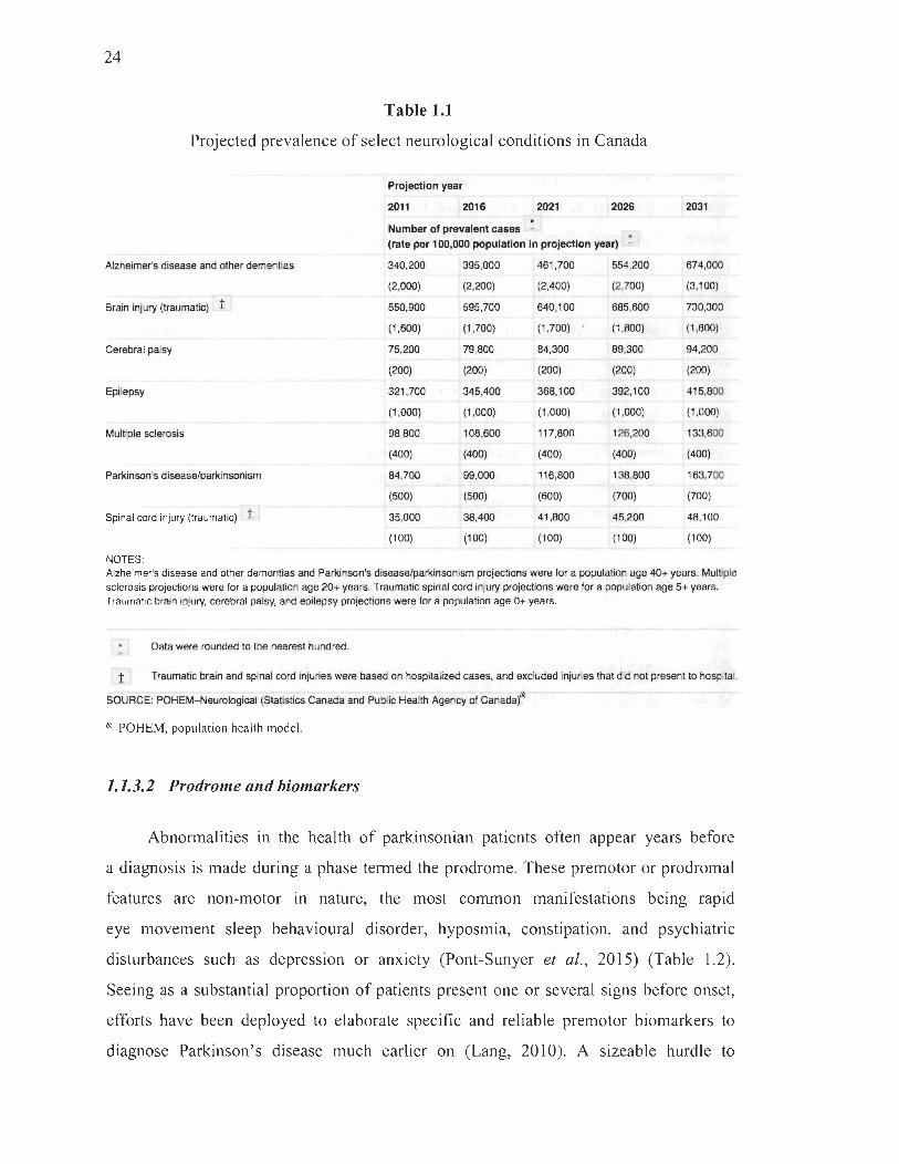
24
Table 1.1
Projected prevalence of select neurological conditions in Canada
Projection year
2011 2016 2021 2026 2031
Number of prevalent cases - . (rate per 100,000 population ln projection year) -
Alzheimer's disease and other dementias 340,200 395,000 461,700 554,200 674,000
(2,000) (2,200) (2,400) (2,700) (3,100)
Brain in jury (traumatic) t 550,900 595 ,700 640,1 00 685,600 730,300
(1,600) (1,700) (1,700) (1,800) (1,800)
Cerebral paisy 75 ,200 79,800 84,300 89,300 94,200
(200) (200) (200) (200) (200)
Epilepsy 321,700 345,400 368,100 392,1 00 415,800
(1,000) (1,000) (1,000) (1 ,000) (1 ,000)
Multiple sclerosis 98,800 108,600 117,800 126,200 133,600
(400) (400) (400) (400) (400)
Parkinson 's disease/parkinsonism 84,700 99,000 116,800 138,800 163,700
(500) (500) (600) (700) (700)
Spinal cord in jury (traumatic) t 35,000 38,400 41,800 45,200 48,100
(1 00) (100) (100) (100) (100)
NOTES: Alzheimer's disease and other dementias and Parkinson's disease/parkinsonism projections were for a population age 40+ years. Multiple sclerosis projections were for a population age 20+ years. Traumatic spinal cord in jury projections were for a population age 5+ years.
Traumatic brain in jury, cerebral paisy, and epilepsy projections were for a population age 0+ years.
Data were rounded to the nearest hundred.
1 Traumatic brain and spinal cord injuries were based on hospitalized cases, and excluded injuries that did not present to hospital.
SOURCE: POHEM-Neurological (Statistics Canada and Public Health Agency of Canada)&
& POHEM, population health mode!.
1.1.3.2 Prodrome and biomarkers
Abnormalities in the health of parkinsonian patients often appear years before
a diagnosis is made during a phase termed the prodrome. These premotor or prodromal
features are non-motor in nature, the most common manifestations being rapid
eye movement sleep behavioural disorder, hyposmia, constipation, and psychiatrie
disturbances such as depression or anxiety (Pont-Sunyer et al., 2015) (Table 1.2).
Seeing as a substantial proportion of patients present one or several signs before onset,
efforts have been deployed to elaborate specifie and reliable premotor biomarkers to
diagnose Parkinson's disease much earlier on (Lang, 2010). A sizeable hurdle to
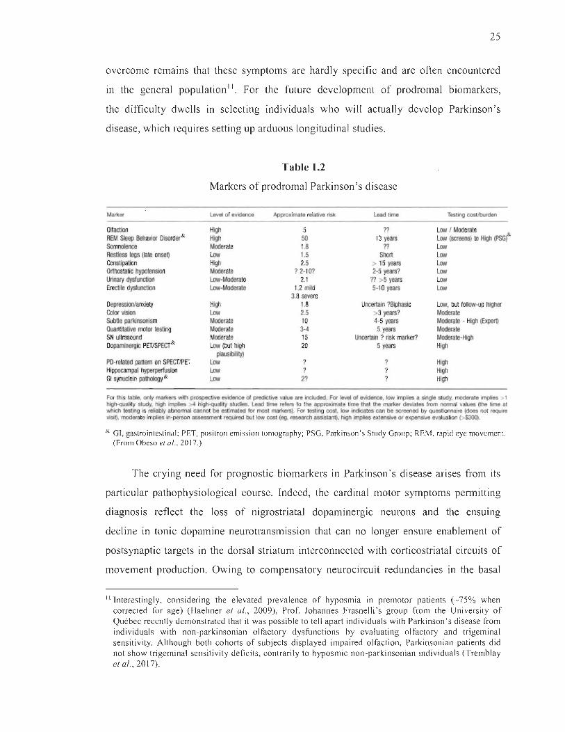
25
overcome remams that these symptoms are hardly specific and are often encountered
in the general population". For the future development of prodromal biomarkers,
the difficulty dwells in selecting individuals who will actually develop Parkinson' s
disease, which requires setting up arduous longitudinal studies.
Table 1.2
Markers of prodromal Parkinson ' s disease
Marker Level of evidence Approxlmate relative risk Lead time Testing costlburden
O~action High 5 ?? Law 1 Moderate & REM Sleep Behavior Disorder& High 50 13 years Law (screens) ta High (PSG) Somnolence Moderate 1.8 ?? Law Restless legs (late onset) Law 1.5 Short Law Constipation High 2.5 > 15 years Law Orthostatic hypotension Moderate ? 2·10? 2·5 years? Law Urinary dysfunction Law-Moderate 2.1 ?? >5 years Law Erectile dysfunction Law-Moderate 1.2 mild 5·10 years Law
3.8 severe Depression/anxiety High 1.8 Uncertain ?Biphasic Law, but follaw·up higher Color vision Law 2.5 >3 years? Moderate Subtle parkinsonism Moderate 10 4-5 years Moderate - High (Expert) Quantitative motor testing Moderate 3-4 5 years Moderate SN ultrasound Moderate 15 Uncertain ? risk marker? Moderate-Hlgh Dopaminergic PETISPECT& Law (but high 20 5 years High
plausibility) PD-related pattern on SPECT/PET Law ? ? High Hippocampal hyperpertusion Law ? ? High GI synuclein palhology & Law 2? ? High
For thls table, only markers wrth prospective evodence of predictove value are oncluded. For level of evidence, low Implies a single study, moderate ompies > 1 high-quality study, high ompies >4 high-quality studoes. Lead time refers to the approximate tome that the marker deviates from normal values (the tome at which testong is reliably abnormal cannot be estimated for rnost markers). For testong cost, Iow indicates can be screened by questoonnaire (does not requira visit), moderate impies in-person assessment requlred but low cost (eg, research aSsistant), hlgh Implies extensive or ex pensive evaluation (>$300).
& GI, gastrointestinal; PET, positron emission tomography; PSG, Parkinson's Study Group; REM, rapid eye movement. (From Obeso et al. , 201 7.)
The crying need for prognostic biomarkers in Parkinson ' s disease arises from its
particular pathophysiological course. lndeed, the cardinal motor symptoms permitting
diagnosis reflect the loss of nigrostriatal dopaminergic neurons and the ensuing
decline in tonic dopamine neurotransmission that can no longer ensure enablement of
postsynaptic targets in the dorsal striatum interconnected with corticostriatal circuits of
movement production. Owing to compensatory neurocircuit redundancies in the basal
II Interestingly, considering the elevated prevalence of hyposmia in premotor patients (~75% when corrected for age) (Haehner et al. , 2009), Prof. Johannes Frasne lli 's group from the Uni versity of Québec recently demonstrated that it was possible to te ll apart individuals with Parkinson's disease from individuals with non-parkinsonian olfactory dysfunctions by evaluating olfactory and trigeminal sensiti vity. Although both cohorts of subjects displayed impaired olfaction, Parkinsonian patients did not show trigeminal sensitivity deficits, contrarily to hyposmic non-parkinsonian individuals (Tremblay et al., 2017).

26
ganglia and adjustments of dopamine receptor numbers, anywhere between 30-70% of
nigrostriatal dopaminergic neurons and 50-80% of their striatal terminaIs have peri shed
before the advent of clear motor symptoms (Bemheimer et al., 1973; Cheng et al., 2010;
Feamley and Lees, 1991), dulling any hope for reversaI of the pathology. Like man y
other neurodegenerative disorders, Parkinson's disease begins many years before
classical symptoms arise. However, to our advantage, it is a rather special pathology in
that neurodegeneration appears to begin in regions outside the substantia nigra pars
compacta whose progressive dysfunction may emerge as a phenotypic non-motor
signature that could one day be exploited for emitting diagnoses at premotor stages.
The following section grants a closer look at the pathophysiological mechanisms
subtending the particular course of progression in Parkinson's disease.
1.1.3.3 Pathophysiology
More th an a century after its identification by Félix Vicq d'Azyr (Vicq d'Azyr,
1786), the substantia nigra was freshly emerging as the neuroanatomical substrate of
the motor syndrome in Parkinson's disease (Blocq and Marinesco, 1892; 1893; Brissaud,
1895). In parallel, Friedrich Heinrich Lewy was conducting histological assessments of
parkinsonian brains, discovering the presence of cytoplasmic inclusions of aggregated
proteins in the dorsal vagal nucleus and substantia innominata (Lewy, 1912),
later identified in surviving nigral neurons of other patients and termed Lewy bodies in
recognition of the former 's seminal work (Trétiakoff, 1919). In the mid 1900s, the role
of the substantia nigra in Parkinson's disease was further bolstered by findings enlisting
dopamine as a key contributor (Carlsson, 1958; Ehringer and Homykiewicz, 1960) and
demonstrating the projection of nigral neurons to the dorsal striatum (Homykiewicz,
1966). From these early discoveries were derived the two pathological requirements to
emit a definitive post-mortem diagnosis, still standing today: first, the marked 10ss of
pigmented dopaminergic neurons in the substantia nigra pars compacta, and second, the
presence of Lewy bodies or their intraneuritic counterparts, Lewy neurites (Dickson
et al. , 2009). To these findings were added, in later years, the contributions of
Langston and colleagues describing parkinsonism in opioid addicts who had
27
accidentally consumed I-methyl-4-phenyl-l,2,3 ,6-tetrahydropyridine (MPTP) (Langston
et al., 1983), a by-product of desmethylprodine synthesis that impedes the mitochondrial
electron transport chain and triggers the production ofROS I2 (Greenamyre et al., 2001).
This discovery also allowed further development of animal models of Parkinson's
disease, complementing physical and 6-hydroxydopamine (6-0HDA) les ion paradigms
(Ungerstedt, 1968), and coaxed investigations on environmental neurotoxins in the
etiopathogenesis of the disease owing to MPTP's resemblance to certain known
pesticides.
The latest significant leap forward in the field dates back to the identification of
the first gene mutation, explicitly a single point mutation in the SNCA gene co ding for
a-synuclein 13, found to cause a dominant familial form of early onset Parkinson 's
disease (Polymeropoulos et al., 1997). This event triggered a sequence of discoveries
regarding the major constituent of Lewy bodies and neurites, a-synuclein (Spillantini
et al., 1997), but also conceming genetic forms of the pathology. Disease-causing
duplications (Chartier-Harlin et al., 2004) and triplications (Singleton et al., 2003) of
the SNCA gene were quickly identified, to which were added in subsequent years several
other genes (Table 1.3). It is important to note that idiopathie Parkinson 's disease still
composes approximately 90-95% of diagnoses and are not always faithfully represented
by genetic forms in terms of symptomatology and pathophysiology (see for review
Houlden and Singleton, 2012).
\2 The generation of ROS by the mitochondrial electron transport chain is later described in section 1.2.4.1.
\3 a-Synuclein is an intrinsically unstructured protein, especially localized to presynaptic terminais wherein it cycles between a cytosolic natively unfolded state and a membrane-bound state. Its function in normal physiological conditions remains unclear to date, but it is thought to regulate synaptic activity, vesicular trafficking and several metabolic and transport enzymes specifie to dopaminergic neurons (see for review Surré, 2015).
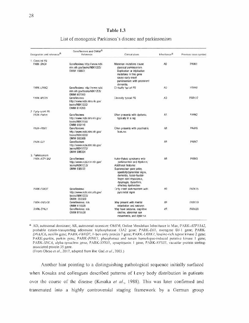
28
Table 1.3
List of monogenic Parkinson's disease and parkinsonism
Designalioo and reference &
1. Classical PO PARK-SNC4
PARK-LRRK2
PARK-VPS35
2. Early-onset PO PARK -parkin
PARK-PINK1
PARK-OJ1
3. Parkinsonism PARK-ATP1342
PARK-FBX07
PARK-ONAJC6
PARK-SYNJI
GeneReviews and OMIM& Reference
GeneReviews hl1p://www.ncbi. nlm.nih.govlbookslNBK1 'l231 DMIM 168601
GeneReviews hl1p://www.ncbi. nlm.nih.govlbookslNBKl2081 OMIM 607060 GeneReviews hI1pJIwww.ncbi.nlm.nih.gov/ books/NBK1223J OMIM 614203
GeneReviews hI1pJIwww.ncbi.nlm.nih.gov/ books/NBKl1551 OMIM 600116 GeneReviews httpJIwww.ncbi.nlm.nih.gov/ books/NBK1223J OMIM 605909 GeneReviews hI1pJIwww.ncbi.nlm.nih.gov/ books/NBK1223J OMIM 606324
GeneReviews hI1pJIwww.ncbi.nlm.nih.gov/ books/NBK1223J OMIM 606693
GeneReviews hI1pJIwww.ncbi.nlm.nih.gov/ books/NBK1223J OMIM: 260300 GeneReviews: nIa OMIM 615528 GeneReviews: nIa OMIM 615530
Clinical cluBs
M issense mutations cause dassical par1<insonism. Duplication IX' triplication mutations in this gene cause early onset parkinsonism with pro minent dementia
Clinically typical PO
Clinically typical PO
Often presents with dystonia, typically in a leg
Often presents with psychiatrie leatures
Kufor-Rakeb syndrŒne with parkinsonism and dystonia;
Additional leatu res: Supranuclear gaze paisy,
spastidty/pyramidal signs, dementia, lacial-laudallinger mini-myodonus, dysphagia, dysarthria, ~Iactory dyslunction
Early onset parkinsonism with pyramidal signs
May present with mental retwdation and seizures
May have seizures, cognitive decline, abnorrnal eye movements, and dystonia
Inheritance&
AD
AD
AD
AR
AR
AR
AR
AR
AR
AR
Previous locus symbol
PARK1
PARK8
PARK17
PARK2
PARK6
PARK7
PARK9
PARK15
PARK19
PARK20
& AD, autosomal dominant; AR, autosomal recessive; OMIM, Online Mendelian Inheritance in Man; PARK-ATPI 3A2, probable cation-transporting adenosine triphosphatase 13A2 gene; P ARK-DJI , oncogene OJ- I gene; P ARKDNAJC6, auxilin gene; PARK-FBX0 7, F-box only protein 7 gene; PARK-LRRK2, leucine-ri ch repeat kinase 2 gene; PARK-parlan, parkin gene; PARK-PINKl , phosphatase and tensin homologue-induced putative kinase 1 gene; PARK-SNCA, alpha-synuclein gene; PARK-SYNJI , synaptojanin 1 gene; PARK- VPS35, vacuolar protein sortingassociated prote in 35 gene. (From Obeso et al., 20 17, adapted From Bar-Gad et al., 2003 .)
Another hint pointing to a distinguishing pathological sequence initially surfaced
when Kosaka and colleagues described patterns of Lewy body distribution in patients
over the course of the disease (Kosaka et al., 1988). This was later confirmed and
transmuted into a highly controversial staging framework by a German group

29
spearheaded by Heiko Braak and Kelly Del Tredici (Braak et al., 2003). They reported
that in many idiopathie Parkinson's disease patients, though not all l4 (Kalaitzakis et al. ,
2008b; Milber et al., 2012; Parkkinen et al., 2005; Zaccai et al., 2008), Lewy bodies and
neurites appear to colonize the olfactory bulb or the brainstem early in the pathology
(stages 1 and 2), spreading sequentially through the midbrain (stages 3 and 4),
and ultimately reaching neocortical and limbic areas (stages 5 and 6) (Braak et al., 2003)
(Figure 1.9). Interestingly, Lewy bodies had been identified in the brainstem almost one
century prior (Lewy, 1912). More recent hypotheses conforming with Braak staging
(Braak et al., 2006) go as far as to suggest that the pathology may begin in the periphery
downstream from the vagal nerve, for instance in the enteric nervous system, and spread
to the CNS as substantiated by evidence in rodents (Holmqvist et al., 2014; Pan-Montojo
et al., 2010, 2012; Phillips et al., 2008; Ulusoy et al., 2013), and as alluded to in human
studies (Stokholm et al., 2016). Of particular interest, apparition of Lewy bodies in
extranigral regions may lead to the dysfunction - but not necessarily to the demise - of
these neurons, partly explaining the prodromal phase and later dopamine-unresponsive
symptoms. Whether this theory holds true or not, the conjecture that Lewy bodies
spread across interconnected neuroanatomical regions has provided a cornerstone for
more advanced theories of trans-synaptic neuron-to-neuron propagation of misfolded
a-synuclein, for example the discordant prion hypothesis 15, supported by experimental
demonstrations (Desplats et al., 2009; Luk et al., 2012; Recasens et al., 2014; Volpicelli
Daley et al., 2011) and observations of host-to-graft spreading of Lewy bodies in
humans 10-14 years after transplants (Kordower et al., 2008; Li et al., 2008).
14 Many parkinsonian post-mortem samples present Lewy body patterns that deviate from Braak's staging framework (Halliday et al., 2012b; Kalaitzakis et al. , 2008a) . In addition, neuroanatomical connectomics do not aid ·in predicting the apparition of Lewy bodies. Exemplarily, striatal medium spiny neurons, highly interconnected with Lewy body-Iaden nigrostriatal dopaminergic neurons, hardly ever display Lewy pathology (Halliday et al. , 20 Il ; Martin et al. , 2008).
15 Today, no doubt remains that fibrillar a-synuclein can spread from one neuron to another upon experimental inoculation. However, the time course (only 50% of patients show Lewy pathology by 13 years of onset) and infectiousness of a-synuclein in humans are significantly different than those observed in preclinical prion models. In fact, no direct evidence of spreading in humans is available. For many, the question remains whether prion-like spreading and toxicity is the primary governing factor in the selective vulnerability of nigrostriatal dopaminergic neurons in Parkinson 's disease (McCann et al. , 2015 ; Surmeier et al. , 2017).

30
Figure 1.9
(~ dm co an me he te
1
2 '.
Braak staging of the progression of Parkinson 's disease-related Lewy body pathology. According to this staging framework, spreading of Lewy body pathology (dm to fc at the top of the graph) begins in the dorsal IX/X motor nucleus (stage 1) but aiso in the anterior olfactory nucleus, sequentially spreading toward coeruleus regions (stage 2), the midbrain (stage 3), mesocortical entorhinal regions (stage 4), high order cortical areas (stage 5) and finally to first order cortical areas (stage 6). Colour intensity indicates the degree of Lewy body pathology: in the schematic representations of the brain, stage 6 is depicted. co, coeruleus/subcoeruleus complex ; dm, dorsal IX/X motor nucleus; fc, first order sensory association areas and premotor areas and/or primary sensory and motor fields of the neocortex; hc, high order sensory association are as and pre frontal areas of the neocortex; mc, transentorhinal region and/or ectorhinal region (anteromedial temporal mesocortex); sn, posterior portion of substantia nigra pars compacta. (Adapted from Braak et al., 2003.)

31
Connecting this constellation of breakthroughs has led to our current appreciation
of the pathophysiology that characterizes Parkinson ' s disease, without however fully
unscrambling its etiopathogenesis. We suspected and have confirmed that the
degeneration of nigrostriatal dopaminergic neurons leads to the depletion of dopamine at
the striatal interface of basal ganglia circuits, accounting for motor symptoms (Calabresi
et al., 2014; Kravitz et al. , 2010; Tritsch and Sabatini, 2012). However, formai
recognition that Parkinson' s disease do es not necessarily start in the substantia nigra
pars compacta leaves us at loss as to why nigrostriatal dopaminergic neurons degenerate
long before other Lewy body-Iaden neurons do. It is likely that various cellular and
molecular mechanisms account for the select vulnerability of nigrostriatal dopaminergic
neurons in Parkinson ' s disease, which only accentuates our awareness of the multiple
dimensions that demarcate this pathology.
1.1.3.4 Vulnerability of the nigrostriatal pathway
Identifying exogenous and endogenous risk factors is a sensible place to start on
the quest for c1ues to unravel one of the most important questions left unsettled in
Parkinson ' s disease, that is, the selective early death of nigrostriatal dopaminergic
neurons. The incidence of idiopathic Parkinson's disease is c1early linked with
environmental factors. The discovery of MPTP as a mitochondria-targeting agent of
parkinsonism broke new ground for the exploration of other chemicals, such as
pesticides (Furlong et al., 2015; Priyadarshi et al. , 2000), solvents (Goldman et al. ,
2012; Pezzoli and Cereda, 2013), metals (Finkelstein and Jerrett, 2007; Gorell et al. ,
1997) and organochlorines (Steen land et al. , 2006; Weisskopf et al. , 2012), subsequently
shown to constitute risk factors acting via bioenergetic disruption and oxidative
mechanisms. Elements of infection were also suggested long ago to promote
Parkinson' s disease (Charcot, 1877; Gowers, 1886), substantiated by the occurrence
of postencephalitic parkinsonism that succeeded the great pandemic of encephalitis
lethargica in the early 1900s (Schwab et al. , 1956; Vilensky et al., 2010). Later, studies
found certain of these exogenous elements to represent a greater hazard in individuals
carrying a specific genetic profile. Indeed, a host of genetic variants are known today
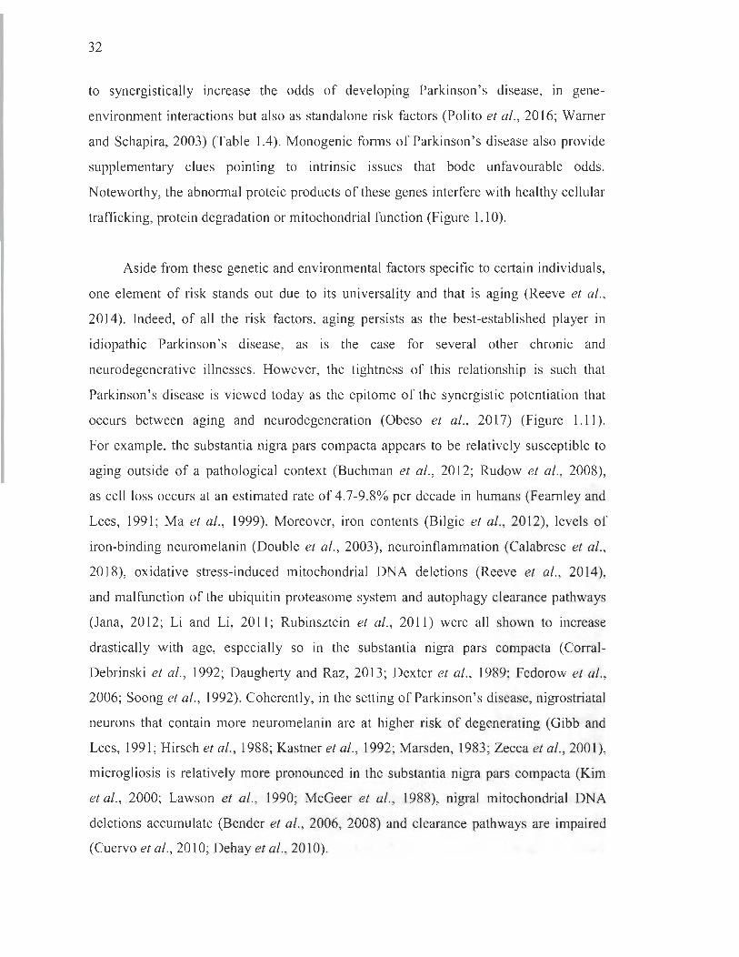
32
to synergistically increase the odds of developing Parkinson 's disease, in gene
environment interactions but also as standalone risk factors (Polito et al., 2016; Wamer
and Schapira, 2003) (Table 1.4). Monogenic forms of Parkinson's disease also provide
supplementary clues pointing to intrinsic issues that bode unfavourable odds.
Noteworthy, the abnormal proteic products of these genes interfere with healthy cellular
trafficking, prote in degradation or mitochondrial function (Figure 1.10).
Aside from these genetic and environmental factors specifie to certain individuals,
one element of risk stands out due to its universality and that is aging (Reeve et al.,
2014). Indeed, of ail the risk factors, aging persists as the best-established player in
idiopathie Parkinson's disease, as is the case for several other chronic and
neurodegenerative illnesses. However, the tightness of this relationship is such that
Parkinson's disease is viewed today as the epitome of the synergistic potentiation that
occurs between aging and neurodegeneration (Obeso et al., 2017) (Figure 1.11).
For example, the substantia nigra pars compacta appears to be relatively susceptible to
aging outside of a pathological context (Buchman et al., 2012; Rudow et al., 2008),
as cellioss occurs at an estimated rate of 4.7-9.8% per decade in humans (Feamley and
Lees, 1991; Ma et al. , 1999). Moreover, iron contents (Bilgic et al., 2012), levels of
iron-binding neuromelanin (Double et al., 2003), neuroinflammation (Calabrese et al. ,
2018), oxidative stress-induced mitochondrial DNA deletions (Reeve et al., 2014),
and malfunction of the ubiquitin proteasome system and autophagy clearance pathways
(Jana, 2012; Li and Li, 2011; Rubinsztein et al., 2011) were ail shown to mcrease
drastically with age, especially so in the substantia nigra pars compacta (Corral
Debrinski et al., 1992; Daugherty and Raz, 2013; Dexter et al., 1989; Fedorow et al. ,
2006; Soong et al., 1992). Coherently, in the setting of Parkinson's disease, nigrostriatal
neurons that contain more neuromelanin are at higher risk of degenerating (Gibb and
Lees, 1991; Hirsch et al., 1988; Kastner et al., 1992; Marsden, 1983; Zecca et al., 2001),
microgliosis is relatively more pronounced in the substantia nigra pars compacta (Kim
et al., 2000; Lawson et al., 1990; McGeer et al., 1988), nigral mitochondrial DNA
deletions accurnulate (Bender et al., 2006, 2008) and clearance pathways are impaired
(Cuervo et al., 2010; Dehay et al., 2010).
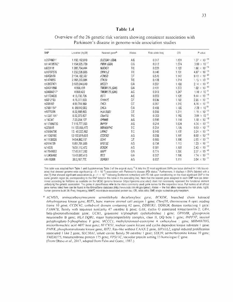
33
Table 1.4
Overview of the 26 genetic risk variants showing consistent association with Parkinson 's di sease in genome-wide association studies
SNP Location (hg38) Nearest gene& AIIeles Risk allele freq OR P value
rs35749011 1 :155,162,810 SLC50Al (GBA) AlG 0.017 1.824 1.37 X 10- 29
rsl14138760* 1 :154,925,709 PMVK(GBA) C/G 0.012 1.574 3.80 X 10- 7
rs823118 1 :205,754,444 NUCKSI TIC 0.559 1.122 1.66 X 10- 16
rsl0797576 1 :232,528,865 SIPA1L2 TIC 0.140 1.131 4.87 X 10- 10
rs6430538 2:134,782,397 ACMSD CfT 0.570 1.143 9.13 X 10- 20
rs1474055 2:168,253,884 STK39 TIC 0.128 1.214 1.15 X 10- 20
rs12637471 3:183,044,649 MCCCI GIA 0.807 1.188 2.14 X 10- 21
rs34311866 4:958,159 TMEM175 (GAI<) GIA 0.191 1.272 1.02 X 10- 43
rs34884217* 4:950,422 TMEM175 (GAI<) AlC 0.913 1.247 1.10 X 10- 6
rsl1724635 4:15,735,728 BSTI AlC 0.553 1.126 9.44 X 10- 18
rs6812193 4:76,277,833 FAM47E CfT 0.636 1.103 2.95 X 10- 11
rs356182 4:89,704,960 SNCA CfT 0.367 1.316 4.16 X 10- 73
rs7681154* 4: 89,842,802 SNCA CfA 0.498 1.189 7.09 X 10.19
rs9275326 6:32,698,883 HLA-DOBI CfT 0.906 1.211 1.19 X 10- 12
rs132011 01 * 6:32,375,827 C6orfl0 TIC 0.053 1.192 3.84 X 10- 6
rs199347 7:23,254,127 GPNMB AlG 0.590 1.110 1.18 X 10- 12
rs117896735 7:119,777,065 INPP5F AlG 0.014 1.624 4.34 X 10- 13
rs329648 11:133,895,472 MIR4697HG TIC 0.354 1.105 9.83 X 10- 12
rs76904798 12: 40,220,882 LRRK2 TIC 0.143 1.155 5.24 X 10- 14
rsll060180 12:122,819,039 CCDC62 AlG 0.558 1.105 6.02 X 10- 12
rsll158026 14:54,882,151 GCHI CfT 0.665 1.106 5.85 X 10- 11
rs2414739 15:61,701 ,935 VPS13C AlG 0.734 1.113 1.23 X 10- 11
rs14235 16:31 ,110,472 BCKDK AlG 0.381 1.103 2.43 X 10- 12
rs17649553 17:45,917,282 MAPT GIA 0.774 1.300 2.37 X 10- 48
rs12456492 18:43,093,415 RIT2 GIA 0.307 1.106 7.74 X 10- 12
rs8118008 20:3,187,770 . DDRGKI AlG 0.657 1.111 3.04 X 10- 11
This table was adapted from Table 1 and Supplementary Table 3 of the original study.13 It lists the 22 most si~nificant SNPs per locus (defined in 1 Mb boundaries) that showed genome-wide signiflcant Ip <5 x 1 0 ~ association with Parkinson's disease (PD) status. Furthermore, it displays 4 SNPs (Iabeled with a star rn that showed significant association (ie, p <1 x 10- 5 following Bonferroni correction) with PD risk upon conditioning on the mest significant SNP in the same genetic region (ie, corresponding to the SNP listed in this table in the preceding line). Note that the nearest gene assigned to each SNP here (as determined according to RefGene as available on the UCSC genome browser (https:lIgenome.ucsc.edul)) does not necessarily represent the functional element undet1ying the genetic association. The genes in parentheses refer to the more commonly used gene names for the respective locus. Full names of ail official gene names listed here can be found in the EntrezGene database (http://www.ncbi.nlm.nih.gov/genel).Alleles = the first allele represents the risk allele. hg38, human genome build 38; Freq, frequency; MAPT, microtubule-associated protein tau; OR, odds ratio; SNP, single nucleotide polymorphism.
& ACMSD, aminocarboxymuconate semialdehyde decarboxylase gene; BCKDK, branched chain ketoacid dehydrogenase kinase gene; BSTl , bone marrow stromal cell antigen 1 gene; C6orfl O, chromosome 6 open reading frame 10 gene; CCDC62, coiled-co il domain containing 62 gene; DDRGKl , DDRGK domain containing 1 gene; FAM47E, fa mily with sequence similari ty 47 member E gene; GAK, cyclin G associated kinase/auxilin -2; GBA, beta-glucocerebrosidase gene; GCR l , guanosine triphosphate cyclohydrolase 1 gene; GPNMB, glycoprote in neuromedin B gene; HLA-DQBl , major histocompatibili ty complex, c lass Il, DQ beta 1 gene; INPP5F, inositol polyphosphate-5-phosphatase F gene; MCCCl, methylcro tonoyl-coenzyme A carboxylase 1 gene; MIR4697HG, microribonucleic acid 4697 host gene; NUCKSl , nuclear casein kinase and cyclin dependent ki nase substrate 1 gene; PMVK, phosphomevalonate kinase gene; RIT2, Ras-like without CAAX 2 gene; SIPA IL2, signal induced proli fe ration associated 1 like 2 gene; SLC50A l , solute carrier family 50 member 1 gene; STK39, serine/threonine kinase 39 gene; TMEM175, transmembrane protein 175 gene; VPS13C, vacuolar protein sorting 13 homologue C gene. (From Obeso et al., 20 17, adapted trom Fahn and Goetz, 1987.)
34
Synaptic/endosomal vesicle/protein recycling
Proteasome
Mitochondrial function Protein degradation
Figure 1.10 Disease mechanisms implicated in Parkinson's disease. This diagram, termed the Bermuda triangle by the authors, highlights three important cellular pathological axes affected in monogenic forms of Parkinson ' s disease, which may also be implicated in idiopathic forms. Importantly, cellular trafficking, mitochondrial homeostasis and protein degradation are tightly interwoven in the pathophysiology of Parkinson 's disease. (From Obeso et al. , 2017.)
In considering the sum of exogenous and endogenous risk factors that come into
play in Parkinson ' s disease, several mechanisms proposed to dwell at the core of this
illness emerge, notably mitochondrial dysfunction, protein aggregation, iron toxicity,
neuroinflammation and impaired cellular trafficking, among others. Despite their
apparent disparity, they converge to cause the preferential early degeneration of
nigrostriatal neurons in Parkinson ' s disease and possibly contribute to oxidative stress to
which these neurons may be more vulnerable (Brichta and Greengard, 2014; Haddad and
Nakamura, 2015; Oliveira et al. , 2017; Surmeier, 2007; Surrneier et al., 2017).
This concept dates back to the 1980s (G6tz et al., 1990; Jenner, 1991; Lenzi et al., 1979;
y oudim et al., 1989), but its popularity has waxed and waned at the rhythm of
discoveries such as the sequential spreading of Lewy bodies (Braak et al. , 2003 ).
Nevertheless, the ide a that metabolic oxidative insults may underlie the specific
vulnerability of nigrostriatal neurons has lately gained momentum as the pro minent
philosophies of the 1900s and beginning of the 2000s are failing to satisfy questions

35
regarding the course of progressIOn of Parkinson ' s disease (Obeso et al. , 2017).
Oxidative stress and the propagation of Lewy bodies are not mutually exclusive theories
either: oxidative stress is a known facilitator of a-synuclein aggregation and Lewy
bodies contribute to oxidative stress (Gupta et al. , 2009). As such, they likely engage in
an intimate relationship in the etiopathogenesis of Parkinson's disease.
Substantla nigra toxins 1-----------,
Figure 1.11 Crosstalk between aging and Parkinson 's disease. Aging leads to several pathological events in neurons that are conducive to the development of Parkinson's disease. In particular, oxidative stress emerges as a pivotai factor. A TP, adenosine triphosphate; PTP, penneability transition pore; UPS, ubiquitin-proteasome system. (From Schapira, 2008.)
In order for oxidative stress to incarnate the noxious interface through which ail
of these risk factors converge onto nigrostriatal dopaminergic neurons, the latter must
exhibit exclusive properties discerning them from other neuronal populations and
rendering them sensitive to each of these contributing elements. It is critical that
nigrostriatal neurons distinguish themselves from other dopaminergic neurons,
especially their mesocorticolimbic analogues, as they ail share the liability of employing
36
dopamine as a neurotransmitter, recognized to generate ROS upon auto-oxidation
(Segura-Aguilar et al., 2014) and to require ROS-producing monoamine oxidases for
their degradation (Andersen et al., 1994; Graham, 1984). In this respect, we argue that
nigrostriatal dopaminergic neurons display the following phenotypic idiosyncrasies that
may afford their vulnerability to oxidative stress instigated by genetic mutations,
environrnental toxins and aging:
1) They are embedded in a particular environrnent, the substantia nigra pars compacta,
which stores considerable amounts of iron ions (Chinta and Andersen, 2008; Hirsch
and Faucheux, 1998) accumulating with age (Daugherty and Raz, 2013) and known
to participate in deleterious Fenton reactions with hydrogen peroxide to produce the
very reactive hydroxyl radical (Youdim et al., 1989);
2) They express lower levels of VMAT2 than most other catecholaminergic neurons,
implying the protracted presence of dopamine in the cytosol leading to the
inevitable formation oftoxic oxidative by-products and, ultimately, potentially toxic
neuromelanin (Liang et al. , 2004);
3) Constituting a very small cluster of cells (German et al., 1988; Percheron et al.,
1989), they are nonetheless required to fully innervate a very large surface,
the dorsal striatum, which entails tremendous bran ching of axons harbouring
numerous vesicular release sites ridden with potentially pathogenic a-synuclein
(Matsuda et al., 2009; Parent and Parent, 2006);
4) To enable corticostriatal circuits, the y constitute the greatest striatal input of
uninterrupted tonic dopamine relying on autonomous pacemaking activity at the
source of high baseline levels of calcium ions, which enhance oxidative
phosphorylation, mitochondrial membrane hyperpolarization and superoxide anion
production (Guzman et al., 2010; Pacelli et al., 2015);
5) To maintain their neurotransmission activities and drawn-out axonal arbours,
these neurons keep an army of mitochondria to fulfil their exorbitant energetic
needs, also irnplying elevated basal rates of oxidative phosphorylation and greater
production of electron transport chain superoxide anion by-products (Pacelli et al. ,
2015);

37
6) Last, they are endowed with a limited calcium-buffering capacity (Foehring et al.,
2009) and scanty endogenous antioxidative defences (Bharath et al. , 2002),
consequent of their low expression of calbindin (Dopeso-Reyes et al. , 2014;
Iacopino and Christakos, 1990; Liang et al., 1996; Yamada et al., 1990) and
glutathione (Kang et al., 1999), respectively.
In aU evidence, these features unite to augment the basal oxidative burden to
which are subjected nigrostriatal neurons. Upon further exposure to environrnental or
genetic insults, endogenous antioxidative defences are eventually overwhelmed:
this turning point typified by the failure of neurons to cope with the added oxidative load
constitutes the origin of oxidative stress. Sustained in time and enhanced by aging
processes, oxidative stress may eventually lead to the death of nigrostriatal neurons in
Parkinson' s disease. Following this premise, studies have employed primary neuron
cultures (Pacelli et al. , 2015), acute rat brain slices (Carbone et al., 2017),
rodents (Vidyadhara et al., 2016) and primate (Dopeso-Reyes et al. , 2014) models to
demonstrate that nigrostriatal dopaminergic neurons are more vulnerable to parkinsonian
neurotoxins in comparison to other neuronal populations, exemplarily dopaminergic
neurons 10dged in the neighbouring ventral tegmental area, and that this vulnerability is
abated by pharmacologically targeting these specific phenotypic risk features (Carbone
etaI. , 2017; Pacelli et aI. , 2015).
If indeed nigrostriatal neurons are relatively more susceptible to insults that
overwhelm their endogenous coping mechanisms, other sources of oxidative stress that
are not specific to Parkinson 's disease may yield similar results and shore up the idea
of their selective vulnerability. Indications of this are provided by epidemiological
evidence showing a higher occurrence of Parkinson ' s disease in patients suffering
from other pathologies with a significant component of oxidative stress, such as
diabetes (Cereda et al. , 2012; Santiago and Potashkin, 2013; Sun et al. , 2012), ischemic
stroke (Huang et al. , 2013) and sleep apnea (Snyder and Cunningham, 2017).
Interestingly, Parkinson ' s disease is significantly less frequent in patients with the
hyperuricemic illness gout wherein the antioxidative effects of uric acid may in fact
protect nigrostriatal neurons (Alonso and Sovell, 2010). Although evidence weighs in

38
favour of the selective vulnerability of the nigrostriatal pathway in pathological states
that induce upwelling of systemic oxidation, for instance in a hyperglycaemic setting,
studies are required to validate this. The present thesis aims to expound on this question.
1.2 Hyperglycaemia in the central nervous system
Before addressing the mechanisms by which hyperglycaemia 16 may instigate
oxidative stress in the CNS, and before laying out the epidemiological evidence in
favour of the particular vulnerability of nigrostriatal dopaminergic neurons in these
conditions, it is critical that we fully grasp glucose dynamics in the brain. The transport
kinetics and metabolism of glucose are highly complex, especially in the CNS, and bear
significant implications in the initiation of oxidative stress in neurons. Progressing in
this direction, a fundamental appraisal of the brain' s energy sources is first required.
1.2.1 Glucose as a preferential fuel for the brain
Long has functional neuroimagery exposed the importance of glucose in the
fulfilment of the adult brain's energetic needs (Landau et al., 1955; Schmidt and Kety,
1947; Siesjo, 1978; Sokoloff et al., 1977). In fact, the brain utilizes 25% of total body
glucose, mostly to power oxidative metabolism (Heiss, 2016; Kety and Schmidt, 1948;
Laughlin and Attwell, 2001; Shulman et al., 2001; Sokoloff et al., 1999). Glucose is
indispensable to neurons and thus plays a significant role in CNS health and disease.
In settings of extreme dietary and pathological depletion of glucose or its transport,
the brain is forced to employ other fuels, although a minimal amount of glucose is
always required to maintain normal cerebral functions (Casazza et al., 1984; Owen
et al., 1967; Vanltallie and Nufert, 2003). In these specifie contexts, blood-borne ketone
bodies can account for up to 70% of brain energy requirements through its conversion to
acetyl coenzyme A, in contrast to fatty acids that inefficiently cross the blood-brain
16 Normoglycaemia is defined in humans as fasting blood glucose < 5.5 mM « 100 mg/dL), postprandial blood glucose < 7.8 mM « 140 mg/dL) or glycated haemoglobin < 5.7%; hyperglycaemia is defined as fasting blood glucose ~ 7.0 mM (~ 126 mg/dL), postprandial blood glucose ~ ILl mM (~ 200 mg/dL) or glycated haemoglobin ~ 6.5% (American Diabetes Association, 2016).
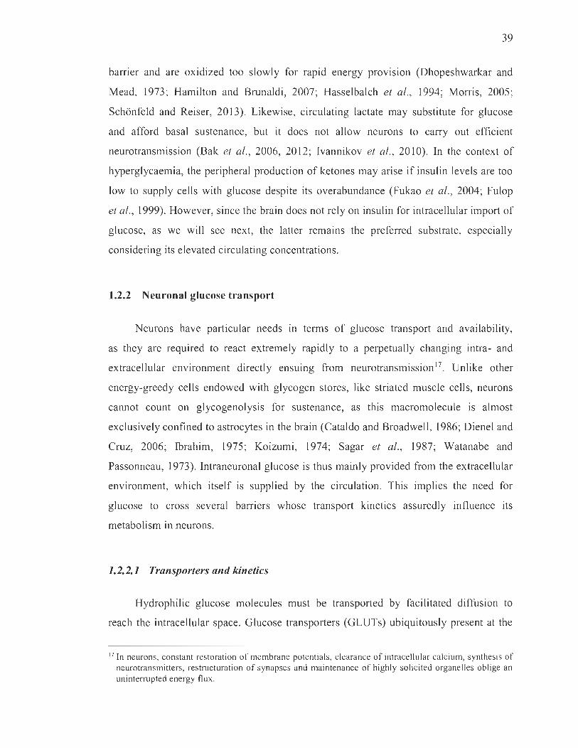
39
barrier and are oxidized too slowly for rapid energy provIsIon (Dhopeshwarkar and
Mead, 1973; Hamilton and Brunaldi, 2007; Hasselbalch et al. , 1994; Morris, 2005;
Schonfeld and Reiser, 2013). Likewise, circulating lactate may substitute for glucose
and afford basal sustenance, but it do es not allow neurons to carry out efficient
neurotransmission (Bak et al., 2006, 2012; Ivannikov et al. , 2010). In the context of
hyperglycaemia, the peripheral production of ketones may arise if insulin levels are too
low to supply cells with glucose despite its overabundance (Fukao et al., 2004; Fulop
et al. , 1999). However, since the brain does not rely on insulin for intracellular import of
glucose, as we will see next, the latter remains the preferred substrate, especially
considering its elevated circulating concentrations.
1.2.2 Neuronal glucose transport
Neurons have particular needs III terrns of glucose transport and availability,
as they are required to react extremely rapidly to a perpetually changing intra- and
extracellular environrnent directly ensuing from neurotransmission l7. Unlike other
energy-greedy cells endowed with glycogen stores, like striated muscle cells, neurons
cannot count on glycogenolysis for sustenance, as this macro mole cule is almost
exclusively confined to astrocytes in the brain (Cataldo and Broadwell, 1986; Dienel and
Cruz, 2006; Ibrahim, 1975; Koizumi, 1974; Sagar et al., 1987; Watanabe and
Passonneau, 1973). Intraneuronal glucose is thus mainly provided from the extracellular
environrnent, which itself is supplied by the circulation. This implies the need for
glucose to cross several barriers whose transport kinetics assuredly influence its
metabolism in neurons.
1.2.2.1 Transporters and kinetics
Hydrophilic glucose molecules must be transported by facilitated diffusion to
reach the intracellular space. Glucose transporters (GLUTs) ubiquitously present at the
17 In neurons, constant restoration of membrane potentials, clearance of intracellular calcium, synthesis of neurotransmitters, restructuration of synapses and maintenance of highl y solicited organelles oblige an uninterrupted energy flux.

40
surface of cells occupy the function of taking up glucose from the extraceIlular
environment (Table 1.5). Excluding the myo-inositol transporter, aIl GLUTs identified
to date mediate energy-independent facilitative transport (see for review Mueckler and
Thorens, 2013; Wood and Trayhum, 2003).
Table 1.5
Glucose transporter expression sites and substrates
Type Prolein (gene)&<
GLUTI (SLC2A/)
GLUTl (SLC2A2)
GL T3 (SLC2A3)
GL T4 (SLC2M)
(Insulin-sensilive)
GLUTS (SLC2A5)
FacilitativelSodium- GLUT6 (SLC2A6)
indcpcndent GLUn (SLC2A7)
GLUn (SLC2A8)
(Insulin-responsive?)
GLUT9 (SLC2A9)
GLUTIO (SLC2A IO)
iles expressed
Brain endothelial and epithelial-Iike brain barriers, glial cells, blood-tissue
barriers, eye, pheripheral nerves, placenta, lactating mammary gland
(Ubiquilous distribution in mosl mammalian cells)
Kidney, small intestine (epithelial cells), Iiver, pancreas (islets), brain
(astrocytes)
Neurons, testis, placenta, brain endothelial cells?
Brown and white adipose tissue, muscle (skeletal), fal, heart (myocardium),
hippocampal neurons, cerebellar neurons
Intestine (jejunum), Iddney, testi , brain microglia
Brain, peripheral and spleen (Ieukocytes)
Small intestine (mamly in brush border mcmbranc-cntcrocytes), colon, testis,
prostate, liver (associated with endoplasmic reticulum)
BlaslOCytes, testis, brain (neurons), muscle, adipocytes, mammary gland?
Liver, kidney (proximal tubule of epithelial cells), placenta?
Liver, pancreas, heart, lung, brain, skeletal mu cie, placenta
Iso-form A: Heart, skeletal muscle, kidney
GLUTII (SLC2A If) lso-form B: Placenta, adipo e ti sue, kidney
GL Tl2 (SLC2A / 2)
(In. ulin-sensitive?)
HMIT (SLC2Al 3)
(co-transporler)
lso-form C: Adipose tissue, heart, keletal muscle, pancreas
Heart, skeletal muscle, fat, prostrate, laetating mammary gland ?, spleen 1,
breast cancer (Ductal cell carcinoma) tissue
Brain (neurons mtracellular vesicles)
ubslrale/lransports
>>Glucose, galactose,
mannose, gl ucosamine,
ascorbic acid
Mannose, galactose,
fructose, glucose,
glucosamme
Glucose, galactose,
mannose, xylose,
dehydroascorbic acid
Glucose,
dehydroascorbic acid,
glucosamine
Fructo e
Glucose
>Fructose, glucose
Gluco e
Glucose, urate
Glucose, galactose
Fructose, glucose
Glucose
H+'myo-inositol
& GLUT, glucose transporter; HMIT, H+/myo-inosito l transporter; SLC2A, solute carrier famil y 2 gene. (Adapted From Simpson et al., 2007.)
Contrary to most peripheral cells that express GLUT4 requmng insulin for
membrane translocation, the vast majority of CNS components employ insulin
independent GLUTs (Huang and Czech, 2007; James et al., 1988) (Figure 1.1 2).
GLUTI is the most widespread isoform in the body and, aside from red blood ce Ils,
it is most abundant in the brain microvasculature, evenly distributed among the luminal
41
and abluminal surfaces of the endothelium (Maher et al., 1994; Simpson et al. , 2001).
It is also the main transporter employed by glial cells, especially astrocytes (Dick et al. ,
1984; Gerhart et al. , 1989; Maher et al., 1994; Simpson et al. , 2001). Neurons, on the
other hand, overwhelmingly employ the GLUT3 isoform densely located across the cell
surface but more intensely so at the neuropil (Leino et al. , 1997; Mantych et al., 1992;
Nagamatsu et al. , 1993; see for review Simpson et al. , 2008). Consequently, to access
the brain parenchyma, circulating glucose must penetrate the endothelium and astrocytic
endfeet of the blood-brain barrier via GLUTI. In turn, glucose diffuses through the
extracellular space where it can be absorbed by neuronal GLUT3 .
• GLUn 55K 8 Mcn • GLUT1 45K MCT2
GLUT3 K MCT3
* GLUT4 MCT4 Â GLUT5
GLUT6 GLUT8
GLUn 55K
1
GLUT5
Figure 1.12 Glucose and monocarboxylate transporters in the mammalian brain. Endothelial cells solely express GLUT1 , whereas other brain celis usually express multitudinous isoforms. Noteworthy, endothelial cells express the highly glycosylated 55-kDa isoform of GLUT1 ,while astrocytes and oligodendrocytes express the 45-kDa variant. Neurons mainly express GLUT3 , but can also express GLUTS 1, 2, 4, 6, 8 and the myo-inositol transporter, depending on brain regions and isoforms. The fructose transporter GLUT5 is almost only found at the surface of microglial cells, but its function remains unclear in the brain due to the virtual absence of its known substrate in the parenchyma (Jurcovicova, 2014; Maher et al. , 1994). MCT, monocarboxylate transporter. (From Simpson et al., 2007, adapted from McKenna et al. , 2005.)

42
Although cellular constituents of the brain do not rely on insulin for glucose
uptake, several kinetic factors come into play in limiting glucose permeation between
compartments. Seeing as GLUTs are facilitative, bidirectional, energy-independent
transporters, they allow not for glucose accumulation but for its equilibration between
compartments in a manner that smoothens concentration gradients. Brain GLUTs
possess elevated affinities for glucose, highest for neuronal GLUT3 (Km ~ 1.5 mM)
(ColviUe et al. , 1993; Maher et al. , 1996) (Table l.6). In fact, GLUT3 also presents the
greatest turnover for glucose, defmed as the number of transport cycles per transporter
per second (Manolescu et al., 2007; Rumsey et al., 1997).
Table 1.6
Glucose transport capacities of brain ce Us
!eeU TRANSPORTER [Transporter[ [Transporter] [Transporter] !!Vmax ~m c ubstratelligand llkcat ~redicted
SPECIES uptake uptake Vmn in
model
pmol/mg membrane carriers/flm 2 pmol/mg total nmol/I06 mM per mmol/sec
protein protein ceUs/min sec
RBC& GLUTI 2000 2083 92 80.4 8 GI CB 1166
Endothelium GLUTI 400 fI 000 gl8 0.21 8 CB 1166 7.8 x 10 -IS
------Astrocyte GLI/TI 7.3 f l8 gO.34 4.8 8 CB 1166 h.4 x 10
-16
!1.9 XIO- IS
Neuron GLUTI 9.S [24 gO.44 hS.O 2.8 3MG/CB h868 2.3 x 10
i34.6 4;S12 - 14
'Cell in which tran pon i measurcd. bMeasured or compuled maximum rate (V maJ or affinity (K.J for transpon .. "Tbe substrate employed in transpon (Glucose (Glc), 3-0-methylglucose (3MG) or lactale) or ligand GLUT quanlitalion experirncnts (Cytochalasin B (CB) binding). dcompuled k Cal ([Vm .. ,I[GLUT]) for transpon. ' Predicled V mu for IranSpon of glucose and lactate for use in the mode!. fAssumes 1 mg membrane prolein is equivalenllo a surface area of 2.4 x IOll flffi2. IAssurnes lhat the ratio of membrane:lotal cellular protein is 0.0462 : 1. hMeasured a12SoC. iCompuled for transpon al 37°C as uming thal GLUT3 and GLUTI hare Ihe same temperature dependence. JOirection basal lamina 10 astrocyte. kOirection inlerstilium to aSlrocyte. Glucose transpon parameters in endothelial cells, aslrocytes and neurons were computed using estimated ccII membrane GLUT conlents and measured k Cil paramelers. MCT concentrations are estimatcd as uming MCT k cal = GLUTI k cal and using measurcd V .... values. &RBC, red blood cell.
(From Simpson el al., 2007.)

43
Since normoglycaemic settings afford much more glucose than transport kinetics
require, one would expect brain cells like neurons to contain approximately as much
glucose as their immediate extracellular environment. Likewise, brain glucose
concentrations should fluctuate as a function of glycaemia, regardless of insulin levels.
According to computational studies performed by Simpson and colleagues, neurons and
astrocytes only contain ~0.9 mM of glucose in normoglycaemic steady-state conditions
(Simpson et al., 2007) (Table 1.7). Similarly, extracellular concentrations of glucose
merely reach ~ 1.4 mM. Even endothelial cells that express GLUTI and are in direct
contact with blood-bome solutes display concentrations of ~3.8 mM. This discrepancy
between expected compartment equilibration and markedly lower levels of glucose in
parenchymal and endothelial cells may be explained by transport inhibition at the level
of the blood-brain barrier. Indeed, endothelial GLUT1 undergoes allosteric inactivation
by adenosine triphosphate (ATP) in settings of metabolic abundance (Carruthers and
Helgerson, 1989; Levine et al., 1998; Lowe and Walmsley, 1986). Consequently,
endothelial ce Ils uptake and provide less glucose to the brain parenchyma in
replete, normoglycaemic settings. Conversely, when glycolytic demands increase and
ATP runs low, GLUT 1 transport is uninhibited and glucose may efficiently be
transferred to surrounding tissues (Carruthers, 1986; Cloherty et al., 1996). Globally,
the parenchyma's apparent Km for glucose remains 1-3 mM, signifying that glycaemia
must significantly drop before brain uptake deficits are perceived (Rao et al. , 2006).
This can be interpreted as an evolutionary adaptation in favour of organisms whose
neurons were uninterruptedly supplied with fuel.
Table 1.7
Computed compartment glucose levels for the core (primary) model or the astrocyte-neuron lactate shuttle (ANLS)
Serum [Gle] 6 30 6 30
EndoIhchal [GIe] 3. 174 3.1 11 .0
B3YlI.m,na [Giel 2.1 10 2.0 10.4
Astroc)tle 1 Gle 1 0.9 1.9 U 9.4
IO'<r!mualIGkl 14 99 U 95
" euro .. 1 [Gkl 1.2 Il 4
BI1IIOIGIeI 1.2 9 12 91
(From Simpson et al. , 2007.)

44
1.2.2.2 Physiological considerations
Notwithstanding the progress made in elucidating how glucose gains entry to the
brain and permeates neurons, the CUITent state of the field leaves many questions
unanswered in physiological settings. Converging data on differential expression of
GLUTs across brain regions, between species, in health and in disease are lacking.
We know that GLUT3 appears to occur in high levels in grey matter, especially in
densely populous zones known to be energetically needy; fittingly, neurons from the
hippocampal region, cerebral cortex, cerebellum, striatum and midbrain express the
greatest amounts in the rat brain (Bondy et al., 1992; Nagamatsu et al., 1993).
However, we know little about the transcriptional regulation of brain GLUTs and the
stabilization of rnRNA transcripts 18. Best studied is the upregulation of GLUTs in the
CNS during hypoglycaemic events (Anitha et al. , 2012; Nagamatsu et al., 1994;
Santiago et al., 2006; Simpson et al., 1999). Moreover, aside from evidence of cerebral
GLUT mobilization in specifie regions of the brain during active firing (Ashrafi et al.,
2017; FeITeira et al., 20 Il; Weisova et al., 2009), information is scarce on the recyc1ing
and trafficking ofthese transporters (McClory et al., 2014). Unlike GLUT4, intracellular
pools of GLUT 1 and 3 are seemingl y small and do not require insulin to be upregulated
at the cell surface 19.
At any rate, hyperglycaemia, especially sustained for long periods oftime, remains
a blind spot in the study of cerebral GLUTs and glucose uptake in the brain.
In opposition to the hypoglycaemia-induced compensatory upregulation of brain
GLUTs, hyperglycaemia has not been demonstrated to afford a c1ear regulatory effect in
rodents, at least on the short term (Anitha et al. , 2012; Nagamatsu et al. , 1994; Santiago
et al., 2006; Simpson et al., 1999). Under normal physiological circumstances, literature
further provides that microvasculature density is mostly uniform across brain regions,
18 There is sorne evidence in the periphery to support a role for cellular stress in the transcriptional upregulation of GLUn , explicitly in contexts of oxidative stress, rnitochondria inhibition and hypoxia (Anitha et al. , 2012; Bashan et al., 1992, 1993; Baumann et al. , 2007; Kozlovsky et al., 1997; McMahon and Frost, 1995; Mobasheri et al., 2005; Nagarnatsu et al., 1994; Santiago et al. , 2006).
19 Sorne have rai sed the possibility that rnernbrane-bound brain GLUTs rnay be rapidly trafficked in and out of lipid rafts to respond to rnetabolic signais (Bames et al., 2004; Rauch et al. , 2006).
45
including the midbrain and the striatum (lelacqua et al., 2016; Kolinko et al., 2016).
However, the possible effects of hyperglycaemia on cerebral blood flow are
contradictory. On the one hand, both acute and chronic hyperglycaemic states are known
to reduce cerebral blood flow (Duckrow et al., 1987; McCall, 1992; Nishimura et al.,
2007). On the other hand, they cause the occurrence of pathological neovascularization,
that is, a rise in the volume of capillaries with deficient pericyte coverage (Ergul et al. ,
2015; Li et al., 2010; Prakash et al., 2013). The ensuing gain of blood-brain barrier
permeability might grant glucose the ability to sidestep endothelial ATP-inhibited
GLUT 1 and to infuse the entire brain more profusely (Carruthers and Helgerson, 1989;
Levine et al., 1998; Lowe and Walmsley, 1986).
Despite these remammg ambiguities, vanous demonstrations strengthen the
idea that hyperglycaemia causes a substantial rise in parenchymal glucose
concentrations. Early studies demonstrated that brain glucose levels were indeed
sensitive to glycaemic circumstances. Indeed, they faU or rise during transient
hypoglycaemic or hyperglycaemic challenges, respectively (Abi-Saab et al., 2002;
Béland-Millar et al., 2017; Macauley et al., 2015; McCall et al., 1986; Osborne et al.,
1997). In rodent models of sustained diabetes-induced20 hyperglycaemia, intracerebral
glucose concentrations are also augmented (de Vries et al., 2003; Gomez and Barros,
2003; Jacob et al., 2002; McCrimmons et al., 2003). These experiments were performed
on who le brain tissues and reliable intraneuronal measurements are still lacking21.
Nevertheless, computational renderings taking into account GLUT kinetics and their
distribution predict greater glucose permeation in ail brain constituents under
hyperglycaemia (Simpson et al., 2007) (Table l.7). We can therefore cautiously submit
to the idea that hyperglycaemia causes an upwelling of intraneuronal glucose likely to
hold implications for cellular fitness.
20 Diabetes mellitus is described in section 1.2.5.1.
2 \ Previous studies reporting these concentrations were recently demonstrated to be tlawed, owing to differing transport kinetics between glucose and the tagged analogues employed for quantifications (Dienel et al., 2017).

46
1.2.3 Neuronal glucose metabolism
Before addressing the metabolic singularities that characterize neurons, a brief
outline of glucose metabolism is warranted (see for review Stryer et al. , 2007)
(Figure 1.13). In the cytosol, glucose is first engaged along a glycolytic fate upon
irreversible phosphorylation by an important rate-limiting, ATP-consuming enzyme,
hexokinase. Resulting glucose 6-phosphate (G6P) is a gateway metabolite, not only to
the glycolytic pathway, but also to glycogenesis, relevant to astrocytes, or to the pentose
phosphate pathway (ppp)22. In glycolysis, G6P undergoes isomerization into fructose
6-phosphate (F6P), which is in turn transformed by phosphofructokinase-l (PFK) into
fructose 1,6-bisphosphate. Importantly, PFK is another rate-limiting, energy-consuming
enzyme that drives glycolysis and is allosterically inhibited by high ATP:adenosine
monophosphate (AMP) ratios, which equally ensures activation of glycolysis in times of
energy depletion. Continuing along the glycolytic pathway, fructose 1,6-bisphosphate is
converted into a pair of triose intermediates, glyceraldehyde 3-phosphate (GA3P),
further metabolized by glyceraldehyde 3-phosphate dehydrogenase (GAPDH)
into 1,3-bisphosphoglycerate. From 1,3-bisphosphoglycerate, a series of enzymatic
transformations yields the final glycolysis metabolite, pyruvate, through irreversible
cleavage of the phosphate group by pyruvate kinase. At this level, pyruvate can enter
mitochondria and undergo catalysis mediated by pyruvate dehydrogenase, yielding the
gateway intermediate to the tricarboxylic acid cycle, acetyl coenzyme A 23 . In certain
circumstances, it can also be metabolized in the cytosol by the bidirectional lactate
dehydrogenase enzyme to give lactate, a process termed the Warburg effecr24 (Warburg,
1956). Exogenous lactate may also be provided as a fuel at this level through its
transformation to pyruvate, once again via lactate dehydrogenase. Generated along the
course of glycolysis and tricarboxylic acid cycling are matrix-dwelling electron donors,
22 The ppp is later discussed in section 1.2.3. 1. Of note, the ppp yields GA3P and F6P, an additional connection between glycolysis and the PPP.
23 This is also the access point for alternate fue ls, like ketone bodies, provided during glucose depletion, which is beyond the scope of this thesis.
24 In mammalian ce lls, the Warburg effect occurs under aerobic conditions, contrarily to the lactateproducing anaerobic glycolytic process. Tt should not be confused with the Warburg effect that occurs in plants, describing decreased photosynthesis in hyperoxic conditions.

47
namely reduced nicotinamide adenine dinucleotide (NADH) and flavin adenine
dinucleotide (F ADH2), whose electrons are funnelled through the electron transport
chain of the inner mitochondrial membrane and are relinquished to the terminal
electron acceptor, oxygen, to yield water. Concurrently, this electron traffic establishes
a transmembrane proton gradient serving to power the synthase that ultimately pro duces
ATP (30-36 per molecule of glucose).
Although this energy production pathway is present in sorne form or other in
aIl ceIls, it is highly ramified and shares intermediates with a plethora of other parallel
metabolic routes. A few of these are indeed relevant in the brain and in a
hyperglycaemic setting, but, for the sake ofbrevity, only the most salient ofthese will be
discussed in the next subsections.
Glucose
(il) ppp - - -~ H' c ~ Glucose- 6P
NAOf>+ X GSSG X ROOH
NAOPH GSH ROH
~ ... ...;;......;..---~, GPI + (iii) ~
Fructose- 6P
PFK ~ Fructose-l.6P,
Aldolil5e ,/'
OHAP ~GA3P TPI
GAP OH 2Aœ+2NAO+
2ATP+ 2 NADH
CO,
Ribulose-SP
~ __ R_ibo_se-_ S_P_XTKXylUIOse-SP
~ Sedoheptulose-7P
TA Erythrose-4P
TK
Pyruvilte ___ ). +- 0, NAOH ~
NAO+ 4 ... _.....;. Lactate
\ ... (l) Glycolysis
Lactate
3G-36ATP ~CO,+H,O ,
(Iv) Oxidiltive Phosphoryliltlon
Figure 1.13 Schematic representation of glucose metabolism and pertinent connecting pathways in neurons and astrocytes. Continued on next page.
48
(Continued.) Glucose is processed principally via glycolysis (i) leading up to oxidative phosphorylation (iv) with the intent of producing ATP for energy. However, glucose metabolites can also be diverted into tangential pathways. In astrocytes, glucose 6-phosphate (G6P) can be transformed into glycogen by glucokinase (iii). In neurons, the pentose phosphate pathway (PPP) is crucially provided by G6P for the maintenance of a proper redox status via the regeneration of the reduced form of glutathione (ii). ADP, adenosine diphosphate; DHAP, dihydroxyacetone phosphate; GA3P, glyceraldehyde 3-phosphate; GAPDH, glyceraldehyde 3-phosphate dehydrogenase; GPI, glucose 6-phosphate isomerase; GSH, glutathione; GSSG, glutathione disulphide; HK, hexokinase; NAD, nicotinamide adenine dinucleotide (NAD+ oxidized, NADH reduced) ; NADP, nicotinamide adenine dinucleotide phosphate (NADP+ oxidized, NADPH reduced); PFK, phosphofructokinase; ROH, alcohol; ROOH, hydroperoxide compound; TPI, triosephosphate isomerase; TA, transaldolase; TCA, tricarboxylic acid; TK, transketolase. (From Magistretti and Allaman, 2015.)
1.2.3.1 Specifie metabolic Jates
Extracellular glucose levels in the CNS are always in excess of demand due to
favourable GLUT 1 kinetics. Yet, to ensure uninterrupted intracellular glucose
availability, it is also crucial that the velocities of uptake and metabolism be tightly
coupled. In neurons, the first step of glycolysis holds a particular importance in this
regard. Indeed, hexokinase phosphorylates glucose more slowly th an it is taken up by
GLUT3 (Lowry and Passonneau, 1964; Whitesell et al., 1995; Wilson, 2003). In other
words, GLUT3 uptake is never limiting and free cytosolic glucose is constantly available
(Dienel, 2012). Similarly, to secure glycolysis with a sustained flow of metabolites,
especially during neuronal activity, engagement of glucose in the pathway must not be
rate-limiting. In this respect, processing of glucose by hexokinase allows neurons to
"stock" fuel in the form of G6P. However, since hexokinase is inhibited by G6P,
its accumulation is somewhat limited. Nevertheless, A TP levels dwindle during neuronal
activation, thereby relieving the inhibition on downstream rate-limiting PFK and
favouring the expenditure of accrued metabolites between hexokinase and PFK. In tum,
hexokinase is disinhibited due to the consequent reduction of G6P pools and can once
again phosphorylate glucose to provide the glycolytic pathway with substrates.
These gating processes allowing the accumulation of glycolytic intermediates act as a
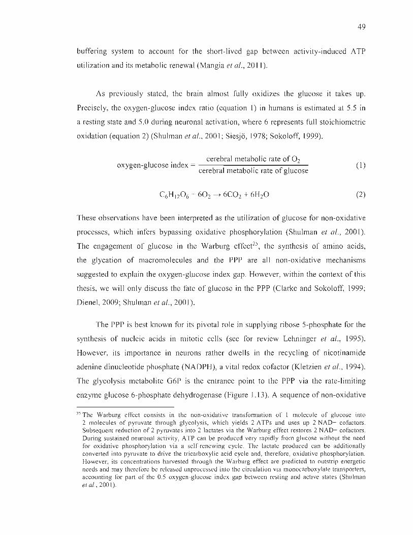
49
buffering system to account for the short-lived gap between activity-induced ATP
utilization and its metabolic renewal (Mangia et al., 20 Il).
As previously stated, the brain almost fully oxidizes the glucose it takes up.
Precisely, the oxygen-glucose index ratio (equation 1) in humans is estimated at 5.5 in
a resting state and 5.0 during neuronal activation, where 6 represents full stoichiometric
oxidation (equation 2) (Shulman et al., 2001; Siesjo, 1978; Sokoloff, 1999).
cerebral metabolic rate of O2 oxygen-glucose index = .
cerebral metaboltc rate of glucose (1)
(2)
These observations have been interpreted as the utilization of glucose for non-oxidative
processes, which infers bypassing oxidative phosphorylation (Shulman et al., 2001).
The engagement of glucose in the Warburg effect25, the synthesis of amino acids,
the glycation of macromolecules and the ppp are al! non-oxidative mechanisms
suggested to explain the oxygen-glucose index gap. However, within the context of this
thesis, we will only discuss the fate of glucose in the ppp (Clarke and Sokoloff, 1999;
Dienel, 2009; Shulman et al., 2001).
The ppp is best known for its pivotaI role in supplying ribose 5-phosphate for the
synthesis of nuc1eic acids in mitotic cells (see for review Lehninger et al., 1995).
However, its importance in neurons rather dwells in the recyc1ing of nicotinamide
adenine dinuc1eotide phosphate (NADPH), a vital redox cofactor (Kletzien et al., 1994).
The glycolysis metabolite G6P is the entrance point to the ppp via the rate-limiting
enzyme glucose 6-phosphate dehydrogenase (Figure 1.13). A sequence of non-oxidative
25 The Warburg effect consists in the non-oxidative transformation of 1 molecule of glucose into 2 molecules of pyruvate through glycolysis, which yields 2 ATPs and uses up 2 NAD+ cofactors. Subsequent reduction of 2 pyruvates into 2 lactates via the Warburg effect restores 2 NAD+ cofactors. During sustained neuronal activity, ATP can be produced very rapidly from glucose without the need for oxidative phosphorylation via a self-renewing cycle. The lactate produced can be additionally converted into pyruvate to drive the tricarboxylic acid cycle and, therefore, oxidative phosphorylation. However, its concentrations harvested through the Warburg effect are predicted to outstrip energetic needs and may therefore be released unprocessed into the circulation via monocarboxylate transporters, accounting for part of the 0.5 oxygen-glucose index gap between resting and active states (Shulman et al., 2001).

50
reactions regenerates 2 molecules of NADPH from G6P and returns the intermediate
back on the glycolytic track through the production of GA3P or F6P. Irnportantly,
NADPH produced by the ppp is employed by glutathione reductase to restore oxidized
glutathione to its reduced and useful form26 (Hothersall et al. , 1979; Kletzien et al.,
1994). Indeed, by virtue of glutathione's detoxifying action on ROS, studies support a
potent antioxidative role for the ppp in neurons and astrocytes (Ben-Yoseph et al.,
1996a, 1996b; Bolafios et al., 2008; Herrero-Mendez et al., 2009; Kussmaul et al., 1999;
Vaughn and Deshmukh, 2008). Pertaining to the oxygen-glucose index gap, attention
must rather be drawn to the shared metabolic intermediate between glycolysis and PPP,
F6P, since it stands upstream from any irreversible reactions in the glycolytic pathway,
as opposed to GA3P found downstream from the unidirectional PFK enzyme and
destined to oxidative phosphorylation. Combined with evidence for a bottleneck effect
at the level of PFK in neurons27 (Bolafios et al., 2010; Herrero-Mendez et al., 2009),
sorne have suggested that F6P is recycled through the PPP by catalytic revers ion to G6P
(Bouzier-Sore and Bolafios, 2015). Therefore, any fraction ofthese metabolites that keep
cycling through the non-oxidative PPP may weil account for a portion of the
0.5 stoichiometric gap between glucose and oxygen use (Bouzier-Sore and Bolafios,
2015).
1.2.3.2 Current hypotheses in neuroenergetics
For the most part, the metabolic processes described above have not been clearly
ascribed to neurons or other brain cells on account of the lack of consensus per the
proportion of glucose absorbed and metabolized by each compartment. For the past
20 years, the field of neuroenergetics has been a battleground of ideas around this
26 Glutathione, or y-L-glutamyl-L-cysteinylglycine, is a pseudo-tripeptide whose thiol moiety acts as a reducing agent. Glutathione peroxidase catalyzes the transfer of electrons between two glutathiones and one hydrogen peroxide molecule, yielding water and one glutathione disulphide, a condensation of two oxidized glutathiones. Glutathione disulphide is reverted to its useful form, reduced glutathione, by glutathione reductase. Glutathione transferase can also catalyze the addition of glutathione onto potentially toxic xenobiotics.
27 The bottleneck effect is described in section 1.2.3 .2.

51
emgma, ansmg mainly from two groups that allot differential importance to these
processes in neurons and astrocytes28.
The first group, spearheaded by Pellerin and Magistretti, defends the position that
astrocytes, and not neurons, are the principal site of glucose uptake, especially during
neuronal activation (Pellerin and Magistretti, 1994). In tum, astrocytes pro vide energy to
neurons in the form of lactate, a hypothetic process hence termed the astrocyte-neuron
lactate shuttle29 (Figure 1.14). According to this the ory, astrocytes are predominantly
glycolytic, meaning that the y utilize glucose to produce lactate through glycolysis and
the Warburg effect. On the other hand, neurons are rather oxidative and transform
astrocyte-derived lactate into pyruvate, which is metabolized through the tricarboxylic
acid cycle to power oxidative phosphorylation. The authors put forward two central
arguments: 1) astrocytes import more glucose than neurons; 2) neurons cannot
upregulate their rate of glycolysis. The first contention hinges on evidence
demonstrating astrocytes as importing more than 50-90% of available brain glucose
(Barros et al., 2009; Nehlig et al., 2004; Vega et al. , 2003). The second is based
on studies revealing that the levels of a specific enzyme, 6-phosphofructo-2-
kinase/fructose-2,6-bisphosphatase 3 (Pfkfb3), are markedly downregulated in neurons
compared to astrocytes due to constant proteasomal degradation (Bolaiios et al., 2010;
Herrero-Mendez et al., 2009). Pfkfb3 phosphorylates F6P to produce fructose
2,6-bisphosphate (Fru-2,6-P2) known to activate one of the key rate-limiting glycolytic
enzymes, PFK. The authors therefore con tend that sparse expression of Pfkfb3 in
neurons explains the bottleneck effect at the level of PFK, liable for its lirnited glycolytic
capacities.
28 A heated debate took place a few years ago between these two groups in the Journal of Cerebral Blood Flow and Metabolism. A succession of commentaries offers very interesting insight into the arguments hired by both parties to support their theories (Jolivet et al., 2010; Mangia et al., 20 II) .
29 Later biophysical computations and physiological applications came in support of this model (Aubert and Costalat, 2005; Aubert et al., 2007; Béland-Millard et al., 2017; Machler et al., 2016).
52
The second theory, termed the core model3o, is advocated by Mangia, Simpson and
Vannucci, and rather posits that the lactate shuttle effect is negligible (Mangia et al.,
2009; Simpson et al., 2007). Their computational model relies on the same variables
used by the former group, with the exception that it considers experimentally measured
transporter kinetics and distribution patterns throughout the brain. Taking into account
these additional parameters, this group arrives at the conclusion that neurons constitute
the main site of glucose uptake. To bolster their position, they recapitulated and
invalidated the two main arguments in support of the former model. First, regarding the
preferred astrocytic uptake of glucose, sorne of the studies demonstrating a greater
glucose influx in favour of astrocytes (80-90%) were performed in cerebellar slices
(Barros et al., 2009) and ex vivo rat vagus nerves (Vega et al., 2003), which may po orly
represent physiological situations. Other reports in freely moving animais indeed
moderate these numbers and rather support equal partitioning or even preferential
neuronal uptake (50-80%) (Nehlig et al., 2004; Zielke et al., 2007). Second, while the
former group argues that glycolytic activity is capped in neurons due to the bottleneck
effect at PFK (Bolafios et al. , 2010; Herrero-Mendez et al., 2009), Mangia and
colleagues contend instead that PFK may be activated by metabolites other th an
Fru-2,6-P2, thus, that glycolysis can be enhanced31 (Ogushi et al. , 1990). In support of
this, other neuronal monosaccharidic metabolites were found to be more potent
activators of PFK, and Fru-2,6-P2 appears to be expendable in the amplification of
glycolysis during neurotransmission (Ogushi et al., 1990; Pauwels and Trouet, 1984).
Based on these arguments, the core model stipulates that neurons, and not astrocytes,
take up more glucose and account for lactate transients during brain activity.
In a final attempt to reach a consensus, one group gathered both models into a
unified computational framework, which ultimately favoured the theory that astrocyte
neuron lactate shuttling is negligible, that neurons are the principal glucose consuming
30 This position is bolstered by physiological evidence that contradicts the studies in support of the astrocyte-neuron lactate shuttle (Diaz-Garcia et al. , 2017; Drulis-Faidasz et al. , 2019; Hall et al., 201 2).
31 Simpson and colleagues further provide reports that indirectly contradict the alleged lack of glycolysis upregulation, showing ri ses in surface GLUT3 in response to neuronal activity a nd, by the same token, proving that neurons di spose of purposeful pathways for controlling internaI energy supplies, Iikely via glycolytic processes (Ferreira et al. , 2011 ; Weisova et al. , 2009) .
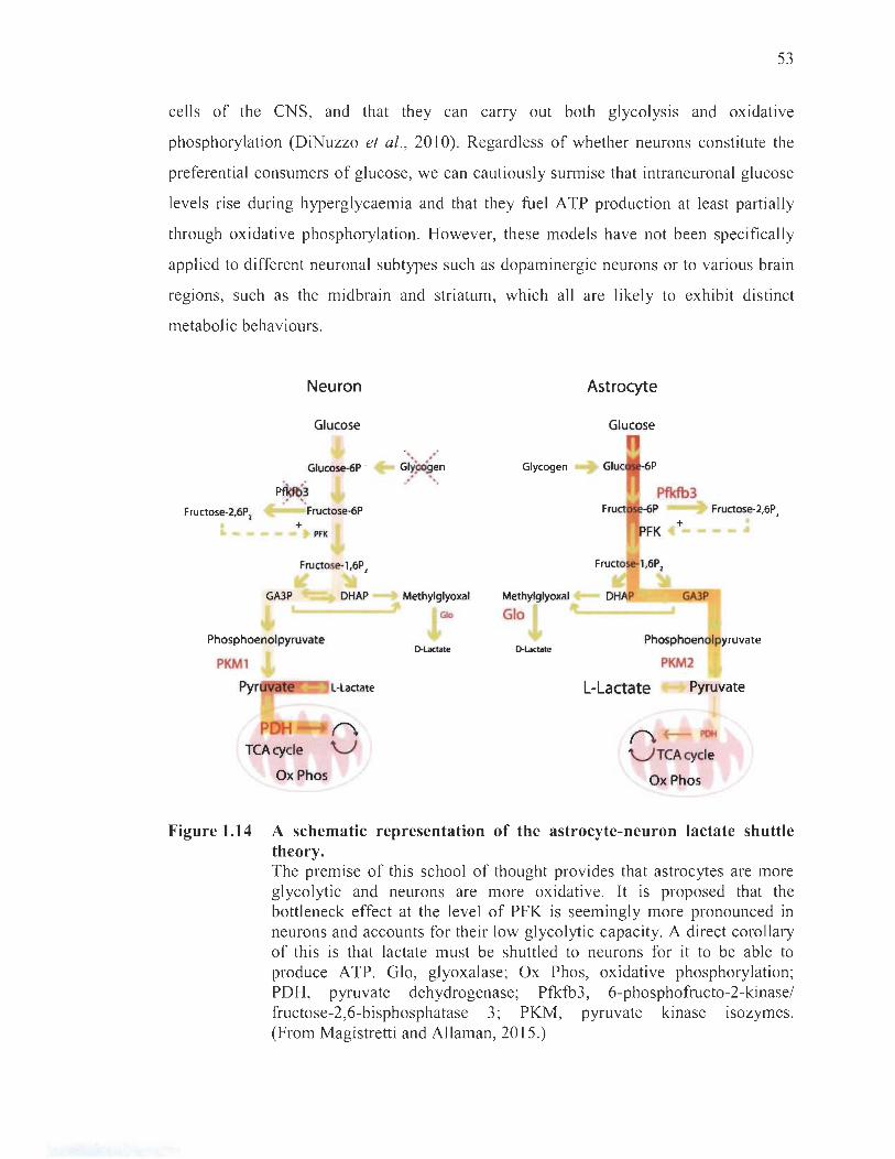
53
cells of the CNS, and that they can carry out both glycolysis and oxidative
phosphorylation (DiNuzzo et al., 2010). Regardless of whether neurons constitute the
preferential consumers of glucose, we can cautiously surmise that intraneuronal glucose
levels rise during hyperglycaemia and that they fuel ATP production at least partially
through oxidative phosphorylation. However, these models have not been specifically
applied to different neuronal subtypes such as dopaminergic neurons or to various brain
regions, such as the midbrain and striatum, which aU are likely to exhibit distinct
metabolic behaviours.
Neuron Astrocyte
Glucose Glucose
., . Glucose-6P .
P~~ ~~~~~n Glycogen
Pfkfb3 Fructose-2,6P, Fructose-6P 6P Fructose-2,6P,
+ PFK
Fructose-l ,6P,
GA3P DHAP Methylglyoxal
GIo
Methylglyoxal
Gia
PFK
Fructose-l,6P,
DHAP
+
GA3P
Phosphoenolpyruvate
PKMl lH.actate D-Lactate
Phosphoenolpyruvate
PKM2
Pyr e L·Lactate L-Lactate Pyruvate
POH TCAcycie
(\
V (\ POH
V TCAcycie Ox Phos Ox Phos
Figure 1.14 A schematic representation of the astrocyte-neuron lactate shuttle theory. The premise of this school of thought provides that astrocytes are more glycolytic and neurons are more oxidative. It is proposed that the bottleneck effect at the level of PFK is seemingly more pronounced in neurons and accounts for their low glycolytic capacity. A direct corollary of this is that lactate must be shuttled to neurons for it to be able to produce ATP. Glo, glyoxalase; Ox Phos, oxidative phosphorylation; PDH, pyruvate dehydrogenase; Pfkfb3 , 6-phosphofructo-2-kinase/ fructose-2 ,6-bisphosphatase 3; PKM, pyruvate kinase lsozymes. (From Magistretti and AUaman, 2015.)

54
1.2.4 Neuronal oxidative stress in hyperglycaemia
The clinical significance of hyperglycaemia emerged upon its formai identification
as the prime culprit in comorbid diabetic complications (Diabetes Control and
Complications Trial Research Group, 1993; Gaede et al. , 2008; Giacco and Brownlee,
2010; UK Prospective Diabetes Study Group, 1998). The most affected collateral targets
in diabetes mellitus are the kidneys, the cardiovascular system and the nervous system
(Nathan, 1993). Within each ofthese tissues dwell cells that are preferentially vulnerable
to hyperglycaemia, namely mesangial cells of renal glomeruli, endothel ial capillary
celIs, and brain cells. Despite their many functional and phenotypic differences,
these cells share the distinct liability of taking up glucose in a manner that is reliant on
extracellular concentrations (Heilig et al. , 1995; Kaiser et al., 1993). Indeed, mesangial
and endothelial cells predominantly express insulin-independent GLUT1 (Brosius and
Heilig, 2005; Mueckler and Thorens, 2013 ; Sone et al. , 2000); likewise, brain cells
express several insulin-independent GLUTs, chiefly GLUT1 and GLUT3 (Mantych
et al. , 1992; Nagamatsu et al. , 1993; Simpson et al. , 2008). Although it is c1ear that
glucose concentrations rise in these ce Ils un der conditions of hyperglycaemia,
how exactly cellular damage occurs is not as forthright. The CUITent concensus is that
oxidative stress explains hyperglycaemia-induced diabetic complications32 (Araki and
Nishikawa, 2010; Brownlee, 2005; Ceriello, 2003 ; Giugliano et al. , 1996) .
Oxidation is a ubiquitous phenomenon common to al! organisms that engage in
aerobic respiration for energy. Indeed, ROS are mainly produced from the leakage of
the mitochondrial electron transport chain during oxidative phosphorylation, but can also
arise from other endogenous (e.g. , monoamine oxidase activity, dopamine auto
oxidation, Fenton reactions in the presence of transition metals, etc.) or exogenous
sources (e.g., exposure to pesticides, smoking, ultraviolet and ionizing radiations,
pollution, etc.). Although required for a number of desirable physiologica1 processes,
the excessive generation of ROS can affect almost aIl cellular components, namely
32 Understanding the link between hyperglycae mia and diabetic complications required the discovery of particular markers in the blood and tissues of diabetic patients indicating a state of oxidative stress (Cheng and Gonzalez, 1986; Jones el al. , 1988; Matkovics et al. , 1982).
55
DNA, lipids and proteins (Barzilai and Yamamoto, 2004; Niki, 2008). To cope with
the unavoidable production of ROS, antioxidative defences are provided endogenously,
by superoxide dismutase (SOD), catalase, urate, ascorbate and glutathione, or
exogenously, by vitamins, polyphenols and other nutrients found in the di et. Per se,
oxidative stress arises from an imbalance between the generation and clearance of ROS,
in favour of the former, and may ultimately provoke cell death. This inability to cope
with an oxidative overload constitutes a key pathological element, not only in
hyperglycaemia (Brownlee, 2005; Wolff et al. , 1991), but also in Parkinson ' s disease
(Hwang, 2013; Schapira and Jenner, 2011 ; Tsang and Chung, 2009) and aging (Finkel
and Holbrook, 2000; Pérez et al. , 2009).
In consummg approximately 20% of total inhaled oxygen (Jain et al. , 20 lO;
Quastel and Wheatley, 1932), the brain is doubtlessly susceptible to undergo oxidative
stress. Indeed, processing of this oxygen implies engagement of oxidative
phosphorylation, which inexorably entails the generation of mitochondrial ROS (Kudin
et al. , 2004; Liu et al. , 2002). Owing to their steep demands in ATP, neurons possess a
high basal oxidative metabolism compared to other types of cells in the brain or
elsewhere, and are thus plausibly more vulnerable to additional sources of stress.
Although the literature do es not provide an adequate account of the mechanisms
underlying hyperglycaemia-induced oxidative stress in the CNS, let alone in nigrostriatal
dopaminergic neurons, we can speculate on the events that contribute to this critical
coping threshold based on experimental data and our knowledge of neuronal glucose
metabolism.
1.2.4.1 Mitochondrial mechanisms
Following an upsurge in intraneuronal glucose, a plausible event that may occur is
an amplified flux of electron donors toward oxidative phosphorylation. In a first
scenario, high intraneuronal glucose concentrations may provide the glycolysis pathway
and tricarboxylic acid cycle with more fuel , culminating in the increased production of
electron donors and the improvement of oxidative phosphorylation. In a second scenario,
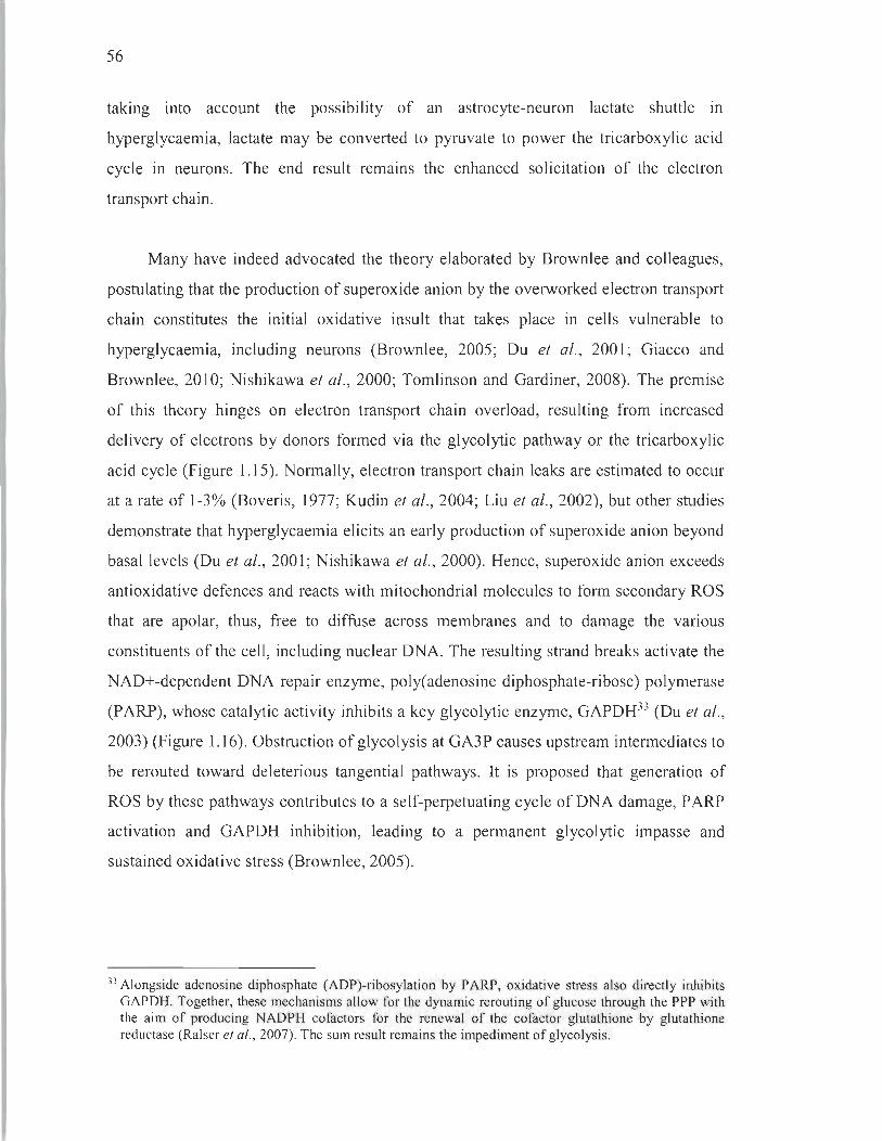
56
ta king into account the possibility of an astrocyte-neuron lactate shuttle in
hyperglycaemia, lactate may be converted to pyruvate to power the tricarboxylic acid
cycle in neurons. The end result remains the enhanced solicitation of the electron
transport chain.
Many have indeed advocated the theory elaborated by Brownlee and colleagues,
postulating that the production of superoxide anion by the overworked electron transport
chain constitutes the initial oxidative insult that takes place in cells vulnerable to
hyperglycaemia, including neurons (Brownlee, 2005; Du et al. , 2001; Giacco and
Brownlee, 2010; Nishikawa et al., 2000; Tomlinson and Gardiner, 2008). The premise
of this theory hinges on electron transport chain overload, resulting from increased
delivery of electrons by donors formed via the glycolytic pathway or the tricarboxylic
acid cycle (Figure 1.15). Normally, electron transport chain leaks are estimated to occur
at a rate of 1-3% (Boveris, 1977; Kudin et al., 2004; Liu et al., 2002), but other studies
demonstrate that hyperglycaemia elicits an early production of superoxide anion beyond
basal levels (Du et al. , 2001; Nishikawa et al. , 2000). Hence, superoxide anion exceeds
antioxidative defences and reacts with mitochondrial molecules to form secondary ROS
that are apolar, thus, free to diffuse across membranes and to damage the various
constituents of the ceIl, including nuclear DNA. The resulting strand breaks activate the
NAD+-dependent DNA repair enzyme, poly(adenosine diphosphate-ribose) polymerase
(PARP), whose catalytic activity inhibits a key glycolytic enzyme, GAPDH33 (Du et al. ,
2003) (Figure 1.16). Obstruction of glycolysis at GA3P causes upstream intermediates to
be rerouted toward deleterious tangential pathways. It is proposed that generation of
ROS by these pathways contributes to a self-perpetuating cycle of DNA damage, P ARP
activation and GAPDH inhibition, leading to a permanent glycolytic impasse and
sustained oxidative stress (Brownlee, 2005).
33 Alongside adenosine di phosphate (ADP)-ribosylation by PARP, oxidative stress also di rectly inhibits GAPDH. Together, these mechanisms a\low for the dynamic rerouting of glucose through the ppp with the aim of producing NADPH co factors for the renewal of the cofactor glutathione by glutathione reductase (Ralser et al., 2007). The sum result remains the impediment of glycolysis.
57
Generate H+ gradient Collapse H+ gradient
1 H+ H+ 1
ATP
Heat
Figure 1.15 Hyperglycaemia-induced generation of superoxide anion at the level of the mitochondrial electron transport chain. Electron traffic through the complexes (I-IV) of the chain allows the pumping of protons into the mitochondrial intermembrane space, which affords an electrochemical potential difference that powers A TP synthase (Trumpower, 1990). Protons are usually trafficked back into the matrix by ATP synthase, paired to its catalytic activity, and by uncoupling protein-1, which generates heat to alleviate overly steep gradients. However, when electron transport is profuse, the transmembrane potential increases faster than it can be dissipated by ATP synthase and uncoupling protein-1. Above a certain potential threshold, certain complexes, especially complex III, of the electron transport chain undergo inhibition and begin to leak their electrons, which are in turn captured by oxygen to pro duce superoxide anion (Adam-Vizi, 2005 ; Korshunov et al., 1997; Kwong and Sohal, 1998; Starkov et al., 2004). Owing to its negative charge that prevents it from crossing phospholipid bilayers, superoxide anion remains trapped within mitochondria. There, it is usually deactivated by the mitochondrial superoxide dismutase (SQD) into peroxide, another reactive oxygen species (ROS) that can cross phospholipid bilayers and damage other components of the cell. An upsurge in oxidative phosphorylation as it occurs in states of high intracellular glucose availability will necessarily increase superoxide anion generation. Cyt c, cytochrome c; e-, electron; 0 2·-, superoxide anion; Pi, inorganic phosphate; Q, quinone; UCP, uncoupling protein. (From Brownlee, 2005.)

58
PARP (active)
>.:' / >-2 - ' . ' H " 0
~~ ~~c;
Figure 1.16 Detrimental activation ofPARP in response to DNA damage. Oxidative stress leads to ROS-induced nuclear DNA damage responsible for activating poly(adenosine diphosphate-ribose) polyrnerase (PARP). Besides repairing single-stranded DNA nicks, it also operates as an adenosine diphosphate-ribosyl transferase (Sawa et al., 1997; Schmidtz, 2001). One of its targets is GAPDH, known to shuttle between the cytosol and the nucleus. Upon ADP-ribosylation, GAPDH is inactivated, thereby contributing to the accrual ofupstream glycolysis rnetabolites. Moreover, if PARP is not inactivated, it can lead to the depletion of the NAD+ cofactor. ADPR, adenosine diphosphate ribosyl; NA, nicotinamide. (From Brownlee, 2005.)
It is important to note that the key results in support of Brownlee's theory were
obtained in bovine aortic endothelial cells treated with 30 mM of glucose for 7 da ys
(Du et al., 2001; Nishikawa et al., 2000). Consequently, these conditions may not reflect
what occurs in the brains of animais exposed to high levels of glucose. A small body of
literature investigating these mechanisms in the CNS and the periphery of rodents does
not clearly validate this model in neurons. In fact, whether at the level of glycolysis or
the tricarboxylic acid cycle, the data inconsistently reports up- or downregulation of
these pathways, both in peripheral nerves or brains of hyperglycaemic rodents (Akude
et al., 2011; Chowdhury et al., 2010; Hinder et al., 2013; Kaur and Bhardwaj, 1998;
Price et al., 2006; Thakran et al., 2003; Thurston et al., 1995; Zheng et al., 20 17) .
However, with regard to oxidative phosphorylation, most studies revealed a decreased
activity or expression of electron transport chain constituents in the brain and in
peripheral sensory neurons (Aghanoori et al., 2017; Akude et al., 2011; Chowdhury
59
et al., 2010; Kaur and Bhardwaj, 1998; Stancié et al. , 2013). A closer look at the
duration of hyperglycaemia affords a clearer understanding of the process. Early events
in hyperglycaemia cause the upregulation of glycolysis and tricarboxylic acid cycling
(Kaur and Bhardwaj, 1998; Price et al., 2006; Thakran et al. , 2003; Thurston et al.,
1995). Later, however, these pathways are impeded (Aghanoori et al., 2017; Akude
et al., 2011; Chowdhury et al., 2010; Hinder et al., 2013; Zheng et al., 2017). At aIl time
points, oxidative phosphorylation is impaired (Aghanoori et al., 2017; Akude et al.,
2011; Chowdhury et al., 2010; Kaur and Bhardwaj, 1998; Stancié et al., 2013). In other
words, dysfunction of mitochondrial respiration likely occurs before glycolysis and
tricarboxylic acid cycle hindrance. This supports the theory that hyperglycaemia induces
an early upsurge in glucose metabolism leading to the initial insult arising from
mitochondrial respiration (Brownlee, 2005; Du et al., 2001; Giacco and Brownlee, 2010;
Nishikawa et al., 2000; Tomlinson and Gardiner, 2008). However, since the model
proposed by Brownlee does not account for precise cellular effects of long-term
hyperglycaemia, it cannot explain the global impairment of glucose metabolism
observed at later time points.
Among these studies, very few groups measured superoxide amon levels and
proper mitochondrial respiration. In these reports, solely performed in peripheral sensory
neurons of long-term hyperglycaemic rats, superoxide anion production and respiration
rates were downscaled even when corrected for mitochondrial numbers, despite overt
manifestations of oxidative stress (Aghanoori et al., 2017; Akude et al., 20 Il;
Chowdhury et al., 2010). This may indicate that sustained hyperglycaemia causes the
waning of mitochondrial respiratory functions over time, sparing superoxide anion
production while ROS generation is maintained by non-mitochondrial sources.
One explanation provides that the initial superoxide anion overload proposed by
Brownlee's model may cause deleterious mutations in mitochondrial DNA, leading to
defects in electron transport chain components that in tum generate more ROS34.
34 Unlike nuclear DNA, mitochondrial DNA lacks protection afforded by histones and repair enzymes. For this reason, the rates of mutagenesis in mitochondria are relatively high. Sorne subunits of complexes l and IV of the electron transport chain are encoded in mitochondrial DNA and, thus, are highly susceptible to mutation-induced impairments (Wei , 1998).
60
In the long term, excessive oxidative damage to mitochondrial DNA may impair
respiratory chain expression altogether, causing the observed shortfall in superoxide
anion35 (see for reviews Guo et al., 2013; Wei, 1998). Although studies employing
proper animal models of hyperglycaemia have not investigated very early mitochondrial
events in neurons, a prompt increase in superoxide anion is observed following
ischaemia-reperfusion, a paradigm similar in sorne respects to a transient nse ln
glycaemia (Iwata et al., 2014; Muranyi and Li, 2006; Won et al. , 20 15).
1.2.4.2 Rerouting mechanisms: polyol pathway and macromolecule glycation
Aside from ROS generated through mitochondrial failure, hyperglycaemia may
trigger the onset of oxidative stress via pathways that dwell upstream in glycolysis.
As previously demonstrated, free glucose is present in the cytosol of neurons and its
concentrations rise during hyperglycaemia. Under normal circumstances, hexokinase is
mandated to transform available glucose into G6P. However, due to substrate-mediated
inhibition, it can only engage a fraction of cytosolic glucose along the glycolytic
pathway before it is saturated. Excess glucose is therefore consumed by other pathways
and is constantly replenished by high extracellular levels. The principal compensatory
outlets are the polyol pathway and macromolecule glycation.
The polyol pathway is driven by aldose reductase whose affinity for glucose is
relatively low (Gabbay et al., 1966). Under normal glycaemia, aldose reductase does not
appoint glucose to the polyol pathway, but high intraneuronal concentrations drive its
catalytic activity. Aldose reductase preferentially employs the NADPH cofactor,
produced by the PPP, to reduce glucose into the polyalcohol sorbitol (Lee and Chung,
35 Another explanation is that the increased glucose availability may provide ample supply of ATP via the non-respiratory Warburg effect, thereby circumventing the need for oxidative phosphorylation and culminating in a downward adjustment of mitochondrial functions , akin to the Crabtree effect (Hamberger and Hyden, 1963; Ibsen, 1961). Accounts of thi s are , however, inconsistent in hyperglycaemic models (Herse and Petchell, 1998; Koziel et al. , 2012 ; Padnick-Silver and Linsenmeier, 2003).

61
1999)36. Sorbitol then undergoes enzymatic converSIOn to fructose by sorbitol
dehydrogenase, which exploits the important co factor NAD+ (Verdin, 2015; Williamson
et al., 1993; Wu et al., 2016) (Figure 1.17). Increased processing of glucose through the
polyol pathway thus drains pools of cofactors required for the activity of a plethora of
enzymes, including glutathione reductase, GAPDH and others in the glycolytic pathway
and tricarboxylic acid cycle (Vander Jagt et al., 1995).
Glucose 1 1
Glycolysis 1
NADH NAD+
NAD+ NADH Pyruvate
'\.. ? dehydrogenase
Fatty acids - -~ - - + Acetyl-CoA NADH ~-Oxidation ~
( NAD;
Krebs cycle NADH
Figure 1.17 NAD in glucose metabolism. This schematic represents the nurnerous enzymes that require the NAD+ cofactor to function and highlights the hazard that its depletion represents for the maintenance of normal metabolism. Acetyl-CoA, acetyl coenzyme A. (From Wu et al., 2016.)
36 A possible noxious effect of driving the polyol pathway was first proposed to be osmotic damage, seeing as cells do not export sorbitol efficiently. However, sorbitol concentrations measured in diabetic nerves and capillaries were too low to endorse thi s effect (Brownlee, 2001).

62
The second outlet of excess glucose is via macromolecule glycation, which may
indirectly contribute to oxidative stress (Vicente Miranda et al., 2016). Glycation
consists of the slow non-enzyrnatic, energy-independent, covalent addition of a
monosaccharide to macromolecules, usually lipids or proteins termed advanced
glycation end-products (AGEs)37 (Figure 1.18). Although glucose is a poor glycating
agent, one of its downstream metabolites, methylglyoxal, exceeds its reactivity
20000 times (Thomalley, 2005). Methylglyoxal is formed from the non-enzyrnatic
degradation of triose phosphates, such as the glycolysis intermediate GA3P, and readily
glycates proteins at the level of arginine and lysine residues (Ahmed et al., 1997;
Oya et al., 1999; Phillips and Thomalley, 1993; Richard, 1984; Shipanova et al., 1997;
Thomalley et al., 1999). Intracellular AGEs produced via this pathway are harmful to
cells because of the loss or alteration of their normal prote in or lipid functions, but also
by virtue of the mechanisms in place to ensure the prophylactic clearance of glycating
agents (Ahmed et al. , 2003). Indeed, methylglyoxal and other agents are enzyrnatically
degraded by glyoxalase and aldose reductase (Izaguirre et al., 1998; Monder, 1967;
Sousa Silva et al., 20 13; Vander Jagt et al., 1992). In the setting of hyperglycaemia,
excess glycating agents may overwhelm these pathways and lead nonetheless to AGEs,
evidenced by the proportional rise in CNS glycation as a function of glycaemia, which
can reach an exorbitant 34-fold increase (Uchiki et al., 2012). Most importantly,
however, glyoxalase employs gluthathione to deactivate molecules, while we know that
aldose reductase depletes pools of NADPH (Schieber and Chandel, 2014; Thomalley,
1988). Therefore, methylglyoxal and other glycating agents derived from glucose or
its metabolites indirectly worsen the oxidative status of neurons by monopolizing
endogenous antioxidative defences or the cofactors necessary for their restoration.
Since glycation is a non-specific and spontaneous reaction, AGEs may also occur
extracellularly. These activate deleterious inflammatory pathways by binding their
designated membrane receptor, RAGE (Jakus and Rietbrock, 2004; Singh et al., 2001).
37 In diabetic patients, the gold standard in the assessment of systemic glycation, thus of the severity of hyperglycaemia, consists in quantification of circulating glycated haemoglobin (Kovatchev, 2017). In fact, on account of the graduaI nature of these reactions, this measure is insensitive to daily glycaemic fluctuations and rather indicates glycaemic control spanning over the previous few weeks.
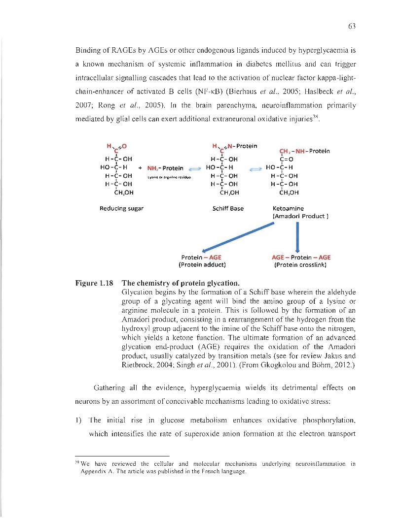
63
Binding of RAGEs by AGEs or other endogenous ligands induced by hyperglycaemia is
a known mechanism of systemic inflammation in diabetes mellitus and can trigger
intracellular signalling cascades that lead to the activation of nuc1ear factor kappa-light
chain-enhancer of activated B cells (NF-KB) (Bierhaus et al., 2005; Haslbeck et al.,
2007; Rong et al., 2005). In the brain parenchyma, neuroinflammation primarily
mediated by glial cells can exert additional extraneuronal oxidative injuries38.
H , ",0 C 1
H-C-OH 1
H , ........ N- Protein C 1
H-C- OH 1
HO-C-H 1
HO-C-H 1
+ NH,- Protein H-C-OH
1 H-C-OH
1
CH,OH
Reducing sugar
H-C-OH 1
H-C-OH 1
CH,OH
Schiff Base
Protein - AGE (Protein adduct)
Figure 1.18 The chemistry of protein glycation.
CH , - NH - Protein 1
c=o 1
• HO-C-H 1
H-C-OH 1
H-C-OH 1
CH,OH
Ketoamine (Amadori Product )
1 AGE - Protein - AGE (Protein crosslink)
Glycation begins by the formation of a Schiff base wherein the aldehyde group of a glycating agent will bind the amino group of a lysine or arginine molecule in a protein. This is followed by the formation of an Amadori product, consisting in a rearrangement of the hydrogen from the hydroxyl group adjacent to the imine of the Schiff base onto the nitrogen, which yields a ketone function. The ultimate formation of an advanced glycation end-product (AGE) requires the oxidation of the Amadori product, usually catalyzed by transition metals (see for review Jakus and Rietbrock, 2004; Singh et al., 2001). (From Gkogkolou and B6hm, 2012.)
Gathering ail the evidence, hyperglycaemia wields its detrimental effects on
neurons by an assortment of conceivable mechanisms leading to oxidative stress:
1) The initial rise in glucose metabolism enhances oxidative phosphorylation,
which intensifies the rate of superoxide anion formation at the electron transport
38 We have reviewed the cellular and molecular mechanisms underlying neuroinflammation 10
Appendix A. The article was published in the French language.

64
chain. On the one hand, superoxide anion damages mitochondrial DNA, causing the
expression of respiratory components prone to generate more ROS, which may lead
to global mitochondrial failure in the long term. On the other hand, superoxide
anion creates more ROS that cause nuclear DNA strand breaks, thereby activating
PARP.
2) Increased expenditure of NAD+ by P ARP slows the rates of glycolysis,
tricarboxylic acid cycling and oxidative phosphorylation operating via enzymes that
require this cofactor. PARP may also ADP-ribosylate and inhibit the glycolytic
enzyme GAPDH, which provokes upstream accumulation of metabolites.
3) Accrued levels of GA3P upstream of GAPDH are non-enzymatically converted into
the glycating agent methylglyoxal whose decomposition by glyoxalase and aldose
reductase expends antioxidative cofactors, such as glutathione and NADPH.
4) Excess glucose can be diverted into the deleterious polyol pathway, powered once
again by aldose reductase that further depletes stocks of NADPH required for the
renewal of the antioxidative co factor glutathione.
What emerges here is a number of different pathways that collectively cause a
shortage of antioxidant defences layered with mitochondrial dysfunction, two pivotai
factors likely responsible for pushing vulnerable cells beyond the critical coping
threshold. The sum of these undesirable processes appears to be particularly relevant in
neurons, in light of their relatively low glutathione levels and their reliance on the ppp
for its rapid regeneration (Bolafios et al., 1995; Kang et al., 1999). In fact, insufficient
provision of the ppp leads to poor regeneration of glutathione and neuronal apoptosis
(Herrero-Mendez et al., 2009). Neurons may reach the coping threshold quicker than
other brain cells, for instance astrocytes that display comparatively greater ppp activity
(Garcia-Nogales et al. , 2003; Herrero-Mendez et al., 2009).
Most importantly, the proposed molecular events leading to neuronal oxidative
stress in hyperglycaemia, explicitly via increased superoxide anion generation (Ziegler
et al. , 2015), mitochondrial dysfunction (see for review Patti and Corvera, 2010),
PARP activation (Obrosova et al., 2005), impairment of the PPP (Ziegler et al., 2017),
65
glycation (Aubert et al., 2014) and glutathione system deficits (Kasznicki et al., 2012 ;
Mendez et al., 2015), are all cogently established in diabetic patients with neuronal
pathology. Remarkably, clinical trials employing pharmacological agents to target sorne
of these pathways, notably aldose reductase inhibitors to diminish polyol pathway influx
(Greene et al., 1999; Kawai et al. , 2010; Obrosova et al., 2002) or exogenous SOD to
quench superoxide anion (Bertolotto and Massone, 2012), are demonstrated to improve
diabetic peripheral nerve damage and oxidative status, further solidifying the importance
of oxidative stress in neuronal affections arising from hyperglycaemia in humans.
Unquestionably, though, glycaemic control remains the most accessible means to
preclude oxidative stress-induced diabetic complications in the brain and in peripheral
nerves (Van den Berghe et al., 2005).
1.2.5 Vulnerability of the nigrostriatal pathway
In light of their comparatively heightened basal oxidative burden39, it may be
surmised that sustained hyperglycaemia may precipitate the death of nigrostriatal
dopaminergic neurons. The validity of this hypothesis transpires from epidemiological
evidence suggesting a link between the prime pathologies involving the nigrostriatal
pathway and hyperglycaemia, explicitly Parkinson 's disease and diabetes mellitus.
Before describing how they are pathologically interlaced, the latter disease will be
defined in more detail for contextual purposes. Bearing in mind that a handful of other
medical conditions also cause hyperglycaemia and may be associated with Parkinson 's
disease, as is the case for metabolic syndrome (Saaksjarvi et al., 2015), we deliberately
hold our focus on diabetes mellitus that offers a more tangible collection of experimental
and clinical evidences to work with.
1.2.5.1 Epidemiological basis: Parkinson 's disease in diabetic patients
Diabetes mellitus is the most widespread chronic metabolic disorder estimated to
affect over 400 million adults worldwide, its prevalence having doubled since 1980 on
39 Refer to the list ofphenotypic liabilities in section 1.1 .3.4.

66
the likely account of the marked ri se in overweight or obese phenotypes in the
population (World Health Organization, 2016). Today, diabetes has joined the infamous
list of leading causes of death, responsible for 8.4% of global mortality in adulthood
(International Diabetes Federation, 2013). Both types l and II diabetes occur from the
progressive failure of the body to manage circulating blood glucose. However,
the etiopathogenesis of type l diabetes (5-10% of diabetic patients) is specifically rooted
in the auto immune destruction of insulin-producing pancreatic ~ cells and usually,
but not always, develops before adulthood (Yoon and Jun, 2001). On the other hand,
type II diabetes ensues from peripheral resistance to and deficient production of insulin,
alongside other secondary metabolic elements. It is more frequently diagnosed in adults
although youth-onset forrns are dramatically on the rise (Correia et al. , 2008; Nadeau
et al., 2016). The perceived relative or absolute lack of insu lin control, which especially
wields its effects on cells harbouring GLUT4 like striated muscle cells and adipocytes,
culminates in prolonged hyperglycaemia and impaired ability to control glycaemic
fluctuations , known as glucose intolerance (see for detailed review of their
pathophysiology Cnop et al., 2005). Other symptoms also include polyuria, polydipsia
and hyperphagia (increased urination, thirst and hunger, respectively).
Granted the multiple aspects of health it impinges on, diabetes nurtures the
development of a myriad Qf diverse comorbid conditions. As previously stated,
a number of central (encephalopathies) and peripheral (neuropathies) complications are
associated with diabetes, regardless of the type (Biessels et al. , 2002). Neuropathies
remain the most prevalent neuronal affections in diabetes, as peripheral nerves are
devoid of blood-brain barrier protection, which, as we know, serves to limit exposure
to circulating glucose (Bansal et al. , 2006; Rajabally et al., 2017). Nonetheless,
encephalopathies are a widely acknowledged feature of diabetes (DeJong, 1950) and
manifest themselves in the forrn of cognitive decline (Brands et al., 2007; Brayne et al.,
2005; Hofman et al., 1997), white and grey matter atrophy (Moran et al. , 2013), altered
cortical connectivity (Lyoo et al. , 2013; van Duinkerken et al., 2012), and
neurodegenerative diseases, predominantly Alzheimer's and Parkinson ' s diseases
(Armstrong et al. , 2005 ; Correia et al. , 2011; Verdile et al., 2015 ; Vicente Miranda
et al. , 2016).

67
The first accounts of a possible association between diabetes and Parkinson's
disease date back to almost 60 years ago. Studies conducted in parallel showed that
diabetes exacerbates the progression of motor and cognitive deficits in Parkinson's
disease (Schwab, 1960), and that non-diabetic parkinsonian patients present impaired
glucose tolerance (Barbeau et al. , 1961) and hyperglycaemia (Boyd et al., 1971).
Since then, these findings have been reiterated numerous times in treated individuals40
(Cereda et al., 2012; Lipman et al., 1974; Sandyk, 1993), but also in drug-naïve patients
who still displayed higher-than-normal levels of fasting blood glucose, within the
prediabetic diagnostic range41 (Santiago and Potashkin, 2015). In addition, dietary habits
featuring an elevated intake of high glycaemic indexed carbohydrates42 have more
recently been associated with greater odds of developing Parkinson's disease (Cheng
et al., 2009; Murakami et al. , 2010; Okubo et al., 2012; Stafstrom and Rho, 2012; Yang
and Cheng, 2010).
In light of accumulating reports showing a possible association between the
two pathologies, numerous prospective cohort studies were elaborated to unravel their
temporal relationship (see for reviews Santiago and Potashkin, 2013; Wirdefeldt et al.,
2011) (Table l.8). Most, though not all (Becker et al., 2008; Cereda et al., 2011; Driver
et al., 2008) showed that pre-existing diabetes is a risk factor for the development of
Parkinson's disease (Arvanitakis et al., 2007; Cereda et al., 2012; Hu et al., 2007;
Xu et al., 20 Il). To dispel any doubt, one group addressed the question in a cohort
10 times larger (1.8 million patients) than previously, and robustly confirmed the risk
that diabetes represents for the future development ofParkinson's disease (Klimek et al.,
2015). Today, little doubt remains as to the hazard that a diabetic prelude represents in
the development ofParkinson's disease.
40 If anything, many dopaminergic drugs employed in the treatment of Parkinson's disease actually exert a blood glucose lowering effect (Cincotta et al., 1999; Lopez Vicchi et al. , 2016).
4\ Prediabetes is defined in humans as fasting blood glucose 5.5-7 mM (100-126 mg/dL), postprandial blood glucose 7.8- 1l.! mM (140-200 mg/dL) or glycated haemoglobin 5.7-6.5% (American Diabetes Association, 2016).
42 The glycaemic index of a food is defined as the rise in glycaemia that it occasions 2 hours after consumption in humans. Low glycaemic indexed nutrients release glucose slowly and steadily, in contrast to high glycaemic indexed foods that cause a rapid hyperglycaemia.

68
Table 1.8
Recent studies investigating the association between diabetes and Parkinson's disease
Hu er al., 2007 (5) Cohort study, Finnish population
Moran er al., 2008 (14) Meta-analysis
D'Amelia er a/., 2009 (122) Case-<:ontrol
Palacios er al .. 2011 (101 ) Cohort Sludy
Schernhammer er al., 2011 [1231 Case-<:ontrol , Danish population
Xu er al., 2011 (4)
Bosco er al., 2012 [181
Menon er al., 2011 [491
Cereda er a/., 2011 (121)
Cereda er a/., 2012 (19)
Cohort study
Case-control , Italian population
Genome-wide association study
Meta-analysis
Case-control
PD: 633 Contrais: 51 552 T2DM is associated wlth an increased risk 01 PD.
NIA Shared blological pathwavs between PD, T2DM, cancer, and inllammation.
PD: 318 Contrais: 318 Inverse association between PD and
PD: 656
PD. 1931 Contrais: 9651
diabetes preeeding PD onset.
No association.
T2DM Is assoclated wlth an increased risk 01 PD, espeeially younger onset PD.
Dlabetes: 21 611 Contrais: T2DM is associated with an increased 267 051 risk 01 PD. PD~emenlia: 53 PD: 57 Insulin resistance is associated with
an increased risk 01 dementla in PD.
NIA Shared pathways between PD and T1 DM.
lA Diabetes appears ta be a risk lactor lor PD.
Diabetes prlor ta PD onset: Dnset 01 T2DM belore onset 01 PD is 89 Contrais: 89 associated wlth an increased severity 01
PD symptoms.
Sun er al., 2012 (124) Case-<:ontrol , Chinese population Diabetes: 603413 Contrais: Diabetes is associated with an increased 472 718 risk 01 PD onset.
WahlqviSl er al. , 2012 (61) Case-control , Talwanese population Diabetes: 64 166 Contrais: T2DM is associated with an increased risk 698587 in PD. Metlormin-sullonylurea therapy
reduces the rlsk 01 PD.
& TlDM, type l diabetes melli tus; T2DM, type Il diabetes mellitus. (From Santiago and Potashkin, 2013 .)
From this point onward, the particuJar bond between diabetes and Parkinson's
disease Joses focus . What exactly triggers nigrostriatal degeneration in Parkinson's
disease among the multifarious metabolic changes that occur in diabetes remains
unclear. The two predominant lines of thought grant importance to the dimensions of
dysfunctional insulin signalling and oxidative stress. Although insulin occupies an
increasingly acknowledged role in the human CNS, by activating neuron-bound insulin
receptors responsible for controlling appetite, reward, cognition and memory (Anthony
et al., 2006; Benedict et al. , 2004, 2007; Craft et al. , 1996; Hallschmid et al. , 2008;
Khanh et al. , 2014; Reger et al. , 2008), we will address these dimensions in the
discussion and maintain our focus on oxidative stress for now.
1.2.5.2 Molecular and cellular bases
Aside from the epidemiological relationship between Parkinson 's disease and
diabetes, further clues support the possible susceptibility of nigrostriatal dopaminergic
neurons in hyperglycaemia. As previously discussed, oxidative stress mediated
by sustained hyperglycaemic conditions arises from 1) mitochondrial dysfunction,

69
2) activation of the polyol pathway, and 3) macromolecule glycation, which together
accentuate ROS production and expenditure of antioxidative resources. We will briefly
illustrate how these features manifest themselves in Parkinson ' s disease and how they
can be traced back to the phenotypic risk factors identified in nigrostriatal dopaminergic
neurons.
One of the most prominent liabilities specific to nigrostriatal dopaminergic
neurons consists in their vulnerability to mitochondrial dysfunction. Indeed,
their characteristic pacemaking activity leads to steep intracellular calcium
fluctuations, known to augment oxidative phosphorylation, mitochondrial membrane
hyperpolarization and superoxide anion production (Guzman et al. , 2010; PaceUi et al. ,
2015). On top of this, nigrostriatal neurons have a limited ability to bind excess
intracellular calcium (Dopeso-Reyes et al. , 2014; Iacopino and Christakos, 1990;
Liang et al., 1996; Yamada et al., 1990) and comprise a relatively high number of
mitochondria to sustain their dense arbours (Pacelli et al., 2015). It is, therefore, not
surprising that mitochondrial dysfunction has long been associated with Parkinson 's
disease (Perier and Vila, 2012). This pathological node first emerged upon identifying
potent parkinsonian toxins (e.g. , MPTP and rote none) whose detrimental effects are
principally conveyed by means of complex l inhibition in the electron transport chain
(BeaI, 2001 , 2003; Betarbet et al. , 2000; Greenamyre et al., 2001; Langston et al. , 1983).
The relevance of these findings was then fully recognized upon disceming complex l
deficiencies and accumulation of mitochondrial DNA deletions in the substantia nigra
pars compacta of parkinsonian patients (Bender et al., 2006, 2008; Schapira et al.,
1989). Later, robust support for impaired respiration was provided by the identification
of genes that cause familial forms of Parkinson ' s disease and that are involved in
mitochondrial homeostasis, for instance leucine-rich repeat kinase 2 (LRRK2)
(Mortiboys et al., 2010; Wang et al. , 2012), DJ-1 (Irrcher et al. , 2010; Krebiehl et al.,
2010), and the mitochondrial duo, PTEN-induced putative kinase 1 (PINK1) and parkin
(Ashrafi et al., 2014; Deng et al. , 2008; Exner et al. , 2007).
Parkinson ' s disease does not directly point to a hyperactive polyol pathway,
although one isolated study showed increased plasma levels of sorbitol in drug-naïve
70
parkinsonian patients, suggested to arise from glucose metabolism via non-oxidative
pathways ensuing from mitochondrial dysfunction (Ahmed et al., 2009). There is sorne
indirect evidence for a link between polyol pathway activation and neurodegeneration,
via downregulation of nerve growth factor (NGF) as demonstrated in hyperglycaemia
induced neuropathies (Hounsom et al., 2001 , 1998; Obrosova et al., 2001). Levels of
brain growth factors , like NGF, that promote neuronal survival and differentiation
indeed de cline in the substantia nigra pars compacta and dorsal striaturn of parkinsonian
patients (Mogi et al. , 1999). More substantially, though, endogenous antioxidant
depletion, provoked by polyol pathway activation, has been repeatedly suggested to
partake in the pathogenesis of Parkinson's disease. Indeed, exhaustion of the reduced
glutathione cofactor is one of the earliest events to occur in the brains of parkinsonian
patients (Jenner, 1993; Pearce et al. , 1997; Sian et al. , 1994), markedly so in
nigrostriatal dopaminergic neurons, which are inherently endowed with limited stocks of
glutathione (Bharath et al. , 2002; Kang et al., 1999).
Spontaneous methylglyoxal formation and macromolecule glycation are
particularly salient elements linking Parkinson's disease to hyperglycaemia. The most
obvious connection dwells in the inhibition of the ubiquitin proteasome system caused
both by protein and ubiquitin glycation, as it occurs in the CNS (Munch et al. , 2012;
Rabbini and Thomalley, 2012; Uchiki et al., 2012; Vicente Miranda et al., 201 6).
In fact, the pathologically relevant a-synuclein protein is glycated in Lewy bodies
(Auburger and Kurz, 2011 ; Cereda et al. , 2012; Kurz et al. , 2011 ; Vincent et al.,
2012). Interference with the ubiquitin proteasome system may therefore trigger a
self-perpetuating cycle, wherein increased levels of glycated a-synuclein further obstruct
proteasome function, consequently contributing to accumulation of the former, but also
to its oligomerization, aggregation and purported toxicity (Chen et al., 2006; Tanaka
et al., 2001). Additionally, RAGE polyrnorphisms have been shown to both offer
protection against and favour the development ofParkinson ' s disease (Gao et al. , 201 4) .
Insightful in vitro studies further demonstrate the shielding effect of ablating the
receptor in a dopaminergic neuronal model of the disease (Sathe et al. , 2012; Teismann
et al., 2012). Interestingly, not only can methylglyoxal glycate proteins and erode stocks
71
of NADPH VIa glyoxalase and al do se reductase activation, it participates like other
aldehydes in deleterious reactions with dopamine to form salsolinol-like compounds
(Deng et al. , 2012; Song et al., 2014; Szent-Gyorgi and McLaughlin, 1975),
which resemble 6-0HDA, MPTP and endogenous dopamine derivatives already
recognized for their toxicity (Kurnik-Lucka et al., 2018; Maruyama et al., 1999; Naoi
et al., 2002; Su et al., 2013). Of prime significance, parkinsonian patients display
particularly elevated levels of salsolinol-like compounds in the substantia nigra pars
compacta and dorsal striatum (Deng et al., 2012). Since nigrostriatal neurons express
relatively low levels of VMAT2 responsible for the vesicular uptake of lingering
cytosolic dopamine, they may also offer ideal temporal conditions for the transformation
of the latter into salsolinol-like compounds in a hyperglycaemic setting (Liang et al.,
2004).
To surnmarize, hyperglycaemia represents an additional cause of superoxide anion
production, mitochondrial dysfunction, antioxidative bankruptcy, ubiquitin proteasome
system failure and dopamine toxicity that may well favour the early demise of
nigrostriatal dopaminergic neurons. Although buttressed by a wealth of epidemiological,
molecular and cellular evidence, this hypothesis has not been directly tested. In the aim
of establishing the selective vulnerability of nigrostriatal neurons to a supplementary
oxidative burden, proper demonstrations would require43 a) the testing of a single
exogenous insult, for instance sustained hyperglycaemia; b) the comparison of
neurodegeneration III each loci involved in the nigrostriatal and mesocorticolimbic
pathways, preferably with concomitant verifications in other brain regions; c) the
assessment of neurodegeneration at more than one time point; d) the evaluation of glial
populations to gain insight into non-neuronal outcomes, and; e) the full appraisal of
behavioural alterations that may arise from nigrostriatal neurodegeneration. It is
precisely in this perspective that were elaborated a series of experiments presented
43 The most pertinent studies in this sense have only demonstrated neurodegeneration in the substantia nigra pars compacta of rodent models of diabetes, without investigating the repercussions on the totality of central doparninergic pathways, on other populations of brain cells, or on the range of behavioural alterations that may arise from neuronal death (Brambilla Bagatini et al., 2014; do Nascimento et al., 2011). Other studies have also combined various parkinsonian insults with a hyperglycaemic challenge, which does not address the intrinsic hazard that hyperglycaernia represents to nigrostriatal neurons in otherwise healthy subjects (Choi et al., 2005; Morris et al., 2010; Roterrnund et al., 2014).

72
herein to fulfil these interrogations that warrant answers, in light of the epidemiological
risk incurred by diabetic patients with regard to the potential development of
Parkinson's disease.
1.3 The antioxidative polyphenol resveratrol
The proposaI that a heightened vulnerability to oxidative stress is at the origin of
the preferential degeneration of nigrostriatal dopaminergic neurons can be tested by
verifying the ability of an exogenous antioxidant to prevent this death. In the present
thesis, we hire this strategy by employing a well-known antioxidative polyphenol,
resveratrol, ln dopaminergic neurons exposed to high glucose conditions.
Although resveratrol and other dietary polyphenols can offer protection in various
disease settings, the scope of our CUITent work does not encompass providing evidence
of its already widely acknowledged neuroprotective potential44. Used here as an
antioxidative tool, the following section thus focuses on resveratrol's ability to preclude
oxidative stress. Before detailing its protective mechanisms in dopaminergic neurons,
we will nonetheless provide sorne background information on resveratrol pertaining to
its origins, its dietary sources and the chemico-structural peculiarities that shape its
antioxidative properties.
1.3.1 Background
1.3.1.1 Historical context
Centuries of traditional medicine practiced in native cultures worldwide have
revolved around the potency of edible medicinal herbs and plants ri ch in polyphenols to
ameliorate human health (Manyam et al., 1999; Song et al., 2012). When dietary habits
and health status were initially associated, the role of polyphenols in human health
44 We have already reviewed the therapeutic potential of polyphenols in Parkinson ' s disease elsewhere (Regl6di, Renaud et al. , 2017; Renaud et al., 2015). A critical appraisal of the employment of polyphenols against neurodegenerative diseases is also presented in Appendix B. Specifically regarding resveratrol, our group has extensively discussed its anti-inflammatory mechanisms in the CNS (Appendix C) and has detailed the various demonstrations of its protective properties in preclinical models (Renaud and Martinoli, 2014).

73
immediately sparked vivid scientific interest. At the root of polyphenol studies,
a mixture of flavonoids extracted from citrus fruit was initially coined "vitamin P"
because of its anti-scurvy, vitamin C-sparing effects (Bentsath et al. , 1936; Rusznyak
and Szent-Gy6rgi, 1936). The term was later abandoned upon disceming that "vitamin
P" was not truly essential , although the observed health benefits still held true. A host of
controlled epidemiological reports have since then supported the protective role of
polyphenols in abating numerous health issues, su ch as cardiovascular diseases (Estruch
et al., 2013 ; Hertog et al. , 1993; Joshipura et al. , 2001 ; von Ruesten et al. , 2013),
diabetes (Carter et al., 2010; Sargeant et al., 2001) and cancer (Lunet et al. , 2005 ;
Masala et al., 2012; Steinmetz and Potter, 1996). As evidence amassed, intervention
studies were warranted to bolster the significance of these findings, and most, though not
all (Anderson et al. , 2002; Conquer et al. , 1998; Simons et al. , 2000), confirmed a
positive function for specific dietary polyphenols in hurnan health (see for review
Williamson and Manach, 2005). As research progressed, certain biofunctional
polyphenols gained more attention than others by virtue of their promising protective
competences.
In the field of polyphenol research, resveratrol holds a special place. Indeed,
its popularity shot through the roof in the 90s following the airing of an episode of
"60 minutes" on the CBS network wherein Dr. Serge Renaud was interviewed on the
topic of the "French Paradox", a term henceforth etemalized (Renaud and de Lorgeril,
1992). The expression was employed to describe the apparently low incidence of
coronary heart diseases in Southem France populations despite their relatively elevated
consumption of saturated fats in comparison to other industrialized countries (Wu et al. ,
2001). A vital food staple in Southem France, red wine was thought to afford
cardioprotection (Renaud and de Lorgeril, 1992) and resveratrol was later proposed to
be one of the key bioactive constituents responsible for these observed health
benefits (Fauconneau et al. , 1997; Frankel et al. , 1993; Pace-Asciak et al. , 1995).
Robust demonstrations that resveratrol exerts antioxidative effects (Frankel et al. , 1993 ;
Kerry and Abbey, 1997; Jang et al. , 1999) and mitigates carcinogenesis (Della Ragione
et al. , 1998; Gehm et al., 1997; Hsieh et al. , 1999; Jang et al., 1997; Lu and Serrero,
1999; Mgbonyebi et al. , 1998) truly launched the field from thereon (Figure 1.19).
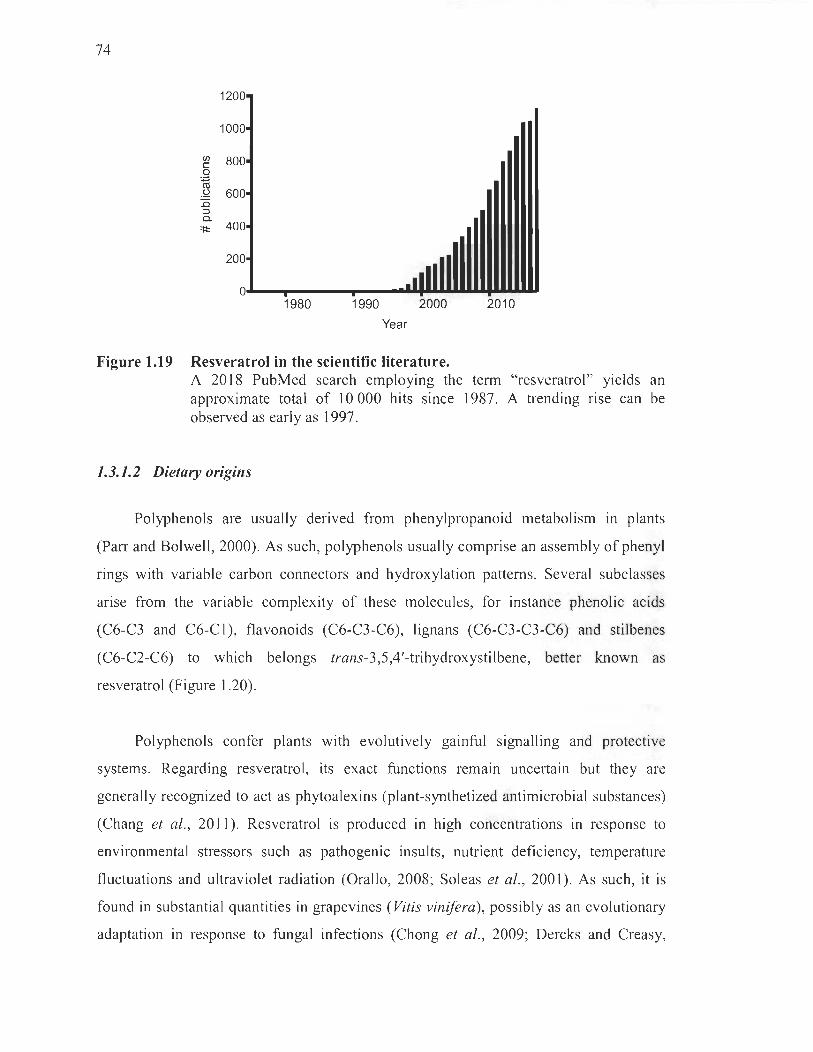
74
fi) c o ~ .2 :0 :::J a. :;t
1980 1990 2000 2010
Year
Figure 1.19 Resveratrol in the scientific Iiterature. A 2018 PubMed search employing the term "resveratrol" yields an approximate total of 10000 hits since 1987. A trending rise can be observed as early as 1997.
1.3.1.2 Dietary origins
Polyphenols are usually derived from phenylpropanoid metabolism in plants
(Parr and Bolwell, 2000). As such, polyphenols usually comprise an assembly of phenyl
rings with variable carbon connectors and hydroxylation patterns. Several subclasses
arise from the variable complexity of these molecules, for instance phenolic acids
(C6-C3 and C6-C 1), flavonoids (C6-C3-C6), lignans (C6-C3-C3-C6) and stilbenes
(C6-C2-C6) to which belongs trans-3,5,4'-trihydroxystilbene, better known as
resveratrol (Figure 1.20).
Polyphenols confer plants with evolutively gainful signalling and protective
systems. Regarding resveratrol, its exact functions remain uncertain but they are
generally recognized to act as phytoalexins (plant-synthetized antimicrobial substances)
(Chang et al., 2011). Resveratrol is produced in high concentrations in response to
environmental stressors such as pathogenic insults, nutrient deficiency, temperature
fluctuations and ultraviolet radiation (Orallo, 2008; Soleas et al., 2001). As such, it is
found in substantial quantities in grapevines (Vitis vinifera), possibly as an evolutionary
adaptation in response to fungal infections (Chong et al., 2009; Dercks and Creasy,
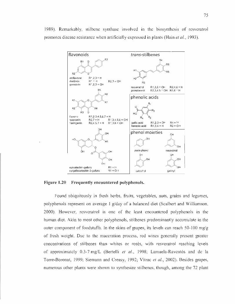
75
1989). Remarkably, stilbene synthase involved in the biosynthesis of resveratrol
promotes disease resistance when artificially expressed in plants (Hain et al., 1993).
flavonoids R3
R1 0
R2 0
isoflavone R1 .2.3 = H daidzein R1 = H R2.3 = OH genistein R1 .2.3 = OH
R5
R6
R1 R7
R2 R4
R3 0
flavone R1 .2.3.4 .5.6.7 = H quercetin R2.7 = H R1 .3.4.5.6 = OH naringenin R2.4.5.7 = H R1 .3.6 = OH
6c0H
OH
Ho'Ç00 ./ "" 1 R1 1
; .~ .• O "" OH
o 1
epicatechin-gallate epigallocatechin-3-gallate
h OH
OH
R1 = H R1 =OH
trans-stil benes R4
R5
R1 "" R6
R2
R3
resveratrol R1 .3.5 = OH R2.4.6 = H piceatannol R2.3.4 .5 = OH R1 .6 = H
phenolic acids R,
O~R2 Hb } -{
R4 R3
gallic acid R1 .2.3 = OH R4 = H R2 = OH benzoic acid R1 .3.4 = H
phenol moieties
~OH
N para-phenol
OH
~OH A) catechol
OH
~OH resorcinol
OH
û : galloyl
Figure 1.20 Frequently encountered polyphenols.
Found ubiquitously in fresh herbs, fruits, vegetables, nuts, grains and legumes,
polyphenols represent on average 1 g/day of a balanced diet (Scalbert and Williamson,
2000). However, resveratrol is one of the least encountered polyphenols in the
human di et. Akin to most other polyphenols, stilbenes predominantly accumulate in the
outer component of foodstuffs . In the skins of grapes, its levels can reach 50-100 mg/g
of fresh weight. Due to the maceration process, red wines generally present greater
concentrations of stilbenes th an whites or rosés, with resveratrol reaching levels
of approximately 0.3-7 mg/L (Bertelli et al., 1998; Lamuela-Ravent6s and de la
Torre-Boronat, 1999; Siemann and Creas y, 1992; Vitrac et al., 2002). Besides grapes,
numerous other plants were shown to synthesize stilbenes, though, among the 72 plant

76
specles identified, mulberries and peanuts are the only other typical sources rich ln
resveratrol in the human diet (Langcake and Pryce, 1977).
1.3.1.3 Structure, chemistry and antioxidative functions
Insofar as the > 8000 polyphenols known to date are structurally diverse,
they share several hall marks that undoubtedly underlie their therapeutic potential in
living organisms, namely phenyl rings, hydroxyl moieties and conjugated double bonds.
These chemico-structural features grant polyphenols like resveratrol the potential to
tackle a keystone of dopaminergic neurodegeneration, explicitly oxidative stress.
Resveratrol wields its antioxidative effects via both direct and indirect mechanisms
(Foresti et al., 2013; Leonard et al., 2003). Its hydroxyl groups are thought to account
for the near totality ofits direct antioxidative actions (Fauconneau et al. , 1997; Frémont,
2000; Kawada et al. , 1998; Stivala et al., 2001). Indeed, resveratrol scavenges free
radical species principally by lending its most acidic hydrogen from the para-phenol
group or other hydrogen atoms from the meta-hydroxyl groups of the resorcinol moiety
(Stivala et al., 2001) (Figure 1.20). It may also cede a 1t bond from the resorcinol ring
to accommodate radicals (Khanduja and Bhardwaj , 2003). The radicalized form of
resveratrol is stabilized by its multiple conjugated double bonds that allow the
delocalization of the unpaired electron (Figure 1.21).
While resveratrol is appreciated for its ability to directly scavenge free radical
species, it is not among the most efficient metal chelators. Since divalent transition
metals like iron are toxic, cells are endowed with tight control systems to regulate their
availability in a free form. When in excess, however, they are known to participate in
deleterious reactions that generate ROS45. Chelation by bidentate molecules possessing
phenol rings is possible, but is best achieved when there are two or three hydroxyl
45 For example, Fenton reactions occur between ferric ions and superoxide anion to yield ferrous ions and oxygen (Jomova et al., 2010). In turn, Haber-Weiss reactions between ferro us ions and hydrogen peroxide further give ferric ions and hydroxyl radicals, the most reactive kind of ROS (Valko et al. , 2004). In the substantia nigra pars compacta endowed with appreciable levels of iron ions, these deleterious reactions are extremely pertinent.
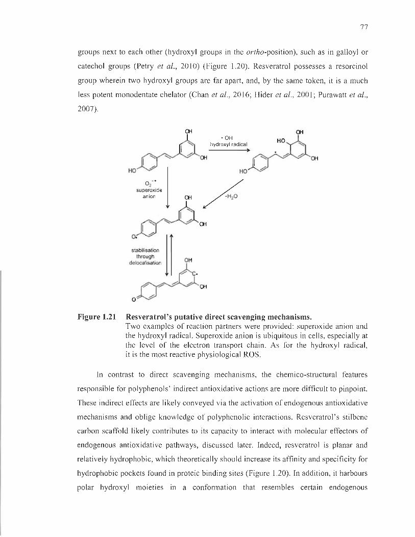
77
groups next to each other (hydroxyl groups in the ortho-position), such as in galloyl or
catechol groups (Petry et al. , 2010) (Figure 1.20). Resveratrol possesses a resorcinol
group wherein two hydroxyl groups are far apart, and, by the same token, it is a much
less potent monodentate chelator (Chan et al. , 2016; Hider et al., 2001 ; Purawatt et al.,
2007).
Oi Oi ' OH
hydroxyl radical
I~ Oi Oi
~ ~ 1 HO
° 2- '
~ supe~oxide anion Oi
I~ Cti
~ A 11 stabilisation through
OH delocalisation
o
Figure 1.21 Resveratrol's putative direct scavenging mechanisms. Two examples of reaction partners were provided: superoxide anion and the hydroxyl radical. Superoxide anion is ubiquitous in cells, especially at the level of the electron transport chain. As for the hydroxyl radical, it is the most reactive physiological ROS.
In contrast to direct scavenging mechanisms, the chemico-structural features
responsible for polyphenols' indirect antioxidative actions are more difficult to pinpoint.
These indirect effects are likely conveyed via the activation of endogenous antioxidative
mechanisms and oblige knowledge of polyphenolic interactions. Resveratrol's stilbene
carbon scaffold likely contributes to its capacity to interact with molecular effectors of
endogenous antioxidative pathways, discussed later. Indeed, resveratrol is planar and
relatively hydrophobic, which theoretically should increase its affinity and specificity for
hydrophobic pockets found in proteic binding sites (Figure 1.20). In addition, it harbours
polar hydroxyl moieties in a conformation that resembles certain endogenous
78
ligands, such as estrogen. These hydroxyl groups participate in hydrogen bonds with
multiple amino acid side chains within hydrophobic pockets. A few studies have also
demonstrated the importance of resveratrol's phenol rings, especially the one bearing
the para-hydroxyl moiety, and its trans stereoisomery in binding certain proteins
(Nwachukwu et al., 2014; Stivala et al., 2001). In other instances, resveratrol may not
require its hydroxyl functions to mediate its neuroprotective effects. Studies tested this
hypothesis by preparing a plethora of resveratrol analogs devoid of hydroxyl functions
(Heynekamp et al., 2006; Solberg et al., 2014). Despite their inability to scavenge ROS,
these stilbene compounds were nonetheless capable of dampening neuroinflammation in
a NF-KB-Iuc reporter ceIl line (Heynekamp et al., 2006) and reducing ~-amyloid plaque
density in the CNS of a mouse model of Alzheimer's disease (Solberg et al., 2014).
Such studies highlight the importance of resveratrol 's multiple chemico-structural
singularities in shaping both the nature and the variety of molecular interactions it can
engage in, which undoubtedly account for its multifarious modes of action.
1.3.2 Protection of dopaminergic neurons against oxidative stress
Resveratrol 's ability to improve various clinical endpoints has been demonstrated
m myriads of disease settings, including neurodegenerative diseases (Moussa et al. ,
2017), diabetes (Goh et al., 2014; Imamura et al., 2017), obesity (Xue et al., 2016) ,
polycystic ovary syndrome (Banaszewska et al., 2016) and ulcerative colitis
(Samsamikor et al., 2016). Its antioxidative potential has been shown to contribute to its
protective effects in preclinical models of diabetes (Jezek et al., 2014; Jiang et al., 2013 ;
Sadi et al., 2014) and neurological disorders, such as ischemic stroke (Rodrigo et al. ,
2013; Saleh et al., 2013; Sun et al., 2010), neurodegenerative diseases (Karuppagounder
et al., 2009; Maher et al., 2011; Varamini et al., 2014) and traumatic brain injuries
(Gatson et al., 2013; Hall et al., 2010; Liu et al., 2011). SpecificaIly re1ated to our work,
resveratrol improves the oxidative status of the CNS in rodent models of diabetes
(Bagatini et al., 2017; Sadi and Konat, 2016), but this parameter has not been addressed
in dopaminergic neurons challenged with high glucose concentrations. Nevertheless,
its antioxidative properties have been verified numerous times in preclinical models of
Parkinson's disease. As a basis for the employment of resveratrol in dopaminergic

79
neurons to antioxidative ends, this section will therefore briefly review the findings
provided by these studies.
To study Parkinson's disease, several models were developed in vitro as well as
in vivo that depend on pro-oxidative mechanisms to induce dopaminergic neuronal
death. Typical parkinsonian toxins include the aforementioned 6-0HDA and I-methyl-
4-phenylpyridinium (MPP+, the active metabolite of MPTP), as well as pesticides like
rotenone and paraquat, which instigate ROS overproduction by various means (Deumens
et al. , 2002; Dut Y and Jenner, 2011 ; Jackson-Lewis et al., 2012; Nagatsu, 1997).
Accordingly, studies consistently show resveratrol to rescue dopaminergic neurons from
the various oxidative assaults that typify these models (Blanchet et al. , 2008; Gélinas
and Martinoli, 2002; Wu et al. , 2011). Neuroprotection is complemented with reduced
levels of ROS, like the hydroxyl radical (Lu et al. , 2008), which prevents neuronal
oxidative damages from arising, as assessed by the measurement of lipid peroxidation
and prote in carbonyl by-products (Anandhan et al. , 2010; Khan et al. , 2010; Okawara
et al. , 2007; Palle and Neerati, 2018). Most remarkable is the recovery of motor
behaviour in animal models, which reflects the concrete protection afforded by
resveratrol on dopaminergic neurons in models of Parkinson 's disease (Anandhan et al. ,
2010; Jin et al. , 2008; Lu et al. , 2008; Makhija and Jagtap, 2014; Palle and Neerati et al. ,
2018; Zhao et al. , 2017).
A recurring the me in parkinsonian models is the apoptotic death46 of dopaminergic
neurons. Indeed, this mode of programmed cell death is particularly relevant in
Parkinson ' s disease (Anglade et al. , 1997; Mochizuki et al. , 1996; Toulorge et al. ,
2016), but also in diabetes (Maiese et al. , 2007; Russell et al., 1999). In this respect,
resveratrol is weil appreciated for its ability to abrogate oxidation-induced apoptosis.
Congruently, resveratrol favourably modulates various pro- or anti-apoptotic proteins,
su ch as p53 , protein kinase B (Akt) and glycogen synthase kinase 3 (GSK-3) (Bournival
et al. , 2009; Feng et al. , 2015; Lin et al. , 2014; Okawara et al. , 2007; Wu et al., 2011 ;
Zeng et al. , 2017). As mitochondria are a nexus of apoptotic processes, studies
46 We address apoptotic mechanisms in greater detail in section 1.5.104.

80
specifically uncovered resveratrol's aptitude to repeal the leakage of mitochondrial pro
apoptotic factors , such as apoptosis inducing factor and cytochrome c, likely through
modulation of proteins involved in the formation of the mitochondrial permeability
transition pore, exemplarily B cell Iymphoma 2 (Bcl-2) and Bcl-2-associated X protein
(Bax) (Bournival et al., 2009; Mud6 et al., 2012; Zeng et al., 2017).
Among these studies, many have reported certain upstream events that may
account for the antioxidative and anti-apoptotic effects afforded by resveratrol.
On the one hand, resveratrol remarkably restores endogenous antioxidative defences,
for instance glutathione, SOD, thioredoxin, heme oxygenase and catalase (Anandhan
et al. , 2010; Khan et al., 2010; Lin et al. , 2014; Mud6 et al. , 2012; Okawara et al. , 2007;
Palle and Neerati, 2018; Srivastava et al., 2012). On the other hand, resveratrol displays
a striking ability to foster mitochondrial homeostasis, by ameliorating respiration (Palle
and Neerati, 2018; Mud6 et al., 2012) and providing protection against ultrastructural
changes (Jin et al., 2008; Peng et al. , 2016). Interestingly, in primary skin fibrob last
cultures from patients with early-onset Parkinson's disease caused by different PARK2
mutations, encoding parkin, resveratrol markedly improved mitochondrial functions,
substantiated by increased ATP production, complex l activity, oxygen consumption,
and decreased generation of lactate and ROS (Ferretta et al. , 2014).
The sum of these results obtained in vanous cell lines, pnmary cultures,
organotypic midbrain slice models and rodent paradigms leaves no doubt as to the
capacity of resveratrol to protect dopaminergic neurons by virtue of its ability to
counteract oxidative stress, a key contributor of apoptosis. These neuroprotective
benefits seem to stem from the improvement of endogenous antioxidative defences
and mitochondrial functions . Taking into consideration the mechanisms by which
hyperglycaemia induces oxidative stress, that is, by expending endogenous antioxidants
and causing mitochondrial dysfunction, it is reasonable to expect resveratrol to confer
protection against high glucose conditions in dopaminergic neurons.

81
1.3.3 Direct putative targets
Although polyphenols like resveratrol are theoretical ROS scavengers, their ability
to do so in a physiological environment remains controversial. First, polyphenol
scavenging dictated by the transfer of a proton or the breaking of a double bond must
occur faster than any reaction between the free radical species and susceptible
constituents of the cell, for instance lipids (Di Meo et al. , 2013). Second, in order to
offer significant antioxidative support, polyphenols are theoretically required to accrue
in similar concentrations to that of endogenous antioxidants such as ascorbate
(30-100 !lM) and urate (140-200 !lM) (Hollman, 2014). However, tissue or plasma
concentrations of polyphenols rarely exceed micromolar magnitudes at any given time
(Del Rio et al., 2013). Consequently, the direct scavenging properties of polyphenols
like resveratrol may be trivial in a physiological context (Benzie et al. , 1999; Hollman
et al., 20 Il). In this regard, it is argued that cellular events occurring at nanomolar
concentrations might better uphold polyphenols ' antioxidative actions. These indirect
effects are likely conveyed via the activation of endogenous antioxidative mechanisms
and require only very low concentrations owing to the diverging and amplifying nature
of many cell signalling cascades.
In an attempt to reconcile resveratrol's bioefficacy with the improbability of
firsthand scavenging, efforts have been deployed to identify these putative direct targets
(see for review Britton et al. , 2015) (Table 1.9). Among the multiple molecules
identified to date, a few are directly or indirectly linked to oxidative stress, namely
ribosyldihydronicotinamide dehydrogenase (quinone) (NQ02) (Buryanovsky et al.,
2004; Wang et al. , 2004), phosphodiesterases (PDEs) (Park et al. , 2012), and the
mammalian target of rapamycin (mTOR) (Park et al. , 2016). In neurons, the se effectors
are notorious for executing noxious processes and their inhibition by resveratro1 may
explain the latter' s beneficial properties in the CNS (Cant6 and Auwerx, 2009;
Chaturvedi and BeaI, 2013; Lee et al. , 2012; Li et al. , 2010; Sharma et al. , 2012;
Wang et al., 2008).

82
Table 1.9
Direct putative targets of resveratrol
Molecular target&
CycJooxygenase-l CycJooxygenase-2 Fatty acid synthase NQ02 GSTP1 AKT-1 HDACs PDE1, 3, and 4
ATM SIRTl PKCa, [3I, and PKD1 Aromatase DNA/RNA Lipoproteins DNA polymerase a and 8 F1-ATPase CBR1
LTA-tH PPAR-y and 8 Various kinases TyrRS
aUnder oxidizing conditions (H202). bDirect binding disproved. 'Cocrystal with cis-resveratrol.
Enz Inhib, Xray Chem Pro t, Enz Inhib
Enz Inhib Chem Prot, Xray, Enz Inhib Chem Prot Chem Prot Enz Inhib Fluor Kinase assay Fluor Kinase assay Enz Inhib, in silico
Spec HPLC Enz Inhib Xray Chem Prot, Enz Inhib Chem Prot, in silico Xray, Affin Chrom Activity assay Enz Inhib, Xray'
Observation&
IC50 = 0.535 !-LM IC50 = 0.996 !-LM IC50 = 8.5 !-LM Binding, KI = 35 nM Binding Binding pan-HDACi
IC50 = 6, ID, and 14 !-LM Activationa
Activationb
IC50 = 2,100, and 800 !-LM
IC50 = 12.8 !-LM Destabilizing Binding Ki = 3.3 and 5.0 !-LM Binding Binding, Ki = 55.8 !-LM Binding Kd = lA and 2.7 !-LM Moderate to no effect
Ki = 22 !-LM
& Affin Ch rom, affinity chromatography; AKT - l , protein kinase B; ATM, ataxia telangiectasia mutated serine/threonine kinase; Chem Prot, chemical proteomics; CBRI , carbonyl reductase 1; Enz lnhib, enzyme inhibition assay; FI-ATPase, FI portion of adenosine triphosphatase; Fluor, fluorescence assay; GSTPI , glutathione S-transferase P; HDAC, hi stone deacetylase; HPLC, high-performance Iiquid chromatography; IC5o, half maximal inhibitory concentration ; Kd, di ssoc iation constant; Ki, inhibition constant, L T A4H, leukotriene A4 hydrolase; NQ02, ribosyldihydron icotinamide dehydrogenase (quinone); PDE, phosphodiesterases; PKC, protein kinase C; PKD, protein kinase D; PPAR, peroxisome proliferator-activated receptor; SIRTl, silent mating type information regulation 2 homologue 1; Spec, spectroscopie assay; TyrRS, tyrosine-transfer RNA ligase; Xray, X-ray cocrystal structure. (Adapted from Britton et al., 2015.)
1.3.3.1 Ribosyldihydronicotinamide dehydrogenase (quinone) and oxidative stress
NQ02 is a detoxifying enzyme mandated to catalyze the reduction of quinones to
hydroquinones (Monks et al., 1992; Vella et al., 2005). At first sight, it appears to
operate as an endogenous antioxidative enzyme, due to its role in reducing quinone

83
ROS. However, it was recently found to produce ROS by-products during quinone
reduction in sorne settings (Gong et al. , 2008; Reybier et al. , 2011). In fact, NQ02
appears to impart the noxious effects of certain parkinsonian toxins and of exogenous
dopamine on dopaminergic neurons (Janda et al., 2013 , 2015 ; Wang et al. , 2008).
Interestingly, NQ02 has emerged as a novel risk factor for Parkinson's disease.
Indeed, genetic polymorphisms associated with the pathology were initially speculated
to result in decreased NQ02 expression (Harada et al. , 2001). However, later findings
showed these polymorphisms to occur in the promoter and to enhance NQ02
expression, which was further sustained in human fibroblasts of individuals with or
without the genetic modification (Wang et al. , 2004). In this light, it was concluded that
amplified expression of the reductase constitutes a risk factor for the development of
Parkinson's disease.
Likely the most relevant target in oxidative stress, NQ02 displays the greatest
affinity for resveratrol to date (nanomolar range) . Its interaction with the polyphenol has
also been ascertained by no less than three methods carried out by two independent
groups (Buryanovsky et al. , 2004; Wang et al. , 2004). It follows that resveratrol ' s
inhibitory action on NQ02 is presumed to confer protection in this setting, but this
remains to be confirmed in neurons.
1.3.3.2 Phosphodiesterases and the energy sensing axis
Many of the abovementioned studies demonstrating resveratrol's antioxidative and
neuroprotective properties in dopaminergic neurons also expose its modulatory effect
on a selection of interrelated bioenergetic power players, namely 5' adenosine
monophosphate-activated prote in kinase (AMPK) , silent mating type information
regulation 2 homologue 1 (SIR T 1) and peroxisome proliferator-activated receptor
gamma coactivator l-alpha (PGC-l a) (Albani et al., 2009; Feng et al., 2015; Ferretta
et al., 2014; Mudè et al., 2012; Wu et al. , 2011). The signalling axis to which they
belong is thought to be responsible for lifespan amelioration provided by exercise and
calorie restriction (Tennen et al. , 2012).
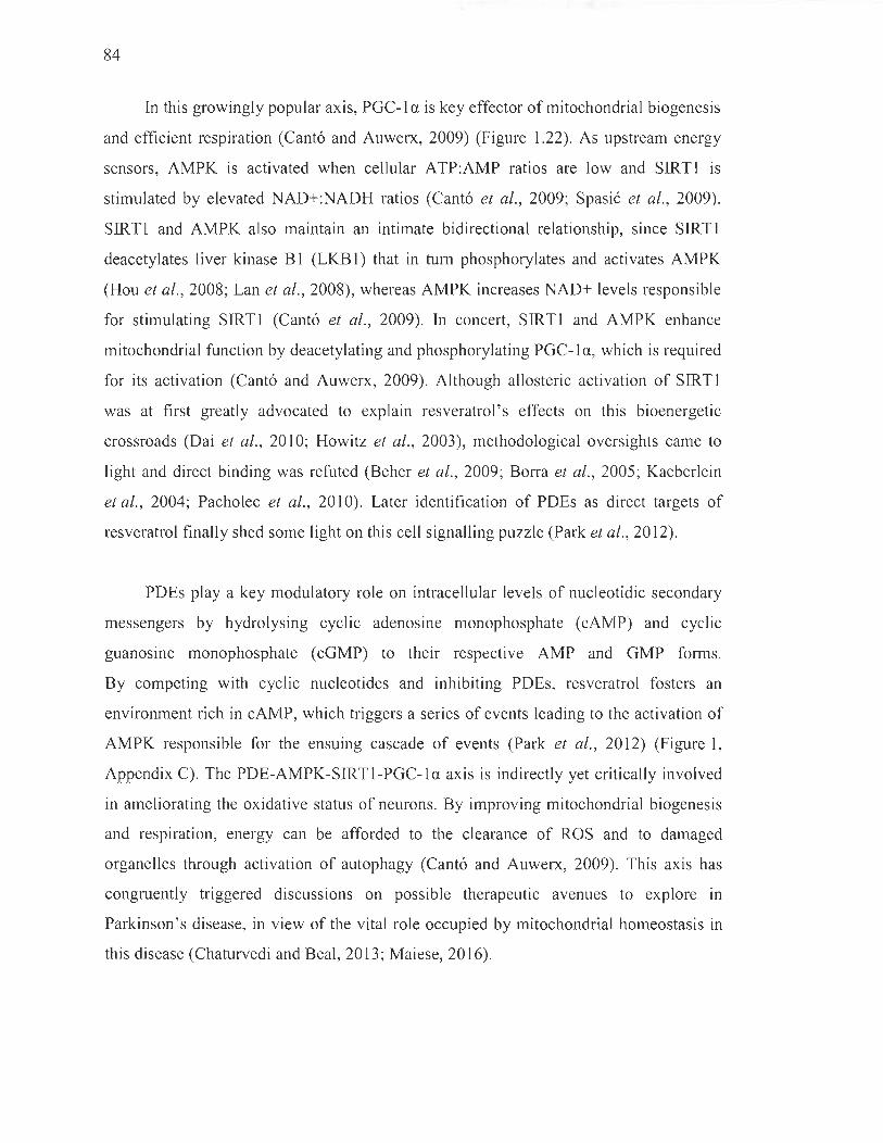
84
In this growingly popular axis, PGC-1 a is key effector of mitochondrial biogenesis
and efficient respiration (Canto and Auwerx, 2009) (Figure 1.22). As upstrearn energy
sensors, AMPK is activated when cellular ATP:AMP ratios are low and SIRTI is
stirnulated by elevated NAD+:NADH ratios (Canto et al., 2009; Spasié et al. , 2009).
SIRT1 and AMPK also maintain an intirnate bidirectional relationship, since SIRTI
deacetylates liver kinase BI (LKB1) that in turn phosphorylates and activates AMPK
(Hou et al., 2008; Lan et al., 2008), whereas AMPK increases NAD+ leve ls responsible
for stirnulating SIRT1 (Canto et al., 2009). In concert, SIRT1 and AMPK enhance
rnitochondrial function by deacetylating and phosphorylating PGC-l a, which is required
for its activation (Canto and Auwerx, 2009). Although allosteric activation of SIRT1
was at first greatly advocated to explain resveratrol's effects on this bioenergetic
crossroads (Dai et al. , 2010; Howitz et al., 2003), methodological oversights came to
light and direct binding was refuted (Beher et al. , 2009; Borra et al. , 2005; Kaeberlein
et al., 2004; Pacholec et al., 2010). Later identification of PDEs as direct targets of
resveratrol finally shed sorne light on this cell signalling puzzle (Park et al., 2012).
PDEs play a key rnodulatory role on intracellular levels of nucleotidic secondary
rnessengers by hydrolysing cyclic adenosine monophosphate (cAMP) and cyclic
guanosine monophosphate (cGMP) to their respective AMP and GMP forms.
By competing with cyclic nucleotides and inhibiting PDEs, resveratrol fosters an
environment rich in cAMP, which triggers a series of events leading to the activation of
AMPK responsible for the ensuing cascade of events (Park et al., 20 12) (Figure 1,
Appendix C). The PDE-AMPK-SIRTI-PGC-la axis is indirectly yet critically involved
in arneliorating the oxidative status of neurons. By improving mitochondrial biogenesis
and respiration, energy can be afforded to the clearance of ROS and to damaged
organelles through activation of autophagy (Canto and Auwerx, 2009). This axis has
congruently triggered discussions on possible therapeutic avenues to explore in
Parkinson's disease, in view of the vital role occupied by mitochondrial homeostasis in
this disease (Chaturvedi and BeaI, 2013; Maiese, 2016).

CALORIE RESTRICTION, FASTING, EXERCISE
~ ----t l Glucagon . High-fat diet Catecholamine
!~00~ " , , , , , , , , ,
~ Q MPV ·, "'"
Resveratrol
, , ~ "" '~
"tNAD+ "... ~ .,' , , , '- , ~ .- '(
OtherNAD+- ~~ " dependent enzymes ,,'~'- '~
: ,',' ŒGC-~
.l.Genome~nstabilitY ~", /
.1. Triglyceride synthesis
.1. Glycolysis
.1. ROS
.1. Inflammation
1
"t Fatty acid oxidation "t Gluconeogenesis
l Mitochondrial biogenesis and respiration
METABOLIC AND AGING-RELATED DISEASE?
Figure 1.22 Molecular mainstays of metabolic homeostasis.
85
By sensing levels of cyclic adenosine monophosphate (cAMP) and NAD+, and by acting on each other, this highly ramified pathway promotes equilibrium in instances of metabolic deficits. Moreover, they protect cells against a slew of noxious molecular events and are thought to mediate the beneficial effects of calorie restriction. AMPK, adenosine monophosphate kinase; CamKK~, calcium/calmodulindependent prote in kinase kinase 2 or beta; Epac l , exchange factor directly activated by cyclic adenosine monophosphate 1; PGC-l a, peroxisome proliferator-activated receptor gamma coactivator l-alpha; PKA, protein kinase A. (From Tennen et al., 2012.)
1.3.3.3 Mammalian target ofrapamycin and autophagy
Autophagy is an intricately structured cascade of events that promotes the
equilibrium between clearance and production of organelles and proteins. Per se,

86
autophagy degrades non-essential or damaged cellular components and is therefore
activated in settings of energy depletion and oxidative stress. Moreover, its role in
Parkinson's disease has been evoked time and again, as it is a process required to clear
misfolded proteins, pathogenic fibrils and full-fledged aggregates expressed in
a-synuclein pathology (Hashimoto et al., 2003; Schapira and Gegg, 20 Il; Schneider and
Zhang, 2010).
Focal to autophagy is mTOR, an inhibitor of the initial stages of this process,
which is of capital importance in tissues disposing of meagre regenerative capacities,
like the CNS (Lee et al., 2012). Akin to AMPK and SIRT1, mTOR is an energy sensor,
but it is activated in instances of energy and nutrient abundance, thus occupying a
central position in bridging energy levels and autophagy (Kundu, 2011) (Figure l.23).
mTOR phosphorylates the initial effector of autophagy, autophagy-related protein 13
(Atg13), thereby obstructing the onset of autophagy (Chan et al., 2009). However,
mTOR is repressed both by AMPK activation (Boister et al., 2002) and oxidative
modifications (Dames et al., 2005), in which context autophagy is accordingly activated
to restore cellular homeostasis. mTOR therefore collaborates narrowly with the
PDE-AMPK-SIRT 1-PGC-l a axis to alleviate oxidative stress and restore favourable
bioenergetic dynamics.
Resveratrol was recently shown to bind and inhibit mTOR by competing with
A TP, thereby promoting autophagy in proper cellular contexts (Park et al., 2016).
Correspondingly, previous reports demonstrating resveratrol 's neuroprotective capacities
in dopaminergic neurons challenged with rotenone concurrently found it to stimulate the
autophagie flux (Lin et al., 2014; Wu et al., 2011). Moreover, suppression of the master
metabolic regulator AMPK dampened these beneficial effects, which bolsters the
pertinence of bioenergetics-autophagy crosstalk in the neuroprotection afforded by
resveratrol against oxidative stress (Wu et al., 2011).
Energy Starvation
NUTRIENT HOMEOSTASIS
Figure 1.23 Metabolic regulation of cell growth and autophagy dynamics.
87
A tri ad of kinases senses nutrient and energy availability, acting in concert to promote cellular homeostasis by modulating cell growth and autophagy. The central bioenergetic power player, AMPK, can have opposite effects in the cell depending on the ATP:adenosine monophosphate (AMP) ratio. ln a replete, high ATP setting, AMPK will activate the tuberous sclerosis complex (TSC) duo that inhibits mammalian target of rapamycin complex 1 (mTORCl), a complex constituted of mammalian target of rapamycin (mTOR) and its regulators, thereby promoting ceU growth and silencing autophagic pathways. ln circurnstances of energy depletion, AMPK will rather activate Unc-51 like autophagy activating kinase (ULKl), which wiU stimulate autophagy and inhibit cell growth. This toggle is highly sensitive and meticulously regulated by feedback loops between the different components of this triad. Atg13, autophagy-related protein 13; FIP200, retinoblastoma l-inducible coiled-coil protein 1; LKB 1, liver kinase BI; Lst8, mTORC subunit lethal with SEC13 protein 8. (From Dunlop and Tee, 2013.)
1.3.3.4 Other targets
While focus has only been granted to three of numerous putative direct targets of
resveratrol, other binding partners may likely play an indirect though significant role in
conveying this polyphenol's antioxidative effects. Worthy of mention is a ho st of

88
important players in the arena of inflammation, for instance cyclooxygenases-l and
-2 (Calamini et al., 2010; Murias et al., 2004; Zykova et al., 2008), leukotriene 4A
hydrolase (Oi et al., 2010) and estrogen receptors (Bowers et al., 2000; Nwachukwu
et al. , 2014), which have been reviewed elsewhere with respect to resveratrol's anti
inflammatory potential (Appendix C). Neuroinflammation is greatly acknowledged to
actively participate in the etiopathogenesis ofParkinson's disease and is interwoven with
oxidative processes, as suggested by microglia-derived oxidative bursts and oxidative
stress-induced microglial activation (Langston et al., 1999; More et al. , 2013; Russo
et al., 2014; Zecca et al., 2008). As such, resveratrol's antioxidative effects in the CNS
of rodents and humans may also be conveyed via impeding pro-oxidative enzymes in
microglial cells that may injure neurons in the substantia nigra pars compacta, su ch as
myeloperoxidase and NADPH oxidase (Chang et al., 2013).
Ali evidence considered, its capacity to modulate a wide array of proteic activities
affords resveratrol tangible pathway-modifying properties that converge toward the
prevention of oxidative stress in dopaminergic neurons. Although mostly tried in
parkinsonian paradigms or in non-neuronal diabetic models, the literature grants a
credible role for resveratrol in protecting cultured dopaminergic neurons against high
glucose conditions.
1.4 Research aims and hypotheses
Provided the pre mIse that dopaminergic neurons of the nigrostriatal pathway
are relatively more susceptible to insults that overwhelm their endogenous coping
mechanisms in Parkinson's disease, it is reasonable to surmise that other sources of
stress, not specific to this pathology, may lead to their death, thereby shoring up this
concept. As already mentioned, Parkinson's disease occurs more frequently in patients
suffering from pathologies featuring a generalized state of oxidative stress, su ch as
diabetes (Cereda et al., 2012; Santiago and Potashkin, 2013; Sun et al., 2012).
Although awareness of these pathological associations has been growing, the selective

89
vulnerability of nigrostriatal dopaminergic neurons in the se settings has never been the
object of investigations.
In this respect, the core work of my thesis addresses the following central
hypothesis: nigrostriatal dopaminergic neurons are more vulnerable to hyperglycaemic
conditions compared to other neuronal populations, expressly the mesocorticolimbic
pathway.
Accordingly, we elaborated a senes of experiments employing dopaminergic
neuronal cultures in high glucose conditions as well as a rat model of hyperglycaemia.
We assessed the degeneration of dopaminergic neurons in vitro and in vivo, and
described the ensuing behavioural alterations in rats. By offsetting the production of
ROS using a well-known antioxidant, resveratrol, we also verified the role of high
glucose-induced oxidative stress in the death of dopaminergic neuronal cultures.
1.4.1 Objective 1: Evaluate the degeneration of cultured dopaminergic neuronal cells in high glucose conditions
We first tested whether dopaminergic neurons in culture undergo degeneration in
sustained high glucose conditions. We employed an in vitro model of dopaminergic
neurons cultured in elevated though physiologically plausible concentrations of glucose.
The production of ROS was measured and dopaminergic neuronal cell death was
assessed. We specifically quantified the superoxide anion radical, as it is thought to
constitute the initial toxic species overproduced in hyperglycaemia and liable for the
cascade of oxidative events leading up to cellular injuries (Brownlee, 2005).
Next, we investigated the mechanisms by which dopaminergic neuronal cells degenerate
in high glucose conditions, focusing on the apoptotic cascade. Particular indices of
apoptosis were examined, expressly the terminal events occurring at the level of the
DNA supported by changes in the expression of various proteins involved in the process.

90
1.4.2 Objective 2: Determine the potential of the antioxidative polyphenol resveratrol to hamper the high glucose-induced degeneration of cultured dopaminergic neuronal cells
Alongside objective 1, we employed a strategy aimed at moderating oxidative
stress to demonstrate its role in dopaminergic neurodegeneration. In this regard,
we selected the stilbene resveratrol whose protective competences were extensively
studied by virtue of its antioxidative potential. In line with objective 1, we verified the
ability of resveratrol to prevent dopaminergic neuronal cell death, diminish superoxide
anion radical production, and impede the apoptotic cascade.
Results for objectives 1 and 2 are reported in the article entitled "Resveratrol
protects dopaminergic PC12 cells against high glucose-induced oxidative stress and
apoptosis: effect on p53 and glucose-regulated protein 75 colocalization" published in
November 2013 in the peer-reviewedjournal Neurotoxicity Research (see Chapter II).
1.4.3 Objective 3: Characterize dopaminergic neurodegeneration in a rat model of long-term hyperglycaemia
On the grounds of results acquired in vitro, the preferential degeneration of the
nigrostriatal pathway compared to the mesocorticolimbic pathway was next assessed
in vivo in a well-established rat model of hyperglycaemia induced by a toxin that targets
insulin-producing pancreatic ~ cells. Neurodegeneration assessments were performed
following long-term hyperglycaemia, allowing the graduai instatement and maintenance
of oxidative stress required to overwhelm susceptible targets. Accordingly, we measured
glucose concentrations at source and terminal regions of the nigrostriatal and
mesocorticolimbic pathways to inquire whether ail areas were equally exposed to
circulating glucose. By immunohistochemical and immunoblotting methods,
we appraised neurodegeneration in both pathways as weil as in the hippocampus for
comparative means. Dopamine was measured at the terminais of these neurocircuits to
corroborate observations of neurodegeneration in the midbrain. We also assessed the
fate of glial cells in these regions to gain a better understanding of the effects of long
term hyperglycaemia in the substantia nigra pars compacta of rats.
91
1.4.4 Objective 4: Assess the behavioural alterations resulting from nigrostriatal neurodegeneration in a rat model of long-term hyperglycaemia
Degeneration of neurocircuits involved in the regulation of behaviours, as is the
case for the nigrostriatal and mesocorticolimbic pathways, often leads to measurable
behavioural alterations. Although both the nigrostriatal and mesocorticolimbic pathways
fulfil highly complex mandates that can in sorne ways overlap, it remains that the former
specializes in regulating motor behaviours. In keeping with our hypothesis, motor
behavioural assessments were performed in the same subjects as in objective 3 to
determine whether observed nigrostriatal neurodegeneration may lead to discernable
motor deficits. Rats executed motor tasks typically employed in models of Parkinson 's
disease. To comparative ends, a cognitive novel object recognition test was performed to
evaluate non-spatial working memory regulated by the hippocampus (Bast, 2007;
Bast et al., 2009), but also involving the mesocorticolimbic components nucleus
accumbens (Annett et al., 1989) and prefrontal cortex (Akirav and Maroun, 2006).
Insights of these behavioural tests in conjunction with the results pertaining to
objective 3 are reported in the article entitled "Dopaminergic neurodegeneration in a
rat model of long-term hyperglycemia: preferential degeneration of the nigrostriatal
motor pathway" published online in May 2018 and issued in September 2018 in the
peer-reviewed journal Neurobiology of Aging (see Chapter III).
Given the role of dopamine in social reward, social cognition and rough-and
tumble play processes (Narvaes and Martins de Almeida, 2014; Plavén-Sigray et al.,
2014; Trezza et al., 2010), we next investigated whether nigrostriatal degeneration
disrupts these behaviours in our rat model of long-term hyperglycaemia. In order to
evaluate social behaviour, interactions between unacquainted conspecifics and emissions
of ultrasonic vocalizations (USVs) were recorded simultaneously. USVs inform on the
emotional state of rats during social contexts such as mating, play or aggression
(Burgdorf et al., 2008) and are largely regulated by dopamine (Brudzynski, 2009).

92
Complementary to the evaluation of motor deficits, these inquiries into the social
behaviours of our model further extend our comprehension of the various ways that
dopaminergic neurodegeneration may manifest itself. These specific results are reported
in the article entitled "Long-term hyperglycaemia modifies social behaviour and
emission of ultrasonic vocalisations in rats: a possible experimental model of altered
sociability in diabetes" (see Chapter IV). The manuscript was submitted to the peer
reviewed journal Scientific Reports on July 9th, 2018.
1.5 Methodology
1.5.1 Objective 1: Evaluate the degeneration of cultured dopaminergic neuronal cells in high glucose conditions
1.5.1.1 Cell culture
To elucidate the possible neurodegenerative effects of a hyperglycaemic state
on dopaminergic neurons in vitro, we employed the well-established model of
dopaminergic neurons, the NGF-differentiated pheochromocytoma (PCI2) cell line.
These cells were initially derived from a neuroendocrine turnour in the adrenal medulla
of a rat (Greene and Tischler, 1976), and as such they can be subcultured almost
indeterminately. PC12 cells both synthesize and store catecholamines, principally
dopamine, in large dense-core vesicles that are released upon depolarization (Greene and
Rein, 1977; Greene and Tischler, 1976). PCI2 cells share embryonic origins with
neuroblastic cells. Thus, they can be differentiated into neuron-like cells with NGF
treatments that phosphorylate and activate the tropomyosin receptor kinase A (Huang
and Reichardt, 2003). Differentiation to a neuronal phenotype is enhanced by low serum
concentrations (Yung et al., 2010). Ensuing signal transduction abrogates cell division
and enhances the activity of the rate-limiting enzyme in dopamine synthesis,
TH (Schubert et al., 1980), as weIl as the outgrowth of neurites and the expression of
neurofilaments (Greene and Tischler, 1976; Lee et al., 1982). The PC12 cell line also
expresses dopamine transport and metabolic machinery, such as DAT, VMATI

93
(preferentially expressed in adrenal glands, as opposed to neuronal VMAT2) and
monoamine oxidase (Kadota et al., 1996; Liu et al., 1994; Youdim et al., 1986).
Figure 1.24 Characterization of dopaminergic neuronal cell cultures. Top: From left to right are shown naïve pheochromocytoma (PCI2) cells and 3-, 5-, 7-day nerve growth factor (NGF)-differentiated neuronal PC12 cells. Bottom: Immunofluorescent assessment of the expression of neuronal and dopaminergic markers. From left to right: 7 -day differentiated neuronal PC12 cells marked for TH (green), neurofilaments (red) and nuclei (blue); dopaminergic N27 cells with Hoechst-stained nuclei marked for neurofilaments (yellow), DAT (green), and TH (red).
In this respect, our group employs 5-1O-day NGF-differentiated neuronal PC12
cells (50 ng/mL) cultured in low concentrations of fetal bovine serum (1%) (Achour
et al., 2016; Bournival et al., 2012a, 2012b; Renaud et al., 2014). Figure 1.24 displays
representative microphotographs of naïve (undifferentiated) PC12 cells and neuronal
PC12 cells cultured in our laboratory. Pertinent to the present project, the y also express
markers necessary for a high glucose context, expressly insulin-independent GLUT347
(Mah~r et al., 1991).
47 PC12 cells also express insulin-dependent GLUT4 (Hudson et al. , 1993), as weil as insulin-independent GLUTl and GLUT8 (Maher et al. , 1991), although the latter dwells in intracellular compartrnents and has not yet been found to translocate to the cell surface (Widmer et al. , 2005). Given the high affinity and capacity of GLUT3 compared to ail other GLUTs (Maher et al., 1996), it is the most relevant glucose transporter in our model in the absence of insulin. Accordingly, midbrain dopaminergic neurons express GLUT3 and only very little GLUT4, at least in the rat brain (El Messari et al., 1998; Maher et al., 1991).
94
1.5.1.2 High glucose conditions
High glucose conditions in vitro alm to emulate the increase in extracellular
glucose concentrations that takes place in hyperglycaemia. Depending on the culture
medium, typical glucose concentrations range between 5.5 mM (1 g/L) to 17.5 mM
(3.5 g/L). Taking into consideration the normal concentrations at which neurons are
cultured, careful optimization of D-glucose concentrations and treatment times are
required to achieve a faithful model of a physiologically hyperglycaemic state in cell
cultures. In this respect, undiagnosed, untreated or uncontrolled diabetes can lead to a
ri se in glycaemia reaching 25 mM (450 mg/dL), qualified as severe hyperglycaemia
(Amblee et al., 2016; Marchese et al., 2017; Saul et al., 2016). In rats exhibiting a
glycaemia of 30 mM48, neurons are predicted to be exposed to ~9 mM of glucose,
while at normoglycaemic values of 6 mM, they are exposed to 1.2 mM (Simpson et al.,
2007) (Table 1.7). This represents a 7.5-fold increase between normal and severe
hyperglycaemic conditions. Since PC12 cells are normally cultivated in I l mM of
glucose, we tested the following concentrations within this physiological 7.5-foid
increase in dose-response and time course studies: 25, 50 and 75 mM (Figure 1.25).
Seeing as we were aiming for a moderate cytotoxic effect averagmg 30-50%
(or 50-70% viability) with the lowest dose of D-glucose, results of these kinetic
dose-response studies allowed for the selection of a concentration of 25 mM (4.5 g/L)
administered for a duration of 96 h in neuronal PC 12 cells. We also tested the effect of
D-glucose on naïve PC 12 cells and irnrnortalized fetal mesencephalic dopaminergic
N27 cells. Akin to PC12 cells, the latter secrete dopamine in addition to expressing the
principal dopaminergic neuronal proteins, namely TH, DAT, nestin, neurofilaments and
a neuron-specific enolate (Adams et al., 1996; Prasad et al., 1994), also characterized in
our laboratory (Figure 1.24). These additional kinetic dose-response investigations ruled
out the use of naïve PC12 cells, in light oftheir proliferative response, at least at 25 mM.
These data also confirm that dopaminergic neuronal cells are vulnerable to high glucose
48 We williater see that this glycaemia was indeed sustainable for 6 months in our rat mode\.

95
conditions, even when these are rather moderate (25 mM), strengthened by experiment
repetitions in dopaminergic N27 cells.
A g-150 B c 8 o 100 ~ ~
~ :ë 50 ro .S;
Qi Ü
0 0 24 48 72 96 120
Treatment time (h)
0 80 E
~60 l.~.:.i ~ .c 1
~ 40 1
/ ,, '
1 •• •• -0 ,. Qi
f
ü 20
0 0 24 48 72 96 120
Treatment time (h)
::::- 150 e ë 0 u o 100 ~ 0
~ :ë 50 ro .S;
Qi Ü
0 0 24 48 72 96
Treatment time (h)
80
~60 ~ .c
~ 40 -0
Qi ü 20
0 0 24 48 72 96
Treatment lime (h)
120
120
C ::::-150 e ë 8
- 25 mM diffPC12 -~_. 50 mM diff PC12 ... ~ ... 75 mM diff PC12 - 25 mM naïve PC12 -~_. 50 mM naïve PC12 ...• ... 75 mM naïve PC12 - 25mM N27 - 4- · 50 mM N27 ... •.. . 75 mM N27
o 100.&.-""F-.:-::-jf'-.-.-<l
~
.. 0+-..... .....,,.....,.. ............
F 80
~60 ~ .c
~ 40 -0
Qi ü 20
o 24 48 72 96 120 Treatment time (h)
o~~--~ ...... --r-~ o 24 48 72 96 120
Treatment lime (h)
Figure 1.25 Time-course and dose-response study of the toxicity of glucose on dopaminergic cells in culture. Cells were cultured in high glucose conditions ranging between 25-75 mM for 0-120 h and cell viability (top A-C) or cell death (bottom D-F) were measured by MTT assay and lactate dehydrogenase quantification, respectively. Differentiated neuronal PCI2 ceIls (red lines) demonstrated a greater vulnerability th an naïve ceIls (blue lines). Dopaminergic N27 ceIls exhibited intermediate susceptibility (green line). There seems to be a slight proliferative effect for non-differentiated ceIls (B, C), likely owing to the fact that these populations were not in a post-mitotic state: glucose may have stimulated ceIl growth at low concentrations. This effect was lost at the highest dose. The chosen concentration, 25 mM, is portrayed as a bold !ine.
As with any ceIl line, the PCI2 model presents limitations. In this regard,
we performed tests in a rat model of hyperglycaemia, detailed later. Nonetheless, among

96
the vanous utilizations of the PC12 cell line, our paradigrn is advantaged by the
differentiation of the cells to a clear neuron-like phenotype with pronounced expression
of dopaminergic markers and cessation of division. In contrast, most other publications
utilizing PC12 cells do not differentiate them, implying a tumoural phenotype and
tapered dopaminergic properties, or use much higher concentrations of D-glucose that
are not physiologically explainable (75-300 mM)49 (Aminzadeh, 2017; Fouda and
Abdel-Rahman, 2017; Rayegan et al. , 2017; Song et al. , 2017; Wang et al. , 2017; Zhao
et al., 2015). Additionally, naïve and NGF-treated PC12 cells are differentially
susceptible to oxidative stress particularly caused by methylglyoxal, a by-product of
sustained exposure to high glucose concentrations (Okouchi et al., 2005). Differences in
cellular redox states, specifically regarding the reduced glutathione-to-oxidized
glutathione disulphide ratio, are likely accountable for these distinct responses.
1.5.1.3 Superoxide anion quantification
The primary goal of this part of the thesis was to verify that oxidative stress likely
underpins the death of dopaminergic neuronal cells cultured in high glucose conditions.
We measured the initial oxidative event that is believed to occur early in response to a
glucose overload, that is, the overproduction of superoxide anion (Brownlee, 2005).
Quantification of superoxide anion at the level of mitochondria is possible using a
modified hydroethidine superoxide anion fluorogenic probe marketed under the name
MitoSOXTM (Robinson et al., 2006) (Figure 1.26). Hydroethidine is a reduced form of
ethidium (3,8-diamino-5-ethyl-6-phenylphenanthridinium), shown to undergo oxidation
by potassium superoxide into a red fluorescent molecule (Rothe and Valet, 1990).
Since its discovery, hydroethidine has been widely used as an intracellular superoxide
anion detector (Perticarari et al., 1994; Rothe and Valet, 1990; Tarpey et al., 2004).
The modified hydroethidine molecule is conjugated with a triphenylphosphonium
moiety that steers molecules toward mitochondria. The three lipophilic phenyl groups
49 Such high concentrations of any solute, in fact, constitute a risk for noxious hypertonie effects that can be mistaken for the toxicity of the molecule per se. Although we remained within a reasonable concentration range, our work also provides D-mannitol control s, scarcely metabolized by mammalian cells, to rule out the possibility ofa toxic osmotic effect in our mode!.

97
facilitate movement across membranes of live cells and the positively charged
phosphonium group further favours accumulation in the mitochondrial matrix endowed
with a negative membrane potential. As such, MitoSOXTM almost exclusively reacts
with mitochondrial superoxide anion (Robinson et al. , 2006) (Figure 1.27).
H2 ~ li ~!J NH2 2-hydroxy-5-- \ JJ=\1 (triphenylphosphonium)
- (CH2)e- PNJ hexylethidium ~ li 3 590 nm
Figure 1.26 Structure and fluorescent mechanism of MitoSOXTM Red. Vpon reacting with superoxide anion, MitoSOXTM adopts a more highly conjugated structure enabling it to produce red fluorescence when it is excited at a wavelength of 510 nm. We can also appreciate the hydroethidine structure to the left and the positively charged triphenylphosphonium responsible for driving MitoSOXTM to mitochondria.
Granted superoxide anion overproduction in response to stress is a very early event
in cell cultures (Carange et al. , 2011; Ronson et al. , 1999), we performed time course
studies and identified 3 h as the optimal incubation time when to perform measurements
in high glucose conditions. We have also previously shown that levels of nitrates and
nitrites, reactive nitrogen species arising from secondary reactions between superoxide
anion and nitric oxide (Pacher et al. , 2007), are increased at a later time point in our
model (Boumival et al., 2012a). In the article presented in Chapter II, the use of
diethyldithiocarbamate, a selective inhibitor of SOD causing the accumulation of
superoxide anion (Khazaei et al. , 2009; Puglia and Loeb, 1984), substantiated the
specificity of MitoSOXTM in our paradigm.

98
10,000 1.6
Ê 1.4 ID c 1.2 ()
1,000 co c: ~ 1.0 ID () QI 0.8 U) 0
ID c t1I 0.6 L.. -e 0 100 0 0.4
::J fi)
c;:: .c 0.2 <t:
ID >
+=# Sodium Nitric Peroxy-CO nitrite oxide nitrite ID 10
Cl::
Figure 1.27 Selectivity of MitoSOXTM Red. The speeifieity of two ROS probes, dihydrorhodamine 123 (white bars) and MitoSOXTM Red (black bars), were tested on a variety of speeies. While dihydrorhodamine 123 was not specifie for any partieular ROS, MitoSOXTM Red more readily seavenged superoxide anion, as evideneed by measures of fluorescence. It remains that this probe emits a weaker signal than dihydrorhodamine 123. The inset graph with grey bars displays Griess nitrite determination results, as a control for the detection of reactive nitrogen species. H202, hydrogen peroxide; HRP, horseradish peroxidase. (From the product information sheet provided by Molecular Probes, 2015.)
1.5.1.4 Evaluation of ap op to tic death
Apoptosis is a key contributor to the degeneration of neurons in Parkinson 's
disease50 (Anglade et al., 1997; Moehizuki et al., 1996; Toulorge et al., 2016) as weB as
in diabetes (Maiese et al., 2007; Russell et al., 1999). Alongside autophagie death,
neeroptosis, ferroptosis, anoikis and many others, apoptosis belongs to a family of
programmed cell death processes that occur in multicellular organisms (Ke et al., 2016)
50 Several apoptotic hall marks were revealed in post-mortem substantia nigra pars compacta samples of parkinsonian patients, for instance chromatin condensation and DNA fragmentation (Tatton, 2000), p53 and Bax overexpression (Mogi et al. , 2007; Tatton, 2000), and elevated caspase-3 activity (Mogi et al. , 2000; Tatton, 2000).

99
(Figure 1.28). Cellular apoptosis is a profitable course of death for an organism owing to
its tight regulation and to the cleanliness of the process that does not induce overt
inflammation, as opposed to necrosis (Alberts et al., 2008). The importance of apoptosis
becomes aIl the more apparent in cancer, wherein its pathological impairment is at the
basis of several forms of the disease (Evan and Vousden, 2001; Green and Kroemer,
2009). However, excessive apoptosis, due to lax cellular control or elevated death
signais in the immediate environment, is also cause for pathology especially in tissues
with scant regenerative potential such as the CNS (Bredesen et al. , 2006).
Extrinsic pathway
Apo2ljTRAIL
DISC
Chemotherapy Radiotherapy
DNAdamage
• @
• [ Puma, Noxa 1
l [ BeI-2, Bel-XL. Mel-l i
l
Growth factor receptors
1 ® PI3K I
l @---+ [ Bax, Bak l .... 4~--- @)
/ Intrin." • pathway
Caspase 8.10
Figure 1.28 The classic intrinsic and extrinsic pathways in apoptosis. Continued on next page.
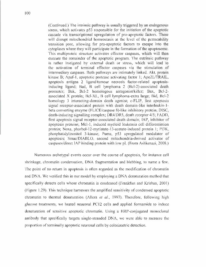
100
(Continued.) The intrinsic pathway is usually triggered by an endogenous stress, which activates p53 responsible for the initiation of the apoptotic cascade via transcriptional upregulation of pro-apoptotic factors. These will disrupt mitochondrial homeostasis at the level of the permeability transition pore, allowing for pro-apoptotic factors to escape into the cytoplasm where they will participate in the formation of the apoptosome. This multiproteic structure activates effector caspases, which will then execute the remainder of the apoptotic program. The extrinsic pathway is rather instigated by external death or stress, which will lead to the activation of terminal effector caspases via the stimulation of intermediary caspases. Both pathways are intimately linked. Akt, protein kinase B; Apaf-l , apoptotic protease activating factor 1; Ap02L/TRAIL, apoptosis antigen 2 ligand/tumour necrosis factor-related apoptosisinducing ligand; Bad, B cell lymphoma 2 (Bcl-2)-associated death promoter; Bak, Bcl-2 homologous antagonist/killer; Bax, Bcl-2-associated X protein; Bcl-XL, B cell lymphoma-extra large; Bid, Bcl-2 homology 3 interacting-domain death agonist; c-FLIP, first apoptosis signal receptor-associated protein with death domain-like interleukin-lbeta converting enzyme (FLICE/caspase 8)-like inhibitory protein; DISC, death-inducing signalling complex; DR4/DR5, death receptor 4/5; F ADD, first apoptosis signal receptor-associated death domain; IAP, inhibitor of apoptosis proteins; Mcl-l, induced myeloid leukemia cell differentiation protein; Noxa, phorbol-12-myristate-13-acetate-induced prote in 1; PI3K, phosphatidylinositol 3-kinase; Puma, p53 upregulated modulator of apoptosis; Smac/DIABLO, second mitochondria-derived activator of caspases/direct IAP binding protein with low pl. (From Ashkenazi, 2008.)
Numerous archetypal events occur over the course of apoptosis, for instance cell
shrinkage, chromatin condensation, DNA fragmentation and blebbing, to name a few.
The point of no retum in apoptosis is often regarded as the modification of chromatin
and DNA. We verified this in our model by employing a DNA denaturation method that
specifically detects cells whose chromatin is condensed (Frankfurt and Krishan, 2001)
(Figure 1.29). This technique harnesses the amplified sensitivity of condensed apoptotic
chromatin to thermal denaturation (Allera et al., 1997). Therefore, following high
glucose treatments, we heated neuronal PC12 cells and applied formamide to induce
denaturation of sensitive apoptotic chromatin. Using a HRP-conjugated monoclonal
antibody that specifically targets single-stranded DNA, we were able to measure the
proportion of terminally apoptotic neuronal cells by colorimetric detection.

101
500
q; -.- APOPTOTIC CEllS ~ CONTROL CEllS ...J 400 W ___ SODIUM AZIDE
Z ~ HYPERTHERMIA Z oC( -.- HYDROGEN PEROXIDE ~ 300 ~ SAPONIN ~ W 0 Z 200 w 0 ln W a: 0 100 :::J ...J IL C)
0 0 ...J
30 40 50 60 70 80 90 100
TEMPERATURE (DEGREES C)
Figure 1.29 Specificity with respect to temperature kinetics of formamideinduced DNA denaturation. Formamide was applied in cells submitted to several kinds of treatments described in the box. DNA denaturation was quantified over the course of rising temperatures. Apoptotic cells begin to exhibit DNA denaturation at manifestly cooler temperatures compared to other non-apoptotic cells, which corroborates the specificity of this test, performed at 70°C. (From Frankfurt and Krishan, 2001.)
To bolster our findings , we also verified other events that accompany the major
phenotypic changes that occur during apoptosis, mainly modifications in the expression,
subcellular localization or post-translational alterations of proteins. Activation of the
caspase cascade, a sequence of proteic cleavage events controlled by caspase proteases,
is a hallmark of apoptosis (Sakahira et al. , 1998). Caspase-3 is the terminal effector of
this cascade that, upon cleavage, is responsible for the activation of a DNA
fragmentation enzyme, caspase-activated deoxyribonuclease. Therefore, we verified
terminal caspase activation and DNA fragmentation in our model, by employing
immunofluorescence for cleaved caspase-3 labeling combined with the terminal
deoxynucleotidyl transferase dUTP nick end labeling (TUNEL) method to isolate cells
displaying DNA fragmentation. To ensure the exclusive counting of apoptotic cells,
we only identified ones that exhibited both fluorescent signais. The specificity of our
results was also substantiated by the use of an irreversible inhibitor of cleaved caspase-3 ,

102
the fluormethy1ketone-conjugated tetrapeptide Z-DEVD-FMK, expected to yie1d cells
with very little or no TUNEL signal.
Next, we evaluated the expressIOn of proteins upstream or downstream from
caspase-3 by immunoblotting. Upstream at the level of the mitochondria, one of the
pro-apoptotic member of the Bcl-2 family, Bax, spearheads the formation of the
mitochondrial permeability transition pore responsible for the cytosolic release of
apoptogenic molecules, such as cytochrome c (Gollapudi et al. , 2003). Bcl-2, an anti
apoptotic member of the Bcl-2 family, interacts with Bax to prevent permeabilization
(Renault et al. , 2013) (Figure 1.30). It follows that an elevated Bax to Bcl-2 expression
ratio is a faithful indicator that apoptosis is on-going (Cory and Adams, 2002).
At the level of the nucleus, the DNA repair enzyme P ARP constitutes a downstream
target of caspase-3 and its cleavage in 24 and 89 kDa fragments is a recognized feature
of apoptosis (Kaufmann et al., 1993). Thus, a low ratio of full-Iength-to-cleaved PARP
constitutes a marker of apoptosis. Farther upstream of caspase activation, the tumour
suppressor p53 was also assessed in light of its role in evoking apoptosis and sensing
oxidative stress. Indeed, p53 is stabilized by oxidative stress and localizes to the nucleus
where it induces the transcription of pro-apoptotic factors (Lee et al., 2008; Macip et al.,
2003; Nair, 2006). Consequently, we studied the cytoplasmic and nuclear localization of
p53 in neuronal PC12 cells following high glucose treatments, a high nuclear-to
cytoplasmic ratio likely indicating its active promotion of apoptosis in our model.
Directly linked to this, we also verified by immunoblotting and
immunofluorescence the subcellular localization of a constitutively expressed chaperone
of the heat shock protein family, glucose-regulated protein 75 (GRP75), known to
sequester p53 in the cytoplasm, thereby preventing apoptosis in multiple models
(Kaul et al., 2005). Fluorescence colocalization analyses were performed to evaluate its
aptitude to bind p53 in the cytoplasm in conditions of oxidative stress. Normally, GRP75
acts as a mitochondrial chaperone and guardian against oxidative stress among a
plethora of other functions. However, its role under conditions of stress is highly
variable (Londono et al., 2012) (Figure 1.31). One of our aims was to define its role in
103
high glucose-induced oxidative stress due to its purported implication III
neurodegeneration. Indeed, GRP75 is depleted in the affected brain regions of
Parkinson's disease patients (Burbulla et al. , 2010; Jin et al. , 2006) and knocking out its
expression leads to neurodegeneration that can be rescued by the parkinsonian gene
product parkin (Yang et al., 2011). In addition, it is a well-recognized binding partner of
other genes mutated in Parkinson ' s disease, namely DJ-l and a-synuclein (Jin et al. ,
2006; Li et al., 2005) .
Bcl-2
inactive Bax
activator BH30
Bad
Figure 1.30 Mitochondrial translocation of Bax followed by the formation of the mitochondrial permeability transition pore. Bax is usually found in an inactive state (light blue) in the cytoplasm. When Bcl-2 homology 3 (BH3)-only proteins such as tBid, Bcl-2-like protein Il (Bim) or Puma are produced in response to an apoptosisinducing stress, these can activate Bax, rende ring it more liable to insert itself in the mitochondrial membrane leading to the generation of the permeability transition pore. Anti-apoptotic members of the Bcl-2 family, on the other hand, prevent Bax from inserting itself in the membrane. Considering the ratio of these proteins can offer insight into events occurring at the level of the mitochondrial membrane during apoptosis. p38MAPK, p38 mitogen-activated protein kinases. (From Renault et al., 2013.)

104
o morùlln
~ FGF-l
• p66Shc
pS3
MVDl
- Alds proteln refoldlng
- Gu.rdI.., ~.Inst oxldMive stress .nd .poptosis
- MetJlbolism .nd rnItochondri. homeostMis
- Intercelul.r trllfllcklng
7.FGF-1 .Â.... C..J -Regul.tlon of cell prollfer.tlon
Figure 1.31 The multiple roles of GRP75.
Unfolded polypeptide
MorUllnu cMperone
...
p21 modlflutlon
S.IL-1R
Glucose-regulated prote in 75 (GRP75 or mortalin) is primarily involved in the mitochondria, where it fulfils chaperone, import, and oxidationsensing functions. In the advent of cellular stress, GRP75 is recruited to the cytoplasm where it can regulate several processes, including apoptosis. By binding p53, it abrogates this transcription factor 's translocation to the nucleus where it usually perforrns pro-apoptotic operations. Nevertheless, much remains to be elucidated pertaining to its functions. FGF-l , fibroblast growth factor 1; Hsp, heat shock protein; IL-IR, interleukin-l receptor; MVD 1, diphosphomevalonate decarboxylase; p66Shc, 66 kDa proto-oncogene Src homologous-collagen homologue adaptor protein; VDAC, voltage-dependent anion channel. (From Londono et al., 2012.)
Further methodological details conceming the utilization of neuronal PC12 cells
cultured in 25 mM of D-glucose for 96 h, the measurement of superoxide anion at 3 h,
or the assessment of apoptosis in our model are provided in Chapter II.
105
1.5.2 Objective 2: Determine the potential of the antioxidative polyphenol resveratrol to hamper the high glucose-induced degeneration of cultured dopaminergic neuronal cells
1.5.2.1 Resveratrol treatments
In addition to measunng superoxide amon production in neuronal PC12 cells
treated with high glucose conditions, we employed an antioxidative strategy to verify the
importance of oxidative mechanisms in dopaminergic neurodegeneration in vitro .
The polyphenol resveratrol was chosen for its potent antioxidative capacities
demonstrated in various cellular (Karlsson et al., 2000; Savaskan et al., 2003; Wu et al. ,
2017; Zhuang et al., 2003) and animal models (Kiziltepe et al., 2004; Palle and Neerati,
2018; Sinha et al., 2002). In neuronal paradigms, resveratrol is best known for its ability
to enhance the activity of SOD, an enzyme that catalyzes superoxide anion
inactivationS! , both in vitro (Bai et al., 2013; Lee et al., 2012; Yuan et al., 2013) and
in vivo (Cheng et al. , 2014; Kesherwani et al., 2013; Nalagoni and Karnati, 2016;
Ren et al., 2011).
Very few studies have used resveratrol on neuronal PC12 cells, solely focusing on
demonstrating its pro-differentiation competences (Lecomte et al., 2017; Ma et al. ,
2014; Sugino et al., 2010). These projects established resveratrol's effects at
concentrations ranging from 0.5 )lM to 20 )lM. Most other applications of resveratrol on
cultured neurons typically provide its use at 1-120)lM (Calabrese et al., 2010; Jardim
et al., 2017). In our paradigm, resveratrol was administered at an optimized
concentration of 0.1 )lM, constituting the lowest dose that reliably protects our model
against various oxidative insults (Bournival et al., 2009; Bureau et al., 2008; Gélinas and
Martinoli, 2002). To verify the ability of resveratrol to modulate superoxide anion
concentrations in mitochondria, neuronal PC 12 cells were co-treated with the
polyphenol for the duration of high glucose treatments.
5\ In purified neuronal mitochondria, resveratrol also demonstrates direct scavenging properties for the superoxide anion (Zini et al., 1999, 2002). One group employed PC 12 cells challenged with high glucose concentrations to demonstrate its protective effects via the activation of the phosphatidylinositol 3-kinase/ Akt/forkhead box 03 (PI3K1 Akt/Fox03a) pathway (Liu et al., 2015). However, the authors used undifferentiated cells, which, as previously discussed, respond differently to oxidative insults (Okouchi et al., 2005).
106
The differentiation of PC12 cells with NGF is important, not only to yield a clear
neuronal phenotype, but also to prevent adverse effects from resveratrol treatments.
Indeed, resveratrol exhibits both protective (Agrawal et al. , 20 Il; Dasgupta and
Milbrandt, 2007; Della-Morte et al. , 2009) and toxic (Muqbil et al., 2012; Trincheri
et al., 2007; Wenner, 2012) properties depending on cellular contexts. In naïve PC1 2
cells, resveratrol is cytotoxic, whereas it promotes neurite outgrowth, mitochondria
renewal and energy balance in NGF-differentiated ones (Hayakawa et al., 2013).
For this reason, PC12 cells were differentiated for at least 5 days with NGF before high
glucose or resveratrol treatments were employed.
The application of polyphenols m cell cultures also reqUlres certain
methodological adjustments. One of these is to ensure the absence of other phenolic
substances in the cell culture medium to mIe out any effect not attributable to the
polyphenol treatrnent. To that end, ail sera used during experiments were charcoal
stripped to remove steroids, the principal phenolic compounds found in these ceU culture
additives (Boumival et al., 2009; Bureau et al., 2008; Cao et al., 2009; Gélinas and
Martinoli, 2002).
Further methodological information concernmg the use of resveratrol ln our
dopaminergic model is provided in Chapter II.
1.5.3 Objective 3: Characterize dopaminergic neurodegeneration in a rat model of long-term hyperglycaemia
1.5.3.1 Rat model oflong-term hyperglycaemia
Selection of an appropriate paradigm to simulate long-term hyperglycaemia in
rodents likely constituted the most critical aspect of the in vivo portion of this thesis.
Attempts to model diabetes and other metabolic diseases as working platforms to
develop treatments and elucidate pathological mechanisms have yielded today a slew of
weU-described paradigms (see for reviews Baxter and Duckworth, 2004; Islam and
Loots, 2009; Kim et al., 1998; Srinivasan and Ramarao, 2007; Van Belle et al., 2009).

107
Our choice was founded on accessibility, operational simplicity and hyperglycaemia
sustainability. While the aim of this thesis is not to provide an extensive description of
the multiple paradigms of diabetes, an overview of the most salient rodent models is
offered next with respect to our selection criteria.
Diabetic rodent models are conveniently separated into two categories: genetic and
experimentally-induced (Table 1.10). Various genetic and transgenic models of diabetes
mellitus have been developed to date, sorne of the most common being Long Evans
Tokushima Fatty rats (cholecystokinin receptor deficiency), Zucker fatty rats (leptin
receptor deficiency), and db/db (leptin receptor deficiency) or ob/ob (leptin deficiency)
strains of mice, reflective of a type II diabetes-like state, as well as the non-obese
diabetic mouse strain (polygenic cause) and BioBreeding diabetes-prone rats (polygenic
cause), rather mimicking a type I-like pathology (Bell and Hye, 1983; Kawano et al. ,
1992; Shafrir, 2003; Verdaguer et al. , 1996). These models bear the advantage of
developing a spontaneous pathology. However, they are costly and require meticulous
maintenance, which diminishes their accessibility. In addition, frank hyperglycaemia is
not observed in sorne instances, as is the case for Zucker fatty rats or ob/ob mice.
Conversely, when hyperglycaemia is readily present, for example in db/db or non-obese
diabetic mice, insulin treatrnent may be required to main tain the animaIs.
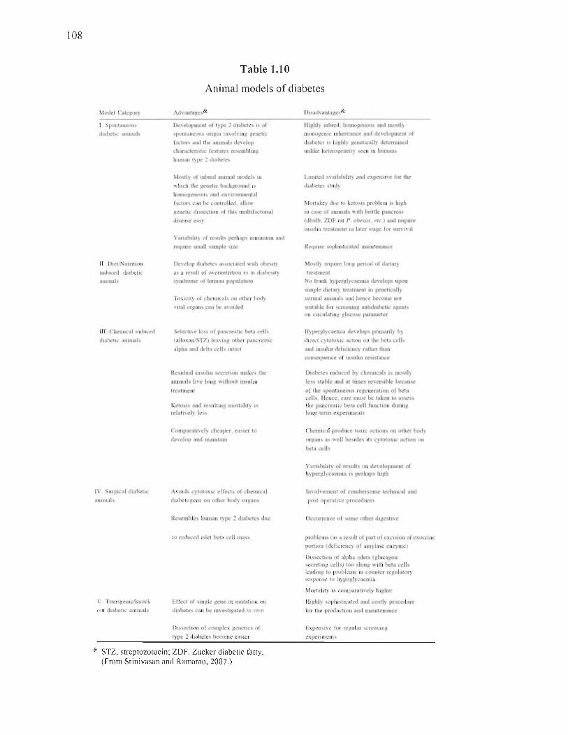
108
Table 1.10
Animal models of diabetes
1 Spolll"Il~OU~ tll<tlxllC ;HlIlltal~
Il Dlct/. ulnllOI1
Induccd dl::tbcllC
allll11als
III (,h<1I1Ical lIIt1uced
dlabellc allllllais
1\ 'urglcal dlabel"
anHnab
V Trans~el1lclknock
out dlabellc aUlIllal!.
Ad\:tlll"p,e~&
De\dopllll!lIl of '}Ile :! <.1I,tbete,>, of
SI>OIlI<UIC:OIIS Ofl!Z1II 1I1'-llh IIlg ~(lIellC
factor, and Ih\.' ;1111111011 clt.!\ dop
charach:n"otlc [cal ures re~elllbhll!l
111III1an I) l>C .1 ct. .. hclc'\
Mostl~ of II1bred <llIlIlIal models 111
\\ luch the: gc:netll.' backgrouud ,.,
hOtllo~eneou <llld eu\ Ir\.llimeniai
(ac:lor~ cali lh! l'ollirolled . 01110\\
llcncllc dl~~Ctl Il of IllIs Innltlfaclonal
dlscasc ea )
\"armbIlH) of reslIlIs ~rhaps IlUIlIUlUI1I and
requlre sl1Iall sautpl..:: Ile
J)e\cIOI) du,hCIC\ ac;\oclalcd \\ 11h ol)C'HI~
as " rCfinlt of Q\'crnnlntlOIl as III dlllhesll)
yndrolllc of human population
TaXlell} of chel1l1cals 011 ollter bod)
\ liai or~ans cali tlc! a\old~d
el~CII\e 10 ~ of panl"realiC bela c~lIs
(alloxan/STZ) lea"n~ olher pancrcallc
alpha and deh" cell~ Inlact
Resldual IIbultn ~~crC!ltoll IIH,kc!, Ihe
aUlll1al~ lI\"e IOUIl \\ tthout lIlsultn
Irc!atllleni
Keto~1 and rc)u!tlup, lIIorfaltt~ 1
r<l"II\<I) les
Comparallvel) chcapcr. e"'ilcr 10
de\"e!ap "nd mallltalll
A\Old~ c~ tolaXle ~ffecls of cheml al
dlabelog<ns on olher bod) or~an,
Resernbles human 1) pe ~ dl"beles due
la reduced Islel beta cdl l11ass
Effect of stn!!le ~enc or nHualion 011
dlabetes cali be 1I1\"e:~ti!laled III \'''"0
Dissection of complex }!eneliCS of
type :2 dtabetes becollle eaSter
& STZ, streptozotocin; ZDF, Zucker diabetic fa tty. (From Srinivasan and Ramarao, 2007.)
Ilt~hl~ lubred hOl1lo~euou, and mo.,1l)
Hlol1o~elllc IIIht:nl<lIlCC allli dc\ c.'luplllell1 01
dl.dlC1e\ l' htJ!hl~ p,t:nel1call~ delewlIued
uullLe helerOlZenell) -,ecu III hllllMIl
LlIllltcd a\'ailalllltty and cxpcn 1\"( for the
lhat~les sind)
~Iortilhl) dtle 10 kelo)l) probleUi 1; II1!lh
III cao;;c of anllnalc, \\ Ilh bnttlc pallcrea
(dbldb . ZDF rai P ob,SII' . rie ) and rcqlllre
tIlsllltn Ireatmclil III later slil~e for colln'I\'al
Re<IUlre "'Ol)hl~tll' al~d lIIalutenance
~lostl) rcql1lr~ 101lp, pcnod of dll:lar~
Ireatl1lCIII
o frank h~ pcr~lycacnHa developco IIpon
~llIlple (helar} Ircaln1c:nt III p,ellellcall)
normal anllnals and hence become 1101
~lIllable lor !'cret!l1Il1~ aUlIdlabelle a~enls 011 clrculalmg gluco~e paramcler
Il)p<rgl)c.elllla dC\elops 1"'11"",1) b)
(hrt:ct C) totOXIC [lCllOI1 011 the bel<l cdh
and 111 111111 dcfICICIIC) ralher Ihan
consequcnce of III~tlltll rcsistancc
Dlflbele~ lIIduced b} chenueal., 1., lIIo)ll~
Ico; rable and al IImc rever.,lblc beeause
of Ihe SpOIlI:tllCOIIS rep.eueralloll of bela cells lIeuce. care must lx talen to as e ., Ihe p:U1crealle lxla cdl fUllelJon dllrlllg long -terni expenments
Chcl1l1eal produce tOXle <tcllons on olher body
or!l ,ms Ole; \\ cil be'lder; lili c~ totoxle action 011
bela cclls
Vanabllll~ of re;lIlt~ 011 dc\elopmenl of
h)p<r)tl)caellllil " p<rh"p' III~h
III\ohemc::nl of clllllbe~OlHe lechlll al :md
pO~1 operall\ e procedures
Occurrence of ,ollie OIher dl!!e lI'e
problelll~ (as a result of part of eXCISion of t!xocnnc
porllon (deficlcncy of amylase enzyme)
DISSCCllon of "11>h" ISle" (~Iucagon secrelmg cells) 100 along \\ Ilh bela cells Il!adlllg 10 problems III cou nIer rep,ulalory response to h}poglycaenllil
Morta il ly I~ comp.uatl\'el) IU!lher
~hghl) SOplllsllc.led and COSII) procedure
for Ihe productIOn and mallileuance
Expensl\'C for regular screcl1In~
experllncnls

109
Experimentally-induced models are, on the other hand, generally inexpensive,
flexible and require little to no special maintenance (Islam and Loots, 2009).
This category can further be subdivided into chemical-, diet- and surgically-induced
models. Partial or total pancreatectomies were formerly common procedures to generate,
respectively, type II or type 1 diabetes-like phenotypes presenting prominent
hyperglycaemia (Foglia, 1950; Pauls and Bancroft, 1950). However, their popularity
dwindled with the recognition that much simpler chemical protocols cou Id efficiently
deplete insulin-producing pancreatic ~ cells. By a large amount, the two most prominent
toxins are alloxan (Goldner and Gomori, 1943) and streptozotocin (Rakieten et al. ,
1963). Both operate via oxidative mechanisms to cause the necrosis of pancreatic ~ cells
(Rerup, 1970; Yamamoto et al. , 1981) (Figure l.32) . Owing to its greater stability and
longer half-life, streptozotocin is sometimes preferred over alloxan in rats and mice,
but not in rabbits who are in sensitive to the former (Rerup, 1970; Srinivasan and
Ramarao, 2007). Depending on the intravenous (i.v.) dose52, these molecules can yield
either type 1- (higher dose) or type II-like (lower dose) diabetic features (Rakieten et al.,
1963). However, most agree that, regardless of the dose, i.v. alloxan and streptozocin
induce a diabetic phenotype that closely resembles type 1 diabetes with manifestations of
acute hypoinsulinaemia instead of insulin resistance. Intraperitoneal (i.p.) injections in
adult or neonatal rodents were later adopted to moderate the outcomes and to enhance
their maintainability (lto et al. , 1999, 2001 ; Kodama et al. , 1993; Portha et al. , 1974,
2007).
Other methods used to generate more sustainable experimentally-induced models
consist in laye ring different treatrnents to modulate extreme phenotypes. One of the most
widely hired strategies in this sense employs single i.p. injections of nicotinamide prior
to streptozotocin administration (Masiello et al., 1998; Nakamura et al. , 2006).
Nicotinamide moderates the necrosis of pancreatic ~ cells induced by streptozotocin,
thereby generating a model that is hypoinsulinaemic and hyperglycaemic, but viable for
52 When administered i.v., alloxan is usually given in doses ranging From 40-200 mg/kg body weight [b.w.] in rats or 50-200 mg/kg b.w. in mice. Streptozotocin, on the other hand, is administered i.v. in doses ranging From 35-65 mg/kg b.w. in rats or 40-200 mg/kg b.w. in mice.
110
several months without the need for insulin treatment53. In recent years, mixed chemical
diet paradigms were implemented that more faithfully emulate type II diabetes (Reed
et al. , 2000; Srinivasan et al., 2005). Indeed, standalone specialized diets (high-fat,
high-carbohydrate, high-fat low-carbohydrate, etc.) present the disadvantage of causing
a very progressive form of metabolic syndrome, characterized mostly by obesity and
insulin resistance, but rarely associated with frank hyperglycaemia or glucose
intolerance (Houssay and Martinez, 1947; Surwit et al., 1988; Winzell and Ahren,
2004). Combined with a low i.p. dose of streptozotocin, high-fat diets induce increased
body weight, hyperglycaemia, hyperinsulinaemia and dyslipidaemia in rodents (Reed
et al. , 2000; Srinivasan et al., 2005). Uniting insults in the form of diets and toxins is
today considered a core framework for the development of long-term type II diabetic
like models (Zhang et al. , 2003). Nevertheless, despite attempts to refine existing
models, none described to date are fully representative of either type l or II diabetes in
humans (Baxter and Duckworth, 2004; Islam and Loots, 2009; Van Belle et al. , 2009).
Granted the purpose of our work dwells in evaluating the effects of a sustained
hyperglycaemic phenotype and not in faithfully simulating a specific disease, we were
principally interested in models displaying this key feature for a long period of time
without the need for ectopic insulin interventions. In this spirit, moderate chemical
induced and diet-chemical mixed paradigms constituted the simplest and most accessible
protocols. Thus, we tested two well-documented paradigms in parallel: moderate dose
nicotinamide-streptozotocin-treated rats and low dose streptozotocin-treated rats fed a
high-fat diet (Reed et al., 2000; Srinivasan et al., 2005). In the first protocol, ovemight
fasted rats were simply injected with nicotinamide (i.p. 100 mg/kg b.w.) 20 min prior to
administration of streptozotocin (i.p. 55 mg/kg b.w.), as previously described (Badole
et al., 2015; Masiello et al. , 1998). In the second protocol, rats were maintained on a
60% high-fat di et adjusted for calories that began two weeks prior to a single i.p.
injection of streptozotocin (35 mg/kg b.w., overnight fasted) (Srinivasan et al. , 2005).
Hyperglycaemia was first assessed 72 h following injections and thereafter measured on
53 Precise ly, nicotinamide inhibits PARP, who se exaggerated activation by streptozotocin-induced DNA alkylation leads to the depletion of NAD+ and ATP, liable for pancreatic P cell necrosis. Nicotinamide also offers protection by serving as a precursor of NAD+ (Szkudelski, 201 2).

111
a regular basis, alongside body weight and food intake. Figure 1.33 surnmarizes these
monitored metabolic parameters in our models.
o 0"" HN)lNH ljIH
o~o HO HNy O
o ..... N~WO
pancreatic J3 cell alloxan/streptozotocin
extracellular
If Ifll fi fi Il Il fi Il fi 11 11 Il 1111 Il !llnr If 11 film 11 Il If TI nn fi IIlfll nîl fi 111111111111 11 111111111111 1111 Il 1111 Il Il Il Il Il Il Il Il Il 1111 111111111111 1111
fI~ n ll ll TI ll ll ml lllr TIln l llfirrlirr lI i l nIr II 1 n ITIi TIn 111 11 111111 1111 1111 1111 1111 1111 1111 1111 11 1111 111111 11 111111 1111 1111 1111
intracellular
Fe2+ Fe3+
/02::":::'.' + W 7 H202~·OH + OH'
O·' ,,_ .. _~
9IUI S.
alloxan '""""T':: ,2 alloxan O2- O2 02fradicals~
dialuric acid alloxan
~alloxan~ GS· GSH radicals GS· GSH
~/
glucokinase
.., ATP and'" insu lin secretion
streptozotocin --------,
GSSH + 2NADPH 7 2GSH + 2NADP+
NO
DNA destruction
nucleus
Figure 1.32 Mechanisms of pancreatic P cell death in chemical-induced diabetes. Alloxan and streptozotocin both enter pancreatic ~ cells via GLUT2, competing with glucose on the way. Alloxan engages in different mechanisms to participate in the death of pancreatic ~ cells. For instance, alloxan fuels a redox cycle with its reduction product, dialuric acid, thereby producing highly reactive ROS, such as the hydroxyl radical (-OH) via Fenton and Haber-Weiss reactions, and depleting antioxidative cofactors, like GSH and NADPH. Alloxan may also inactivate glucokinase, leading to hampered A TP production and insu lin secretion, a mechanism inhibited by the presence of glucose. Contrary to alloxan, streptozotocin's primary mechanism is quite straightforward and implicates firsthand alkylation of DNA. Exposure to either toxin leads to damaged DNA and the activation of the reparation enzyme PARP whose overstimulation will deplete stocks of the NAD+ cofactor: pancreatic ~ cells thereby undergo necrosis. Fe2
+, ferrous iron ions; Fe3+ , ferric iron ions; GS-, glutathione radical ; GSSH, glutathione disulphide; NO, nitric oxide. (Adapted from Radenkovié et al. , 2016.)
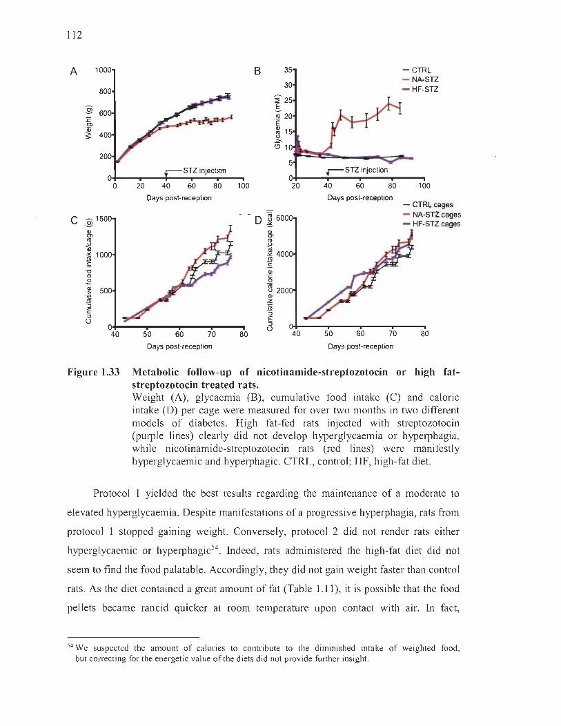
112
A 100
800
.9 600 1: 0) 'CD 3: 400
200
STZ injectJon 0
0 20 40 60 80 Days post-reception
C :9 1500
& ri "al S 1000 ,ç "0 0 .E Q) 500 > = ~ ::l E ::l U
0 40 50 60 70
Days post-reception
B 35
30
:2 25 .§. ,~ 20 E Q) 15 ~ ~ (!) 10
5
0 100 20
~
- - D I6000
80
& ~ ~ 4000 ,ç Q)
'~
~ 2000 ~
.~
"S E :::l
U
-CTRL - NA-STZ - HF-STZ
~ STZ injection
40 60 80 100 Days post-reception
- CTRLcages - NA-STZ cages - HF-STZ cages
50 60 70 80
Days post-reception
Figure 1.33 Metabolic follow-up of nicotinamide-streptozotocin or high fatstreptozotocin treated rats. Weight (A), glycaemia (B), cumulative food intake (C) and calorie intake (D) per cage were measured for over two months in two different models of diabetes. High fat-fed rats injected with streptozotocin (purple tines) clearly did not develop hyperglycaemia or hyperphagia, while nicotinamide-streptozotocin rats (red lines) were manifestly hyperglycaemic and hyperphagic. CTRL, control; HF, high-fat diet.
Protocol 1 yielded the best results regarding the maintenance of a moderate to
elevated hyperglycaemia. Despite manifestations of a progressive hyperphagia, rats from
protocol 1 stopped gaining weight. Conversely, protocol 2 did not render rats either
hyperglycaemic or hyperphagic54. Indeed, rats administered the high-fat diet did not
seem to find the food palatable. Accordingly, they did not gain weight faster than control
rats. As the diet contained a great amount of fat (Table 1.11), it is possible that the food
pellets became rancid quicker at room temperature upon contact with air. In fact,
54 We suspected the amount of calories to contribute to the diminished intake of weighted food, but correcting for the energetic value of the diets did not provide further insight.
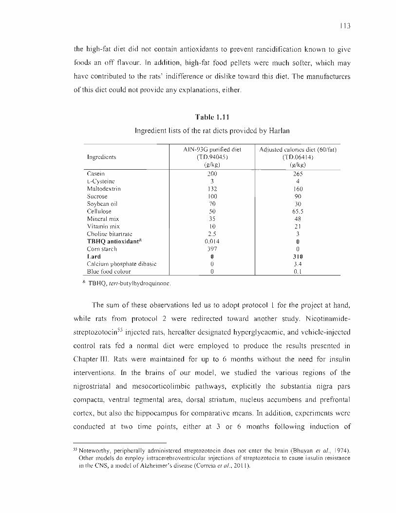
113
the high-fat di et did not conta in antioxidants to prevent rancidification known to give
foods an off flavour. In addition, high-fat food pellets were much softer, which may
have contributed to the rats' indifference or dislike toward this diet. The manufacturers
of this diet could not provide any explanations, either.
Table 1.11
Ingredient lists of the rat diets provided by Harlan
Ingredients
Casein L-Cysteine Maltodextrin Sucrose Soybean oil Cellulose Mineral mix Vitamin mix Choline bitartrate TBHQ antioxidant& Corn starch Lard Calcium phosphate dibasic Blue food colour
& TBHQ, tert-butylhydroquinone.
AfN-93G purified diet (TD.94045)
(g/kg)
200 3
132 100 70 50 35 10 2.5
0.014 397 o o o
Adjusted calories diet (60/fat) (TD.064 14)
(g/kg)
265 4
160 90 30
65.5 48 21 3 o o
310 3.4 0.1
The sum of these observations led us to adopt protocol 1 for the project at hand,
while rats from protocol 2 were redirected toward another study. Nicotinamide
streptozotocin55 injected rats, hereafter designated hyperglycaemic, and vehic1e-injected
control rats fed a normal diet were employed to produce the results presented in
Chapter III. Rats were maintained for up to 6 months without the need for insulin
interventions. In the brains of our mode l, we studied the various regions of the
nigrostriatal and mesocorticolimbic pathways, explicitly the substantia nigra pars
compacta, ventral tegmental area, dorsal striatum, nucleus accumbens and prefrontal
cortex, but also the hippocampus for comparative means. In addition, experiments were
conducted at two time points, either at 3 or 6 months following induction of
55 Noteworthy, peripherally administered streptozotocin does not enter the brain (Bhuyan et al., 1974). Other models do employ intracerebroventricular injections of streptozotocin to cause insulin resistance in the CNS, a model of Alzheimer's disease (Correia et al., 20 Il) .

114
hyperglycaemia, in order to gam a clearer insight regarding the time necessary for
neurodegeneration to manifest itself in our mode!. Moreover, many experiments were
repeated in different cohorts and in different institutions (Université du Québec à
Trois-Rivières, and University of Cagliari, Italy) , thereby solidifying our findings.
Chapter III offers further details pertaining to the experimental design and the various
metabolic parameters monitored along the course of this project, including haemoglobin
glycation measurements and a terminal oral glucose tolerance test commonly used in
humans and in animal models to assess the severity of hyperglycaemia and glucose
intolerance, respectively.
1.5.3.2 Intracerebral glucose measurements
The central hypothesis of this thesis requires firsthand observations of a rise in
glucose concentrations in the brain regions of interest, namely the substantia nigra pars
compacta and dorsal striatum of the nigrostriatal pathway, and the ventral tegmental
area, nucleus accumbens and prefrontal cortex of the mesocorticolimbic pathway.
As previously stated, precise quantifications of intraneuronal glucose are lacking in the
literature. Extracellular levels of glucose have, however, been appraised in rodents,
but reports usually coyer one single brain region at a time during specific tasks56.
More pertinent to our project, several studies demonstrated that hyperglycaemic or
hypoglycaemic challenges in otherwise healthy rodents provoke same-direction changes
in extracellular glucose levels, as previously discussed (Abi-Saab et al., 2002; Béland
Millar et al. , 2017; Macauley et al., 2015; Osborne et al., 1997). In diabetic paradigms,
rises in extracellular glucose concentrations were only measured in the inferior
colliculus of the brainstem (Jacob et al., 2002; McCrimmons et al., 2003), the striaturn
(Gomez and Barros, 2003) and the ventromedial hypothalamus (de Vries et al., 2003).
Several reports in humans account for cerebral glucose levels but were mostly conducted
in pathological states, for instance in partial seizures (Abi-Saab et al. , 2002), following
56 Many experiments have been conducted during various tasks or in response to several stimuli, such as anaesthetics, tail pinching or hypo/hyperoxia. The brain regions targeted during the se studies are the striatum (Osborne et al. , 1997; Fellows and Boutelle, 1993; Lowry et al., 1998a, 1998b, 1 998c), hippocampus (Macauley et al. , 2015; McNay et al. , 2000, 2001a, 2001b, 2004; Rex et al., 2009; Su et al., 2015) and motor cortex (Béland-Millar et al. , 2017).

115
a cardiac arrest (Hifumi et al. , 2017) and especially in traumatic brain injuries (Jalloh
et al. , 2013 ; Rostami, 2014).
Despite these reports indicating a nse In extracellular glucose levels in
hyperglycaemic or diabetic states, no adequate account exists to support the supposition
that concentrations increase evenly throughout several neuroanatomical locations.
We therefore performed glucose measurements in the multiple regions of interest in our
hyperglycaemic model by two different methods allowing us to estimate both
intracellular and extracellular concentrations. For intracellular glucose assessments,
brain homogenates were prepared from the striatal, midbrain, prefrontal cortex and
hippocampal regions, the latter serving for comparative means. We used a standard
glucose measurement kit that employs a mutarotase enzyme to transform a-D-glucose to
~-D-glucose, further oxidized by glucose oxidase. This generates hydrogen peroxide that
can be colorimetrically quantified upon its reaction with a chromogenic agent (Crystal
Chem, Downers Grove, IL, USA). Since rats were perfused upon sacrifice with ice-cold
phosphate-buffered saline, it is possible that the results obtained may provide an
underestimation of intracellular glucose concentrations.
For extracellular assessments, the intracerebral microdialysis technique in freely
moving and awake rats was employed, later described in greater detail. Seeing as
separation of the different subregions of the striatum and midbrain was not possible in
the previous method, intracerebral microdialysis advantageously allowed us to
specifically target the ventral tegmental area, substantia nigra pars compacta, nucleus
accumbens and dorsal striatum by stereotactic vertical insertion of dialysis probes that
coUect interstitial fluid solutes in real time. Given the various glycaemic and feeding
profiles of our rats, especiaUy between control and hyperglycaemic individuals,
our protocol required to standardize these parameters. We therefore fasted aU rats
ovemight and administered a specific amount of food diluted in water by intragastric
gavage precisely 30 min before the beginning of microdialysis experiments. To obtain a
reliable baseline measurement, samples were collected over a I-hour lapse of time and
glucose was quantified using the same kit employed for brain homogenates.
116
Combining these methods ensured a robust confirmation of the increase in
intracerebral glucose levels in our model. It also allowed us to verify that glucose
concentrations rise evenly across the brain regions of interest.
1.5.3.3 Assessment ofneurodegeneration
Objective 3 aimed to characterize the effects of long-term hyperglycaemia on the
nigrostriatal and mesocorticolimbic pathways with a keen focus on neurodegeneration.
We combined immunoblotting and immunohistochemical techniques to strengthen any
findings of neurodegeneration upon which hinges this part of the project. TH and DAT
were employed as dopaminergic markers expressed in the whole length of dopaminergic
neurons of either the nigrostriatal or mesocorticolimbic pathways. NeuN served as a
general neuronal marker, as it is expressed in ail neuron ceil bodies, but not in processes,
regardless of their type. Harvesting one brain hemisphere for immunoblotting and the
other for immunohistochemistry maximized the use of animaIs.
Immunoblotting assays allowed for a semi-quantitative measurement of the
expression levels of dopaminergic and neuronal markers in the midbrain, striatum,
prefrontal cortex and hippocampus of our model, 3 and 6 months following induction of
hyperglycaemia. Altered TH and DA T expression levels in our model provided hints of
dopaminergic modifications, perhaps indicative of neurodegeneration, whereas NeuN
measurements served to support these results. We exercised caution when analysing
NeuN expression in the striatum, since dopaminergic neuronal cell bodies are not found
in this region but rather in the midbrain.
The immunohistochemical method served to confirm whether changes in
expression were attributable to a proper loss of dopaminergic neurons. It also permitted
the precise neuroanatomical identification of these changes. Since expression levels of
TH, DAT and NeuN were altered only at 6 months in the striatum and midbrain of our
hyperglycaemic model, we performed immunohistochemical analyses at this time point.
Post-fixed frozen hemispheres were sliced rostrocaudally in coronal sections and

117
neuronal markers were revealed immunohistochemicaIly. In the substantia mgra
pars compacta and ventral tegmental area, TH-positive dopaminergic neurons or
NeuN-positive overall neurons were counted. In the various regions of the striatum,
the density of TH-positive dopaminergic fibres originating from the midbrain was
measured by optical densitometry. As such, we were able to verify proper
neurodegeneration of either neuronal cell bodies or terminal arbours in both pathways.
NeuN-positive neurons were also counted in the striatum, prefrontal cortex and
hippocampus to make sure that other non-dopaminergic populations remained
unchanged. TH-positive dopaminergic fibre staining was not seen In the prefrontal
cortex, as it is only lightly innervated by the ventral tegmental area. Likewise, the
hippocampus, which receives minor dopaminergic input from the ventral tegmental
area57 (Lisman and Grace 2005), did not display any staining.
1.5.3.4 Assessment of glial profiles
In an attempt to gain better insight into the changes that take place in the CNS of .
our hyperglycaemic rat model, the fate of glial populations was studied alongside that of
neurons. Astrocyte proliferation, or astrogliosis, is a well-appreciated feature of the
diabetic nervous system, both in humans (Araszkiewicz and Zozulinska-Ziolkiewicz,
2016; DeJong, 1977; Lu et al., 2014) and in models (Alomar et al., 2016; Baydas et al.,
2003; Duarte et al., 2009; Nagayach et al., 2014; Rostami et al., 2017; Saravia et al. ,
2002). However, studies have not addressed astrogliosis in multiple regions at a time nor
have they tackled this feature in long-term hyperglycaemia. Likewise, microgliosis is an
appreciated feature of retinal and peripheral nerve pathologies in models of diabetes
(Gaucher et al., 2007; Krady et al., 2005; Mazzeo et al., 2017; Zeng et al., 2000) as weIl
as in patients (Altmann and Schmidt, 2018; Zeng et al., 2008), but many gaps remain
pertaining to the CNS (Oliveira et al., 2016; Nagayach et al., 2014). Conversely, the loss
of microglial cells or astrocytes may also be indicative of the general distress expressed
57 The hippocampus and ventral tegmental area form a functional loop. When the hippocampus detects new information that is not stored in long-term memory, it sends a novelty signal through the nucleus accumbens and ventral pallidum to the ventral tegmental area. Then, the ventral tegmental are a releases dopamine in the hippocampus, thereby enhancing long-term potentiation and leaming. (Lisman and Grace, 2005).

118
by resident cells of the CNS. Although under-acknowledged, degeneration of glial cells
may in fact occur in the context of sustained oxidative stress (Streit et al. , 2008).
The importance of addressing glial populations in our models lies in their
purported role with regard to neurodegeneration, especially in the context ofParkinson 's
disease (McGeer et al., 1988; More et al., 2013; Russo et al., 2014). Indeed, glial
alterations may imply a neuroinflammatory state likely connected to the death of
dopaminergic neurons (Cabezas et al., 2014; Hirsch et al., 2003), as micro glial cells
were shown to be more abundant in the substantia nigra pars compacta of post-mortem
brains of parkinsonian patients (Kim et al., 2000; Lawson et al., 1990).
The knowledge gaps highlighted above alongside the relevance of glial
populations in neurodegeneration stress the pertinence of addressing the fate of glial
populations as an indicator of the severity of the hyperglycaemic insult in our model.
To that end, microglial cells and astrocytes were immunohistochemically detected using
antibodies raised against ionized calcium-binding adapter molecule 1 (Iba1) or glial
fibrillary acidic protein (GF AP), respectively. Cells were counted in all of the
aforementioned brain regions using the method previously described.
1.5.3.5 Intracerebral dopamine measurements
ldeally, observed losses of neurons or their terminaIs should be supported by
further neurochemical investigations. To that end, we measured extracellular dopamine
in the terminal regions of the nigrostriatal and mesocorticolimbic pathways, that is,
the dorsal striatum, the nucleus accumbens and the prefrontal cortex. The microdialysis
technique, also used to measure extracellular brain glucose, allows for the accurate
assessment of these regions. However, it entails complicated surgical procedures that
could not be performed in our model at the 6-month time point. Instead, we conducted
experiments at 3 months to evaluate the possibility that a functional loss of
dopaminergic neurotransmission could emerge before the neurodegeneration observed at
6 months. Information provided at this time point was nonetheless relevant to our

119
interpretation of several behavioural manifestations observed at 3 months in our model,
presented next in objective 4.
Ever since its design was optimized (Ungerstedt and Pycock, 1974; Un gers te dt
et al., 1982), intracerebral microdialysis has been extensively used to measure small
solutes in the interstitial fluid of the brain (see for review Chefer et al., 2009)
(Figure 1.34). This technique employs a minute probe composed of a metal cannula that
contains in its centre a tubular dialysis membrane permitting free transport of solutes
smaller than the molecular weight cut-off, typically of 20 000 Da. The probe is equipped
with an inlet, to transport the perfusate, and an outlet, to collect the microdialysate.
Referring to an atlas of neuroanatomical coordinates, the probe is vertically inserted in
the brain region of choice with the assistance of a stereotactic apparatus. A defined
portion of the semipermeable dialysis membrane at the very tip of the probe is directly
exposed to the parenchyma; only this surface participates in molecular exchanges and
can be modified to accommodate the dorsoventral thickness of the region of interest.
Perfusion begins as soon as the probe is inserted and the automatic syringe dispenser is
activated. Typical perfusion rates range between 0.3-3 ~Llmin and have a direct
incidence on the recovery rate, that is, the fraction of solute collected by the probe found
to be inversely proportional to the speed of its production, diffusion and replacement in
the interstitial fluid (Wages et al., 1986; Zetterstrom et al., 1988). Aiso depending on the
perfusion rate as well as on the sensitivity of the analytical apparatus, sample collection
times range from 1-20 min. The perfusate c1i1ssically consists of a solution that most
faithfully represents the extracellular environrnent in which the probe is inserted, except
that it is completely depleted in the solute meant to be sampled. Indeed, it is usually
accepted that uptake occurs by diffusion along a concentration gradient58. In our
experiments, we prepared the probes according to the widely employed protocol
established in Prof. Gaetano Di Chiara's laboratory (Bassareo et al., 2011; Di Chiara
et al., 1993; Tanda et al., 1996). This protocol presents the advantage of employing a
dialysis membrane mounted on a tungsten fibre and covered in an impermeable silicone
58 This also means that retrodialysis is possible if a certain solute is included in the perfusate, but found in lower concentrations in the brain parenchyma (Khan-Dawood et al., 1994; Wei et al., 1997). Such strategies allow for the delivery of drugs to very precise neuroanatomical loci.

120
sheath. As they are rigid and do not reqmre a cannula to guide them, the brain
parenchyma is not in direct contact with metal.
.yringo pump (aCSF, 1 !JI/min)
signai ttansduetlon
fradion colledor (10·20 min)
ventriculaf space CSF
Figure 1.34 Schematic representation of solute exchanges during microdialysis experiments. Not only do microdialysis probes uptake neurotransmitters, they can also sample most any molecule present in the environment that is smaller than the molecular weight cut-off. These can come from glial cells and blood capillaries, in addition to neurons. Likewise, solutes can be infused in discrete brain regions employing a retrodialysis paradigm, wherein a greater concentration of the substance is supplied in the perfusate (H6cht et al., 2007). aCSF, artificial cerebrospinal fluid; CSF cerebrospinal fluid. (From Bossers et al., 2013.)
Our protocol did not involve behavioural assessments or pharmacological
interventions sensitive to the state of the animal that, in these circumstances, must
recover for several days. As such, baseline dopamine was measured on the day
following acute cannulations in well-rested, awake and healthy rats. An intermediate
perfusate rate of 1 ilL/min was employed and 9 samples were collected over 20-min
121
periods for a total of 3 h. Microdialysates were collected and analyzed by high
perfonnance liquid chromatography. This technique separates the various molecules
present in a liquid according to their affinity for a stationary (solid column) phase that is
usually polar, for instance silica. However, microdialysates usually conta in rather
hydrophilic solutes whose retention times on the column are too long, hence the use of
a reverse-phase high-performance liquid chromatography wherein the stationary phase is
non-polar to allow the movement of these molecules (Irnperato and Di Chiara, 1984).
Solutes are electrochemically detected and their identity is confinned by appraisal of
their signature elution time.
Although microdialysis offers great neuroanatomical precision, it does not offer
very good temporal resolution. The sensitivity of the analytical apparatus usually
requires samples to be at least a few microlitres, which, at best, implies collection times
of 1 min. Consequently, phasic bursts are difficult to quantify by microdialysis as they
occur below the l-second threshold and require voltammetric or electrophysiological
methods that are sensitive to infrasecond fluctuations caused by behaviour or stimuli
(Hauber, 2010; Schultz, 2010; Segovia et al., 2011 ; Wightman and Robinson, 2002).
Tonic release, however, is feasibly quantified by intracerebral microdialysis experiments
that measure events dependent on time constants greater than 1 min (Di Chiara, 1990).
Extracellular concentrations of dopamine measured in our experiments therefore
represent dopaminergic tonic firing59 at baseline and hold important implications for
behavioural interpretations that we will address in the discussion.
To fulfil objective 3, the joint evaluation of neurodegeneration, glial disturbances
and dopamine release in the various brain regions of the nigrostriatal and
mesocorticolimbic pathways offered a broad appreciation of the dopaminergic neuronal
alterations present in our hyperglycaemic model. Supplementary information, especially
pertaining to probe insertion locations in microdialysis experiments, can be found in the
article presented in Chapter III.
59 We must bear in mind that a loss of tonie firing in the dorsal striatum is most eonspieuously observed in Parkinson's disease and leads to bradyki nesia and rigidity.
J

122
1.5.4 Objective 4: Assess the behavioural alterations resulting from nigrostriatal neurodegeneration in a rat model of long-term hyperglycaemia
Video samples of the behavioural experiments described below can be viewed
at the following address: goo.gl/9HF6ux. Copy and paste this shortened URL in your
browser to access the Dropbox file.
1.5.4.1 Assessment ofmotor deficits
In consonance with the three previous objectives, we wanted to achieve a broad
perception of the effects of long-term exposure to high concentrations of glucose on
dopaminergic neurons. Working with a rodent model allowed us to appreciate
behavioural consequences arising from dopaminergic neurodegeneration. lndeed, several
well-established tests exist to evaluate nigrostriatal or mesocorticolimbic functions.
As previously mentioned, the nigrostriatal pathway is prominently involved in the
production of movement owing to its important pacemaker activity that provides the
striatum, especially the dorsal region, with tonic dopamine (Lanciego et al., 2012) .
Rat models ofParkinson's disease are commonly subjected to specifically designed tests
to uncover motor symptoms that may arise from the degeneration of the nigrostriatal
pathway (see for reviews Dunnett and Lelos, 2010; Meredith and Kang, 2006; Pinna and
Morelli, 2014; Plowman and Kleim, 2011). They are also used to develop treatments to
alleviate motor symptoms that are thought to arise from loss of dopamine tonicity
(Bergstrom and Garris, 2003; Bergstrom et al., 2011).
Among the great many experimental tools developed to assess motor deficits in
rodents, only a few were applicable to our model. Indeed, many tests are designed for
asymmetrically lesioned animais induced, for instance, by a unilateral injection of
6-0HDA into the ascending nigrostriatal bundle of rats (Schwarting and Huston, 1996;
Ungerstedt and Arbuthnott, 1970; Vellucci et al., 1993). These tests are usually based on
the evaluation of tuming behaviour in response to stimuli that is due to the enhanced
sensitivity of postsynaptic receptors following striatal denervation rather th an the proper
123
loss of dopamine tonicity (Creese et al., 1977). Unilateral utilization of limbs in tasks is
another behaviour that can be scored in hemiparkinsonian rodents. However, the CNS
of our model is evidently subjected to a wide bilateral exposure to high glucose
concentrations and therefore asymmetric neurodegeneration is unlikely. In light of this,
we chose tests typically employed in bilateral models ofParkinson's disease or ones that
could be adapted to our needs. We did not expect our rats to express overt motor
symptoms and, as such, we performed the most sensitive tests possible to uncover
bradykinesia or akinesia, as weil as gait disturbances and sensorimotor deficits (Pinna
and Morelli, 2014). We also selected tests that could be performed at multiple time
points, expressly before the induction of hyperglycaemia, then at 3 and 6 months
following that mark so as to link results with our neurobiological evaluations.
The stepping test, a form of beam traversai task, is used in rats to assess three
different parameters: time of initiation, time to cross and numbers of steps made
. (Meredith and Kang, 2006; Olsson et al., 1995; Pinna et al., 2007, 2010). The test
employs a 20 cm-wide beam inclined at 15 degrees leading to the home cage.
Briefly, rats are trained to depart from the hands of the experimenter with one forelimb
placed at the beginning of the beam. Protracted initiation of movement toward the home
cage and time to cross the beam both uncover bradykinesia or akinesia. The numbers of
steps made, usually greater in parkinsonian rodents, inform on the gait. The stepping test
therefore reliably picks up subtle basal ganglia alterations and has been used in genetic
models and aged animais that present little nigrostriatal damage (Drucker-Colin and
Garcia-Hemandez, 1991). In our model based on a bilateral hyperglycaemic insult to the
CNS, differences between starting forelimbs were not found and results were pooled.
We also employed the horizontal bar test typically used to measure
pharmacologically-induced catalepsy in rodents (Alvarez-Cervera et al., 2005; Ciucci
and Connor, 2009; Kuschinsky and Homykiewicz, 1972; Sanberg et al. , 1981 , 1988).
In our model, however, this test rather served to identify bradykinetic or akinetic
individuals. In this task, the experimenter places untrained rats habituated to the test
cage in an unusual posture by placing both its front limbs on a cylindrical bar placed at a
1
J
124
convenient height. Normal rodents naturally seek to correct imposed postures and will
quickly remove both limbs. Latency to disengage from the bar is thought to expose basal
ganglia dysfunction (Duvoisin, 1976; Sanberg, 1980), although it is less sensitive than
the previous method, at least in certain genetic models (Kelm-Nelson et al. , 2015).
This test is however criticized for its sensitivity to recurring trials (Costall and Olley,
1971 ; Stanley and Click, 1976) likely ensuing from repeated handling that may cause
tonic immobility (Sanberg et al., 1980). Since we performed tests at wide intervals,
at 0, 3 and 6 months, these confounding events were unlikely. Nevertheless, overt
manifestations of proper catalepsy in otherwise active control or hyperglycaemic rats
were extremely rare and were omitted from data, as they could have been the result of
tonic immobility.
We conducted a third test to evaluate limb gait and bradykinesia, termed the
forelimb adjusting step test (Chang et al., 1999; Meredith and Kang, 2006; Pinna et al.,
2007, 2010). Briefly, trained rats are held by the torso with only one forelimb placed on
a surface and bearing weight. During trials, rats are laterally moved back and forth
across the surface at a fixed speed, and forehand (forelimb adjusts toward the torso) or
backhand (forelimb adjusts away from the torso) steps are counted (Chang et al., 2003 ;
Kirik et al., 2000, 2002; Olsson et al. , 1995; Schallert et al. , 2003; Tillerson and Miller,
2002). Akin to the stepping test, the forelimb adjusting step test is performed unilaterally
but results were combined upon confirmation that both limbs responded similarly in all
animaIs .
Many other tests were considered for their application in our model but either
failed to expose any deficits or were impractical. This was the case for the vibrissae
elicited forelimb placement test wherein a rat's whiskers are brushed upon a surface,
which normally .evokes a paw placement response (Meredith and Kang, 2006; Pinna
et al., 2007, 2010). Sensorimotor integrative deficits are identified when rats fail to
respond to brushing of their vibrissae. Our hyperglycaemic model did not perform
differently compared to control rats at any time point. We also attempted to perform an
adapted pole test for rats, which evaluates coordination and motor skills (Chompoopong

125
et al., 2016; Zaitone et al., 2012), as weil as an adhesive removal task, highly sensitive
for uncovering sensorimotor deficits and fine motor impairment in minimally dopamine
deficient animais (SchaUert et al., 1982), but rats failed to perform the tasks correctly for
vanous reasons.
Nonetheless, results obtained with the three abovementioned tests were
sufficient to draw up a picture of the key motor deficits displayed by our long-term
hyperglycaemic model, especiaUy with respect to bradykinesia/akinesia and gait
disturbances. Detailed experimental designs foUowed to fulfil this part of the objective
are provided in Chapter III.
1.5.4.2 Evaluation of social behaviour
Although best appreciated in the regulation of motor processes, nigrostriatal
dopamine also partakes in the modulation of many other behaviours. Owing to its role in
goal-directed conducts and habit leaming (Bromberg-Martin et al. , 2011 ; Faure et al.,
2005; Gunaydin et al. , 2014; Haber et al. , 2000; Redgrave et al. , 2010; Saddoris et al.,
2013; Schultz, 2002; Seger and Spiering, 2011 ; Wang et al. , 2011), nigrostriatal
dopamine has recently emerged as a regulator of social interactions and affective-based
decisions, which were hitherto exc1usively lent to the mesocorticolimbic pathway (Burke
et al. , 2010; King-Casas et al. , 2005; Lamichhane et al. , 2014; Narvaes and Martins
de Almeida, 2014; Ong et al. , 2011 ; Palmeri et al., 2017; Plavén-Sigray et al., 2014;
Stoeckel et al. , 2014; Trezza et al. , 2010). Although a proper correlation with
nigrostriatal degeneration has not been drawn, Parkinson ' s disease patients do
experience an altered quality of social life very early in the pathology. In particular,
social interactions, communication and emotion recognition are impaired to variable
extents in these individuals (PeU et al. , 2006; Schrag et al. , 2000; Y oshimura et al. ,
2005).
For this reason, we designed experiments wherein social behaviours were scored
concomitantly to the measurement of an outwardly accessible indicator of dopaminergic
1
J

126
functions modulated in social encounters, namely USV caUs (Figure 1.35). In social
behaviour research, US Vs constitute a growingly valuable instrument in the assessment
of the valence of encounters, as they convey information on the affective state of rodents
during courtship, rough-and-tumble play, cooperation and aggression (Brudzynski,
20l3 ; Burgdorf et al., 2008; Knutson et al., 1998; Lopuch and Popik, 2011 ; Wëhr and
Schwarting, 2013). Most importantly, however, they provide information on the
activation of midbrain dopaminergic pathways involved in their production. The two
principal neurotransmitters involved in the emission of ultrasonic calls are dopamine and
acetylcholine, which function together as a switch that can quickly activate positive or
negative affective states, respectively (Brudzynski, 2007; Cragg, 2006; Rakovska et al.,
2003). Dopamine and acetylcholine are sensitively released in response to salient and
novel environmental stimuli, inc1uding social encounters, and cooperate at the striatal
interface to select the appropriate affective state. This is accompanied by the emission of
USVs at different ranges of frequency6o. Dopamine is principally associated with the
emission of 50-kHz caUs, which are positively valenced and produced during social
interactions, su ch as tickling (Panksepp and Burgdorf, 2000), playing (BruneUi et al. ,
2006) or mating (Bialy et al., 2000). Acetylcholine is rather linked to 22-kHz USVs
reflecting negative affective states in various contexts, for instance social isolation
(Francis, 1977) and aggression (Kaltwasser, 1990). Sorne suggest that 22-kHz caUs are
also emitted in paradigms of chronic pain (Calvino et al. , 1996; Jourdan et al. , 1995),
but this association was disproven in the streptozotocin-treated rat model of neuropathic
pain (Jourdan et al. , 2002).
Affiliative/exploratory and aggression-related behaviours were scored in our rats
and associated with the emissions of 50- and 22-kHZ USVs. Rats interacted in a novel
and neutral arena, and not in resident-intruder paradigms (Hilakivi-Clarke et al., 1990;
Meehan et al. , 1986), to obtain insight into the reactions driven by socialization, which
more faithfuUy represent encounters between humans in day-to-day life. Behaviours and
USVs were related to the degree of striatal dopaminergic denervation and to insulin
levels and function. This allowed us to draw up the socioaffective profiles of our rat
60 Examples ofUSV audiograms are presented in Figure 4.7 of Chapter IV.
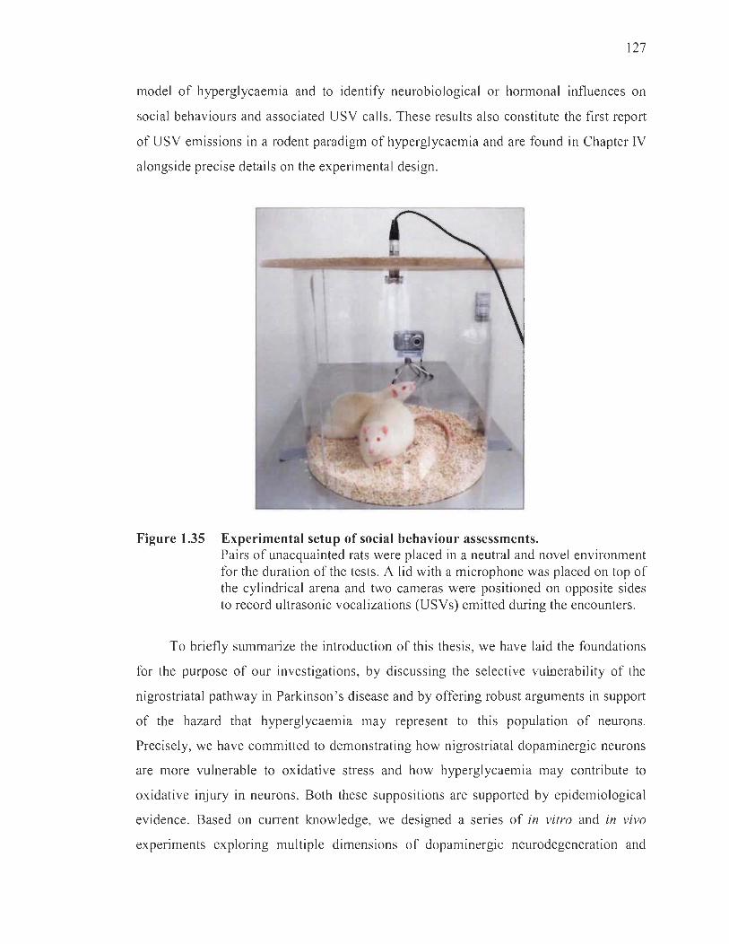
127
model of hyperglycaemia and to identify neurobiological or hormonal influences on
social behaviours and associated USV caUs. These results also constitute the first report
of USV emissions in a rodent paradigm of hyperglycaemia and are found in Chapter IV
alongside precise details on the experimental design.
Figure 1.35 Experimental setup of social behaviour assessments. Pairs of unacquainted rats were placed in a neutral and novel environment for the duration of the tests. A !id with a microphone was placed on top of the cy!indrical arena and two cameras were positioned on opposite sides to record ultrasonic vocalizations (USVs) emitted during the encounters.
To briefly summarize the introduction of this thesis, we have laid the foundations
for the purpose of our investigations, by discussing the selective vulnerability of the
nigrostriatal pathway in Parkinson's disease and by offering robust arguments in support
of the hazard that hyperglycaemia may represent to this population of neurons.
Precisely, we have committed to demonstrating how nigrostriatal dopaminergic neurons
are more vulnerable to oxidative stress and how hyperglycaemia may contribute to
oxidative injury in neurons. Both these suppositions are supported by epidemiological
evidence. Based on CUITent knowledge, we designed a series of in vitro and in vivo
experiments exploring multiple dimensions of dopaminergic neurodegeneration and

128
have also used a strategy employing an antioxidant, resveratrol, to offer further
validation of the pertinence of oxidative stress in high glucose-induced neuronal death.
The results of these experiments are presented in Chapters II, III and IV, and offer
molecular, cellular, neurochemical, neuroanatomical and behavioural evidence in
support of our central hypothesis that nigrostriatal dopaminergic neurons are more
vulnerable to hyperglycaemic conditions compared to other neuronal populations,
expressly the mesocorticolimbic pathway.
CHAPTERII
RESVERATROL PROTECTS DOPAMINERGIC PC12 CELLS FROM HIGH GLUCOSE-INDUCED OXIDATIVE STRESS AND APOPTOSIS: EFFECT ON
P53 AND GLUCOSE-REGULATED PROTEIN 75 LOCALIZATION
Renaud, J., Bournival, J., Zottig, X., Martinoli, M. G. (2014).
Neurotox. Res. 14, 110-23.
The content of this chapter was published online in November 2013 and issued in
January 2014 in the peer-reviewedjournal Neurotoxicity Research.
2.1 Author contributions
Justine Renaud contributed to the design of the study and performed 80% of
the manipulations. She wrote 80% of the manuscript, and prepared and edited aU the
figures. Julie Bournival, a master's student in Maria-Grazia Martinoli's laboratory at the
time, contributed to the design of the study and optimized experiments. Ximena Zottig,
an undergraduate student in Maria-Grazia Martinoli 's laboratory at the time,
provided assistance for the irnrnunoblotting experiments. Maria-Grazia Martinoli,
Justine Renaud ' s research supervisor at the Université du Québec à Trois-Rivières,
was the guarantor of the work and provided supervision, preparation and editing of
the manuscript.
130
2.2 Résumé
Le resvératrol est un composé polyphénolique dont les propriétés
cardioprotectrices et anti-inflammatoires sont déjà bien connues. Il détient également
des capacités antioxydantes permettant de protéger des neurones dopaminergiques
contre l'apoptose engendrée par le stress oxydant dans des paradigmes de la maladie de
Parkinson. De récentes études ont démontré qu'une hyperglycémie soutenue peut causer
un stress oxydant pouvant être nocif pour le système nerveux central. À la lumière de ce
qui précède, notre étude avait pour but a) d'évaluer le potentiel antioxydant du
resvératrol contre des concentrations physiologiquement élevées de glucose dans des
neurones dopaminergiques en culture, b) d'étudier les capacités anti -apoptotiques du
resvératrol dans ce contexte, et c) d'analyser la relation particulière entre le facteur de
transcription pro-apoptotique p53 et son séquestreur cytoplasmique GRP75 dont les
niveaux sont faibles dans le cerveau de patients parkinsoniens. Nos résultats démontrent
que le resvératrol protège les neurones dopaminergiques en culture contre un stress
oxydant induit par de fortes concentrations de glucose, précisément en diminuant les
niveaux d'anion superoxyde. De plus, les neurones dopaminergiques en culture traités
avec le resvératrol ne succombaient pas à l'apoptose qu'engendrait le glucose à des
doses élevées. En particulier, le resvératrol prévenait la fragmentation de l'ADN et
l'altération des profils d'expression de plusieurs marqueurs de l'apoptose. De fortes
concentrations de glucose causaient une translocation de p53 vers le noyau, et une forte
augmentation des niveaux de GRP75 dans le cytoplasme qui répondait à un besoin d'y
séquestrer le facteur de transcription pro-apoptotique. En effet, une augmentation du
taux de colocalisation entre GRP75 et p53 était observée. À cet égard, le resvératrol était
en mesure de prévenir la translocation nucléaire de p53. Ces résultats appuient les études
épidémiologiques soulignant que les patients diabétiques courent un plus grand risque de
développer la maladie de Parkinson que la population générale. Notre étude suggère
également que le resvératrol détient un rôle neuroprotecteur pouvant se prêter au
développement de thérapies préventives ou complémentaires contre le développement de
complications neuronales dans le diabète.

131
2.3 Full article in English: Resveratrol protects dopaminergic pe12 cells from high glucose-induced oxidative stress and apoptosis: effect on p53 and glucose-regulated protein 75 localization
Abstract
Resveratrol (RESV), a polyphenolic natural compound, has long been
acknowledged to have cardioprotective and anti-inflammatory actions. Evidence
suggests that RESV has antioxidant properties that reduce the formation of reactive
oxygen species leading to oxidative stress and apoptotic death of dopaminergic
(DAergic) neurons in Parkinson's disease (PD). Recent literature has recognized
hyperglycemia as a cause of oxidative stress reported to be harmful for the nervous
system. In this context, our study aimed a) to evaluate the effect of RESV against high
glucose (HG)-induced oxidative stress in DAergic neurons, b) to study the anti-apoptotic
properties of RESV in HG condition, and c) to analyze RESV's ability to modulate p53
and GRP75, a p53 inactivator found to be under-expressed in post-mortem PD brains.
Our results suggest that RESV protects DAergic neurons against HG-induced oxidative
stress by diminishing cellular levels of superoxide anion. Moreover, RESV significantly
reduces HG-induced apoptosis in DAergic cells by modulating DNA fragmentation and
the expression of several genes implicated in the apoptotic cascade, such as Bax, Bcl-2,
cleaved caspase-3, and cleaved PARP-I. RESV also prevents the pro-apoptotic increase
of p53 in the nucleus induced by HG. Such data strengthens the correlation between
hyperglycemia and neurodegeneration while providing new insight into the high
occurrence of PD in patients with diabetes. This study enlightens potent neuroprotective
roles for RESV that should be considered as a nutritional recommendation for
preventive and/or complementary therapies ln controlling neurodegenerative
complications in diabetes.
Introduction
Glucose is the essential energy substrate of the central nervous system and large
amounts of it are required to fill the high energetic needs of neurons. Unlike muscle cells
132
or adipocytes that depend on insulin, glucose uptake in neurons depends mainly on
its extracellular concentration (Tom lins on and Gardiner, 2008). Persistent episodes of
long-term glucose exposure may induce oxidative stress that results in cellular damage
(Giaccari et al. , 2009), such as neuropathic complications resulting from hyperglycemia
in uncontrolled diabetes (Rajabally, 2011). Accumulating evidence has enlightened the
relationship between diabetes and neurodegenerative disorders, including Alzheimer's
disease (AD) (Vignini et al. , 2013) and Parkinson ' s disease (PD) (Jagota et al. , 201 2) .
Recent literature has reported an increased risk of developing PD in patients with type 2
diabetes mellitus (Hu et al., 2007; Sun et al., 2012) .
PD is a neurodegenerative disorder characterized by the progressive loss of
nigrostriatal dopaminergic (DAergic) neurons in the substantia nigra pars compacta
(SNpc). DAergic neurons in this region are selectively lost due to the high activity of
monoamine oxidase and elevated levels of iron which both lead to increased generation
of reactive oxygen species (ROS) (Cui et al., 2012; Pearce et al. , 1997). At the cellular
level, mechanisms of high glucose (HG)-induced toxicity are similarly sustained by
oxidative stress in vitro (Boumival et al., 2012; Cao et al. , 2012) as weIl as in vivo
(Styskal et al. , 2012). By increasing aerobic respiration, raised sugar metabolism
promotes excessive formation of ROS that, jointly with insufficient antioxidant
defences, may damage cells (Apel and Hirt, 2004). Indeed, generation of mitochondrial
superoxide is increased and is thought to be at the origin of several HG-induced
complications (Brownlee, 2001). Currently, it is weIl known that oxidative stress may
lead to apoptosis (Circu and Aw, 2010) and increased production of ROS in HG
conditions may account for glucose neurotoxicity duly observed.
In addition, several genes are known to be implicated in the pathogenesis of
PD, such as PINK 1 and DJ-l. Glucose-regulated protein 75 (GRP75, also called
mortalinimtHSP70/mot-2), a member of the cytoprotective Hsp70 family of chaperon es,
interacts with both PINKI (Jin et al., 2006, 2007; Li et al. , 2005; Rakovic et al., 2011 )
and DJ-l (Jin et al. , 2005; Li et al. , 2005). GRP75 is mainly localized within the
mitochondria matrix of neurons where it accomplishes several functions such as

133
mitochondrial import and oxidative stress management (Yaguchi et al., 2007).
Overexpression of GRP75 leads to the extension of life span in nematodes and human
cells. On the other hand, it serves as a major target for oxidation and it was shown to be
involved in aging of nerve cells and in particular in the degeneration of DAergic neurons
(Burbulla et al., 2010). In mitotic ceIls, GRP75 localized in the cytoplasm sequestrates
and inactivates p53 preventing its nuclear translocation and apoptosis (Kaul et al. , 2001 ,
2005; Wadhwa et al., 2002). Indeed, p53 is a tumor suppressor protein known to play an
important role in evoking apoptosis when located in the nucleus by encouraging the
transcription of several pro-apoptotic genes such as Bax (Macip et al. , 2003).
p53 activity is stabilized in response to oxidative stress through posttranslational
modifications disrupting interactions with negative regulators (Neilson et al., 2008).
It is also a recurrent target in PD given the involvement of oxidative stress in p53
activation (Nair, 2006) and the evidence of DNA fragmentation and chromatin
condensation in DAergic neurons of the SNpc in PD patients (Hartmann and Hirsch,
2001; Tatton, 2000).
Prevention of neuronal loss in PD has not yet been addressed by existing
syrnptomatic treatments. Neuroprotection by dietary polyphenols may be an interesting
avenue in CUITent attempts to overcome oxidative stress induced by hyperglycemia.
We have recently shown that quercetin and sesamin, antioxidant polyphenols, exert
neuroprotective effects in neurons exposed to HG (Bournival et al. , 2012). The stilbene
resveratrol (RESV) is another polyphenol, primarily found in red wine, known for its
potent cardioprotective, anti-inflammatory and anticarcinogenic actions (Aluyen et al. ,
2012; Rosa et al., 2012). Our group, as weIl as others, have highlighted its potential in
defending neurons against oxidative assaults induced by a spectrum of treatments,
including neurotoxins (Blanchet et al. , 2008; Boumival et al. , 2009; Gélinas and
Martinoli, 2002; Peritore et al., 2013) or cerebral ischemic injury (MOITis et al. , 2011;
Simào et al. , 2012). Abundant literature suggests that RESV plays a protective role in
several neurodegenerative diseases including PD, AD and Huntington's disease (Albani
et al. , 2010; Hung et al., 2010) as weIl as against neuroinflammation (Foti Cuzzola
et al. , 2011).

134
Although the beneficial properties of RESV in neurodegenerative diseases are
extensively depicted in the literature, its role in defending neurons against HG-induced
damage has yet to be elucidated. The present study was designed to examine the
neuroprotective effects of the polyphenol RESV in differentiated DAergic PC12 cells
maintained in HG condition. NGF-differentiated PC12 cells are a reliable model for the
investigation of oxidative stress and neuroprotection of DAergic neurons. They express
tyrosine hydroxylase (TH), high-affinity dopamine transporter, estrogen receptor-a and
-~ , neurofilaments and secrete high levels of dopamine (Gélinas and Martinoli, 2002;
Kadota et al. , 1996; Nilsen et al. , 1998). In this comprehensive investigation, we outline
the roles of RESV in preventing neural parameters of cellular oxidative stress and
apoptosis induced by HG exposure in a cellular DAergic system. Our results
demonstrate that RESV can modulate the expression and localization of GPR75 and thus
might mediate mitochondria pathways of ce Il stress.
Materials and methods
Drugs and chemicals
All reagents and chemicals were purchased from Sigma (St. Louis, MO) unless
noted otherwise. Mouse anti-GRP75 (raised against amino acids 525-679 of GRP75 of
human origin), rabbit anti-p53 (raised against full length p53 of human origin,
for immunofluorescence), rabbit anti-Bcl-2 (raised against a peptide mapping at the
N-terminus of Bcl-2 of human origin), rabbit anti-Bax (raised against a peptide mapping
near the N-terminus of Bax of mouse origin), mouse anti-cleaved PARP-l (poly(ADP
ribose) polymerase, raised against C-terminal purified thymus PARP-l of calf origin),
goat anti-HDAC 1 (histone deacetylase 1, raised against amino acids 450 to
C-terminus of human HDACl), and mouse anti-GAPDH (glyceraldehyde 3-phosphate
dehydrogenase, raised against recombinant GAPDH of human origin) antibodies were
purchased from Santa Cruz Biotechnology (Santa Cruz, CA). Mouse anti-p53 (raised
against amino acids surrounding Ser20 of hum an p53 , for Western blotting) and rabbit
anti-cleaved caspase-3 (raised against amino-terminal residues surrounding Asp 175 in
135
human caspase-3) antibodies were purchased from Cell Signaling (Boston, MA).
Rabbit anti-VDAC (voltage-dependent anion channel , raised against amino acids
152-169 of VDAC of human origin), mouse anti-TH (raised against rat TH) primary
antibodies, and anti-mouse and -rabbit horseradish peroxidase-conjugated secondary
antibodies were purchased from Sigma. Anti-mouse Cy3 (cyanine 3)-conjugated
secondary antibody was purchased from Medicorp (Montreal, QC, Canada). Goat anti
rabbit FITC (fluorescein isothiocyanate)-conjugated secondary antibody was purchased
from Millipore (Temecula, CA).
Cell culture and treatments
PC 12 cells, obtained from American Type Culture Collection (ATCC, Rockville,
MD), were maintained in a humidified environment at 37°C and 5% C02 atmosphere.
Cells were grown in Roswell Park Memorial Institute medium 1640 (RPMI 1640)
supplemented with 10% (v/v) heat-inactivated horse serum, 5% (v/v) heat-inactivated
fetal bovine serum (FBS) and gentamicin (50 Jlg/mL). PC 12 cell neuronal differentiation
was evoked by administration of nerve growth factor-7S (NGF, 50 ng/mL) in
Dulbecco's Modified Eagle medium (DMEM) supplemented with 1 % FBS for 7 days, as
already described (Bournival et al., 2009, 2012). The DMEM containing 1.0 g/L of
D-glucose (Sigma D5523) is further called control (CTRL) medium, whereas HG
DMEM containing 4.5 g/L of D-glucose (Sigma D7777) is named HG medium. DAergic
PC12 cells were incubated with CTRL or HG medium for 96 h, unless stated otherwise.
We previously performed lactate dehydrogenase-based cytotoxicity assays to determine
the appropriate time of treatment in order to study the apoptotic process in the remaining
live cells (Bournival et al., 2012). For the last 24 h of treatment, DAergic PC12 cells
were incubated with or without RESV (0.1 JlM). RESV concentration was selected after
dose-response and kinetic studies (Boumival et al. , 2009; Bureau et al. , 2008).
An osmotic control consisting of CTRL medium supplemented with 3.5 g/L of
D-mannitol (MANN) was used to rule out a hypertonic effect of HG medium on PC12
ceUs. Charcoal-stripped serum was used in aU experiments to ensure that media were
free from steroids. For each experiment, initial seeding density was 30 000 ceUs/cm2.
1
J

136
Detection of mitochondrial superoxide radical
DAergic PC12 cells were grown and treated on collagen-coated circular glass
coverslips (Fisher Scientific, Ottawa, ON, Canada). Intracellular superoxide anion ("02-)
production was measured with MitoSOX™ Red (Invitrogen, Burlington, ON, Canada),
a fluorogenic dye used for the selective detection of superoxide in the mitochondria of
live cells. After treating cells with CTRL or HG medium for 3 h with or without RESV,
the cells were incubated with MitoSOX™ Red (5 mM) for 10 min at 37°C. MitoSOX™
Red is rapidly and selectively targeted to the mitochondria. Once in the mitochondria,
it is oxidized by superoxide and exhibits red fluorescence. Cells were washed with
Hank's balanced salt solution (HBSS, Invitrogen), and Hoechst 33342 counterstained
all nuclei. Cells were fixed in 4% paraformaldehyde for 6 min at 37°C. Coverslips were
mounted with Molecular Probes ' ProLong® Antifade Kit (Invitrogen). Images were
acquired by a Leica SD AF confocal microscope, and analyzed with Leica Application
Suite 3.l.3 software (Leica Microsystems, Concord, ON, Canada). To demonstrate
MitoSOX™ Red selectivity, a positive control was performed using sodium
diethyldithiocarbamate (DDC), a superoxide dismutase (SOD) inhibitor, in CTRL
medium.
Immunojluorescence and terminal deoxynucleotidyl transferase dUTP nick end labeling assay
Apoptotic cells were also detected by both terminal deoxynucleotidyl transferase
dUTP nick end labeling (TUNEL) assay (Roche Diagnostics, Laval, QC, Canada) and
activated caspase-3 immunofluorescence. DAergic PC12 cells were grown and
treated on collagen-coated circular glass coverslips. Cells were then fixed in 4%
paraforma1dehyde for 15 min at 37°C, washed with phosphate-buffered saline (PBS) and
further incubated in a blocking and permeabilizing solution (1 % bovine serum albumin
[BSA], 0.18% fish skin gelatin, 0.1% Triton-X and 0.02% sodium azide) for 30 min
at RT. Fixed cells were incubated with polyclonal anti-cleaved caspase-3 antibody 1:500
in PBS overnight. The slides were washed and treated with Cy3-conjugated secondary
antibody diluted 1 :500 in PBS for 4 h and then incubated with the TUNEL enzyme and

137
fluorescent dUTP mixture for 1 h at 37°C. Nuclei were counterstained 4',6-diamidino-2-
phenylindole (DAPI). Coverslips were mounted with ProLong® Antifade Kit.
Images were acquired by a Leica SD AF confocal microscope. DAergic PC 12 cells were
considered to be apoptotic when they were positive for cleaved caspase-3 and their
nuclei were stained with TUNEL. The number of apoptotic DAergic PC12 ce ll s among
300 randomly chosen neuronal was counted on 10 different optical fie lds from three
slides per group, as already reported (Bournival et al. , 2009, 2012), with Leica
Application Suite 3.1.3 software. In each experiment 50 !lM of fluormethylketone
conjugated tetrapeptide Z-DEVD-FMK (Bachem, Torrance, CA), a cell-permeable
caspase-3 inhibitor, was used on DAergic PC12 cells in HG and HG RESV conditions as
internai control for caspase-3 activation (Bournival et al., 2009, 2012).
Specifie apoptotic DNA denaturation analysis
Specific DNA denaturation in apoptotic cells was assessed with a single-stranded
DNA (ssDNA) apoptosis ELISA kit (Chemicon International, Temecula, CA).
This procedure is based on the selective denaturation ofDNA by formamide in apoptotic
cells but not in necrotic cells (Frankfurt and Krishan, 2001). After treatment with CTRL
or HG medium with or without RESV, denatured DNA was detected with a monoclonal
antibody highly specific to ssDNA and a peroxidase-Iabeled secondary antibody.
The reaction was then stopped with a hydrochloric acid solution and ssDNA
fragmentation was quantified by measuring absorbance at 405 nm with a Multiscan
Ascent microplate reader (Thermolab System, Franklin, MA). ssDNA was quantified
with reference to CTRL conditions. Absorbance of positive (wells coated with ssDNA)
and negative controls (wells treated with SI nuclease) served as quality control for the
ELISA.
Protein extraction
DAergic PC12 cells were grown and treated in collagen-coated 6-well plates.
Total proteins were extracted using a nuclear extraction kit (Active Motif, Carlsbad,

138
CA). Briefly, cells were washed with a mixture of ice-cold PBS and phosphatase
inhibitors, and then harvested in centrifuge tubes. Cell lysis was performed using the
supplied buffer and samples were centrifuged to obtain membrane-free supernatants
containing total proteins.
Cytoplasmic-nuclear fractionation was achieved using the nuclear extraction kit.
Briefly, cells were washed with a mixture of ice-cold PBS and phosphatase inhibitors,
and then harvested in centrifuge tubes. Cytoplasmic membranes were ruptured by
treatment with a hypotonic buffer and detergent. Samples were centrifuged to pellet the
intact nuclei, and soluble material was preserved as the cytoplasmic fraction. Nuclei
were then lysed and conserved in the provided lysis buffer.
Mitochondrial-cytoplasmic fractionation was achieved usmg a mitochondrial
extraction kit (Active Motif, Carlsbad, CA). Cells were washed with a mixture of
ice-cold PBS and phosphatase inhibitors, and then harvested in centrifuge tubes.
Cells were incubated on ice with isotonic cytosol buffer for 15 min. Cell membranes
were ruptured with a pestle homogenizer. Intact cells and nuclei were pelleted after two
centrifugations and discarded. Supernatants containing cytoplasm and mitochondria
were centrifuged twice to obtain a pellet of mitochondria. The resulting supernatant was
preserved as the cytoplasmic fraction. Mitochondria were washed with cytosol buffer
and lysed with detergent.
Electrophoresis and Western blotting analysis
Protein dosage was performed with a bicinchoninic acid-based sodium dodecyl
sulfate (SDS)-compatible Protein Assay Kit (Pierce, Rockfort, IL) for each fraction of
every sample. Equal amounts of protein were loaded onto 12% SDS polyacrylamide
gels. After electrophoretic separation, the gels were transferred to polyvinylidene
difluoride membranes (0 .22)lm pore size, BioRad, Hercules, CA). The blots were
blocked for 1 h at RT in Blotto B (1 % non-fat powdered milk, 1 % BSA, 0.05% Tween
20, 0.5 mg/mL sodium azide, in Tris buffered saline). Dilution of primary anti-GRP75,

139
anti-p53 , anti-Bax, anti-Bcl-2, anti-cleaved PARP-1 , anti-GAPDH, anti-HDAC1 ,
anti-VDAC and anti-TH (1 :200, 1:200, 1 :50, 1 :50, 1: 1000, 1:50, 1 :50, 1 :500 and
1: 10000 respectively) antibodies was prepared in Blotto B. The blots were then
incubated with peroxidase-conjugated secondary antibody (l : 10 000) in Blotto B for 2 h
at RT and finally developed with an enhanced chemiluminescence substrate solution
(Haan and Behrmann, 2007).
Glucose-regulated protein 75-p53 colocalization
DAergic PC12 cells were grown and treated on collagen-coated circular glass
coverslips. Then, the y were fixed in 4% paraformaldehyde for 15 min at 37°C, washed
with PBS and further incubated in a blocking and permeabilizing solution for 30 min
at RT. Fixed cells were incubated with both rabbit anti-p53 antibody 1: 100 and mouse
anti-GRP75 1: 100 in PBS ovemight. The slides were washed with and subsequently
treated with anti-rabbit Cy3-conjugated and anti-rabbit FITC-conjugated secondary
antibodies both diluted 1 :500 in PBS for 4 h. Nuclei were counterstained with DAPI.
Coverslips were mounted with Molecular Probes ' ProLong® Antifade Kit. Images were
acquired by a Leica SD AF confocal microscope. Colocalization was assessed for
100 randomly chosen PC12 cells on 6 different optical fields from three slides per group
with Leica Application Suite 3.1.3 software.
Statistical analysis
Significant differences between groups were ascertained by one-way analysis of
variance (ANOVA), followed by Tukey's post-hoc analysis with the GraphPad Instat
program, version 3.06 for Windows (San Diego, CA; www.graphpad.com). Ail data,
analyzed at the 95% confidence interval, are expressed as means ± standard error of
the mean (SEM) from at least 3 independent experiments. Asterisks indicate statistical
differences between the treatrnent and CTRL condition (***p < 0.001 , **p < 0.01 and
*p < 0.05); plus signs show statistical differences between the treatment and HG
condition (+++p < 0.001 , ++p < 0.01 and +p < 0.05).
140
Results
Resveratrol rescues high glucose-induced production of superoxide
To study the mechanisms underlying the neuroprotective effects of RESV against
HG, we measured the production of superoxide with a derivative of ethidium bromide,
MitoSOX™ Red, after administration of HG with or without RESV for 3 h. This time
period was considered since free radical generation and eventually oxidative stress
are early events in the causative process of cellular death (Carange et al. , 2011;
Pérez-De La Cruz et al. , 2010; Zhou et al. , 2008). Figure 2.1A disc10ses low
fluorescence levels in CTRL and MANN conditions as well as in cells treated with
RESV in CTRL medium after 24 h, whereas a marked signal was detected in HG- and
DDC-treated cells. When RESV was added to HG medium, fluorescence was strongly
reduced. Figure 2.1B also reports the semi-quantitative analysis of mitochondrial
superoxide anion presented in Figure 2.1 A, revealing high fluorescence levels with HG
and positive control DDC as well as a very significant reduction (p < 0.001) when
DAergic PC12 ce Ils in HG medium were treated with RESV. In the DDC condition,
inhibition of SOD supports the specific detection of superoxide anion. AlI nuc1ei are
stained blue by Hoechst 33342 (Figure 2.1A).
A

B
Figure 2.1
55 50
_ 45
~ 40 LL - 35 ~ ëi; 30 c: Q) 25 -c: 20 Q)
.~ 15 -.!2 10 Q)
~ 5 o
CTRL RESV HG HG RESV
+++
***
MANN CTRL+ DDC
141
RESV reduces HG-induced superoxide anion production in DAergic PC12 cells. Resveratrol (RESV) reduces high glucose (HG)-induced superoxide anion production in dopaminergic (DAergic) PC12 cells. (A) Fluorescence microphotographs. Blue: DAergic PC12 nuclei counterstained with Hoechst 33342. Red: MitoSOX™ Red superoxide indicator signal. A marked red signal is evident in DAergic PC12 cells treated with HG or diethyldithiocarbamate (DDC) (control [CTRL] + DDC). Red fluorescence was less intense in cells treated with CTRL medium, RESV alone or when RESV was added in HG medium (HG RESV). (B) Semi-quantitative image analysis. ***p < 0.001 compared with CTRL, +++p < 0.001 compared with HG, as determined by one-way one-way analysis of variance (ANOV A), followed by Tukey's multiplecomparison test. Fluorescence units (F.U).
Resveratrol reduces high glucose-induced apoptosis
We measured DNA denaturation induced by formamide, a specific hallmark
of apoptosis (Frankfurt and Krishan, 2001), using a ssDNA specific antibody
(Figure 2.2A). Specific apoptotic DNA denaturation is observed in early as well as
in late apoptotic cells. HG condition showed a 43% increase in apoptotic cells in
comparison to CTRL wells. This increment was fully reversed by RESV treatment in
HG medium (Figure 2.2A). MANN medium did not yield significant apoptosis.

142
We then examined the effect of HG and RESV on later events of the apoptotic
cascade leading to DNA fragmentation. Detection of cleaved caspase-3, the terminal
effector caspase responsible for late apoptosis-mediated DNA fragmentation (Fan et al.,
2005), was conducted by immunofluorescence alongside a TUNEL assay measuring
DNA degradation (Figure 2.2B and C). In the HG condition, immunofluorescence
revealed the presence of cleaved caspase-3 positive cells (Figure 2.2C, red signal),
while TUNEL assay stained numerous nuclei undergoing DNA fragmentation
(Figure 2.2C, green signal). Total nuclei were stained with DAPI (Figure 2.2C, blue
signal). DAergic PCI2 cells were considered to be in late apoptosis when they hosted
both caspase-3 activation and DNA fragmentation events (Figure 2.2C, cells pointed by
white arrows). Treatment with RESV for 24 h clearly reduced the presence of apoptotic
nuclei as implied by the lower number ofDAergic PCI2 cells exhibiting both green and
red fluorescence . The number of apoptotic cells was also counted (Figure 2.2B),
as described in the Materials and Methods section. Administration of RESV decreased
the number of apoptotic cells compared to the HG condition. MANN medium did not
yield a significant rise in apoptotic cells compared to CTRL. To show that caspase-3
activation is a key step in the HG-induced apoptotic pathway, DAergic PCI2 cells were
pretreated with 50 ).lM Z-DEVD-FMK, a cell-permeable selective caspase-3 inhibitor,
followed by treatment with HG with or without RESV (Figure 2.2B and C).
A _160 "0 ~ 140 o ~ 120 o ê 100 c: ,g 80
~ 60 -.; c: 40 CI>
1:1 c( 20 <
z c 0
+++
CTRL RESV HG HG MANN RESV
B 70
!!!. 60 Qi u u 50 ~ Q. 40 &. CIl 30 ô :D 20 .c E :i 10
o
+ ++
1 ++
n CTRL RESV HG HG HG HG MANN
RESV 1 RESV J
Z-DEVD-FMK

> CI) W 0:: (!) ~
Figure 2.2
CLEAVED
RESV reduces HG-induced apoptosis in DAergic PC12 cells. Continued on next page.
143

144
(Continued.) (A) Histogram of specific apoptotic DNA denaturation by formamide in DAergic PC12 cells as detected with a monoclonal antibody against single-stranded DNA (ssDNA). CTRL, MANN and RESV alone do not affect specific apoptotic DNA denaturation. HG increases apoptotic DNA denaturation. Treatment of HG-exposed cells with RESV elicits a significant decrease in specific apoptotic DNA denaturation (HG RESV). (B) The number of apoptotic DAergic cells among 300 randomly chosen DAergic cells was counted on 10 different optical fields from 3 slides per group, as illustrated in panel C on a subsequent page. (C) Microphotographs of immunofluorescence detection of apoptotic DAergic PC12 cells. Blue: DAergic PC12 nuclei counterstained with 4',6-diamidino-2-phenylindole (DAPI). Red: anticleaved caspase-3 signal. Green: Terminal deoxynucleotidyl transferase dUTP nick end labeling (TUNEL) staining of nuclei exhibiting DNA fragmentation. Triple-staining (cells points by white arrows) clearly reveals several apoptotic cells on si ides treated with HG and fewer apoptotic cells when DAergic PC12 cells are treated with CTRL medium, RESV alone or wh en RESV is administered in HG conditions (HG RESV). To show that caspase-3 activation is a key step in the HG-induced apoptotic pathway, DAergic PC12 cells were pretreated with 50)lM of fluormethylketone-conjugated tetrapeptide Z-DEVD-FMK, a cell-permeable caspase-3 inhibitor, followed by treatment with HG, with or without RESV (HG- Z-DEVD-FMK and HG RESV- Z-DEVDFMK, respectively). MANN condition is similar to CTRL cells. Enlarged microphotographs: HG and HG RESV merge microphotographs, to show apoptotic nuclei in these key conditions. **p < 0.01 and ***p < 0.001 compared with CTRL, +p < 0.05, ++p < 0.01 and +++p < 0.001 compared with HG, as determined by one-way ANOVA, followed by Tukey's multiple-comparison test.
In order to further support these findings, we analyzed the expression of several
proteins acting in the apoptotic cascade. Western blotting was performed on total
proteins extracted from DAergic PC 12 cells treated with HG or CTRL medium, with or
without RESV (Figure 2.3). We analyzed the pro-apoptotic Bax and anti-apoptotic Bcl-2
protein ratio (Figure 2.3A) reported to be correlated with apoptosis (Cory and Adams,
2002). A high BaxlBcl-2 ratio favors the release of mitochondrial factors leading to the
activation of effector caspases in the apoptotic cascade (Kang and Reynolds , 2009).
Our results demonstrate that the administration of HG medium for 96 h increases the
Bax/Bcl-2 ratio two-fold compareq to CTRL medium, supporting that HG-induced
apoptosis in DAergic PC12 cells is mediated, at least in part, by the mitochondrial
pathway (Figure 2.3A, histogram full grey line). The HG-induced raise of the Bax/Bcl-2
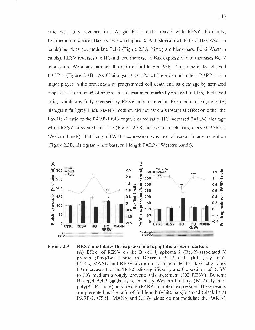
145
ratio was fully reversed in DAergic PCl2 cells treated with RESV. Explicitly,
HG medium increases Bax expression (Figure 2.3A, histogram white bars, Bax Western
bands) but does not modulate Bcl-2 (Figure 2.3A, histogram black bars, Bcl-2 Western
bands). RESV reverses the HG-induced increase in Bax expression and increases Bcl-2
expression. We also examined the ratio of full-Iength PARP-l on inactivated cleaved
PARP-l (Figure 2.3B). As Chaitanya et al. (2010) have demonstrated, PARP-l is a
major player in the prevention of programmed cell death and its cleavage by activated
caspase-3 is a hallmark of apoptosis. HG treatment markedly reduced full-length/cleaved
ratio, which was fully reversed by RESV administered in HG medium (Figure 2.3B,
histogram full grey line) . MANN medium did not have a substantial effect on either the
Bax/Bcl-2 ratio or the P ARP-l full-lengthlcleaved ratio. HG increased PARP-l cleavage
while RESV prevented this rise (Figure 2.3B, histogram black bars, cleaved P ARP-l
Western bands). Full-length PARP- Iexpression was not affected in any condition
(Figure 2.3B, histogram white bars, full-length PARP-l Western bands).
A B _ o Bax
2.5 :::- 0 Full-Iength ~ 400 - Cleaved 1.2
0 +++ .-
ni ë 300 - Bcl-2 ë - Ratio
~ 250 Î\... ~ :~ 0
~ 200 ----/ ~ 1.0 ~
'E - Ratio o 350 u '0 300
ê 250 c
k ... 1 ....
1
0.8 Il.. 0:::
0.6 ~ 't:I
r= 1 ~ r 111 ~5: 1 Il.. 0 -1.5
.2 200 1/1
= 150 ... ~ 100 ~Il
;;:: 50
~ 0 CTRL RESV HG
0.4 ~ ni
+ 0.2 QI
~ :2' 1 1 -0.4 =
HG MANN ~ CTRL RESV HG HG MANN RESV RESV
Bax Bcl-2
Figure 2.3
Full-Iength Cleaved
RESV modulates the expression of apoptotic protein markers. (A) Effect of RESV on the B cell lymphoma 2 (Bcl-2)-associated X protein (Bax)/Bcl-2 ratio in DAergic PC12 cells (full grey line). CTRL, MANN and RESV al one do not modulate the Bax/Bcl-2 ratio. HG increases the Bax/Bcl-2 ratio significantly and the addition of RESV to HG medium strongly prevents this increment (HG RESV). Bottom: Bax and Bcl-2 bands, as revealed by Western blotting. (B) Analysis of poly(ADP-ribose) polymerase (PARP-l) protein expression. These results are presented as the ratio of full-length (white bars)/cleaved (black bars) PARP-l. CTRL, MANN and RESV alone do not modulate the PARP-l
146
ratio in DAergic PC12 cells (full grey line). A decrease ofPARP-l ratio is apparent in HG condition. When RESV is delivered in HG condition, a significant increase of full-Iength/cleaved P ARP-l was evident (HG RESV). Bottom: Western blot bands of full-Iength and cleaved PARP-l. **p < 0.01, ***p < 0.001 compared with CTRL and +p < 0.05, +++p < 0.001 compared with HG, as determined by one-way ANDV A, followed by Tukey's multiple-comparison test.
Resveratrol modulates p53 and glucose-regulated protein 75 subcellular localization and colocalization
We studied the expression levels ofp53, a tumor suppressor, and GRP75, a stress
response protein (Figure 2.4). In several models, GRP75 binds and inactivates pro
apoptotic p53 in the cytosol, therefore helping to prevent apoptosis. In order to elucidate
this alleged relationship between both markers, protein levels were measured in the
cytoplasm and the nucleus (p53) or in the cytoplasm and the mitochondria (GRP75)
(Figure 2.4A and B). Treatment of DAergic PC12 ce lis with HG medium for 96 h
noticeably decreased p53 cytoplasmic/nuclear ratio (Figure 2.4A, histogram full grey
line). This was prevented by administration with RESV. Expressly, HG increased p53
expression in the nucleus (Figure 2.4A, histogram black bars, p53 nuclear Western
bands) while it did not seem to affect cytoplasmic levels (Figure 2.4A, histogram white
bars, p53 cytoplasmic Western bands). RESV in HG medium preserved p53 levels
at CTRL range in both compartments. HG administration for 96 h increased GRP75
expression both in the cytoplasm and in the mitochondria (Figure 2.4B, histogram white
and black bars, GRP75 mitochondrial and cytoplasmic Western bands). Treatment with
RESV in HG medium prevented GRP75 levels from rising in the cytoplasmic
fraction only. The result is a small but significant decrease m the GRP75
cytoplasmic/mitochondrial ratio (Figure 2.4B, histogram full grey line). MANN medium
did not affect the expression of either GRP75 or p53.

147
A B 0 ~ Cytoplasmic
1.5 _ Cytoplasmic ;;
350 _ Nuclear
'0 ~ Ratio
~ 300 0
~ 250 0
ê200 C
.2 150 1/1 1/1
~ 100
1 Q, )( QI
50 C') Il) Q,
o CTRL
p53 TH -
HDACl C N
Figure 2.4
1.5 ni 0 '0 300 - Mitochondrial ... ;; ...
- Ratio Il) 1\1 - .... ... c
+ ++ 1.0 8 250
Q, C')
1.0 0: Il) Q, .... Cl ... 0 .... ni ê 200 'ii
• 0.5 CIl 0.5 .: ... Ü 'C C C ~ .2 150 .. 0 c U 1/1 1 .r:.
++ 0 1/1 Il + 0 U
1 'Ë QI 0
'1 · li 100
1 1 ~.51 1 -0.5 -a. )( QI
0 Il) 50 >. .... Q,
(,) 0:
RESV HG HG MANN -1.0 Cl o CTRL RESV HG HG MANN -1.0 g. RESV RESV ~
GAPDH - - VDAC GRP75
C N C N C N C N C M C M C M C M C M
Effeet of RESV on the subeellular loealization of p53 and GRP75. (A) HG medium significantly increases nuclear localization of p53 (black bars) and administration of RESV in HG medium prevents this increase (HG RESV). RESV and MANN alone do not modulate p53 cellular localization. Cytoplasmic p53 (white bars) is not affected in any condition. Ratio of cytoplasmic/nuclear p53 is decreased in HG condition, which is prevented by RESV administration (HG RESV, full grey line). Bortom: p53 , cytoplasmic fraction (C) purity marker tyrosine hydroxylase (TH) and nuclear fraction (N) purity marker histone deacetylase 1 (HDAC1) bands as revealed by Western blorting. (B) Effect of RESV on the cellular localization of glucose-regulated protein 75 (GRP75) in DAergic PC12 cells. HG treatment increases both cytoplasmic and mitochondrial content of GRP75 . Administration of RESV in HG medium (HG RESV) significantly reduces cytoplasmic levels of GRP75 (white bars) while it does not amend mitochondrial levels (black bars). Ratio of cytoplasmic/mitochondria GRP75 is decreased in HG condition, which is prevented by RESV administration (HG RESV, full grey line). Bottom: GRP75, cytoplasmic fraction purity marker glyceraldehyde 3-phosphate dehydrogenase (GAPDH) and mitochondrial fraction (M) purity marker voltage-dependent anion channel (VDAC) bands, as revealed by Western blotting. **p < 0.01 , ***p < 0.001 compared with CTRL and +p < 0.05, ++p < 0.01 compared with HG, as determined by one-way ANOV A, followed by Tukey' s multiple-comparison test.
Finally, to evaluate the potential for GRP75 and p53 to interact in the cytoplasm,
immunofluorescence colocalization measurements were performed following treatment
ofDAergic PCI2 cells with HG medium with or without RESV. Scatter plots show that
p53 (Figure 2.5A, green signal distribution) and GRP75 (Figure 2.5A, red signal
distribution) signaIs are mainly independent from one another except for slight

148
colocalization (Figure 2.5A, plots and micrographs, Figure 2.5B). However, treatment
with HG medium still appears to yield more colocalization on the scatter plot
(Figure 2.5A, plots), which is also supported by the colocalization rate histogram
(Figure 2.5B). Overlaid pictures ofp53 and GRP75 staining (Figure 2.5A, micrographs)
in CTRL, MANN, and RESV condition show dispersed punctual staining (white signal)
in the cytoplasm, while in the HG condition a perinuclear dense staining is clearly
visible. Administration of RESV in HG medium reveals a more scattered staining than
in HG condition alone.
A
B 14
- 12 ~ 0 -cu 10 -~ c 8 0 :;: cu .~ 6 +++ cu
1 C.> 0 4
n 0 "0 u
2
0 CTRL RESV HG HG MANN
RESV
Figure 2.5 RESV modulates p53 and GRP75 colocalization. Continued on next page.

Discussion
149
(Continued.) (A) Scatter plots and corresponding overlaid micrographs of p53 and GRP75 . Scatter plots show signal intensity for p53 (green signal) on the y axis and GRP75 (red signal) on the x axis. Each dot represents one event of fluorescent signal. Thresholds for green and red signal are optimized at 85% (two white lines) and mean background at 10% (arched delimitation), in each condition. Signal is colocalized when signais in scatter plots are located between the threshold lines on the outside of the background delimitation. White signal in microphotographs indicates high probability of colocalization. CTRL, RESV and MANN conditions are similar in that p53 and GRP75 colocalization is scarce and scattered in the cell cytoplasm. HG increases the white signal around the perinuclear area as well as the nurnber of dots in the region of interest on the plot. Treatment with RESV yields a scatter plot and overlaid white signal (HG RESV) similar to the CTRL condition, suggesting its potential to diminish colocalization between GRP75 and p53. (B) Histogram depicting the colocalization rate of GRP75 and p53 observed in panel A. HG increases the colocalization rate significantly. RESV administration in HG medium reverses this increase in colocalization rate (HG RESV). Ail other conditions are similar to CTRL. ***p < 0.001 compared with CTRL and ++p < 0.001 compared with HG, as determined by one-way ANOVA, followed by Tukey' s multiple-comparison test.
We previously reported that several natural polyphenols, including the stilbene
RESV, exert powerful neuroprotective activity in DAergic PC12 cells against the
oxidative burden triggered by the administration of the potent parkinsonian toxin
1-methyl-4-phenyl-1 ,2,3,6-tetrahydropyridine (MPTP) in vivo (Blanchet et al. , 2008) or
its active metabolite I-methyl-4-phenylpyridinium (MPP+) in vitro (Bournival et al. ,
2009; Gagné et al. , 2003 ; Lahaie-Collins et al. , 2008). Since hyperglycemia has also
been listed as a growing risk factor for PD (Hu et al. , 2007; Jagota et al., 2012;
Sun et al. , 2012), we focused our study on the neuroprotective effect of RESV on
HG-induced oxidative stress and apoptosis in DAergic PC12 cells with regard to GRP75
and p53 localization.
The Diabetes Control and Complications Trial (1993) together with the u.K. Prospective Diabetes Study (1998) have determined that hyperglycemia is the culprit to
blame for tissue damage in type l and type II diabetes. Currently, we know that

150
overproduction of superoxide is the single upstream event leading to the following
pathways involved in glucose toxicity (Giacco and Brownlee, 2010): 1) increased flux
of glucose and other sugars through the polyol pathway; 2) increased intracellular
formation of advanced glycation end-products (AGEs); 3) increased expression of the
receptor for AGEs and its activating ligands; 4) activation of protein kinase C (PKC)
isoforms; 5) overreactivity of the hexosamine pathway. The formation of AGEs and
activation of AGE receptors (Shaikh and Nicholson, 2008), the activation of PKC (Aoki
and Li, 20 Il) and the dysfunction of the polyol pathway (Ahrned et al., 2009) have been
identified as contributors in the development of PD. These mechanisms suggest a strong
link between neuronal apoptosis observed in PD and hyperglycemic damage in diabetes
(Klein et al., 2004; Li et al., 2002; Li et al., 2008) .
In this study, we demonstrated the defensive role of RESV in counteracting
cellular distress parameters evoked by HG in DAergic PC12 cells. We tested
NGF-differentiated PC 12 cells, a known, reliable and efficient model for the
investigation of oxidative stress, apoptosis and neuroprotection of DAergic neurons
(Bournival et al., 2012; Gélinas and Martinoli, 2002; Lahaie-Collins et al., 2008).
Since oxidative stress is an essential factor in glucose toxicity and in the pathogenesis of
PD, we investigated whether RESV protects DAergic PC12 cells by reducing levels of
mitochondrial superoxide in HG condition. Our results show that RESV effectively
diminishes superoxide production after as early as 3 h. In PD, superoxide reacts with
iron cations to form hydroxyl radical (Ramas arma, 2012), known to exert very
deleterious effects on DNA, lipids and proteins. This ROS can also react with nitric
oxide, an important signaling molecule in the brain, to form peroxynitrite, a powerful
oxidant shown to play a significant role in protein aggregation pertinent to PD
(Danielson and Andersen, 2008).
It is currently weil known that oxidative stress may cause apoptosis through
several pathways: 1) ROS-induced expression or activation of nuclear factor-kappa B
(NF-KB) (Gloire et al. , 2006); 2) mitochondria-mediated cell apoptosis (Circu and Aw,
2010); 3) ROS-mediated DNA damage and p53 activation (Liu and Xu, 2011);

151
4) stress-activated protein kinases pathway to apoptosis (Johnson and Nakamura, 2007).
We performed a set of experiments to investigate the apoptotic cascade in DAergic
PC 12 cells ensuing oxidative stress to further demonstrate the preventive role of RESV.
A specific apoptotic DNA denaturation assay demonstrated that RESV significantly
prevents apoptosis in cells exposed to HG. We further examined markers of late
apoptosis to determine whether the prote in cascade leads to terminal events such as the
irreversible fragmentation of DNA. RESV in HG clearly reduced the number of
apoptotic PC12 cells in comparison to the HG condition alone as shown by the decline
in TUNEL and cleaved caspase-3 double-positive cells. Another target of activated
caspase-3 is PARP-l , a protein known to participate in the repair of damaged DNA
(Wang et al. , 2012). Our findings reveal that the PARP-l protein ratio, full-Iength versus
cleaved, was decreased after HG treatment and was then improved by RESV
administration, hence supporting once more the neuroprotective anti-apoptotic role of
RESV in a HG paradigm. In addition, the Bax and Bcl-2 expression was studied to
determine the apoptotic events surrounding the mitochondria. Bax contributes to the
leakiness of the outer mitochondrial membrane, while Bcl-2 blocks the permeability
transition pore, thus inhibiting mitochondria-mediated programmed cell death (Smith
et al., 2008). The rise in the Bax to Bcl-2 ratio is a characteristic feature in apoptosis
(Cory and Adams, 2002) equally observed in glucose toxicity (Allen et al. , 2005) and in
several models of PD including hum an post-mortem brains (Vila and Perier, 2008).
Our data reveal that the Bax/Bcl-2 protein ratio is increased after HG administration, and
is decreased by RESV treatment in the HG condition, strongly suggestive of a role for
mitochondrial dysfunction in the mechanisms underiying the apoptosis of DAergic
neurons in our cellular paradigm of hyperglycemia.
GRP75 has often been linked to PD pathogenesis as reported in studies showing
binding properties to PD-associated proteins in the mitochondria (Jin et al., 2005 , 2006,
2007; Li et al. , 2005; Rakovic et al., 2011) and reduced levels of the protein in
post-mortem PD brain samples (Burbulla et al. , 2010; Jin et al. , 2005; Shi et al. , 2008).
While GRP75 is mainly confined to the outer membrane of mitochondria, several studies
have shown that it may bind and sequestrate pro-apoptotic p53 in the cytosol thereby

152
preventing its entry ln the nucleus, impeding apoptosis and ultimately promoting
p53 degradation by the MDM2 proteasome degradation pathway (Kaul et al., 2001,
2005; Wadhwa et al., 2002). Such studies were mainly conducted in cancer cells (Kaul
et al., 2001 , 2005; Wadhwa et al., 2002) or in naïve, mitotic PC12 cells (Guo et al. ,
2009; Li et al., 2011). Our results obtained in post-mitotic PC12 cells, show that
HG treatment increases GRP75 expression in the cytoplasm as weIl as in mitochondria
thus suggesting that GRP75 is induced by HG cellular stress. While RESV reduced
GRP75 levels in the cytoplasm, it did not ensure a significant effect in diminishing
mitochondria GRP75 localization. Apparently, in our cellular paradigm, RESV
modulates the subcellular distribution of GRP75 by preventing cytoplasmic levels from
rising. RESV may be responsible for quenching HG-induced stress signais that promote
the induction of GRP75 in the cytoplasm. Moreover, p53 localization is increased in the
nucleus, which points toward a pro-apoptotic effect of HG on DAergic PC12 cells.
RESV in HG medium maintains the cellular distribution of p53 , which partially accounts
for its anti-apoptotic properties. Altogether, these results show an increase of GRP75 in
the cytoplasm while p53 levels rise significantly in the nucleus in HG condition,
suggesting relatively weak interaction between both markers in post-mitotic cells.
Colocalization studies deepened our understanding of the relationship between GRP75
and p53 in our cellular model. We show that GRP75 and p53 have a potential to bind in
the cytoplasm but to a limited extent. Binding in HG condition is significantly enhanced,
perhaps due to increased expression of both pro teins in the cytoplasm, but still remains
limited. We show for the first time that post-mitotic DAergic PC12 cells exert weak
binding of GRP75 and p53, which contrasts with findings in non-differentiated mitotic
PC12 cells (Guo et al. , 2009; Li et al. , 2011). Moreover, GRP75 levels are decreased in
post-mortem PD brains (Burbulla et al., 2010; Jin et al., 2005; Shi et al. , 2008) while we
see an increase of expression in HG condition. Perhaps long-term mitochondrial
dysfunction is responsible for the depletion of stress markers such as GRP75 in PD.
Altogether, our results demonstrate that HG-induced oxidative stress and apoptosis
of DAergic PC12 cells can be improved by RESV, sustaining an important role for this
naturally occurring polyphenol in diabetes treatment. RESV has been the object of

153
several diabetes studies because of its ability to improve insulin sensitivity, protect
pancreatic ~ cells and control glycaemia (Lee et al., 2012; Milne et al., 2007; Szkudelski
and Szkudelska, 2011). lndeed, RESV protects against retinopathy in rats with diabetes
(Soufi et al., 2012) and prevents nephropathy in db/db mice by inhibiting lipotoxicity
related apoptosis and oxidative stress in the kidney (Kim et al., 2013). Additional
beneficial effects of the stilbene RESV may contribute to alleviate obesity-induced
metabolic complications (Rosenow et al., 2012) often related with diabetes. A recent
clinical study has found oral administration of RESV to be effective in improving
glycaemia in type 2 diabetes mellitus (Bhatt et al. , 2012).
Even though RESV is principally metabolized into its glucoronide and sulfate
conjugates, recent data show that these metabolites may possess beneficial properties
(Delmas et al. , 20 Il). lncreased bioavailability due to a synergistic effect with other
polyphenols or compounds, such as curcumin or the glycemic control drug metformin,
must also be taken into account (Bruckbauer and Zernel, 2013 ; Du et al. , 2013). Besides,
recent pharmacological advances have improved bioavailability of RESV (for details see
Amiot et al. , 2013; Neves et al., 2013). Finally, the potential beneficial properties of
RESV on hurnan health are broadly displayed in the literature and justify the need to
further unravel the powerful cellular role of this dietary polyphenol.
Acknowledgments
This work was funded by a Natural Sciences and Engineering Research Council
(NSERC) Canada grant to MGM. JR is a NSERC-Vanier student fellow.

154
References
Ahmed SS, Santosh W, Kumar S, Christlet RT (2009) Metabolic profiling of Parkinson's disease: evidence of biomarker from gene expression analysis and rapid neural network detection. J Biomed Sei 16:63 .
Albani D, Polito L, Signorini A, Forloni G (2010) Neuroprotective properties of resveratrol in different neurodegenerative disorders. Biofactors 36:370-6.
Allen DA, Yaqoob MM, Rarwood SM (2005) Mechanisms of high glucose-induced apoptosis and its relationship to diabetic complications. J Nutr Biochem 16:705-13 .
Aluyen JK, Ton QN, Tran T, Yang AE, Gottlieb RB, Bellanger RA (2012) Resveratrol : potential as anticancer agent. J Diet Suppl 9:45-56 .
Amiot MJ, Romier B, Anh Dao TM, Fanciullino R, Ciccolini J, Burcelin R, Pechere L, Emond C, Savouret JF, Seree E (2013) Optimization of trans-resveratrol bioavailability for human therapy. Biochimie pii: S0300-9084(1 3)00011-4.
Aoki R, Li YR (2011) a-Synuclein promotes neuroprotection through NF-KB-mediated transcriptional regulation of protein kinase CD. Sci Signal 4:jc6.
Apel K, Rirt R (2004) Reactive oxygen species: metabolism, oxidative stress, and signal transduction. Annu Rev Plant Biol 55:373-99.
Bhatt JK, Thomas S, Nanjan MJ (2012) Resveratrol supplementation improves glycemic control in type 2 diabetes mellitus. Nutr Res 32:537-41.
Blanchet J, Longpré F, Bureau G, Morissette M, DiPaolo T, Bronchti G, Martinoli MG (2008) Resveratrol, a red wine polyphenol, protects dopaminergic neurons ln
MPTP-treated mice. Prog Neuropsychopharmacol Biol Psychiatry 32: 1243-50.
Bournival J, Quessy P, Martinoli MG (2009) Protective effects of resveratrol and quercetin against MPP+ -induced oxidative stress act by modulating markers of apoptotic death in dopaminergic neurons. Cell Mol Neurobiol 29: 1169-80.
Bournival J, Francoeur MA, Renaud J, Martinoli MG (2012) Quercetin and sesamin protect DAergic PC12 cells from high-glucose-induced oxidation, nitrosative stress, and apoptosis. Rejuvenation Res 15:322-33 .
Brownlee M (2001) Biochemistry and molecular cell biology of diabetic complications. Nature 414:813-20 .

155
Brownlee M (2005) The pathobiology of diabetic complications: a unifying mechanism. Diabetes 54: 1615-25.
Bruckbauer A, Zemel MB (2013) Synergistic effects of metformin, resveratrol, and hyderoxymethylbutyrate on insulin sensitivity. Diabetes Metab Syndr Obes 6:93-102.
Burbulla LF, Schelling C, Kato H, Rapaport D, Woitalla D, Schiesling C, Schulte C, Sharma M, Illig T, Bauer P, Jung S, Nordheim A, Sch61s L, Riess 0 , Krüger R (2010) Dissecting the role of the mitochondrial chaperone mortalin in Parkinson's disease: functional impact of disease-related variants on mitochondrial homeostasis. Hum Mol Genet 19:4437-52.
Camins A, Sureda FX, Junyent F, Verdaquer E, Folch J, Beas-Zarate C, Pallas M (2010) An overview of investigational antiapoptotic drugs with potential application for the treatment of neurodegenerative disorders. Expert Opin Investig Drugs 19:587-604.
Chaitanya GV, Steven AJ, Babu PP (2010) PARP-1 eleavage fragments: signatures of cell-death proteases in neurodegeneration. Cel! Commun Signal 8:31 .
Cao M, Jian J, Du Y, Yan P (2012) Mitochondria-targeted antioxidant attenuates high glucose-induced P38 MAPK pathway activation in human neuroblastoma cells. Mol Med Rep 5:929-34.
Carange J, Longpré F, Daoust B, Martinoli MG (2011) 24-Epibrassinolide, a phytosterol from the brassinosteroid family, protects dopaminergic cells against MPP+induced oxidative stress and apoptosis. J Toxieol 2011 :392859.
Circu ML, Aw TY (2010) Reactive oxygen species, cellular redox systems and apoptosis. Free Radie Biol Med 48:746-62 .
Cory S, Adams JM (2002) The Bel2 family: regulators of the cellular life-or-death switch. Nat Rev Cancer 2:647-56.
Cui H, Kong Y, Zhang H (2012) Oxidative stress, mitochondrial dysfunction, and aging. J Signal Transduet 2012:646354.
Danielson SR, Andersen JK (2008) Oxidative and nitrative protein modifications ln
Parkinson's disease. Free Radie Biol Med 44: 1787-94.
Delmas D, Aires V, Limagne E, Dutarte P, Mazué F, Ghiringhelli F, Latruffe N (2011) Transport, stability, and biological activity of resveratrol. Ann N Y Aead Sei 1215:48-59.
156
Ding HF, Lin YL, McGill G, Juo P, Zhu H, Blenis J, Yuan J, Fisher DE (2001) Essential role for caspase-8 in transcription-independent apoptosis triggered by p53. J Biol Chem 275:38905-11.
Du Q, Hu B, An HM, Shen KP, Xu L, Deng S, Wei MM (2013) Synergistic anticancer effects of curcumin and resveratrol in Hepal-6 hepatocellular carcinoma. Oncol Rep 29:1851-8.
Fan TJ, Han LH, Cong RS, Liang J (2005) Caspase family proteases and apoptosis. Acta Biochim Biophys Sin (Shanghai) 37:719-27.
Fiordaliso F, Leri A, Cesselli D, Limana F, Safai B, Nadal-Ginard B, Anversa P, Kajstura J (2001) Hyperglycemia activates p53 and p53-regulated genes leading to myocyte cell death. Diabetes 50:2363-75.
Foti Cuzzola V, Ciurleo R, Giacoppo S, Marino S, Bramanti P (2011) Role of resveratrol and its analogues in the treatment of neurodegenerative diseases: focus on recent discoveries. CNS Neurol Disord Drug Targets 10:849-62.
Frankfurt OS, Krishan A (2001) Identification of apoptotic cells by formamide-induced DNA denaturation in condensed chromatin. J Histochem Cytochem 49:369-78.
Gagné B, Gélinas S, Bureau G, Lagacé B, Ramassamy C, Chiasson K, Valastro B, Martinoli MG (2003) Effects of estradiol, phytoestrogens, and Ginkgo biloba extracts against I-methyl-4-phenyl-pyridine-induced oxidative stress. Endocrine 21:89-95.
Gélinas S, Martinoli MG (2002) Neuroprotective effect of estradiol and phytoestrogens on MPP+-induced cytotoxicity in DAergic PC12 cells. J Neurosci Res 70:90-6 .
Giaccari A, Sorice G, Muscogiuri G (2009) Glucose toxicity: the leading actor in the pathogenesis and clinical history of type 2 diabetes - mechanisms and potentials for treatment. Nutr Metab Cardiovasc Dis 19:365-77.
Giacco F, Brownlee M (2010) Oxidative stress and diabetic complications. Circ Res 107:1058-70.
Gloire G, Legrand-Poels S, Piette J (2006) NF-kappaB activation by reactive oxygen species: fifteen years later. Biochem Pharmacol 72: 1493-505.
Guo WW, Yang L, Liu XY, Liu W, Zuo J (2009) Construction of eukaryotic expression vector of glucose-regulated prote in 75 gene deletion mutant and its expression in PC12 cells. Sheng Li Xue Bao 61:533-8.

157
Haan C, Behrmann l (2007) A co st effective non-commercial ECL-solution for Western blot detections yielding strong signaIs and low background. J Immunol Methods 318: 11-9.
Hartmann A, Hirsch EC (2001) Parkinson's disease. The apoptosis hypothesis revisited. Adv Neurol 86: 143-53.
Hu G, Jousilahti P, Bidel S, Antikainen R, Tuomilehto J (2007) Type 2 diabetes and the risk ofParkinson's disease. Diabetes Care 30:842-47.
Hung CW, Chen YC, Hsieh WL, Chiou SH, Kao CL (2010) Ageing and neurodegenerative diseases. Ageing Res Rev 9:S36-46.
Jagota P, Bhidayasiri R, Lang AE (2012) Movement disorders in patients with diabetes melIitus. J Neurol Sei 314:5-1l.
Jin J, Meredith GE, Chen L, Zhou Y, Xu J, Shie FS, Lockhart P, Zhang J (2005) Quantitative proteomic analysis of mitochondrial proteins: relevance to Lewy body formation and Parkinson's disease. Brain Res Mol Brain Res 134: 119-38.
Jin J, Hulette C, Wang Y, Zhang T, Pan C,Wadhwa R, Zhang J (2006) Proteomic identification of a stress protein, mortalin/mthsp70/GRP75 : relevance to Parkinson disease. Mol Cel! Proteomics 5: 1193-204.
Jin J, Li GJ, Davis J, Zhu D, Wang Y, Pan C, Zhang J (2007) Identification of novel pro teins associated with both alpha-synuclein and DJ-l. Mol Cel! Proteomics 6:845-59.
Johnson GL, Nakamura K (2007) The c-jun kinase/stress-activated pathway: regulation, function and role in human disease. Biochim Biophysic Acta 1773: 1341-8.
Kadota T, Yamaai T, Saito Y, Akita Y, Kawashima S, Moroi K, Inagaki N, Kadota K (1996) Expression of dopamine transporter at the tips of growing neurites of PC12 celIs. J Histochem Cytochem 44:989-96.
Kang MH, Reynolds CP (2009) Bcl-2 inhibitors: targeting mitochondrial apoptotic pathways in cancer therapy. Clin Cancer Res 15: 1126-32.
Kaul SC, Reddel RR, Mitsui Y, Wadhwa R (2001) An N-terminal region ofmot-2 binds to p53 in vitro. Neoplasia 3: 110-4.
158
Kaul SC, Aida S, Yaguchi T, Kaur K, Wadhwa R (2005) Activation of wild type p53 function by its mortalin-binding, cytoplasmically localizing carboxyl terminus peptides. J Biol Chem 280:39373-9.
Kim MY, Lim JH, Youn HH, Hong VA, Yang KS, Park HS, Chung S, Koh SH, Shin SJ, Choi BS, Kim HW, Kim YS, Lee JH, Chang YS, Park CW (2013) Resveratrol prevents renal lipotoxicity and inhibits mesangial cell glucotoxicity in a manner dependent on the AMPK-SIRT1-PGCla axis in db/db mice. Diabetologia 56:204-17.
Klein JP, Hains BC, Craner MJ, Black JA, Wax man SG (2004) Apoptosis of vasopressinergic hypothalamic neurons in chronic diabetes mellitus. Neurobiol Dis 15:221-8.
Lahaie-Collins V, Boumival J, Plouffe M, Carange J, Martinoli MG (2008) Sesamin modulates tyrosine hydroxylase, superoxide dismutase, catalase, inducible NO synthase and interleukin-6 expression in dopaminergic cells under MPP+-induced oxidative stress. Oxid Med Cel! Longev 1 :54-62.
Lee YE, Kim JW, Lee EM, Ahn YB, Song KR, Yoon KR, Kim HW, Park CW, Li G, Liu Z, Ko SH (2012) Chronic resveratrol treatment protects pancreatic islets against oxidative stress in db/db mice. PLoS One 7:e50412.
Li HM, Niki T, Taira T, 19uchi-Ariga SM, Ariga H (2005) Association of DJ-1 with chaperones and enhanced association and colocalization with mitochondrial Hsp70 by oxidati ve stress. Free Radie Res 39: 1091-9.
Li HY, Yang L, Liu W, Zuo J (2011) GRP75 overexpression inhibits apoptosis induced by glucose deprivation via Raf/Mek/Erk1 /2 signaling pathway. Sheng Li Xue Bao 63:69-74.
Li YH, Zhuo YH, Lü L, Chen L Y, Huang XH, Zhang JL, Li SV, Wang XG (2008) Caspase-dependent retinal ganglion cell apoptosis in the rat model of acute diabetes. Chin Med J (Engl) 121 :2566-71.
Li ZG, Zhang W, Grunberger G, Sima AA (2002) Hippocampal neuronal apoptosis in type 1 diabetes. Brain Res 946:221-31.
Liu D, Xu y (20 Il) p53 , oxidative stress, and aging. Antioxid Redox Signal 15: 1669-78.
Macip S, Igarashi M, Berggren P, Yu J, Lee SW, Aaronson SA (2003) Influence of induced reactive oxygen species in p53-mediated cell fate decision. Mol Cel! Biol 23:8576-85.
159
Milne JC, Lambert PD, Schenk S, Camey DP, Smith JJ, Gagne DJ, Jin L, Boss 0 , Pemi RB, Vu CB, Bernis JE, Xie R, Disch JS , Ng PY, Nunes JJ, Lynch AV, Yang H, Galonek H, Isrealian K, Choix W, Iffland A, Lavu S, Medvedik 0 , Sinclair DA, Olefsky JM, Jirousek MR, Elliott PJ, Westphal CH (2007) Small molecule activators of SIRT1 as therapeutics for the treatment of type 2 diabetes. Nature 450:712-6.
Morris KC, Lin HW, Thompson JW, Perez-Pinzon MA (2011) Pathways for ischemic cytoprotection: role of sirtuins in calorie restriction, resveratrol, and ischemic preconditioning. J Cereb Blood Flow Metab 31: 1003-19.
Nair VD (2006) Activation of p53 signaling initiates apoptotic death in a cellular model ofParkinson's disease. Apoptosis 6:955-66.
Nakata R, Takahashi S, Inoue H (2012) Recent advances in the study on resveratrol. Biol Pharm Bull 35:273-9.
Neilson PM, Cheney KM, Li CW, Chen JD, Cawrse JE, Schulz RB, Powell JA, Kumar R, Callen DF (2008) Identification of ANKRD Il as a p53 coactivator. JCeli Sei 121:3541-52.
Nelson LE, Valentine RJ, Cacicedo JM, Gauthier MS, Ido Y, Ruderman NB (2012) A novel inverse relationship between metformin-triggered AMPK-SIRT1 signaling and p53 protein abundance in high glucose-exposed HepG2 cells. Am J Physiol Cell Physiol 303:C4-C13 .
Neves AR, Lucio M, Martins S, Lima JL, Reis S (2013) Novel resveratrol nanodelivery systems based on lipid nanoparticles to enhance its oral bioavailability. Int J Nanomedieine 8: 177-87.
Pearce RK, Owen A, Daniel S, Jenner P, Marsden CD (1997) Alterations in the distribution of glutathione in the substantia nigra in Parkinson ' s disease. J Neural Transm 104:661-77.
Pérez-De La Cruz V, Elinios-Calder6n D, Carrillo-Mora P, Silva-Adava D, Konigsberg M, Monin J, Ali SF, Chanez-Cardenas ME, Pérez-De La Cruz G, Santamaria A (2010) Time-course correlation of early toxic events in three models of striatal damage: modulation by proteases inhibition. Neurochem Int 56:834-42 .
Peritore CS, Ho A, Yamamoto BK, Schaus SE (2012) Resveratrol attenuates L-DOPAinduced hydrogen peroxide toxicity in neuronal cells. Neuroreport 17: 989-94.
Rajabally Y A (20 Il) Neuropathy and impaired glucose tolerance: an updated review of the evidence. Acta Neurol Scand 124:1-8 .

160
Rakovic A, Grünewald A, Voges L, Hofmann S, Orolicki S, Lohmann K, Klein C (2011 ) PINKI-Interacting Proteins: Proteomic Analysis of Overexpressed PINK l. Parkinsons Dis 2011:153979 .
Rosa FT, Zulet MA, Marchini lS, Martinez lA (2012) Bioactive compounds with effects on inflammation markers in humans. Int J Food Sei Nutr 63:749-65.
Rosenow A, Noben lP , locken l , Kallendrusch S, Fischer-Posovszky P, Mariman EC, Renes 1 (2012) Resveratrol-induced changes of the human adipocyte secretion profile. J Proteome Res 11 :4733-43 .
Schuler M, Green DR (2001) Mechanisms of p53-dependent apoptosis. Bioehem Soc Trans 29:684-8 .
Shaikh S, Nicholson LF (2008) Advanced glycation end products induce in vitro crosslinking of alpha-synuclein and accelerate the process of intracellular inclusion body formation . J Neurosei Res 86:2071-82.
Shi M, lin l , Wang Y, Beyer RP, Kitsou E, Albin RL, Gearing M, Pan C, Zhang 1 (2008) Mortalin: a protein associated with progression of Parkinson disease? J Neuropathol Exp Neurol 67: 117 -24.
Simào F, Matté A, Pagnussat AS, Netto CA, Salbego CG (2012) Resveratrol prevents CAl neurons against ischemic injury by parallel modulation of both GSK-3~ and CREB through PI3-KlAkt pathways. Eur J Neurosei 36:2899-905 .
Smith Dl, Ng H, Kluck RM, Nagley P (2008) The mitochondrial gateway to ceil death. IUBMB Life 60:383-9.
Soufi FG, Mohammed-Nejad D, Ahmedieh H (2012) Resveratrol improves diabetic retinopathy possibly through oxidative stress - nuclear factor KB - apoptosis pathway. Pharmaeol Rep 64:1505-14.
Styskal l , Van Remmen H, Richardson A, Salmon AB (2012) Oxidative stress and diabetes: what can we learn about insu lin resistance from antioxidant mutant mouse models? Free Radie Biol Med 52:46-58.
Sun Y, Chang YH, Chen HF, Su YH, Su HF, Li CY (2012) Risk of Parkinson disease onset in patients with diabetes: a 9-year population-based cohort study with age and sex stratifications. Diabetes Care 35: 1047 -9.
Szkudelski T, Szkudelska K (2011) Anti-diabetic effects of resveratrol. Ann N Y Aead Sei 1215:34-9.
161
Tatton NA (2000) Increased caspase 3 and Bax immunoreactivity accompany nuclear GAPDH translocation and neuronal apoptosis in Parkinson's disease. Exp Neurol 166:29-43.
The Diabetes Control and Complications Trial Research Group (1993) The effect of intensive treatment of diabetes on the development and progression of long-term complications in insulin-dependent diabetes mellitus. N Engl J Med 329:977-86.
Tomlinson DR, Gardiner NJ (2008) Glucose neurotoxicity. Nat Rev Neurosei 9:36-45.
UK Prospective Diabetes Study (UKPDS) Group (1998) Intensive blood-glucose control with sulphonylureas or insulin compared with conventional treatment and risk of complications in patients with type 2 diabetes (UKPDS 33). Lancet 352:837-53.
Vignini A, Giulietti A, Nanetti L, Raffaelli F, Giusti L, Mazzanti L, Provinciali L (2013). Alzheimer' s disease and diabetes: new insights and unifying therapies. Curr Diabetes Rev 9:218-27.
Vila M, Perier C (2008) Molecular pathways of programmed cell death in experimental Parkinson ' s disease. Parkinsonism Relat Disord 14:S 176-S179.
Wadhwa R, Yaguchi T, Hasan MK, Mitsui Y, Reddel RR, Kaul SC (2002) Hsp70 family member, mot-2/mthsp70/GRP75, binds to the cytoplasmic sequestration domain of the p53 prote in. Exp Cell Res 274:246-53.
Wang Z, Wang F, Tang T, Guo C (2012) The role of PARPI in the DNA damage response and its application in tumor therapy. Front Med 6: 156-64.
Yaguchi T, Aida S, Kaul SC, Wadhwa R (2007) Involvement of mortalin in cellular senescence from the perspective of its mitochondrial import, chaperone, and oxidative stress management functions. Ann N Y Aead Sei 1100:306-11.
Zhou C, Huang Y, Przedborski S (2008) Oxidative stress in Parkinson ' s disease: a mechanism of pathogenic and therapeutic significance. Ann N Y Aead Sei 1147:93-104.


CHAPTER III
DOPAMINERGIC NEURODEGENERA TION IN A RAT MODEL OF LONG-TERM HYPERGL YCEMIA: PREFERENTIAL DEGENERA TION
OF THE NIGROSTRIA T AL MOTOR PATHW AY
Renaud, 1. , Bassareo, V., Beaulieu, J. , Pinna, A., Schlich, M., Lavoie, C., Murtas, D.,
Simola, N., Martinoli, M. G. Neurobiol. Aging, 69:117-128.
The content of this chapter was published online in May 2018 and issued ln
September 2018 in the peer-reviewed journal Neurobiology of Aging.
3.1 Author contributions
Justine Renaud designed most of the study and performed 90% of the
manipulations at the Université du Québec à Trois-Rivières and at the University of
Cagliari, Italy, during a 6-month stay. She wrote 95% of the manuscript, and
prepared and edited aIl the figures. Valentina Bassareo, a researcher affiliated to the
National Institute of Neuroscience at the University of Cagliari, performed the
microdialysis experiments with Justine Renaud's assistance in preparing the probes,
conducting surgeries and collecting the samples. Jimmy Beaulieu, master's student in
Maria-Grazia Martinoli's laboratory, provided assistance for the immunoblotting
experiments. Annalisa Pinna helped with the motor behaviour tests performed at
the University of Cagliari and reviewed the manuscript. Michele Schlich and
Daniela Murtas performed glucose measurements in microdialysis samples at the
University of Cagliari . Carole Lavoie, a professor from the Department of Medical
Bio logy at the Université du Québec à Trois-Rivières, offered expertise and assistance
with the hyperglycaemic mode!. Nicola Simola supervised the project at the University
of Cagliari and reviewed the manuscript. Maria-Grazia Martinoli , Justine Renaud's
research supervisor, was the guarantor of the work and provided supervision, preparation
and editing of the manuscript.

164
3.2 Résumé
De nombreuses études épidémiologiques soutiennent une corrélation entre le
diabète et les maladies neurodégénératives liées à l'âge, telles les maladies d'Alzheimer
et de Parkinson. En effet, l'hyperglycémie engendre un stress oxydant auquel est
vulnérable le système nerveux central. Nous avons récemment démontré que des
concentrations physiologiquement élevées de glucose causent un stress oxydant menant
à la mort de neurones dopaminergiques en culture. Les altérations dopaminergiques
survenant avec l'âge dans le diabète demeurent méconnues. Dans cette optique, le but de
notre étude était de caractériser l'état des deux voies dopaminergiques principales du
système nerveux central dans l 'hyperglycémie chronique. Nous avons donc employé un
modèle modéré de rat diabétique, soit le paradigme nicotinamide-streptozotocine
permettant le maintien d' une hyperglycémie à long terme. Spécifiquement, nous nous
sommes attardés à la voie nigrostriée, bien connue pour son rôle dans le contrôle de
la motricité et dans la maladie de Parkinson, ainsi qu'à la voie mésocorticolimbique,
un circuit voisin davantage associé au système de la récompense. Les neurones et
les cellules gliales ont fait l'objet d'analyses 3 et 6 mois suivant l'induction de
l'hyperglycémie. Nos résultats démontrent une neurodégénérescence préférentielle de
la voie nigrostriée, celle-ci étant accompagnée d'une prolifération marquée des
astrocytes et de la mort des cellules micro gliales à 6 mois. Des tests comportementaux
ont confirmé l'existence de déficits moteurs évoquant les symptômes de la maladie
de Parkinson, survenant de cette neurodégénérescence. La somme de ces résultats
démontre une relation entre l'hyperglycémie et la mort préférentielle des neurones
dopaminergiques de la VOle nigrostriée, appuyant de surcroît les études
épidémiologiques soulignant le risque que comporte un diabète préexistant pour le
développement de la maladie de Parkinson.
165
3.3 Full article in English: Dopaminergic neurodegeneration in a rat model of long-term hyperglycemia: preferential degeneration of the nigrostriatal motor pathway
Abstract
Epidemiological evidence suggests a correlation between diabetes and
age-related neurodegenerative disorders, including Alzheimer's and Parkinson ' s
diseases. Hyperglycemia causes oxidative stress in vulnerable tissues such as the brain.
We recently demonstrated that elevated levels of glucose lead to the death of
dopaminergic neurons in culture through oxidative mechanisms. Considering the lack of
literature addressing dopaminergic alterations in diabetes with age, the goal of this study
was to characterize the state of two critical dopaminergic pathways in the nicotinamide
streptozotocin rat model of long-term hyperglycemia, specifically the nigrostriatal motor
pathway and the reward-associated mesocorticolimbic pathway. Neuronal and glial
alterations were evaluated 3 and 6 months after hyperglycemia induction, demonstrating
preferential degeneration of the nigrostriatal pathway complemented by a noticeable
astrogliosis and loss of microglial cells throughout aging. Behavioral tests confirmed the
existence of motor impairments in hyperglycemic rats that resemble early parkinsonian
syrnptomatology in rats, ensuing from nigrostriatal alterations. These results solidify the
relation between hyperglycemia and nigrostriatal dopaminergic neurodegeneration,
providing new insight on the higher occurrence of Parkinson's disease in diabetic
patients.
Introduction
Glucose is the obligate energy substrate of adult neurons. Owing to the
preponderant expression of glucose transporter 1 (GLUTl) at the blood-brain
barrier (Anraku et al. , 2017) and GLUT3 at the neuron plasma membrane (Patching
et al. , 2017, Simpson et al. , 2007), uptake overwhelmingly occurs in an insulin
independent fashion. Thus, intraneuronal glucose concentrations directly depend on
extracellular concentrations, i.e. , on plasma glucose concentrations (Jacob et al., 2002) .

166
Indeed, neurons belong to the most severely hit targets in hyperglycemia, along
with mesangial and capillary endothelial ceUs (for review see Brownlee, 2005).
Well appreciated is the fact that persistent hyperglycemia induces oxidative stress
occasioning damage in the peripheral nervous system, as is the case in comorbid
neuropathies of uncorttrolled diabetes (Rajabally, 2017). Fewer studies have focused
their attention on hyperglycemia in the human central nervous system (Moulton et al.,
2015) but go as far as to draw relationships between diabetes and age-related
neurodegenerative disorders, including Alzheimer's and Parkinson's diseases (Jagota
et al., 2012, Vicente Miranda et al., 2016, Vieira et al., 2017, Vignini et al., 2013).
Notwithstanding the wealth of evidence supporting a toxic role for glucose ln
neurons of the central nervous system (see for review Tomlinson and Gardiner 2008,
Vicente Miranda et al., 2016), details are lacking in the bigger picture. Most studies
have addressed neurodegeneration in discrete brain regions on a short-term scale
(Do Nascimento et al., 2011, Kamboj and Sandhir, 2011), which do not represent
temporal or spatial breadths of hyperglycemic distress in aging diabetic patients.
Moreover, literature describing dopaminergic neurodegeneration in hyperglycemic
models remains modest. Our group demonstrated a role for chronic glucose exposure in
apoptotic death of dopaminergic neurons through oxidative mechanisms (Bournival
et al., 2012, Renaud et al., 2014) and several studies report dopaminergic alterations in
diabetes or acute hyperglycemia (Lozovsky et al., 1981 , Murzi et al. , 1996, Sevak et al. ,
2007). Nevertheless, the state of entire dopaminergic pathways in the long-term has not
been inquired.
In order to improve our understanding of central dopaminergic neurodegeneration
in hyperglycemia, we focused on two critical central dopaminergic systems, namely
the nigrostriatal and mesocorticolimbic pathways (for review see Haber, 2014).
The nigrostriatal pathway is primarily involved in the generation of movement.
Cell bodies of nigrostriatal neurons are found in the substantia nigra pars compacta
(SNe), lodged in the ventral midbrain, and extend their dopaminergic fiber terminaIs to
the dorsal striatum (DS). The mesocorticolimbic pathway is often termed the reward
system. Cell bodies of mesocorticolimbic neurons are also found in the ventral midbrain,

167
though in a neighboring subregion named the ventral tegmental area (VT A).
This pathway innervates both the ventral striatum (VS, mesolimbic pathway),
which contains the nucleus accumbens (NAcc) and the olfactory tubercle (ûT), as weil
as the prefrontal cortex (PFC, mesocortical pathway) (Squire et al. , 2008). We thus
evaluated neurodegeneration and analyzed glial populations in these regions to obtain a
better appreciation of the global state of the dopaminergic pathways in long-term
hyperglycemia. Finally, behavioral assessments were carried out to uncover motor and
cognitive alterations possibly expressed as a consequence of neurodegeneration.
Research design and methods
Subjects
A total of 91 rats were used ln this study. For immunoblotting,
immunohistochemical and behavioral studies, two different cohorts totaling 61 male
Sprague-Dawley rats (Charles River, St-Constant, Canada) weighing 175-200 g and
aged 5-6 weeks were housed under standard laboratory conditions (12 h light/dark
cycle). In Italy, motor and cognitive behavior analyses were repeated and
complementary microdialysis studies were carried out in 30 male Sprague-Dawley rats
(Harlan Italy, Udine, Italy) of the same weight and age, also housed under standard
laboratory conditions. In all experiments, standard food and water were available
ad libitum. Rats were acclimated for 2 weeks and were therefore 7 -8 weeks of age upon
induction of hyperglycemia described below.
AH experiments were conducted in accordance with the guidelines for animal
experimentation of the EU directives (2010/63/EU; L.276; 22/09/2010), of the Ethical
Committee of the University of Cagliari , of the AnimaIs for Research Act and following
the legislation and policies of the Canadian Council on Animal Care, as well as with the
guidelines established by the Animal Care Committee of the Université du Québec
(Trois-Rivières) (2014-M.G.M.5) . Maximal efforts were made to minimize discomfort
and numbers of animaIs used.
168
Induction of long-term hyperglycemia
Rats were randomly divided Ïnto two groups; control (CTRL) and hyperglycemic
(HG). Long-term hyperglycemia was induced in fasted rats (overnight, 12-16 h) by a
single i.p. injection of freshly dissolved streptozotocin (STZ, 0.1 M cold citrate buffer,
pH 4.5, 55 mg/kg b.w.). Nicotinamide (NA, 100 mg/kg b.w.) dissolved in physiological
saline was administered i.p. exactly 20 min prior to the STZ injection to minimize the
destruction of insulin-producing pancreatic beta cells, yielding HG rats that would
survive for 6 months without the need for glycemia-Iowering treatments (Badole et al.,
2015, Masiello et al., 1998). CTRL rats received i.p. injections ofvehicles. All rats were
injected on mornings within a time frame of 3 h. Hyperglycemia was confirmed 72 h
after NA-STZ injections using a digital glucose meter (UltraMini with One Touch Ultra
strips) to analyze blood collected from the tail vein. Only rats with a glycemia steadily
above 10 mM were used in this study. Exhaustive metabolic follow-ups were conducted
regularly throughout experiments (Supplementary Figure SI).
Motor behavior assessments
We investigated the possibility that the degeneration of neurons and neuronal
fibers may be accompanied by alterations in motor behavior. Rats were submitted to
motor behavior assessments, beginning with baseline evaluations prior to hyperglycemia
induction, followed by trials 3 and 6 months later.
Stepping test and forepaw adjusting step test: Both tests were adapted for
non-parkinsonian models, implying bilateral rende ring of results, and conducted as
previously described (Pinna et al., 2007). The stepping test measures the rapidity of rats
to initiate movement and cross a beam. Briefly, rats were held with one forelimb placed
at the beginning of a beam leading to the home cage. The initiation time was measured
from the moment the free forelimb was placed on the beam until the rat started to step
using the same forelimb. Time to reach the home cage and nurnbers of strides were
also recorded. The adjusting step test measures akinesialbradykinesia (Chang et al.,
1999) in rats by dragging their forepaw sideways on a surface at a constant speed.

169
Briefly, rats were moved s10wly sideways along the table surface by an experimenter,
first in a forehand and then in a backhand direction. Adjusting steps made in forward
and backward directions were counted.
Horizontal bar test: Healthy rats typically correct their position quickly when
placed in an awkward posture. To evaluate the ability of rats to correct an extemally
imposed posture, we performed a horizontal bar test (Sanberg et al. , 1981). Following a
10-min habituation phase to the test cage, rats were tested by gently placing both
forepaws on a horizontal bar (0.8 cm diameter, 3 cm from floor). Descent latency was
measured as the time span from placing the animal on the bar until both paws were
removed. The test cage was washed after each animal.
Cognitive behavior
Novel object recognition task: Evaluation of novel object recognition (NOR)
performance is widely used for assessing non-spatial working memory in rodents
(Ennaceur, 2010), a function regulated by limbic and cortical areas such as the
hippocampus (HPC), NAcc and PFC. NOR experiments were performed as previously
described (Simola et al., 2008). The experimental procedure consisted of habituation,
acquisition and testing phases. Habituation to the test cage took place on the day before
tests during a single 5-min trial. The following day, the acquisition phase was performed
by placing the rat in the test cage together with two identical copies of an object
(familiar objects). Rats were left to freely explore the objects for 3 min. The testing
phase took place 60 min after acquisition and consisted in exposing the rat to one copy
of the objects presented previously and a novel object. Results were obtained 3 months
after injections but not after 6 months since HG rats develop cataracts and their sight is
likely to be impaired.
Sacrifices and tissue harvest
At 3 or 6 months, rats were intracardially perfused with ice-cold PBS containing
protease and phosphatase inhibitors. The brain was sectioned into hemispheres. One was
170
irnmediately used to sequester the striatum, the midbrain and the PFC, which were
frozen in dry ice-cooled isopentane and stored at -80°C for later irnmunoblotting
analyses or intraceUular glucose quantification in tissues. The HPC was also harvested
for comparison purposes, as it constitutes a secondary DA hub receiving dopaminergic
input from the VT A through the hippocampal-VT A loop (Lisman and Grace, 2005).
The other hemisphere was post-fixed in 4% paraformaldehyde, cryoprotected through
gradients of sucrose, and conserved at -80°C for immunohistochemical analyses.
The Paxinos and Watson rat brain atlas (1998) served as a reference for aU tissue
harvests.
Immunohistochemistry
Frozen post-fixed brain hemispheres were cut into 20 I-lm-thick coronal free
floating seriaI sections. By systematic random sampling (West, 2012), one out of every
six sections was immunoreacted with antibodies raised against either the specific
dopaminergic neuron marker anti-tyrosine hydroxylase (anti-TH), the general neuronal
marker anti-NeuN, the astrocyte marker anti-glial fibriUary acidic protein (anti-GFAP),
or the microglial ceU marker anti-ionized calcium-binding adapter molecule l (anti
Thal) (for list of antibodies, concentrations and manufacturers see Supplementary
Table SI). Then, sections were incubated with a HRP-conjugated secondary antibody,
revealed with 3,3' -diaminobenzidine, mounted on microscope slides, dehydrated, and
analyzed under a microscope in brightfield mode (MBF Bioscience, WiUiston, VT,
USA). Neuroanatomical loci were determined according to the atlas of Paxinos and
Watson (1998) and were identicaUy delimited in aU sections of the same anteriority
between animaIs. Because of the systematic random sampling method, the same
numbers of sections at the same anteriorities per region were consistently analyzed in
each animal, foUowing guidelines already established for stereology (West, 2012).
Total counts of overaU neurons (NeuN+), dopaminergic neurons (TH+), astrocytes
(GFAP+) and microglial ceUs (Tha1+) were performed in the SNc and in the VTA
(bregma -4.80 to -6.30 mm) using the manual ceU counting marker function provided by
the NIH ImageJ software version 1.49. In the DS (lateral and media l, bregma 2.2 to
171
-1.0 mm), the VS (NAcc and OT, bregma 2.7 to 0.5 mm), the PFC (bregma 4.0 to
2.0 mm) and the HPC (bregma -2.3 to -6.0 mm), we counted total numbers of NeuN+,
GF AP+ and Iba+ cells. Density of TH+ dopaminergic terminais in the striatum (Iateral,
medial, NAcc and OT, refer to aforementioned bregmas) was evaluated by densitometric
assays using ImageJ. These were systematically performed on non-overlapping spherical
regions of interest (3 x 1 mm2 for lateral or medial DS; 5 x 0.5 mm2 for NAcc or OT).
Immunoblotting
Frozen tissues were homogenized by steel bail milling in RIPA buffer and proteins
were quantified by the bicinchoninic acid method before running them in a SDS-PAGE.
Proteins were transferred cnte polyvinylidene difluoride membranes, which in tum
were incubated with primary antibodies raised against the dopaminergic markers TH
or dopamine transporter (DAT), or against the general neuronal marker NeuN.
Following incubation with HRP-conjugated secondary antibodies, blots were finally
developed with an enhanced chemiluminescence substrate solution and immunopositive
chemiluminescent signaIs were visualized using the AlphaEase FC imaging system and
software (San Leandro, CA, USA). Densitometric blot analyses were performed using
ImageJ. ~-tubulin blots served as a loading comparative standard and ail immunoblotting
results have been accordingly normalized to ~-tubulin levels.
Intracerebral microdialysis in freely moving rats
Two microdialysis trials in separate experiment-naïve groups of rats were
conducted to verify extracellular glucose or DA concentrations in discrete brain regions
in order to support and complete data obtained by the other techniques in this study.
Probe preparation: Vertical microdialysis probes were prepared with AN69
membranes (Hospal Dasco, Bologna, Ital y) according to the method described by
Di Chiara et al. (1993) further modified by Tanda et al. (1996). Probes had a dialyzing
portion of 1.5 mm for the NAce, the SNe and the VT A, and of 3 mm for the DS and
the PFC.

172
Surgery: Three months after hyperglycemia induction, rats underwent stereotaxic
acute probe-insertion surgery as previously described by Bassareo et al. (2011).
Briefly, the probes were inserted vertically under the following coordinates:
NAcc (A: 1.9, L: 1.1 from bregma; V: -7.6 from dura), DS (A: 0.7, L: 2.8 from bregma;
V: -6.5 from dura), PFC (A: 3.8, L: 0.9 from bregma; V: -5.0 from dura), SNc (A: -5.8,
L: 1.7 from bregma; V: -8.3 from dura) and VTA (A: -6.0, L: 0.5 from bregma; V: -8.2
from dura) according to the atlas of Paxinos and Watson (1998). A maximum of
two regions on opposite hemispheres were targeted in each rat (for glucose: VTA and
NAcc, VTA and DS, SNc and NAcc, or SNc and DS; DA: DS and NAcc, NAcc and
PFC, or PFC and DS), and each subject was tested only once during the experimental
session. We collected data in 3-month HG rats for glucose and DA microdialysis
experiments. Rats did not undergo surgery at 6 months due to their delicate health.
Methods specific to microdialysis experiments dedicated to glucose
measurements: Prior to analytical procedures, rats were fasted overnight to normalize
the contribution of feeding on glycemia. Thirty minutes before the start of experiments,
each rat was fed with normal chow diluted in water (1.5 g/kg b.w. by intragastric
gavage). Glycemia was monitored before, during and after microdialysis experiments
to ensure its stability.
Microdiaysis experiments: On the day following surgery, microdialysis
experiments were conducted in freely moving and awake rats, as previously described
(Bassareo et al., 2011). Briefly, dialysate samples (20 ~L) were taken every 20 min,
for a total of 9 samples (180 min) for DA quantification and 3 samples (60 min)
for glucose measurements. For DA quantification, 20 ~L samples were immediately
injected into a high-performance liquid chromatography, whereas, for glucose
measurements, samples were pooled and immediately frozen for later quantification.
Basal dialysate DA was calculated as the area under the curve of the 9 consecutive
samples collected during the 180-min session, varying by no more than 10% (for time
course see Supplementary Figure S2).

173
Histology: At the end of experiments, rats were sacrificed and transcardially
perfused with 50 mL of saline and 50 mL of a 4% forrnaldehydell % calcium
acetatellOO mM NaCI solution. Sections were cut with a vibratome and probe location
was ensured as reported by Bassareo et al. (2011) (Supplementary Figure S3).
Brain tissue and microdialysate glucose concentrations
In order to investigate whether glucose concentrations may be altered differentially
in the various brain regions of interest following long-terrn hyperglycemia, glucose
concentrations in homogenized brain tissue and intracerebral microdialysis samples were
measured according to the manufacturer' s protocol using a Rat Glucose Assay kit
(Crystal Chem, Downers Grove, IL, USA). For homogenized brain tissue, frozen
striatum, midbrain, PFC or HPC tissues were homogenized by steel baIl milling in
volume/weight equivalents of RIPA buffer and immediately deproteinized by
trichloroacetic acid precipitation before glucose quantification.
Statistical analyses
Statistical analyses were carried out using GraphPad Prism 7 software (San Diego,
CA, USA; http: //www.graphpad.com). Significant differences between groups were
ascertained by Student's unpaired two-tailed t-test, or by two-way ANOV A followed
by Sidak's post-hoc analysis. Ali data, analyzed at the 95% confidence interval,
are expressed as means ± SEM. T and F statistics for each figure are described in
Supplementary Table S2.
Results
Glucose concentrations increase in ail brain regions of interest
At 3 (Figure 3.1 A) and 6 months (Figure 3.1 B), glucose concentrations in HG rats
(black bars) were increased compared to CTRL rats (white bars) in homogenized tissues
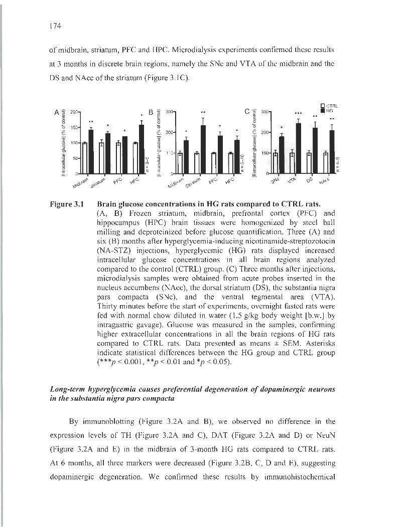
174
of midbrain, striatum, PFC and HPC. Microdialysis experiments confirmed these results
at 3 months in discrete brain regions, namely the SNc and VTA of the midbrain and the
DS and NAcc of the striatum (Figure 3.1C).
D CTRL
A ~ 200 B ~ 300- ** c ~ 300- *** . HG
* .= ë' ë' c: ** ** 8 ** 8 8 0 150- * 0 * 0 * ~ * ê 200- * * ~ 200-Q) Q) Q)
'" 100- ~ ~ '" '" 8 8 8 " " "
~ Cl Cl 100- ~ ~ ~
Cl ri1 li; ~ a ~ 100
'" ~ 50 .,., ~ 1" "
~ .,l,
~ .,., ~
~ " ~ " ~ c: c: .§. 0 .§. !:!:!. 0
. ~ <" ç~c, y-,çc, !iP . ~v<" ,?~c, y-,,?c, S ..... " ~I>- OS ..... 1>-,," . Ôv;,I'1>' . '1>,-:; ","' S'''' ~,Ôv;, S,I''I>
Figure 3.1 Brain glucose concentrations in HG rats compared to CTRL rats. (A, B) Frozen striatum, midbrain, prefrontal cortex (PFC) and hippocampus (HPC) brain tissues were homogenized by steel ball milling and deproteinized before glucose quantification. Three (A) and six (B) months after hyperglycemia-inducing nicotinamide-streptozotocin (NA-STZ) injections, hyperglycemic (HG) rats displayed increased intracellular glucose concentrations in all brain regions analyzed compared to the control (CTRL) group. (C) Three months after injections, microdialysis samples were obtained from acute probes inserted in the nucleus accumbens (NAcc), the dorsal striatum (DS), the substantia nigra pars compacta (SNc), and the ventral tegmental area (VTA). Thirty minutes before the start of experiments, ovemight fasted rats were fed with normal chow diluted in water (1.5 g/kg body weight [b.w.] by intragastric gavage). Glucose was measured in the samples, confirming higher extracellular concentrations in ail the brain regions of HG rats compared to CTRL rats . Data presented as means ± SEM. Asterisks indicate statistical differences between the HG group and CTRL group (***p < 0.001, **p < 0.01 and *p < 0.05).
Long-term hyperglycemia causes preferential degeneration of dopaminergic neurons in the substantia nigra pars compacta
By immunoblotting (Figure 3.2A and B), we observed no difference in the
expression levels of TH (Figure 3.2A and C), DAT (Figure 3.2A and D) or NeuN
(Figure 3.2A and E) in the midbrain of 3-month HG rats compared to CTRL rats .
At 6 months, all three markers were decreased (Figure 3.2B, C, D and E), suggesting
dopaminergic degeneration. We confirmed these results by immunohistochemical
<D .j.
" c:
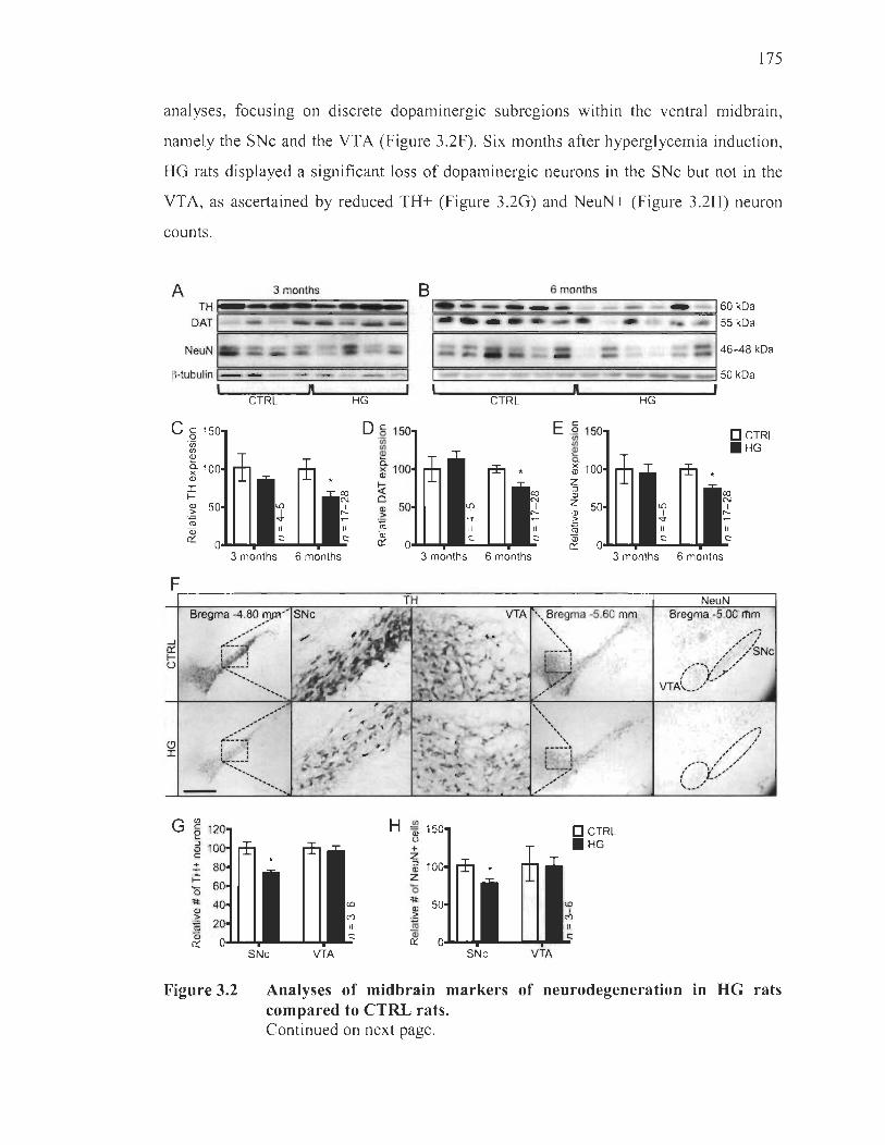
175
analyses, focusing on discrete dopaminergic subregions within the ventral midbrain,
namely the SNc and the YTA (Figure 3.2F). Six months after hyperglycemia induction,
HG rats displayed a significant loss of dopaminergic neurons in the SNc but not in the
VTA, as ascertained by reduced TH+ (Figure 3.2G) and NeuN+ (Figure 3.2H) neuron
counts.
A 3 months
TH~==~~~~~~ DAT
F==-~~==-======-~
NeuN
~-tubu l in 1- -
Cc 1 o "in II)
~ a. )( Q)
J: f-
~ ~ Qi 0::
1 ft CTRL
3 months 6 months
HG
Da 1 "in II)
~ ~1 Q)
~ o ~ ~ ~
6 months
ft CTRL
E .§ 1 II) II)
~ a. ~ 1 z :J Q)
Z Q) > :; Qi 0::
3 months 6 months
HG
3 months 6 months
D CTRL . HG
F~ __________________ -=~ ____________________ ~ __ ~~ __ ~ NeuN
Bregma -4.80 ~" SNe " "" ,..
r'!·::'- .. :
,,' " .. ",
,-'
(9 t'A'!.:_--, ' , ' J: , '
~:.-.~~ .... '. ".
G II) c 1 e :J Q) 1 c + J: f-0 ~ Q) > "" lU Qi 0::
SNe VTA
H lE 1 Qi U
+ Z :J 1 Q)
Z 0 ~ Q)
.~ ni Qi 0::
SNe
VTA " , Bregma -5.60 mm "~\~
'loI\.\
~----~ , ' , ' :
VTA
D CTRL . HG
Bregma -5.00 rflm
Figure 3.2 Analyses of midbrain markers of neurodegeneration in HG rats compared to CTRL rats. Continued on next page.

176
(Continued.) (A-E) Immunoblotting analyses of midbrain homogenates. A SDS-PAGE was performed on steel baIl milled frozen midbrain homogenates. The midbrain contains the SNc and the VT A. (A, B) Representative immunoblots of dopaminergic markers TH and dopamine transporter (DAT), and the general neuronal marker NeuN 3 (A) and 6 (B) months following hyperglycemia-inducing NA-STZ injections. (C-E) TH (C), DAT (D) and NeuN (E) expression levels are lower in HG rats 6 months, but not 3 months, after injections compared to CTRL rats. Expression levels are normalized to ~-tubulin. Blots were independently repeated at least 3 times each. (F-H) Immunohistochemical analyses of the midbrain region. Six months after hyperglycemia-inducing NA-STZ injections, frozen post-fixed brain hemispheres were cut into 20 )..lm-thick coronal seriai sections and stained for the dopaminergic neuronal marker TH or the general neuronal marker NeuN. (F) Representative midbrain microphotographs of TH staining at 2 different anteriorities and of NeuN staining. The midbrain subregions include the SNc and the VTA, isolated by dotted lines in the NeuN microphotograph. Scalebar 1 mm. (G, H) Cell counts in the SNc and VTA demonstrated lower numbers of both TH+ (G) and NeuN+ (H) cells in the SNc of HG rats compared to CTRL rats. No differences were found in the VT A. AlI data presented as means ± SEM. Asterisk indicates statistical differences between the HG group and CTRL group (*p < 0.05).
Long-term hyperglycemia causes preferential degeneration of dopaminergic fiber terminais in the dorsal striatum
By immunoblotting (Figure 3.3A and B), we observed no difference in the
expression levels of TH (Figure 3.3A and C), DAT (Figure 3.3A and D) or NeuN
(Figure 3.3A and E) in the striatum of 3-month HG rats compared to CTRL rats.
At 6 months, HG rats expressed lower levels of the dopaminergic markers TH
(Figure 3.3B and C) and DAT (Figure 3.3A and D), indicative of dopaminergic fiber
terminal degeneration in the striatum. NeuN, mainly expressed in the ceIl bodies of
neurons, remained unchanged in HG rats at 6 months (Figure 3.3B and E), suggesting
that no neurons were lost in the striatum. Irnmunohistochemical assays in the various
subregions of the striatum (Figure 3.3F) confirmed a significant loss of dopaminergic
fiber terminaIs in 6-month HG rats, especiaIly in the DS, both in the DMS and DLS
subregions, as ascertained by quantification of the density of TH+ fibers (Figure 3.3G).
No distinctions between 6-month HG and CTRL rats were detected in the ventral
regions of the striatum, namely the NAcc and the OT (Figure 3.3G). NeuN+ ceIl counts

177
did not differ between CTRL and HG groups in any of the reglOns investigated
(Figure 3.3H), corroborating previous immunoblotting observations of unchanged
striatal NeuN expression levels.
Degeneration of dopaminergic fiber terminaIs may be accompanied by alterations
in regional DA release (G6rska et al., 2017). To address this possibility, microdialysis
samples were collected at 3 months in the main central DA terminaIs, namely the DS,
the N Acc and the PFC. We noticed that, even in the absence of TH loss made apparent
at 3 months by immunoblotting (Figure 3.3A and C), HG rats displayed impaired basal
DA release in the DS, but not in the other brain regions (Figure 3.31). Together, these
data suggest early (3 months) dopaminergic fiber terminal dysfunction, and later
(6 months) degeneration in the DS coinciding with loss of neurons in the SNc where the
fibers originate. It is also possible that a moderate amount of neurodegeneration was
present at 3 months that we failed to detect by immunoblotting, due to the fewer
numbers of rats employed at this time point. If this were the case, dampened dopamine
release at' 3 months wou Id coincide with this loss of dopaminergic neurons or fibers.
Immunohistochemical analyses of brains at 3 months are required to confirm either of
these hypotheses. The main finding remains that neurodegeneration was observed in
6-month HG rats, as corroborated by 2 different techniques.
A 3 months
THt DAT r.:, ~~===~~::;~
NeuN 1
f{-tubulin EI-~~ëai~~===:::=::~~!!!!~
Cc 1 o ïii fJ)
~ ~1 Q)
I ~ Q) > ~ Qi 0::
, ft CTRL HG
3 months 6 months
6 months
~~~~=======================960kDa 55 kDa
46--48 kDa
~ ____________________________ ~50kDa
ft CTRL
3 months 6 months
HG
3 months 6 months
OCTRL . HG
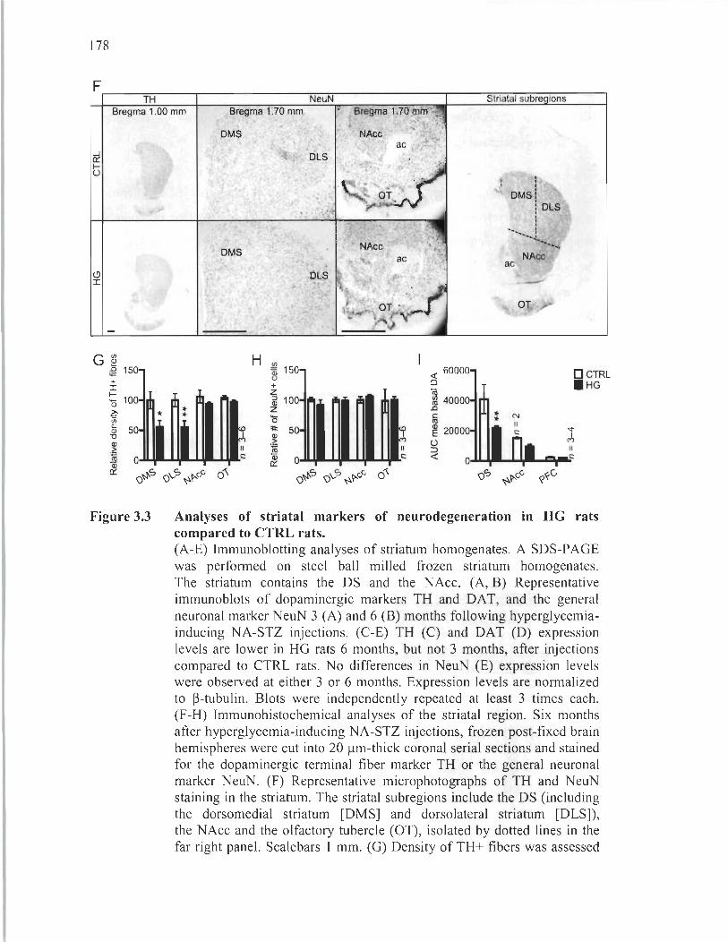
178
F~ __ ~ ____ ~ __________ ~~ __________ -. ____ ~~-.-~ ____ ~ TH NeuN Striatal subre ions
Bregma 1.00 mm Bregma 1.70 mm
...J oc 1-t)
+ I
!: 10 o j?;'in 5i "0
~ ro Qi oc
Figure 3.3
* ... ...
OMS
Il c:
H
Il c:
oC{ 60000 Q
ë5 ~ 40000 .0 c: ro ~ 20000 t) => oC{
1
: OMS:
: OLS 1 1
: __ 1
.............. ~ ........... "'~
NAcc ac
OT
: N Il c:
D CTRL HG
Analyses of striatal markers of neurodegeneration in HG rats compared to CTRL rats. (A-E) Immunoblotting analyses of striatum homogenates. A SDS-PAGE was performed on steel ball milled frozen striatum homogenates. The striatum contains the DS and the NAcc. (A, B) Representative immunoblots of dopaminergic markers TH and DAT, and the genera1 neuronal marker NeuN 3 (A) and 6 (B) months following hyperglycemiainducing NA-STZ injections. (C-E) TH (C) and DAT (D) expression levels are lower in HG rats 6 months, but not 3 months, after injections compared to CTRL rats. No differences in NeuN (E) expression levels were observed at either 3 or 6 months. Expression levels are normalized to ~-tubu1in. B10ts were independently repeated at least 3 times each. (F-H) Immunohistochemical analyses of the striatal region. Six months after hyperglycemia-inducing NA-STZ injections, frozen post-fixed brain hemispheres were cut into 20 Ilm-thick coronal seriaI sections and stained for the dopaminergic terminal fiber marker TH or the general neuronal marker NeuN. (F) Representative microphotographs of TH and NeuN staining in the striatum. The striatal subregions include the DS (including the dorsomedial striatum [DMS] and dorsolateral striatum [DLS]), the NAcc and the olfactory tubercle (OT), iso1ated by dotted lines in the far right panel. Scalebars 1 mm. (G) Density of TH+ fibers was assessed

179
by densitometric assays in the DMS, DLS, NAcc and the OT, showing less dense dopaminergic terminal fibers in the DMS and DLS, but not in the NAcc or the OT, of HG rats compared to CTRL rats. (H) Cell counts in the DMS, DLS, NAcc and OT demonstrated no differences between HG and CTRL rats. (1) Three months after injections, microdialysis samples were obtained from the NAcc, the DS and the PFC. Measurements of DA in the samples were performed by highperformance liquid chromatography and revealed lower extracellular concentrations in the DS, but not in the NAcc or the PFC, in HG rats compared to CTRL rats. Ali data presented as means ± SEM. Asterisk indicates statistical differences between the HG group and CTRL group (*p < 0.05, **p < 0.01). ac, anterior commissure; AUC, area under curve; DA, dopamine.
Long-term hyperglycemia does not cause substantial neurodegeneration in the prefrontal cortex or in the hippocampus
In the PFC and the HPC, we report no sensible changes of TH or DAT expression
at either 3 (Figure 3.4A, C and D; Figure 3.5A, C and D) or 6 months (Figure 3.4B, C
and D; Figure 3.5B, C and D) as revealed by immunoblotting assays (Figure 3.4A
and B; Figure 3.5A and B). In addition, hyperglycemia sustained for 6 months did not
modulate NeuN expression (Figure 3.4A, Band E; Figure 3.5A, Band E), suggesting no
visible degeneration in neuronal cell bodies housed in the PFC or the HPC.
Immunohistochemical analyses confirmed these findings since no changes in NeuN+
cell counts were observed in HG rats at 6 months compared to the CTRL group
(Figure 3.4F and G; Figure 3.5F and G). Lack of neuronal degeneration in the HPC is
corroborated by another study in long-term diabetic mice (Duarte et al. , 2012).

180
A 3 months 6 months
-160kOa - 155 kOa
TH~======~==========~ OAT
_ \ 46-48 kOa NeuN
F==================9
~-tu bulin L----'-__ -=-=-____ ---J 150 kOa
Cc 1 o "iii Vl
~ ~1 <Il :r: 1-<Il
.~ ro Qi ex:
CTRL
3 months
n HG CTRL
6 months 3 months 6 months
n 1 HG
E .Q 1 D CTRL Vl Vl . HG ~ a. ~ 1 z :::J <Il Z <Il
.~ ro Qi ex:
3 months 6 months
F~--~==----~------~----~ G ~ 1
Figure 3.4
u + z a: 1 z '0 'II: <Il
.~ ro Qi ex:
CTRL HG
Analyses of prefrontal markers of neurodegeneration in HG rats compared to CTRL rats. (A-E) Irnmunoblotting analyses of PFC homogenates. A SDS-PAGE was perfonned on steel baIl milled frozen PFC homogenates. (A, B) Representative immunoblots of dopaminergic markers TH and DAT, and the general neuronal marker NeuN 3 (A) and 6 (B) months following hyperglycemia-inducing NA-STZ injections. ~-tubulin blots served as a loading standard. (C-E) No differences in TH (C), DAT (D) or NeuN (E) expression levels were observed at either 3 or 6 months. Expression levels are nonnalized to ~-tubulin . Blots were independently repeated at least 3 times each. (F, G) Irnmunohistochemical analyses of the PFC region. Six months after hyperglycemia-inducing NA-STZ injections, frozen post-fixed brain hemispheres were cut into 20 Ilm-thick coronal seriaI sections and stained for the general neuronal marker NeuN. (F) Representative microphotographs of NeuN staining. Prelimbic (PL) and infralimbic (IL) subregions of the medial PFC are isolated by dotted lines. Scalebar 1 mm. (G) No differences in NeuN+ cell counts in the PFC of HG rats compared to CTRL rats. Ali data presented as means ± SEM. cc, corpus callosum.

A 3 months
DAT F=================~
NeuN
181
B.-__ ~ ________ 6_m_o~n~th~s __ ~~ ______ ~
~============================~160kDa ~============================~155kDa
146-48 kDa ~-tubulin L...-____ -'--______ -=----':........:~ F=============================~150kDa
n CTRL
C c 1 0 ëi) III
~ ~1 <Il
I f-<Il > fil Qi ex:
HG
o § 1 'iii III
~ ~1 <Il
~ a <Il
.~ Cil Qi ex:
~ ____ ~~ ____ ~n~ ____ ~~ ____ ~, CTRL HG
O CTRL .HG
3 months 6 months 3 months 6 months 3 months 6 months
F G III
~ 1 Bregma -3.00 mm
o + z
Figure 3.5
al 1 z o 'II:
.~ Cil Qi ex:
CTRL HG
Analyses of hippocampal markers of neurodegeneration in HG rats compared to CTRL rats. (A-E) Immunoblotting analyses ofHPC. A SDS-PAGE was performed on steel bail milled frozen HPC homogenates. (A, B) Representative immunoblots of dopaminergic markers TH and DAT, and the general neuronal marker NeuN 3 (A) and 6 (B) months following hyperglycemiainducing NA-STZ injections. B-tubulin blots served as a loading standard. (C-E) No differences in TH (C), DAT (D) or NeuN (E) expression levels were observed at either 3 or 6 months. Expression levels are normalized to B-tubulin. Blots were independently repeated at least 3 times each. (F, G) Immunohistochemical analyses of the HPC region. Six months after hyperglycemia-inducing NA-STZ injections, frozen post-fixed brain hemispheres were cut into 20 J..lm-thick coronal seriai sections and stained for the general neuronal marker NeuN. (F) Representative microphotographs of NeuN staining in the dentage gyms (DG), Cornu ammonis (CA) 1 and 3 regions of the HPC. Scalebars 0.5 mm. (G) No differences in NeuN+ cell counts in the HPC of HG rats compared to CTRL rats. Ali data presented as means ± SEM.

182
Long-term hyperglycemic rats display astrogliosis and loss of microglial cells in degenerated dopaminergic regions
Six months following hyperglycemia induction, HG rats exhibitedgreater GF AP+
staining revealed by immunohistochemistry in the SNc and DS (Figure 3.6A),
both regions displaying dopaminergic degeneration. Interestingly, these same regions
also showed a significant decrease of Tha+ staining (Figure 3.6A). In 6-month HG rats,
this apparent astrogliosis and 10ss of microglial cells was confirmed by GF AP+
(Figure 3.6B) and Thal + (Figure 3.6C) cell counts. Although we could not demonstrate
neurodegeneration in the NAcc (Figure 3.3F, Gand H), we detected an increase in
numbers of astroglial cells (Figure 3.6A and B) together with a 10ss of microglial cells
(Figure 3.6A and C) in HG rats compared to CTRL rats. The PFC, on the other hand,
displayed signs of astrogliosis (Figure 3.6A and B) without a decrease in Thal + cells
(Figure 3.6A and C). No effects were identified in the HPC, the VTA (Figure 3.6A, B
and C) or the OT (Figure 3.6B and C).
E E
E o
UM Cl. ,
l '" E
~
CTRL GFAP
HG
"
CTRL
' . . ..
Iba1
. .,,~" yI' ~
/' SNr
' .. '.
HG
"

B ~ 200 u + Cl. 150 II ~ 100 o
=Il:
~ 50 ~
• •
'f C')
Il
c ~ 150 al u + .-:8 100 •
'0 =Il: al >
50
~
183
O CTRL . HG
te ~ Il
~ O~,.. ...... - Qi - - .. c: cr 0 - - c:
0'<" 0'<" ~u Cj
Figure 3.6
-t-'?" ~t:-.J Nt:-.J UU f<v g,v ~u r<-'?" ~t:-.J Nt:-.J UU f<v g,v Q Q ~'?' q ~ Cj ~ Q Q ~'?' q ~
Immunohistochemical analyses of astrocytes and microglial cells in the brains of HG rats compared to CTRL rats. Six months after hyperglycemia-inducing NA-STZ injections, frozen post-fixed brain hemispheres were cut into 20 f.lm-thick coronal seriai sections and stained for the astrocytic marker glial fibrillary acidic protein (GFAP) or the microglial marker ionized ca1cium-binding adapter molecule 1 (Tha 1). (A) Representative microphotographs of GF AP or Thal staining in the different brain regions analyzed in this article with neuroanatomical landmarks isolated by dotted lines. The HPC microphotographs are taken from the CAl region and the PFC microphotographs are taken from the PL region. Not shown: OT and subregions of the DS (DMS and DLS). Scalebar 100 f.lm. (B) Cell counts in the various regions demonstrated higher numbers of GF AP+ cells in the SNc, DMS, DLS, NAcc and PFC of HG rats compared to CTRL rats. No differences were observed in other regions. (C) Cell counts in the various regions demonstrated lower numbers of Thal + cells in the SNc, DMS, DLS and NAcc of HG rats compared to CTRL rats. No differences were observed in other regions. Ail data presented as means ± SEM. Asterisk indicates statistical differences between the HG group and CTRL group (* **p < O.OOI , **p < O.OI and *p < 0.05) . SNr, substantia nigra pars reticulata.
Long-term hyperglycemic rats show altered motor behaviour
In the stepping test, HG rats took longer to cross the beam (Figure 3.7B) and made
more strides (Figure 3.7C) at 3 and 6 months compared to the CTRL group. Time to
initiate movement was not different between CTRL and HG groups at any time point
(Figure 3.7A). While no changes were detected at the 3-month mark in the horizontal
bar test, 6-month HG rats exhibited an increase in the latency to remove paws compared
to their baseline results and in relation to the CTRL group (Figure 3.7D). In the adjusting
step test, HG rats performed lower numbers of adjustments with either forepaw, in both
forehand (white bars for CTRL or black bars for HG) and backhand (horizontally lined

184
white bars for CTRL or checkered bars for HG) directions, compared to CTRL rats at
both time points (Figure 3.7E). In the NOR test, we did not find any difference in the
am ou nt of time HG rats spent exploring novel objects compared to the CTRL group
(Figure 3.7F). This was confirmed by expressing novel object exploration times in
relation to old object exploration times, yielding a discrimination index that did not
significantly contrast between CTRL and HG rats (Figure 3.7G).
A B 1.5 4
<il * '; 3 ~
CI)
g .9 2 al E F 1
al 1.0 E :;::; + + + + c: + + .Q 115 0.5 :g (0
Il
0.0 c: o
DCTRL . HG
+ ,.. ,.. ,..
(0 ..-
c CI)
al "0
15
~ 10 a Qi .0 E 5 :J Z
D 15 ,.. ,.. ,.. ,..
+,.. +,.. + + û) + ~10
()' c: 2 ro -l 5
(0
+ + +
*
(0
Il c:
O...olol.j~'"
Il
O·...I6o ..... ......,. __ ......... c:
~\0e ~\S'~ ~\S'~ <o'O~ (00 (00
e\\0e ~\S'~ ~\S'~ <o'O"i '!> (00 ~ (00
E CI)
~ 15 ûi Ol c:
"ëii 10 :J '5" ro a li 5 E :J
Z 0
-
-
-
-
'!> ~
-
-
o CTRL forehand a CTRL backhand • HG forehand Il HG backhand
+ + + +
~ + + + + + + + ~
,.. + ~ * ,.. ,..
1- .. * ri" ,.. ,.. - ,.. : ~ : ~ :
: ~ : ~
: :
Baseline 3 months 6 months
(0
1 1 c: ..
F 25
al 20 E ~ 15 o
~ 10 o ëi ~ 5
o
G
CTRL HG
Figure 3.7 Behavioral assessments of HG rats compared to CTRL rats. Three or six months after hyperglycemia-inducing NA-STZ injections, rats were subjected to behavioral tests to evaluate the presence of motor (A-E) or memory impairments (F, G). (A-C) A stepping test was conducted, consisting in a beam-crossing task. Both groups improved their time to initiate movement at 3 and 6 months compared to baseline, although no differences were observed between CTRL and HG rats (A). At 3 and 6 months, however, HG rats took longer than CTRL rats to cross the beam, and their results at 6 months grew significantly different from baseline (B). HG rats also made more strides to cross the beam than

Discussion
185
CTRL rats at 3 and 6 months, and only the latter performed better than baseline tests at both 3 and 6 months (C). (D) For the horizontal bar test, descent latency was measured as the time taken for rats to remove both forepaws from a horizontal bar. At 6 months, but not 3 months, HG rats took longer to remove both paws compared to CTRL rats and their own baseline. (E) During the forepaw adjusting step test, the rat was moved slowly sideways along the table surface by an experimenter, first in a forehand and then in a backhand direction. Numbers of adjusting steps made were fewer in HG rats at 3 and 6 months compared to CTRL rats and their own baseline. (F, G) A novel object recognition task, based on exploratory behavior of newly introduced elements in an environment, was used to assess non-spatial working memory in rats at 3 months. Rats were not tested at 6 months due to loss of eyesight (cataracts) of HG rats. Both CTRL and HG groups explored the nover object for a longer period of time than the old object (F). No differences were seen between groups in their capacity to discriminate old and nover objects (G). AU data presented as means ± SEM. Asterisk indicates statistical differences between the HG group and CTRL group (***p < 0.001 and *p < 0.05). Plus signs indicate statistical differences between results at the 3- or 6-month time point and the baseline evaluation (+++p < 0.001 , ++p < 0.01 and +p < 0.05). For the novel object recognition (NOR) task, circles indicate statistical differences between exploration time of the novel object and the old object (OOp < 0.01 and Op < 0.05).
The CUITent study sheds a new light on neuronal degeneration in long-term
hyperglycemia, innovative in that the condition was extended to a penultimate length of
6 months in the NA-STZ rat model without the need for glycemia-Iowering treatments.
The noxious effects of long-term hyperglycemia were evaluated in the nigrostriatal
and mesocorticolimbic pathways. Our findings are relevant to elucidate the interplay
between hyperglycemia and age-related neurodegenerative conditions, such as
Parkinson' s disease.
To the best of our knowledge, this is the first time that hyperglycemia-induced
neuropathological manifestations have been related to glucose measurements in brain
tissue homogenates and extracellular intracerebral microdialysates. Since glucose
overload is one of the major contributors to diabetic complications (Brownlee, 2005 ,
Tomlinson and Gardiner, 2008), the sole observation that glucose levels are significantly

186
increased indiscriminately in all brain regions studied herein raises the question as to
why sorne structures degenerate preferentially over time. It is now well appreciated that
oxidative stress is a key player in the nigrostriatal degeneration observed in Parkinson ' s
disease (Schapira and Jenner, 20 Il), a relative susceptibility attributable to various
factors: 1) All dopaminergic neurons are submitted to oxidative insults from dopamine
auto-oxidation and the elevated activity of mono amine oxidase (Goldstein et al., 2014);
2) Specifically, the SNc holds high levels of iron ions that generate highly reactive
hydroxyl radicals, but also possesses low levels of the ubiquitous antioxidant molecule
glutathione (Chinta and Andersen, 2008); 3) Dopaminergic nigrostriatal neurons are
endowed with an exceptionally dense dendritic arborization, rich in mitochondria and
constantly requiring costly metabolic sustenance (Surmeier et al., 2017). On the basis of
these physiological peculiarities, we argue that the relative vulnerability of the
nigrostriatal pathway, as compared here to the mesocorticolimbic pathway, stems from
its elevated basal oxidative burden paired with its scanty survival mechanisms faced to
sustained hyperglycemia over the course of aging.
In our long-term model of hyperglycemia, other pathological indices manifested
themselves concurrently to dopaminergic neurodegeneration, expressly a prominent
astrogliosis and loss of micro glial cells both in the SNc and the DS. Astrogliosis has
been observed in several animal models of diabetes (Nagayach et al., 2014, Rostami
et al., 2017), with implications for dopaminergic neuron death (Cabezas et al., 2014).
However, our present study constitutes an original report of the fate of glial cells
following long-term hyperglycemia, moreover demonstrating astrogliosis in the SNc of
STZ-treated rats for the first time. Microgliosis is well appreciated in retinas and
peripheral nerves of diabetic models (Mazzeo et al., 2017) though less well known in the
central nervous system (Nagayach et al., 2014, Oliveira et al., 2016). Yet, microglial cell
degeneration is an under-recognized feature in diabetes as well as in neuropathologies.
In our model, it is likely that microglial cells are lost due to the sustained oxidative
burden, supported by observations that this population is susceptible to oxidative stress
in health and in disease (Streit et al. , 2008). In fact, iron accumulation in the aging
human brain promotes oxidative stress and may lead to microglial degeneration
187
(Streit et al., 2008). lnterestingly, in patients deceased from septic shock, microglial
apoptosis was correlated to hyperglycemia, possibly due to lack of GLUT5
downregulation (Polito et al. , 2011). Other studies have found that advanced-glycation
end-products (AGEs), abundant in diabetic and aging brains, cause microglial apoptosis
in a dose-dependent manner (Khazaei et al., 2008, Sabokdast et al., 2015). Microglial
derived AGEs were further shown to promote dopaminergic neuron death (Bayarsaikhan
et al. , 2016). lnasmuch as previous reports reveal microgliosis in rodent models of
diabetes within 2 months (Nagayach et al., 2014, Oliveira et al. , 2016), we suggest that
microglial cells are activated early in hyperglycemia, in turn causing the activation and
proliferation of astrocytes (Liddelow et al. , 2017). The latter persist in high numbers at
6 months while microglial ce Ils degenerate over time due to the overwhelming oxidative
load of sustained hyperglycemia. Although confirmation of this tentative timeline is
required, it remains interesting that this astrogliosis paired to microglial cell loss
preferentially targets dopaminergic regions.
Motor deficits possibly ansmg from this nigrostriatal neurodegeneration
expressed themselves in tests employed to characterize motor impairment in rat models
of Parkinson's disease (Pinna and Morelli , 2014). ln particular, HG rats showed
bradykinesia and made greater numbers of sm ail steps, bearing likeness to motor
symptoms found in parkinsonian patients (Pinna and Morelli , 2014). Together with
immunohistochemical, immunoblotting and microdialysis studies, these data provide
new insight on the higher occurrence of Parkinson's disease in aging diabetic patients
(Cereda et al. , 2011 , Santiago and Potashkin, 2013, Sun et al. , 2012). One study
investigating blood biomarkers in 99 de nova patients of Parkinson ' s disease even found
their average fasting glycemia (5.64 mM) comparable to that of pre-diabetic individuals
(5.55 mM) (Santiago and Potashkin, 2015). Of high interest, recent clinical trials (Barker
et al. , 2013) are employing drug-repurposing strategies in Parkinson ' s disease patients
using anti-diabetic treatments such as exenatide (Aviles-Olmos et al., 2013) and
thiazolidinediones (NET-PD FS-ZONE, 2015). Whereas the link between Parkinson 's
disease and diabetes remains foggy, sorne have suggested shared dysfunctional cellular
pathways (Santiago and Potashkin, 2013) or neurochemical hypotheses (Deng et al.,

188
2012). Indeed, high reactivity between the glycation metabolite methylglyoxal and DA
was recently shown to yield a salsolinol-like toxin specifie to dopaminergic neurons
(Song et al., 2014). Interestingly, this toxin can be found in brains of parkinsonian
patients (Deng et al. , 2012).
In summary, our results mend our appreciation of the relationship that may exist
between diabetes and Parkinson's disease, by demonstrating the preferential nigrostriatal
dopaminergic neurodegeneration that occurs in long-term hyperglycemia. As it is
expressed in uncontrolled diabetes, hyperglycemia may cause premature aging of the
central nervous system, fostering the development of age-related neurodegenerative
disease.
Acknowledgments
Acknowledgments: The authors thank Dr. Michel Cyr (U Québec) for the use of
equipment and infrastructure for immunohistochemical studies, Dr. Gaetano Di Chiara
(U Cagliari) for the use of equipment and infrastructure for microdialysis experiments,
and Mr. Roberto Frau (U Cagliari) for assisting in microdialysis studies. We would like
to thank our local pharmacist in Trois-Rivières, Dr. L. Trudel, for providing us with
thousands of glucose meter strips at a reduced priee.
Funding: This research was funded by grants from the Natural Sciences and
Engineering Research Council (NSERC) of Canada no. 04321 to M.G.M. and by funds
from the Autonomous Region of Sardinia (L.R. n 7/2007-2015) to N.S. J.R. is recipient
of a Vanier Graduate Scholarship from the NSERC and a Doctoral Training Scholarship
from the Fonds de recherche en santé du Québec.

189
References
Anraku Y, Kuwahara H, Fukusato Y, Mizoguchi A, Ishii T, Nitta K, Matsumoto Y, Toh K, Miyata K, Uchida S, Nishina K, Osada K, ltaka K, Nishiyama N, Mizusawa H, Yamasoba T, Yokota T, Kataoka K. Glycaemic control boosts glucosylated nanocarrier crossing the BBB into the brain. Nat Commun 2017;8:1001.
Aviles-Olmos l, Dickson J, Kefalopoulou Z, Djamshidian A, EU P, Soderlund T, Whitton P, Wyse R, Isaacs T, Lees A, Limousin P, Foltynie T. Exenatide and the treatment of patients with Parkinson's disease. J Clin In vest 2013 ; 123 :2730-6.
Badole SL, Chaudhari SM, Jangam GB, Kandhare AD, Bodhankar SL. Cardioprotective Activity of Pongamia pinnata in Streptozotocin-Nicotinamide Induced Diabetic Rats. Biomed Res Int 2015 ;2015:403291.
Barker RA, Stacy M, Brundin P. A new approach to disease-modifying drug trials in Parkinson's disease. J Clin Invest 2013 ; 123 :2364-5.
Bassareo V, Musio P, Di Chiara G. Reciprocal responsiveness of nucleus accumbens sheU and core dopamine to food- and drug conditioned stimuli. Psychopharmacol 2011 ;214:687-97.
Bayarsaikhan E, Bayarsaikhan D, Lee J, Son M, Oh S, Moon J, Park HJ, Roshini A, Kim SU, Song BJ, Jo SM, Byun K, Lee B. Microglial AGE-albumin is critical for neuronal death in Parkinson's disease: a possible implication for theranostics. Int J Nanomedicine 2016;10 Spec Iss:281-92.
Bournival J, Francoeur MA, Renaud J, Martinoli MG. Quercetin and sesamin protect neuronal PC12 ceUs from high-glucose-induced oxidation, nitrosative stress, and apoptosis. Rejuvenation Res 2012 ;15 :322-33.
Brownlee M. The pathobiology of diabetic complications: a unifying mechanism. Diabetes 2005;54: 1615-25.
Cabezas R, Avila M, Gonzalez J, EI-Bacha RS, Beiez E, Garcia-Segura LM, Jurado Coronel JC, Capani F, Cardona-Gomez GP, Barreto GE. Astrocytic modulation of blood brain barrier: perspectives on Parkinson's disease. Front CeU Neurosci 2014;8:211.
Cereda E, BaricheUa M, Cassani E, Caccialanza R, Pezzoli G. Clinical features of Parkinson disease when onset of diabetes came first: A case-control study. Neurology 2012;78: 1507-11.
190
Chang JW, Wachtel SR, Young D, Kang UJ. Neuroseience 1999;88:617-28.
Chinta SJ, Andersen JK. Redox imbalance in Parkinson's disease. Biochim Biophys Acta 2008; 1780: 1362-7.
Deng Y, Zhang Y, Li Y, Xiao S, Song D, Qing H, Li Q, Rajput AH. Occurrence and distribution of salsolinol-like compound, 1-acetyl-6,7-dihydroxy-1,2,3,4-tetrahydroisoquinoline (ADTIQ) in parkinsonian brains. J Neural Transm (Vien na) 2012;119:435-41.
Di Chiara G, Tanda G, Frau R, Carboni E. On the preferential release of dopamine in the nucleus accumbens by amphetamine: further evidence obtained by vertically implanted concentric dialysis probes. Psychopharmacology 1993;112:398-402.
Do Nascimento PS, Lovatel GA, Barbosa S, Ilha J, Centenaro LA, Malysz T, Xavier LL, Schaan BD, Achaval M. Treadmill training improves motor skills and increases tyrosine hydroxylase immunoreactivity in the substantia nigra pars compacta in diabetic rats. Brain Res 2011;1382:173-80.
Duarte JM, Agostinho PM, Carvalho RA, Cunha RA. Caffeine consumption pre vents diabetes-induced memory impairment and synaptotoxicity in the hippocampus of NONcZNOI0/LTJ mice. PLoS One 2012;7:e21899.
Ennaceur A. One-trial object recognition in rats and mlce: methodological and theoretical issues. Behav Brain Res 2010;215:244-54.
Goldstein DS, Kopin 11, Sharabi y. Catecholamine autotoxicity. Implications for pharmacology and therapeutics of Parkinson disease and related disorders. Pharmacol Ther 2014; 144:268-82.
Gorska AM, Kaminska K, Wawrzczak-Bargiela A, Costa G, Morelli M, Przewlocki R, Kreiner G, Go!embiowska K. Neurochemical and Neurotoxic Effects of MDMA (Ecstasy) and Caffeine After Chronic Combined Administration in Mice. Neurotox Res 2017;33:532-48.
Haber SN. The place of dopamine in the cortico-basal ganglia circuit. Neuroseience 2014;282 :248-57.
Jacob RJ, Fan X, Evans ML, Dziura J, Sherwin RS. Brain glucose levels are elevated in chronically hyperglycemic diabetic rats: no evidence for protective adaptation by the blood brain barrier. Metabolism 2002;51:1522-4.
Jagota P, Bhidayasiri R, Lang AE. Movement disorders In patients with diabetes mellitus. J Neurol Sei 2012;314:5-11.

191
Kamboj SS, Sandhir R. Protective effect of N-acetylcysteine supplementation on mitochondrial oxidative stress and mitochondrial enzymes in cerebral cortex of streptozotocin-treated rats. Mitochondrion 20 Il; 11:214-22.
Khazaei MR, Habibi-Rezaei M, Karimzadeh F, Moosavi-Movahedi AA, Sarrafnejhad AA, Sabouni F, Bakhti M. Microglial cell death induced by glycated bovine serum albumin: nitric oxide involvement. J Biochem 2008;144:197-206.
Liddelow SA, Guttenplan KA, Clarke LE, Bennett FC, Bohlen CJ, Schirmer L, Bennett ML, Münch AE, Chung WS, Peterson TC, Wilton DK, Frouin A, Napier BA, Panicker N, Kumar M, Buckwalter MS, Rowitch DH, Dawson VL, Dawson TM, Stevens B, Barres BA. Neurotoxic reactive astrocytes are induced by activated microglia. Nature 2017;541 :481-7.
Lisman JE, Grace AA. The hippocampal-VTA loop: controlling the entry of information into long-term memory. Neuron 2005;46:703-13.
Lozovsky D, Saller CF, Kopin Il. Dopamine receptor binding is increased in diabetic rats. Science 1981;214:1031-3.
Masiello P, Broca C, Gross R, Roye M, Manteghetti M, Hillaire-Buys D, Novelli M, Ribes G. Experimental NIDDM: development of a new model in adult rats administered streptozotocin and nicotinamide. Diabetes 1998;47 :224-9.
Mazzeo A, Arroba AI, Beltramo E, Valverde AM, Porta M. Somatostatin protects human retinal pericytes from inflammation mediated by microglia. Exp Eye Res 2017;164:46-54.
Moulton CD, Costafreda SG, Horton P, Ismail K, Fu CH. Meta-analyses of structural regional cerebral effects in type 1 and type 2 diabetes. Brain Imaging Behav 2015;9:651-62.
Murzi E, Contreras Q, Teneud L, Valecillos B, Parada MA, De Parada MP, Hernandez L. Diabetes decreases limbic extracellular dopamine in rats. Neurosci LeU 1996;202: 141-4.
Nagayach A, Patro N, Patro 1. Astrocytic and microglial response in experimentally induced diabetic rat brain. Metab Brain Dis 2014;29:747-61.
NINDS Exploratory Trials in Parkinson Disease (NET -PD) FS-ZONE Investigators. Pioglitazone in early Parkinson's disease: a phase 2, multicentre, double-blind, randomised trial. Lancet NeuroI2015;14:795-803.

192
Oliveira WH, Nunes AK, França ME, Santos LA, Los DB, Rocha SW, Barbosa KP, Rodrigues GB, Peixoto CA. Effects of metforrnin on inflammation and short-terrn memory in streptozotocin-induced diabetic mice. Brain Res 2016; 1644: 149-60.
Patching SG. Glucose Transporters at the Blood-Brain Barrier: Function, Regulation and Gateways for Drug Delivery. Mol Neurobiol2017; 54: 1046-1077.
Paxinos G, Watson C. The Rat Brain in Stereotaxic Coordinates. 4th ed. Academic Press; 1998.
Pinna A, Morelli M. A critical evaluation of behavioral rodent models of motor impairrnent used for screening of antiparkinsonian activity: The case of adenosine A(2A) receptor antagonists. Neurotox Res 2014;25:392-401.
Pinna A, Pontis S, Borsini F, Morelli M. Adenosine A2A receptor antagonists improve deficits in initiation of movement and sensory motor integration in the unilateral 6-hydroxydopamine rat model ofParkinson's disease. Synapse 2007;61:606-14.
Polito A, Brouland JP, Porcher R, Sonneville R, Siami S, Stevens RD, Guidoux C, Maxime V, de la Grandmaison GL, Chrétien FC, Gray F, Annane D, Sharshar T. Hyperglycaemia and apoptosis of microglial cells in human septic shock. Cri! Care 2011;15:R131.
Rajabally YA, Stettner M, Kieseier BC, Hartung HP, Malik RA. CIDP and other inflammatory neuropathies in diabetes - diagnosis and management. Nat Rev NeuroI2017;13:599-611.
Renaud J, Bournival J, Zottig X, Martinoli MG. Resveratrol protects DAergic PC12 cells from high glucose-induced oxidative stress and apoptosis: effect on p53 and GRP75 localization. Neurotox Res 2014;25: 110-23.
Rostami F, Javan M, Moghimi A, Haddad-Mashadrizeh A, Fereidoni M. Streptozotocininduced hippocampal astrogliosis and insulin signaling malfunction as experimental scales for subclinical sporadic Alzheimer model. Life Sei 2017; 188: 172-85.
Sabokdast M, Habibi-Rezaei M, Moosavi-Movahedi AA, Ferdousi M, Azimzadeh-Irani E, Poursasan N. Protection by beta-Hydroxybutyric acid against insulin glycation, lipid peroxidation and microglial cell apoptosis. Daru 2015;23:42.
Sanberg PR, Pisa M, Fibiger HC. Kainic acid injections in the striatum alter the cataleptic and locomotor effects of drugs influencing dopaminergic and cholinergic systems. EurJ PharmaeoI1981;74:347-57.
193
Santiago lA, Potashkin lA. Shared dysregulated pathways lead to Parkinson' s disease and diabetes. Trends Mol Med 2013 ;19: 176-86.
Santiago lA, Potashkin lA. Blood Biomarkers Associated with Cognitive Decline in Early Stage and Drug-Naive Parkinson's Disease Patients. PLoS One 2015;10:eOI42582.
Schapira AH, Jenner P. Etiology and pathogenesis of Parkinson's disease. Mo v Disord 2011 ;26: 1049-55.
Sevak Rl, Koek W, Galli A, France CP. Insulin replacement restores the behavioral effects of quinpirole and raclopride in streptozotocin-treated rats. J Pharmacol Exp Ther 2007;320: 1216-23 .
Simola N, Bustamante D, Pinna A, Pontis S, Morales P, Morelli M, Herrera-Marschitz M. Acute perinatal asphyxia impairs non-spatial memory and alters motor coordination in adult male rats. Exp Brain Res 2008;185:595-60l.
Simpson lA, Carruthers A, Vannucci Sl. Supply and demand in cerebral energy metabolism: The role of nutrient transporters. J Cereb Blood Flow Metab 2007; 27: 1766-91.
Song DW, Xin N, Xie Bl, Li Yl , Meng L Y, Li HM, SchHippi M, Deng YL. Formation of a salsolinol-like compound, the neurotoxin, l-acetyl-6,7-dihydroxy-l ,2,3,4-tetrahydroisoquinoline, in a cellular model of hyperglycemia and a rat model of diabetes. Int J Mol Med 2014;33:736-42.
Squire LR, Berg D, Bloom FE, du Lac S, Ghosh A, Spitzer NC. Fundamental Neuroscience. 3rd ed. Academic Press; 2008.
Streit Wl, Miller KR, Lopes KO, Njie E. Microglial degeneration in the aging brain -bad news for neurons? Front Biosci 2008;13:3423-38.
Sun Y, Chang YH, Chen HF, Su YH, Su HF, Li CY. Risk of Parkinson disease onset in patients with diabetes: a 9-year population-based cohort study with age and sex stratifications. Diabetes Care 2012;35: 1047-9.
Surmeier Dl, Obeso lA, Halliday GM. Selective neuronal vulnerability in Parkinson disease. Nat Rev Neurosci 2017;18: 101-13.
Tanda G, Bassareo V, Di Chiara G Mianserin markedly and selectively increases extracellular dopamine in the prefrontal cortex as compared to the nucleus accumbens of the rat. Psychopharmacology 1996;123 :127-30.
194
Tomlinson DR, Gardiner NJ. Glucose neurotoxicity. Nat Rev Neurosci 2008;9:36-45 .
Vicente Miranda H, EI-Agnaf OM, Outeiro TF. Glycation in Parkinson's disease and Alzheimer's disease. Mov Disord 2016;31:782-90.
Vieira MNN, Lima-Filho RAS, De Felice FG. Connecting Alzheimer's disease to diabetes: Underlying mechanisms and potential therapeutic targets. Neuropharmacology 2017;pii:S0028-3908(17)30523-3 .
Vignini A, Giulietti A, Nanetti L, Raffaelli F, Giusti L, Mazzanti L, Provinciali L. Alzheimer's disease and diabetes: new insights and unifying therapies. Curr Diabetes Rev 2013;9:218-27.
West Ml. Introduction to stereology. Cold Spring Harb Pro toc 2012;8:pii: pdb.top070623.
195
Supplementary data
Metabolic follow-up and disease progression
Each individual rat's weight (Supplementary Figure SlA), food and water intake
(Supplementary Figure SlB), glycemia (Supplementary Figure SIC), and general health
were monitored on a regular basis. Glycemia was estimated twice a week and weight
was measured weekly. Food and water intake was assessed for group-paired cages,
since rats were not housed individually.
Oral glucose tolerance test: On the moming of their sacrifice, 6 months after
NA-STZ injections, we performed an oral glucose tolerance test (OGTT; Supplementary
Figure SIE and F) on 6 CTRL rats and 16 HG rats. Fasted rats (12-16 h) were
administered 2 glkg of a D-glucose solution by intragastric gavage. Approximately
0.5 mL of blood was collected by the jugular vein right before the OGTT (0 min) and
at 30, 60 and 120 min after administration of the bolus. Samples were kept on ice and
plasma glucose and insulin were quantified immediately after. Rats were lightly
anesthetized by isoflurane inhalation prior to blood sampling. Plasma glucose
concentrations following the terminal OGTT were measured using the Rat Glucose
Assay kit provided by Crystal Chem (Downers Grove, IL, USA), whereas plasma insulin
was quantified using a Milliplex Adipokine Magnetic Bead Multiplex Assay (Millipore;
Etobicoke, ON, Canada).
Glycated hemoglobin: In a different group of rats, plasma hemoglobin A1c
(HbA1c) levels (Supplementary Figure SlD) were assessed terminally at 3 or 6 months
following NA-STZ injections. On the moming of their sacrifice, intracardiac blood
samples from CTRL (3 months n = 4; 6 months n = 10) and HG (3 months n = 5;
6 months n = 16) rats were drawn when under deep anesthesia by isoflurane inhalation.
Glycated Hb was measured immediately using a rat HbAlc assay kit (Crystal Chem;
Downers Grove, IL, USA).
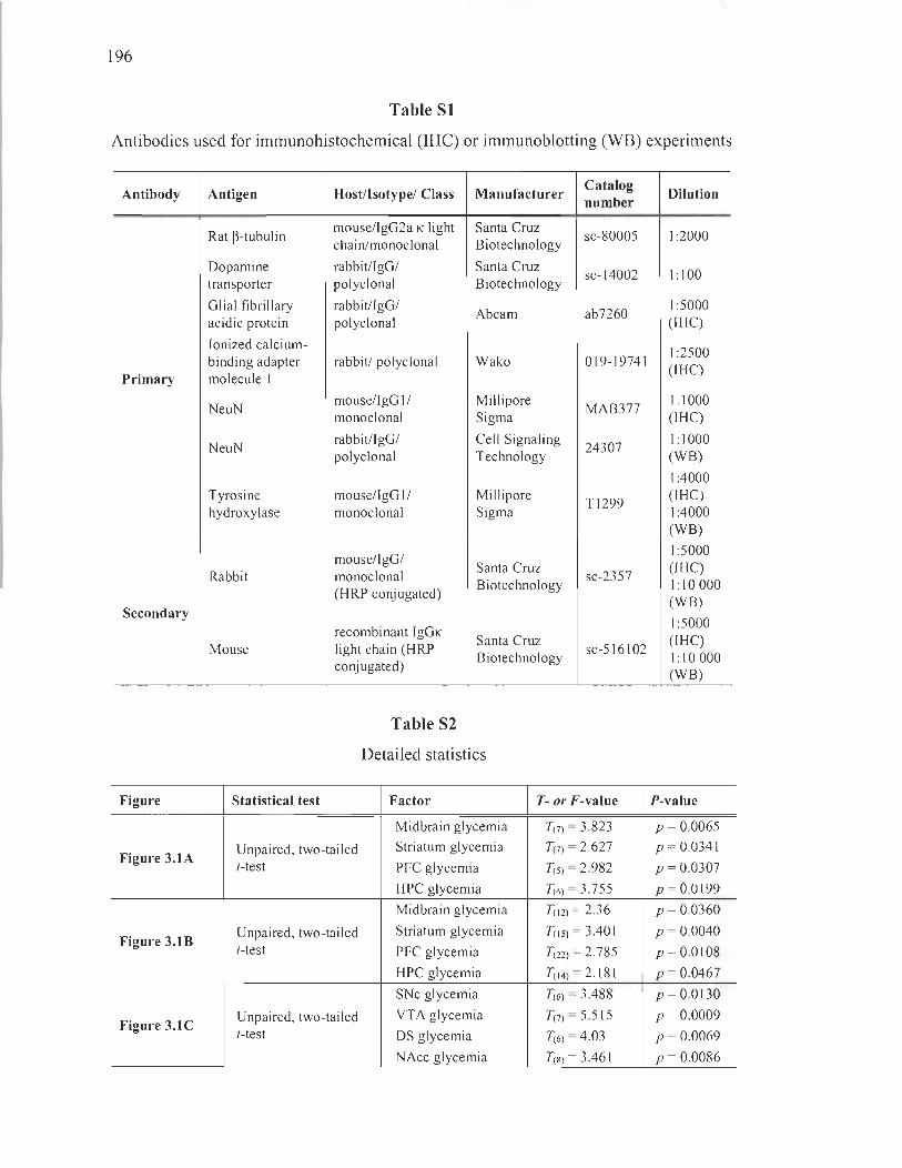
196
Table 81
Antibodies used for immunohistochemical (IRC) or immunoblotting (WB) experiments
Antibody Antigen Host/Isotype/ Class Manufacturer Catalog
Dilution number
Rat ~-tubulin mouselIgG2a K light Santa Cruz
sc-80005 1:2000 chain/monoclonal Biotechnology
Dopamine rabbit/IgG/ Santa Cruz sc-14002 1:100
transporter polyclonal Biotechnology
Glial fibrillary rabbit/IgG/ Abcam ab7260
1:5000 acidic protein polyclonal (IHC)
Ionized calcium-1 :2500
binding adapter rabbit/ polyclonal Wako 019-19741 (IHC)
Primary molecule 1
NeuN mouse/IgG 11 Millipore
MAB377 1:1000
monoclonal Sigma (IBC)
NeuN rabbit/IgG/ Cell Signaling
24307 1:1000
polyclonal Technology (WB)
1:4000 Tyrosine mouse/IgG 11 Millipore
Tl299 (IBC)
hydroxylase monoclonal Sigma 1 :4000 (WB)
mouse/IgG/ 1:5000
Rabbit monoclonal Santa Cruz
sc-2357 (IBC)
(HRP conjugated) Biotechnology 1: 10 000
(WB) Secondary
1:5000 recombinant IgGK
Santa Cruz (IBC) Mouse light chain (HRP
Biotechnology sc-516102
1:10000 conj ugated)
(WB)
Table 82
Detailed statistics
Figure Statistical test Factor T- or F-value P-value
Midbrain glycemia T(7) = 3.823 p = 0.0065
Figure 3.1A Unpaired, two-tailed Striatum glycemia T(7) = 2.627 p = 0.0341 (-test PFC glycemia T(5) = 2.982 P = 0.0307
HPC glycemia T(4) = 3.755 p = 0.0199
Midbrain glycemia T(12) = 2.36 P = 0.0360
Figure 3.1B Unpaired, two-tailed Striatum glycemia T(15) = 3.401 P = 0.0040 (-test PFC glycemia T(22) = 2.785 p = 0.0108
HPC glycemia T(14) = 2.181 P = 0.0467
SNe glycemia T(6) = 3.488 p = 0.0130
Figure 3.1C Unpaired, two-tailed VTA glycemia T(7)= 5.515 p = 0.0009 (-test DS glycemia T(6) = 4.03 P = 0.0069
NAcc glycemia T(8) = 3.461 P = 0.0086

197
Figure Statistical test Factor T- or F-value P-value
Figure 3.2C Unpaired, two-tailed 3 months glyeemia T(7) = 0.9727 P = 0.3631 (-test 6 months glyeemia T(43) = 2.589 p = 0.013
Figure 3.2D Unpaired, two-tailed 3 months glyeemia T(6) = 0.6376 P = 0.5473 (-test 6 months glyeemia T(17) = 2.274 P = 0.036
Figure 3.2E Unpaired, two-tailed 3 months glycemia T(7) = 0.2629 p = 0.8002 (-test 6 months glyeemia T(43) = 2.462 p = 0.018
Figure 3.2G Unpaired, two-tailed SNe glyeemia T(7)= 3.471 p = 0.0104 (-test YT A glyeemia T(7) = 0.4764 P = 0.6483
Figure 3.2H Unpaired, two-tailed SNe glyeemia T(7) = 2.439 p = 0.0448 (-test YTA glyeemia T(7) = 0.1207 P = 0.9073
Figure 3.3C Unpaired, two-tailed 3 months glyeemia T(7) = 0.5223 p = 0.6176 (-test 6 months glyeemia T(43) = 2.432 P = 0.019
Figure 3.3D Unpaired, two-tailed 3 months glyeemia T(7) = 0.9898 p = 0.3552 (-test 6 months glyeemia T(lo) = 2.256 P = 0.048
Figure 3.3E Unpaired, two-tailed 3 months glyeemia T(7) = 0.4726 p = 0.6509 (-test 6 months glyeemia T(42) = 0.5358 P = 0.595
DMS glyeemia T(7) = 2.443 P = 0.0446
Figure 3.3G Unpaired, two-tailed DLS glyeemia T(7) = 2.628 P = 0.0340 (-test NAee glyeemia T(7) = 1.495 p = 0.1785
OT glyeemia T(7) = 1.462 p = 0.1871
DMS glyeemia T(7) = 0.7135 P = 0.4986
Figure 3.3H Unpaired, two-tailed DLS glyeemia T(7) = 0.1507 P = 0.8845 (-test NAee glyeemia T(7) = 1.134 P = 0.2942
OT glyeemia T(7) = 0.0886 p = 0.9319
Region x glyeemia F(2 ,14) = 3.60 P = 0.0546
Figure 3.31 Two-way ANOY A Region F(2, 14) = 34.3 P < 0.0001
G1yeemia F(I ,14) = 7.59 P = 0.0155
Figure 3.4C Unpaired, two-tailed 3 months glyeemia T(7) = 0.4794 P = 0.6463 (-test 6 months glyeemia T(44) = 0.9042 P = 0.3708
Figure 3.4D Unpaired, two-tailed 3 months glyeemia T(7) = 0.30 13 p = 0.7719 (-test 6 months glyeemia T(l5) = 0.7387 P = 0.472
Figure 3.4E Unpaired, two-tailed 3 months glyeemia T(7) = 1.705 p = 0.1319 (-test 6 months glyeemia T(44) = 0.8821 P = 0.3825
Figure 3.4G Unpaired, two-tailed
PFC glyeemia T(7) = 1.168 p = 0.2810 (-test
Figure 3.5C Unpaired, two-tailed 3 months glycemia T(5) = 0.6179 P = 0.5637 (-test 6 months glyeemia T(45) = 0.6687 P = 0.5071
Figure 3.5D Unpaired, two-tailed 3 months glyeemia T(5) = 0.8448 P = 0.4368 (-test 6 months glyeemia T(25) = 1.272 p = 0.215
Figure 3.5E Unpaired, two-tailed 3 months glyeemia T(5) = 1.365 P = 0.2303 (-test 6 months glyeemia T(47) = 0.4349 P = 0.6656
Figure 3.5G Unpaired, two-tailed
HPC glyeemia T(7) = 0.9185 P = 0.3889 (-test
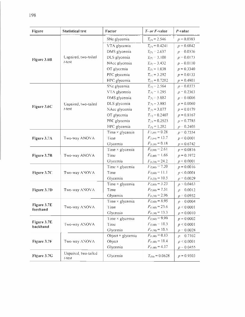
198
Figure Statistical test Factor T- or F-value P-value
SNc glycemia T(7) = 2.546 P = 0.0383
YT A glycemia T(7) = 0.4241 P = 0.6842
DMS glycemia T(7) = 2.637 P = 0.0336
Figure 3.6B Unpaired, two-tailed DLS glycemia T(7) = 3.100 p = 0.0173 (-test NAcc glycemia T(7) = 3.432 p = O.OllO
OT glycemia T(7) = 1.038 p = 0.3340
PFC glycemia T(7) = 3.292 p = 0.0133
HPC glycemia T(7) = 0.7282 p = 0.4901
SNc glycemia T(7) = 2.564 P = 0.0373
YTA glycemia T(7) = 1.295 p = 0.2363
DMS glycemia T(7) = 5.882 p = 0.0006
Figure 3.6C Unpaired, two-tailed DLS glycemia T(7) = 3.885 p = 0.0060 (-test NAcc glycemia T(7) = 3.077 P = 0.0179
OT glycemia T(7) = 0.2407 p = 0.8167
PFC glycemia T(7) = 0.2923 P = 0.7785
HPC glycemia T(7) = 1.282 P = 0.2405
Time x glycemia F (2,60) = 0.28 P = 0.7554
Figure 3.7A Two-way ANOY A Time F (2 ,60) = 13.7 P < 0.0001
Glycemia F(I ,30) = 0.18 P = 0.6742
Time x glycemia F (2 ,60) = 2.61 p = 0.0816
Figure 3.7B Two-way ANOY A Time F (2,60) = 1.66 p = 0.1972
Glycemia F (I ,30) = 24.2 p < O.OOOI
Time x glycemia F (2 ,60) = 7.20 p = 0.0016
Figure 3.7C Two-way ANOY A Time F (2 ,60) = 11 .1 P < 0.0001
Glycemia F(I ,30) = 10.5 P = 0.0029
Time x glycemia F (2 ,60) = 3.23 P = 0.0463
Figure 3.7D Two-way ANOY A Time F (2 ,60) = 7.51 p = 0.0012
Glycemia F(I ,30) = 2.96 P = 0.0952
Time x glycemia F (2 ,60) = 8.95 P = 0.0004 Figure 3.7E
Two-way ANOY A Time F (2 ,60) = 23 .6 p < O.OOO I forehand
Glycemia F(I ,30) = 13 .3 p = O.OOIO
Time x glycemia F (2,60) = 9.99 p = 0.0002 Figure 3.7E
Two-way ANOY A Time F (2 ,60) = 18.3 P < 0.0001 backhand Glycemia F(I ,30) = 10.5 P = 0.0028
Object x glycemia F (I ,60) = 0.13 P = 0.7102
Figure 3.7F Two-way ANOY A Object F(I ,60) = 18.4 P < 0.0001
Glycemia F(I ,60) = 4.17 P = 0.0455
Figure 3.7G Unpaired, two-tailed
Glycemia T(3o) = 0.0628 p = 0.9503 [-test
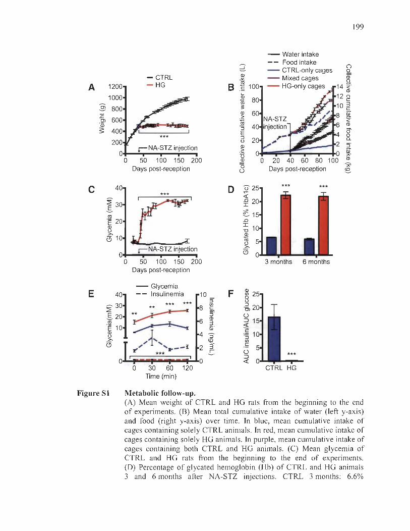
A 1200
1000
..9 800 E
600 Cl "iD s: 400
200
0
C 40
:2: 30 .S-CIl ·E 20 Q) u >. (5 10
0
- CTRL - HG 8
***
r NA-STZ injection
50 100 150 200 Days post-reception
0
199
- Water intake
2: -- Food intake
() - CTRL-only cages Q.
Q) - Mixed cages ëD ..l<:
CIl 0
ë 100 - HG-only cages 14 ~ ~ CD ....
12 8 Q) 80 -CIl 10 :3 :l: c
Q) 60 8 §I > ~
<. 40 6 CD
:::J i3' E 0 :::J 20 a. u 2 Q) :::J > 0 0 0) U ;;<;
~ 0 20 40 60 80 100 CD -"0 Days post-reception ;;<;
Ü ca
û ..--< .n I
0
~1 .n I 1 "0 Q) -CIl u >.
o o
r NA-STZ injection
50 100 150 200 Days post-reception
(5
3 months 6 months
E 40
-30 :2: .S- 20 CIl
·E 10 Q) u >. (5
0
Figure SI
- Glycemia -- Insulinemia 10 F
Q) CIl 0
*** :::J *** CIl
** 8 c
*~ :::J
6 CD :3
~ aï
~1', S-ca --J!~ ' !!" _- r :3 r *** -
0
U :::J Cl Ü ::J ~ C
:::J CIl C
Ü *** ::J -<
0 30 60 120 CTRL HG Time (min)
Metabolic follow-up. (A) Mean weight of CTRL and HG rats from the beginning to the end of experiments. (B) Mean total cumulative intake of water (left y-axis) and food (right y-axis) over time. In blue, mean cumulative intake of cages containing solely CTRL animais . In red, mean cumulative intake of cages containing solely HG animais. In purple, me an cumulative intake of cages containing both CTRL and HG animaIs. (C) Mean glycemia of CTRL and HG rats from the beginning to the end of experiments. (D) Percentage of glycated hemoglobin (Hb) of CTRL and HG animais 3 and 6 months after NA-STZ injections. CTRL 3 months: 6.6%
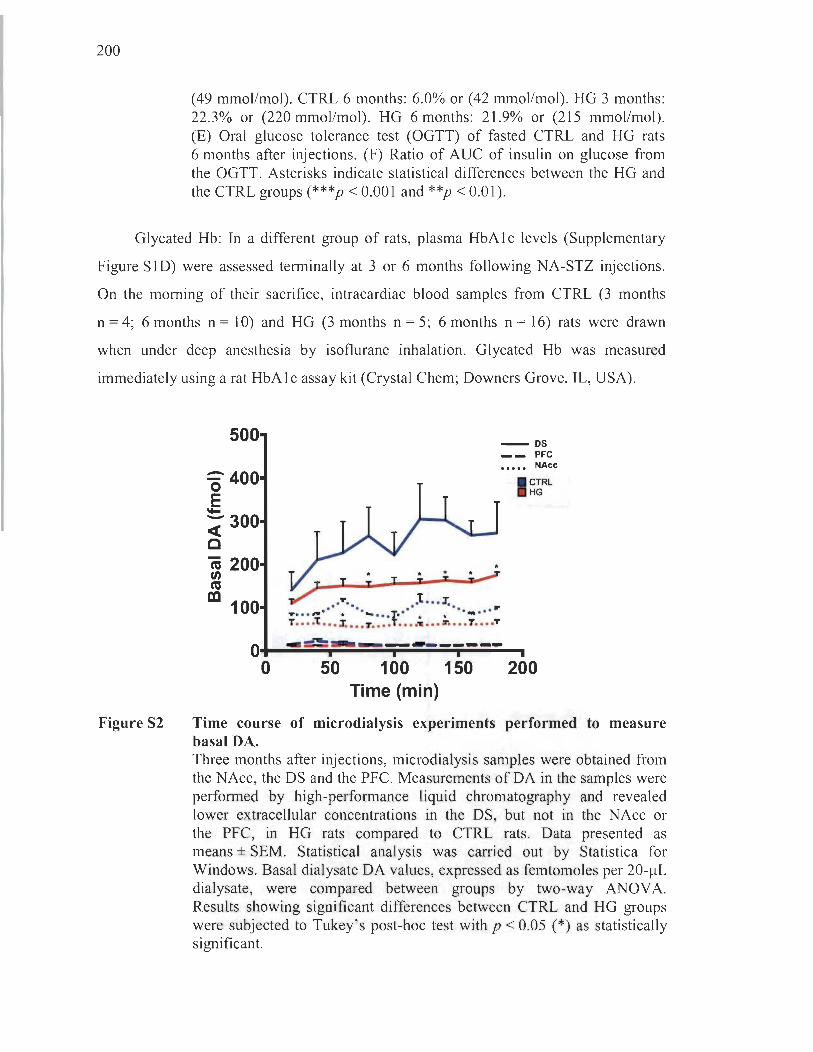
200
(49 mmol/mol). CTRL 6 months: 6.0% or (42 mmol/mol). HG 3 months: 22.3% or (220 mmol/mol). HG 6 months: 21.9% or (215 mmol/mol). (E) Oral glucose tolerance test (OGTT) of fasted CTRL and HG rats 6 months after injections. (F) Ratio of AUC of insulin on glucose from the OGTT. Asterisks indicate statistical differences between the HG and the CTRL groups (***p < 0.001 and **p < 0.01).
Glycated Hb: In a different group of rats, plasma HbAlc levels (Supplementary
Figure SlD) were assessed terminally at 3 or 6 months following NA-STZ injections.
On the moming of their sacrifice, intracardiac blood samples from CTRL (3 months
n = 4; 6 months n = 10) and HG (3 months n = 5; 6 months n = 16) rats were drawn
wh en under deep anesthesia by isoflurane inhalation. Glycated Hb was measured
immediately using a rat HbAlc assay kit (Crystal Chem; Downers Grove, IL, USA).
Figure 82
500
:-400 o ~ -300 ~ c ca 200 fi) cu al 100
* * * * ~ T T T T " .. ~
~ .or. .t ... x. ...... ,._ •• * ••• ~ ••• T.··· • * ... .. ... ". T •• • :t ... 3. ••• 7 •••• T •••• -,; ••• r .... r .... T
- os __ PFC
••••• NAce
I CTRL HG
O+-----~~-----~--.. -------r---------. o 50 100 150 200 Time (min)
Time course of microdialysis experiments performed to measure basal DA. Three months after injections, microdialysis samples were obtained from the NAcc, the DS and the PFC. Measurements of DA in the sampi es were performed by high-performance liquid chromatography and revealed lower extracellular concentrations in the DS, but not in the NAcc or the PFC, in HG rats compared to CTRL rats. Data presented as means ± SEM. Statistical analysis was carried out by Statistica for Windows. Basal dialysate DA values, expressed as femtomoles per 20-j.!L dialysate, were compared between groups by two-way ANOV A. Results showing significant differences between CTRL and HG groups were subjected to Tukey's post-hoc test with p < 0.05 (*) as statistically significant.
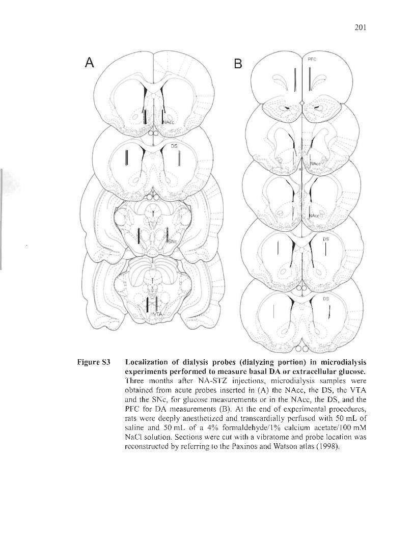
201
A B PFC
, . (
Î:-
( }~r ,
Il INA~C /"
Figure S3
~« - • ........ -o!
r )-.. /
Localization of dialysis probes (dialyzing portion) in microdialysis experiments performed to measure basal DA or extracellular glucose. Three months after NA-STZ injections, microdialysis samples were obtained from acute probes inserted in (A) the NAcc, the DS, the VTA and the SNc, for glucose measurements or in the NAcc, the DS, and the PFC for DA measurements (B). At the end of experimental procedures, rats were deeply anesthetized and transcardially perfused with 50 mL of saline and 50 mL of a 4% formaldehydell % calcium acetatell 00 mM NaCI solution. Sections were cut with a vibratome and probe location was reconstructed by referring to the Paxinos and Watson atlas (1998).

CHAPTERIV
LONG-TERM HYPERGL YCAEMIA MODIFIES SOCIAL BEHA VIOUR AND EMISSION OF ULTRASONIC VOCALISATIONS IN RATS : A POSSIBLE EXPERIMENT AL M O DEL OF AL TERED SOCIABILITY IN DIABETES
Renaud, 1. , Beaulieu, 1., Martinoli, M. G. , Simola, N.
The content of this chapter was submitted July 9th, 2018, to the peer-reviewed
journal Scientific Reports (SREP-18-25599).
4.1 Author contributions
Justine Renaud designed most of the study and perforrned 90% of the
manipulations. She wrote 95% of the manuscript, and prepared and edited ail the
figures. Jimmy Beaulieu, master's student in Maria-Grazia Martinoli's laboratory,
provided assistance for immunohistochemistry experiments. Nicola Simola mentored
Justine Renaud at the University of Cagliari during a short I-month training pro gram to
perforrn USV recordings. He offered expertise and assistance with the design of
USV experiments, and analyzed the audiograms. He also provided Figure 4.7.
Maria-Grazia Martinoli, Justine Renaud's research supervisor, was the guarantor of the
work and provided supervision, preparation and editing of the manuscript.
4.2 Résumé
Il ne fait plus de doute que le diabète soit un facteur de risque contribuant au
déclin cognitif et aux troubles de l 'humeur se manifestant avec l'âge. Néanmoins,
des études adressant les effets de l'hyperglycémie chronique sur les comportements
sociaux, tels que le jeu et l'agressivité, se font toujours attendre. Dans cette étude,
nous avions pour but d'évaluer les comportements sociaux manifesté par des rats
204
chroniquement hyperglycémiques. Dans cette optique, nous avons examiné les
interactions de type affiliation/exploration et agression entre des paires de rats dans une
enceinte neutre, et ce, 5 mois suivant l'induction de l'hyperglycémie dans le paradigme
nicotinamide-streptozotocine. Simultanément, les vocalisations ultrasoniques, porteuses
de l'état affectif des sujets, étaient enregistrées. Nos résultats démontrent que les rats
chroniquement hyperglycémiques sont manifestement agressifs et hyper-sociables dans
des contextes de nouveauté sociale. Ceux-ci émettent également beaucoup plus de
vocalisations ultrasoniques régulées en partie par le neurotransmetteur dopamine.
En fait, ces comportements corrélaient avec le degré de perte d'innervation
dopaminergique du striatum, mais pas avec des paramètres liés aux concentrations ou
aux fonctions de l' insul ine. En somme, nos résultats suggèrent un lien entre
l'hyperréactivité sociale causée par l'hyperglycémie chronique et la neurotransmission
dopaminergique dans le striatum, déjà reconnu pour son rôle modulateur sur les
comportements sociaux. Cette étude jette une nouvelle lumière sur de possibles substrats
neurobiologiques, soit la dopamine, pouvant sous-tendre les difficultés psychiatriques et
socioaffectives auxquelles font face les patients diabétiques.
205
4.3 Full article in English: Altered social behaviour in long-term hyperglycemic rats displaying dopaminergic striatal denervation: an ultrasonic vocalisation study
Abstract
Diabetes is a well-known risk factor of cognitive decline and mood disorders .
However, there is no adequate account of the effects of long-term hyperglycaemia on the
various dimensions of social behaviours, such as play and aggression. In this study,
we evaluated the social behaviour presented by the nicotinamide-streptozotocin
rat model of long-term hyperglycaemia. Five months following induction of
hyperglycaemia, we scored affiliative/exploratory or aggressive social interactions
between pairs of unacquainted rats in a neutral arena. Concurrently, we recorded
ultrasonic vocalisations (USVs), envisaged as behavioural markers of emotional states in
rats. Our results demonstrate alterations in the behaviour of long-term hyperglycaemic
rats faced with social novelty. Specifically, hyperglycaemic rats engaged in hyper
sociable and hyper-aggressive encounters, while emitting greater numbers of USVs.
The magnitude of social interactivity and vocalisations was associated with the degree of
striatal denervation observed in the brains of long-term hyperglycaemic rats, but not
with insulin levels or functions . Altogether, our data suggest a lack of social
appropriateness in long-term hyperglycaemic rats, which may be parti y linked to
changes in striatal dopamine known to regulate social behaviour. This study exposes
additional dopaminergic neural substrates that may underlie the social dimensions of the
psychiatric challenges faced by diabetic patients.
Introduction
The ability to suitably engage in social encounters occupies a significant role in
the lives of individuals, epitomized by the debilitating nature of the behavioural
inappropriateness manifested in neuropsychiatric disorders like autism, schizophrenia
and attention deficit hyperactivity disorder [1-4]. Although less acknowledged, altered
social behaviours are also a feature of diabetes. lndeed, recent studies link diabetes with

206
increased aggression and agitation in patients [5-7]. In addition, both hypoglycaemic
« 70 mg/dL or 3.88 mM) and hyperglycaemic (> 120 mg/dL or 6.66 mM) states are
known to foster hostility and aggression [8-11]. To date, very few studies have sought to
investigate social interactions in diabetic-like rodents and these have yielded highly
inconsistent results [12-14] . Moreover, none have employed long-term paradigms of
hyperglycaemia that could better represent the chronicity of diabetes in humans.
Perhaps for this reason, the possible neural substrates of inappropriate behaviours in
diabetes remain iU defined.
Ultrasonic vocalisations (USVs) are a valuable tool in social behavioural research,
carrying information on the affective state of rats during aggressive and playful
interactions [15-18]. USV scan be categorized based on average frequency and
frequency modulation in the two large families of 22-kHz caUs and 50-kHz caUs, which
are thought to have different behavioural significance [19]. USVs of 22-kHz are
considered markers of negative affective states, since rats emit these calls in aversive
situations, for example, during aggressive social interactions with conspecifics or upon
exposure to predators [20]. On the other hand, rats mainly emit 50-kHz USVs in
pleasurable situations such as mating and playful interactions with conspecifics or
familiar humans [15]. Moreover, 50-kHz US Vs can be further classified in the
two subcategories of frequency modulated (FM) and fiat caUs based on the modulation
of sound frequency [16]. FM caUs have a sound frequency that varies substantially,
whereas fiat caUs present a sound frequency that remains nearly unchanged within
individual caUs [16]. Moreover, FM 50-kHz USVs are thought to be tightly linked to
positive emotional states, and to function as affiliative calls to elicit approach behaviour
and to maintain playful interactions [21, 22]. Flat 50-kHz caUs, on the other hand, are
unclear in the exact functions they serve [23 , 24], but appear to bear a role in pacing
social behaviour, supposedly acting as ambivalent contact caUs [25,26].
In addition to conveymg information on the affective state of rats, USVs are
intimately linked with the activation of specific neuronal circuits [15]. In particular,
activation of the ascending mesolimbic dopaminergic system produces 50-kHz US Vs

207
[27, 28], whereas activation of the ascending mesolimbic cholinergie system produces
22-kHz caBs [15]. Notably, dopamine and acetylcholine extensively interact at the
striatal interface to dynamically regulate the affective state of rodents [15]. As such,
dopamine and acetylcholine antagonize each other in a cooperative system wherein one
or the other prevails in a context-dependent manner, allowing for the rapid and
appropriate activation of one of two states: positive or negative [29]. These states are
thus mutually exclusive and are rendered specifically in rats by the categories of USVs
emitted [15, 30]. From this perspective, it is reasonable to believe that alterations in
mesostriatal dopaminergic neurotransmission may be reflected at the behavioural level
by modifications in social interactions and USV emission.
A recent study by our group demonstrated the occurrence of mesostriatal
dopaminergic neurodegeneration in a rat model of long-term hyperglycaemia [31].
In light of these results, we evaluated herein the presence of social behavioural
abnormalities that may arise from hyperglycaemia-induced dopaminergic denervation
III the striatum. In particular, we measured the occurrences of specifie
affiliative/exploratory as weIl as aggressive behaviours in pairs of unacquainted rats in
a neutral, novel environment. Since no other studies have investigated USVs in
hyperglycaemic rats in a social context, we quantified the numbers of 22-kHz,
FM 50-kHz and flat 50-kHz calls emitted during these encounters. Lastly, we evaluated
the impact of the degree of striatal denervation on the magnitude of social interactivity
and USV emissions.
Results
Occurrences of social behaviours and ultrasonic vocalisations
The occurrences of affiliative/exploratory (dorsal contacts, allogrooming,
anogenital sniffmg and crawl-overs) and aggression-related (freezing, nose-offs, boxing,
pins) behaviours were scored during 10-min encounters between pairs of unacquainted
rats of the same group, namely control (CTRL) or hyperglycaemic (HG). Noticeably,
many behaviours occurred more often between pairs of HG rats compared to duos of
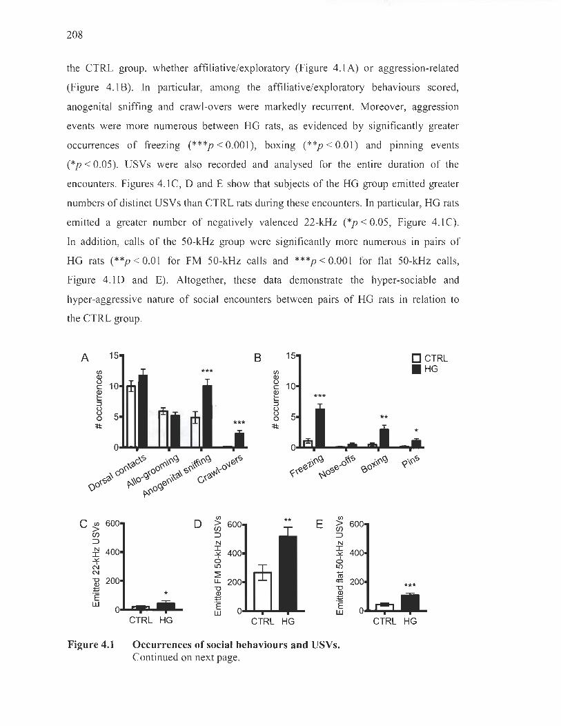
208
the CTRL group, whether affiliative/exploratory (Figure 4.1A) or aggression-related
(Figure 4.1B). In particular, among the affiliative/exploratory behaviours scored,
anogenital sniffing and crawl-overs were markedly recurrent. Moreover, aggression
events were more numerous between HG rats, as evidenced by significantly greater
occurrences of freezing (***p < 0.001), boxing (**p < 0.01) and pmnmg events
(*p < 0.05). USVs were also recorded and analysed for the entire duration of the
encounters. Figures 4. 1 C, D and E show that subjects of the HG group emitted greater
numbers of distinct USVs than CTRL rats during these encounters. In particular, HG rats
emitted a greater number of negatively valenced 22-kHz (*p < 0.05, Figure 4.1 C).
In addition, caUs of the 50-kHz group were significantly more numerous in pairs of
HG rats (**p < 0.01 for FM 50-kHz caUs and ***p < 0.001 for fiat 50-kHz caUs,
Figure 4.1D and E). Altogether, these data demonstrate the hyper-sociable and
hyper-aggressive nature of social encounters between pairs of HG rats in relation to
the CTRL group.
A 15
CIl Q) t)
10 c Q) 10... 10... :J t) t)
5 0 'lt:
c~ 600 Cf)
::J N 400 I
..:t::. 1
N N "0 200 Q) ~
·E w
0
Figure 4.1
B 15 DCTRL CIl . HG Q) t)
10 c Q) 10... 10... :J t) t)
5 ** 0 'lt:
0
1>~~ o~<:> ~~~ (;;~ee ~o<:>e- (()o
«\~<:>
CIl
0 > Cf) ::J N I ..:t::.
1 0 L()
:?: u.
* "0 ID ~ ·E w
CTRL HG
E
CTRL HG
~ Cf) ::J N I ..:t::.
1 o L() -ct! c;:: "0
~ ·E w
Occurrences of social behaviours and USVs. Continued on next page.
CTRL HG
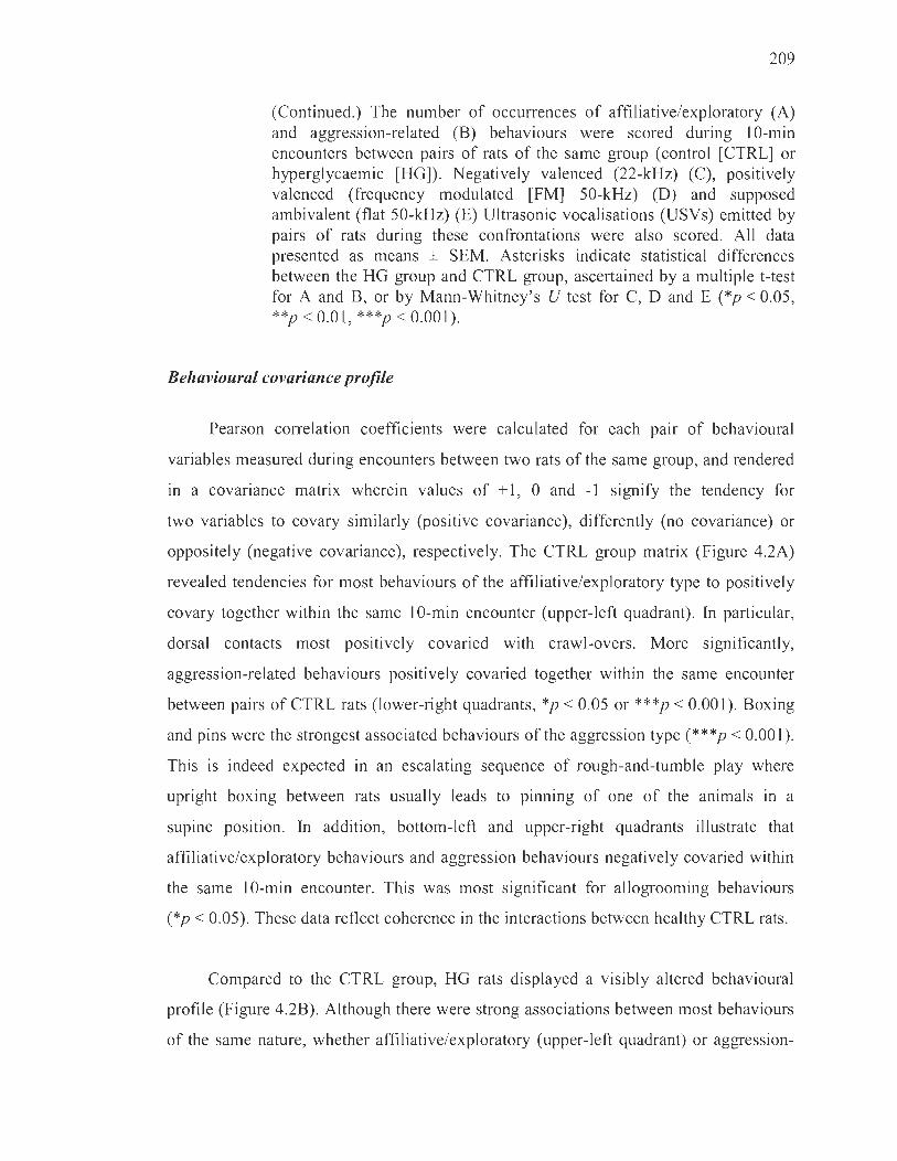
209
(Continued.) The number of occurrences of affiliative/exploratory (A) and aggression-related (B) behaviours were scored during 10-min encounters between pairs of rats of the same group (control [CTRL] or hyperglycaemic [HG]). Negatively valenced (22-kHz) (C), positively valenced (frequency modulated [FM] 50-kHz) (D) and supposed ambivalent (flat 50-kHz) (E) Ultrasonic vocalisations (USVs) emirted by pairs of rats during these confrontations were also scored. AIl data presented as means ± SEM. Asterisks indicate statistical differences between the HG group and CTRL group, ascertained by a multiple t-test for A and B, or by Mann-Whitney's U test for C, D and E (*p < 0.05, **p < 0.01, ***p < 0.001).
Behavioural covariance profile
Pearson correlation coefficients were calculated for each pair of behavioural
variables measured during encounters between two rats of the same group, and rendered
in a covariance matrix wherein values of + l, 0 and -1 signify the tendency for
two variables to covary similarly (positive covariance), differently (no covariance) or
oppositely (negative covariance), respectively. The CTRL group matrix (Figure 4.2A)
revealed tendencies for most behaviours of the affiliative/exploratory type to positively
covary together within the same 10-min encounter (upper-Ieft quadrant). In particular,
dorsal contacts most positively covaried with crawl-overs. More significantly,
aggression-related behaviours positively covaried together within the same encounter
between pairs of CTRL rats (lower-right quadrants, *p < 0.05 or ***p < 0.001). Boxing
and pins were the strongest associated behaviours of the aggression type (***p < 0.001).
This is indeed expected in an escalating sequence of rough-and-turnble play where
upright boxing between rats usuaIly leads to pinning of one of the animaIs in a
supine position. In addition, bortom-left and upper-right quadrants iIlustrate that
affiliative/exploratory behaviours and aggression behaviours negatively covaried within
the same 10-min encounter. This was most significant for allogrooming behaviours
(*p < 0.05). These data reflect coherence in the interactions between healthy CTRL rats.
Compared to the CTRL group, HG rats displayed a visibly altered behavioural
profile (Figure 4.2B). Although there were strong associations between most behaviours
of the same nature, whether affiliative/exploratory (upper-left quadrant) or aggression-
210
related (lower-right quadrant), other behaviours not shown to similarly covary in
CTRL rats within the same encounter were markedly positively associated in HG rats.
Specifically, the affiliative/exploratory anogenital sniffing behaviour tended to
positively covary with aggressive behaviours such as boxing and nose-offs, but was
most tightly associated to pins (***p < 0.001). Relatedly, we found a strong positive
covariance between pins and dorsal contacts (**p < 0.01). Despite their higher rate of
occurrence (Figure 4.2B), freezing events did not consistently positively covary with any
other behaviour during encounters between pairs of HG rats. With respect to the pattern
of association displayed by the CTRL group, rats of the HG group seem to engage in
incoherent social interactions characterized by episodes of play and fight intertwining
within the same encounter.
A CTRL B HG 1.0
Dorsal contacts
Allogrooming
Anogenital sniffing
Crawl-overs
* * *
Dorsal contacts
Allogrooming
Anogenital sniffing
Crawl-overs
Figure 4.2
Freezing
Nose-off * Boxing *
Pins *
Freezing
Nose-off
Boxing
ln Cl Cl ln Cl ln Cl ln -cc'-c:t:cC U .- lE Q) . - 0 .- .-~ E .- ~ :il ci> ~ a.. c 0 c , Q) ln CIl 8e~~'-o _ Clro roli. z :Jl g 'Ë ü '- « Q) o Cl Q 0
~
o
-0.5
-1 .0
Affiliative/exploratory and aggression behavioural covariance matrices. Matrices show strength of association, expressed as Pearson correlation coefficients, for each combination of behaviour laid out on the x- and y-axes as measured during 10-min encounters between two rats of the same group. Values of 1 (deep red), 0 (white) and -1 (deep blue) signify the tendency for two variables to covary similarly (positive covariance), di fferentl y (no covariance) or oppositely (negative covariance), respectively. Affiliative/exploratory behaviours are dorsal contacts, allogrooming, anogenital sniffing and crawl-overs. Aggression behaviours are freezing, nose-offs, boxing and pins. Behavioural profiles are provided for the CTRL (A) and HG (B) groups. Asterisks indicate statistical significance of the positive or negative covariance of two variables (*p < 0.05, **p < 0.01, ***p < 0.001).

211
Behaviour-vocalisation covariance profile
A similar strategy was employed to evaluate whether the emlSSlOn of certain
categories of USVs covaried with distinct types of behaviours within the same 10-min
encounter between pairs of same-group rats (Figure 4.3). Between pairs of CTRL rats
(left quadrants), the emission of negatively valenced 22-kHz caUs and aggression
behaviours of aU types positively covaried (***p < 0.001). ln addition, FM 50-kHz
USVs positively covaried with one of the most positively valenced behaviours, that is,
aUogrooming [32] (*p < 0.05). Accordingly, 22-kHz US Vs negatively covaried with this
behaviour (*p < 0.05). Flat 50-kHz caUs, on the other hand, were more often emitted
during encounters wherein anogenital sniffing activities were nurnerous (**p < 0.01),
but also slightly positively covaried with aUogrooming and pins. This is in line with
the proposed ambivalent nature of flat calls, which may have served here to pace
intrusive exploratory behaviours, su ch as anogenital sniffing and allogrooming, or to
signal discomfort or uncertainty during pinning events.
With respect to the CTRL group, HG rats (Figure 4.3, right quadrants) presented
pronounced distortions in the covariance relationships between the USVs they emitted
and the behaviours they exhibited during the same 10-min encounter. Most striking is
the lack of a c1ear positive covariance between 22-kHz calls and behaviours likely to
induce these USV s, such as nose-offs, boxing and pins. ln addition, FM 50-kHz and
affiliative/exploratory behaviours did not positively covary. The most coherent pattern
of positive covariance remained the significantly nurnerous emissions of 22-kHz calls
during encounters that also featured numerous freezing events between HG rats
(***p < 0.001).

212
Figure 4.3
CTRL HG 1.0
Dorsal contacts
Allogrooming * * 0.5 Anogenital sniffing
Crawl-overs 0
Freezing
Nose-off -0.5
Boxing
Pins N N N N N N
-1.0 J: J: J: J: J: J: ~ ~ ~ ~ ~ ~ ,
2> 2> N 2> 2> N N II) II) N II) II)
~ co ~ co Il.. Li: Il.. Li:
Behaviour-vocalisation covariance matrix. Matrices show strength of association, expressed as Pearson correlation coefficients, for each combination of USV category (x-axis) and type of behaviour (y-axis) as measured during 10-min encounters between two rats of the same group. Values of 1 (deep red), 0 (white) and -1 (deep blue) signify the tendency for two variables to covary similarly (positive covariance), differently (no covariance) or oppositely (negative covariance), respectively. Affiliative/exploratory behaviours are dorsal contacts, aUogrooming, anogenital sniffing and crawl-overs. Aggression behaviours consist in freezing, nose-offs, boxing and pins. Twenty-two-kHz USVs are negatively valenced, FM 50-kHz US Vs are positively valenced, and flat 50-kHz USVs are supposed ambivalent caUs. Behaviour-vocalisation profiles are provided for the CTRL (left quadrants) and HG (right quadrants) groups. Asterisks indicate statistical significance of the positive or negative covariance of two variables (*p < 0.05, **p < 0.01, ***p < 0.001).
Magnitude of social interactivity and ultrasonic vocalisations in relation to the degree of striatal denervation, hypoinsulinaemia and glucose intolerance
We performed densitometric immunohistochemical analyses of striatal tyrosine
hydroxylase (TH)-positive terminal fibres in the brains of certain CTRL (n = 3) and
HG rats (n = 6) utilized in the experiments described above. This allowed for the
division of these HG subjects into two distinct subgroups: low denervation and high
denervation (Figure 4.4A). Figure 4.4B displays representative microphotographs of the
striatal denervation observed in HG brains compared to healthy CTRL rats. The baseline
insulinaemia and glucose tolerance of certain CTRL (n = 5-6) and HG (n = 17-20) rats
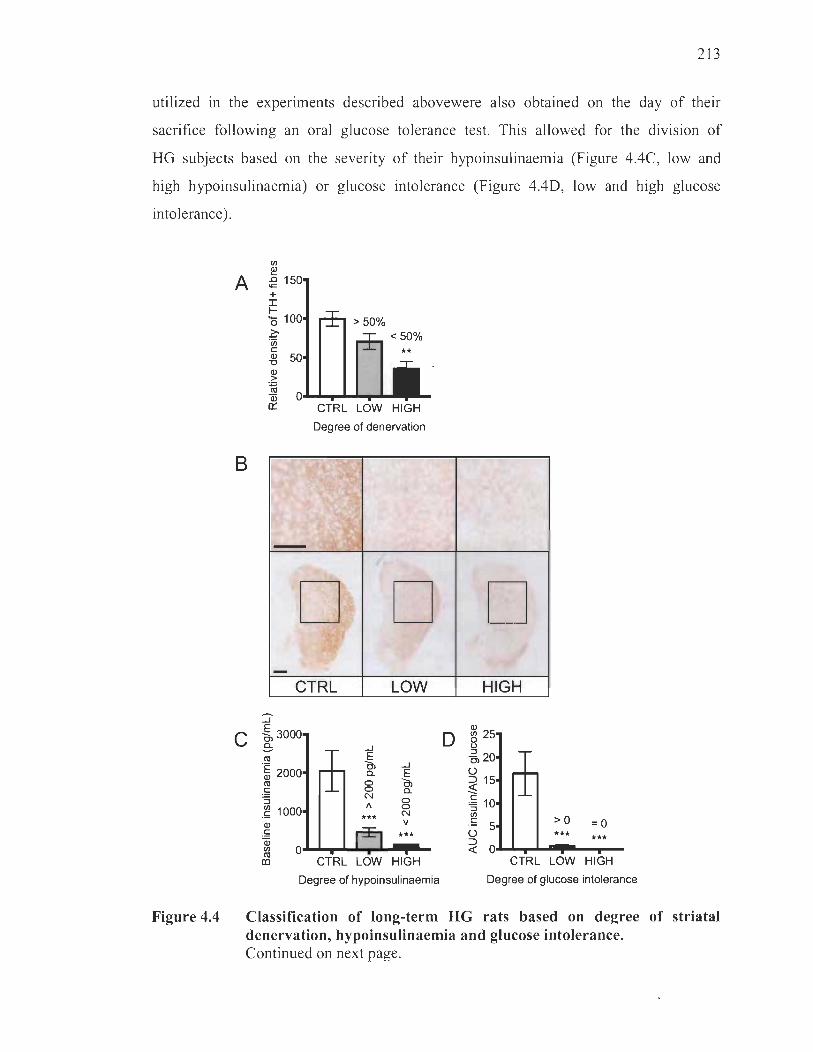
213
utilized in the experiments described abovewere also obtained on the day of their
sacrifice following an oral glucose tolerance test. This allowed for the division of
HG subjects based on the severity of their hypoinsulinaemia (Figure 4.4C, low and
high hypoinsulinaemia) or glucose intolerance (Figure 4.4D, low and high glucose
intolerance ).
Figure 4.4
A
B
If)
2! ï§ 1 + I ~
ë5 1 ~ "iii c: al "0 al > ~ Qi Il::
>50% <50%
**
CTRL LOW HIGH
Degree of denervation
-
{
:'1 _ ~.,
,
D D -
CTRL LOW
::J C -§, 3000
.3: ro
.~ 2000 ro ~ :l
.~ 1000 al
.~ Qi
...J E 0, a. g N V
*** ~ O~~--~~--.. --
(Il CTRL LOW HIGH
Degree of hypoinsulinaemia
D HIGH
CTRL LOW HIGH
Degree of glucose intolerance
Classification of long-term HG rats based on degree of striatal denervation, hypoinsulinaemia and glucose intolerance. Continued on next page.

214
(Continued.) (A, B) Irnmunohistochemical analyses of the striatal region of CTRL and HG rat brains. At the end of behavioral experiments, frozen post-fixed brain hemispheres were cut into 20 Ilm-thick coron al seriai sections and stained for the dopaminergic terminal fiber marker, tyrosine hydroxylase. A. Densitometric analyses allowed for the distinction of two subgroups, LOW (> 50% of CTRL) or HIGH « 50% of CTRL), within the HG group concurring to the degree of striatal dopaminergic denervation. B. Representative microphotographs of tyrosine hydroxylase staining in the striatum (Bregma ~ 1 mm, scalebar 1 mm). C: Baseline insulinemia measurements upon sacrifices allowed for the separation of HG rats between LOW hypoinsulinemia (> 200 pg!mL) and HIGH hypoinsulinemia « 200 pg/mL). D: Following an oral glucose tolerance test performed on the moming of their sacrifice, are as under the curve of glycemias on areas under the curve of insulinemias yielded indices of glucose intolerance. Rats were separated on this basis between LOW (> 0) and HIGH (0) glucose intolerance. AlI data presented as means ± SEM. Asterisks indicate statistical differences between the HG group and CTRL group (**p < 0.01).
The mean rate of occurrence of specific affiliativelexploratory and aggression
related behaviours was obtained for each abovementioned subgroup ranked by degree
of striatal denervation, hypoinsulinaemia and glucose intolerance (Figure 4.5).
In particular, HG rats exhibiting the highest levels of striatal denervation engaged in
more numerous affiliativelexploratory (Figure 4.5A) and aggression-like (Figure 4.5B)
behaviours than rats from the other two subgroups, significantly for dorsal contacts
(***p < 0.001 compared to CTRL and +p < 0.05 compared to the low denervation
subgroup), anogenital sniffing (***p < 0.001 compared to CTRL and ++p < 0.01
compared to the low denervation subgroup), boxing (***p < 0.001 compared to CTRL
and +++p < 0.001 compared to the low denervation subgroup) and pinning
(***p < 0.001 compared to CTRL and ++p < 0.01 compared to the low denervation
subgroup). No c1ear trend for increased affiliativelexploratory or aggression behaviour in
relation to the degree of hypoinsulinaemia (Figure 4.5C and D) or glucose intolerance
(Figure 4.5E and F) was revealed. In fact, dorsal contacts were less frequent in the high
hypoinsulinaemia subgroup than in the low insulinaemia subgroup (+p < 0.05).

A 20 <Il
~ 15 c: ~ :; 10 u u o
'*1: 5
C 20 <Il
~ 15 !li 5 10 8 o
'*1: 5
** *
*
D CTRL IJ LOW denervation • HIGH denervation
B 8 ***
<Il
~ 6 *** c: ~ :; 4 u u o '*1:2
0 ........ .."...
***
Dorsal Allo- Anogenital Crawl- Freezing Nose-offs Boxing contacts grooming sniffing overs
*
***
D CTRL IJ LOW hypoinsulinaemia • HIGH hypoinsulinaemia o lU
~ 8 u
~ 6 :; 8 4 o
'*1: 2
***
***
* *
215
Pins
0 ........... 0 .............
E 20 <Il
~ 15 c: ~ :; 10 u g
'*1: 5
Dorsal Allo- Anogenital Crawl- Freezing Nose-offs Boxing contacts grooming sniffing overs
** *
D CTRL LOW glucose intolerance
• HIGH glucose intolerance F 10
*** ~ u
~ :; 8 4 o
'*1: 2
*
Pins
0 ........... 0 ........ .."... Dorsal Allo- Anogenital Crawl- Freezing Nose-offs Boxing Pins
contacts grooming sn iffi ng overs
Figure 4.5 Occurrences of social behaviours in relation to the degree of striatal denervation, hypoinsulinaemia or glucose intolerance. Occurrences of affiliative/exploratory (A, C, E) or aggression-related (B, D, F) behaviours performed by pairs ofCTRL rats or pairs of HG rats belonging to the low or high striatal denervation (A, B), hypoinsulinaemia (C, D) or glucose intolerance (E, F) groups were plotted. AH data presented as means ± SEM. Asterisks indicate statistical differences between groups and the CTRL group, ascertained by two-way ANOVA followed by Tukey's post-hoc analysis (*p < 0.05, **p < 0.01 , ***p < 0.001). Plus signs indicate statistical differences between high and low groups, ascertained by two-way ANOVA followed by Tukey' s post-hoc analysis (+p < 0.05, ++p < 0.01 , +++p < 0.001).
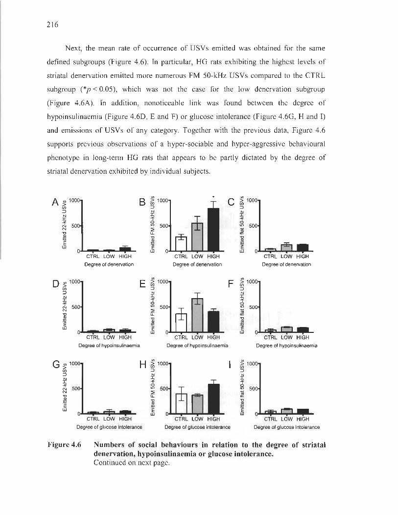
216
Next, the mean rate of occurrence of USVs emitted was obtained for the same
defined subgroups (Figure 4.6). In particular, HG rats exhibiting the highest levels of
striatal denervation emitted more numerous FM 50-kHz USVs compared to the CTRL
subgroup (*p < 0.05), which was not the case for the low denervation subgroup
(Figure 4.6A). In addition, nonoticeable link was found between the degree of
hypoinsulinaemia (Figure 4.6D, E and F) or glucose intolerance (Figure 4.6G, H and 1)
and emissions of USVs of any category. Together with the previous data, Figure 4.6
supports previous observations of a hyper-sociable and hyper-aggressive behavioural
phenotype in long-term HG rats that appears to be partly dictated by the degree of
striatal denervation exhibited by individual subjects.
A ~ 1000 B ~ 1 :::> N I
C/)
:::> N
I .>t:. N 500 N "0 .l!l E w
0.&..1 .... - ......... 1....
o ~ 1000 C/)
:::> N I .>t:. N 500 N "0
~ "Ë w
CTRL LOW HIGH
Degree of denervation
0--................. .... CTRL LOW HIGH
Degree of hypoinsulinaemia
G ~ 1000 C/)
:::>
~ .>t:. N 500 N "0
~ E w
0.&..1 • ...,.; ............... CTRL LOW HIGH
Degree of glucose intolerance
.>t:. o l{)
:E u.. "0
~ "Ë w
* c ~ 1
CTRL LOW HIGH
Degree of denervation
:::>
~ -'f a l{)
i;j <;::
al E E w
CTRL LOW HIGH
Degree of de nervation
F ~ 1000 :::> N I .>t:.
CTRL LOW HIGH
Degree of hypoinsulinaemia
Degree of glucose intolerance
o l{) 500 i;j 0= "0
~ "Ë W Ol~~~~~~~
CTRL LOW HIGH
Degree of hypoinsulinaemia
CTRL LOW HIGH
Degree of glucose intolerance
Figure 4.6 Numbers of social behaviours in relation to the degree of striatal denervation, hypoinsulinaemia or glucose intolerance. Continued on next page.

Discussion
217
(Continued.) Numbers of US Vs emitted by CTRL rats or HG rats belonging to the low or high striatal denervation (A-C), insulinaemia (D-F) or glucose intolerance (G-I) groups were plotted. (A, D, G) Negatively valenced 22-kHz USVs. (B, E, H) Positively valenced FM 50-kHz USVs. (C, F, 1) Supposed ambivalent flat 50-kHz USVs. Ali data presented as means ± SEM. Asterisks indicate statistical differences between groups and the CTRL condition, ascertained by Kruskal-Wallis ANOVA followed by Dunn's post-hoc analysis (*p < 0.05).
Although awareness of behavioural alterations in diabetic patients has been on the
rise lately, research offers limited insight on the physiological factors that may impinge
on these issues. While acute hypo or hyperglycaemic states are acknowledged to foster
more aggressive states in humans [8-11], little is known about the effects of long-terrn
dysglycaemia. Prior studies have employed acutely hyperglycaemic mice (duration of
hyperglycaemia < 1 month) in resident-intruder paradigms [13 , 14] or hyperglycaemic
rats in mating paradigms [12] , but have yielded inconsistent findings on the social
effects of hyperglycaemia in paradigms that, further, poorly reflect the chronicity of
the diabetic condition.
To the best of our knowledge, the present report constitutes the fIfst detailed
account of alterations in social behaviour and USV communications in a rat model of
long-terrn hyperglycaemia. In aIl evidence, rats of the HG group were hyper-sociable
and hyper-aggressive, as supported by marked increases in the occurrences of both
affiliative/exploratory and aggression-related behaviours. Accordingly, compared to
CTRL rats, they emitted many more USVs thought to operate both as social contact caIls
[21 , 33] and expressions of affective states [15]. In addition to the rise in occurrences of
social behavioural or vocalisation events, the covariance profiles of these variables were
radically different between CTRL and HG groups. Coherent associations between
behaviours of the same kind or between behaviours and USVs that supposedly have the
same valence were aimost completely lost in HG rats.
218
ln an attempt to better understand the neurobiological underpinnings of impaired
social behaviour and disjointed USV communications in our mode l, results were related
to the degree of TH-positive fibres in the striaturn. lndeed, focal to social behaviour
are a variety of neurotransmitters acting via circuits that can be modulated by
pharmacological interventions [34, 35]. One such neurotransmitter is dopamine,
exemplarily targeted by dopaminergic antagonists to treat abnormal aggresslve
behaviours in autism, schizophrenia and excessive stress [36-40]. By releasing dopamine
in the striatum, midbrain neurons partake in the regulation of social behaviours but also
of other goal-directed operations ranging from movement production to reward-seeking
processes [41-44]. ln agreement with the purported role of mesostriatal dopamine in
social conduct, we found the altered social behaviours between pairs of unacquainted
rats to be predicted by the degree of striatal TH-positive fibre loss. This is supported by
the differences in 50-kHz USV emissions between CTRL and HG rats, also importantly
modulated by mesostriatal dopamine [15]. Nevertheless, we cannot exclude that our
results may have been partly influenced by toxic and/or adaptive modifications induced
by long-term hyperglycaemia in non-dopaminergic neurotransmitter systems, which also
critically regulate social behaviour and emission of USVs [35, 45-50]. For example,
based on the loss of striatal TH-positive fibres, it could be hypothesized that
hyperglycaemic damage of the noradrenergic system may have participated in the
behavioural abnormalities observed here. Further studies will be necessary to precisely
determine the interplay between social and vocal behaviour and neurotransmitter
systems in HG rats.
The role for increased mesostriatal dopamine neurotransmission in the promotion
of impulsive, social and reward-related behaviours as weIl as in the emission of 50-kHz
USVs seems to challenge our results showing an apparent correlation between striatal
denervation and increased sociability, aggression and vocalisation. Nevertheless, our
findings on the emission of 50-kHz USVs by HG rats are consistent with results from a
previous study in male rats bearing a unilateral terminal denervation of the dopaminergic
nigrostriatal system [51] . lndeed, the latter study showed that dopamine-denervated rats
showed a trend towards an increase in the numbersof 50-kHz USVs emitted during

219
mating, compared with neurologically intact rats. It is possible that the occurrence of
dopamine receptor supersensitivity due to deafferentiation and/or an imbalance in tonic
and phasic firing may account for the increased reactivity to social novelty in our model
[52]. Indeed, we have previously shown a decrease in tonic dopaminergic transmission
in the striatum oflong-term HG rats [31]. Tonic firing consists in the slow and irregular
single-spike release of dopamine produced by the baseline pacemaker activity of
mesostriatal dopaminergic neurons, which main tains a certain level of striatal dopamine
"noise" [53]. Tonic dopamine is required to enable striatal postsynaptic targets
embedded in corticostriatal circuits involved in the selection of pre-Ieamed programs
[54-56] . Phasic firing, on the other hand, involves a stimulus-mediated discharge of
spikes in a short amount of time [57] . These bursts raise the levels of striatal dopamine
above the noisy tonicity, acting as a "signal" that directs attention to a salient stimulus.
When striatal tonicity is low, phasic dopamine becomes much more relevant over the
reduced noisy background; thus, when produced in response to salient stimuli like
exposure to a novel social context or emission of 50-kHz USVs [58], phasic firing might
cause inappropriately intense reactions as observed in our mode!. Notably, transcending
neurodegeneration, receptor supersensitivity, altered dopamine concentrations or
imbalances in tonic-to-phasic (noise-to-signal)firing have aIl been proposed to explain
various psychiatric disorders such as schizophrenia [59, 60] and attention deficit
hyperactivity disorder [61 , 62]. Importantly, hyper-sociable and hyper-aggressive traits
are typical of the latter and very similar to those present in our model [63-65].
The overt hyper-sociable and hyper-aggressive behaviours exhibited by HG rats
are also reminiscent of impulsive traits found in the streptozotocin model, albeit in
other contexts. Indeed, drugs that modulate mesostriatal dopamine are more rewarding
in streptozotocin-treated rats than in untreated conspecifics [66-68]. In addition,
hyperphagia, a hallmark of the model, is at least partly regulated by mesostriatal
dopamine [69, 70]. To aIl appearances, the streptozotocin model increases the rewarding
properties of several environmental stimuli. We show here that this could include social
interactions as supported by the concomitant increase in the emission of 50-kHz USVs,
which are a marker of positive affective states [15]. While metabolic hormonal
220
imbalances may be implicated to sorne extent in the hyper-reactive phenotype of
streptozotocin-treated rats, as suggested by sorne [66, 69], it remains that the extent of
behavioural alterations was tightly associated with striatal denervation and not with
insu lin concentrations or functions in long-term hyperglycaemia. Given its implication
in modulating behaviour in short- and medium-term [71-73], a role for the
hypothalamic-pituitary-adrenal axis cannot be ruled out in the behavioural abnormalities
observed here in streptozotocin-treated rats and should be addressed in future works
[14, 74] .
In summary, the present results demonstrate explicit manifestations of hyper
sociable and hyper-aggressive traits in our model of long-term hyperglycaemia.
The striking overall increases in positively and negatively valenced events, either
behavioural or vocal, combined with the loss of coherence between the affective states
represented by USVs and the social behaviour displayed may arise from neurobiological
alterations. This is supported by the tight association between striatal denervation as
opposed to the lack of a clear link between insulin and hyper-reactivity. These findings
may ultimately lead to the formaI identification of dopaminergic neural substrates that
may be targetable by pharmacological treatments, both to prevent striatal denervation
and to palliate neurochemical imbalances in diabetic patients faced with psychiatric
challenges.
Methods
Subjects
Fort y male Sprague-Dawley rats (Charles River, St-Constant, Canada) weighing
175-200 g were housed under standard laboratory conditions (12 h light/dark cycle).
In all experiments, standard food and water were available ad libitum. Rats were housed
two per cage to rule out any effect of social isolation on their subsequently measured
behaviours [75]. Experiments were conducted in accordance with legislation and
policies of the Canadian Council on Animal Care, as weIl as with the guidelines

221
established by the Animal Care Committee of the Université du Québec (Trois-Rivières)
(2014-M.G.M.5).
Induction of hyperglycaemia
Rats were randomly assigned to the control or hyperglycaemic group (CTRL
n = 18; HG n = 22). Hyperglycaemia was induced as previously described [31].
Briefly, fasted rats of the HG group were intraperitoneally injected with nicotinamide
(100 mg/kg b.w.) followed by the beta cell toxin streptozotocin (55 mg/kg b.w.,
Enzo Life Sciences, Farmingdale, NY, USA). Pre-injections with nicotinamide restrain
the death of insulin-producing pancreatic beta cells and yield long-term HG rats that
do not require glycaemia-Iowering treatments [31 , 76]. CTRL rats received vehicle
injections. Throughout experiments, glycaemia was estimated using an UltraMini digital
glucose meter and matching strips (One Touch Ultra) both purchased at a local Brunet
pharmacy (Trois-Rivières, QC, Canada). Hyperglycaemia was confirmed 3 days after
nicotinamide-streptozotocin injections and rats from the HG group that did not display a
glycaemia steadily above 10 mM were discarded from this study. Importantly, rats from
both groups were matched for age and experience, meaning that they were handled in
the same manner and at the same frequency, in light of studies showing the role of age
and experience on the emission ofUSVs [26].
Oral glucose intolerance test and baseline insulinemia
On the morning of their sacrifice, we performed an oral glucose tolerance test
(OGTT) on fasted rats, as previously described [31] . Briefly, rats were intragastrically
administered 2 glkg of a D-glucose solution. Glycaemia and insulinaemia were measured
right before the OGTT (0 min, baseline) and at 30, 60 and 120 min after administration
of D-glucose using the Rat Glucose Assay kit provided by Crystal Chem (Downers
Grove, IL, USA) and the Milliplex Adipokine Magnetic Bead Multiplex Assay
(Millipore; Etobicoke, ON, Canada), respectively. The ratio of the area under the curve

222
of plasma insulin concentrations on the area under the curve of plasma glucose
concentrations yielded indices of glucose intolerance.
Ultrasonic vocalisations and social behaviour
We began experiments 5 months following the induction of hyperglycaemia, at the
time when rats were fit to engage in social interactions. Experiments consisted in 3 trials
separated by 7 days each in order to prevent the behavioural habituation of rats to
multiple encounters in a short amount of time [77]. Experiments were performed in
3 different quiet rooms to rule out any environmental effects on the behaviour of the
rats. USV recordings were performed as previously described [78]. Two group-paired
unacquainted rats (CTRL 9 pairs times 3 trials; RG Il pairs times 3 trials) were placed
in a Plexiglas cylinder (diameter, 25 cm; height, 30 cm) with fresh bedding, surrounded
by walls to enhance the feeling of security, and topped with a sound-insulating lido
An ultrasonic microphone (CM16/CMPA, Avisoft) was placed through a tight-fitting
hole in the lid and was connected to an ultrasound-recording device (UltraSoundGate
116 Rb, Avisoft). During USV recordings, constant gain was maintained. Figure 4.7
shows examples of 22- and 50-kHz USVs recorded during this study. Two video
cameras were positioned on opposite sides of the cylinder to record the encounters.
USVs and images were recorded for 10 min, starting immediately after both rats were
simultaneously placed in the cylinder. Rence, neither rat was an intruder in this context.
Recording time was selected based on previous studies that evaluated the emission of
US Vs by rats that engaged in social contacts [49, 71]. A blinded experimenter scored
eight types of interactions provided by the video recordings. Affiliative/exploratory
behaviours consisted in dorsal contacts (with both paws), allogrooming, anogenital
sniffing, and crawl-overs (one rat crawls over the other rat) [79-81]. Aggression-related
behaviours consisted in freezing (a submissive rat remains 5 consecutive seconds or
more without movement except sniffing), nose-offs (both rats are immobile, facing each
other, four paws on the ground or front paws raised, but do not box), boxing (one or both
rats stand on their hind legs and push or paw their opponent), pins (one rat tops another
one that is in supine position) [16, 82]. Occurrences of any of these behaviours were
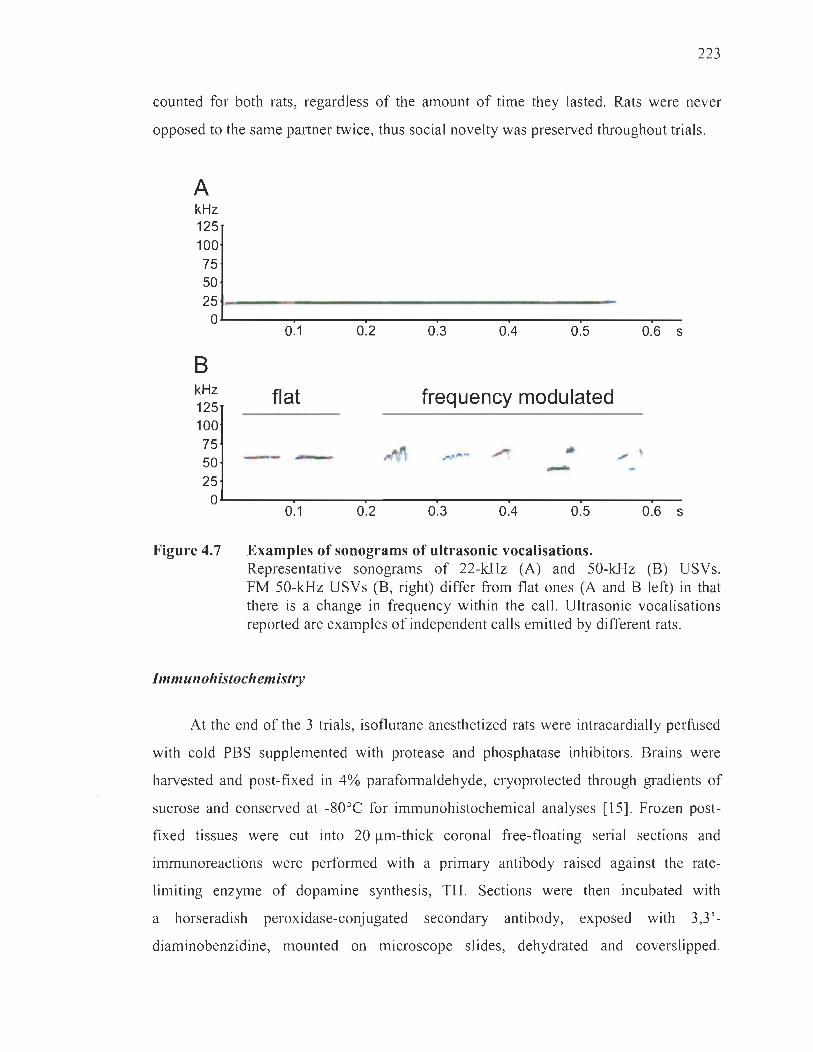
223
counted for both rats, regardless of the amount of time the y lasted. Rats were never
opposed to the same partner twice, thus social novelty was preserved throughout trials.
A kHz 125 100 75 50
251- ---------------------------------------O~------~------~------~------~------~------~--
B kHz 125 100 75 50 25
0.1
fiat
-- -
0.2 0.3 004 0.5 0.6 5
frequency modulated
OL-------~------~------~------~------~------~--
Figure 4.7
0.1 0.2 0.3 004 0.5 0.6 5
Examples of sonograms of ultrasonic vocalisations. Representative sonograms of 22-kHz (A) and 50-kHz (B) USVs. FM 50-kHz USVs (B, right) differ from flat ones (A and B left) in that there is a change in frequency within the cali. Ultrasonic vocalisations reported are examples of independent calls emitted by different rats.
Immunohistochemistry
At the end of the 3 trials, isoflurane anesthetized rats were intracardially perfused
with cold PBS supplemented with protease and phosphatase inhibitors. Brains were
harvested and post-fixed in 4% paraformaldehyde, cryoprotected through gradients of
sucrose and conserved at -80oe for immunohistochemical analyses [15]. Frozen post
fixed tissues were cut into 20 )..lm-thick coronal free-floating seriaI sections and
immunoreactions were performed with a primary antibody raised against the rate
limiting enzyme of dopamine synthesis, TH. Sections were then incubated with
a horseradish peroxidase-conjugated secondary antibody, exposed with 3,3'
diaminobenzidine, mounted on microscope slides, dehydrated and coverslipped.
224
Using a microscope ln brightfield mode (MBF Bioscience, Williston, VT, USA),
the density of striatal terminais positive for TH was quantified densitometricaUy using
the NIH ImageJ software version 1.49, and averaged in 4 slices per animal. A total of
3 brains from the CTRL group and 6 brains from the HG group were randomly selected
and analysed. Due to normal interindividual variability, degrees of denervation differed
across the 6 HG rats, allowing for the separation of these individuals into high (n = 3) or
low (n = 3) denervation subgroups.
Statistical analyses
USV recordings were converted into spectrograms using the software SASLab
Pro 4.52 (Avisoft) with the foUowing settings: 512 FFT-Iength, Hamming window and
75% overlap frame set-up [83]. A ski lied experimenter visually inspected and manuaUy
cleaned aU signaIs that could not be univocaUy classified as vocalisations. Nurnbers of
22-kHz and 50-kHz USVs were then counted using the SASLab Pro 4.52 software.
Moreover, an experienced experimenter unaware of treatments manually counted
numbers of FM and fiat 50-kHz USVs. USVs of 22-kHz were defined as caUs with
sound frequency maintained between 20- and 35-kHz and long duration, higher than
300 ms for a single caU [19]. In the present study, no 22-kHz USVs of duration shorter
than 300 ms were observed [84]. USVs of 50-kHz were defmed as caUs with sound
frequency maintained between 35- and 80-kHz and short duration, usually 10-15 ms for
a single caU [19]. USV s of 50-kHz were further divided in fiat and FM USV s, based on
the modulation of their sound frequency. Thus, fiat calls have the sound frequency
within 35-50-kHz that remains nearly unchanged over the duration of the individual
USVs [26]. Conversely, FM 50-kHz caUs have the sound frequency within 40-80-kHz
that shows substantial changes within individual USVs [16].
Ail data were submitted to statistical analyses performed using GraphPad Prism 7
software (San Diego, CA, USA; http://www.graphpad.com). Data were tested for
normality and homoscedasticity, and were analysed accordingly. Behaviour-behaviour
and behaviour-USV covariation matrices (Figure 4.2 and Figure 4.3) were derived from

225
Pearson correlation coefficients calculated for each pair of variables, where + 1, 0, and
-1 respectively indicate the tendency for two variables to covary similarly (positive
covariance), differently (no covariance) or oppositely (negative covariance) [85].
Occurrences of social behaviours, relative density of TH fibres , and measures of
insulinaemia and glucose intolerance were parametrically distributed. Thus, significant
differences between groups shown in Figure 4.lA and B were ascertained by multiple
t-tests , ones in Figure 4.4A, C and D were ascertained by one-way ANOV A followed by
Tukey's post-hoc analysis, and those shown in Figure 4.5 were determined by two-way
ANOVA followed by Tukey's post-hoc analysis. On the other hand, emissions of VSVs
were non-parametrically distributed. Therefore, significant differences between groups
shown in Figure 4.1C, D and E were ascertained by Mann-Whitney's U test,
while significant differences between groups shown in Figure 4.6 were ascertained by
the Kruskal-Wallis ANOVA, followed by Dunn' s post-hoc analysis. All data were
analysed at the 95% confidence interval and are expressed as means ± SEM.
Acknowledgments
The authors thank Dr Michel Cyr (V. Québec) for the use of equipment and
infrastructure. This research was funded by grants from the Natural Sciences and
Engineering Research Council (NSERC) of Canada no. 04321 to M.G.M. N.S. gratefully
acknowledges the financial support of Autonomous Region of Sardinia (L.R. n. 7/2007-
2015) and of Fondazione di Sardegna (EsercizioFinanziario 2017). J.R. is recipient of a
Vanier Graduate Scholarship from the NSERC and a Doctoral Training Scholarship
from the Fonds de recherche en santé du Québec.
226
References
1. Alessandri, S. M. Attention, play, and social behavior in ADHD preschoolers. J Abnorm Child Psychol, 20, 289-302 (1992).
2. Fava, M. Psychopharmacologic treatment of pathologic aggresslOn. Psychiatr Clin North Am, 20, 427-51 (1997).
3. Jordan, R. Social play and autistic spectrum disorders: a perspective on the ory, implications and educational approaches. Autism, 7,347-60 (2003).
4. Nevels, R. M., Dehon, E. E., Alexander, K., Gontkovsky, S. T. Psychopharmacology of aggression in children and adolescents with primary neuropsychiatricdisorders: a review of CUITent and potentially promlsmg treatrnent options. Exp Clin Psychopharmacol, 18, 184-201 (2010).
5. Lau, C. G., Tang, W. K., Liu, X. X., Liang, H. J., Liang, Y., Wong, A., Mok, V., Ungvari, G. S., Wong, K. S. , Kim, J. S., Paradiso, S. Poststroke agitation and aggression and social quality of life: a case control study. Top Stroke Rehabil, 24, 126-133 (2017).
6. Nagata, T., Nakajima, S., Shinagawa, S., Plitman, E., Nakayama, K., Graff-GueITero, A., Mimura, M. Baseline Predictors of Antipsychotic Treatrnent Continuation and Response at Week 8 in Patients with Alzheimer's Disease with Psycho sis or Aggressive Symptoms: An Analysis of the CATIE-AD Study. J Alzheimers Dis, 60, 263-272 (2017).
7. Tilov, B., Semerdzhieva, M., Bakova, D., Tomyova, B., Stoyanov, D. Study of the relationship between aggression and chronic diseases (diabetes and hypertension). J Eval Clin Pract, 22, 421-4 (2016).
8. Benton, D., Kumari, K. , Brain, P. F. Mild hypoglycemia and questionnaire measures ofaggression. Biol Psychol, 14,129-135 (1982).
9. McCrimmon, R. J. , Ewing, F. M. E., Frier, B. M., Deary, 1. 1. Anger state during acute insulin-induced hypoglycemia. Physiol Behav, 67, 35-39 (1999).
10. Surwit, R. S. , Williams, R. B., Siegler, 1. C., Lane, 1. D., Helms, M. , Applegate, K. L., Zucker, N., Feinglos, M. N., McCaskill, C. M. , Barefoot, 1. C. Hostility, race, and glucose metabolism in nondiabetic individuals. Diabetes Care, 25, 835-839 (2002).

227
Il. Virkkunen, R. Reactive hypoglycemic tendency among habituaUy violent offenders. A further study by means of the glucose tolerance test. Neuropsychobiology, 8, 35-40 (1982).
12. Hernandez-Munive, A. K., Rebolledo-Solleiro, D ., Ventura-Aquino, E., Fernandez-Guasti, A. Reduced Lordosis and Enhanced Aggression in Paced and Non-Paced Mating in Diabetic Female Rats. J Sex Med, 15, 124-135 (2018) .
13. Hilakivi-Clarke, L. A., Wozniak, K. M. , Durcan, M . J., Linnoila, M. Behavior of streptozotocin-diabetic mice in tests of exploration, locomotion, anxiety, depression and aggression. Physiol Behav, 48, 429-433 (1990).
14. Meehan, W . P ., Leedom, L. 1. , Nagayama, T. , Zeidler, A. Agonistic behavior patterns in mi ce with streptozotocin-induced diabetes meUitus. Physiol Behav, 38, 301-306 (1986) .
15. Brudzynski, S. M. Ethotransmission: communication of emotional states through ultrasonic vocalization in rats. Curr Opin Neurobiol, 23, 310-317 (2013).
16. Burgdorf, J. , Kroes, R. A. , Moskal , 1. R. , Pfaus, J . G., Brudzynski, S. M. , Panksepp, J . Ultrasonic vocalizations of rats (Rattusnorvegicus) during mating, play, and aggression: Behavioral concomitants, relationship to reward, and self-administration of playback. J Comp Psychol, 122, 357-367 (2008) .
-17. Knutson, B. , Burgdorf, 1., Panksepp, J. Anticipation ofplay elicits high-frequency
ultrasonic vocalizations in young rats. J Comp Psychol, 112, 65-73 (1998).
18. Wohr, M. , Schwarting, R. K. W . Affective communication in rodents: ultrasonic vocalizations as a tool for research on emotion and motivation. Cel! Tissue Res, 354, 81-97 (2013).
19. Simola, N ., Brudzynski, S. M. Repertoire and biological function of ultrasonic vocalizations in adolescent and adult rats. In: Stefan M. Brudzynski, editor, Handbook of Behavioral Neuroscience, Vol. 25 , Amsterdam: Academic Press, 2018, pp. 177-186.
20. Blanchard, R. J. , Blanchard, D. C. , Agullana, R. , Weiss, S. M. Twenty-two kHz alarm cries to presentation of a predator, by laboratory rats living in visible bUITOW systems. Physiol Behav, 50, 967-972 (1991).
21 . Brudzynski, S. M ., Pniak, A. Social contacts and production of 50-kHz short ultrasonic caUs in adult rats. J Comp Psycho l, 116, 73-82 (2002).

228
22. Kisko, T. M. , Himmler, B. T. , Himmler, S. M. , Euston, D. R. , PeUis, S. M. Are 50-kHz caUs used as play signaIs in the playful interactions of rats? II. Evidence from the effects of devocalization. Behav Process, 111 , 25-33 (201 5).
23 . Simola, N. , Costa, G. Emission of categorized 50-kHz ultrasonic vocalizations in rats repeatedly treated with amphetamine or apomorphine: Possible relevance to drug-induced modifications in the emotional state. Behav Brain Res, 347, 88-98 (2018).
24. Taylor, J. O. , Urbano, C. M., Cooper, B. G. Differentiai patterns of constant frequency 50 and 22 khzusv production are related to intensity of negative affective state. BehavNeurosci, 131, 115-126 (2017).
25 . Burke, C. J. , Kisko, T. M. , PeUis, S. M ., Euston, D. R. Avoiding escalation from play to aggression in adult male rats: The role of ultrasonic caUs. Behav Processes, 144, 72-81 (2017).
26. W6hr, M., Houx, B. , Schwarting, R. K. W., Spruijt, B. Effects of experience and context on 50-kHz vocalizations in rats. Physiol Behav, 93, 766-776 (2008).
27. Burgdorf, J. , Knutson, B., Panksepp, J. , Ikemoto, S. Nucleus accumbens amphetamine miCrOl~)ections unconditionally elicit 50-kHz ultrasonic vocalizations in rats. Behav Neurosci, 115, 940-944 (2001).
28. Thompson, B. , Leonard, K. C., Brudzynski, S. M. Amphetamine-induced 50 kHz caUs from rat nucleus accumbens: a quantitative mapping study and acoustic analysis. Behav Brain Res, 168, 64-73 (2006).
29. Cragg, S. J. Meaningful silences: how dopamine listens to the ACh pause. Trends Neurosci. 29, 125-131 (2006).
30. Simola, N. Rat ultrasonic vocalizations and behavioral neuropharmacology: From the screening of drugs to the study of disease. Curr Neuropharmacol, 13, 164-79 (2015).
31. Renaud, J. , Bassareo, V. , Beaulieu, J. , Pinna, A. , Schlich, M., Lavoie, C. , Murtas, D. , Simola, N. , Martinoli, M. G. Dopaminergic neurodegeneration in a rat model of long-term hyperglycemia: preferential degeneration of the nigrostriatal motor pathway. Neurobiol Aging, 69, 117-128 (201 8).
32. Boissy, A. , Manteuffel, G. , Jensen, M. B., Moe, R. O. , Spruijt, B. , Keeling, L. J. , Winckler, C. , Forkman, B. , Dimitrov, 1. , Langbein, J. , Bakken, M., Veissier, 1. , Aubert, A. Assessment of positive emotions in animais to improve their welfare. Physiol Behav, 92, 375-97 (2007).

229
33. Smith, 1. W. The study of ultrasonic communication. Am Zool, 19, 531-538 (1979).
34. Narvaes, R. , and Martins de Almeida, R. M. Aggressive behavior and three neurotransmitters: dopamine, GABA, and serotonin - A review of the last 10 years. Psychology & Neuroscience, 7, 601-607 (2014).
35 . Vanderschuren, L. J. , Achterberg, E. J. , Trezza, V. The neurobiology of social play and its rewarding value in rats. Neurosci Biobehav Rev, 70, 86-105 (2016).
36. Bronsard, G. , Botbol, M. , Tordjman, S. Aggression in low functioning children and adolescents with autistic disorder. PLoS One, 5, e14358 (2010).
37. Nelson, R. J. , Trainor, B. C. Neural mechanisms of aggression. Nat Rev Neurosci, 8, 536-546 (2007).
38. Saez, L, Zhu, L. , Set, E. , Kayser, A. , Hsu, M. Dopamine modulates egalitarian behavior in humans. Curr Biol, 25, 912-919 (2015) .
39. Seo, D. , Patrick, C. J. , Kennealy, P. 1. Role of Serotonin and Dopamine System Interactions in the Neurobiology of Impulsive Aggressionand its Comorbidity with other Clinical Disorders. Aggress Violent Behav, 13, 383-395 (2008).
40. Soyka, M. , Graz, C. , Bottlender, R., Dirschedl, P., Schoech, H. Clinical correlates of later violence and criminal offences in schizophrenia. Schizophrenia Research, 94, 89-98 (2007).
4l. Gunaydin, L. A. , Grosenick, L. , Finkelstein, J. C. , Kauvar, 1. V. , Fenno, L. E., Adhikari , A. , Lammel, S. , Mirzabekov, 1. J., Airan, R. D., Zalocusky, K. A. , Tye, K. M. , Anikeeva, P. , Malenka, R. C. , Deisseroth, K. Natural neural projection dynamics underlying social behavior. CeU, 157, 1535-1551 (2014).
42. Haber, S. N. , Fudge, J. L. , McFarland, N. R. Striatonigrostriatal pathways in primates form an ascending spiral from the shell to the dorsolateral striaturn. J Neurosci, 20, 2369-2382 (2000).
43. Saddoris, M. P. , Sugam, 1. A. , Cacciapaglia, F., Carelli, R. M. Rapid dopamine dynamics in the accumbens core and shell: learning and action. Front Biosci (Elite Ed) , 5, 273-288 (2013).
44. Schultz, W. Getting formai with dopamine and reward. Neuron , 36, 241-63 (2002) .

230
45. Simola, N., Brudzynski, S. M. Rat 50-kHz ultrasonic vocalizations as a tool in studying neurochemical mechanisms that regulate positive emotional states. J Neurosci Methods, S0165-0270, 30197-3 (2018) .
46. Achterberg, E. 1. M., Damsteegt, R., Vanderschuren, L. J. M. J. On the central noradrenergic mechanism underlying the social play-suppressant effect of methylphenidate in rats. Behav Brain Res, 347, 158-166 (2018).
47. Costa, G., Morelli, M., Simola, N. Involvement of glutamate NMDA receptors in the acute, long-term, and conditioned effects of amphetamine on rat 50 kHz ultrasonic vocalizations. Int. J Neuropsychopharmacol, 18, pyv057 (2015).
48. Nunes, E. J. , Randall , P. A. , Podurgiel, S. , Correa, M., Salamone, J. D. Nucleus accumbens neurotransmission and effort-related choice behavior in food motivation: effects of drugs acting on dopamine, adenosine, and muscanmc acetylcholine receptors. Neurosci Biobehav Rev, 37, 2015-25 (2013).
49. Simola, N., Costa, G., Morelli, M. Activation of adenosine A2A receptors suppresses the emission of pro-social and drug-stimulated 50-kHz ultrasonic vocalizations in rats: possible relevance to reward and motivation. Psychopharmacology (Berl), 233,507-19 (2016).
50. Wohr, M., Rippberger, H., Schwarting, R. K., van Gaalen, M. M. Critical involvement of 5-HT2C receptor function in amphetamine-induced 50-kHz ultrasonic vocalizations in rats. Psychopharmacology (Berl), 232, 1817-29 (2015).
51 . Ciucci, M. R. , Ma, S. T. , Fox, c., Kane, J. R. , Ramig, L. O., Schallert, T. Qualitative changes in ultrasonic vocalization in rats after unilateral dopamine depletion or haloperidol: a preliminary study. Behav Brain Res, 182, 284-289 (2007).
52. Grace, A. A. The tonic/phasic model of dopamine system regulation and its implications for understanding alcohol and psychostimulant craving. Addiction, 95, S 119-28 (2000).
53. Grace, A. A., Bunney, B. S. The control of firing pattern in nigral dopamine neurons: single spike firing. J Neurosci, 4, 2866-2876 (1984).
54. Ellwood, LT. , Patel, T., Wadia, V., Lee, A. T., Liptak, A. T., Bender, K. 1., Sohal, V. S. Tonic or Phasic Stimulation of Dopaminergic Projections to Prefrontal Cortex Causes Mice to Maintain or Deviate from Previously Learned Behavioral Strategies. J Neurosci, 37,8315-8329 (2017).

231
55 . Hikosaka, O. , Takikawa, Y. , Kawagoe, R. Role of the basal gang lia in the control of purposive saccadic eye movements. Physiol Rev, 80, 953-978 (2000).
56. Redgrave, P. , Prescott, T ., Gurney, K. N. The basal ganglia: a vertebrate solution to the selection problem? Neuroscience, 89, 1009-1023 (1999).
57. Grace, A. A. , Bunney, B. S. The control of firing pattern in nigral dopamine neurons: burst firing. J Neurosci, 4, 2877-2890 (1984).
58. Willuhn, I. , Tose, A., Wanat, M . J. , Hart, A. S., Hollon, N. G., Phillips, P. E. , Schwarting, R. K. , Wëihr, M. Phasic dopamine release in the nucleus accumbens in response to pro-social 50 kHz ultrasonic vocalizations in rats. J Neurosci, 34, 10616-10623 (2014).
59. Grace, A. A. Phasic versus tonic dopamine release and the modulation of dopamine system responsivity: a hypothesis for the etiology of schizophrenia. Neuroscience, 41 , 1-24 (1991).
60. Grace, A. A. Dysregulation of the dopamine system in the pathophysiology of schizophrenia and depression. Nat Rev Neurosci, 17, 524-32 (2016).
61. Badgaiyan, R. D. , Sinha, S. , Sajjad, M. , Wack, D. S. Attenuated Tonic and Enhanced Phasic Release of Dopamine in Attention Deficit Hyperactivity Disorder. PLoS One, 10, e0137326 (2015).
62. Sikstrëim, S. , Sëiderlund, G. Stimulus-dependent dopamine release in attention deficit/hyperactivity disorder. Psychol Rev, 114,1047-75 (2007).
63. Jhang, C. L. , Huang, T. N ., Hsueh, Y. P., Liao, W. Mice lacking cyclin-dependent kinase-like 5 manifest autistic and ADHD-like behaviors. Hum Mol Genet, 26, 3922-3934 (2017).
64. King, J. A ., Tenney, 1. , Rossi , V., Colamussi, L. , Burdick, S. Neural substrates underlying impulsivity. Ann N Y Acad Sci, 1008, 160-169 (2003).
65. Ko, I. G. , Kim, S. E. , Kim, T. W., Ji , E. S. , Shin, M. S. , Kim, C. 1. , Hong, M. H. , Bahn, G. H. Swimming exercise alleviates the symptoms of attention-deficit hyperactivity disorder in spontaneous hypertensive rats. Mol Med Rep , 8, 393-400 (2013).
66. Bayat, A. H., Haghparast, A. Effect of insulin deficiency on the rewarding properties of methamphetamine in streptozotocin-induced diabetic rats. Pharmacol Biochem Behav, 128, 8-13 (2014).

232
67. O'Dell, L. E., Natividad, L. A., Pipkin, 1. A., Roman, F., Torres, L, Jurado, J., Torres, O. V., Friedman, T. C., Tenayuca, J. M. , Nazarian, A. Enhanced nicotine self-administration and suppressed dopaminergic systems in a rat model of diabetes. Addict Biol, 19, 1006-1019 (2014).
68. Samandari, R. , Chizari, A. , Hassanpour, R., Mousavi , Z. , Haghparast, A. Streptozotocin-induced diabetes affects the deve10pment and maintenance of morphine reward in rats. Neurosci LeU, 543, 90-94 (2013).
69. Carr, K. D., Kim, G. , Cabeza de Vaca, S. Hypoinsulinemia may mediate the lowering of self-stimulation thresholds by food restriction and streptozotocin induced diabetes. Brain Res, 863, 160-168, (2000).
70. Pa1miter, R. D. Is dopamine a physiologically relevant mediator of feeding behavior? Trends Neurosci, 30, 375-381 (2007).
71. Simola, N., Paci, E., Serra, M. , Costa, G., Morelli, M. Modulation of Rat 50-kHz Ultrasonic Vocalizations by Glucocorticoid Signaling: Possible Relevance to Reward and Motivation. Int J Neuropsychopharmacol, 21 , 73-83 (2018).
72. Vares, M., Metelitsa, M. , K5iv, K., Kaldmae, M. , Laugus, K. , Tiitsaar, K., Shimmo, R., Harro J. Chronic stress sensitizes amphetamine-elicited 50-kHz calls in the rat: Dependence on positive affective phenotype and effects of long-term fluoxetine pretreatment. Pharmacol Biochem Behav, 171, 10-19 (2018).
73. Walker, S. E., Papilloud, A. , Huzard, D., Sandi, C. The link between aberrant hypothalamic-pituitary-adrenal axis activity during development and the emergence of aggression-Animal studies. Neurosci Biobehav Rev, S0149-7634, 30193-2 (2016).
74. Ates, M. , Dayi, A. , Kiray, M., Sisman, A. R., Agilkaya, S. , Aksu, 1., Baykara, B., Buyuk, E., Cetinkaya, C., Cingoz, S., Uysal, N. Anxiety- and depression-like behaviour are correlated with leptin and leptin receptor expression in prefrontal cortex of streptozotocin-induced diabetic rats. Biotech Histochem, 89, 161-171 (2014).
75. Keesom, S. M., Finton, C. J. , Sell, G. L., Hurley, L. M. Early-Life Social Isolation Influences Mouse Ultrasonic Vocalizations during Male-Male Social Encounters. PLoS One, 12, e0169705 (2017).
76. Badole, S. L., Chaudhari, S. M., Jangam, G. B., Kandhare, A. D., Bodhankar, S. L. Cardioprotective Activity of Pongamiapinnata in Streptozotocin-Nicotinamide Induced Diabetic Rats. Biomed Res Int, 2015,403291 (2015).

233
77. Robinson, D. L., Heien, M. L., Wightman, R. M. Frequency of dopamine concentration transients increases in dorsal and ventral striatum of malerats during introduction of conspecifics. J Neurosci, 22, 10477-86 (2002).
78. Simola, N., More1li, M. Repeated amphetamine administration and long-term effects on 50-kHz ultrasonic vocalizations: possible relevance to the motivational and dopamine-stimulating properties of the drug. Eur Neuropsychopharmacol, 25, 343-55 (2015).
79. Lukas, M., W6hr, M. Endogenous vasopressin, innate anxiety, and the emission of pro-social 50-kHz ultrasonic vocalizations during social play behavior in juvenile rats. Psychoneuroendocrinology. 56, 35-44 (2015).
80. Miczek, K. A., de Boer, S. F. Aggressive, defensive, and submissive behavior. ln Whishaw, 1. Q. and Kolb, B. (eds), The behavior of the laboratory rat: a handbook with tests. Oxford University Press, 2004. p. 520.
81. Taylor, G. T., Bartko, G., FaIT, S. Gonadal hormones and conspecific marking in male rats. Horm Behav, 21, 234-244 (1987).
82. Blanchard, R. J., Blanchard, D. C. Aggressive behavior in the rat. Behav Biol, 21. 197-224 (1977).
83. Simola, N., Fenu, S., Costa, G., Pinna, A., Plumitallo, A., Morelli, M. Pharmacological characterization of 50-kHz ultrasonic vocalizations in rats: comparison of the effects of different psychoactive drugs and relevance in drug induced reward. Neuropharmacology, 63, 224-234 (2012) .
84. Brudzynski SM, Bihari F, Ociepa D, Fu XW. Analysis of 22 kHz ultrasonic vocalization in laboratory rats: long and short calls. Physiol Behav, 54, 215-221 (1993).
85. Nica, L, Deprez, M., Nuttin, B., Aerts, J. M. Automated Assessment of Endpoint and Kinematic Features of Skilled Reaching in Rats. Front Behav Neurosci, 11 , 255 (2018).

CHAPTERV
DISCUSSION
Based on evidence of a unique phenotypic liability, and supported by the greater
prevalence of Parkinson ' s disease in diabetic patients, the work presented in this thesis
aimed to verify the hypothesis that nigrostriatal dopaminergic neurons are more
vulnerable to hyperglycaemic conditions compared to other neuronal populations,
expressly the mesocorticolimbic pathway. In the process, we attempted to coyer as
man y dimensions of dopaminergic functioning as possible, harvesting evidence on a
molecular, cellular, neurochemical, neuroanatomical and behavioural level to render a
comprehensive picture of hyperglycaemia-induced neurodegeneration. To that end, four
objectives were fulfilled and will be examined in light of elements brought to the table in
the introduction. l will conclude these discussions by translating the significance of our
results to the realities of diabetic and parkinsonian patients.
5.1 Objectives 1 and 2: In vitro, high glucose-induced oxidative stress leads to the death of dopaminergic neurons avertible by 'resveratrol treatments
In Chapter II, we successfully demonstrated that dopaminergic neurons degenerate
when cultured in high glucose conditions. Using neuronal PC12 ceUs treated with the
antioxidant resveratrol, we confirmed the implication of oxidative stress in high glucose
induced apoptosis. This first step of the project provided a crucial comerstone upon
which we built subsequent in vivo work.
5.1.1 Drawing parallels with Brownlee's theory
Brownlee's theory proposes that superoxide anion production is the earliest event
leading up to oxidative stress in a hyperglycaemic setting (Brownlee, 2005; Du et al.,
2001; Giacco and Brownlee, 2010; Nishikawa et al., 2000). As previously stated,

236
early superoxide amon generation has not been investigated in animal models of
hyperglycaemia. Our group was the first to verify the production of this specifie ROS
in cultured neurons exposed to high concentrations of glucose (Boumival et al. , 2012a).
In the present work, we further confirmed that this event occurs as early as 3 h following
high glucose treatments, thereby supporting Brownlee's hypothesis.
5.1.1.1 From oxidative stress to apoptosis
Trailing this theory's mechanistic progression, superoxide anion should lead to the
formation of other ROS that can damage nuclear DNA. In high glucose conditions,
p53 was indeed markedly upregulated61 (Figure 5.1A and B) and it strongly localized to
the nucleus at 96 h, indicating its probable transcriptional activation, stabilization and
recruitment to damaged DNA, ail ensuing from oxidative insults (Lee et al. , 2008; Liu
and Xu, 2011 ; Macip et al. , 2003; Norbury and Zhivotovsky, 2004; Sun et al. , 1995).
Following its nuclear localization, p53 can either induce DNA reparation or prompt the
activation of apoptosis when injuries are too substantial (Liu and Kulesz-Martin, 2000).
Accordingly, our results show that neuronal PC12 cells undergo apoptosis in high
glucose conditions, as evidenced by chromatin modifications, caspase-3-induced DNA
fragmentation, PARP cleavage and a rise in Bax-to-Bcl-2 expression ratios.
Supplementary evidence that oxidative stress is ongoing in our model is the rise in
GRP75 expression under high glucose conditions (Figure 5.1 C and D), which confirms
other reports of its induction during cellular stress (Londono et al. , 2012). Furthermore,
we observe an increase in GRP75 and p53 colocalization in the cytoplasm62, which
likely occurs as a result of oxidative stress (Lee et al., 2007). Indeed, in response to
oxidative insults, p53 is phosphorylated, which enables its interaction with GRP75 in
the cytoplasm (Figure 5.2). Bound by GRP75 , p53 cannot enter the nucleus and induce
61 Other groups also found the induction of p53 in response to DNA damage to consti tute a key event in the provocation of apoptosis. Indeed, p53 upregulation in dopaminergic neurons challenged with parkinsonian toxins seems to occur before any other apoptotic event (Li et al. , 2016; Nair, 2006).
62 During cytoplasmic sequestration, GRP75-p53 colocalization is noticeably peri nuclear in our model, which corroborates the findings of others (Taurin et al., 2002) .

237
apoptosis ln otherwise lethal conditions. However, when p53 is steeply expressed,
as in our model, it may partially evade GRP75 sequestration in the cytoplasm and enter
the nucleus to induce apoptosis.
A
Figure 5.1
25
~ 20 il u
'" ~ 15 ~ QI
~ 10 ., e ~ 5 Q.
o
B
.........
1 0 CTRL RESV HG HG MANN
RESV
_ 160
"2 ë 140 o u '0 120
~ 100 c: o .u; 80
'" f ~ 60 QI
~ 40 Q.
;; 20 cs 1- o
...
CTRL RESV HG HG MANN RESV
30 ~200
o CTRL RESV HG
D g 180
~ 160
HG MANN RESV
o ~ 140
g 120
-= 100 QI
:t 80 )(
:: 60 ... Q. 40 0: CI 20 ;; o 0 1- CTRL RESV HG HG MANN
RESV
Total levels of p53 and GRP75 in neuronal pe12 cells treated with high glucose concentrations. Previously, we showed subcellular modulation of p53 and GRP75 levels. Here, total cellular levels are addressed. Neuronal PCl2 cells were treated with 25 mM of glucose for 96 h, with or without 0.1 !lM of resveratrol. Isotonie mannitol served as an osmotic control. (A, C) Relative total p53 or GRP75 immunofluorescence levels. (B, D) Relative total p53 or GRP75 expression measured by immunoblotting. High glucose concentrations increased total p53 expression levels, which was rescued by resveratrol treatments. In response to the glucose-induced rise in p53 levels, GRP75 expression is accordingly enhanced, with or without resveratrol treatments. *p < 0.05 , ***p < 0.001 compared with CTRL and +p < 0.05, +++p < 0.001 compared with HG, as determined by one-way analysis of variance (ANOVA), followed by Tukey' s multiplecomparison test. Refer to Chapter II for methodological details.

238
Figure 5.2
NOnNIl: • Maint_ne. of mltochondriallntegrlty Mitochonclrbi • MItCKhondrlai biog.-sls
\ 05 InOfUIIln
3.Non-stressecl Mlldly stressed
(§t)2. Ah.,ed ",,---+
r :f No actIv:. survlval
pSJ arres-.clln dM cytopIasm Aglng1
N ... ~ativ. 01_ Apoptosis
Role of GRP75 in oxidative stress-induced apoptosis. As a sensor of oxidative stress and protein chaperone, GRP75 (mortalin) fulfils numerous functions in cells that are dependent on context. Under normal circumstances, GRP75 is thought to promote homeostasis in the subcellular localizations it occupies, expressly mitochondria. In conditions of stress, GRP75 is upregulated and translocates to the cytoplasm where it can also convey protection by preventing apoptosis (1). In particular, GRP75 can bind the phosphorylated form of the proapoptotic effector p53. Under sustained amounts of stress, GRP75 can undergo deleterious alterations, which impede its protective functions (2). When the cell is only mildly stressed, GRP75 is poorly recruited to the cytoplasm, likely owing to a lack of significant p53 activation (3). OS, oxidative stress; P, phosphate group. (From Londono et al., 2012.)
5.1.1.2 Paradoxical poly(adenosine di phosphate-ribose) polymerase inactivation
Brownlee's model provides that the activity of the DNA repair enzyme PARP is
augmented early in hyperglycaemic conditions, in response to ROS-induced DNA
damage. This leads to the ADP-ribosylation of GAPDH and its consequent inhibition,
inducing an accumulation of upstream glycolysis metabolites responsible for the
rerouting of glucose into deleterious pathways. However, we observe that activated
caspase-3 cleaves P ARP in its 24 and 89 kDa fragments, thereby preventing its
239
ADP-ribosylation function (Kaufmann et al., 1993). In fact, PARP cleavage is required
in order to carry out apoptosis (Boulares, 1999). Irreversible binding of the 24 kDa
portion to damaged DNA inhibits DNA repair and conserves energy pools in
anticipation of apoptosis (D'Amours, 2001). Indeed, in its activated form, PARP is a
heavy user of NAD+ whose exhaustion can lead to energy depletion-induced necrosis
(Boulares, 1999). Since the full-Iength-to-cleaved ratio was measured in our model after
96 h of exposure to high glucose conditions, it is probable that P ARP inactivation occurs
late in the apoptotic cascade, which does not contradict a possible transient early
increase in its activity.
5.1.1.3 Validating Brownlee's model
Although our results corroborate the implication of oxidative stress in the high
glucose-induced apoptosis of dopaminergic neurons in culture, a deeper mechanistic
insight is warranted to verify the neuronal suitability of Brownlee's theory, originally
elaborated in bovine aortic endothelial cells. In this view, a step-by-step analysis of
the events leading up to the death of dopaminergic neurons in culture is presently
under way.
The strategy we envision calls upon the inhibition of the most salient checkpoints
in glucose metabolism dysfunction in the neuronal PC12 and N27 dopaminergic models.
First, we will characterize by immunofluorescence the regulation of GLUT localization
at the surface of neuronal cultures in response to glucose in a series of kinetic
dose-response studies. In parallel, we will appraise the effect of varying glucose
concentrations on its uptake at various time points, keeping in mind that accurate
quantifications are not possible with CUITent fluorescent glucose analogues (Dienel et al.,
2017). Next, we will ver if y if GLUT inhibitors, such as cytochalasin B63, can fully
abrogate high glucose-induced neuronal death. Early events of mitochondrial
63 Cytochalasin B is a competitive inhibitor of ail GLUT isoforms. In fact, this inhibitor is used to estimate absolute concentrations of GLUT proteins at the surface of cells, by measuring its equilibrium binding (Kalaria et al. , 1988; Maher and Simpson, 1994; Maher et al. , 1994). Such measurements were utilized in computing glucose and lactate transport capacities of brain cells in the core model (Simpson et al., 2007) (Table 1.6).
240
dysfunction will be investigated as per the strategy hired by Brownlee and colleagues,
which employed inhibitors of the electron transport chain, like rotenone (complex l
inhibitor) and antimycin A (complex III inhibitor), to confirm the site of superoxide
anion generation (Du et al., 2001; Nishikawa et al., 2000). Ideally, application of
a mitochondrial uncoupler or upregulation of mitochondrial SOD should also be
performed to verify, respectively, that dissipating the elevated transmembrane potential
induced by the overworked electron transport chain and that quenching superoxide anion
at the site of production are individually sufficient to prevent the death of neurons
(Du et al., 2001; Nishikawa et al., 2000). We also plan to employ PJ34 and INO-I00l,
specific P ARP inhibitors, to uncover the importance of enhanced P ARP activity in the
death of neurons, especially in view of our current results demonstrating its cleavage in
late apoptosis. Correspondingly, we will assess the level of GAPDH ADP-ribosylation
and measure this enzyme's activity in conditions of high glucose. It would also be
interesting to verify whether the inhibition of GAPDH by koningic acid is sufficient to
elicit the death of neurons in a way that emulates high glucose conditions. Last, the
polyol pathway will be investigated, on the one hand, by weighing the benefits of
inhibiting aldose reductase with sorbinil in high glucose conditions, and on the other, by
measuring the state of reduced glutathione-to-oxidized glutathione in our mode\.
Considering the lack of evidence pertaining to the chain of early events that lead to
neuronal degeneration in high glucose or hyperglycaemic conditions, it would be highly
pertinent to determine whether neuronal apoptosis occurs before a tangible loss of
mitochondrial respiration. For the moment, the literature only reports a drop in the
respiratory rate of cultured peripheral nerves from diabetic rodents at later time points
(Aghanoori et al. , 2017; Akude et al., 2011; Chowdhury et al. , 2010). This question
should be investigated both in vitro and in vivo in order to determine whether cultured
neurons are faithful mechanistic models of what occurs in the CNS. The elaboration of
proper in vitro frameworks provided with a valid precinct of applicability would
therefore allow for more cautious and robust conclusions to be drawn from cultured
neurons. In our case, we simply aimed to verify the death of cultured dopaminergic

241
neurons in high glucose conditions, permitting a careful inference of their probable
demise in a rodent model ofhyperglycaemia.
5.1.2 Resveratrol: partial antioxidative effects, but full neuroprotection
In addition to demonstrating that high glucose concentrations trigger the oxidative
stress-induced apoptosis of dopaminergic neurons, Chapter II also provides evidence for
the protection conferred by the antioxidant resveratrol in these conditions. These results
constitute the first report of the protective effect of resveratrol on neurons challenged
with high glucose concentrations or hyperglycaemia. Studies performed in the CNS of
diabetic rodents were issued later and support our results (Bagatini et al. , 2017; Sadi and
Konat, 2016).
Although we did not aim to unravel the mechanistic underpinnings of resveratrol 's
effects, it is possible to surmise that direct scavenging of ROS occurred in our model,
as it was shown to be possible in vitro (Fauconneau et al. , 1997; Frémont, 2000; Kawada
et al. , 1998; Stivala et al., 2001). Additional antioxidative mechanisms may have been
mediated indirectly through inhibition of PDE, mTOR or NQ02, respectively leading to
activation of the AMPK-SIRTI-PGC-la axis, stimulation of autophagy, and reduced
production of cytoplasmic ROS. Ali of these events may have contributed to mitigate
oxidative stress and to prevent the apoptotic death of neuronal PC 12 cells in high
glucose conditions.
It remains that sorne of the protective effects exerted by resveratrol treatments
were admittedly limited. This was the case for the inhibition of superoxide anion
generation and the reversai of GRP75 , s compensatory overexpression. Our results show
that resveratrol did not completely prevent the formation of superoxide anion at 3 h.
However, we did not verify this effect at later time points, and the antioxidant may weil
have exerted its full effect by 96 h. More plausible is that resveratrol's antioxidative
effect was indeed partial, but sufficient to confer total neuroprotection, as evidenced by
the complete abrogation of apoptosis. The lack of mitochondrial GRP75 downregulation
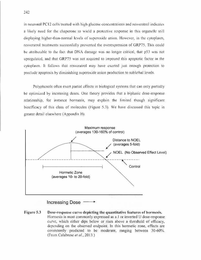
242
in neuronal PC12 cells treated with high glucose concentrations and resveratrol indicates
a likely need for the chaperone to wield a protective response in this organelle still
displaying higher-than-norrnal levels of superoxide anion. However, in the cytoplasm,
resveratrol treatments successfully prevented the overexpression of GRP75. This could
be attributable to the fact that DNA damage was no longer critical, that p53 was not
upregulated, and that GRP75 was not required to impound this apoptotic factor in the
cytoplasm. It follows that resveratrol may have exerted just enough protection to
preclude apoptosis by diminishing superoxide anion production to sublethallevels.
Polyphenols often exert partial effects in biological systems that can only partially
be optimized by increasing doses. One theory provides that a biphasic dose-response
relationship, for instance horrnesis, may explain the limited though significant
bioefficacy of this class of molecules (Figure 5.3). We have discussed this topic in
greater detail elsewhere (Appendix B).
Figure 5.3
Maximum response (averages 130-160% of control)
/
Hormetic Zone (averages 10- to 20-fold)
Increasing Dose
Distance to NOEL (averages 5-fold)
NOEL (No Observed Effect Level)
Control
Dose-response curve depicting the quantitative features of hormesis. Horrnesis is most commonly expressed as a J or inverted U dose-response curve, which either dips below or rises above a threshold of efficacy, depending on the observed endpoint. In this horrnetic zone, effects are consistently predicted to be moderate, ranging between 30-60%. (From Calabrese et al. , 2013.)
243
5.1.3 The relevance of glucose-regulated protein 75 in Parkinson 's disease
Reports showed the depletion of GRP75 in post-mortem substantia nigra pars
compacta samples of patients before our paper was published (Burbulla et al. , 2010;
Jin et al., 2006). Gaining momentum as a possible novel genetic risk factor, reports
today are rather equivocal regarding its association with the disease. Indeed, one group
found two point mutations, albeit rare, in the GRP75 gene of parkinsonian patients
(Wadhwa et al. , 2015), whereas another study concluded that this chaperone was not
genetically associated to Parkinson ' s disease (Chung et al. , 2017). Nevertheless,
evidence converges toward the reduced expression of GRP75 in specific affected brain
regions, expressly the substantia nigra pars compacta, both in patients and in rodent
models of Parkinson ' s disease (Cook et al. , 2016; Koo et al. , 2017; Liu et al., 2015).
In this view, our results rather show a rise in GRP75 expression levels in response
to a cellular stress. What occurs in our in vitro model challenged with high
concentrations of glucose is likely not representative of these physiological observations
harvested in brain samples having sustained months, in rodents, or years, in humans,
of Parkinson's disease-related stress. It is nevertheless interesting to see how GRP75 is
systematically downregulated in the substantia nigra pars compacta and invites the
question of whether this protective chaperone may be a supplementary phenotypic
liability specific to the neurons harboured in this structure.
5.2 Objectives 3 and 4: In vivo, long-term hyperglycaemia causes preferential nigrostriatal dopaminergique neurodegeneration and consequential behavioural alterations
Upon validating that dopaminergic neurons cultured in high glucose conditions
undergo cell death, we elaborated a rodent paradigm wherein neurodegeneration
could be assessed following long-term hyperglycaemia. Through immunoblotting,
immunohistochemical, neurochemical and behavioural analyses, we obtained data
presented in Chapter III demonstrating a decrease in dopamine neurotransmission in the
dorsal striatum, the degeneration of nigrostriatal neurons, motor impairments and glial

244
alterations in long-term hyperglycaemic rodents. Chapter IV provided further
information on the behavioural manifestations displayed by hyperglycaemic rats In a
social context, correlated to the level of striatal denervation.
5.2.1 Intracerebral glucose concentrations
The first element discussed in Chapter III was the nse ln CNS glucose
concentrations in hyperglycaemic rats. This firsthand demonstration was required to
solidify the argument that sustainably elevated intracerebral glucose concentrations may
induce neurodegeneration. Precisely, providing proof that glucose concentrations were
not altered differentially in the various brain regions of interest was crucial.
As previously stressed, aIl studies reporting intracerebral glucose measurements to date
have provided quantifications in, at best, two brain regions at a time (Abi-Saab et al.,
2002; Béland-Millar et al., 2017; de Vries et al., 2003; Gomez and Barros, 2003; Jacob
et al., 2002; Macauley et al., 2015; McCrimmons et al., 2003; Osborne et al., 1997).
Granted inter- and intra-individual variability ainong hyperglycaemic models, we thus
sought to rely on our own measurements to confirm - instead of presuming, like others
have - a uniform rise in glucose concentrations throughout various brain regions at a
time64.
In this Vlew, we aimed to verify the rise in brain glucose by more than one
single method. Using the microdialysis technique, we were able to obtain estimations of
extracellular glucose concentrations in uniformly-fed rats. This technique also allowed
us to distinguish midbrain and striatal subareas, owing to the spatial precision afforded
by the probes. By homogenizing brain tissues, we also appraised intracellular glucose
concentrations, although we were fully aware that terminal perfusion of the animaIs
constituted a cause for underestimations due to efflux through bidirectional GLUTs
driven by concentration gradients. Nevertheless, the purpose of these procedures was to
assess if there was a significant rise in glucose concentrations throughout the brain,
64 Speculative computations performed by Simpson and colleagues (2007) sti ll provided a theoretical basis for an initial assumption that glucose concentrations may rise in the brain in hyperglycaemic conditions and for justifying the purpose of our in vivo investigations (Table l .7).

245
and if there were substantial differences between neuroanatomical loci. We also aimed
to verify that intra- and extracellular observations coincided. For these reasons,
ail results were reported in relative and not absolute values.
Our results provided a firm validation that intracerebral glucose concentrations
increase significantly and uniformly throughout the midbrain, striatum, prefrontal cortex
and hippocampus of hyperglycaemic rats. Elevated concentrations were also sustained
over the course of 6 months. These results were important to unravel several remaining
interrogations with respect to a possible downregulation of glucose uptake in long-term
hyperglycaemia. Indeed, there is no adequate account of the regulation of GLUTs over
long periods of hyperglycaemia (Anitha et al. , 2012; Nagamatsu et al., 1994; Santiago
et al. , 2006; Simpson et al. , 1999), nor is there evidence to sustain reduced glucose
uptake due to reductions in cerebral blood flow arising from abnormal vascular features
in this chronic setting (Carruthers and Helgerson, 1989; Duckrow et al., 1987; Levine
et al. , 1998; Lowe and Walmsley, 1986; McCall, 1992; Nishimura et al. , 2007). We are
currently quantifying GLUT expression in brain slices from our model in the aim of
filling these knowledge gaps. There is, nevertheless, reason to believe that intracellular
glucose concentrations remain significantly elevated after 6 months of sustained
hyperglycaemia in our rat mode\.
5.2.2 Altered glial profiles as an indicator of oxidative stress
On the quest to coyer as many aspects of CNS dysfunction III our paradigm,
we reported the effects of long-term hyperglycaemia not only in neurons, but also in
glial populations. These glial alterations are discussed first on account of the information
they can convey with respect to the oxidative status of the CNS, directly linked to our
in vitro work.
By immunohistochemical staining, we discovered a marked proliferation of
astrocytes in the substantia nigra pars compacta, the dorsal striatum, the nucleus
accumbens and the prefrontal cortex. Conversely, there was a noticeable loss of

246
microglial ceIls in the substantia nigra pars compacta, the dorsal striatum, and the
nucleus accumbens. A first appraisal of these results aIlowed us to conclude that both
pathological features are present in the substantia nigra pars compacta, but not in the
ventral tegmental area, further underlining a regional vulnerability to pathological
alterations ensuing from hyperglycaemia. They also manifested themselves throughout
the striatum.
In Chapter III, we discussed the well-appreciated association between astrogliosis
and diabetes. We also proposed a role for oxidative stress in causing the death of
microglial ceIls, an under-recognized feature in CNS affections. We can further
elaborate on the tentative timeline proposed in this article, which stipulated that an initial
microglial ceIl activation in turn conduces to astroglial activation, foIlowed by the death
of the former, which are not equipped to cope with sustained oxidative stress. lndeed,
CNS microgliosis occurs early in hyperglycaemic rodent models (Nagayach et al. , 2014;
Oliveira et al., 2016). Importantly, microgliosis can be triggered by oxidative stress, as
demonstrated in various paradigms (Bordt and Poister, 2014; Kang et al., 2001; Mander
et al., 2006; Rojo et al., 2010). This event th en directly contributes to the activation of
astrocytes (Liddelow et al., 2017) whose elevated levels can be appraised in our model
6 months following hyperglycaemia induction. One question that dweIls here is the
physiological pertinence of upregulating astrocyte proliferation by oxidative stress
activated microglial cells. Recalling the importance of the ppp in protecting against
oxidative stress offers one clue. With neurons, astrocytes constitute the main ceIl type to
process important amounts of G6P through the ppp for the recovery ofNADPH serving
to regenerate the antioxidative co factor glutathione. In fact, the ppp is 4-5 times more
active in astrocytes th an in neurons and is upregulated in high glucose conditions65
(Ben-Yoseph et al., 1996a, 1996b; Garcia-Nogales et al., 2003; Takahashi et al., 2012)
(Figure 5.4). Remarkably, microglial cells can also mediate the increase in astroglial
ppp activity, independently of its influence on the proliferation of these cells (Iizurni
et al., 2016). It is therefore reasonable to surmise that microglial activation serves to
65 Similarly, the ppp is downregulated when glucose levels are reduced, thereby dulling the antioxidative potential of neural cells (Takahashi et al., 2012). Sorne have suggested this phenornenon to explain the risks that sharp glycaernic variations represent in diabetic patients.

247
promote the proliferation of astrocytes, alongside their ppp flux , in order to respond to
oxidative insults. Interestingly, the ppp also serves to generate ribose 5-phosphate for
the synthesis of nucleic acids required for mitosis, such as in proliferating astrocytes,
constituting yet another way by which this compensatory effect is coupled with
purposeful cellular functions (Lehninger et al. , 1995).
Figure 5.4
A B c
Metabolic activities of astrocytes and neurons. In primary neuronal (black) and astroglial (white) cultures, the rate of ppp activity (C) was obtained by measuring the difference between radio-carbon dioxide ('4C02) production ensuing from the ppp and the tricarboxylic acid cycle (A) or the tricarboxylic acid cycle alone (B) by employing glucose isotopes labelled at the Cl or C6 positions. Focusing on the graph on the right, astroglial cultures display ppp activities 4-5-fold times greater than neuronal cultures. Means ± standard deviation. *p < 0.05 ; ***p < 0.001 between groups, according to t-test. (From Takahashi et al., 2012 .)
The functional relationship between oxidative stress-induced microglial activation
and astrogliosis clearly emerges upon disceming the near-perfect correspondence
between loci displaying high levels of astrocytes and those harbouring significantly
fewer micro glial cells. If indeed microglial cells perish due to sustained oxidative stress,
as granted by several lines of evidence (Khazaei et al., 2008; Sabokdast et al. , 2015 ;
Streit et al. , 2008), it follows that these identified loci were prone to oxidative events in
our mode!. It is therefore likely that, before their demise, microglial cells were indeed
activated by this oxidative stress, as supported by previous demonstrations (Bordt and
Poister, 2014; Kang et al., 2001 ; Mander et al., 2006; Rojo et al. , 2010). In these
J

248
reglOns, we can infer that activated microglial cells may have initiated a reactive
astrogliosis, still perceptible 6 months foilowing hyperglycaemia. In other words,
regions displaying high levels of oxidative stress led to consequent changes in glial
profiles to promo te ROS clearance via the purposeful activation of the ppp and the
generation of antioxidative cofactors. Several supplementary firsthand verifications
would be required to support this proposed timeline of events occurring in glial
populations. Nonetheless, we can say without a doubt that this possible coping
mechanism provided by a reactive astrogliosis did not afford sufficient protection to
dopaminergic neurons of the nigrostriatal pathway nor to micro glial cells in oxidation
prone areas, which ail visibly degenerated in our model 6 months following
hyperglycaemia induction.
What remains unclear is why the substantia nigra pars compacta and the striaturn
were auspicious to oxidative stress-related events, precisely astrogliosis and microglial
death, whereas the hippocampus was relatively spared. One explanation might be that
these regions share the liability of processing large amounts of dopamine66, known to
react with hyperglycaemia-induced methylglyoxal to form oxidative salsolinol-like
compounds (Deng et al., 2012; Song et al., 2014; Szent-Gyorgi and McLaughlin, 1975).
Another option is that the substantia nigra pars compacta is rich in iron (Chinta and
Andersen, 2008; Hirsch and Faucheux, 1998), whereas the entire striatum is rich in
midbrain dopaminergic fibres that harbour a wealth of mitochondria (Pacelli et al. ,
2015), two independent elements conducive to ROS generation. These lines of thought
in fact boil down to the risk phenotypes characteristic of nigrostriatal neurons. We are
currently in the process of measuring advanced glycation end-products in these various
regions employing antibodies raised against glycated arginine and lysine residues.
By these means, we aim to identify differential levels of glycation throughout the CNS,
an indirect marker of oxidative stress, and to determine vulnerable cell types between
neurons, astrocytes or micro glial ceils.
66 In fact, the prefrontal cortex contains smaller amounts of dopamine than these two regions, as evidenced by our own microdialysis studies (Figure 3.31), and was also partially spared as it did not display signs of microglial cell death (Figure 3.6A and C); perhaps the proliferation of PPP-utilizing astrocytes in this region was sufficient to partially protect microglial cells against oxidative stress.
249
5.2.3 Nigrostriatal dopaminergic neuronal death: beyond the validation of our hypothesis
The work presented ln Chapter III was pivotaI to this project, as it provided
a direct confirmation of the core hypothesis. Indeed, our results show that the
nigrostriatal pathway is preferentially targeted in a hyperglycaemic setting, compared to
mesocorticolimbic neurons. Bridging these findings with observed motor deficits and
tracing the sequence of events offer further insight into how nigrostriatal dopaminergic
neurodegeneration may have operated in our rat model of long-term hyperglycaemia.
5.2.3.1 Subtle neurodegeneration and motor deficits
Upon inspecting the results presented in Chapter III, it is possible to observe
that most changes occurring because of long-term hyperglycaemia are subtle. Indeed,
the reduction in the expression of dopaminergic markers in the midbrain and striaturn,
the relative loss of dopaminergic nigrostriatal neurons, and the dampened tonic
dopamine release in the dorsal striatum do not exceed 50% of age-matched controls.
Other groups also observed these modest effects in a similar rodent paradigm
(Brambilla Bagatini et al., 2014; do Nascimento et al., 2011). Both groups found small
amounts of neurodegeneration in the substantia nigra pars compacta in streptozotocin
treated animais. However, these results were obtained 60 (Brambilla Bagatini et al.,
2014) or 90 days (do Nascimento et al., 2011) following streptozotocin injections, while
we did not observe neurodegeneration at the 3-month mark, at least wh en considering
expression levels of dopaminergic and neuronal markers67• This could be explained by
the fact that these studies employed considerably harsher chemical-induced rodent
paradigms of diabetes, thereby accelerating the neurodegenerative process. It is unclear
whether these models would exhibit more dopaminergic neuronal death at 6 months or
67 Brambilla Bagatini and colleagues employed i.p. injections of 65 mg/kg b.w. of streptozotocin without nicotinamide, a fairly high concentration in rats affording complete insulin depletion (Brambilla Bagatini et al. , 2014). In an even harsher paradigm, the other group administered rats with streptozotocin at a dose of 50 mg/kg b.w. i.v. , once again without nicotinamide (do Nascimento et al., 2011).
250
whether neurodegeneration would slow down to reach a stable degree of modest
denervation that could resemble the levels found in our paradigm. However, granted the
severity of the hyperglycaemic phenotype, it is unlikely that such models could survive
to the 6-month time point.
Other findings corroborating our work contrast with studies performed
ln paradigms combining parkinsonian and diabetic insults in which overt
neurodegeneration can be observed (Choi et al., 2005; Morris et al., 2010; Rotermund
et al., 2014). For example, after one week on a high-fat diet following MPTP treatments,
mice display almost complete nigrostriatal neurodegeneration (Choi et al., 2005).
Although such studies provide information suggesting that metabolic alterations
exacerbate the deleterious effects of parkinsonian toxins, they do not provide insight into
the natural processes that may underlie neurodegeneration in a longstanding metabolic
syndrome. Our model thus presents the advantage of mimicking the dopaminergic
neurodegeneration that occurs in a hyperglycaemic setting over the course of aging
without other exogenous effects: this neuronal loss is realistically modest.
Most importantly, our findings support the moderate motor deficits observed in
our rat model of hyperglycaemia, recalling bradykinesia and gait disturbances in
parkinsonian patients. In fact, as already mentioned, sorne tests did not reveal any
differences between control and hyperglycaemic rats, such as in the vibrissae-elicited
forelimb placement test used to assess sensorimotor integrative deficits.
These observations evoke the compensatory neurocircuit redundancies in the basal
ganglia of parkinsonian patients that account for the fact that 30-70% of nigrostriatal
dopaminergic neurons and 50-80% of their striatal projections have usually perished
before the initiation of gross motor symptoms (Bernheimer et al., 1973; Cheng et al. ,
2010; Feamley and Lees, 1991). Accordingly, we found approximately 30% of neuronal
loss in the substantia nigra pars compacta and 50% of denervation in the striatum of
long-term hyperglycaemic rats. This level of neurodegeneration stands at the threshold
of symptomatic detection and likely explains why we observe modest, yet significant,
indices ofbradykinesia and gait disturbances.

251
5.2.3.2 Time course of neurodegeneration
In Chapter III, we investigated dopaminergic and motor alterations at 3 and
6 months in our model of hyperglycaemic rats. At 6 months, hyperglycaemic rats
displayed clear neurodegeneration of the nigrostriatal pathway, as substantiated by
immunoblotting and immunohistochemical assays. However, sorne indications of
dopaminergic dysfunction were already apparent at the 3-month mark before
immunoblotting measurements could attest of any neurodegeneration.
Specifically, tonic dopamine release in the dorsal striatum was already dampened
at 3 months. We cannot exclude the possibility that the immunoblotting method
may not have allowed for the detection of inconspicuous neurodegeneration in
striatal dopaminergic fibres, which could have been picked up by more sensitive
immunohistochemical analyses at 3 months. Nevertheless, the literature consistently
shows a dulling of dopamine neurotransmission occurring without visible
neurodegeneration. Indeed, our observations bolster other reports showing that neither
TH activity or expression are reduced in the striatum of streptozotocin rats, despite
visible downregulation of dopamine neurotransmission as early as 2 weeks following the
induction of hyperglycaemia (Bitar et al. , 1986; Lim et al., 1994). Depreciation of
striatal dopamine neurotransmission despite undetectable neurodegeneration is also
typical of sorne transgenic models of Parkinson ' s disease (Abeliovich et al. , 2000;
Li et al. , 2009).
Although neurodegeneration was unnoticeable at 3 months, several motor deficits
were already apparent, as highlighted by the stepping and forepaw adjusting step tests.
In support of our observations, others have reported modest impairments of motor
functions in rodent models of Parkinson's disease despite the absence of nigrostriatal
neurodegeneration (Abeliovich et al. , 2000; Li et al. , 2009). Yet, the horizontal bar test
did not uncover any motor abnormalities in our model at 3 months. As previously
mentioned, it is the least sensitive of the 3 tests, which may explain why modest
dopaminergic dysfunctions at 3 months may not have manifested themselves in this
specific task (Kelm-Nelson et al., 2015). Another group reported horizontal bar deficits
252
in a similar model 60 days following the induction of hyperglycaemia. However, these
rats were submitted to a harsher streptozotocin regimen and also displayed substantial
immobility in the open field test (Brambi lla Bagatini et al. , 2014). In contrast, although
we did not perform this specific test, our rats did not manifest signs of hypolocomotion,
on the contrary68 .
Ail evidence considered, it appears that dopaminergic neurotransmission
dysfunctions occur before a detectable loss of nigrostriatal neuron bodies or their
projections in our hyperglycaemic rat mode!. Dampened tonic dopamine release and
manifestations of motor deficits preceding neurodegeneration sustain this proposed time
course. Nevertheless, a proper immunohistochemical appraisal of the density of striatal
dopaminergic fibres at 3 months is warranted to confirm this. It would also be interesting
to measure dopamine release at 6 months to assess the extent of neurotransmission
deterioration once neurodegeneration is perceptible. However, the surgical procedures
inherent to the microdialysis method are risky in our model at that time point, explaining
why we didn ' t perform these analyses.
5.2.4 Hyper-aggressive and hyper-sociable manifestations
In addition to motor deficits highlighted in Chapter III, we discovered hyper
aggressive and hyper-sociable traits in our model of long-term hyperglycaemia. In fact,
these results presented in Chapter IV constitute the first adequate account of social
behaviour in hyperglycaemic rats obtained in neutral circumstances69. In addition,
we report the first USV recordings ever performed in a diabetic model, whether
chemical-induced, diet-induced or genetic.
68 Observations of hyperactivity in our hyperglycaemic model were partly responsible for sparking our interest in their atypical social behaviour, related in Chapter IV.
69 Ali other behavioural investigations in similar models relied on resident-intruder paradigms that may uncover submissive or dominant traits, but the y do not report normal interspecies interactions (File and Seth, 2003 ; Hilakivi-Clarke et al. , 1990; Meehan et al. , 1986).

253
5.2.4.1 A possible relationship with nigrostriatal dopaminergic neurodegeneration
A rapid analysis of our results reveals much more frequent emissions of all types
of USVs and manifestations of affiliation/exploration or aggression-related behaviours
made by long-term hyperglycaemic rats. Covariance profiles between kinds of
behaviours or between behaviours and USVs are also noticeably altered. Our results
converge toward the conclusion that long-term hyperglycaemic rats are hyper-aggressive
and hyper-sociable, displaying behaviours that do not coincide with the affective valence
of their communications. Importantly, we found a correlation between the degree of
striatal denervation and the severity of these phenotypes. Because the article presented in
Chapter IV did not precisely address nigrostriatal neurodegeneration, we separated our
rats into groups according to overall striatal denervation. However, as we know from
Chapter III, this denervation is greatest in the dorsal area; separating the animaIs on the
basis of dorsostriatal denervation still yields the same 3 groups (Figure 5.5). Therefore,
although not within the scope of the article presented in Chapter IV, we can still say in
the context of this thesis that hyper-aggressive and hyper-sociable manifestations
alongside USV emissions were specifically associated with the degree of nigrostriatal
dopaminergic neurodegeneration.
Figure 5.5
~ Q)
@ 1 + I 1-a 1 ç "iiï c Q) "0 Q)
> ~ Qi 0::: DM DL
D CTRL denervation LOW de nervation
• HIGH denervation
NAee OT
Classification of long-term hyperglycaemic rats based on the degree of striatal denervation in multiple regions. In Figure 4.4, hyperglycaemic rats were already divided in groups according to total striatal denervation. Using the same animaIs in identical groups, we show here that dorsomedial and dorsolateral denervation are responsible for this total observed striatal denervation reported in Chapter IV. Ali data presented as means ± SEM. Asterisks indicate statistical differences between the HG group and CTRL group (**p < 0.01). Refer to Chapters III and IV for methodological details.
254
5.2.4.2 A possible relationship with phasie and tonie dopaminergie neurotransmission
Our findings raise the legitimate question of how dampened dorsostriatal
dopaminergic neurotransmission can account both for a rise in motor deficits and for
abnormal social reward-related behaviours. The link between insufficient tonic firing in
the dorsal striatum and manifestations of motor deficiencies reported in our model and
manifested in parkinsonian patients in the form of bradykinesia and rigidity is weil
appreciated (Dreyer, 2014). However, the association between a loss of dopamine tone
and increased reactivity to novel or rewarding elements in the environment is much less
obvious. In fact, parkinsonian patients usually exhibit apathy or motivational deficits,
which starkly contrast with the hyper-sociable and hyper-aggressive traits found in our
model (Drui et al., 2014; Magnard et al., 2016). A challenge therefore emerges upon
attempting to reconcile these two seemingly divergent behavioural manifestations in
light of evidence of depleted dorsostriatal dopamine in our model of long-term
hyperglycaemia.
Identifying neuropathologies bearing similar social abnormalities is a sensible
place to start in the aim of unravelling this daunting question. Our model quite
noticeably exhibits archetypal behavioural features of attention-deficit hyperactivity
disorder70. This neuropathology is strongly associated with hyper-reactivity to novel
elements in the environment, whether social or not, which can manifest itself in the form
of inappropriate impulsivity, curiosity and aggression, both in animal models and
in humans (Hopkins et al., 2009; Jhang et al., 2017; King et al., 2003; Ko et al., 2013 ;
Rosa-Neto et al., 2005). Like Parkinson's disease, its pathophysiological underpinnings
are tightly linked to dopaminergic dysfunction7 \ (Levy, 1991; 2004; Sikstrom and
Soderlund, 2007). Recent studies have slowly begun to extricate the neuroanatomical
loci implicated in this disease, and the dorsal striatum and substantia nigra pars
compacta have emerged among the most important regions. Indeed, patients with
70 The highly complex epidemiological relationship between Parkinson ' s disease and attention-deficit hyperactivity disorder is discussed in a short commentary provided in Appendix D.
71 In fact, the dopamine reuptake inhibitor methylphenidate remains the benchmark treatment.
255
attention-deficit hyperactivity disorder present an altered nigral morphology (deI Campo
et al., 2013; Romanos et al., 2010). In addition, others have reported diminished tonic
and enhanced phasic dopaminergic neurotransmission in the dorsal striatum of these
subjects (Badgaiyan et al., 2015; Sikstrom and Soderlund, 2007).
This last piece of evidence provides important insight into what may occur in our
modei of long-term hyperglycaemia. There is cause to believe that low dopamine
tonicity, or background noise, favours the saliency of phasic signaIs. As previously
stated, the dorsal striaturn is normally submitted to higher noise-to-signal ratios than the
ventral striatum 72, owing to the marked pacemaking activity of nigrostriatal neurons
(Dreyer, 2014; Zhang et al., 2009). In a context of partial tonic depletion, signais
provided by phasic dopamine are overstated when apposed to a diminished background
of tonic noise, and also by means of a compensatory upregulation of postsynaptic
dopamine receptors 73. However, when dopaminergic denervation is substantial, as m
Parkinson's disease, neither phasic nor tonic dopaminergic neurotransmission IS
properly fulfilled, giving rise to motor symptoms and loss of hyper-reactivity, or even to
the apparition of apathy (Dreyer, 2014). On this basis, we can carefully submit to the
idea that our hyperglycaemic paradigm yields a model that is sufficiently denervated to
allow the timely overlap between a moderately hypotonic phenotype responsible for
subtle motor deficits and a relative hyperphasicity accounting for social hyper-reactivity.
We could suppose that if the nigrostriatal pathway had undergone greater levels of
degeneration, our model would have more closely resembled the apathetic, motor
deficient parkinsonian phenotype. Noteworthy, proper demonstrations of enhanced
dorsostriatal phasic signalling are warranted to confirm this, and would require the
employment of methods allowing the infrasecond detection of dopamine transients,
72 In fact, the low noise-to-signal ratio in the ventral striatum is conducive to proper phasic signalling by ventral tegmental area neurons, which underlies the mesocorticolimbic pathway's prominent role in encoding reward saliency and value.
73 Recently emerged complementary evidence in humans and rodents supports a role for nigrostriatal hyperphasicity in behavioural alterations. In particular, self-driven curiosity, motivation, and aggression-related behaviours, ail applicable to social settings, are enhanced by phasic nigrostriatal dopaminergic neurotransmission (Di Domenico and Ryan, 2017; Rossi et al., 2013; Skibsted et al., 2017).
256
for instance fast-scan cyc1ic voltarnrnetry or electrophysiological techniques (Hauber,
2010; Schultz, 2010; Segovia et al., 2011; Wightman and Robinson, 2002).
5.2.5 Effects attributable to hypoinsulinaemia
To obtain a long-term hyperglycaemic phenotype, rats were rendered permanently
hypoinsulinaemic. Given insulin's highly complex roles in the healthy and diseased
CNS, extensively reviewed elsewhere (Blazquez et al. , 2014; Ghasemi et al., 2013 ;
Gray et al., 2014; Porte et al., 2005), our challenge arises upon attempting to pick apart
the effects attributable to hypoinsulinaemia or hyperglycaemia in the multiple neuronal
and behavioural alterations observed in our mode!.
5.2.5.1 Insulin in neurodegeneration
As described in section 1.2.2, insulin is not required for glucose transport in the
brain parenchyma or in most resident cells. Nevertheless, this hormone is ubiquitous in
the CNS where it mediates developmental, differentiation, plasticity and survival
signaIs. In a normal setting, insulin 's concentrations are highest in the pons, medulla and
hypothalamus, and lowest in the occipital cortex and thalamus (Banks et al., 1998),
our brain regions of interest falling within the average. As well, the insulin receptor is
also widely expressed throughout the CNS, especially so at the neuronal surface
(Havrankova et al. , 1978a; Hill et al. , 1986; Schulirigkamp et al., 2000; Unger et al.,
1991), and possesses similar kinetic properties to its peripheral counterpart (LeRoith
et al. , 1988; Zahniser et al., 1984). Insulin signalling in neurons is quite similar to that in
peripheral cells and leads to the activation of PI3K1Akt and RAS-extracellular signal
related kinase (ERK) pathways responsible for the stimulation of synaptic plasticity and
gene transcription, among many other effects (Figure 5.6). By modulating these
pathways, it is generally accepted that insulin acts as a neurotrophic factor,
promoting the survival of neurons. Accordingly, several demonstrations of insulin 's
neuroprotective competences have been carried out in vitro, for instance in settings of
excitotoxicity or oxidative stress (Duarte et al., 2006; Kim and Han, 2005; Ribeiro et al.,
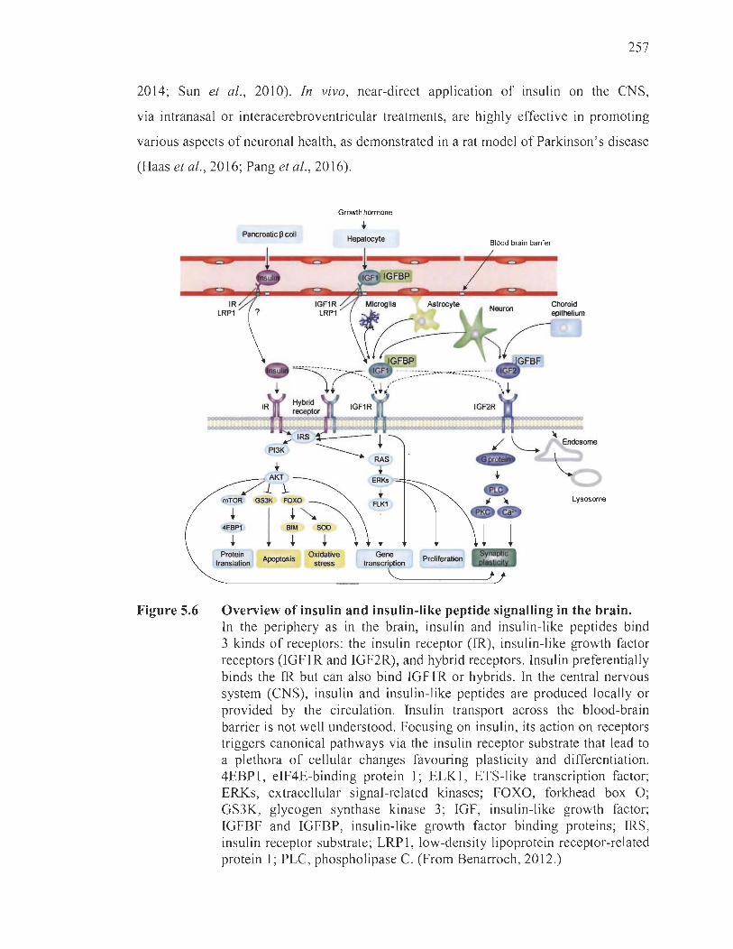
257
2014; Sun et al., 2010). In vivo, near-direct application of insulin on the CNS,
via intranasal or interacerebroventricular treatments, are highly effective in promoting
various aspects of neuronal health, as demonstrated in a rat model of Parkinson's disease
(Haas et al., 2016; Pang et al., 2016) .
Figure 5.6
Pancreatic Il cell
IR LRPI
AKT
/ .1. 1-I!!TOR GS3K FOXO
+ 1 4EBPI BIM
+ + Protein
Apoptosis translation
Growth hormone
t Hepatocyte
t ElKl
l SOD
+ Oxidative Gene
stress transcription
'--
Blood brain barrier
IGF2R
Choroid epithelium
,/ \ le '-- Endosome
Lysosome
Overview of insulin and insulin-Iike peptide signalling in the brain. In the periphery as in the brain, insulin and insulin-like peptides bind 3 kinds of receptors: the insulin receptor (IR), insulin-like growth factor receptors (IGFIR and IGF2R), and hybrid receptors. Insulin preferentially binds the IR but can also bind IGFIR or hybrids. In the central nervous system (CNS), insulin and insulin-like peptides are produced locally or provided by the circulation. Insulin transport across the blood-brain barrier is not weil understood. Focusing on insulin, its action on receptors triggers canonical pathways via the insulin receptor substrate that lead to a plethora of cellular changes favouring plasticity and differentiation. 4 EBP l , eIF 4 E-binding protein 1; ELK l , ETS-like transcription factor; ERKs, extracellular signal-related kinases; FOXO, forkhead box 0 ; GS3K, glycogen synthase kinase 3; IGF, insulin-like growth factor; IGFBF and IGFBP, insulin-like growth factor binding proteins; IRS, insulin receptor substrate; LRP1, low-density lipoprotein receptor-related protein 1; PLC, phospholipase C. (From Benarroch, 2012.)

258
Although the neuroprotective competences of in situ insulin treatment are
undeniable, the abovementioned reports do not directly address the possible harmful
effects of hypoinsulinaemia pertinent to our project. Other studies attempting to
demonstrate that neuronal death directly ensues from hypoinsulinaemia employ
peripheral insulin administration to reverse cellular or structural damage induced by
streptozotocin treatments in the CNS of rodents 74 (Hung et al., 2014; Moreira et al.,
2005, 2006; Ramanathan et al., 1999). However, they do not take into account the
various peripheral benefits exerted by this hormone replacement therapy, for instance on
glycaemic control. Such strategies exploiting organism-wide paradigms to establish
causality between hypoinsulinaemia and CNS neuronal death are based on the
misconception that Intracerebral insulin is solely provided from the circulation,
itself supplied by the pancreas. As a matter of fact, insulin concentrations are
considerably greater in the brain parenchyma than in the plasma, and are unexpectedly
upregulated in hypoinsulinaemic, hyperglycaemic streptozotocin-treated rats (Banks
et al., 1997b; Gupta et al., 1992; Havrankova et al., 1978a, 1978b, 1979) (Figure 5.7).
In support of this, insulin receptors are modulated in the periphery but not in the brain
of streptozotocin models (Pacold and Blackard, 1979; Pezzino et al. , 1996).
These preliminary findings led to the discovery that, besides being taken up through the
blood-brain barrier (Banks et al., 1997 a; Margol is and Altszuler, 1967; Woods and
Porte, 1977), insulin is synthesized by neurons within the mammalian brain (Birch et al. ,
1984; Deltour et al., 1993; Devaskar et al., 1994; Dom et al., 1983; Frolich et al., 1998;
Mehran et al., 2012; Molnar et al. , 2014; Schechter et al., 1994; Young, 1986).
Importantly, one group demonstrated that high glucose conditions enhance insulin
production by cortical neurons (Molnar et al., 2014), which may be a mechanism to
maintain appropriate Intracerebral concentrations of insu lin when pancreatic supplies
plunge (Banks et al., 1997b; Gupta et al., 1992; Havrankova et al., 1979). As the
aforementioned studies have not quantified insulin in the injured regions of the
74 One group did c1aim to show that streptozotocin-induced hypoinsulinaemia to cause CNS injuries, albeit without cell loss, and to further uncover the protective effects of intranasal insulin treatments (Francis et al. , 2008). However, this article, along with 7 others from the same group on the sa me subject, was retracted in 2014.

259
CNS following its peripheral depletion, there is no way to assertively avow a direct
negative influence of hypoinsulinaemia on neurons.
54 • BRAIN STREPTOZOTOCIN
TREA~D CONTROLS
CONTROlS
Figure 5.7
PANCREAS
STREPTOZOTOCIN TREA~D
o z ~ 1 z ::J ::J
~ ( 1
2 OlfACTORY DIENCE. CEREBRAl. BULB PHALON CORTEX
BRAIN CEREBELLUM STEM
UVER
Insulin content and binding in the brain of streptozotocin-treated rats. Left, top: Brain insulin levels (black bars) are not diminished one month after the induction of hyperglycaemia by streptozotocin compared to control conditions (hatched bars), although serum insulin is clearly reduced (white bars). Left, bottom: Pancreatic insulin levels are depleted in the pancreas of streptozocin-treated rats. Right: Insulin binding does not differ between control (C, hatched bars) and streptozotocin-treated (S, black bars) rats across various brain regions. Hepatic insu lin binding, on the other hand, is greatly increased in hyperglycaemic rats, owing to an enhanced sensitivity of receptors. White bars indicate non-specific binding. (From Havrankova et al. , 1979.)
Regarding our studies, we did not measure insulin concentrations in the brain
regions of interest in our long-terrn hyperglycaemic mode!. However, faced with the
unverified possibility that hypoinsulinaemia can trigger neuronal death, we re-exarnined
our data in a similar manner to Chapter IV by factoring in our rats' insulin profiles.
In our model, hypoinsulinaemia and glucose intolerance clearly do not correlate with
the loss of nigrostriatal neurons (Figure 5.8). Yet, we strongly agree that future
investigations would require quantifications of insulin and its receptor in targeted brain
reglOns to corroborate hypoinsulinaemia' s apparent lack of influence on
neurodegeneration in our mode!.
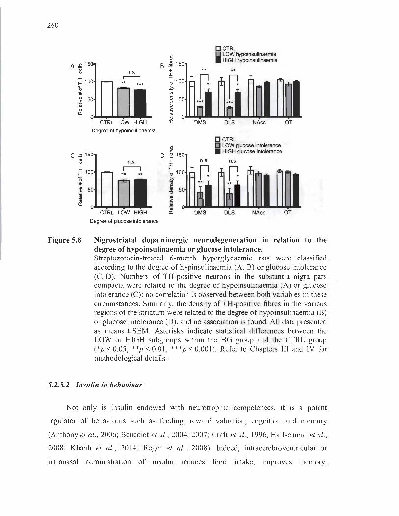
260
A ~ 1 2l + ~ 1 '0 =t:I: al
.?! ra ëii a::
C !!2 1 ~ + ~ 1 '0 =t:I: al
.?! ra ëii a::
Figure 5.8
n.s.
*~ ***
CTRL LOW HIGH
Degree of hypoinsulinaemia
n.s.
** *.
CTRL LOW HIGH
Degree of glucose inlolerance
fi)
~ B iê 150
+ l 1-'0 100 Z:-fn c: ~ 50
.~
**
ra ëii 0.&.1. ...... ,.. a::
fi)
~ D iê 150
+ l 1-'0 100 Z:-en ai 50 '0 al .?! ra & 0
DMS
n.s.
DMS
D CTRL D LOW hypoinsulinaemia • HIGH hypoinsulinaemia
**
DLS NAcc
i CTRL LOW glucose inlolerance HIGH glucose inlolerance
n.s.
DLS NAcc
OT
OT
Nigrostriatal dopaminergic neurodegeneration in relation to the degree of hypoinsulinaemia or glucose intolerance. Streptozotocin-treated 6-month hyperglycaemic rats were classified according to the degree of hypinsulinaemia (A, B) or glucose intolerance (C, D). Numbers of TH-positive neurons in the substantia nigra pars compacta were related to the degree of hypoinsulinaemia (A) or glucose intolerance (C): no correlation is observed between both variables in these circumstances. Similarly, the density of TH-positive fibres in the various regions of the striatum were related to the degree ofhypoinsulinaemia (B) or glucose intolerance (D), and no association is found. All data presented as means ± SEM. Asterisks indicate statistical differences between the LOW or HIGH subgroups within the HG group and the CTRL group (*p < 0.05, **p < 0.01 , ***p < 0.001). Refer to Chapters III and IV for methodological details .
5.2.5.2 Insulin in behaviour
Not only is insulin endowed with neurotrophic competences, it is a potent
regulator of behaviours such as feeding, reward valuation, cognition and memory
(Anthony et al., 2006; Benedict et al., 2004, 2007; Craft et al., 1996; Hallschmid et al.,
2008; Khanh et al., 2014; Reger et al. , 2008). Indeed, intracerebroventricular or
intranasal administration of insu lin reduces food intake, Improves memory,
261
and ameliorates cognitive performances in humans and rodents (Babri et al. , 2007;
Benedict et al., 2004; Haj-ali et al. , 2009; Park et al., 2000; Stockhorst et al. , 2004).
Under physiological circumstances, postprandial peripheral insulin most significantly
regulates the hypothalamus, directly at the level of the median eminence devoid of
blood-brain barrier protection, where it normally conveys satiety signais. Ail of these
regulatory effects, wh ether physiological or exogenous, are mediated by insulin ' s ability
to promote neuronal plasticity, but also by its modulatory role on neurotransmission
itself (Gupta et al. , 1992; Ohtani et al., 1997). An emerging and important
neurotransmitter on which insulin exercises its control is striatal dopamine. Granted the
central position it holds in our project, we will focus here on the relationship between
insulin75 and mesostriatal (nigrostriatal and mesolimbic) dopaminergic signalling,
especially pertaining to ensuing effects on food intake behaviours, which are overtly
altered in streptozotocin-treated models. This will allow us to draw parallels with the
abnormal behaviours we uncovered in our hypoinsulinaemic, hyperglycaemic rat mode!.
lnsulin ' s effect on the midbrain is primarily indirect. By inhibiting neuropeptide Y
/GABA/agouti-related protein (NPY/GABA/AgRP or NGA) neurons and by activating
pro-opiomelanocortinlcocaine and amphetamine regulated transcript (POMC/CART)
neurons, both located in the arcuate hypothalamus, insulin allows for the stimulation of
anorexigenic neurons and the silencing of orexigenic neurons (Abizaid et al. , 2006;
Cone et al. , 2005; Morton et al. , 2006) (Figure 5.9). The sum of these effects converges
toward decreased food intake and increased catabolism. Pertinent to our project,
orexigenic neurons in the lateral hypothalamus innervate many targets including
midbrain dopaminergic neurons in the ventral tegrnental area and the substantia nigra
pars compacta (Sakurai et al., 1998). lmpeding orexigenic signaIs onto midbrain neurons
leads to decreased dopaminergic neurotransmission in the striatum and diminished food
seeking behaviours. Although insulin ' s effects are broadly conveyed to mesostriatal
circuits, dopaminergic neurotransmission in the dorsal striatum appears to be essential
for feeding, whereas signalling to the nucleus accumbens plays only a secondary role in
75 In an attempt to simplify this part of the di scussion, we will disregard other hormonal changes that occur in our model, namely the depletion of leptin and the rise in ghrelin, which more or less mimic the effects of insulin on behaviour (see for review Palmiter, 2007).

262
modulating food anticipation behaviours (Baldo and Kelley, 2007; Cannon et al., 2004;
Drago et al. , 1994; Salamone and Correa, 2002; Ungerstedt, 1971).
Figure 5.9
Cf) , ,
insulin leptin
PVN paraventricular nucleus LHA l.ter.1 hypoth.lamic .re. PFA perifomical area Arc arcuate nucleus
regulation of food intake
vagal nerve
ghrelin
Insulin 's action on the hypothalamus. The arcuate nucleus is directly exposed to blood-bome hormones, as it is located at the level of the median eminence devoid of the blood-brain barrier. As such, insulin has a direct effect on the two principal populations that dwell therein. Neuropeptide Y (NPY)/gammaaminobutyric acid (GABA)/agouti-related protein (AgRP) neurons (NGA) neurons innervate the paraventricular nucleus (PVN) and inhibit anorexigenic neurons. Its projections onto orexigenic neurons of the lateral hypothalamic area (LHA) rather activates them. Pro-opiomelanocortin/cocaine and amphetamine regulated transcript (POMC/CART) neurons innervate the same targets but have opposite effects than NGA neurons. Orexigenic and anorexigenic neurons project to the nucleus of the solitary tract (NTS), thereby regulating food intake. Since insulin inhibits NGA neurons and stimulates POMC/CART ones, its sum effect is to inhibit food intake and promote energy expenditure. Not shown here are the LHA's projections to the midbrain. GSHR, growth hormone secretagogue receptor or ghrelin receptor; INSR, insulin receptor; LepR, leptin receptor; MC4R, melanocortin 4 receptor; a-MSH, alpha-melanocyte-stimulating hormone; YIR, neuropeptide Y receptor y 1. (From http: //www.cellbiol.net/ste/alpobesity2.php.)

263
In this vlew, one wou Id expect a hypoinsulinaemic setting, resulting from
fasting or streptozotocin treatments for instance, to lead to the disinhibition of orexigenic
neurons and the enhancement of midbrain dopaminergic neurotransmission;
the consequential rise in striatal dopamine, encoding the subjective reward value of
food, would compel the organism to feed itself, as it is a goal-directed behaviour. In fact,
hyperphagia is archetypal in the spectrurn of abnormal behaviours displayed by
streptozotocin-treated rodents, confirmed by our own findings (Figure 1.33). However,
what actually occurs at the level of striatal dopamine is highly inconsistent with these
theoretical extrapolations. 1ndeed, almost ail studies 76 performed in genetic or chemical
induced hypoinsulinaemic models, including ours (Figure 3.31), report low basal levels
of striatal dopamine (Bradberry et al. , 1989; Chen and Yang, 1991 ; Crandall and
Femstrom, 1983; Kono and Takada, 1994; Kowk and Jurio, 1986; Kwok et al. , 1985;
Lim et al., 1994; O'Dell et al. , 2014; Saller, 1984; Samandari et al. , 2013; Trulson and
Himmel, 1983). Once again, we are faced with ostensibly irreconcilable and opposite
outcomes evoked by - what appears to be - one factor.
To date, no adequate explanation has accounted for this dualism. However, we and
others propose that, since striatal dopamine transients have ne ver been measured in
hypoinsulinaemic models by techniques sensitive enough to detect infrasecond events,
it is probable that we are missing very important pieces of the puzzle pertaining to
phasic neurotransmission77 (Palmiter, 2007, 2008). Sorne indications of a role for phasic
firing that could explain overt feeding behaviours in hypoinsulinaemic subjects despite
low levels of dopamine are provided by experiments employing psychostimulants.
Indeed, streptozotocin-treated rodents manifest a greater sensitivity to addictive
substances like nicotine78 (O 'Dell et al. , 2014; Samandari et al. , 2013). In addition,
76 Sorne reports have shown enhanced dopamine levels in the brains of streptozotocin-treated rodents, but today the consensus remains that concentrations are in fact lower in the striatum (S itar et al., 1986; Gupta et al., 1992; Lackovié et al. , 1990).
77 One study did measure phasic dopamine in striatal neurons by fast-scan cyclic voltammetry, but data were obtained in organotypic slices to which insulin was applied (Stouffer et al. , 2015). As we know, it is not clearly known whether peripheral insulin may have a direct effect on regions protected by the blood-brain barri er.
78 In fact, smoking rates in diabeti c pati ents are higher than in the general population (Bishop et al. , 2009).

264
amphetamine and apomorphine, which flood the striatum with dopamine thereby raising
the noise-to-signal ratio, exert a hypophagic effect on fasted, hypoinsulinaemic rats that
would normally engage in feeding (Cannon et al., 2004; Sotak et al., 2005). Moreover,
knocking out DAT, especially important in clearing phasic dopamine transients from the
extracellular space, enhances motivation for food rewards and yields a hyperphagic
phenotype (Cagniard et al., 2006; Pecifia et al., 2003). Without providing a clear answer,
these data do converge toward a heightened production of or sensitivity to phasic
dopamine bursts in streptozotocin-treated models like ours.
This proposai bears significant implications for other behaviours mediated by
mesostriatal dopaminergic neurotransmission. We observed marked hyper-sociable and
hyper-aggressive behaviours in our long-term hypoinsulinaemic, hyperglycaemic rats,
which we explained by a possible drop in the noise-to-signal ratio. Admittedly, it is
unlikely that the sole decrease in tonic firing in the dorsal striatum arising from
hyperglycaemia-induced nigrostriatal neurodegeneration could have accounted for a
low-enough noise-to-signal ratio conducive to these hyper-reactive behaviours.
Chronic hypoinsulinaemia favouring enhanced mesostriatal dopamine phasicity may
well have participated in reducing this ratio. However, our data presented in Chapter IV
evokes a lack of correlation between the degree of hypoinsulinaemia or glucose
intolerance and the intensity of behavioural abnormalities (Figure 4.5 and Figure 4.6).
This can either signify that long-term hypoinsulinaemia do es not hold a role in
heightening phasicity and hyper-reactivity, or that the low and high subgroups were not
different enough to yield a perceptible gradation of behaviour. We reiterate here the
need for experiments addressing phasic dopamine transients in hypoinsulinaemic rats.
At any rate, substantial gaps remam m our understanding of the role held by
insulin, or lack thereof, on the CNS. Investigations are warranted to explain low
dopamine levels in hypoinsulinaemic rats, apparent even after a few days of treatment
and, in our model, observed before nigrostriatal neurons had perished at 3 months.
It would be imprudent not to contemplate the prospect of a plethora of peripheral and

265
central substances modulating the effects of insulin on the brain, likely exerting a
compensatory effect in times of induced depletion.
5.2.6 Improving the model
The nicotinamide-streptozotocin rat model of long-term hyperglycaemia has
allowed us to verify our main hypothesis, but still leaves certain questions pending.
Uncertainties remain over the influence that a systemic defect can have on the CNS,
a caveat shared by ail experimental strategies employing complex peripheral disease
models and attempting to ascribe noxious effects on the brain to a single pathological
component. In our model , much more than a single variable is tampered with;
thus, while it is tempting to attribute selective nigrostriatal dopaminergic
neurodegeneration solely to a hyperglycaemic state, we can truly only interpret that the
sum of physiological disruptions arising from nicotinamide-streptozotocin injections in
our rats maintained over 6 months leads to the preferential death of this subpopulation of
neurons. It remains that this multilayered physiological condition constitutes a stress
conducive to CNS oxidative stress to which nigrostriatal neurons were manifestly more
susceptible. Demonstrations achieved in the introduction section firmly support the
legitimacy of this basis upon which hinges our conclusion.
The literature offers a collection of astutely constructed experiments aiming to
circurnvent this problem. Ideally, the need for tampering with peripheral hormones
should be avoided in creating a high glucose environment. In addition, concentrations of
glucose should be controlled between individuals. In this view, it is reasonable to
propose verifying the selective degeneration of certain populations of neurons
throughout the brain by chronic infusions of glucose via the intracerebroventricular route
(Park et al. , 2009). Worthy of mention, glucose infusion in the brain al one can have
peripheral effects by modulating central homeostasis hubs, thus pilot studies would be
required to identify these outcomes (Carey et al., 2013 ; Park et al., 2009). In the same
vein, retrodialysis, described earlier, could address certain issues with widespread effects
of high or low circulating levels of glucose or insulin on behavioural endpoints
266
(see for review H6cht et al., 2007). Although this has never been attempted before,
glucose could easily be administered directly in the brain regions of interest following
a chronic daily regimen. Behaviour or neurotransmission responses could then be
measured in acute or chronic paradigms. This would dissipate any doubt regarding the
neuroanatomical loci responsible for the behavioural effects observed. Advantageously,
in situ measurement of oxidative stress could also be conducted in parallel using the
salicylate trapping method, which implies administering salicylic acid through the
microdialysis cannula and measuring stable oxidized adducts by high-performance
liquid chromatography (Mertens et al. , 20 Il) .
These kinds of experiments are appealing on the account that they specifically
target the CNS or precise regions therein, allowing for the identification of clear-cut
causal relationships between high glucose levels, oxidative stress, neuronal death and
behavioural outcomes. However, since such experiments imply maintaining a chronic
cannula in the CNS of rodents, they cannot be performed for more than a couple of
months on end. A methodological compromise would dwell in the employment of
a more moderate peripheral phenotype via the maintenance of appreciable levels of
circulating insu lin without sacrificing the hyperglycaemic condition. To that end,
Femyhough ' s group designed a strategy utilizing streptozotocin-treated rodents injected
with subcutaneous insu lin implants into the nape of the neck that can be renewed
monthly (Aghanoori et al. , 2017; Akude et al. , 2011). Using a small implant limits the
amount of insulin dispensed and yields a low-maintenance model of long-term
hyperglycaemia that do es not display overt hypoinsulinaemia. Peripheral alterations are
accordingly less pronounced than in the classic streptozotocin model, reducing the risk
of inferring inaccurate associations between hyperglycaemia and CNS affections.
5.3 Therapeutic perspectives
This thesis addressed the selective vulnerability of dopaminergic neurons to
hyperglycaemia-induced oxidative stress. In the process, we touched upon Parkinson 's
disease and diabetes whose pathophysiological facets and epidemiological relationship
267
are directly linked to the core question. We can extend the significance of our findings
to these pathologies.
5.3.1 Implications for diabetic patients
Diabetic patients present highly heterogeneous clinical profiles, on account of high
inter-individual variations in lifestyle, in disease management and in the presence of
additional pathological conditions. However, whether they suffer from type 1 or type II
diabetes mellitus, medical authorities have reached the consensus that glycaemic control
constitutes the topmost priority for the prevention of severe comorbidities. Our data
provide further support for this approach by demonstrating that uncontrolled
hyperglycaemia can, in the long term, lead to dopaminergic neurodegeneration.
Importantly, hyperglycaemia on its own was sufficient to cause this modest neuronal
death.
The importance of glycaemic control emerged from large trials that marked a
milestone in diabetes research by demonstrating that tight blood glucose management
could delay the ons et of complications (Diabetes Control and Complications Trial
Research Group, 1993; Holman et al., 2008; Nathan et al. , 2005; UK Prospective
Diabetes Study Group, 1998). Most striking was the discovery that these benefits
persisted long after efforts of glycaemic control had been abandoned with respect to a
cohort of patients who had never undergone tight blood glucose management.
This phenomenon is termed "glycaemic memory" and is thought to at least partially
arise from oxidative damage to mitochondrial DNA that is vulnerable to permanent
mutations leading to irreversible respiratory defects (Giacco and Brownlee, 2010).
Seeing as certain neuronal subtypes are intrinsically susceptible to oxidative stress,
as we have demonstrated here, there is an imperative need to stress the importance of
early blood glucose control in diabetic patients: the mindfulness of practitioners has
accordingly evolved in this direction. Because the existence of a glycaemic memory
implies that damage appearing at any moment of the disease is irretrievable and
accumulative, the current state of the field is more actively striving for the development

268
of therapies that could reverse cellular defects thought to underlie this phenomenon
(Giacco and Brownlee, 2010; Lovre and Fonseca, 2015; Misra and Bloombarden, 2018).
One legitimate question remams concernmg the relative risk that constitutes
pre-existing diabetes for the development of Parkinson's disease. As previously stated,
certain epidemiological studies did not find diabetic patients to present an increased
chance of developing this neurodegenerative disease (Becker et al., 2008; Cereda et al.,
20 Il; Driver et al., 2008). However, the sum of the other positive reports converges
toward a mode st two-fold increase in this risk (Arvanitakis et al., 2007; Cereda et al.,
2012; Hu et al., 2007; Klimek et al., 2015; Xu et al., 2011). It is possible to surmise that
the life expectancies of patients with type 1 or II diabetes, respectively shortened by as
much as 15 or 5-10 years (Canadian Diabetes Association, 2009), may account for these
moderate findings 79. If diabetic patients lived longer, one could expect the incidence of
age-related neurodegenerative disorders like Parkinson's disease to be substantially
superior than in the general population. Bearing in mind the idiopathic and multifaceted
nature of Parkinson's disease, uncontrolled glycaemia may interact with other risk
factors besides aging to additively or synergistically hasten the ons et of oxidative stress
induced neurodegeneration; this is plausibly what is picked up by epidemiological
studies.
5.3.2 Implications for parkinsonian patients
The keynote finding of our studies provides that hyperglycaemia can lead to the
selective degeneration of the nigrostriatal pathway. On the one hand, these results
support the hypothesis that nigrostriatal dopaminergic neurons present a characteristic
phenotype that rend ers them susceptible to what we suspect is oxidative stress. On the
other, they warn of a possible additional risk factor, especially present in diabetic
patients, which could contribute to the development of Parkinson's disease.
These results are important in light of persisting doubts regarding the significance of
79 0ther explanations for the heterogeneity of these data could be variations in the type of studies employed (case control vs. cohort), the diagnosis of Parkinson's disease and diabetes (medical examination vs. self-reported) and, as previously stated, the sizes of the populations evaluated.
269
reports stating that parkinsonian patients display elevated glycaemias (Boyd et al. , 1971 ;
Cereda et al. , 2012; Lipman et al., 1974; Sandyk, 1993; Santiago and Potashkin, 2015).
It is unclear whether hyperglycaemia appears after the onset of the disease or if it
manifests itself upstream; a direct corollary of the latter would be that hyperglycaemia
may contribute to the development of Parkinson's disease. Our work offers strong
support in favour of this second option, without however refuting a possible
bidirectional relationship between parkinsonian neurodegeneration and blood glucose
control (Brunerova et al. , 2013).
For parkinsonian patients who already suffer from this disease, war must be waged
on further nigrostriatal dopaminergic neurodegeneration; sorne rather prioritize tackling
the deterioration of their syrnptoms, which is not always directly linked to further
neurodegeneration. Our findings, though not obtained in a parkinsonian model (Choi
et al. , 2005; Morris et al. , 2010; Rotermund et al., 2014), clearly expose the hazard that
uncontrolled hyperglycaemia represents for the survival of nigrostriatal dopaminergic
neurons. Moreover, other studies provide evidence supporting a possible link between
diabetes and the severity of the syrnptoms in Parkinson's disease (Cereda et al., 2012;
Schwab, 1960). Likewise, in older persons without Parkinson's disease, parkinsonian
syrnptoms like gait disturbances are more intimately correlated to existing diabetes
(Arvanitakis et al., 2007). Considering the elevated rates of hyperglycaemia in
parkinsonian patients (Barbeau et al., 1961 ; Boyd et al., 1971; Cereda et al., 2012 ;
Lipman et al. , 1974; Sandyk, 1993), exercising a tighter glycaemic control constitutes an
achievable target to prevent the exacerbation of both neurodegeneration and worsening
of syrnptoms.
Among the most recent clinical advances, anti-diabetic drugs have emerged as
strong candidates for the development of disease-modifying therapies. A sharp interest
in these substances surfaced following reports evoking a decreased risk of
developing Parkinson 's disease in diabetic patients employing such drugs. Specifically,
the incidence of Parkinson's disease was found to be lower in diabetic patients treated
with metformin or thiozolidinediones, two glucose-lowering medications (Brauer et al.,

270
2015; Wahlqvist et al. , 2012). For example, CUITent clinical trials are investigating the
anti-diabetic and glucose-Iowering drug exenatide, a glucagon-like peptide 1 (GLP-l)
receptor agonist that largely mimi cs insulin's action (see for review Athauda and
Foltynie, 2017). An initial open label trial carried out in 44 medicated parkinsonian
patients showed that twice-daily injections of exenatide for 12 months improved
ovemight off-medication motor symptoms and dementia, rated according to the unified
Parkinson's disease rating scale and the Mattis dementia rating scale, respectively
(Aviles-Olmos et al. , 2013). This trial employed a washout design wherein exenatide
treatments were ceased at 12 months and patients were re-evaluated at later time points.
Remarkably, the benefits observed at 12 months still held at 14 (Aviles-Olmos et al.,
2013) and 24 months (Aviles-Olmos et al. , 2014). This encouraged the elaboration of
subsequent double-blinded, placebo-controlled trials once aga in designed to include a
washout period and to measure symptoms in off-medication patients. The benefits of
exenatide were reiterated and supplementary DaTscan neuroimaging results, which
conveyed information on the amount of presynaptic DAT left in the striatum of patients,
confirmed a significant positive difference between treated and control parkinsonian
patients (Athauda et al., 2017).
These results are very encouragmg and are pavmg the way for further
investigations that will perhaps address the mechanistic underpinnings of exenatide's
efficacy. In this regard, it is surprising that glycaemic profiles were not drawn
throughout these clinical trials . We can nonetheless suppose that glycaemic control was
improved in parkinsonian patients based on robust evidence provided by trials in
diabetic patients (Bergenstal et al., 2010; Buse et al. , 2009). However, a link between
improved glycaemia and symptomatic amelioration in these investigations cannot be
presumed. Indeed, GLP-l and exenatide are neurotrophic factors, akin to insulin
(Athauda and Foltynie, 2016b; Perry et al., 2002). Exenatide may therefore wield its
disease-modifying effects independently of glycaemic ameliorations. It remains that our
data grant indirect evidence of the benefits of reducing hyperglycaemia, which we
believe is a contributing factor in the efficacy of exenatide treatments in Parkinson 's
disease.

271
5.3.3 Employing resveratrol to therapeutic ends
There is compelling evidence to suggest that oxidative stress occuples a
central position in both diabetic complications and nigrostriatal dopaminergic
neurodegeneration in Parkinson's disease. Accordingly, antioxidants like the polyphenol
resveratrol have been the object of much scientific and public interest for their
therapeutic potential, either as complementary or preventive treatments. Although not
the aim of this thesis, we previously introduced the idea of utilizing resveratrol as a
neuroprotective strategy. The many sensitive elements that impinge on the employment
of polyphenols in the CNS are reviewed in Appendix B.
5.4 Concluding remarks
The etiopathogenesis of Parkinson 's disease remains largely enigmatic, in part
due to our lack of comprehension regarding the seemingly selective vulnerability of
the nigrostriatal pathway to undergo degeneration, despite the neuroanatomical breadth
of neuropathological manifestations in this illness. Ascending hypotheses grant a
preeminent role to the existence of a particular phenotypic liability that characterizes
neurons of the substantia nigra pars compacta and that may render them more sensitive
to oxidative stress. In the aim of verifying the preferred susceptibility of nigrostriatal
dopaminergic neurons to oxidative stress, we hired a strategy that employed high
glucose conditions to induce the graduaI death of neurons in vitro and in vivo. For the
first time, we provided robust evidence for the selective demise of the nigrostriatal
pathway compared to mesocorticolimbic neurons over the course of 6 months of
sustained hyperglycaemia, attended by alterations in glial profiles that indirectly
supported a role for oxidative stress as a key conspirator in this neuronal death, further
evinced by in vitro groundwork. Despite mode st neurodegeneration, hyperglycaemia
evoked motor deficits strikingly reminiscent of parkinsonian motor symptoms. We also
reported noticeably abnormal social behaviours in a hyperglycaemic mode l, which
further informed us on a possible imbalance between phasic and tonic dopaminergic
signalling arising from nigrostriatal neurodegeneration and, perhaps, hypoinsulinaemia.

272
These findings also direct our attention toward a rising need to better understand how
diabetes may alter the quality of patients' social encounters.
Across the se experiments, we demonstrated the various advantages that a long
term hyperglycaemic model presents, especial1y regarding the physiological significance
of inducing a graduaI and moderate neuronal death without layering additional
parkinsonian insults. Indeed, by revealing that hyperglycaemia is sufficient to induce
nigrostriatal dopaminergic neurodegeneration, we lend vigorous support to rather
modest epidemiological accounts of the relationship between pre-existing diabetes and
the development of Parkinson's disease in later years. According to our findings, it is
critical that the efforts recently deployed to tighten glycaemic control in diabetic patients
be firmly maintained. We further wish to express the tangible risk that an inappropriate
glycaemic health may represent for the survival of remaining nigrostriatal dopaminergic
neurons in the CNS of parkinsonian patients.
From this wealth of information, we can also fully appreciate the complexity of
several dimensions specifie to the CNS that, in remaining unsettled, dwell at the core of
numerous longstanding debates. There is a profound lack of clarity pertaining to the
bond shared between the brain and the periphery, especially in pathological settings.
In our project, this was apparent upon attempting to identify the elements responsible for
neurodegeneration in a model presenting multifaceted peripheral alterations.
Dopaminergic systems also exposed their intricate nature that emerged on our daunting
venture to draw connections between neurodegeneration, dopamine functions and
behaviour. Most evidently, our understanding of the se pending issues is at its infancy,
but formaI identification of these chasms in our knowledge draws us closer to their
unravelling.
REFERENCES
Abeliovich, A. , Schmitz, Y. , Farifias, L, Choi-Lundberg, D. , Ho, W. H. , Castillo, P. E. , Shinsky, N., Verdugo, J. M., Annanini, M. , Ryan, A., Hynes, M. , Phillips, H. , Sulzer, D. , Rosenthal, A. (2000) . Mice lacking alpha-synuclein display functional deficits in the nigrostriatal dopamine system. Neuron, 25, 239-252.
Abi-Saab, W. M. , Maggs, D. G. , Jones, T. , Jacob, R., Srihari, v., Thompson, 1. , Kerr, D., Leone, P. , Krystal , J. H. , Spencer, D. D. , During, M. 1. , Sherwin, R. S. (2002). Striking differences in glucose and lactate levels between brain extracellular fluid and plasma in conscious human subjects: effects of hyperglycemia and hypoglycemia. J Cereb. Blood Flow Metab., 22, 271-279.
Abizaid, A. , Gao, Q. , Horvath, T. L. (2006). Thoughts for food: brain mechanisms and peripheral energy balance. Neuron, 51, 691-702.
Achour, L, Arel-Dubeau, A. M., Renaud, J., Legrand, M., Attard, E. , Germain, M. , Martinoli, M. G. (2016). Oleuropein prevents neurodegeneration, mitigates mitochondrial superoxide production and modulates autophagy in a dopaminergic cellular model. Int. J Mol. Sei. , 17, pii:E1293 .
Adam-Vizi, V. (2005). Production of reactive oxygen species in brain mitochondria: contribution by electron transport chain and non-electron transport chain sources. Antioxid. Redox Signal., 7, 1140-1149.
Adams, F. S. , La Rosa, F. G. , Kumar, S. , Edwards-Prasad, J. , Kentroti, S. , Vernadakis, A., Freed, C. R. , Prasad, K. N. (1996). Characterization and transplantation of two neuronal celilines with dopaminergic properties. Neurochem. Res., 21, 619-627.
Aghanoori, M. R., Smith, D. R. , Roy Chowdhury, S., Sabbir, M. G. , Calcutt, N. A. , Fernyhough, P. (2017). Insulin pre vents aberrant mitochondrial phenotype in sensory neurons of type 1 diabetic rats . Exp. Neurol., 29 7, 148-157.
Agrawal, M. , Kumar, V. , Kashyap, M. P. , Khanna, V. K. , Randhawa, G. S., Pant, A. B. (2011) . Ischemic insult induced apoptotic changes in PC12 cells: protection by trans resveratrol. Eur. J Pharmacol. , 666, 5-11 .
Ahmed, M. u., Brinkmann Frye, E. , Degenhardt, T. P. , Thorpe, S. R. , Baynes, 1. W. (1997). N-epsilon-( carboxyethyl)lysine, a product of the chemical modification of proteins by methylglyoxa1, increases with age in human lens proteins. Biochem. J , 324, 565-570.

274
Ahrned, S. S. , Santosh, W. , Kumar, S., Christlet, H. T. (2009). Metabolic profiling of Parkinson's disease: evidence of biomarker from gene expression analysis and rapid neural network detection. J Biomed. Sei. 16, 63.
Ahrned, N., Thomalley, P. J. , Dawczynski, 1. , Franke, S. , Strobel, J., Stein, G., Haik, G. M. (2003). Methylglyoxal-derived hydroimidazolone advanced glycation end-products ofhuman lens proteins. Invest Ophthalmol. Vis. Sei., 44, 5287-5292.
Akirav, L, Maroun, M. (2006). Ventromedial prefrontal cortex is obligatory for consolidation and reconsolidation of object recognition memory. Cerebral Cortex, 16, 1759-1765.
Akude, E., Zherebitskaya, E. , Chowdhury, S. K. , Smith, D. R., Dobrowsky, R. T., Femyhough, P. (2011). Diminished superoxide generation is associated with respiratory chain dysfunction and changes in the mitochondrial proteome of sensory neurons from diabetic rats. Diabetes, 60, 288-297.
Albani, D. , Polito, L. , Batelli, S. , De Mauro, S., Fracasso, C., Martelli, G., Colombo, L., Manzoni, C. , Salmona, M. , Caccia, S. , Negro, A., Forloni, G. (2009). The SIRT 1 activator resveratrol protects SK-N-BE cells from oxidative stress and against toxicity caused by alpha-synuc1ein or amyloid-beta (1-42) peptide. J Neurochem., 110, 1445-1456.
Alberts, B. , Johnson, A., Lewis, J., Raff, M., Roberts, K., Walter, P. (2008). Chapter 18 Apoptosis: Programmed Cell Death Eliminates Unwanted Cells. In: Molecular Biology of the Cel! (textbook), (5 th ed.). Garland Science. p. 1115.
Albin, R. L. , Young, A. B. , Penney, J. B. (1989). The functional anatomy of basal ganglia disorders. Trends Neurosci., 12, 366-375.
Alexander, G. E., Crutcher, M. D. , DeLong, M. R. (1990). Basal ganglia-thalamocortical circuits: parallel substrates for motor, oculomotor, "prefrontal" and " limbic" functions. Prog. Brain Res., 85, 119-146.
Allaman, L, Magistretti, P. J. , (2013). Brain energy metabo1ism. In Squire, L. R. , Berg, D. , Bloom, F. E., du Lac, S., Ghosh, A. , Spitzer, N. C. (eds), Fundamental Neuroscience, Academic Press, San Diego, pp. 261-284.
Allera, C., Lazzarini, G. , Patrone, E. , Alberti, L, Barboro, P., Sanna, P. , Melchiori, A. , Parodi, S., Balbi, C. (1997). The condensation of chromatin in apoptotic thyrnocytes shows a specific structural change. J Biol. Chem. , 272, 10817-10822.
275
Alliot, F., Godin, I. , Pessac, B. (1999). Microglia derive from progenitors, originating from the yolk sac, and which proliferate in the brain. Brain Res. Dev. Brain Res. , 11 7, 145-152.
Alomar, F., Singh, J., Jang, H. S., Rozanzki, G. 1. , Shao, C. H. , Padanilam, B. 1., Mayhan, W . G., Bidasee, K. R. (2016). Smooth muscle-generated methylglyoxal impairs endothelial cell-mediated vasodilatation of cerebral microvessels in type 1 diabetic rats. Br. J Pharmacol. , 1 73, 3307-3326.
Alonso, A. , Sovell, K. A. (2010). Gout, hyperuricemia, and Parkinson's disease: a protective effect? Curr. Rheumatol. Rep. , 12, 149-155.
Altmann, C. , Schmidt, M. H. H. (2018). The Role of Microglia in Diabetic Retinopathy: Inflammation, Microvasculature Defects and Neurodegeneration. Int. J Mol. Sei. , 19, pii: E110.
Alvarez, E. , Martinez, M. D. , Roncero, I. , Chowen, J. A. , Garcia-Cuartero, B. , Gispert, J. D. , Sanz, C., Vazquez, P. , Maldonado, A. , de Caceres, 1., Desco, M ., Pozo, M. A. , Blazquez, E. (2005). The expression of GLP-I receptor rnRNA and protein allows the effect of GLP-1 on glucose metabolism in the human hypothalamus and brainstem. J Neurochem., 92, 798-806.
Alvarez-Cervera, F. 1. , Villanueva-Toledo, J., Moo-Puc, R. E. , Heredia-Lopez, F. J., Alvarez-Cervera, M. , Pineda, J. C. , Gongora-Alfaro, 1. L. (2005). A novel automated rat catalepsy bar test system based on a RISC microcontroller. J Neurosci. Methods, 146, 7683 .
Amblee, A. , Lious, D., Fogelfeld, L. (2016). Combination of Saxagliptin and Metformin Is Effective as Initial Therapy in New-Onset Type 2 Diabetes Mellitus With Severe Hyperglycemia. J Clin. Endocrinol. Metab., 101, 2528-2535 .
American Diabetes Association. (2016). Diagnosing Diabetes and Learning About Prediabetes. http://www . diabetes. org/ diabetes-bas ics/ diagnosis/
Aminzadeh, A. (2017). Protective effect of tropisetron on high glucose induced apoptosis and oxidative stress in PC12cells: roles of JNK, P38 MAPKs, and mitochondria pathway. Metab. Brain Dis., 32, 819-826.
Amiot, M. J., Romier, B. , Anh Dao, T. M. , Fanciullino, R. , Ciccolini, 1. , Burcelin, R. , Pechere, L. , Emond, C., Savouret, J . F., Seree, E. (2013). Optimization of trans-resveratrol bioavailability for human therapy. Biochimie, pü: S0300-9084, 00011-4.
276
Anandhan, A., Tamilselvam, K. , Vijayraja, D., Ashokkumar, N., Rajasankar, S., Manivasagam, T. (2010). Resveratrol attenuates oxidative stress and improves behaviour in l-methyl-4-phenyl-1,2,3,6-tetrahydropyridine (MPTP) challenged mice. Ann. Neurosci., 17, 113-119.
Andén, N. E. , Dahlstr6m, A., Fuxe, K., OIson, L., Ungerstedt, U. (1966). Ascending noradrenaline neurons from the pons and the medulla oblongata. Exp erien tia, 22,44-45.
Anderson, J. J., Chen, X. , Boass, A., Symons, M., Kohlmeier, M., Renner, J. B., Garner, S. C. (2002). Soy isoflavones: no effects on bone minerai content and bone mineraI density in healthy, menstruating young adult women after one year. J Am. Coll. Nutr., 21, 388-393 .
Andersen, J. K., Frim, D. M., Isacson, O., Beai, M. F. , Breakefield, X.O. (1994). Elevation of neuronal MAO-B activity in a transgenic mouse model does not increase sensitivity to the neurotoxin 1-methyl-4-phenyl-1 ,2,3,6-tetrahydropyridine (MPTP). Brain Res., 656, 108-114.
Andres-Lacueva, c., Shukitt-Hale, B., Galli, R. L., Jauregui, O., Lamuela-Raventos, R. M., Joseph, J. A. (2005). Anthocyanins in aged blueberry-fed rats are found centrally and may enhance memory. Nutr. Neurosci. , 8, 111-120.
Anglade, P., Vyas, S., Javoy-agid, F. , Herrero, M. T., Marquez, J. , Mouatt-Prigent, A., Ruberg, M., Hirsch, E. c., Agid, y. (1997) . Apoptosis and autophagy in nigral neurons of patients with Parkinson's disease. Histol. Histopathol., 12, 25-31.
Anitha, M. , Abraham, P. M. , Paulose, C. S. (2012). Striatal dopamine receptors modulate the expression of insu lin receptor, IGF-1 and GLUT-3 in diabetic rats: effect of pyridoxine treatment. Eur. J Pharmacol. , 696, 54-61.
Annett, L. E., McGregor, A., Robbins, T. W. (1989). The effects ofibotenic acid lesions of the nucleus accumbens on spatial learning and extinction in the rat. Behavioural Brain Research, 31, 231-242.
Anthony, K. , Reed, L. J., Dunn, J. T. , Bingham, E., Hopkins, D., Marsden, P.K, Amiel, S. A. (2006). Attenuation of insulin-evoked responses in brain networks controlling appetite and reward in insulin resistance: the cerebral basis for impaired control of food intake in metabolic syndrome? Diabetes, 55,2986-2992.
Aquino, C. C., Fox, S. H. (2014). Clinical spectrum of levodopa-induced complications. Mov. Disord. , 30, 80-89.

277
Araki, E., Nishikawa, T. (2010) . Oxidative stress: A cause and therapeutic target of diabetic complications. J Diabetes Investig. , 1, 90-96.
Araszkiewicz, A., Zozulinska-Ziolkiewicz, D . (2016) . Retinal Neurodegeneration in the Course of Diabetes-Pathogenesis and Clinical Perspective. Curr. Neuropharmacol., 14, 805-809.
Arenas, E. , Denham, M., Villaescusa, J. C. (2015). How to make a midbrain dopaminergic neuron. Development, 142, 1918-1936.
Arluison, M ., Quignon, M., Nguyen, P. , Thorens, B. , Leloup, C. , Penicaud, L. (2004). Distribution and anatomical localization of the glucose transporter 2 (GLUT2) in the adult rat brain - an immunohistochemical study. J Chem. Neuroanat. , 28, Il 7 -13 6.
Armstrong, R. A. , Lantos, P. L. , Cairns, N. J. (2005). Overlap between neurodegenerative disorders. Neuropathology, 25, 111-124.
Arvanitakis, Z. , Wilson, R. S. , Bienias, J. L. , Bennet, D. A. (2007). Diabetes and parkinsonian signs in older persons. Alzheimer Dis. Assoc. Disord. , 21, 144-149.
Ashkenazi, A. (2008). Directing cancer cells to self-destruct with pro-apoptotic receptor agonists. Nat. Rev. Drug Discov., 7, 1001-1012.
Ashrafi , G. , Schlehe, 1. S. , LaVoie, M. J. , Schwarz, T. L. (2014). Mitophagy of damaged mitochondria occurs locally in distal neuronal axons and requires PINK1 and Parkin. J Cel!. Biol., 206, 655-670.
Ashrafi, G., Wu, Z., Farrell, R. J. , Ryan, T. A. (2017). GLUT4 Mobilization Supports Energetic Demands of Active Synapses. Neuron. , 93, 606-615.
Athauda, D., Foltynie, T. (2016a). Challenges in detecting disease modification In
Parkinson's disease clinical trials. Parkinsonism Relat. Disord. , 32, 1-11.
Athauda, D., Foltynie, T . (2016b). The glucagon-like peptide 1 (GLP) receptor as a therapeutic target in Parkinson ' s disease: mechanisms of action. Drug Discov. Today, 21, 802-818.
Athauda, D. , Foltynie, T. (2017). Protective effects of the GLP-l mimetic exendin-4 in Parkinson's disease. Neuropharmacology, pii: S0028-3908(17)30440-9.
278
Athauda, D., Maclagan, K. , Skene, S. S. , Bajwa-Joseph, M. , Letchford, D., Chowdhury, K. , Hibbert, S. , Budnik, N., Zampedri, L. , Dickson, J., Li, Y., Aviles-Olmos, I. , Wamer, T. T. , Limousin, P., Lees, A. J., Greig, N . H., Tebbs, S., Foltynie, T. (2017). Exenatide once weekly versus placebo in Parkinson's disease: a randomised, double-blind, placebo-controlled trial. Lancet, 390, 1664-1675.
Aubert, A., Costalat, R. (2005). Interaction between astrocytes and neurons studied using a mathematical model of compartmentalized energy metabolism. J Cereb. Blood Flow Metab., 25, 1476-1490.
Aubert, A. , Pellerin, L. , Magistretti, P. J., Costalat, R. (2007). A coherent neurobiological framework for functional neuroimaging provided by a model integrating compartmentalized energy metabolism. Proc. Nat/. Acad. Sei. USA, 104,4188-4193.
Aubert, C. E., Michel, P. L. , Gillery, P., Jaisson, S., Fonfrede, M ., Morel, F., Hartemann, A. , Bourron, O. (2014). Association of peripheral neuropathy with circulating advanced glycation end products, soluble receptor for advanced glycation end products and other risk factors in patients with type 2 diabetes. Diabetes Metab. Res. Rev., 30, 679-685.
Auburger, G., Kurz, A. (2011) . The role of glyoxalases for sugar stress and aging, with relevance for dyskinesia, anxiety and Parkinson's disease. Aging, 3, 5-9.
Aviles-Olmos, I., Dickson, J. , Kefalopoulou, Z. , Djamshidian, A. , Ell, P., Soderlund, T., Whitton, P., Wyse, R, Isaacs, T., Lees, A., Limousin, P. , Foltynie, T. (2013). Exanatide and the treatment of patients with Parkinson 's disease. J Clin. In vest. , 123, 2730-2736.
A viles-Olmos, I. , Dickson, J. , Kefalopoulou, Z., Djamshidian, A., Kahan, 1. , Fmedsci, P. E. , Whitton, P. , Wyse, R, Isaacs, T. , Lees, A., Limousin, P., Foltynie, T. (2014). Motor and cognitive advantages persist 12 months after exenatide exposure in Parkinson 's disease. J Parkinsons Dis. , 4, 337-344.
Babri, S. , Badie, H. G. , Khamenei, ~. , Ordikhani-Seyedlar, M. (2007). Intrahippocampal insulin improves memory in a passive-avoidance task in male wistar rats. Brain Cogn. , 64, 86-91.
Badgaiyan, RD., Sinha, S., Sajjad, M., Wack, D. S. (2015) . Attenuated Tonic and Enhanced Phasic Release of Dopamine in Attention Deficit Hyperactivity Disorder. PLoS One, 10, e0137326.
279
Badole, S. L. , Chaudhari, S. M. , Jangam, G. B., Kandhare, A. D., Bodhankar, S. L. (2015). Cardioprotective Activity of Pongamia pinnata in StreptozotocinNicotinamide Induced Diabetic Rats. Biomed. Res. Int., 2015, 403291.
Bagatini, P. B. , Xavier, L. L. , Bertoldi, K. , Moysés, F., Lovatel, G., Neves, L. T. , Barbosa, S. , Saur, L. , de Senna, P. N. , Souto. A. A., Siqueira, 1. R., Achaval, M. (2017). An evaluation of avers ive memory and hippocampal oxidative status in streptozotocin-induced diabetic rats treated with resveratrol. Neurosci. Leu., 636, 184-189.
Bai , T., Dong, D. S. , Pei, L. (2013). Resveratrol mltlgates isoflurane-induced neuroapoptosis by inhibiting the activation of the Akt-regulated mitochondrial apoptotic signaling pathway. Int. J Mol. Med., 32, 819-826.
Bak, L. K. , Obel, L. F. , Walls, A. B. , Schousboe, A. , Faek, S. A. , Jajo, F. S., Waagepetersen, H. S. (2012). Novel model of neuronal bioenergetics: Postsynaptic utilization of glucose but not lactate correlates positively with Ca2+ signalling in cultured mouse glutamatergic neurons. ASN Neuro., 4, pii: e00083.
Bak, L. K. , Schousboe, A. , Sonnewald, U., Waagepetersen, H. S. (2006). Glucose is necessary to maintain neurotransmitter homeostasis during synaptic activity in cultured glutamatergic neurons. J Cereb. Blood Flow Metab. , 26, 1285-1297.
Baldo, B. A. , Kelley, A. E . (2007). Discrete neurochemical coding of distinguishable motivational processes: insights from nucleus accumbens control of feeding. Psychopharmacol. (Berl), 191, 439-459.
Baldessarini, R. 1. (1985). Principles and practice. In: Chemotherapy in psychiatry. Harvard University Press.
Banaszewska, B. , Wrotynska-Barczyilska, J. , Spaczynski, R . Z., Pawelczyk, L. , Duleba, A. J. (2016). Effects of Resveratrol on Polycystic Ovary Syndrome: A Double-blind, Randomized, Placebo-controlled Trial. J Clin. Endocrinol. Metab. , 101, 4322-4328.
Banks, W. A. , Jaspan, 1. B., Huang, W. , Kastin, A. J. (1997a) . Transport of insulin across the blood-brain barrier: saturability at euglycemic doses of insulin. Peptides, 18, .1423-1429.
Banks, W. A. , Jaspan, J. B. , Kastin, A. J. (1997b) . Effect of diabetes mellitus on the permeability of the blood-brain barrier to insulin. Peptides, 18, 1577-1584.
Banks, W. A. , Kastin, A. J. (1998) . DifferentiaI permeability of the blood-brain barrier to two pancreatic peptides: insulin and amylin. Peptides, 19, 883-889.

280
Bansal, V., Kalita, J., Misra, U. K. (2006). Diabetic neuropathy. Postgrad. Med. J , 82, 95-100.
Bar-Gad, L, Heimer, G., Ritov, Y., Bergman, H. (2003). Functional correlations between neighboring neurons in the primate globus pallidus are weak or nonexistent. J Neurosci. 23,4012-4016.
Barbeau, A., Giguere, R., Hardy, J. (1961). Clinical experience with tolbutamide in Parkinson ' s disease. Union Med. Can. , 90, 147 -15l.
Bames, K. , Ingram, J. C. , Bennett, M. D., Stewart, G. W., Baldwin, S.A. (2004). Methyl-beta-cyclodextrin stimulates glucose uptake in Clone 9 cells: a possible role for lipid rafts. Biochem. J, 378, 343-351.
Barros, L. F., Courjaret, R., Jakoby, P. , Loaiza, A., Lohr, C. , Deitmer, J. W. (2009) . Preferential transport and metabolism of glucose in Bergmann glia over Purkinje cells: a multiphoton study of cerebellar slices. Glia, 5 7, 962-970.
Barzilai , A. , Yamamoto K. (2004). DNA damage responses to oxidative stress. DNA Repair (Amst), 3, 1109-1115.
Bashan, N., Burdett, E., Gumà, A. , Sargeant, R., Tumiati, L., Liu, Z. , Klip, A. (1993). Mechanisms of adaptation of glucose transporters to changes in the oxidative chain of muscle and fat cells. Am. J Physiol. , 264, 430-440.
Bashan, N., Burdett, E., Hundal, H. S., Klip, A. (1992). Regulation of glucose transport and GLUT 1 glucose transporter expression by 02 in muscle cells in culture. Am. J Physiol., 262, 682-690.
Bassareo, V., Musio, P., Di Chiara, G. (2011). Reciprocal responsiveness of nucleus accumbens shell and core dopamine to food- and drug conditioned stimuli. Psychopharmacol. , 214, 687-697.
Bast, T. (2007). Toward an integrative perspective on hippocampal function: from the rapid encoding of experience to adaptive behavior. Rev Neurosci., 18, 253-281.
Bast, T., Wilson, 1. A., Witter, M. P., Morris, R. G. (2009). From rapid place leaming to behavioral performance: a key role for the intermediate hippocampus. PLoS Bio!. , 7, elO00089.
Batandier, C., Fontaine, E., Kériel , c., Leverve, X. M. (2002). Determination of mitochondrial reactive oxygen species: methodological aspects. J Cel!. Mol. Med., 6, 175-187.

281
Baumann, M. u., Zamudio, S. , Illsley, N. P. (2007). Hypoxic upregu1ation of glucose transporters in BeWo choriocarcinoma cells is mediated by hypoxiainducible factor-1. Am. J Physiol. Cel!. Physiol., 293, 477-485.
Baxter, A. G., Duckworth, R. C. (2004). Models of type 1 (auto immune) diabetes. Drug Discov. Today Dis. Models, 1, 451-455.
Baydas, G. , Nedzvetskii, V. S. , Tuzcu, M. , Yasar, A. , Kirichenko, S. V. (2003). Increase of glial fibrillary acidic protein and S-100B in hippocampus and cortex of diabetic rats: effects ofvitamin E. Eur. J Pharmacol. , 462, 67-71.
Bazelton, M., Fenichel, G M., Randall, J. (1967). Studies on neuromelanin. 1. a melanin system in the adult human brainstem. Neurology, 17, 512-519.
Beai, M. F. (2001). Experimental models of Parkinson's disease. Nat. Rev. Neurosci. 2, 325-334.
Beai, M. F. (2003). Mitochondria, oxidative damage, and inflammation in Parkinson 's disease. Ann. NY Acad. Sei., 991, 120-131.
Becker, C. , Brobert, G. P., Johansson, S. , Jick, S. S., Meier, C. R. (2008). Diabetes in patients with idiopathic Parkinson's disease. Diabetes Care, 31, 1808-1812.
Beeler, J. A. (2011). Preservation of function in Parkinson's disease: what's leaming got to do with it? Brain Res., 1423, 96-113.
Beher, D. , Wu, J., Cumine, S. , Kim, K. W., Lu, S. C., Atangan, L., Wang, M. (2009). Resveratrol is not a direct activator of SIRT1 enzyme activity. Chem. Biol. Drug Des., 74,619-624.
Béland-Millar, A. , Larcher, J., Courtemanche, J. , Yuan, T. , Messier, C. (2017). Effects of Systemic Metabolic Fuels on Glucose and Lactate Levels in the Brain Extracellular Compartment of the Mouse. Front. Neurosci., Il, 7.
Bell, R. H. , Hye, R. J. (1983). Animal models of diabetes mellitus: physiology and pathology. J Surg. Res. , 35, 433-460.
Belluzzi, E., Bisaglia, M. , Lazzarini, E., Tabares, L. C., Beltramini, M., Bubacco, L. (2012). Human SOD2 modification by dopamine quinones affects enzymatic activity by promoting its aggregation: possible implications for Parkinson 's disease. PLoS One, 7, e38026.
282
Ben-Yoseph, O. , Boxer, P. A., Ross, B. D. (1 996a). Assessment of the role of the glutathione and pentose phosphate pathways in the protection of primary cerebrocortical cultures from oxidative stress. J Neurochem., 66, 2329-2337.
Ben-Yoseph, O., Boxer, P. A., Ross, B. D. (1996b). Noninvasive assessment of the relative roles of cerebral antioxidant enzymes by quantitation of pentose phosphate pathway activity. Neurochem. Res., 21, 1005-1012.
Benabid, A. L., Chabardès, S. , Seigneuret, E . (2005). Deep-brain stimulation in Parkinson's disease: long-term efficacy and safety - What happened this year? Curr. Opin. Neurol., 18, 623-630.
Benarroch, E. E. (2012). Insulin-like growth factors in the brain and their potential clinical implications. Neurology, 79, 2148-2153.
Bender, A., Krishnan, K. 1., Morris, C. M., Taylor, G. A., Reeve, A. K., Perry, R. H., Jaros, E., Hersheson, J. S., Betts, 1., Klopstock, T., Taylor, R. W., Turnbull, D. M. (2006). High levels of mitochondrial DNA deletions in substantia nigra neurons in aging and Parkinson disease. Nat. Genet., 38, 515-517.
Bender, A., Schwarzkopf, R. M. , McMillan, A., Krishnan, K. J. , Rieder, G. , Neumann, M. , Eistner, M., Turnbull , D. M., Klopstock, T. (2008). Dopaminergic midbrain neurons are the prime target for mitochondrial DNA deletions. J Neurol., 255, 1231-1235.
Benedict, C., Hallschmid, M., Hatke, A., Schultes, B., Fehm, H. L., Born, J., Kern, W. (2004) . lntranasal insulin improves memory in humans. Psychoneuroendocrinol. , 29, 1326-1334.
Benedict, C., Hallschmid, M. Schmitz, K. , Schultes, B., Ratter, F., Fehm, H. L., Born, J. , Kern, W. (2007). Intranasal insulin improves memory in humans: superiority of insulin aspart. Neuropsychopharmacology, 32, 239-243 .
Benes, F. M. (2001). Carlsson and the discovery of dopamine. Trends in Pharmacological Sciences, 22, 46-47.
Bentsath, A., Rusznyak, S.T. , Szent-Gyorgyi, A. (1936). Vitamin nature of flavones . Nature (London), 138, 798.
Benzie,1. F., Szeto, Y. T. , Strain, J. J., Tomlinson, B. (1999). Consumption of green tea causes rapid increase in plasma antioxidant power in humans. Nutr. Cancer., 34,83-87.

283
Bernheimer, H. , Birkmayer, W ., Hornykiewicz, O. , Jellinger, K. , Seitelberger, F. (1973). Brain dopamine and the syndromes of Parkinson and Huntington. Clinical, morphological and neurochemical correlations. J Neurol. Sei., 20, 415-455 .
Bergenstal, R. M. , Wysham, c., Macconell, L. , Malloy, J ., Walsh, B. , Yan, P., Wilhelm, K., Malone, J ., Porter, L. E. (2010). Efficacy and safety of exenatide once weekly versus sitagliptin or pioglitazone as an adjunct to metformin for treatment of type 2 diabetes (DURATION-2): a randomised trial. Lancet, 376, 431-439.
Bergstrom, B. P. , Garris, P. A. (2003). "Passive stabilization" of striatal extracellular dopamine across the les ion spectrum encompassing the presymptomatic phase of Parkinson's disease: a voltammetric study in the 6-0HDA-Iesioned rat. J Neurochem., 87, 1224-1236.
Bergstrom, B. P., Sanberg, S. G. , Andersson, M., Mithyantha, J., Carroll, F. L, Garris, P . A. (2011). Functional reorganization of the presynaptic dopaminergic terminal in parkinsonism. Neuroseience, 193, 310-322.
Bertelli, A. , Bertelli, A. A. E ., Gozzini, A., Giovannini, L. (1998). Plasma and tissue resveratrol concentrations and pharmacological activity. Drugs Exp . Clin. Res. , 24, 133-138.
Bertolotto, F., Massone, A. (2012) . Combination of alpha lipoic acid and superoxide dismutase leads to physiological and symptomatic improvements in diabetic neuropathy. Drugs R. D., 12, 29-34.
Bertran-Gonzalez, 1. , Bosch, C. , Maroteaux, M. , Matamales, M., Hervé, D. , Valjent, E., Girault, J. A. (2008). Opposing patterns of signaling activation in dopamine Dl and D2 receptor-expressing striatal neurons in response to cocaine and haloperidol. J Neurosei. 28, 5671-5685.
Betarbet, R. , Sherer, T. B. , MacKenzie, G., Garcia-Osuna, M. , Panov, A. V., Greenamyre, 1. T. (2000). Chronic systemic pesticide exposure reproduces features ofParkinson ' s disease. Nat. Neurosei. , 3, 1301-1306.
Bharath, S. , Hsu, M. , Kaur, D., Rajagopalan, S. , Andersen, J. K. (2002). Glutathione, iron and Parkinson's disease. Biochem. Pharmacol., 64, 1037-1048.
Bhuyan, B. K. , Kuentzel, S. L. , Gray, L. G. , Fraser, T. J., Wallach, D. , Neil, G. L. (1974) . Tissue distribution of streptozotocin (NSC-85998). Cancer Chemother. Rep ., 58, 157-165.

284
Bialy, M., Rydz, M., Kaczmarek, L. (2000). Precontact 50-kHz vocalizations in male rats during acquisition of sexual experience. Behav. NeurosCÎ., 114, 983-990.
Bierhaus, A., Hurnpert, P. M., Morcos, M., Wendt, T., Chavakis, T., Arnold, B., Stern, D. M., Nawroth, P. P. (2005). Understanding RAGE, the receptor for advanced glycation end-products. J Mol. Med., 83, 876-886.
Biessel, G. J., van der Heide, L. P. , Kamal, A., Bleys, R. L., Gispen, W. H. (2002). Ageing and diabetes: implications for brain function. Eur. J Pharmacol. , 441,1-14.
Bilgic, B., Pfefferbaum, A., Rohlfing, T., Sullivan, E. V., Adalsteinsson, E. (2012). MRI estimates of brain iron concentration in normal aging using quantitative susceptibility mapping. Neuroimage, 59, 2625-2635.
Birch, N. P., Christie, D. L., Renwick, A. G. C. (1984). Proinsulin-like material ln
mouse foetal brain cell cultures. FEBS LeU., 168, 299-302.
Bishop, F. K., Maahs, D. M., Snell-Bergeon, J. K., Ogden, L. G., Kinney, G. L., Rewers, M. (2009). Lifestyle risk factors for atherosc1erosis in adults with type 1 diabetes. Diab. Vasco Dis. Res. , 6, 269-275.
Bitar, M., Koulu, M., Rapoport, S. L, Linnoila, M. (1986). Diabetes-induced alteration in brain monoamine metabolism in rats. J Pharmacol. Exp. Ther., 236, 432-437.
Bjorklund, A., Dunnett, S. (2007). Dopamine neuron systems in the brain: an update. Trends NeurosCÎ. , 30, 194-202.
Blanchet, J. , Longpré, F., Bureau, G., Morissette, M., DiPaolo, T., Bronchti, G., Martinoli, M. G. (2008). Resveratrol, a red wine polyphenol, protects dopaminergic neurons in MPTP-treated mice. Prog. Neuropsychopharmacol. Biol. Psychiatry, 32, 1243-1250.
Blâzquez, E., Velâzquez, E. , Hurtado-Carneiro, V., Ruiz-Albusac, J. M. (2014). Insulin in the brain: its pathophysiological implications for States related with central insulinresistance, type 2 diabetes and Alzheimer's disease. Front. Endocrinol. (Lausanne), 5, 161 .
Blocq, P., Marinesco, G. (1892). Sur les lésions de la pathogénie de l'épilepsie essentielle. Sem. Med. , 12, 445-446.
Blocq, P., Marinesco, G. (1893). Sur un cas de tremblement parkinsonien hémiplégique symptomatique d 'une tumeur du pédoncule cérébral. C. R. Soc. Biol. , 5, 105-111.
285
Boissy, A., Manteuffel, G. , Jensen, M. B. , Moe, R. O., Spruijt, B. , Keeling, L. 1., Winckler, C., Forkman, B., Dimitrov, L, Langbein, J. , Bakken, M., Veissier, L, Aubert, A. (2007). Assessment of positive emotions in animais to improve their welfare. Physiol. Behav., 92, 375-397.
Bolafios, J. P., Almeida, A. , Moncada, S. (2010). Glycolysis: a bioenergetic or a survival pathway? Trends Biochem. Sei., 35, 145-149.
Bolafios, J . P., Delgado-Esteban, M. , Herrero-Mendez, A. , Femandez-Femandez, S., Almeida, A. (2008). Regulation of glycolysis and pentose-phosphate pathway by nitric oxide: impact on neuronal survival. Biochim. Biophys. Acta. , 1777, 789-793.
Bolster, D. R., Crozier, S. J ., Kimball , S. R. , Jefferson, L. S. (2002). AMP-activated protein kinase suppresses protein synthesis in rat skeletal muscle through downregulated mTOR signaling. J Biol. Chem., 2 77, 23977-23980.
Bondy, C. A. , Lee, W. H. , Zhou, J. (1992). Ontogeny and cellular distribution of brain glucose transporter gene expression. Mol. Cel!. Neurosci. , 3, 305-314.
Bordt, E. A. , Polster, B. M. (2014). NADPH oxidase- and mitochondria-derived reactive oxygen species in proinflammatory microglial activation: a bipartisan affair? Free Radic. Biol. Med. , 76, 34-46.
Borra, M. T. , Smith, B. C., Denu, 1. M. (2005). Mechanism of human SIRTI activation by resveratrol. J Biol. Chem., 280, 17187-17195.
Borroto-Escuela, D. O., Carlsson, 1. , Ambrogini, P. , Narvaez, M. , Wydra, K. , Tarakanov, A. O., Li, X., Mi1l6n, C., Ferraro, L., Cuppini, R., Tanganelli, S. , Liu, F. , Filip, M., Diaz-Cabiale, Z., Fuxe, K. (2017). Understanding the Role of GPCR Heteroreceptor Complexes in Modulating the Brain Networks in Health and Disease. Front Cel!. Neurosci., Il, 37.
Bossers, S. M., de Boer, R. D., Boer, C., Peerdeman, S. M. (2013). The diagnostic accuracy of brain microdialysis during surgery: a qualitative systematic review. Acta Neurochir. (Wien). , 155, 345-353.
Boulares, A. H. , Yakovlev, A. G. , Ivanova, V. , Stoica, B. A. , Wang, G., Iyer, S. , Smulson, M. (1999). Role of poly(ADP-ribose) polymerase (PARP) cleavage in apoptosis. Caspase 3-resistant P ARP mutant increases rates of apoptosis in transfected ceUs. J Biol. Chem., 2 74, 22932-22940.
Boumival, J. , Francoeur, M. A. , Renaud, J. , Martinoli, M. G. (2012a). Quercetin and sesamin protect neuronal PC12 ceUs from high-glucose-induced oxidation, nitrosative stress and apoptosis. Rejuvenation Res., 15, 322-333.

286
Boumival, J., Plouffe, M., Renaud, J. , Provencher, C., Martinoli, M. G. (2012b). Quercetin and sesamin protect dopaminergic cells from MPP+-induced neuroinflammation in a micro glial (N9)-neuronal (PC 12) coculture system. Oxid. Med. Cel! Longev., 2012, 921941.
Boumival, J., Quessy, P., Martinoli, M. G. (2009). Protective effects of resveratrol and quercetin against MPP+-induced oxidative stress act by modulating markers of apoptotic death in dopaminergic neurons. Cel!. Mol. Neurobiol., 29, 1169-1180.
Bouzier-Sore, A. K., Bolafios, J. P. (2015). Uncertainties in pentose-phosphate pathway flux assessment underestimate its contribution to neuronal glucose consumption: relevance for neurodegeneration and aging. Front. Aging Neurosci., 7, 89.
Boveris, A. (1977) . Mitochondrial production of superoxide radical and hydrogen peroxide. Adv. Exp. Med. Biol., 78, 67-82.
Bowers, J. L., Tyulmenkov, V. V., Jemigan, S. C., Klinge, C. M. (2000). Resveratrol acts as a mixed agonist/antagonist for estrogen receptors alpha and beta. Endocrinology, 141, 3657-3667.
Boyd, A. E. 3rd, Lebovitz, H. E., Feldman, J. M. (1971). Endocrine function and glucose
metabolism in patients with Parkinson 's disease and their altemation by L-dopa. J Clin. Endocrinol. Metab., 33, 829-837.
Braak, H. , de Vos, R. A. I. , Bohl, J. , Del Tredici , K. (2006). Gastric a-synuclein immunoreactive inclusions in Meissner's and Auerbach 's plexuses in cases staged for Parkinson 's disease-related brain pathology. Neurosci. LeU., 396, 67-72.
Braak, H., Tredici, K. D., Rüb, u., de Vos, R. A. I., Jansen Steur, E. N. H., Braak, E. (2003). Staging of brain pathology related to sporadic Parkinson's disease. Neurobiol. Aging, 24, 197-211 .
Bradberry, C. W. , Karasic, D. H. , Deutch, A. Y., Roth, R. H. (1989). Regionally-specific alterations in mesotelencephalic dopamine synthesis in diabetic rats: association with precursor tyrosine . J Neural Transm. Gen. Sect., 78, 221-229.
Brambilla Bagatini, P., Xavier, L. L. , Neves, L. , Saur, L. , Barbosa, S., Baptista, P. P. , Augustin, O. A., Nunes de Senna, P. , Mestriner, R. G., Souto, A. A., Achaval, M. (2014). Resveratrol pre vents akinesia and restores neuronal tyrosine hydroxylase immunoreactivity in the substantia nigra pars compacta of diabetic rats. Brain Res., 1592, 101-112.

287
Brands, A. M. , Biessels, G. J. , Kappelle, L. J., de Haan, E. H. , de Valk, H. W. , Algra, A. , Kessels, R. P. , Utrecht Diabetic Encephalopathy Study Group. (2007). Cognitive functioning and brain MRI in patients with type 1 and type 2 diabetes mellitus: a comparative study. Dement. Geriatr. Cogn. Disord. , 23, 343-350.
Brayne, c., Gao, L., Matthews, F., MRC Cognitive Function and Ageing Study. (2005). Challenges in the epidemiological investigation of the relationships between physical activity, obesity, diabetes, dementia and depression. Neurobiol. Aging, 1, 6-10.
Brauer, R. , Bhaskaran, K. , Chaturvedi, N. , Dexter, D . T., Smeeth, L. , Douglas, 1. (2015). Glitazone treatment and incidence of Parkinson's disease among people with diabetes: a retrospective cohort study. PLoS Med. , 12, p.e1001854.
Bredesen, D. E. , Rao, R. V. , Mehlen, P . (2006). Cell death in the nervous system. Nature, 443, 796-802.
Bremer, T. (1920). Encephalite lethargique avec syndrome parkinsonien et catatonie. Rev. Neurol., 27, 772-770.
Brichta, L. , Greengard, P. (2014). Molecular determinants of selective dopaminergic vulnerability in Parkinson's disease: an update. Front. Neuroanat., 8, 152.
Brightman, M. W. , Reese, T.S. (1969). Junctions between intimately apposed cell membranes in the vertebrate brain. J Cel!. Bio!. , 40, 648-677.
Brissaud, E. (1895). Nature et pathogénie de la maladie de Parkinson. In Meige, H. (ed), Leçon sur les maladies nerveuses (Salpêtrière, 1893-1894), Vol. 1, leçon 23. Paris: Masson. p. 488-501 .
Britton, R. G. , Kovoor, C., Brown, K. (2015). Direct molecular targets of resveratrol: identifying key interactions to unlock complex mechanisms. Ann. N. Y Acad. Sei., 1348, 124-133.
Bromberg-Martin, E. S. , Matsumoto, M ., Hikosaka, O. (2010). Dopamine III
motivational control: rewarding, aversive, and alerting. Neuron, 68, 815-834.
Brosius, F. C. , Heilig, C. W. (2005). Glucose transporters in diabetic nephropathy. Pediatr Nephrol, 20, 447-451.
Brown, K. A. (2001). Factors modifying the migration of lymphocytes across the bloodbrain barrier. Int. Immunopharmacol., 1, 2043-2062.
288
Brownlee, M. (2001). Biochemistry and molecular cell biology of diabetic complications. Nature, 414, 813-820.
Brownlee, M. (2005). The pathobiology of diabetic complications: a unifying mechanism. Diabetes, 54, 1615-1625.
Brudzynski, S. M. (2007). Ultrasonic calls of rats as indicator variables of negative or positive states: acetylcholine-dopamine interaction and acoustic coding. Behav. Brain Res., 182,261-273.
Brudzynski, S. M. (2009) . Communication of adult rats by ultrasonic vocalization: biological, sociobiological, and neuroscience approaches. ILAR J,50, 43-50.
Brudzynski, S. M. (2013). Ethotransmission: communication of emotional states through ultrasonic vocalization in rats. Curr. Opin. Neurobiol. , 23,310-317.
Brunelli, S. A. , Nie, R. , Whipple, C., Winiger, V., Hofer, M. A., Zimmerberg, B. (2006). The effects of selective breeding for infant ultrasonic vocalizations on play behavior in juvenile rats. Physiol. Behav., 87, 527-536.
Brunerova, L. , Potockova, J. , Horacek, J., Suchy, J., Andel, M. (2013). Central dopaminergic activity influences metabolic parameters in healthy men. Neuroendocrinology, 97, 132-138.
Buchman, A. S. , Shulman, J. M., Nag, S., Leurgans, S. E., Arnold, S. E., Morris, M. C. , Schneider, J . A., Bennett, D. A. (2012). Nigral pathology and Parkinsonian signs in eiders without Parkinson disease. Ann. Neurol. , 71, 258-266.
Burbulla, L. F., Schelling, C., Kato, H., Rapaport, D., Woitalla, D. , Schiesling, C., Schulte, C. , Sharma, M., Illig, T. , Bauer, P., Jung, S. , Nordheim, A., Schols, L. , Riess, O., Krüger, R. (2010). Dissecting the role of the mitochondrial chaperone mortalin in Parkinson 's disease: Functional impact of disease-related variants on mitochondrial homeostasis . Hum. Mol. Genet., 19, 4437-4452.
Bureau, G. , Longpré, F., Martinoli, M. G. (2008). Resveratrol and quercetin, two natural polyphenols, reduce apoptotic neuronal cell death induced by neuroinflammation. J Neurosci. Res., 86, 403-410.
Burgdorf, J. , Kroes, R. A., Moskal, J. R., Pfaus, J. G., Brudzynski, S. M. , Panksepp, J. Ultrasonic vocalizations of rats (Rattus norvegicus) during mating, play, and aggression: Behavioral concomitants, relationship to reward, and self-administration ofplayback. (2008). J Comp. Psycho!., 122,357-367 .

289
Burke, C. J. , Kisko, T. M. , Pellis, S. M., Euston, D. R. (2017). Avoiding escalation from play to aggression in adult male rats: The role of ultrasonic calls. Behav. Processes., 144, 72-81.
Burke, C. J., Tobler, P. N., Schultz, W. , Baddeley, M. (2010). Striatal BOLD response reflects the impact of he rd information on financial decisions. Front. Hum. Neurosci., 4, 48 .
Burré, J. (2015). The Synaptic Function of a-Synuclein. J Parkinsons Dis., 5, 699-713.
Buryanovskyy, L. , Fu, Y. , Boyd, M., Ma, Y. , Hsieh, T. C., Wu, J. M. , Zhang, Z. (2004). Crystal structure of quinone reductase 2 in complex with resveratrol. Bioehemistry, 43, 11417-11426.
Buse, J. B. , Rosenstock, J., Sesti, G. , Schmidt, W. E., Montanya, E. , Brett, J. H., Zychma, M. (2009). Liraglutide once a day versus exenatide twice a day for type 2 diabetes: a 26-week randomised, parallel-group, multinational, open-label trial (LEAD-6). Lancer (London, England), 374, 39-47.
Cabezas, R, Avila, M., Gonzalez, J. , EI-Bacha, R S. , Baez, E. , Garcia-Segura, L. M. , Jurado Coronel, J. C., Capani, F., Cardona-Gomez, G. P. , Barreto, G. E. (2014). Astrocytic modulation of blood brain barrier: perspectives on Parkinson's disease. Front. Cell. Neurosci. 8, 211.
Cagniard, B. , Balsam, P. D., Brunner, D. , Zhuang, X. (2006). Mice with chronically elevated dopamine exhibit enhanced motivation, but not leaming, for a food reward. Neuropsyehopharmaeology, 31, 1362-1370.
Canadian Diabetes Association. (2009) . An Economic Tsunami: The Co st of Diabetes in Canada. http://www.diabetes.ca/publications-newsletters/advocacyreports/ economic-tsunami-the-cost-of-diabetes-in-canada.
Calabrese, E. J. (2013). Biphasic dose responses in biology, toxicology and medicine: accounting for their generalizability and quantitative features. Environ. Po llut., 182, 452-460.
Calabrese, E. J. , Mattson, M. P., Calabrese, V. (2010). Resveratrol commonly displays hormesis: occurrence and biomedical significance. Hum. Exp. Toxieol., 29, 980-1015.
Calabrese, V. , Santoro, A., Monti, D., Crupi, R , Di Paola, R. , Latteri, S. , Cuzzocrea, S. , Zappia, M. , Giordano, J. , Calabrese, E. J. , Franceschi, C. (2018). Aging and Parkinson's Disease: Inflammaging, neuroinflammation and biological remodeling as key factors in pathogenesis. Free Radie. Biol. Med., 115, 80-91.

290
Calabresi, P., Picconi, B. , Tozzi A. , Ghiglieri, V., Di Filippo, M. (2014). Direct and indirect pathways of basal ganglia: a critical reappraisal. Nat. Neurosci., 17, 1022-1030.
Calamini, B. , Ratia, K., Malkowski, M. G., Cuendet, M., Pezzuto, J. M., Santarsiero, B. D. , Mesecar, A. D. (2010). Pleiotropic mechanisms facilitated by resveratrol and its metabolites. Biochem. J , 429, 273-282.
Calvino, B. , Besson, J. M. , Boehrer, A. , Depaulis, A. (1996). Ultrasonic vocalization (22-28 kHz) in a model of chronic pain, the arthritic rat: effects of analgesic drugs. Neuroreport, 7,581 -584.
Cannon, C. M., Abdallah, L., Tecott, L. H., During, M. J., Palmiter, R. D. (2004). Dysregulation of striatal dopamine signaling by amphetamine inhibits feeding by hungry mice. Neuron, 28, 509-520.
Canto, C., Auwerx, J. (2009). PGC-1alpha, SIRT1 and AMPK, an energy sensing network that controls energy expenditure. Curr. Opin. Lipidol. , 20, 98-105.
Canto, c., Gerhart-Hines, Z., Feige, J. N., Lagouge, M., Noriega, L., Milne, J. C., Elliott, P. J. , Puigserver, P., Auwerx, J. (2009). AMPK regulates energy expenditure by modulating NAD+ metabolism and SIRT1 activity. Nature, 458, 1056-1060.
Cao, Z. , West, C., Norton-Wenzel, C. S., Rej , R., Davis, F. B., Davis, P. J., Rej , R. (2009). Effects of resin or charcoal treatment on fetal bovine serum and bovine calf serum. Endocr. Res. , 34, 101-108.
Carange, J. , Longpré, F., Daoust, B. , Martinoli, M. G. (2011). 24-Epibrassinolide, a Phytosterol from the Brassinosteroid Family, Protects Dopaminergic Cells against MPP-Induced Oxidative Stress and Apoptosis. J Toxicol. , 2011, 392859.
Carbone, C., Costa, A. , Provensi, G., Mannaioni, G. , Masi, A. (2017). The Hyperpolarization-Activated CUITent Determines Synaptic Excitability, Calcium Activity and Specific Viability of Substantia Nigra Dopaminergic Neurons. Front. Cel!. Neurosci. , 11, 187.
Carey, M., Kehlenbrink, S., Hawkins, M. (2013). Evidence for central regulation of glucose metabolism. J Biol. Chem., 288, 34981-34988.
Carlsson, A., Lindqvist, M. , Magnusson, T., Waldeck, B. (1958). On the presence of 3- hydroxytyramine in brain. Science, 127, 471.
Carruthers, A. (1986). A TP regulation of the hum an red cell sugar transporter. J Biol. Chem., 261, 11028-11037.

291
Carruthers, A. , Helgerson, A. L. (1989). The human erythrocyte sugar transporter is also a nucleotide binding protein. Biochemistry. 28, 8337-8346.
Carter, P ., Gray, L. J ., Troughton, J. , Khunti , K. , Davies, M. J. (2010). Fruit and vegetable intake and incidence of type 2 diabetes mellitus: systematic review and meta-analysis. BMJ, 341, c4229.
Cartier, E. A. , Parra, L. A. , Baust, T. B. , Quiroz, M., Salazar, G. , Faundez, V., Egafia, L. , Torres, G. E . (2010) . A biochemical and functional protein complex involving dopamine synthesis and transport into synaptic vesicles . J Biol. Chem., 285, 1957-1966.
Casazza, 1. P. , Felver, M. E. , Veech, R. L. (1984) . The metabolism of acetone in rat. J Biol. Chem. , 259, 231-236.
Castro, M. A., Pozo, M ., Cortes, C., Garcia, M. de L. , Concha, II. , Nualart, F. (2007). Intracellular ascorbic acid inhibits transport of glucose by neurons, but not by astrocytes. J Neurochem., 102, 773-782.
Cataldo, A. M ., Broadwell, R. D. (1986). Cytochemical identification of cerebral glycogen and glucose-6-phosphate activity under normal and experimental conditions. 1. Neurons and glia. J Electron Microsc. Techn. 3, 413-437.
Cereda, E., Barichella, M. , Cassani, E. , Caccialanza, R. , Pezzoli, G. (2012). Clinical features of Parkinson disease when onset of diabetes came first: A case-control study. Neurology, 78, 1507-1511.
Cereda, E. , Barichella, M., Pedrolli, C. , Klersy, c., Cassani, E. , Caccialanza, R. , Pezzoli, G. (2011). Diabetes and risk of Parkinson's disease: a systematic review and meta-analysis. Diabetes Care, 34, 2614-2623 .
Ceriello, A. (2003). New insights on oxidative stress and diabetic complications may lead to a "causal" antioxidant therapy. Diabetes Care, 26, 1589-1596.
Centonze, D. , Grande, C. , Usiello, A. , Gubellini, P. , Erbs, E. , Martin, A. B ., Pisani, A. , Tognazzi, N., Bernardi, G., Moratalla, R. , Borrelli, E. , Calabresi, P. (2003). Receptor subtypes involved in the presynaptic and postsynaptic actions of dopamine on striatal interneurons. J Neurosci. , 23, 6245-6254.
Chan, E . Y. , Longatti , A. , McKnight, N. C., Tooze, S. A. (2009). Kinase-inactivated ULK proteins inhibit autophagy via their conserved C-terminal domains using an Atgl3-independent mechanism. Mol. Cel!. Biol. , 29, 157-171.
292
Chan, S., Kantham, S., Rao, V. M. , Palanivelu, M. K., Pham, H. L. , Shaw, P. N., McGeary, R. P., Ross, B. P. (2016). Metal chelation, radical scavenging and inhibition of AB42 fibrillation by food constituents in relation to Alzheimer's disease. Food Chem. , 199, 185-194.
Chang, C. Y., Choi, D. K., Lee, D. K., Hong, Y. J., Park, E. J. (2013). Resveratrol confers protection against rotenone-induced neurotoxicity by modulating myeloperoxidase levels in glial cells. PLoS One, 8, e60654.
Chang, J. W., Wachtel, S. R., Young, D., Kang, U. 1. (1999). Biochemical and anatomical characterization of forepaw adjusting steps in rat models of Parkinson's disease: studies on medial forebrain bundle and striatal lesions. Neuroscience, 88, 617-628.
Chang, J. Y. , Shi, L. H. , Luo, F., Woodward, D. J. (2003). High frequency stimulation of the subthalamic nucleus improves treadmill locomotion in unilateral 6-hydroxydopamine lesioned rats. Brain Res., 983, 174-184.
Chang, X., Heene, E., Qiao, F., Nick, P. (2011). The phytoalexin resveratrol regulates the initiation of hypersensitive cell death in Vitis cell. PLoS One, 6, e26405 .
Charcot, J. M. (1877). Lectures on the Diseases of the Nervous System, Delivered at La Salpêtrière. London: The New Sydenham Society.
Chartier-Harlin, M. c., Kachergus, J., Roumier, c., Mouroux, V., Douay, X. , Lincoln, S., Levecque, c., Larvor, L., Andrieux, J., Hulihan, M. , Waucquier, N., Defebvre, L., Amouyel, P., Farrer, M., Destée, A. (2004). Alpha-synuclein locus duplication as a cause of familial Parkinson's disease. Lancet, 364, 1167-1169.
Chaturvedi, R. K. , Beai, M. F. (2013). Mitochondria targeted therapeutic approaches in Parkinson's and Huntington's diseases. Mol. Cell. Neurosci., 55, 101-114.
Chaudhuri, K. R., Jenner, P. (2017). 200 years since James Parkinson ' s essay on "The Shaking PaIsy" Have we made progress? Mov. Disord. , 32, 1311-1315.
Chaudhury, D., Walsh, J. J., Friedman, A. K., Juarez, B., Ku, S. M. , Koo, J. W., Ferguson, D., Tsai, H. C., Pomeranz, L., Christoffel, D. J., Nectow, A. R., Ekstrand, M., Domingos, A. , Mazei-Robison, M. S., Mouzon, E., Lobo, M. K. , Neve, R. L. , Friedman, J. M. , Russo, S. J., Deisseroth, K., Nestier, E. J. , Han, M. H. (2013). Rapid regulation of depression-related behaviours by control ofmidbrain dopamine neurons. Nature, 493, 532-536.
Chefer, V. L, Thompson, A. C., Zapata, A., Shippenberg, T. S. (2009). Overview of brain microdialysis. Curr. Protoc. Neurosci., 7.

293
Chen, C. c., Yang, 1. C. (1991). Effects of short and long-lasting diabetes mellitus on mouse brain monoamines. Brain Res. , 552, 175-179.
Chen, L., Thiruchelvam, M. J., Madura, K ., Richfield, E. K. (2006). Proteasome dysfunction in aged human alpha-synuclein transgenic mice. Neurobiol. Dis., 23, 120-126.
Cheng, B. , Yang, X. , An, L. , Gao, B. , Liu, X. , Liu, S. (2009) . Ketogenic diet protects dopaminergic neurons against 6-0HDA neurotoxicity via up-regulating glutathione in a rat mode) ofParkinson's disease. Brain Res., 1286, 25-31.
Cheng, H. C., Ulane, C. M. , Burke, R. E. (2010). Clinical progression in Parkinson disease and the neurobiology of axons. Ann. Neurol., 67, 715-725 .
Cheng, H. M ., Gonzalez, R. G. (1986). The effect of high glucose and oxidative stress on lens metabolism, al do se reductase, and senile cataractogenesis. Metabolism, 35, 10-14.
Cheng, P. W. , Ho, W. Y, Su, Y T. , Lu, P. J. , Chen, B. Z. , Cheng, W. H. , Lu, W. H., Sun, G. C., Yeg, T. C. , Hsiao, M. , Tseng, C. 1. (2014). Resveratrol decreases fructose-induced oxidative stress, mediated by NADPH oxidase via an AMPKdependent mechanism. Br. J Pharmacol. , 171, 2739-2750.
Chinta, S. J., Andersen, 1. K. (2008). Redox imbalance in Parkinson's disease. Biochim. Biophys. Acta. , 1780, 1362-1367.
Choi, J. Y , Jang, E. H. , Park, C. S. , Kang, J. H. (2005). Enhanced susceptibility to I-methyl-4-phenyl-l ,2,3,6-tetrahydropyridine neurotoxicity in high-fat dietinduced obesity. Free Radie. Biol. Med. , 38, 806-816.
Chong, J. , Poutaraud, A. , Hugueney, P. (2009). Metabolism and roles of stilbenes in plants. Plant Sei. , 177, 143-155.
Chompoopong, S. , Jarungjitaree, S., Punbanlaem, R. , Rungruang, T. , Chongthammakun, S., Kertawan, A. , Taechowisan, T. (2016). Neuroprotective Effects of Germinated Brown Rice in Rotenone-Induced Parkinson's-Like Disease Rats. Neuromolecular Med. , 18, 334-346.
Chow, H. S. , Cai, Y , Hakim, 1. A., Crowell, 1. A. , Shahi, F. , Brooks, C. A. , DOIT, R. T., Hara, Y ., Alberts, D. S. (2009). Pharmacokinetics and safety of green tea polyphenols after multiple-dose administration of epigallocatechin gallate and polyphenon E in healthy individuals. Clin. Cancer Res., 9, 3312-3319.
294
Chow, 1. c., Young, D. W., Golenbock, D. T. , Christ, W. J., Gusovsky, F. (1999). Toll-like receptor-4 mediates lipopolysaccharide-induced signal transduction. J Biol. Chem., 274, 10689-10692.
Chowdhury, S. K., Zherebitskaya, E., Smith, D. R. , Akude, E., Chattopadhyay, S., Jolivalt, C. G., Calcutt, N. A., Femyhough, P. (2010). Mitochondrial respiratory chain dysfunction in dorsal root ganglia of streptozotocin-induced diabetic rats and its correction by insulin treatment. Diabetes, 59, 1082-1091.
Chuhrna, N., Mingote, S., Kalmbach, A., Yetnikoff, L., Rayport, S. (2017). Heterogeneity in Dopamine Neuron Synaptic Actions Across the Striatum and Its Relevance for Schizophrenia. Biol. Psychiatry. 81, 43-51.
Chung, S. 1. , Kim, M. J. , Ryu, H. S., Kim, 1. , Kim, Y. J., Kim, K., You, S., Kim, S. Y., Lee, J. H. (2017). Lack of association of mortalin (HSPA9) and other mitochondria-related genes with risk of Parkinson's and Alzheimer's diseases. Neurobiol. Aging, 49, 215:e9-215.elO
Cilia, R. (2012). How neurodegeneration, dopamine and maladaptive behavioral leaming interact to produce impulse control disorders in Parkinson's disease. Basal Ganglia, 2, 195-199.
Cincotta, A. H., Meier, A. H., Cincotta, M. Jr. (1999). Bromocriptine improves glycaemic control and serum lipid profile in obese Type 2 diabetic subjects: a new approach in the treatment ofdiabetes. Expert Opin. Investig. Drugs, 8,1683-1707.
Ciucci, M. R. , Connor, N. P. (2009). Dopaminergic influence on rat tongue function and limb movement initiation. Exp. Brain Re., 194, 587-596.
Clarke, D. D. , Sokoloff, L. (1999). Circulation and energy metabolism of the brain. In Siegel, G., Agranoff, B. , Albers, R. W., Fisher, S. (eds), Basic Neurochemistry: Molecular, Cellular, and Medical Aspects. (6th ed), LippincottRaven, Philadelphia, pp. 637-669.
Clayton, D. F., George, 1. M. (1998). The synucleins: a family of proteins involved in synaptic function, plasticity, neurodegeneration and disease. Trends Neurosei. , 21, 249-254.
Clifford, M. N. (2000). Anthocyanins-nature, occurrence and dietary burden. J Food Sei. Agric., 80, 1063-1072.
Clifford, M. N., Scalbert, A. (2000). Ellagitannins-occurrence in food, bioavailability and cancer prevention. J Food. Sci. Agric., 80, 1118-1125.

295
Cloherty, E. K. , Heard, K. S. , Carruthers, A. (1996). Human erythrocyte sugar transport is incompatible with available carrier models. Biochemistry, 35, 10411-10421.
Cnop, M. , Welsh, N. , Jonas, J. C. , J6rns, A. , Lenzen, S. , Eizirik, D. L. (2005). Mechanisms of pancreatic beta-cell death in type 1 and type 2 diabetes: many differences, few similarities. Diabetes, 54, 97-107.
Colville, C. A. , Seatter, M. J., Jess, T. J. , Gould, G. W. , Thomas, H. M. (1993). Kinetic analysis of the liver-type (GLUT2) and brain-type (GLUT3) glucose transporters in Xenopus oocytes: substrate specificities and effects of transport inhibitors. Biochem. J , 290, 701-706.
Cone, R. D. (2005). Anatomy and regulation of the central melanocortin system Nat. Neurosci. , 8, 571-578.
Conquer, J. A., Mainai, G. , Azzini, E. , Raguzzini, A. , Holub, B. 1. (1998). Supplementation with quercetin markedly increases plasma quercetin concentration without effect on selected risk factors for he art disease in healthy subjects. J Nutr., 128, 593-597.
Cook, T. J., Hoekstra, J . G., Eaton, D. L. , Zhang, J. (2016). Mortalin is Expressed by Astrocytes and Decreased in the Midbrain of Parkinson's Disease Patients. Brain Pathol., 26, 75-81.
Corral-Debrinski, M. , Horton, T. , Lott, M. T., Shoffner, J. M. , Beai, M. F. , Wallace, D. C. (1992) . Mitochondrial DNA deletions in human brain: regional variability and increase with advanced age. Nat. Genet. , 2, 324-329.
Correia, S. C. , Santos, R. X. , Perry, G. , Zhu, X. , Moreira, P. I., Smith, M. A. (2011) . Insulin-resistant brain state: the culprit in sporadic Alzheimer's disease? Ageing Res. Rev., 10, 264-273.
Correia, S. , Carvalho, c., Santos, M. S., Seiça, R. , Oliveira, C. R. , Moreira, P. I. (2008). Mechanisms of action of metformin in type 2 diabetes and associated complications: an overview. Mini Rev. Med. Chem., 8, 1343-1354.
Cory, S. , Adams, J. M. (2002) . The Bc12 Family: Regulators of the Cellular Life-orDeath Switch. Nat. Rev. Cancer, 2, 647-656.
Cossette, L. J. , Gaumond, I., Martinoli, M. G. (2002). Combined effect of xenoestrogens and growth factors in two estrogen-responsive celllines. Endocrine, 18, 303-308.
296
Costall, B., Olley, 1. E. (1971). Cholinergic- and neurolepticinduced catalepsy: Modificationtoy les ions in the caudate-putamen. Neuropharmacol., la, 297-306.
Cotzias, G. C. , Van Woert, M. H., Schiffer, L. M. (1967). Aromatic amine acids and modification ofparkinsonism. N. Engl. J. Med., 276,374-379.
Craft, S., Newcomer, 1., Kanne, S., Dagogo-Jack, S., Cryer, P., Sheline, Y., Luby, J., Dagogo- Jack, A. , and Alderson, A. (1996). Memory improvement following induced hyperinsulinemia in Alzheimer's disease. Neurobiol. Aging, 1 7, 123-130.
Cragg, S. 1. (2006). Meaningful silences: how dopamine listens to the ACh pause. Trends Neurosci., 29,125-131.
Crandall, E. A., Femstrom, 1. D. (1983). Effect of experimental diabetes on the levels of aromatic and branched-chain amino acids in rat blood and brain. Diabetes, 32, 222-230.
Creese, L, Burt, D. R. , Snyder, S. H. (1977). Dopamine receptor binding enhancement accompanies lesion-induced behavioral supersensitivity. Science, 197, 596-598.
Crow, T. J. (1972). Catecholamine-containing neurones and electrical self-stimulation. 1. A review ofsome data. Psychol. Med. , 2, 414-421.
Cuervo, A. M., Wong, E. S. , Martinez-Vicente, M. (2010). Prote in degradation, aggregation, and misfolding. Mov. Disord. , 25, 49-54.
Cummings, 1., Isaacson, S. , Mills, R. , Williams, H. , Chi-Burris, K., Corbett, A., Dhall, R., Ballard, C. (2014). Pimavanserin for patients with Parkinson's disease psychosis: a randomised, placebo-controlled phase 3 trial. Lancet, 383, 533-540.
D'Amours, D., Sallmann, F. R. , Dixit, V. M., Poirier, G. G. (2001). Gain-of-function of poly(ADP-ribose) polymerase-l upon c1eavage by apoptotic proteases: implications for apoptosis. J. Cell. Sci. , 114, 3771-3778.
da Rocha Lindner, G. , Bonfanti Santos, D., Colle, D., Gasnhar Moreira, E. L., Daniel Prediger, R., Farina, M., Khalil, N. M. , Mara Mainardes, R. (2015). Improved neuroprotective effects of resveratrol-Ioaded polysorbate 80-coated poly(lactide) nanopartic1es in MPTP-induced Parkinsonism. Nanomedicine (Lond.), la, 1127-1138.
Dahlin, A. , Xia, L. , Kong, W., Hevner, R. , Wang, J. (2007). Expression and immunolocalization of the plasma membrane mono amine transporter in the brain. Neuroscience, 146, 1193-1211.

297
Dahlstrôm, A., Fuxe, K. (1964). Localization of monoamines in the lower brain stem. Experientia, 20, 398-399.
Dai, H ., Kustigian, L., Camey, D., Case, A., Considine, T ., Hubbard, B. P., Pemi, R. B. , Riera, T. V., Szczepankiewicz, B., Vlasuk, G. P ., Stein, R. L. (2010). SIRT1 activation by small molecules: Kinetic and biophysical evidence for direct interaction of enzyme and activator. J Biol. Chem., 285, 32695-32703.
Dames, S. A. , Mulet, 1. M., Rathgeb-Szabo, K., Hall, M. N., Grzesiek, S. (2005). The solution structure of the FA TC domain of the protein kinase target of rapamycin suggests a role for redox-dependent structural and cellular stability. J Biol. Chem., 280, 20558-20564.
Dantzer, F. , Amé, 1. C., Schreiber, V. , Nakamura, 1. , Ménissier-de-Murcia, 1., de Murcia, G. (2006). Poly(ADP-ribose) polymerase-1 activation during DNA damage and repair. Methods Enzymol., 409, 493-510.
Dasgupta, B., Milbrandt, 1. (2007). Resveratrol stimulates AMP kinase activity ln
neurons Proc. Nat!. Acad. Sei. U.S.A., 104, 7217-7222.
Daubner, S. c., Le, T. , Wang, S. (2011). Tyrosine hydroxylase and regulation of dopamine synthesis. Arch. Biochem. Biophys., 508, 1-12.
Daugherty, A., Raz, N. (2013). Age-related differences in iron content of subcortical nuclei observed in vivo: a meta-analysis. Neuroimage, 70, 113-121.
de Vries, M. G., Arseneau, L. M., Lawson, M. E., Beverly, J. L. (2003) . Extracellular glucose in rat ventromedial hypothalamus during acute and recurrent hypoglycemia. Diabetes, 52, 2767-2773.
Dehay, B. , Bové, 1. , Rodriguez-Muela, N. , Perier, C., Recasens, A., Boya, P. , Vila, M. (2010). Pathogenic lysosomal depletion in Parkinson's disease. J Neurosei., 30, 12535-12544.
DeJong, R. N. (1950). The nervous system complications in diabetes mellitus with special reference to cerebrovascular changes. J Nerv. Ment. Dis. , 111, 181-206.
dei Campo, N. , Fryer, T. D. , Hong, Y. T. , Smith, R., Brichard, L. , Acosta-Cabronero, J., Chamberlain, S. R. , Tait, R. , Izquierdo, D. , Regenthal, R. , Dowson, J. , Suckling, 1. , Baron, J. C., Aigbirhio, F. I. , Robbins, T. W ., Sahakian, B. 1. , Müller, u., (2013). A positron emission tomography study of nigrostriatal dopaminergic mechanisms underlying attention: implications for ADHD and its treatment. Brain, 136, 3252-3270.

298
Del Rio, D., Rodriguez-Mateos, A., Spencer, J. P., Tognolini, M., Borges, G., Crozier, A. (2013). Dietary (poly)phenolics in human health: structures, bioavailability, and evidence of protective effects against chronic diseases. Antioxid. Redox Signal., 18,1818-1892.
Della-Morte, D. , Dave, K. R, DeFazio, RA., Bao, Y. C., Raval, A. P., Perez-Pinzon, M. A. (2009). Resveratrol pretreatment protects rat brain from cerebral ischemic damage via a sirtuin l-uncoupling protein 2 pathway. Neuroseience, 159, 993-1002.
Della Ragione, F. , Cucciola, V., Borriello, A., Della Pietra, V., Racioppi , L. , Soldati, G., Manna, C., Galletti, P., Zappia, V. (1998). Resveratrol arrests the cell division cycle at S/G2 phase transition. Biochem. Biophys. Res. Commun., 250, 53-58 .
Deltour, L., Leduque, P., Blume, N., Madsen, O., Dubois, P., Jami, 1., Bucchini, D. (1993). Differentiai expression of the two nonallelic proinsulin genes in the developing mouse embryo. Proc. Natl. Acad. Sei. U.S.A., 90, 527-531.
Deng, H., Le, W., Shahed, 1., Xie, W., Jankovic, J. (2008). Mutation analysis of the parkin and PINKI genes in American Caucasian early-onset Parkinson disease families. Neurosei. LeU., 430, 18-22.
Deng, Y., Zhang, Y. , Li, Y., Xiao, S., Song, D., Qing, H., Li, Q. , Rajput, A. H. (2012). Occurrence and distribution of a salsolinol-like compound, l-acetyl-6.7-dihydroxy-l,2,3,4-tetrahydroisoquinoline (ADTIQ) in Parkinsonian brains. J Neural. Transm. (Vienna), 119,435-441.
Dercks, W., Creasy, L. L. (1989). The significance of stilbene phytoalexins in the Plasmopara viticola-grapevine interaction. Physiol. Mol. Plant Pathol., 34, 189-202.
Descarries, L. , Watkins, K. C., Garcia, S., Bosler, O., Doucet, G. (1996). Dual character, asynaptic and synaptic, of the dopamine innervation in adult rat neostriatum: a quantitative autoradiographie and immunocytochemical analysis. J Comp. Neurol., 375, 167-186.
Desplats, P., Lee, H. J., Bae, E. J., Patrick, C., Rockenstein, E., Crews, L., Spencer, B., Masliah, E., Lee, S. J., (2009). Inclusion formation and neuronal cell death through neuron-to-neuron transmission of alpha-synuclein. Proc. Nat!. Acad. Sei. U.S.A., 106, 13010-13015.
Deumens, R, Blokland, A., Prickaerts, J. (2002) . Modeling Parkinson's disease in rats : an evaluation of 6-0HDA lesions of the nigrostriatal pathway. Exp. Neurol., 175, 303-317.

299
Devaskar, S. D., Giddings, S. J., Rajakumar, P. A. , Camaghi, L. R. , Menon, R K. , Zahm, D. S. (1994). Insulin gene expression and insulin synthesis in mammalian neuronal cells. J Biol. Chem. , 269, 8445-8454.
Dexter, D. T. , Wells, F. R, Lees, A. J., Agid, F. , Agid, Y. , Jenner, P., Marsden, C. D. (1989). Increased nigral iron content and alterations in other metal ions occurring in brain in Parkinson's disease. J Neurochem. , 52, 1830-1836.
Dhopeshwarkar, G. A. , Mead, J. F. (1973). Dptake and transport of fatty acids into the brain and the role of the blood-brain barrier system. Adv. Lipid Res., Il, 109-142.
Di Chiara, G. (1990). In-vivo brain dialysis of neurotransmitters. Trends Pharmacol. Sei. , 11, 116-121.
Di Chiara, G., Imperato, A. (1988). Drugs abused by humans preferentially increase synaptic dopamine concentrations in the mesolimbic system of freely moving rats. Proc. Natl. Acad. Sei. U.S. A., 85, 5274-5278.
Di Chiara, G., Tanda, G., Frau, R. , Carboni, E. (1993). On the preferential release of dopamine in the nucleus accumbens by amphetamine: further evidence obtained by vertically implanted concentric dialysis probes. Psychopharmacology, 112, 398-402.
Di Domenico, S. L, Ryan, R. M. (2017). The Emerging Neuroscience of Intrinsic Motivation: A New Frontier in Self-Determination Research. Front. Hum. Neurosci., Il, 145.
Di Meo, F., Lemaur, V. , Cornil, J. , Lazzaroni, R , Duroux, J. L. , Olivier, Y. , Trouillas, P. (2013). Free radical scavenging by natural polyphenols: atom versus electron transfer. J Phys. Chem. A., 11 7, 2082-2092.
Diabetes Control and Complications Trial Research Group, Nathan, D. M. , Genuth, S., Lachin, 1. , Cleary, P. , Crofford, O. , Davis, M. , Rand, L. , Siebert, C. (1993). The effect of intensive treatment of diabetes on the development and progression of long-term complications in insulin-dependent diabetes mellitus. N Engl. J Med. , 329, 977-986.
Diaz-Garcia, C. M., Mongeon, R , Lahmann, C., Koveal, D., Zucker, H., Yellen, G. (2017) . Neuronal Stimulation Triggers Neuronal Glycolysis and Not Lactate Dptake. Cel!. Metab. , 26, 361-374.
300
Dick, A. P. , Harik, S. L, Klip, A. , Walker, D. M. (1984). Identification and characterization of the glucose transporter of the blood-brain barrier by cytochalasin B binding and irnrnunological reactivity. Proc. Natl. Acad. Sei U. S.A., 81, 7233-7237.
Dickinson, A., Balleine, B. (1994) . Motivational control of goal-directed action. Anim. Learn. Behav., 22, 1-18.
Dickson, D. W. , Braak, H. , Duda, J. E., Duyckaerts, c., Gasser, T., Halliday, G. M., Hardy, J. , Leverenz, 1. B. , Del Tredici, K. , Wszolek, Z. K. , Litvan, 1. (2009). Neuropathological assessment of Parkinson 's disease: refining the diagnostic criteria. Lancet Neurol. , 8, 1150-1157.
Dienel, G. A. (2009). Energy metabolism in the brain. In Byrne, J. H. , Roberts, J. L. (eds), From Molecules to Networks: an Introduction to Cellular and Molecular Neuroseience, (2nd ed), Academic Press, London, pp. 49-110.
Dienel, G. A. (2012). Brain lactate metabolism: the discoveries and the controversies. J Cereb. Blood Flow Metab., 32, 1107-1138.
Dienel, G. A., Cruz, N. F. (2006). Astrocyte activation in working brain: energy supplied by minor substrates. Neurochem. Int. , 48, 586-595 .
Dienel, G. A., Cruz, N. F. , Sokoloff, L. , Driscoll, B. F. (2017). Determination of Glucose Utilization Rates in Cultured Astrocytes and Neurons with [14C]deoxyglucose: Progress, Pitfalls, and Discovery of Intracellular Glucose Compartmentation. Neurochem. Res., 42,50-63 .
DiNuzzo, M., Giove, F., Maraviglia, B., Mangia, S. (2013). Glucose metabolism downregulates the uptake of 6-(N-(7 -nitrobenz-2-oxa-l ,3-diazol-4-yl)amino )-2-deoxyglucose (6-NBDG) mediated by glucose transporter 1 isoform (GLUT1 ): the ory and simulations using the symmetric four-state carrier mode!. J Neurochem. 125, 236-246.
DiNuzzo, M. , Mangia, S., Maraviglia, B., Giove, F. (2010) . Changes in glucose uptake rather than lactate shuttle take center stage in subserving neuroenergetics: evidence from mathematical modeling. J Cereb. Blood Flow Metab., 30, 586-602.
Do Nascimento, P. S. , Lovatel , G. A. , Barbosa, S., Ilha, J. , Centenaro, L. A. , Malysz, T., Xavier, L. L. , Schaan, B. D. , Achaval, M. (2011). Treadmill training improves motor skills and increases tyrosine hydroxylase irnrnunoreactivity in the substantia nigra pars compacta in diabetic rats. Brain Res., 1382, 173-180.

301
Dobkin RD, Menza M, Allen LA, Gara, M. A. , Mark, M. H. , Tiu, J. , Bienfait, K. L. , Friedman, J. (2011). Cognitive-behavioral therapy for depression in Parkinson ' s disease: a randomized, controlled trial. Am. J Psychiatry, 168, 1066-1074.
Donoghue, J. P. , Herkenham, M. (1986). Neostriatal projections from individual cortical fields conform to histochemically distinct striatal compartments in the rat. Brain Res. , 365, 397-403.
Dopeso-Reyes, 1. G. , Rico, A.1. , Roda, E ., Sierra, S., Pignataro, D. , Lanz, M., Sucunza, D. , Chang-Azancot, L. , Lanciego, 1. L. (2014). Calbindin content and differential vulnerability of midbrain efferent dopaminergic neurons in macaques. Front. Neuroanat., 8, 146.
Dom, A., Bernstein, H. G., Rinne, A. , Ziegler, M., Hahn, H. J., Ansorge, S. (1983). Insulin and glucagonlike peptides in the brain. Anat. Rec. , 207, 69-77.
Double, K. L. , Gerlach, M. , Schunemann, V. , Trautwein, A. X. , Zecca, L. , Gallorini, M. , Youdim, M. B. , Riederer, P ., Ben-Shachar, D. (2003). Iron-binding characteristics of neuromelanin of the human substantia nigra. Biochem. Pharmacol., 66, 489-494.
Drago, J. , Gerfen, C. R. , Lachowicz, J. E. , Steiner, H. , Hollon, T. R. , Love, P. E. , Ooi, G. T. , Grinberg, A. , Lee, E. 1. , Huang, S. P. (1994). Altered striatal function in a mutant mouse lacking DIA dopamine receptors. Proc. Natl. Acad. Sei. U S. A., 91, 12564-12568.
Dreyer, J. K. (2014). Three mechanisms by which striatal denervation causes breakdown of dopamine signaling. J Neurosei., 34, 12444-12456.
Dreyer, J. K. , Herrik, K. F. , Berg, R. W. , Hounsgaard, J. D. (2010). Influence of phasic and tonic dopamine release on receptor activation. J Neurosei., 30, 14273-14283.
Driver, J. A. , Smith, A., Buring, J. E., Gaziano, J. M., Kurth, T. , Logroscino, G. (2008). Prospective cohort study of type 2 diabetes and the risk of Parkinson's disease. Diabetes Care, 31, 2003-2005 .
Drucker-Colin, R. , Garcia-Hernandez, F. (1991). A new motor test sensitive to aging and dopaminergic function. J Neurosci. Methods, 39, 153-161.
Drui, G. , Carnicella, S., Carcenac, C., Favier, M. , Bertrand, A. , Boulet, S., Savasta, M. (2014) . Loss of dopaminergic nigrostriatal neurons accounts for the motivational and affective deficits in Parkinson's disease. Mol. Psychiatry, 19, 358-367.

302
Drulis-Fajdasz, D., Gizak, A., W6jtowicz, T., Wisniewski, J. R., Rakus, D. (2018). Aging-associated changes in hippocampal glycogen metabolism in mice. Evidence for an against astrocyte-to-neuron lactate shuttle. Glia, 66, 1481-1495.
Du, X. L., Edelstein, D. , Dimme1er, S., Ju, Q., Sui, C., Brownlee, M. (2001). Hyperglycemia inhibits endothelial mtnc oxide synthase actlvlty by posttranslational modification at the Akt site. J Clin. Invest., 108, 1341-1348.
Du, X., Matsumura, T., Edelstein, D., Rossetti, L., Zsengeller, Z. , Szabo, C., Brownlee, M. (2003). Inhibition of GAPDH activity by poly(ADP-ribose) polymerase activates three major pathways of hyperglycemic damage in endothelial cells. J Clin. Invest., 112, 1049-1057.
Duarte, A. L, Proenca, T., Oliveira, C. R., Santos, M. S., Rego, A. C. (2006). lnsulin restores metabolic function in cultured cortical neurons subjected to oxidative stress. Diabetes, 55, 2863-2870.
Duarte, J. M. N., Carvalho, R. A., Cunha, R. A., Gruetter, R. (2009). Caffeine consumption attenuates neurochemical modifications in the hippocampus of streptozotocin-induced diabetic rats. J Neurochem. , 111,368-379.
Ducat, L., Rubenstein, A., Philipson, L. H., Anderson, BJ. (2015). A review of the mental health issues of diabetes conference. Diabetes Care, 38, 333-338.
Duckrow, R. B. , Beard, D. C., Brennan, R. W. (1987). Regional cerebral blood flow decreases during chronic and acute hyperglycemia. Stroke, 18, 52-58.
DueUi, R., Enerson, B.E., Gerhart, D.Z., Drewes, L.R. (2000). Expression of large neutral amine acid transporter LAT 1 in rat brain endothelium. J Cereb. Blood Flow Metab. , 20, 1557-1562.
Dunlop, E. A., Tee, A. R. (2013). The kinase triad, AMPK, mTORCI and ULK1 , maintains en erg y and nutrient homeostasis. Biochem. Soc. Trans. , 939-943 .
Dunnett, S. B., Lelos, M. (2010). Behavioral analysis ofmotor and non-motor symptoms in rodent models ofParkinson's disease. Prog. Brain Res. , 184, 35-5l.
Dut y, S., Jenner, P. (2011). Animal models of Parkinson's disease: a source of novel treatments and cIues to the cause of the disease. Br. J Pharmacol., 164, 1357-139l.
Duvoisin, R. C. (1976). Parkinsonism: Animal analogues of human movement disorder. In Yahr, M. D. (ed), The basal ganglia, New York: Raven Press, pp. 293-303.
303
Ehringer, H. , Homykiewicz, O. (1960). Veteilung von Noradrenalin und Dopamine (3-hydroxytyramin) im Gehim des Menschen und ihr Verhalten bei Erkankungen des Extrapyramidalen Systems. KUn. Wochenschr, 38, 1236-1239.
Eiden, L. E. , Schafer, M. K. , Weihe, E., Schutz, B. (2004). The vesicular amine transporter family (SLCI8): amine/proton antiporters required for vesicular accumulation and regulated exocytotic secretion of monoamines and acetylcholine. Pfliigers Arch., 447, 636-640.
Eisenhofer, G. , Kopin, 1. J. , Goldstein, D. S. (2004). Catecholamine metabolism: a contemporary view with implications for physiology and medicine. Pharmacological Reviews, 56, 331-349.
Ekman, P., Davidson, R. J. , Friesen, V. W. (1990). The Duchenne smile: emotional expression and brain physiology II. J Pers. Soc. Psychol. , 58, 342-353 .
El Messari, S., Leloup, C. , Quignon, M. , Brisorgueil, M. 1., Penicaud, L. , Arluison, M. (1998). Immunocytochemical localization of the insulin-responsive glucose transporter 4 (Glut4) in the rat central nervous system. J Comp. Neurol. , 399, 492-512.
Ellwood, 1. T., Patel, T., Wadia, v., Lee, A. T. , Liptak, A. T. , Bender, K. J., Sohal, V. S. (2017). Tonic or Phasic Stimulation of Dopaminergic Projections to Prefrontal Cortex Causes Mice to Maintain or Deviate from Previously Leamed Behavioral Strategies. J Neurosci. , 3 7, 8315-8329.
Emre M, Aarsland D, Albanese A, Byme, E. J. , Deuschl, G., De Deyn, P. P. , Durif, F. , Kulisevsky, J., van Laar, T. , Lees, A. , Poewe, W. , Robillard, A. , Rosa, M. M., Wolters, E., Quarg, P. , Tekin, S. , Lane, R. (2004). Rivastigmine for dementia associated with Parkinson ' s disease. N. Engl. J Med. , 351, 2509-2518.
Engelhardt, B., Vajkoczy, P. , WeIler, R. O. (2017). The movers and shapers in immune privilege of the CNS. Nat. lmmunol. , 18, 123-131.
Ergul, A. , Abdelsaid, M. , Fouda, A. Y. , Fagan, S. C. (2014) . Cerebral neovascularization in diabetes: implications for stroke recovery and beyond. J Cereb. Blood Flow Metab. , 34, 553-583.
Ergul, A. , Valenzuela, J. P. , Fouda, A. Y. , Fagan, S. C. (2015). Cellular connections, microenvironment and brain angiogenesis in diabetes: Lost communication signais in the post-stroke period. Brain Res., 623, 81-96.
Espafia, R. A. , Jones, S. R. (2013) . Presynaptic dopamine modulation by stimulant selfadministration. In: Front. Biosci. (Schol Ed), 5, 261-276.
304
Espin, J. C. , Gonzalez-Sarrias, A., Tomas-Barberan, F. A. (2017). The gut microbiota: A key factor in the therapeutic effects of (poly)phenols. Bioehem. Pharmaeol. , 139, 82-93 .
Estrela, J. M., Ortega, A. , Mena, S., Rodriguez, M. L. , Asensi, M. (2013). Pterostilbene: biomedical applications. Crit. Rev. Clin. Lab. Sei. , 50, 65-78.
Estruch, R., Ros, E. , Salas-Salvad6, J. , Covas, M. L, Corella, D. , Ar6s, F. , G6mezGracia, E. , Ruiz-Gutiérrez, V., Fiol, M. , Lapetra, 1. , Lamuela-Raventos, R. M., Serra-Majem, L. , Pint6, X. , Basora, 1. , Muiloz, M. A. , Sorli, J. V. , Martfnez, J. A., Martinez-Gonzalez, M. A. , PREDIMED Study Investigators. (2013). Primary prevention of cardiovascular disease with a Mediterranean diet. N. Engl. J Med., 368, 1279-1290.
Evan, G. L, Vousden, K. H. (2001). Proliferation, cell cycle and apoptosis in cancer. Nature, 411, 342-348.
Exner, N. , Treske, B. , Paquet, D., Holmstrôm, K. , Schiesling, C. , Gispert, S. , CarballoCarbajal, L, Berg, D., Hoepken, H. H., Gasser, T., Krüger, R., Winklhofer, K. F., Vogel, F. , Reichert, A. S., Auburger, G. , Kahle, P. J., Schmid, B. , Haass, C. Loss-of-function of human PINKI results in mitochondrial pathology and can be rescued by parkin. J Neurosei. , 27, 12413-12418.
Fahn, S. (2008). The history of dopamine and levodopa in the treatment of Parkinson's disease. Movement Disorders, 23, 497-508.
Fahn, S., Goetz, C. G. ( 1987). Charcot, the clinician: the Tuesday lessons: excerpts from nine case presentations on general neurology delivered at the Salpêtrière Hospital in 1887-88 by Jean-Martin Charcot. Mov. Disord. 4, 123-140.
Fauconneau, B. , Waffo-Teguo, P. , Huguet, F. , Barrier, L. , Decendit, A., Merillon, 1. M. (1997) . Comparative study of radical scavenger and antioxidant properties of phenolic compounds from Vitis vinifera cell cultures using in vitro tests. Life Sei., 61, 2103-2110.
Faure, A. , Haberland, u., Conde, F. , El Massioui, N. (2005). Lesion to the nigrostriatal dopamine system disrupts stimulus-response habit formation. J Neurosei., 25, 2771-2780.
Fearnley, J. M. , Lees, A. J. (1991) . Ageing and Parkinson's disease: substantia nigra regional selectivity. Brain, 114, 2283-2301 .

305
Fedorow, H., Halliday, G. M. , Rickert, C. H., Gerlach, M ., Riederer, P. , Double, K. L. (2006). Evidence for specific phases in the development of human neuromelanin. Neurobiol. Aging. , 27, 506-512.
Fellows, L. K. , Boutelle, M. G. (1993). Rapid changes in extracellular glucose levels and blood flow in the striatum of the freely moving rat. Brain Res., 604, 225-231 .
Fellows, L. K. , Boutelle, M. G. , Fillenz, M. (1993). ATP-sensitive potassium channels and local energy demands in the rat hippocampus: an in vivo study. J Neurochem. , 61, 949-954.
Feng, Y., Liu, T. , Dong, S. Y. , Guo, Y. 1., Jankovic, 1., Xu, H., Wu, Y. C. (2015). Rotenone affects p53 transcriptional activity and apoptosis via targeting SIRT1 and H3K9 acetylation in SH-SY5Y cells. J Neurochem., 134, 668-676.
Femândez-Moncada, L, Ruminot, L, Robles-Maldonado, D., Alegria, K. , Deitmer, J . W., Barros, L. F. (2018). Neuronal control ofastrocytic respiration through a variant of the Crabtree effect. Proc. Natl. Acad. Sci. US.A ., 115, 1623-1628.
Ferre ira, J. M., Bumett, A. L. , Rameau, G. A. (20 Il). Activity-dependent regulation of surface glucose transporter-3 . J Neurosci. , 31, 1991-1999.
Ferretta, A., Gaballo, A., Tanzarella, P., Piccoli, c., Capitanio, N. , Nico, B., Annese, T., Di Paola, M., Dell'aquila, C., De Mari, M., Ferranini, E. , Bonifati, V. , Pacelli, C., Cocco, T. (2014). Effect of resveratrol on mitochondrial function: implications in parkin-associated familiar Parkinson's disease. Biochim. Biophys. Acta, 1842,902-915.
Figlewicz, D. P. , Evans, S. B. , Murphy, 1. , Hoen, M. , Baskin, D. G. (2003). Expression of receptors for insulin and leptin in the ventral tegmental arealsubstantia nigra (VT A/SN) of the rat. Brain Res. , 964, 107-115.
File, S. E. , Seth, P. (2003). A review of 25 years of the social interaction test. Eur. J Pharmacol., 463, 35-53.
Finkel, T., Holbrook, N . J. (2000) Oxidants, oxidative stress and the biology of ageing. Nature, 408, 239-247.
Finkelstein, M. M. , Jerrett, M. (2007). A study of the relationships between Parkinson's disease and markers of traffic-derived and environmental manganese air pollution in two Canadian cities. Environmental Research, 104, 420-432.

306
Foehring, R. C. , Zhang, X. F. , Lee, J . c., Callaway, J. C. (2009). Endogenous calcium buffering capacity of substantia nigral dopamine neurons. J Neurophysiol., 102,2326-2333 .
Foglia, V. G. (1950). Experimental diabetes. Rev. Asoc. Med. Argent., 64, 124-127.
Folbergrova, J. , Jesina, P. , Kubova, H., Otahal, J. (2018). Effect of Resveratrol on Oxidative Stress and Mitochondrial Dysfunction in Immature Brain during Epileptogenesis. Mol. Neurobiol., 55, 7512-7522.
Foresti, R. , Bains, S. K. , Pitchurnony, T. S., de, L. E., Drago, F., Dubois-Randé, 1. L. , Bucolo, c., Motterlini, R. (2013). Small molecule activators of the Nrf2-HO- l antioxidant axis modulate heme metabolism and inflammation in BV2 microglia cells. Pharmacol. Res., 76, 132-148.
Fouda, M. A. , Abdel-Rahman, A. A. (2017). Endothelin Confers Protection against High Glucose-Induced Neurotoxicity via Alleviation of Oxidative Stress. J Pharmacol. Exp . Ther., 361, 130-139.
Francis, R. L. (1977). 22-kHz calls by isolated rats. Nature, 265, 236-238.
Francis, G. 1., Martinez, J. A. , Liu, W. Q. , XU, K. , Ayer, A. , Fine, J ., Tuor, U, L, Glazner, G. , Hanson, L. R. , Frey, W. H. 2nd
, Toth, C. (2008). Intranasal insulin prevents cognitive decline, cerebral atrophy and white matter changes in murine type l diabetic encephalopathy. Brain, 131, 3311-3334.
Frank, M. J. , Seeberger, L.C. , O 'Reilly, R. C. (2004). By carrot or by stick: cognitive reinforcement learning in Parkinsonism. Science, 306, 1940-1943,
Frankel, D ., Schipper, H. M. (1999). Cysteamine pretreatment of the astroglial substratum (mitochondrial iron sequestration) enhances PC12 cell vulnerability to oxidative injury. Exp. Neurol., 160, 376-385.
Frankel, E. N. , Waterhouse, A. L. , Kinsella, J. E. (1993) . Inhibition of human LDL oxidation by resveratrol. Lancet, 341, 1103-1104.
Frankfurt, O. S. , Krishan, A. (2001). Identification of apoptotic cells by formamideinduced DNA denaturation in condensed chromatin. J Histochem. Cytochem., 49, 369-378.
Fray, A. E. , Boutelle, M. , Fillenz, M. (1997). Extracellular glucose turnover in the striatum of unanaesthetized rats measured by quantitative microdialysis. J Physiol. , 504, 721-726.

307
Freitas, M. E. , Ruiz-Lopez, M. , Fox, S. H. (2016). Novel levodopa formulations for Parkinson ' s disease. CNS Drug, 30, 1079-1095 .
Frémont, L. (2000). Biological effects ofresveratrol. Life Sci., 66, 663-673 .
Frolich, L. , Blum-Degen, D., Bernstein, H. G., Engelsberger, S., Hurnrich, J., Laufer, S., Muschner, D. , Thalheimer, A. , Türk, A. , Hoyer, S. (1998). Brain insulin and insulin receptors in aging and sporadic Alzheimer's disease. J Neural Transm. , 105, 423-438 .
Fu, Y. , Yuan, Y. , Halliday, G. , Rusznak, Z. , Watson, c., Paxinos, G. (2012). A cytoarchitectonic and chemoarchitectonic analysis of the dopamine cell groups in the substantia nigra, ventral tegmental area, and retrorubral field in the mouse. Brain Struct. Funct. , 21 7, 591-612.
Fukao, T. , Lopaschuk, G. D. , Mitchell , G. A. (2004). Pathways and control of ketone body metabolism: on the fringe of lipid biochemistry. Prostaglandins Leukot. Essent. Fatty Acids. 70, 243-251.
Fulop, M., Murthy, V. , Michilli, A. , Nalamati, J. , Qian, Q. , Saitowitz, A. (1999). Serum betahydroxybutyrate measurement in patients with uncontrolled diabetes mellitus. Arch. Intern. Med. , 159, 381-384.
Furlong, M. , Tanner, C. M ., Goldman, S. M. , Bhudhikanok, G. S. , Blair, A. , Chade, A. , Comyns, K. , Hoppin, J . A. , Kasten, M. , Korell , M. , Langston, 1. W. , Marras, C. , Meng, C. , Richards, M. , Ross, G. W. , Umbach, D. M. , Sandler, D. P. , Kamel, F. (2015). Protective glove use and hygiene habits modify the associations of specific pesticides with Parkinson's disease. Environ. Int. , 75, 144-150.
Gabbay, K. H. , Merola, L. O. , Field, R. A. (1966). Sorbitol pathway: presence in nerve and cord with substrate accumulation in diabetes. Science, 151, 209-210.
Gaede, P., Lund-Andersen, H. , Parving, H. H., Pedersen, O. (2008). Effect of a multifactorial intervention on mortality in type 2 diabetes. N. Engl. J Med. , 358, 580-591.
Gagné, B. , Gélinas, S. , Bureau, G. , Lagacé, B. , Ramassamy, c., Chiasson, K. , Valastro, B. , Martinoli, M. G. (2003) . Effects of estradiol, phytoestrogens, and Ginkgo biloba extracts against I-methyl-4-phenyl-pyridine-induced oxidative stress. Endocrine, 21, 89-95.
Gao, J., Teng, J. , Liu, H., Han, X., Chen, B. , Xie, A. (2014). Association of RAGE gene polymorphisms with sporadic Parkinson's disease in Chine se Han population. Neurosci. LeU., 559, 158-162.

308
Garcia-Nogales, P., Almeida, A. , Bolafios, J . P. (2003). Peroxynitrite protects neurons against nitric oxide-mediated apoptosis. A key role for glucose-6-phosphate dehydrogenase activity in neuroprotection. J Biol. Chem., 278, 864-874.
Gatson, J. W., Liu, M. M., Abdelfattah, K., Wigginton, J. G., Smith, S. , Wolf, S., Minei, 1. P. (2013). Resveratrol decreases inflammation in the brain of mice with mild traumatic brain injury. J Trauma Acute Care Surg., 74, 470-474.
Gaucher, D. , Chiappore, J. A., Pâques, M., Simonutti, M., Boitard, C., Sahel, J . A., Massin, P., Picaud, S. (2007). Microglial changes occur without neural cell death in diabetic retinopathy. Vision Res., 47, 612-623.
Gauuan, P. 1., Trova, M. P., Gregor-Boros, L., Bocckino, S. B., Crapo, J. D. , Day, B. 1. (2002). Superoxide dismutase mimetics: synthesis and structure-activity relationship study of MnTBAP analogues. Bioorg. Med. Chem., JO, 3013-3021.
Gehm, B. D., McAndrews, J. M., Chien, P. Y., Jameson, J. L. (1997). Resveratrol, a polyphenolic compound found in grapes and wine, is an agonist for the estrogen receptor. Proc. Nat!. Acad. Sei. U.S.A., 94, 14138-14143.
Gélinas, S. , Bureau, G., Valastro, B. , Massicotte, G., Cicchetti, F., Chiasson, K., Gagne, B. , Blanchet, J., Martinoli, M. G. (2004). Alpha and beta estradiol protect neuronal but not native PC12 cells from paraquat-induced oxidative stress. Neurotox. Res., 6, 141-148.
Gélinas, S. , Martinoli, M. G. (2002). Neuroprotective effect of estradiol and phytoestrogens on MPP+-induced cytotoxicity in neuronal PC12 cells. J Neurosci. Res., 70, 90-96.
Gerhart, D. Z., LeVasseur, R. 1., Broderius, M. A. , Drewes, L. R. (1989). Glucose transporter localization in brain using light and electron immunocytochemistry. J Neurosei. Res. , 22, 464-472.
German, D. C. , Dubach, M., Askari, S. , Speciale, S. G., Bowdon, D. M. (1988). 1-methyl-4-phenyl- l ,2,3,6-tetrahydropyridine (MPTP)-induced parkinsonian syndrome in macaca fascicularis: which midbrain dopaminergic neurons are [ost? Neuroscience, 24, 161-174.
Ghasemi, R. , Haeri, A., Dargahi, L., Mohamed, Z., Ahmadiani, A. (2013). Insulin in the brain: sources, localization and functions. Mol. Neurobiol. , 47, 145-171.
Giacco, F., Brownlee, M. (2010) . Oxidative stress and diabetic complications. Circ. Res. , 107, 1058-1070.
309
Gibb, W. R., Lees, A. J. (1991). Anatomy, pigmentation, ventral and dorsal subpopulations of the substantia nigra, and differential cell death in Parkinson's disease. J Neurol. Neurosurg. Psychiatry, 54, 388-396.
Giovannini, C., Masella, R. (2012). Role of polyphenols III cell death control. Nutr. Neurosci., 15, 134-149.
Giovinazzo, G., Ingrosso, 1. , Paradiso, A. , Gara, L., Santino, A. (2012). Resveratrol biosynthesis: Plant metabolic engineering for nutritional improvement of food. Plant Foods Hum. Nutr., 67, 191-199.
Giugliano, D., Ceriello, A., Paolisso, G. (1996). Oxidative stress and diabetic vascular complications. Diabetes Care, 19, 257-267.
Gkogkolou, P. , B6hm, M. (2012). Advanced glycation end products: Key players in skin aging? Dermatoendocrinol. , 4, 259-270.
Goh, K. P., Lee, H. Y, Lau, D. P. , Supaat, W. , Chan, Y H., Koh, A. F. (2014). Effects of resveratrol in patients with type 2 diabetes mellitus on skeletal muscle SIRTI expression and energy expenditure. Int. J Sport Nutr. Exerc. Metab., 24, 2-13.
Goldberg, D. M. , Yan, J., Soleas, G. J. (2003). Absorption of three wine-related polyphenols in three different matrices by healthy subjects. Clin. Biochem., 36, 79-87.
Goldman, S. M. , Quinlan, P. J., Ross, G. W. , Marras, C., Meng, C., Bhudhikanok, G. S., Comyns, K., Korell, M. , Chade, A. R , Kasten, M. , Priestley, B. , Chou, K. L. , Femandez, H. H. , Cambi, F., Langston, J. W., Tanner, C. M. (2012). Solvent exposures and Parkinson disease risk in twins. Ann. Neurol., 71, 776-784.
Goldner, M. G., Gomori, G. (1943). Alloxan diabetes in the dog. Endocrinology, 33, 297-308.
Gollapudi, S., McCormick, M. J. , Gupta, S. (2003). Changes in Mitochondrial Membrane Potential and Mitochondrial Mass Occur Independent of the Activation of Caspase-8 and Caspase-3 during CD95-Mediated Apoptosis in Peripheral Blood T Cells. International Journal of On co logy, 22, 597-600.
Gomez, R, Barros, H. M. (2003). Clonazepam increases in vivo striatal extracellular glucose in diabetic rats after glucose overload. Pharmacol. Biochem. Behav., 76, 443-450.
Gong, X., Gutala, R, Jaiswal, A. K. (2008). Quinone oxidoreductases and vitamin K metabolism. Vitam. Horm. , 78,85-101.
310
Gorell , J. M. , Johnson, C. c., Rybicki, B. A. , Peterson, E. L. , Kortsha, G. X., Brown, G. G., Richardson, R. J. (1997). Occupational exposures to metals as risk factors for Parkinson's disease. Neurology, 48, 650-658 .
Gotz, M. E. , Freyberger, A. , Riederer, P. (1990). Oxidative stress: a role in the pathogenesis ofParkinson's disease. J. Neural Transm. Suppl., 29, 241-249.
Gowers, W. R. (1886). A Manual of Diseases of the Nervous System. London: 1. & A. Churchill.
Grace, A. A. (2000). The tonic/phasic model of dopamine system regulation and its implications for understanding alcohol and psychostimulant craving. Addiction, 95, 119-128.
Grace, A. A. (2016). Dysregulation of the dopamine system in the pathophysiology of schizophrenia and depression. Nat. Rev. Neurosci., 1 7, 524-532.
Grace, A. A. , Bunney, B. S. (1984a). The control of firing pattern in nigral dopamine neurons: burst firing. J. Neurosci., 4, 2877-2890.
Grace, A. A. , Bunney, B. S. (1984b). The control of firing pattern in nigral dopamine neurons: single spike firing. J. Neurosci. , 4, 2866-2876.
Graham, D.G. (1984). Catecholamine toxicity: a proposaI for the molecular pathogenesis ofmanganese neurotoxicity and Parkinson's disease. Neurotoxicology, 5, 83-95.
Grattan, D. R. (2015). 60 Years of neuroendocrinology: The hypothalamo-prolactin axis. J. Endocrinol. , 226, 101-122.
Gray, S. M. , Meijer, R. I. , Barrett, E. J. (2014). Insulin regulates brain function, but how does it get there? Diabetes, 63, 3992-3997.
Graybiel, A. M. (1990). Neurotransmitters and neuromodulators in the basal ganglia. Trends Neurosci., 13, 244-254.
Green, D. R. , Kroemer, G. (2009). Cytoplasmic functions of the tumour suppressor p53. Nature, 458, 11 27-1130.
Greenamyre, J. T. , Sherer, T. B. , Betarbet, R., Panov, A. V. (2001 ). Complex l and Parkinson ' s disease. IUBMB Life, 52, 135-141.

311
Greene, D. A., Arezzo, 1. C., Brown, M. B. (1999). Effect of aldose reductase inhibition on nerve conduction and morphometry in diabetic neuropathy. Zenarestat Study Group. Neurology, 53, 580-591.
Greene, L. A. , Rein, G. (1977). Release, storage and uptake of catecholamines by a clonai cell line of nerve growth factor (NGF) responsive pheo-chromocytoma cells. Brain Res., 129, 247-263.
Greene, L. A., Tischler, A. S. (1976) . Establishment of a noradrenergic clonai line of rat adrenal pheochromocytoma cells which respond to nerve growth factor. Proc. Natl. Acad. Sei. U.S.A. , 73, 2424-2428.
Grundy, S. M. , Benjamin, 1. 1., Burke, G. L., Chait, A. , Eckel, R. H. , Howard, B. V., Mitch, W. , Smith, S. C. Jr., Sowers, J. R. (1999). Diabetes and cardiovascular disease: a statement for healthcare professionals from the American Heart Association. Circulation, 100, 1134-1146.
Gunaydin, L. A., Grosenick, L. , Finkelstein, 1. C. , Kauvar, 1. V., Fenno, L. E. , Adhikari, A. , Lammel, S. , Mirzabekov, J. 1. , Airan, R. D. , Zalocusky, K. A., Tye, K. M. , Anikeeva, P., Malenka, R. C., Deisseroth, K. (2014). Natural neural projection dynamics underlying social behavior. CeU, 15 7, 1535-155l.
Guillot, T. S., Miller, G. W. (2009). Protective actions of the vesicular monoamine transporter 2 (VMAT2) in monoaminergic neurons. Mol. Neurobiol. , 39, 149-170.
Guo, C. , Sun, L., Chen, x., Zhang, D. (2013) . Oxidative stress, mitochondrial damage and neurodegenerative diseases. Neural Regen. Res., 8, 2003-2014.
Gupta, G. , Azam, M. , Baquer, N. Z. (1992). Effect of experimental diabetes on the catecholamine metabolism in rat brain. J Neurochem. , 58, 95-100.
Gupta, A. , Dawson, V.L. , Dawson, T.M. (2009). What causes cell death in Parkinson's disease? Ann. Neurol. , 64, 3-15.
Guzman, J. N., Sanchez-Padilla, J. , Wokosin, D., Kondapalli, J. , Ilijic, E. , Schumacker, P. T. , Surmeier, D. 1. (2010). Oxidant stress evoked by pacemaking in dopaminergic neurons is attenuated by DJ-l. Nature, 468, 696-700.
Haas, C. B. , Kalinine, E., Zimmer, E. R. , Hansel, G. , Brochier, A. W. , Oses, J. P. , Portela, L. v., Muller, A. P. (2016). Brain Insulin Administration Triggers Distinct Cognitive and Neurotrophic Responses in Young and Aged Rats. Mol. Neurobiol. , 53, 5807-5817.

312
Haber, S. N. (2014). The place of dopamine In the cortico-basal ganglia circuit. Neuroscience, 282,248-257.
Haber, S. N., Fudge, J. L., McFariand, N. R. (2000). Striatonigrostriatal pathways in primates form an ascending spiral from the shell to the dorsolateral striatum. J. Neurosci., 20, 2369-2382.
Haber, S. N. , Groenewegen, H. J. , Grove, E. A., Nauta, W. J. (1985). Efferent connections of the ventral pallidum: evidence of a dual striato pallidofugal pathway. J. Comp. Neurol., 235, 322-335 .
Habemy, S. L., Carr, K. D. (2005). Food restriction increases NMDA receptor-mediated calcium-calmodulin kinase II and NMDA receptor/extracellular signal-regulated kinase 1/2-mediated cyclic AMP response element-binding protein phosphorylation in nucleus accumbens upon D-l dopamine receptor stimulation. Neuroscience, 132, 1035-1043.
Haddad, D., Nakamura, K. (2015). Understanding the susceptibility of dopamine neurons to mitochondrial stressors in Parkinson's disease. FEES LeU. , 589, 3702-3713.
Haehner, A. , Boesveldt, S., Berendse, H. W. , Mackay-Sim, A., Fleischmann, J., Silbum, P. A., Johnston, A. N., Mellick, G. D., Herting, B. , Reichmann, H., Hummel, T. (2009). Prevalence of smell loss in Parkinson's disease-a multicenter study. Parkinsonism Relat. Disord. , 15, 490-494.
Hain, R. , Reif, H. J., Krause, E., Langebartels, R., Kindl, H., Vomam, B., Wiese, W. , Schmelzer, E., Schreier, P. H., Stocker, R. H. , Stenzel, K. (1 993). Disease resistance results from foreign phytoalexin expression in a novel plant. Nature, 361, 153-156.
Haj-ali, V. , Mohaddes, G., Babri, S. (2009) Intracerebroventricular insulin improves spatialleaming and memory in male Wistar rats. Behav. Neurosci. , 123, 1309.
Hall, C. N., Klein-Flügge, M. C., Howarth, C., Attwell, D. (2012). Oxidative phosphorylation, not glycolysis, powers presynaptic and postsynaptic mechanisms underlying brain information processing. J. Neurosci. , 32, 8940-8951 .
Hall, E. D. , Vaishnav, R., Mustafa, A. G. (2010). Antioxidant therapies for traumatic brain injury. Neurotherapeutics, 7, 51-61.

313
Hall, H., Reyes, S., Landeck, N., Landeck, N ., Bye, C., Leanza, G., Double, K. , Thompson, L. , Halliday, G. , Kirik, D. (2014). Hippocampal Lewy pathology and cholinergic dysfunction are associated with de menti a in Parkinson's disease. Brain, 137, 2493-2508.
Halliday, G. , McCann, H., Shepherd, C. (2012b). Evaluation of the Braak hypothesis : how far can it explain the pathogenesis of Parkinson's disease? Expert Rev. Neurother., 12, 673-686.
Halliday, G., Reyes, S., Double, K. (2012a). Chapter 13 - Substantia Nigra, Ventral Tegmental Area, and Retrorubral Fields. In: The Human Nervous System (3rd ed), pp. 439-455.
Halliday, G. M., Holton, J. L. , Revesz, T. , Dickson, D. W. (2011). Neuropathology underlying clinical variability in patients with synucleinopathies. Acta Neuropathol., 122, 187-204.
Hallschrnid, M., Benedict, C., Schultes, B., Born, J. , Kern, W. (2008) . Obese men respond to cognitive but not to catabolic brain insulin signaling. Int. J Obes. (Lond.), 32, 275-282.
Hamberger, A., Hyden, H. (1963). Inverse enzymatic changes in neuros and glia during increased function and hypoxia. J Cel!. Bio., 16, 521-525.
Hamilton, J. A., Brunaldi, K. (2007). A model for fatty acid transport into the brain. J Mol. Neurosci., 33, 12-17.
Han, c., Rice, M. W. , Cai, D. (2016). Neuroinflammatory and autonomic mechanisms in diabetes and hypertension. Am. J Physiol. Endocrinol. Metab. , 311, 32-41.
Hansson, E., Sellstrom, A. (1983). MAO, COMT and GABA-T activities in primary astroglial cultures. J Neurochem., 40, 220-225.
Harada, S., Fujii, C., Hayashi, A., Ohkoshi, N. (2001). An association between idiopathic Parkinson's disease and polymorphisms of phase II detoxification enzymes: glutathione S-transferase Ml and quinone oxidoreductases 1 and 2. Biochem. Biophys. Res. Commun., 288, 887-892.
Haroon, S. , Vermulst, M. (2016). Linking mitochondrial dynamics to mitochondrial protein quality control. Curr. Opin. Genet. Dev., 38, 68-74.
Hashimoto, M., Rockenstein, E., Crews, L., Masliah, E. (2003). Role of protein aggregation in mitochondrial dysfunction and neurodegeneration in Alzheimer's and Parkinson's diseases. Neuromolecular Med., 4, 21-36.

314
Haslbeck, K. M., Neundorfer, B. , Sch16tzer-Schrehardtt, o., Bierhaus, A., Schleicher, E., Pauli, E., Haslbeck, M., Hecht, M., Nawroth, P ., Heuss, D. (2007). Activation of the RAGE pathway: a general mechanism in the pathogenesis of polyneuropathies? Neurol. Res., 29, 103-110.
Hasselbalch, S. G. , Knudsen, G. M., Jakobsen, J., Hageman, L. P. , Holm, S., Paulson, O. B. (1994). Brain metabolism during short-term starvation in humans. J Cereb. Blood Flow Metab., 14, 125-131.
Hassler, R. (1939). Zur pathologischen anatomie des senilen und des parkinsonistischen tremor. Journal fitr psychologie und neurologie, 13-15.
Hauber, W. (2010). Dopamine Release in the Prefrontal Cortex and Striatum: Temporal and Behavioural Aspects. Pharmacopsychiatry, 43, 32-41.
Havrankova, J ., Roth, J. , Brownstein, M. (1978a). Insulin receptors are widely distributed in the central nervous system of the rat. Nature, 272, 827-829.
Havrankova, J. , Schmechel, D., Roth, J., Brownstein, M. (1978b). Identification of insulin in rat brain. Proc. Nat!. Acad. Sei. U.S.A., 75, 5737-5741.
Havrankova, J. , Roth, J., Brownstein, M. J. (1979). Concentrations of insulin and insulin receptors in the brain are independent of peripheral insulin levels: studies of obese and streptozotocin-treated rodents. J Clin. Invest., 64, 636-642.
Hayakawa, N. , Shiozaki, M., Shibata, M., Koike, M., Uchiyama, Y., Matsuura, N., Gotow, T. (2013). Resveratrol affects undifferentiated and differentiated PC1 2 ceUs differently, particularly with respect to possible differences in mitochondrial and autophagic functions. Eur. J Cel!. Biol. , 92, 30-43 .
Heilig, C. W., Concepcion, L. A. , Riser, B. L. , Freytag, S. O., Zhu, M., Cortes, P. (1995). Overexpression of glucose transporters in rat mesangial ceUs cultured in a normal glucose milieu mimics the diabetic phenotype. J Clin. In vest. , 96, 1802-1814.
Heimer, L. (1978). The olfactory cortex and the ventral striatum. In Livingston, K. E. , Homykiewicz, O. (eds), Limbic Mechanisms. New York: Plenum Press, pp. 95-187.
Heiss, W. D. (2016). Cerebral Glucose Metabolism. In Dierckx, R. A. J. O. , Otte, A. , de Vries, E. F. J., van Waarde, A., Luiten, P. G. M. (eds), PET and SPECT of neurobiological systems. Springer-Verlag, Berlin An.

315
Hendrich, S. (2002). Bioavailability of isoflavones. J Ch rom a togr. BAnal. Technol. Biomed. Life. Sei. , 777, 203-210.
Hemandez-Munive, A. K. , Rebolledo-Solleiro, D., Ventura-Aquino, E., FemandezGuasti, A. (2018). Reduced Lordosis and Enhanced Aggression in Paced and Non-Paced Mating in Diabetic Female Rats. J Sex. Med. , 15, 124-135.
Herraiz, T., Guillén, H. (2011). Inhibition of the bioactivation of the neurotoxin MPTP by antioxidants, redox agents and monoamine oxidase inhibitors. Food Chem. Toxicol. , 49, 1773-1781.
Herrero-Mendez, A. , Almeida, A. , Femandez, E. , Maestre, C. , Moncada, S. , Bolanos, J. P. (2009). The bioenergetic and antioxidant status of neurons is controlled by continuous degradation of a key glycolytic enzyme by APC/ C-Cdhl. Nat. Cell. Biol. , Il, 747-752.
Herse, P. , Petchell, M. (1998). Oxygen consumption and A TPase activity in the comeal epitheliurn of rabbits with alloxan induced hyperglycemia. Acta. Ophthalmol. Scand., 76, 528-532.
Hertog, M. G. , Feskens, E. J. , Hollman, P. C. , Katan, M. B., Krornhout, D. (1993). Dietary antioxidant flavonoids and risk of coronary heart disease: The Zutphen Elderly Study. Lancet, 342, 1007-101l.
Hertog, M. G. L. , Hollman, P. C. H. , Katan, M. B. (1992). Content of potentially anticarcinogenic flavonoids of 28 vegetables and 9 fruits cornmonly consumed in the Netherlands. J Agric. Food Chem. , 40, 2379-2383 .
Heynekamp, J.1., Weber, W. M. , Hunsaker, L. A. , Gonzales, A. M. , Orlando, R. A., Deck, L. M., Jagt, D. L. (2006). Substituted trans-stilbenes, including analogues of the natural product resveratrol, inhibit the human tumor necrosis factor alphainduced activation of transcription factor nuclear factor kappaB. J Med. Chem., 49, 7182-7189.
Hider, R. C., Liu, Z. D., Khodr, H. H. (2001) .. Metal chelation of polyphenols. Methods Enzymol. , 335, 190-203.
Hifumi, T. , Kawakita, K. , Yoda, T. , Okazaki , T. , Kuroda, Y. (2017). Association of brain metabolites with blood lactate and glucose levels with respect to neurological outcomes after out-of-hospital cardiac arrest: A preliminary microdialysis study. Resuscitation, 110, 26-3l.
Hikosaka, O. , Takikawa, Y. , Kawagoe, R. (2000). Role of the basal ganglia in the control ofpurposive saccadic eye movements. Physiol. Rev., 80, 953-978.

316
Hilakivi-Clarke, L. A. , Wozniak, K. M., Durcan, M. J. , Linnoila, M. (1990). Behavior of streptozotocin-diabetic mice in tests of exploration, locomotion, anxiety, depression and aggression. Physiol. Behav. , 48, 429-433.
Hill, J. , Lesniak, M. , Pert, C., Roth, J. (1986). Autoradiographic localization of insulin receptors in rat brain: prominence in olfactory and limbic areas. Neuroseience, 17, 1127-1138.
Hinder, L. M., Vivekanandan-Giri, A., McLean, L. L. , Pennathur, S., Feldman, E. L. (2013). Decreased glycolytic and tricarboxylic acid cycle intermediates coincide with peripheral nervous system oxidative stress in a murine model of type 2 diabetes. J Endocrinol., 216, 1-1 1.
Hirsch, E. C., Breidert, T., Rousselet, E., Hunot, S., Hartmann, A., Michel, P. P. (2003). The role of glial reaction and inflammation in Parkinson's disease. Ann. N Y Acad. Sei. , 991,214-228.
Hirsch, E. c., Faucheux, B. A. (1998). Iron metabolism and Parkinson's disease. Mov. Disord., 1, 39-45.
Hirsch, E., Graybiel, A. M. , Agid, Y. A. (1988). Melanized dopaminergic neurons are differentially susceptible to degeneration in Parkinson 's disease. Nature, 334, 345-348.
H6cht, C., Opezzo, J. A., Taira, C. A. (2007). Applicability of reverse microdialysis in pharmacological and toxicological studies. J Pharmacol. Toxicol. Methods., 55, 3-15.
Hofman, A. , Ott, A. , Breteler, M. M. , Bots, M. L. , Slooter, A. J. , van Harskamp, F. , van Duijn, C. N., Van Broeckhoven, C. , Grobbee, D. E. (1997). Atherosclerosis, apolipoprotein E, and prevalence of dementia and Alzheimer' s disease in the Rotterdam Study. Lancet., 349, 151-154. .
H6kfelt, T. , Martensson, R., Bj6rklund, A. , Kleinau, S. , Goldstein, M. (1984). Distribution maps of tyrosine-hydroxylase-immunoreactive neurons in the rat brain. In Bj6rklund, A. , H6kfelt, T. , (eds), Classical Transmitters in the CNS. Handbook of Chemical Neuroanatomy. Vol. 2, Part 1. Amsterdam, Netherlands: Elsevier Press, pp. 277-379.
Hollman, P. C. (2014). Unravelling of the health effects of polyphenols is a complex puzzle complicated by metabolism. Arch. Biochem. Biophys., 559, 100-105.

317
Hollman, P. c., Cassidy, A. , Comte, B. , Heinonen, M. , Richelle, M., Richling, E. , Serafini, M., Scalbert, A. , Sies, H. , Vidry, S. (2011). The biological relevance of direct antioxidant effects of polyphenols for cardiovascular health in humans is not established. J Nutr., 141 , 989S-1 009S.
Holman, R. R. , Paul, S. K. , Bethel, M. A. , Matthews, D. R., Neil, H. A. (2008). 10-year follow-up of intensive glucose control in type 2 diabetes. N. Engl. J Med. , 359, 1577-1589.
Holmqvist, S., Chutna, O. , Bousset, L. , Aldrin-Kirk, P. L. W. , Bj6rklund, T. , Wang, Z.-Y. , Roybon, L. , Melki, R. , Li, J.-Y. (2014) . Direct evidence of Parkinson pathology spread from the gastrointestinal tract to the brain in rats. Acta Neuropa th 01. , 128, 805-820.
Hopkins, M. E. , Sharma, M., Evans, G. C., Bucci, D. J. (2009). Voluntary physical exercise alters attentional orienting and social behavior in a rat model of attentiondeficit/hyperactivity disorder. Behav. Neurosci. , 123, 599-606.
Hom, T., Klein, J. (2010). Lactate levels in the brain are elevated upon exposure to volatile anesthetics: a microdialysisstudy. Neurochem. Int. , 5 7, 940-947.
Hornykiewicz, O. (1966). Metabolism of brain dopamine in human Parkinsonism: Neurochemical and c1inical aspects. In Costa, E. , et al. , (eds), Biochemistry and pharmacology of the basal ganglia. Hewlett, N. Y: Raven Press, pp. 171-185.
Hothersall, J. S. , Baquer, N . Z., Greenbaum, A. L. , McLean, P. (1979). Alternative pathways of glucose utilization in brain. Changes in the pattern of glucose utilization in brain during development and the effect of phenazine methosulphate on the integration ofmetabolic routes. Arch. Biochem. Biophys. 198, 478-492.
Hou, X. , Xu, S., Maitland-Toolan, K. A. , Sato, K., Jiang, B. , Ido, Y. , Lan, F. , Walsh, K. , Wierzbicki, M., Verbeuren, T. J., Cohen, R. A. , Zang, M. (2008) . SIRTI regulates hepatocyte lipid metabolism through activating AMP-activated protein kinase. J Biol. Chem., 283, 20015-20026.
Houlden, H. , Singleton, A. B. (2012).The genetics and neuropathology of Parkinson ' s disease. Acta Neuropathol., 124, 325-338.
Hounsom, L. , Corder, R., Patel, J., Tomlinson, D. R. (2001). Oxidative stress participates in the breakdown of neuronal phenotype in experimental diabetic neuropathy. Diabetologia, 44, 424-428.

31 8
Hounsom, L. , Horrobin, D. F. , Tritschler, H. , Corder, R. , Tomlinson, D. R. (1998). A lipoic acid-gamma linolenic acid congugate is effective against multiple indices of experimental diabetic neuropathy. Diabetologia, 41, 839-843.
Houssay, B.A. , Martinez, C. (1947). Experimental diabetes and diet. Science, 105, 548-549.
Howitz, K. T., Bitterman, K. J., Cohen, H. Y. , Lamming, D. W., Lavu, S., Wood, 1. G., Zipkin, R. E. , Chung, P. , Kisielewski, A., Zhang, L. L., Scherer, B., Sinclair, D. A. (2003). Small molecule activators of sirtuins extend Saccharomyces cerevisiae lifespan. Nature, 425, 191-196.
Hsieh, T. C., Burfeind, P. , Laud, K. , Backer, J. M. , Traganos, F. , Darzynkiewicz, Z. , Wu, J. M. (1999). Cell cycle effects and control of gene expression by resveratrol in human breast carcinoma cell lines with different metastatic potentials. Int. J Oncol. , 15, 245-252.
Hu, W., Yang, E., Ye, 1. , Han, W. , Du, Z. L. (2018) . Resveratrol protects neuronal cells from isoflurane-induced inflammation and oxidative stress-associated death by attenuating apoptosis via Akt/p38 MAPK signaling. Exp. Ther. Med. , 15, 1568-1573.
Huang, E. J., Reichardt, L. F. (2003). Trk receptors : roles In neuronal signal transduction. Annu. Rev. Biochem. , 72, 609-642.
Huang, S. , Czech, M. P. (2007) . The GLUT4 glucose transporter. Cel!. Metab., 5, 237-252.
Huang, T. J. , Priee, S. A., Chilton, L. , Calcutt, N . A. , Tomlinson, D. R. , Verkhratsky, A., Femyhough, P. (2003). Insulin prevents depolarization of the mitochondrial inner membrane in sensory neurons of type 1 diabetic rats in the presence of sustained hyperglycemia. Diabetes, 52, 2129-2136.
Huang, Y. P., Chen, L. S. , Yen, M. F. , Fann, C. Y. , Chiu, y. H. , Chen, H. H., Pan, S. L. (2013). Parkinson's disease is related to an increased risk of ischemic stroke-a population-based propensity score-matched follow-up study. PLoS One, 8, e68314.
Hudson, A. W., Fingar, D. C. , Seidner, G. A. , Griffiths, G., Burke, B. , Birnhaum, M. J. (1993). Targeting of the "insulin-responsive" glucose transporter (GLUT4) to the regulated secretory pathway in PC12 cells. J Cel!. Biol. , 122, 579-588.
319
Huhn, S., Kharabian Masouleh, S., Stumvoll, M., Villringer, A. , Witte, A. V. (2015). Components of a Mediterranean diet and their impact on cognitive functions in aging. Front. Aging. Neurosei., 7, 132.
Hung, L. M. , Huang, 1. P. , Liao, J. M., Yang, M. H. , Li, D. E. , Day, Y. J. , Huang, S. S. (2014). Insulin renders diabetic rats resistant to acute ischemic stroke by arresting nitric oxide reaction with superoxide to form peroxynitrite. J Biomed. Sei., 21, 92.
Hwang, O. (2013). Role of oxidative stress in Parkinson's disease. Exp. Neurobiol., 22, 11-17.
Iacopino, A. M. , Christakos, S. (1990). Specific reduction of calcium-binding protein (28-kilodalton calbindin-D) gene expression in aging and neurodegenerative diseases. Proe. Natl. Aead. Sei. U.S.A. , 8 7, 4078-4082.
Ibrahim, M. Z. M. (1975). Glycogen and its related enzymes of metabolism in the central nervous system. Adv. Anat. Embryol. Cel!. Biol., 52, 1-85.
Ibsen, H. K. (1961). The Crabtree effect: a review. Cancer Res., 21, 829-841.
Ielacqua, G. D., Schlegel, F., Füchtemeier, M., Xandry, J. , Rudin, M., Klohs, J. (2016). Magnetic Resonance Q Mapping Reveals a Decrease in Microvessel Density in the arcA~ Mouse Model of Cerebral Amyloidosis. Front. Aging Neurosci., 7, 241.
Iizumi, T. , Takahashi, S. , Mashima, K. , Minami, K. , Izawa, Y. , Abe, T., Hishiki, T. , Suematsu, M. , Kajimura, M. , Suzuki, N. (2016). A possible role of microgliaderived nitric oxide by lipopolysaccharide in activation of astroglialpentosephosphate pathway via the Keapl/Nrf2 system. J Neuroinjlammation, 13, 99.
Ikemoto, S. Dopamine reward circuitry: two projection systems from the ventral midbrain to the nucleus accumbens-olfactory tubercle complex. Brain Res. Rev., 56, 27-78.
Imamura, H. , Yamaguchi, T. , Nagayama, D., Saiki, A. , Shirai, K. , Tatsuno, 1. (2017). Resveratrol Ameliorates Arterial Stiffness Assessed by Cardio-Ankle Vascular Index in Patients with Type 2 Diabetes Mellitus. Int. Heart J , 58, 577-583.
Imperato, A., Di Chiara, G. (1984). Trans-striatal dialysis coupled to reverse-phase high performance liquid chromatography with electrochemical detection: A new method for study of the in vivo release of endogenous dopamine and metabolites. J Pharmaeol. Exp. Ther., 4, 966-977.
320
International Diabetes Federation, (2013). In: Diabetes Atlas. 6th ed. Brussels, Belgium: International Diabetes Federation.
Irrcher, L, Aleyasin, H., Seifert, E. L., Hewitt, S. 1., Chhabra, S. , Phillips, M., Lutz, A. K., Rousseaux, M. W., Bevilacqua, L., Jahani-AsI, A., Callaghan, S., MacLaurin, J. G., Winklhofer, K. F., Rizzu, P., Rippstein, P. , Kim, R. H., Chen, C. X., Fon, E. A., Slack, R. S., Harper, M. E., McBride, H. M. , Mak, T. W. , Park, D. S. (2010). Loss of the Parkinson's disease-linked gene DJ-1 perturbs mitochondrial dynamics. Hum. Mol. Genet., 19,3734-3746.
Islam, M. S., Loots du, T. (2009). Experimental rodent models of type 2 diabetes: a review. Methods Find. Exp. Clin. Pharmacol., 31, 249-261.
Ito, M. , Kondo, Y, Nakatani, A., Hayashi, K., Naruse, A. (2001) . Characterization of low dose streptozotocin-induced progressive diabetes in mice. Environment Toxicol. Pharmacol. , 9, 71-78.
Ito, M., Kondo, Y, Nakatani, A., Naruse, A. (1999). New model of progressive noninsu1in- dependent diabetes mellitus in mice induced by streptozotocin. Biol. Pharm. Bull. , 22, 988-989.
Ivannikov, M. V., Sugimori, M. , Llinas, R. R. (2010). Calcium clearance and its energy requirements in cerebellar neurons. Cell Calcium, 47, 507-513.
Iwasaki, S. , Narabayashi, Y , Hamaguchi, K., Iwasaki, A., Takakusagi, M. (1990) . Cause of death among patients with Parkinson's disease: a rare mortality due to cerebral haemorrhage. J Neurol. , 237, 77-79.
Iwata, N., Okazaki, M., Xuan, M., Kamiuchi, S., Matsuzaki, H. , Hibino, Y (2014). Orally administrated ascorbic acid suppresses neuronal damage and modifies expression of SVCT2 and GLUT1 in the brain of diabetic rats with cerebral ischemia-reperfusion. Nutrients, 6, 1554-1577.
Izaguirre, G., Kikonyogo, A., Pietruszko, R. (1998). Methylglyoxal as substrate and inhibitor of human aldehyde dehydrogenase: comparison of kinetic properties among the three isozymes. Comp. Biochem. Physiol. B. Biochem. Mol. Biol., 119, 747-754.
Jackson-Lewis, V., Blesa, J., Przedborski, S. (2012). Animal models of Parkinson's disease. Parkinsonism Relat. Disord., 18, 183-185.
Jacob, R. J., Fan, x., Evans, M. L., Dziura, J., Sherwin, R. S. (2002). Brain glucose levels are elevated in chronically hyperglycemic diabetic rats: no evidence for protective adaptation by the blood brain barrier. Metabolism, 51, 1522-1524.

321
Jain, V., Langham, M. C. , Wehrli, F. W. (2010). MRI estimation of global brain oxygen consumption rate. J Cereb. Blood Flow Metab. , 30, 1598-1607 .
Jakus, V. , Rietbrock, N. (2004) . Advanced glycation end products and the progress of diabetic vascular complications. Physiol. Rev., 53, 131-142.
Jalloh, L, Helmy, A. , Shannon, R. 1., Gallagher, C. N., Menon, D. K. , Carpenter, K. L. , Hutchinson, P. 1. (20l3). Lactate uptake by the injured human brain: evidence from an arteriovenous gradient and cerebral microdialysis study. J Neurotrauma, 30, 2031-2037.
James, D. E. , Brown, R. , Navarro, 1., Pi1ch, P. F. (1988). Insulin-regulatable tissues express a unique insulin-sensitive glucose transport protein. Nature, 333, 183-185.
Jomova, K. , Vondrakova, D. , Lawson, M. , Valko, M. (2010) . Metals, oxidative stress and neurodegenerative disorders. Mol. Cel!. Biochem., 345, 91-104.
Jana, N. R. (2012). Prote in homeostasis and aging: role of ubiquitin protein ligases. Neurochem.lnt. 60, 443-447.
Janda, E. , Lascala, A. , Carresi, C. , Parafati, M., Aprigliano, S., Russo, V., Savoia, C. , Ziviani, E. , Musolino, V., Morani, F. , Isidoro, C. , Mollace, V. (2015). Parkinsonian toxin-induced oxidative stress inhibits basal autophagy in astrocytes via NQ02/quinone oxidoreductase 2: Implications for neuroprotection. Autophagy, 11, 1063-1080.
Janda, E. , Parafati, M. , Aprigliano, S. , Carresi , C. , Visalli , V. , Sacco, L, Ventrice, D. , Mega, T. , Vadala, N. , Rinaldi , S. , Musolino, V. , Palma, E., Gratteri, S. , Rotiroti , D. , Mollace, V. (20l3). The antidote effect of quinone oxidoreductase 2 inhibitor against paraquat-induced toxicity in vitro and in vivo. Br. J Pharmacol, 168,46-59.
Jang, D. S. , Kang, B. S. , Ryu, S. Y. , Chang, 1. M., Min, K. R. , Kim, Y. (1999) . Inhibitory effects of resveratrol analogs on unopsonized zymosan-induced oxygen radical production. Biochem. Pharmacol. , 5 7, 705-712.
Jang, M. , Cai, L. , Udeani, G. O., Slowing, K. V. , Thomas, C. F. , Beecher, C. W. , Fong, H. H. , Famsworth, N. R. , Kinghom, A. D., Mehta, R. G., Moon, R. C., Pezzuto, J. M. (1997). Cancer chemopreventive activity of resveratrol, a natural product derived from grapes. Science, 2 75, 218-220.
322
Jankovic, J. , McDennott, M., Carter, J., Gauthier, S., Goetz, c., Golbe, L. , Huber, S., Koller, W., Olanow, c., Shoulson, I., et al.' (1990). Variable expression of Parkinson's disease: a base-line analysis of the DA TATOP cohort. The Parkinson Study Group. Neurology, 40, 1529-1534.
Jardim, F. R. , de Rossi , F. T ., Nascimento, M. X., da Silva Barros, R. G., Borgesm P. A., Prescilio, I. C. , de Oliveira, M. R. (2018). Resveratrol and Brain Mitochondria: a Review. Mol. Neurobiol. , 55, 2085-2101.
Jenner, P. (1991). Oxidative stress as a cause of Parkinson's disease. Acta Neurol. Scand. Suppl. , 136,6-15.
Jenner, P. (1993). Altered mitochondrial function, iron metabolism and glutathione levels in Parkinson's disease. Acta Neurol. Scand. Suppl. 146, 3-13.
Jezek, P., Olejâr, T. , Smolkovâ, K. , Jezek, 1., Dlaskovâ, A., Plecitâ-Hlavatâ, L., Zelenka, 1., Spacek, T., Engstovâ, H., Pajuelo Reguera, D. , Jabürek, M. (2014). Antioxidant and regulatory role of mitochondrial uncoupling protein UCP2 in pancreatic beta-cells, Physiol. Res., 63, 73-91.
Jhang, C. L. , Huang, T. N. , Hsueh, Y. P. , Liao, W. (2017). Mice lacking cyc1independent kinase-like 5 manifest autistic and ADHD-like behaviors. Hum. Mol. Genet., 26, 3922-3934.
Jiang, B. Guo, L. , Li, B. Y. , Zhen, J. H., Song, 1., Peng, T. , Yang, X. D. , Hu, Z., Gao, H. Q. (2013). Resveratrol attenuates early diabetic nephropathy by down-regulating glutathione s-transferases Mu in diabetic rats. J Med. Food, 16, 481-486.
Jin, F., Wu, Q., Lu, Y. F. , Gong, Q. H., Shi, 1. S. (2008). Neuroprotective effect of resveratrol on 6-0HDA-induced Parkinson's disease in rats. Eur. J Pharmacol., 600, 78-82.
Jin, J. , Hulette, C. , Wang, Y. , Zhang, T., Pan, C., Wadhwa, R. , Zhang, J. (2006). Proteomic identification of a stress protein, mortalinlmthsp70/GRP75: relevance to Parkinson disease. Mol. Cel!. Proteomics, 5, 1193-1204.
Jolivet, R. , Allaman, I. Pellerin, L., Magistretti, P. J. , Weber, B. (2010). Comment on recent modeling studies of astrocyte-neuron metabolic interactions. J Cereb. Blood Flow Metab., 30, 1982-1986.
Jones, A. F., Winkles, J. W., Jennings, P. E., Florkowski, C. M., Lunec. J. , Barnett, A. H. (1988) . Serum antioxidant activity in diabetes mellitus. Diabetes Res., 7, 89-92.

323
Jones, C. A., Watson, D. J., Fone, K. C. (2011). Animal models of schizophrenia. Br. J Pharmacol., 164, 1162-1194.
Jones, H. M., Pilowski , L. S. (2002). Dopamine and antipsychotic drug action revisited. Br. J Psychiatry, 181, 271-275.
Joshipura, K. J. , Hu, F. B. , Manson, J. E. , Stampfer, M. J. , Rimm, E. B. , Speizer, F. E., Colditz, G. , Ascherio, A., Rosner, B., Spiegelman, D. , Willett, W. C. (2001). The effect of fruit and vegetable intake on risk for coronary heart disease. Ann. Intern. Med., 134, 1106-1114.
Jourdan, D. , Ardid, D. , Chapuy, E., Eschalier, A., Le Bars, D. (1995). Audible and ultrasonic vocalization elicited by single electrical nociceptive stimuli to the tail in the rat. Pain, 63, 237-249.
Jourdan, D., Ardid, D. , Eschalier, A. (2002). Analysis of ultrasonic vocalisation do es not allow chronic pain to be evaluated in rats. Pain, 95, 165-173.
Jurcovicova, J. (2014). Glucose transport in brain - effect of intlammation. Endocr. Regul., 48, 35-48.
Justesen, o., Knuthsen, P., Leth, T. (1998). Quantitative analysis of tlavonols, flavones, and tlavanones in fruits , vegetables and beverages by highperforrnance liquid chromatography with photo-diode array and mass spectrometric detection. J Chromatogr. A., 799, 101-110.
Kadota, T. , Yamaai, T. , Saito, Y. , Akita, Y. , Kawashima, S., Moroi, K. , Inagaki, N. , Kadota, K. (1996). Expression of dopamine transporter at the tips of growing neurites ofPC12 cells. J Histochem. Cytochem., 44, 989-996.
Kaeberlein, M. , Kirkland, K. T., Fields, S., Kennedy, B. K. (2004). Sir2- independent life span extension by calorie restriction in yeast. PLoS Biol., 2, E296.
Kaiser, N., Sasson, S., Feener, E. P., Boukobza-Vardi, N. , Higashi, S., Moller, D. E., Davidheiser, S. , Przybylski, R. J. , King, G. L. (1993). Differentiai regulation of glucose transport and transporters by glucose in vascular endothelial and smooth muscle ceUs. Diabetes, 42, 80-89.
Kalaitzakis, M. E., Graeber, M. B., Gentleman, S. M. , Pearce, R. K. (2008a) . Controversies over the staging of alpha-synuclein pathology in Parkinson's disease. Acta Neuropathol., 116, 125-128.
324
Kalaitzakis, M. E., Graeber, M. B., Gentleman, S. M., Pearce, R. K. (2008b). The dorsal motor nucleus of the vagus is not an obligatory trigger site of Parkinson's disease: a critical analysis of a-synuclein staging. Neuropathol. Appl. Neurobiol., 34, 284-295.
Kalaria, R. N., Gravina, S. A. , Schmidley, J. W., Perry, G., Harik, S. 1. (1988). The glucose transporter of the human brain and blood-brain barrier. Annals of Neurology, 24, 757-764.
Kaltwasser, M. T. (1990). Acoustic signaling in the black rat (Rattus rattus). J Comp. Psychol., 104,227-232.
Kang, J., Park, E. J. , Jou, L, Kim, J. H., Joe, E. H. (2001). Reactive oxygen species mediate A beta(25-35)-induced activation of BV -2 microglia. Neuroreport, 12, 1449-1452.
Kang, Y., Viswanath, V., Jha, N. , Qiao, x., Mo, J. Q. , Andersen, J. K. (1999). Brain gamma-glutamyl cysteine synthetase (GCS) rnRNA expression patterns correlate with regional-specific enzyme activities and glutathione levels J. Neurosci. Res., 58, 436-441.
Kar, P., Chawla, H., Saha, S., Tandon, N. , Goswami, R. (2016). Identification of reference housekeeping-genes for mRNA expression studies in patients . with type 1 diabetes. Mol. Cell. Biochem., 417, 46-56.
Karlsson, J., Emgard, M., Brundin, P. , Burkitt, M. J. (2000) . trans-resveratrol protects embryonic mesencephalic cells from tert-butyl hydroperoxide: electron paramagnetic resonance spin trapping evidence for a radical scavenging mechanism. J Neurochem., 75, 141-150.
Karuppagounder, S. S., Pinto, J. T., Xu, H., Chen, H. L., BeaI, M. F., Gibson, G. E. (2009) . Dietary supplementation with resveratrol reduces plaque pathology in a transgenic model of Alzheimer's disease. Neurochem. Int. , 54, 111-118.
Kastner, A. , Hirsch, E. C., Lejeune, O., Javoy-Agid, F., Rascol, O. , Agid, Y. (1992). Is the vulnerability of neurons in the substantia nigra of patients with Parkinson's disease related to their neuromelanin content? J Neurochem., 59, 1080-1089.
Kasznicki, J., Kosmalski, M., Sliwinska, A., Mrowicka, M., Stanczyk, M. , Majsterek, L, Drzewoski, J. (2012). Evaluation of oxidative stress markers in pathogenesis of diabetic neuropathy. Mol. Biol. Rep., 39, 8669-8678.
325
Kaufmann, S. H., Desnoyers, S. , Ottaviano, Y. , Davidson, N . E. , Poirier, G. G. (1993). Specific proteolytic cleavage of poly(ADP-ribose) polymerase: an early marker of chemotherapy-induced apoptosis. Cancer Res., 53, 3976-3985.
Kaul, S. c., Aida, S. , Yaguchi, T. , Kaur, K. , Wadhwa, R. (2005). Activation ofwild type p53 function by its mortalin-binding, cytoplasmicaUy localizing carboxyl terminus peptides. J Biol. Chem. , 280, 39373-39379.
Kaur, G., Bhardwaj, S. K. (1998). The impact of diabetes on CNS. Role of bioenergetic defects. Mol. Chem. Neuropathol. , 35, 119-31.
Kawada, N. , Seki, S. , Inoue, M. , Kuroki , T. (1998). Effect of antioxidants, resveratrol, quercetin, and N-acetylcysteine, on the functions of cultured rat hepatic steUate ceUs and Kupffer ceUs. Hepatology, 2 7, 1265-1274.
Kawai, T. , Takei, L, Tokui, M. , Funae, O. , Miyamoto, K., Tabata, M. , Hirata, T. , Saruta, T. , Shimada, A., Itoh, H. (2010). Effects of epalrestat, an aldose reductase inhibitor, on diabetic peripheral neuropathy in patients with type 2 diabetes, in relation to suppression of N(c)-carboxymethyl lysine. J Diabetes Complications, 24, 424-432.
Kawano, K. , Hirashima, T. , Mori, S. , Saitoh, Y. , Kurosumi, M. , Natori, T. (1992). Spontaneous long-term hyperglycemic rat with diabetic complications, Otsuka Long-Evans Tokushima Fatty (OLE TF) strain. Diabetes, 41, 1422-1428.
Ke, B. , Tian, M. , Li, J. , Liu, B. , He, G. (2016). Targeting Programmed CeU Death Using Small-Molecule Compounds to Irnprove Potential Cancer Therapy. Med. Res. Rev., 36, 983-1035.
Keeler, J . F., Pretsell, D. O. , Robbins, T. W. (2014). Functional implications of dopamine Dl vs. D2 receptors: A ' prepare and select' model of the striatal direct vs. indirect pathways. Neuroscience, 282, 156-175.
Ke1m-Nelson, C. A. , Yang, K. M. , Ciucci, M. R . (2015) . Exercise Effects on Early Vocal Ultrasonic Communication Dysfunction in a PINKI Knockout Model of Parkinson's Disease. J Parkinsons Dis., 5, 749-763 .
Kempster, P. A. , Williams, D. R. , Selikhova, M. , HoIton, J., Revesz, T. , Lees, A. J. (2007). Patterns of levodopa response in Parkinson ' s disease: a clinicopathological study. Brain, 130, 2123-2128.
Kerry, N. L. , Abbey, M. (1997). Red wine and fractionated phenolic compounds prepared from red wine inhibit low density lipoprotein oxidation in vitro. Atherosclerosis, 135, 93-102.
326
Kesherwani, V., Atif, F. , Yousuf, S., Agrawal, S. K. (2013). Resveratrol protects spinal cord dorsal colurnn from hypoxic injury by activating Nrf-2. Neuroscience, 241, 80-88.
Kety, S. S. , Schmidt, C. F. (1948) . The nitrous oxide method for the quantitative determination of cerebral blood flow in man: theory, procedure and normal values. J Clin. Invest. , 27, 476-483.
Khan, M. M., Ahmad, A. , Ishrat, T. , Khan, M. B., Hoda, M. N., Khuwaja, G., Raza, S. S. , Khan, A. , Javed, H., Vaibhav, K. , Islam, F. (2010). Resveratrol attenuates 6-hydroxydopamine-induced oxidative damage and dopamine depletion in rat model of Parkinson's disease. Brain Res., 1328, 139-151 .
Khan-Dawood, F. S. , Gargiulo, A. R. , Dawood, M. Y. (1994). Baboon corpus lutem: Autonomous pulsatile progesterone secretion and evidence for an intraluteal oscillator demonstrated by in vitro microretrodialysis. J Clin. Endocrinol. Metab., 79, 1790-1796.
Khanduja, K. L. , Bhardwaj , A. (2003). Stable free radical scavengmg and antiperoxidative properties of resveratrol compared in vitro with sorne other bioflavonoids.lndian J Biochem. Biophys., 40, 416-422 .
Khanh, D.V., Choi, y. H. , Moh, S. H., Kinyua, A. W., Kim, K. W. (2014). Leptin and insulin signaling in dopaminergic neurons: relationship between energy balance and reward system. Front. Psychol. , 5, 846.
Khayat, Z. A. , McCaIl, A. L. , Klip, A. (1998). Unique mechanism of GLUT3 glucose transporter regulation by prolonged energy demand: increased protein half-life. Biochem. J , 333, 713-718.
Khazaei, M., Moien-Afshari, F. , Elmi, S. , Mirdamadi, A. , Laher, 1. (2009). The effects of diethyldithiocarbamate, a SOD inhibitor, on endothelial function in sedentary and exercised db/db mice. Pathophysiology, 16, 15-18.
Khazaei, M. R. , Habibi-Rezaei , M., Karimzadeh, F., Moosavi-Movahedi, A. A., Sarrafnejhad, A. A. , Sabouni, F. , Bakhti, M. (2008). Microglial ceIl death induced by glycated bovine serum albumin: nitric oxide involvement. J Biochem., 144, 197-206.
Kim, J. H., Nishina, P. M. , Naggert, J. K. (1998). Genetic models for non insulin dependent diabetes mellitus in rodents. J Basic Clin. Physiol. Pharmacol. , 9, 325-345 .

327
Kim, S. J., Han, Y. (2005). Insulin inhibits AMPA-induced neuronal damage via stimulation of protein kinase B (Akt) . J Neural Transm. (Vien na), 112, 179-191 .
Kim, W., Mohney, R. P., Wilson, B., Jeohn, G., Liu, B., Hong, J. (2000). Regional difference in susceptibility to lipopolysaccharide-induced neurotoxicity in the rat brain: role of microglia. J Neurosci., 20, 6309-6316.
King, J. A., Tenney, J. , Rossi, V., Colamussi, L. , Burdick, S. (2003). Neural substrates underlying impulsivity. Ann. N Y. Acad. Sci., J008, 160-169.
King-Casas, B., Tomlin, D., Anen, C., Camerer, C. F., Quartz, S. R., Montague, P. R. (2005). Getting to know you: reputation and trust in a two-person economic exchange. Science, 308, 78-83.
Kirik, D., Georgievska, B. , Burger, c., Winkler, C., Muzyczka, N., Mandel, R. J. , Bjorklund, A. (2002). Reversai of motor impairments in parkinsonian rats by continuous intrastriatal delivery of L-dopa using rAA V -mediated gene transfer. Proc. Natl. Acad. Sci. US.A., 99, 4708-4713.
Kirik, D., Rosenblad, c., Bjorklund, A. (2000). Preservation of a functional nigrostriatal dopamine pathway by GDNF in the intrastriatal 6-0HDA les ion model de pends on the site of administration of the trophic factor. Eur. J Neurosci., 12,3871-3882.
Kiziltepe, U., Turan, N. N., Han, U ., Ulus, A. T., Akar, F. (2004). Resveratrol , a red wine polyphenol, protects spinal cord from ischemia-reperfusion injury. J Vasco Surg., 40, 138-145.
Kletzien, R.F., Harris, P.K.W., Foellmi, L.A. (1994). Glucose-6-phosphate dehydrogenase: a housekeeping enzyme subject to tissue-specific regulation by hormones, nutrients, and oxidant stress, F ASEB J, 8, 174-181.
Klimek, P., Kautzky-Willer, A., Chmiel, A., Schiller-Frühwirth, I., Thumer, S. (2015). Quantification of diabetes comorbidity risks across life using nation-wide big c\aims data. PLoS Comput. Bio!. , 11, el004125.
Knutson, B. , Burgdorf, J. , Panksepp, J. (1998). Anticipation of play elicits highfrequency ultrasonic vocalizations in young rats. J Camp. Psycho!. , 112, 65-73.
Ko, I. G., Kim, S. E., Kim, T. W., Ji, E. S. , Shin, M. S., Kim, C. 1., Hong, M. H., Bahn, G. H. (2013). Swimming exercise alleviates the symptoms of attentiondeficit hyperactivity disorder in spontaneous hypertensive rats. Mol. Med. Rep., 8, 393-400.
328
Kodama, T., Iwase, M. , Nunoi, K. , Maki, Y., Yoshinari, M., Fujishima, M. (1993). A new diabetes model induced by neonatal alloxan treatment in rats. Diabetes Res. Clin. Pract. , 20, 183-189.
Koizumi, J. (1974). Glycogen in the central nervous system. Prog Histochem, 6, 1-35.
Kolinko, Y. , Cendelin, J. , Kralickova, M. , Tonar, Z. (2016). Smaller absolute quantities but greater relative densities of microvessels are associated with cerebellar degeneration in lurcher mice. Front. Neuroanat., 10, 35.
Kono, T., Takada, M. (1994). Dopamine depletion in nigrostriatal neurons 111 the genetically diabetic rat. Brain Res., 634, 155-158.
Koo, J. H., Cho, J. Y. , Lee, U. B. (2017). Treadmill exercise alleviates motor deficits and improves mitochondrial import machinery in an MPTP-induced mouse model ofParkinson's disease. Exp. Gerontol. , 89, 20-29.
Koranyi, L. , Bourey, R. E., James, D. , Mueckler, M., Fiedorek, F. T. Jr., Permutt, M. A (1991). Glucose transporter gene expression in rat brain: Pretranslational changes associated with chronic insulin-induced hypoglycemia, fasting, and diabetes. Mol. CeU. Neurosci., 2, 244-252.
Kordower,1. H., Chu, Y., Hauser, R . A , Freeman, T. B., Olanow, C. W. (2008). Lewy body-like pathology in long-term embryonic nigral transplants in Parkinson 's disease. Nat. Med., 14, 504-506.
Korshunov, S. S., Skulachev, V. P. , Starkov, A A (1997). High protonic potential actuates a mechanism of production of reactive oxygen species in mitochondria. FEBS Lett., 416, 15-18.
Kosaka, K. , Tsuchiya, K. , Y oshimura, M. (1988). Lewy body disease with and without dementia: a clinicopathological study of 35 cases. Clin. Neuropathol., 7, 299-305.
Kovatchev, B. P. (20l7). Metrics for glycaemic control - from HbAlc to continuous glucose monitoring. Nat. Rev. Endocrinol., 13, 425-436.
Koziel, A , Woyda-Ploszczyca, A , Kicinska, A , Jarmuszkiewicz, W. (2012) . The influence of high glucose on the aerobic metabolism of endothelial EA.hy926 cells. Pflugers Arch., 464, 657-669.
Kozlovsky, N. , Rudich, A., Potashnik, R., Ebina, Y. , Murakami, T. , Bashan, N. (1997). Transcriptional activation of the Glut1 gene in response to oxidative stress 111
L6 myotubes. J Biol. Chem., 272, 33367-33372.
329
Krady, 1. K. , Basu, A. , Allen, C. M., Xu, Y. , LaNoue, K. F. , Gardner, T. W., Levison, S. W. (2005). Minocycline reduces proinflammatory cytokine expression, microglial activation, and caspase-3 activation in a rodent model of diabetic retinopathy. Diabetes, 54, 1559-1565 .
Kravitz, A. V., Freeze, B. S. , Parker, P. R. , Kay, K. , Thwin, M. T. , Deisseroth, K. , Kreitzer, A. C. (2010). Regulation of parkinsonian motor behaviours by optogenetic control of basal ganglia circuitry. Nature, 466, 622-626.
Krebiehl, G. , Ruckerbauer, S., Burbulla, L. F. , Kieper, N. , Maurer, B. , Waak, J., Wolburg, H., Gizatullina, Z., Gellerich, F. N. , Woitalla, D. , Riess, O. , Kahle, P. 1., Proikas-Cezanne, T. , Krüger, R. (2010). Reduced basal autophagy and impaired mitochondrial dynamics due to loss of Parkinson's disease-associated protein DJ-l. PLoS One, 5, e9367.
Kudin, A. P. , Bimpong-Buta, N. Y. , Vielhaber, S. , Eiger, C. E. , Kunz, W. S. (2004). Characterization of superoxide-producing sites in isolated brain mitochondria. J Bio. Chem. , 279, 4127-4135 .
Kumawat, K. L. , Kaushik, D.K. , Goswami, P ., Basu, A. (2014). Acute exposure to lead acetate activates microglia and induces subsequent bystander neuronal death via caspase-3 activation. Neurotoxicology, 41, 143-153.
Kundu, M. (2011). ULK1 , mammalian target of rapamycin, and mitochondria: linking nutrient availability and autophagy. Antioxid. Redox. Signa!. , 14, 1953-1958.
Kumik-Lucka, M., Panula, P. , Bugajski , A. , Gil, K. (2018). Salsolinol: an Unintelligible and Double-Faced Molecule-Lessons Leamed from In Vivo and In Vitro Experiments. Neurotox. Res., 33, 485-514.
Kurz, A. , Rabbani, N. , Walter, M. , Bonin, M. , Thomalley, P. , Auburger, G., Gispert, S. (2011). a-Synuclein deficiency leads to increased glyoxalase 1 expression and glycation stress. Cel!. Mol. Life Sci., 68, 721-733 .
Kuschinsky, K. , Homykiewicz, O. (1972). Morphine catalepsy in the rat. Relation to striatal dopamine metabolism. Eur. J Pharmaco!. , 19, 119-122.
Kussmaul, L. , Hamprecht, B. , Dringen, R. (1999). The detoxification of cumene hydroperoxide by the glutathione system of cultured astroglial cells hinges on hexose availability for the regeneration of NADPH. J Neurochem., 73, 1246-1253.
330
Kwok, R. P., Juorio, A. V. (1986). Concentration of striatal tyramine and dopamine metabolism in diabetic rats and effect of insulin administration. Neuroendocrinology, 43, 590-596.
Kwok, R. P., Walls, E. K., Juorio, A. V. (1985). The concentration of dopamine, 5-hydroxytryptamine, and sorne of their acid metabolites in the brain of genetically diabetic rats. Neurochem. Res., 10, 611-616.
Kwong, L. K. , Sohal, R. S. (1998). Substrate and site specificity of hydrogen peroxide generation in mouse mitochondria. Arch. Biochem. Biophys., 350, 118-126.
Lackovié, Z., Salkovié, M., Kuci, Z. , Relja, M. (1990). Effect of long-lasting diabetes mellitus on rat and hum an brain monoamines. J Neurochem. , 54, 143-147.
Lamichhane, B., Adhikari, B. M., Brosnan, S. F., Dhamala, M. (2014). The neural basis ofperceived unfaimess in economic exchanges. Brain Connect., 4, 619-630.
Lamuela-Ravent6s, R. M., de la Torre-Boronat, M. C. (1999). BeneficiaI effects of white wines. Drugs Exp. Clin. Res., 25, 121-124.
Lan, F., Cacicedo, J. M., Ruderman, N., Ido, Y. (2008). SIRT1 modulation of the acetylation status, cytosolic localization, and activity of LKBl. Possible role in AMP activated prote in kinase activation. J Biol. Chem. , 283, 27628-27635.
Lanciego, J. L., Luquin, N. , Obeso, J. A. (2012). Functionai neuroanatomy of the basal ganglia. Cold Spring Harb. Perspect. Med., 2, a009621.
Landau, W. M. , Freygang, W. H. Jr., Roland, L. P., Sokoloff, L., Kety, S. S. (1955). The local circulation of the living brain; values in the unanesthetized and anesthetized cat. Trans. Am. Neurol. Assoc., 80th Meeting, 125-129.
Lang, A. E. (2010). Clinical trials of disease-modifying therapies for neurodegenerative diseases: the challenges and the future. Nat. Med., 16, 1223-1226.
Lang, A. E., Houeto, J. L., Krack, P. , Kubu, C., Lyons, K. E. , Moro, E. , Ondo, W., Pahwa, R., Poewe, W., Troster, A. L, Uitti, R., Voon, V. (2006). Deep brain stimulation: preoperative issues. Mov. Disord., 21, 171-196.
Lang, A. E., Obeso, J. A. (2004). Time to move beyond nigrostriatal dopamine deficiency in Parkinson's disease. Ann. Neurol., 55, 761-765.
Langcake, P., Pryce, R. (1977). A new c1ass of phytoalexins from grapevmes. Experentia, 33, 1151-1152.

331
Langston, J. W., Ballard, P. , Tetrud, 1. , Irwin, 1. (1983). Chronic Parkinsonism in humans due to a product of meperidine-analog synthesis. Science, 219, 979-980.
Langston, J. W. , Forno, L. S., Tetrud, J. , Reeves, A. G. , Kaplan, 1. A. , Karluk, D. (1999). Evidence of active nerve cell degeneration in the substantia nigra of humans years after I-methyl-4-phenyl-l,2,3,6-tetrahydropyridine exposure. Ann. Neurol. , 46, 598-605.
Lau, C. G. , Tang, W. K. , Liu, X. x., Liang, H.1. , Liang, Y., Wong, A. , Mok, V., Ungvari, G. S. , Wong, K. S. , Kim, J. S., Paradiso, S. (2017) . Poststroke agitation and aggression and social quality of life: a case control study. Top. Stroke Rehabil. , 24, 126-133.
Laughlin, S. B. , Attwell, D. (2001). The metabolic cost of neural information: from fly eye to marnrnalian cortex. In Frackowiak, R. S. J. , Magistretti, P. J. , Shulman, R. G., Altman, J. S. , Adams, M. (eds), Neuroenergetics: relevance for functional brain imaging. HFSP - Worshop XI, Strasbourg, pp. 54-64.
Lawson, L. J. , Perry, V. H., Dri, P., Gordon, S. (1990). Heterogeneity in the distribution and morphology of microglia in the normal adult mouse brain. Neuroscience. , 39, 151-170.
Lecomte, S., Lelong, M. , Bourgine, G. , Efstathiou, T., Saligaut, C., Pakdel, F. (2017). Assessment of the potential activity of major dietary compounds as selective estrogen receptormodulators in two distinct cell models for proliferation and differentiation. Toxicol. Appl. Pharmacol. , 325, 61-70.
Lee, A. Y. , Chung, S. S. (1999). Contributions of polyol pathway to oxidative stress in diabetic cataract. FASEB J , 13, 23-30.
Lee, J. G. , Yon, 1. M. , Lin, c., Jung, A. Y. , Jung, K. Y. , Nam, S. y. (2012). Combined treatment with capsaicin and resveratrol enhances neuroprotection against glutamate-induced toxicity in mouse cerebral cortical neurons. Food Chem. Toxicol., 50, 3877-3885.
Lee, J., Giordano, S. , Zhang, 1. (2012). Autophagy, mitochondria and oxidative stress: cross-talk and redox signalling. Biochem. J , 441, 523-540.
Lee, 1. H. , Jeong, M. W. , Kim, W. , Choi , Y. H. , Kim, K. T. (2008). Cooperative roles of c-Abl and Cdk5 in regulation of p53 in response to oxidative stress. J Biol. Chem., 283, 19826-19835.
332
Lee, J. H., Kim, H. S., Lee, S. 1., Kim, K. T. (2007). Stabilization and activation of p53 induced by Cdk5 contributes to neuronal cell death. J Cell. Sci, 120, 2259-2271.
Lee, V., Trojanowski, J. Q., Schlaepfer, W. W. (1982). Induction ofneurofilament triplet proteins in PC12 cells by nerve growth factor. Brain Res., 238, 169-180.
Lehninger, A. L., Nelson, D. L., Cox, M. M. (1995). Principles of Biochemistry. Worth Publishers Inc., New York.
Leino, R. L., Gerhart, D. Z., van Bueren, A. M., McCall, A. L., Drewes, L. R. (1997). Ultrastructural localization of GLUT 1 and GLUT 3 glucose transporters in rat brain. J Neurosci. Res., 49, 617-626.
Lenzi, G. L., Jones, T., Reid, J. L., Moss, S. (1979). Regional impairment of cerebral oxidative metabolism in Parkinson's disease. J Neurol. Neurosurg. Psychiatry, 42, 59-62.
Leonard, S. S., Xia. C., Jiang, B. H., Stinefelt, B., Klandorf, H., Harris, G. K., Shi, X. (2003). Resveratrol scavenges reactive oxygen species and effects radical-induced cellular responses. Biochem. Biophys. Res. Commun., 309, 1017-1026.
LeRoith, D., Lowe, W. L. Jr, Shemer, 1., Raizada, M. K., Ota, A. (1988). Development ofbrain insulin receptors. Int. J Biochem., 20, 225-230.
Levine, K. B., Cloherty, E. K., Fidyk, N. J., Carruthers, A. (1998). Structural and physiologic determinants of human erythrocyte sugar transport regulation by adenosine triphosphate. Biochemistry, 37, 12221-12232.
Levy, F. (1991). The dopamine theory of attention deficit hyperactivity disorder (ADHD). Aust. NZ J Psychiatry, 25, 277-283.
Levy, F. (2004). Synaptic gating and ADHD: a biological theory of comorbidity of ADHD and anxiety. Neuropsychopharmacology, 29, 1589-1596.
LeWitt, P. A., Fahn, S. (2016). Levodopa therapy for Parkinson disease: a look backward and forward. Neurology, 86, 3-12.
Lewy, F. H. (1912). Paralysis agitans. 1. Pathologische Anatomy. In Lewandowsky, M., (ed), Handbuch der Neurologie, Vol. III, spez Neurol II. Berlin: Springer; pp. 920-933.

333
Li, D. W., Li, G. R., Zhang, B. L. , Feng, J. 1. , Zhao, H. (2016) . Damage to dopaminergic neurons is mediated by proliferating cel! nuclear antigen through the p53 pathway under conditions of oxidative stress in a cel! model of Parkinson's disease. Int. J Mol. Med. , 3 7, 429-435 .
Li , H. M. , Niki , T. , Taira, T. , 19uchi-Ariga, S. M. , Ariga, H. (2005) . Association of DJ-l with chaperones and enhanced association and colocalization with mitochondrial Hsp70 by oxidative stress. Free Radic. Res., 39, 1091-1099.
Li, J.-Y., Englund, E., Holton, J . L. , Soulet, D. , Hagel!, P. , Lees, A. J. , Lashley, T. , Quinn, N. P. , Rehncrona, S., Bj6rklund, A., Widner, H. , Revesz, T. , Lindvall, O. , Brundin, P. (2008). Lewy bodies in grafted neurons in subjects with Parkinson ' s disease suggest host-to-graft disease propagation. Nat. Med. , 14, 501-503 .
Li, R. , Jen, N ., Yu, F. , Hsiai, T. K. (2011). Assessing Mitochondrial Redox Status by Flow Cytometric Methods: Vascular Response to Fluid Shear Stress. Curr. Protoc. Cytom., Chapter 9: Unit 9.3 7.
Li, W. , Prakash, R. , Kelly-Cobbs, A. L, Ogbi, S. , Kozak, A. , EI-Remessy, A. B. , Schreihofer, D. A. , Fagan, S. C., Ergul, A. (2010). Adaptive cerebral neovascularization in a model of Type 2 diabetes : Relevance to focal cerebral ischemia. Diabetes, 59, 228-235.
Li, W. , Wu, S. , Ahmad, M. , Jiang, J. , Liu, H. , Nagayama, T. , Rose, M. E. , Tyurin, V. A., Tyurina, Y. Y. , Borisenko, G. G., Belikova, N. , Chen, J. , Kagan, V. E. , Graham, S. H. (2010) . The cyclooxygenase site, but not the peroxidase site of cyclooxygenase-2 is required for neurotoxicity in hypoxic and ischemic injury. J Neurochem., 113, 965-977.
Li, X. J., Li, S. (2011). Proteasomal dysfunction ln aging and Huntington disease. Neurobiol. Dis., 43, 4-8.
Li, Y. , Liu, W. , 00, T. F., Wang, L. , Tang, Y. , Jackson-Lewis, V., Zhou, C., Geghman, L. , Bogdanov, M. , przedborski, S. , BeaI, M . F., Burke, R. E. , Li, C. (2009) . Mutant LRRK2(RI441G) BAC transgenic mice recapitulate cardinal features ofParkinson's disease. Nat. Neurosci. , 12, 826-828.
Li, Z. S. , Pham, T. D. , Tamir, H. , Chen, J. 1. , Gershon, M . D. (2004). Enteric dopaminergic neurons: definition, developmental lineage, and effects of extrinsic denervation. J Neurosci., 24, 1330-1339.

334
Li, Z. S. , Schmauss, c., Cuenca, A., Ratcliffe, E., Gershon, M. D. (2006). Physiological modulation of intestinal motility by enteric dopaminergic neurons and the D2receptor: analysis of dopamine receptor expression, location, development, and function in wild-type and knock-out mice. J Neurosei., 26, 2798-2807.
Liang, C. L. , Nelson, O., Yazdani, U., Pasbakhsh, P. , German, D. C. (2004) . Inverse relationship between the contents of neuromelanin pigment and the vesicular mono amine transporter-2: human midbrain dopamine neurons. J Comp. Neurol., 473, 97-106.
Liang, C. L., Sinton, C. M., Sonsalla, P. K, German, D. C. (1996). Midbrain dopaminergic neurons in the mouse that contain calbindin-D28k exhibit reduced vulnerability to MPTP-induced neurodegeneration. Neurodegeneration, 5, 313-318.
Liddelow, S. A. , Guttenplan, K A., Clarke, L. E., Bennett, F. C., Bohlen, C. 1., Schirmer, L., Bennett, M. L., Münch, A. E., Chung, W. S., Peterson, T. C., Wilton, D. K , Frouin, A., Napier, B. A., Panicker, N., Kumar, M., Buckwalter, M. S., Rowitch, D. H. , Dawson, V. L. , Dawson, T. M., Stevens, B., Barres, B. A. (2017). Neurotoxic reactive astrocytes are induced by activated microglia. Nature, 541, 481-487.
Lim, D. K, Lee, K M., Ho, 1. K (1994). Changes in the central dopaminergic systems in the streptozotocin-induced diabetic rats. Arch. Pharm. Res., 17, 398-404.
Lin, T. K, Chen, S. D., Chuang, y. C., Lin, H. Y., Huang, C. R., Chuang, J. H., Wang, P . W., Huang, S. T., Tiao, M. M. , Chen. 1. B., Liou. C. W. (2014). Resveratrol partially prevents rotenone-induced neurotoxicity in dopaminergic SH-SY5Y cells through induction of he me oxygenase-1 dependent autophagy. Int. J Mol. Sci., 15, 1625-1646.
Linnane. A. W., Kovalenko, S., Gingold, E. B. (1998). The universality of bioenergetic disease. Age-associated cellular bioenergetic de gradation and amelioration therapy. Ann. N Y Acad. Sei., 854, 202-213.
Lipman, 1. J. , Boykin, M. E., Flora, R. E. (1974). Glucose intolerance in Parkinson 's disease. J Chronic. Dis., 27, 573-579.
Lisman, J. E., Grace, A. A. (2005). The hippocampal-VTA loop: controlling the entry of information into long-term memory. Neuron, 46, 703-713.
Liu, C., Shi, Z. , Fan, L., Zhang, C., Wang, K, Wang, B. (2011). Resveratrol improves neuron protection and functional recovery in rat model of spinal cord injury. Brain Res. , 1374, 100-109.

335
Liu, D. , Xu, Y. (2011) . p53 , oxidative stress, and aging. Antioxid. Redox Signa!. , 15, 1669-1678.
Liu, F. T., Chen, Y , Yang, Y. 1. , Yang, L. , Yu, M. , Zhao, J. , Wu, J. 1., Huang, F. , Liu, W. , Ding, Z. T. , Wang, J. (2015). Involvement ofmortalin/GRP75/mthsp70 in the mitochondrial impairments induced by A53T mutant a-synuc1ein. Brain Res., 1604, 52-61.
Liu, M. H., Yuan, C., He, J. , Tan, T. P., Wu, S. J. , Fu, H. Y , Liu, J. , Yu, S., Chen, Y D., Le Q. F. , Tian, W. , Hu, H. J. , Zhang, Y., Lin, X. L. (2015) . Resveratrol protects PC12 cells from high glucose-induced neurotoxicity via PI3KJAkt/Fox03a pathway. Cel!. Mo!. Neurobiol., 35, 513-522.
Liu, X. , Xie, J. , Liu, Z., Gong, Q., Tian, R., Su, G. (2016). Identification and validation of reference genes for quantitative RT -PCR analysis of retinal pigment epithelium cells under hypoxia and/or hyperglycemia. Gene, 580, 41-46.
Liu, Y. , Fiskum, G. , Schubert, D . (2002). Generation of reactive oxygen species by the mitochondrial electron transport chain. J Neurochem., 80, 780-787 .
Liu, Y , Kulesz-Martin, M. (2000). P53 regulation and function in normal cells and turnors. Medicina (B Aires), 60, 9-11 .
Liu, Y. , Schweitzer, E. S., Nirenberg, M. 1. , Pickel, V. M. , Evans, C. 1., Edwards, R. H. (1994). Preferential localization of a vesicular monoamine transporter to dense core vesic1es in PC12cells. J Cell. Biol., 12 7, 1419-1433.
Lofrumento, D. D. , Nicolardi, G., Cianciulli, A., De Nuccio, F., La Pesa, V. , Carofiglio, V ., Dragone, T. , Calvello, R., Panaro, M. A. (2014). Neuroprotective effects of resveratrol in an MPTP mouse model of Parkinson 's-like disease: possible role of SOCS-l in reducing proinflammatory responses. Innate Immun. , 20, 249-260.
Londono, C. , Osorio, C., Gama, v., Alzate, O. (2012). Mortalin, apoptosis, and neurodegeneration. Biomolecules, 2, 143-164.
Lopez Vicchi, F. , Luque, G. M. , Brie, B. , Nogueira, J. P. , Garcia Tornadu, L, Becu-Villalobos, D. (2016). Dopaminergic drugs in type 2 diabetes and glucose homeostasis. Pharmacol. Res., 109, 74-80.
Lopuch, S., Popik, P. (2011). Cooperative behavior of laboratory rats (Rattus norvegicus) in an instrumental task. J Comp. Psychol., 125, 250-253 .
336
Lotia, M., Jankovic, J. (2016). New and emerging medical therapies in Parkinson 's disease. Expert Opin. Pharmacother. , 1 7, 895-909.
Lovre, D., Fonseca, V. (2015). Benefits of timely basal insulin control in patients with type 2 diabetes. J Diabetes Complications, 29, 295-301 .
Lowe, A. G., Walmsley, A. R. (1986). The kinetics of glucose transport in human red blood cells. Biochim Biophys. Acta, 85 7, 146-54.
Lowry, J. P. , Demestre, M., Fillenz, M. (1998c). Relation between cerebral blood flow and extracellular glucose in rat striatum during mild hypoxia and hyperoxia. Dev. Neurosci., 20, 52-58.
Lowry, J. P. , Miele, M. , O'Neill, R. D. , Boutelle, M. G., Fillenz, M. (l998b). An amperometric glucose-oxidase/poly( o-phenylenediamine) biosensor for monitoring brain extracellular glucose: in vivo characterisation in the striatum offreely-moving rats. J Neurosci. Methods, 79,65-74.
Lowry, J. P., O'Neill, R. D., Boutelle, M. G. , Fillenz, M. (l998a) . Continuous monitoring of extracellular glucose concentrations in the striatum of freely moving rats with an implanted glucose biosensor. J Neurochem., 70, 391-396.
Lowry, O. H., Passonneau, J. V. (1964). The relationship between substrates and enzymes of glycolysis in brain. J Biol. Chem., 239, 31-42.
Lozano, A. M., Lipsman, N. (2013). Probing and regulating dysfunctional circuits using deep brain stimulation. Neuron, 77, 406-424.
Lu, K. T., Ko, M. C. , Chen, B. Y , Huang, J. C., Hsieh, C. W., Lee, M. C., Chiou, R. Y , Wing, B. S. , Peng, C. H. , Yang, Y L. (2008). Neuroprotective effects of resveratrol on MPTP-induced neuron loss mediated by free radical scavenging. J Agric. Food Chem., 56, 6910-6913.
Lu, Q. , Ma, Y., Xu, Y S., Jiang, Y R. (2014). Apelin in epiretinal membranes of patients with proliferative diabetic retinopathy. Mol. Vis., 20, 1122-113l.
Lu, R. , Serrero, G. (1999). Resveratrol, a natural product derived from grape, exhibits antiestrogenic activity and inhibits the growth of hum an breast cancer cells. J Cel!. Physiol., 179, 297-304.
Lu, x., Xu, H. , Sun, B., Zhu, Z. , Zheng, D., Li, X. (2013). Enhanced neuroprotective effects of resveratrol delivered by nanoparticles on hydrogen peroxide-induced oxidative stress in rat cortical ceIl culture. Mol. Pharm., 10, 2045-2053.

337
Luk, K. C. , Kehm, V. , Carroll, J. , Zhang, B. , O'Brien, P., Trojanowski, J. Q., Lee, V. M. (2012). Pathological alpha-synuc1ein transmission initiates parkinson-like neurodegeneration in nontransgenic mice. Science, 338, 949-953.
Lunet, N. , Lacerda-Vieira, A. , Barros, H. (2005). Fruit and vegetables consumption and gastric cancer: a systematic review and meta-analysis of cohort studies. Nutr. Cancer, 53, 1-10.
Lynd-Balta, E. , Haber, S. N. (1994). The organization of midbrain projections to the striatum in the primate: Sensorimotor-related striatum versus ventral striatum. Neuroscience, 59, 625-640.
Lyoo, 1. K. , Yoon, S., Renshaw, P. F., Hwang, J., Bae, S., Musen, G. , Kim, J. E., Bolo, N. , Jeong, H. S. , Simonson, D. C. , Lee, S. H. , Weinger, K. , Jung, J. 1., Ryan, C. M. , Choi, Y. , Jacobson, A. M. (2013). Network-level structural abnormalities of cerebral cortex in type 1 diabetes mellitus. PLoS One, 8, e7l304.
Ma, C. Y. , Yao, M. J., Zhai, Q. W., Jiao, J. W., Yuan, X. B. , Poo, M. M. (2014). SIRTI suppresses self-renewal of adult hippocampal neural stem cells. Development, 141, 4697-4709.
Ma, S. Y. , Roytt, M. , Collan, Y. , Rinne, 1. O. (1999). Unbiased morphometrical measurements show loss of pigmented nigral neurones with ageing. Neuropathol. Appl. Neurobiol. , 25, 394-399.
Macauley, S. L., Stanley, M., Caesar, E. E. , Yamada, S. A. , Raichle M. E., Perez, R. , Mahan, T. E. , Sutphen, C. L. , Holtzman, D. M. (2015). Hyperglycemia modulates extracellular amyloid-~ concentrations and neuronal activity in vivo. J Clin. In vest., 125, 2463-2467.
Machler, P. , Wyss, M. T., Elsayed, M. , Stobart, 1. , Gutierrez, R. , von Faber-Castell, A. , Kaelin, V., Zuend, M. , San Martin, A. , Romero-Gomez, L, Baeza-Lehnert, F. , Lengacher, S. , Schneider, B. L. , Aebischer, P., Magistretti, P. J. , Barros, L. F., Weber, B. (2016). In Vivo Evidence for a Lactate Gradient from Astrocytes to Neurons. CeU. Metab., 23, 94-102.
Macip, S. , Igarashi, M., Berggren, P. , Yu, J. , Lee, S. W. , Aaronson, S. A. (2003). Influence of Induced Reactive Oxygen Species in p53-Mediated Cell Fate Decision. Mol. CeU. Biol. , 23, 8576-8585.
Macpherson, T., Morita, M. , Hikida, T. (2014) . Striatal direct and indirect pathways control decision-making behavior. Front. Psycho/. 5, 1301.

338
Madsen-Bouterse, S., Mohammad, G., Kowluru, R. A. (2010) . Glyceraldehyde-3-phosphate dehydrogenase in retinal microvasculature: implications for the development and progression of diabetic retinopathy. In vest. Ophthalmol. Vis. Sei., 51, 1765-1772.
Magistretti, P. 1., Allaman, 1. (2015) . A cellular perspective on brain energy metabolism and functional imaging. Neuron, 86, 883-901.
Magnard, R., Vachez, Y., Carcenac, C., Krack, P. , David, O. , Savasta, M. , Boulet, S., Camicella, S. (2016). What can rodent models tell us about apathy and associated neuropsychiatric symptoms in Parkinson's disease? Transi. Psychiatry, 6:e753.
Maher, F., Davies-Hill, T. M., Lysko, P. G., Henneberry, R. C. , Simpson, 1. A. (1991) . Expression of two glucose transporters, GLUTI and GLUT3, in cultured cerebellar neurons: Evidence for neuron-specific expression of GLUT3. Mol. Cell. Neurosco. , 2, 351-360.
Maher, F., Davies-Hill, T. M., Simpson, 1. A. (1996). Substrate specificity and kinetic parameters of GLUT3 in rat cerebellar granule neurons. Biochem. J, 315, 827-831.
Maher, F., Simpson, 1. A. (1994). Modulation of expression of glucose transporters GLUT3 and GLUT1 . by potassium and N-methyl-D-aspartate in cultured cerebellar granule neurons . Mol. CeU. Neurosei. , 5, 369-375.
Maher, F., Vannucci, S. J., Simpson, 1. A. (1994). Glucose transporter proteins in brain. FASEB J, 8, 1003-1011.
Maher, F., Vannucci, S., Takeda, J., Simpson, 1. A. (1992). Expression of mouse-GLUT3 and human-GLUT3 glucose transporter proteins in brain. Biochem. Biophys. Res. Commun., 182, 703-711.
Maher, P., Dargusch, R., Bodai, L., Gerard, P. E., Purcell, J. M., Marsh, J. L. (2011). ERK activation by the polyphenols fisetin and resveratrol provides neuroprotection in multiple models of Huntington's disease. Hum. Mol. Genet. , 20,261 -270.
Maiese, K. (2016). Targeting molecules to medicine with mTOR, autophagy and neurodegenerative disorders. Br. J Clin. Pharmacol., 82, 1245-1266.
Maiese, K., Morhan, S. D., Chong, Z. Z. (2007). Oxidative stress biology and cell injury during type 1 and type 2 diabetes mellitus. Curr. Neurovasc. Res., 4, 63-71.
339
Makhija, D. T., Jagtap, A. G. Studies on sensitivity of zebrafish as a model organism for Parkinson's disease: Comparison with rat model. J Pharmacol. Pharmacother. , 5,39-46.
Mammucari, C., Rizzuto, R. (2010). Signaling pathways in mitochondrial dysfunction and aging. Mech. Ageing. Dev., 131, 536-543.
Mander, P. K. , Jekabsone, A. , Brown, G. C. (2006). Microglia proliferation is regulated by hydrogen peroxide from NADPH oxidase. J lmmunol., 176, 1046-1052.
Mangia, S. , DiNuzzo, M. , Giove, F., Carruthers, A. , Simpson, 1. A. , Vannucci, S. J. (2011). Response to 'comment on recent modeling studies of astrocyte-neuron metabolic interactions': much ado about nothing. J Cereb. Blood Flow Metab., 31, 1346-1353.
Mangia, S. , Simpson, 1. A., Vannucci, S. J. , Carruthers, A. (2009). The in vivo neuronto-astrocyte lactate shuttle in human brain: evidence from modeling of measured lactate levels during visual stimulation. J Neurochem., 109, 55-62.
Manolescu, A. R., Augustin, R. , Moley, K. , Cheeseman, C. (2007) . A highly conserved hydrophobic motif in the exofacial vestibule of fructose transporting SLC2A proteins acts as a critical determinant of their substrate selectivity. Mol. Membr. Biol. , 24, 455-463.
Manolopoulos, K. N., Klotz, L. O., Korsten, P. , Bornstein, S. R., Barthel, A. (2010). Linking Alzheimer's disease to insu lin resistance: the FoxO response to oxidative stress, Mol. Psychiatry, 15, 1046-1052.
Mantych, G. J. , James, D. E. , Chung, H. D. , Devaskar, S. U. (1992). Cellular localization and characterization of Glut 3 glucose transporter isoform in human brain. Endocrinology, 131, 1270-1278.
Manyam, B. V., Sanchez-Ramos, J. R. (1999). Traditional and complementary therapies in Parkinson ' s disease. Adv. Neurol. , 80, 565-574.
Marchese, M. , Leinung, M., Shawa, H. (2017) . Drug-induced hypersensitivity reaction: A case of simultaneous thyroiditis and fulminant type 1 diabetes. Avicenna J Med. , 7, 67-70.
Margolis, R. U., Altszuler, N. (1967). Insulin m the cerebrospinal fluid. Nature, 215, 1375-1376.
340
Marklund, P., Larsson, A., Elgh, E., Linder, 1., Riklund, K. A., Forsgren, L., Nyberg, L. (2009). Temporal dynamics of basal ganglia under-recruitment in Parkinson's disease: transient caudate abnormalities during updating of working memory. Brain, 132, 336-346.
Marras, C. , Chaudhuri, K. R. (2016). Nonmotor features of Parkinson's disease subtypes. Mov. Disord., 31, 1095-1102.
Marras, C., Lang, A. (2008). Invited Article: Changing concepts in Parkinson diseasemoving beyond the decade of the brain. Neurology, 70, 1996-2003.
Marsden, C. D. (1983). Neuromelanin and Parkinson's disease. J Neural Transm. Suppl. , 19, 121-141.
Martin, W. R. W., Wieler, M. , Gee, M. (2008). Midbrain iron content in early Parkinson disease: A potential biomarker of disease status. Neurology, 70, 1411-1417.
Maruyama, W. , Abe, T., Tohgi, H., Naoi, M. (1999) . An endogenous MPTP-like dopaminergic neurotoxin, N-methyl(R)salsolinol, in the cerebrospinal fluid decreases with progression of Parkinson's disease. Neurosci. LeU., 262, 13-16.
Masala, G. , Assedi, M., Bendinelli, B. , Ermini, L, Sieri, S., Grioni, S., Sacerdote, C., Ricceri, F., Panico, S. , Mattiello, A., Tumino, R. , Giurdanella, M. C., Berrino, F., Saieva, C. , Palli, D. (2012). Fruit and vegetables consumption and breast cancer risk: the EPIC Italy study. Breast Cancer Res. Treat., 132, 1127-1136.
Masiello, P. , Broca, c., Gross, R. , Roye, M., Manteghetti, M. , Hillaire-Buys, D., Novelli, M. , Ribes, G. (1998). Experimental NIDDM: development of a new model in adult rats administered streptozotocin and nicotinamide. Diabetes, 147, 224-229.
Mathiisen, T. M., Lehre, K. P. , Danbolt, N. C. , Ottersen, O. P. (2010). The perivascular astroglial sheath provides a complete covering of the brain microvessels: an electron microscopie 3D reconstruction. Glia, 58, 1094-1103.
Matkovics, B. , Varga, S. L, Szab6, L. , Witas, H. (1982). The effect of diabetes on the activities of the peroxide metabolism enzymes. Horm. Metab. Res., 14, 77-79.
Matsuda, W., Furuta, T., Nakamura, K. C. , Hioki, H., Fujiyama, F., Arai, R. , Kaneko, T. (2009) . Single nigrostriatal dopaminergic neurons form widely spread and highly dense axonal arborizations in the neostriatum. J Neurosci., 29, 444-453.

341
Mazzeo, A., Arroba, A. L, Beltramo, E., Valverde, A. M., Porta, M. (2017). Somatostatin protects human retinal pericytes from inflammation mediated by microglia. Exp. Eye Res., 164, 46-54.
McCall, A. L. (1992). The impact of diabetes on the CNS. Diabetes, 41, 557-570.
McCall, A. L. , Fixman, L. B. , Fleming, N. , Tornheim, K., Chick, W. , Ruderman, N. B. (1986). Chronic hypoglycemia increases brain glucose transport. Am. J Physiol. , 251, 442-447.
McCann, H. , Cartwright, H. , Halliday, G. M. (2015). Neuropathology of a-synuc1ein propagation and braak hypothesis. Mov. Disord. , 31, 152-160.
McClory, H. , Williams, D. , Sapp, E. , Gatune, L. W., Wang, P. , DiFiglia, M. , Li, X. (2014). Glucose transporter 3 is a rab II-dependent trafficking cargo and its transport to the cell surface is reduced in neurons of CAG 140 Huntington's disease mice. Acta Neuropathol. Commun. , 2, 179.
McCrimmon, R. J. , Jacob, R. J., Fan, x., McNay, E. C. , Sherwin, R. S. (2003). Effects of recurrent antecedent hypoglycaemia and chronic hyperglycaemia on brainstem extra-cellular glucose concentrations during acute hypoglycaemia in conscious diabetic BB rats. Diabetologia, 46, 1658-1661.
McElvain, J. S. , Schenk, J. O. (1992) . A multisubstrate mechanism of striatal dopamine uptake and its inhibition by cocaine. Bioehem. Pharmaeol., 43, 2189-2199.
McGeer, P. L. , Itagaki, S. , Boyes, B. E. , McGeer, E. (1988). Reactive microglia are positive for HLA-DR in the substantia nigra of Parkinson's and Alzheimer's disease brains. Neurology, 38, 1285-1291.
McMahon, R. J. , Frost, S. C. (1995). Nutrient control of GLUTI processmg and turnover in 3T3-Ll adipocytes. J Biol. Chem., 270, 12094-12099.
McNay, E. C. , Fries, T. M. , Gold, P. E. (2000). Decreases in rat extracellular hippocampal glucose concentration associated with cognitive demand during a spatial task. Proe. Natl. Aead. Sei. U.S. A., 9 7, 2881-2885. .
McNay, E. C. , Gold, P. E. (2001a). Age-related differences in hippocampal extracellular fluid glucose concentration during behavioral testing and following systemic glucose administration. J Gerontol. A Biol. Sei. Med. Sei., 56, 66-71.

342
McNay, E. C. , McCarty, R. C., Gold, P. E. (2001b) Fluctuations in brain glucose concentration during behavioral testing: dissociations between brain areas and between brain and blood. Neurobiol. of Learn. Mem., 75, 325-437.
McNay, E. c., Sherwin, R. S. (2004). Effect of recurrent hypoglycemia on spatial cognition and cognitive metabolism in normal and diabetic rats. Diabetes, 53, 418-425.
McNay, E. C., Williamson, A. , McCrimmon, R. J., Sherwin, R. S. (2006). Cognitive and neural hippocampal effects of long-term moderate recurrent hypoglycemia. Diabetes, 55, 1088-1095.
Meehan, W. P., Leedom, L. J. , Nagayama, T. , Zeidler, A. (1986). Agonistic behavior patterns in mice with streptozotocin-induced diabetes mellitus. Physiol. Behav., 38, 301-306.
Mehran, A. E ., Templeman, N . M., Brigidi, G. S., Lim, G. E., Chu, K. Y , Hu, x., Botezelli, 1. D., Asadi, A., Hoffman, B. G., Kieffer, T. 1. , Bamji, S. x., Clee, S. M., Johnson, J. D. (2012). Hyperinsulinemia drives diet-induced obesity independently of brain insulin production. Cell. Metab. , 16, 723-737.
Membrez, M., Hummler, E., Beermann, F., Haefliger, J. A., Savioz, R. , Pedrazzini, T., Thorens, B. (2006). GLUT8 is dispensable for embryonic development but influences hippocampal neurogenesis and heart function. Mol. Cell. Biol., 26, 4268-4276.
Mendez, M. M., Folgado, J., Tormo, C., Artero, A. , Ascaso, M. , Martinez-Hervas, S. , Chaves, F. J. , Ascaso, J. F., Real, J. T. (2015) . Altered glutathione system is associated with the presence of distal symmetric peripheral polyneuropathy in type 2 diabetic subjects . J Diabetes Complications, 29, 923-927.
Meng, X. L. , Yang, 1. Y, Chen, G. L., Wang, L. H., Zhang, L. J. , Wang, S. , Li, J ., Wu, C. F. (2008). Effects of resveratrol and its derivatives on lipopolysaccharideinduced microglial activation and their structure-activity relationships. Chem. Biol. Interact. , 174, 51-59.
Menza, M. , Dobkin, R. D., Marin, H. , Mark, M. H., Gara, M., Buyske, S., Bienfait, K. , Dicke, A. (2008). A controlled trial of antidepressants in patients with Parkinson disease and depression. Neurology, 72, 886-892.
Meredith, G. E., Kang, U. J. (2006). Behavioral models of Parkinson's disease In
rodents : a new look at an oid problem. Mov. Disord. , 21, 1595-1606.

343
Mertens, B. , Varcin, M., Michotte, Y. , Sarre, S. (2011). The neuroprotective action of candesartan is related to interference with the early stages of 6-hydroxydopamineinduced dopaminergic ceil death. Eur. J Neurosci., 34, 1141-1148.
Mgbonyebi, O. P. , Russo, J. , Russo, 1. H. (1998). Antiproliferative effect of synthe tic resveratrol on human breast epithelial cells. [nt. J Oncol. , 12, 865-869.
Milber, J. M., Noorigian, J. V. , Morley, J. F. , Petrovitch, H., White, L. , Ross, G. W. , Duda, 1. E. (2012). Lewy pathology is not the first sign of degeneration m vulnerable neurons in Parkinson disease. Neurology, 79, 2307-2314.
Miller, N . S., Chou, K. L., Bohnen, N. L, Müller, M. L. T. M., Seidler, R. D. (2017). Dopaminergic polymorphisms associated with medication responsiveness of gait in Parkinson's disease. Parkinsonism Relat. Disord., pii: S 1353-8020(17)30836-2.
Misra, A., Bloomgarden, Z. (2018). Metabolic memory: Evolving concepts. J Diabetes, 10, 186-187.
Missale, C. , Nash, S. R., Robinson, S. W. , Jaber, M. , Caron, M. G. (1998). Dopamine receptors: from structure to function. Physiol. Rev., 78, 189-225 .
Mittal, R. , Debs, L. H. , Patel, A. P. , Nguyen, D., Pate l, K. , O'Cormor, G., Grati, M. , Mittal, J., Yan, D. , Eshraghi, A. A. , Deo, S. K. , Daunert, S., Liu, X. Z. (2017) . Neurotransmitters: The Critical Modulators Regulating Gut-Brain Axis. J Cell. Physiol. , 232, 2359-2372.
Mobasheri, A. , Richardson, S. , Mobasheri, R. , Shakibaei, M. , Hoyland, J. A. (2005). Hypoxia inducible factor-l and facilitative glucose transporters GLUTI and GLUT3: putative molecular components of the oxygen and glucose sens mg apparatus in articular chondrocytes. Histol. Histopathol. , 20, 1327-1338.
Mochizuki, H. , Goto, K. , Mori, H. , Mizuno, Y. (1996). Histochemical detection of apoptosis in Parkinson's disease. J Neurol. Sei. , 120-123.
Mogenson, G. 1. , Jones, D. L. , Yim, C. y. (1980) . From motivation to action: functional interface between the limbic system and the motor system. Prog. Neurobiol. , 14, 69-97.
Mogi, M ., Kondo, T. , Mizuno, Y. , Nagatsu, T. (2007). p53 protein, interferon-gamma, and NF-kappaB levels are elevated in the parkinsonian brain. Neurosci. LeU., 414, 94-97.
344
Mogi, M., Togari, A., Kondo, 1., Mizuno, Y., Komure, O., Kuno, S., Ichinose, H., Nagatsu, T. (1999). Brain-derived growth factor and nerve growth factor concentrations are decreased in the substantia nigra in Parkinson's disease. Neurosci. LeU., 270, 45-48.
Mogi, M., Togari, A. , Kondo, T., Mizuno, Y., Komure, O., Kuno, S. , Ichinose, H., Nagatsu, T. (2000). Caspase activities and tumor necrosis factor receptor RI (P55) level are elevated in the substantia nigra from parkinsonian brain. J Neural Transm. (Vienna), 107, 335-341.
Molnar, G., Farag6, N., Kocsis, Â. K, R6zsa, M., Lovas, S., Boldog, E. , Baldi, R. , Csajb6k, É., Gardi, J. , Puskas, L. G., Tamas, G. (2014). GABAergic neurogliaform cells represent local sources of insulin in the cerebral cortex. J Neurosci., 34, 1133-1137.
Monder, C. (1967). Alpha-keto aldehyde dehydrogenase, an enzyme that catalyzes the enzymic oxidation ofmethylglyoxal to pyruvate. J Biol. Chem., 242, 4603-4609.
Monks, T. 1., Hanzlik, R. P., Cohen, G. M., Ross, D., Graham, D. G. (1992). Quinone chemistry and toxicity. Toxicol. Appl. Pharmacol., 112, 2-16.
Montagu, K A. (1957). Catechol compounds in rat tissues and in brains of different animais. Nature, 180, 244-245.
Moran, c., Phan, T. G., Chen, J., Blizzard, L. , Beare, R., Venn, A. , Münch, G. , Wood, A. G., Forbes, J., Greenaway, T. M. , Pearson, S., Srikanth, V. (2013). Brain atrophy in type 2 diabetes: regional distribution and influence on cognition. Diabetes Care, 36, 4036-4042.
More, S. V., Kumar, H. , Kim, 1. S., Song, S. Y., Choi, D. K (2013). Cellular and molecular mediators of neuroinflammation in the pathogenesis of Parkinson's disease. Mediators Inflamm. 2013,952375.
Moreira, P. 1., Rolo, A. P. , Sena, C. , Seiça, R., Oliveira, C. R., Santos, M. S. (2006). Insulin attenuates diabetes-related mitochondrial alterations: a comparative study. Med. Chem. , 2, 299-308.
Moreira, P. 1., Santos, M. S., Sena, C., Seiça, R., Oliveira, C. R. (2005). Insulin protects against amyloid beta-peptide toxicity in brain mitochondria of diabetic rats. Neurobiol. Dis., 18, 628-637.
Morris, A. A. (2005). Cerebral ketone body metabolism. J lnherit. Metab. Dis., 28, 109-121.
345
Morris, J. K. , Bomhoff, G. L. , Stanford, J. A. , Geiger, P. C. (2010). Neurodegeneration in an animal model of Parkinson ' s disease is exacerbated by a high-fat diet. Am. J Physiol. Regul. Integr. Comp. Physiol., 299, 1082-1090.
Mortiboys, H. , Johansen, K. K. , Aasly, 1. O. , Bandmann, O. (2010). Mitochondrial impairment in patients with Parkinson disease with the G2019S mutation in LRRK2. Neurology, 75, 2017-2020.
Morton, G. J. , Cummings, D. E. , Baskin, D. G. , Barsh, G. S. , Schwartz, M. W. (2006). Central nervous system control of food intake and body weight. Nature, 443, 289-295.
Moussa, C., Hebron, M., Huang, X. , Ahn, J ., Rissman, R. A., Aisen, P . S., Turner, R. S. (2017). Resveratrol regulates neuro-inflammation and induces adaptive immunity in Alzheimer's disease. J Neuroinflammation, 14, 1.
Mudà, G. , Miikelii, 1. , Di Liberto, V. , Tselykh, T. V. , Olivieri, M. , Piepponen, P. , Eriksson, O., Miilkiii, A. , Bonomo, A. , Kairisalo, M. , Aguirre, J. A. , Korhonen, L. , Belluardo, N. , Lindholm, D. (2012) Transgenic expression and activation of PGC-l a protect dopaminergic neurons in the MPTP mouse model of Parkinson's disease. Cell. Mol. Life Sei. , 69, 1153-1165.
Mueckler, M., Thorens, B. (2013) . The SLC2 (GLUT) family of membrane transporters. Mol. Aspects Med. , 34, 121-138.
Munch, G., Westcott, B. , Menini, T., Gugliucci, A. (2012). Advanced glycation end products and their pathogenic roles in neurological disorders. Amino Acids, 42, 1221-1226.
Muqbil, 1. , Beck, F. W. , Bao, B. , Sarkar, F. H., Mohammad, R. M., Hadi, S. M. , Azmi , A. S. (2012). Old wine in a new bottle: the Warburg effect and anticancer mechanisms of resveratrol Curr. Pharm. Des., 18, 1645-1654.
Murakami , K. , Miyake, Y. , Sasaki, S. , Tanaka, K. , Fukushima, W. , Kiyohara, C., Tsuboi, Y. , Yamada, T. , Oeda, T. , Miki, T. , Kawamura, N. , Sakae, N., Fukuyama, H. , Hirota, Y. , Nagai, M., Fukuoka Kinki Parkinson's Disease Study Group. (2010). Dietary glycemic index is inversely associated with the risk of Parkinson's disease: a case-control study in Japan. Nutrition, 26, 515-52l.
Muranyi, M. , Li, P. A. (2006). Hyperglycemia increases superoxide production in the CAl pyramidal neurons after global cerebral ischemia. Neurosei. LeU. , 393, 119-12l.

346
Murias, M., Handler, N., Erker, T., Pleban, K., Ecker, G. , Saiko, P. , Szekeres, T., Jager, W. (2004). Resveratrol analogues as selective cyclooxygenase-2 inhibitors: synthesis and structure-activity relationship. Bioorg. Med. Chem., 12, 5571-5578.
Nadeau, K. 1., Anderson, B. J., Berg, E. G., Chiang, J. L., Chou, H., Copeland, K. C., Hannon, T. S., Huang, T. T., Lynch, J. L., Powell, J., Sellers, E., Tamborlane, W. V. , Zeitler, P. (2016). Youth-Onset Type 2 Diabetes Consensus Report: CUITent Status, Challenges, and Priorities. Diabetes Care, 29, 1635-1642.
Nagamatsu, S. , Sawa, H., Inoue, N., Nakamichi, Y, Takeshima, H. , Hoshino, T. (1994). Gene expression of GLUT3 glucose transporter regulated by glucose in vivo in mouse brain and in vitro in neuronal cell cultures from rat embryos. Biochem. J,300, 125-131.
Nagamatsu, S., Sawa, H., Kamada, K., Nakamichi, Y , Yoshimoto, K., Hoshino, T. (1993). Neuron-specific glucose transporter (NSGT): CNS distribution of GLUT3 rat glucose transporter (RGT3) in rat central neurons. FEBS LeU., 334, 289-295.
Nagata, T. , Nakajima, S., Shinagawa, S. , Plitrnan, E. , Nakayama, K. , Graff-GueITero, A., Mimura, M. (2017). Baseline Predictors of Antipsychotic Treatrnent Continuation and Response at Week 8 in Patients with Alzheimer's Disease with Psychosis or Aggressive Symptoms: An Analysis of the CATIE-AD Study. J Alzheimers Dis., 60, 263-272.
Nagatsu, T. (1997). Isoquinoline neurotoxins m the brain and Parkinson's disease. Neurosci. Res., 29, 99-111.
Nagayach, A. , Patro, N., Patro, 1. (2014). Astrocytic and microglial response m experimentally induced diabetic rat brain. Metab. Brain Dis., 29, 747-61.
Nair, V. D. (2006). Activation of p53 signaling initiates apoptotic death in a cellular model ofParkinson's disease. Apoptosis, 11, 955-966.
Nakamura, T., Terajima, T., Ogata, T. , Ueno, K., Hashimoto, N., Ono, K., Yano, S. (2006). Establishment and pathophysiological characterization of type 2 diabetic mouse model produced by streptozotocin and nicotinamide. Biol. Pharm. Bull. , 29, 1 167 -1 1 74.
Nalagoni, C. S. R., Kamati, P. R. (2016). Protective effect of resveratrol against neuronal damage through oxidative stress in cerebral hemisphere of aluminum and fluoride treated rats. Interdiscip. Toxicol., 9, 78-82.

347
Naoi, M. , Maruyama, W., Akao, Y. , Yi, H. (2002) . Dopamine-derived endogenous N-methyl-(R)-salsolinol: its role in Parkinson's disease. Neurotoxicol. Teratol. , 24, 579-591.
Narvaes, R. , Martins de Almeida, R. M. (2014). Aggressive behavior and three neurotransmitters: dopamine, GABA, and serotonin- A review of the last 10 years. Psychology & Neuroscience, 7, 601-607.
Nathan, D. M. (1993). Long-term complications of diabetes mellitus. N. Engl. J Med. , 328, 1676-1685.
Nathan, D. M. , Cleary, P. A. , Backlund, J. Y., Genuth, S. M. , Lachin, 1. M. , Orchard, T. J. , Raskin, P. , Zinrnan, B. Diabetes Control and Complications TriallEpidemiology of Diabetes Interventions and Complications (DCCTIEDIC) Study Research Group. (2005). Intensive diabetes treatment and cardiovascular disease in patients with type 1 diabetes. N. Engl. J Med. , 353, 2643-2653 .
Nehlig, A. , Wittendorp-Rechenmann, E. , Lam, C. D. (2004). Selective uptake of [14C]2-deoxyglucose by neurons and astrocytes: high-resolution microautoradiographic imaging by cellular 14C-trajectography combined with immunohistochemistry. J Cereb. Blood Flow Metab., 24, 1004-1014.
Neves, A. R. , Lucio, M. , Martins, S. , Lima, 1. L. , Reis, S. (2013) . Novel resveratrol nanodelivery systems based on lipid nanopartic1es to enhance its oral bioavailability. Int. J Nanomedicine, 8, 177-187.
Niki, E. (2008). Lipid peroxidation products as oxidative stress biomarkers. Biofactors, 34, 171-180.
Nishimura, N., Schaffer, C. B. , Friedman, B. , Lyden, P. D ., Kleinfeld, D. (2007). Penetrating arterioles are a bottleneck in the perfusion of neocortex. Proc. Natl. Acad. Sci. U.S. A., 104, 365-370.
Nishikawa, T. , Edelstein, D. , Du, X. L. , Yamagishi, S., Matsurnura, T. , Kaneda, Y. , Yorek, M. A. , Beebe, D. , Oates, P. J. , Hammes, H. P ., Giardino, L, Brownlee, M. (2000). Normalizing mitochondrial superoxide production blocks three pathways ofhyperglycaemic damage. Nature, 404, 787-790.
Norbury, C. J., Zhivotovsky, B. (2004). DNA damage-induced apoptosis. Oncogene, 23, 2797-2808.
Nutt, J. G. (2016). Motor subtype in Parkinson's disease: different disorders or different stages ofdisease? Mov. Disord. , 31, 957-961.

348
Nwachukwu, J. C., Srinivasan, S., Bruno, N. E., Parent, A. A., Hughes, T. S., Pollock, J. A., Gjyshi, O. , Cavett, V., Nowak, 1., Garcia-Ordonez, R. D., Houtman, R., Griffin, P. R. , Kojetin, D. J. , Katzenellenbogen, J. A. , Conkright, M. D. , Nettles, K W. (2014). Resveratrol modulates the inflammatory response via an estrogen receptor-signal integration network. Elife. 3, :e02057.
O'Dell, L. E., Natividad, L. A., Pipkin, J. A., Roman, F. , Torres, L, Jurado, J., Torres, O. V., Friedman, T. C. , Tenayuca, J. M. , Nazarian, A. (2014). Enhanced nicotine self-administration and suppressed dopaminergic systems in a rat model of diabetes. Addict. Biol. , 19, 1006-1019.
O 'Donnell, P. (2003). Dopamine gating offorebrain neural ensembles. Eur. J Neurosci. , 17, 429-435.
Obeso, J. A., Stamelou, M., Goetz, C. G., Poewe, W., Lang, A. E. , Weintraub, D., Bum, D., Halliday, G. M., Bezard, E., Przedborski, S., Lehericy, S., Brooks, D. 1., Rothwell, J. C., Hallett, M., DeLong, M. R., Marras, C. , Tanner, C. M., Ross, G. W., Langston, 1. W., Klein, C., Bonifati, v., Jankovic, J. , Lozano, A. M., Deuschl, G., Bergman, H., Tolosa, E., Rodriguez-Violante, M., Fahn, S., Postuma, R. B., Berg, D., Marek, K, Standaert, D. G., Surmeier, D. J. , Olanow, C. W., Kordower, J. H., Calabresi, P., Schapira, A. H. V., Stoessl, A. J. (2017). Past, present, and future of Parkinson's disease: A special essay on the 200th Anniversary of the Shaking PaIsy. Mov. Disord., 32,1264-1310.
Obrosova, 1. G., Drel, V. R. , Pacher, P., Ilnytska, O., Wang, Z. Q., Stevens, M. J. , Yorek, M. A. (2005). Oxidative-nitrosative stress and poly(ADP-ribose) polymerase (P ARP) activation in experimental diabetic neuropathy: the relation is revisited. Diabetes, 54, 3435-3441.
Obrosova, 1. G. , Fathallah, L. , Stevens, M. 1. (2001). Taurine counteracts oxidative stress and nerve growth factor deficit in early experimental diabetic neuropathy. Exp. Neurol., 172,211-219.
Obrosova,1. G., Van Huysen, C., Fathallah, L., Cao, X. C., Greene, D. A. , Stevens, M. J. (2002). An aldose reductase inhibitor reverses early diabetes-induced changes in peripheral nerve function, metabolism, and antioxidative defense. F ASEB J, 16, 123-125.
Ogushi, S. , Lawson, J. W. R., Dobson, G. P. , Veech, R. L. , Uyeda, K (1990) . A new transient activator of phosphofructokinase during Initiation of rapid glycolysis in brain. J Biol. Chem., 265, 10943-10949.

349
Ohtani, N. , Ohta, M., Sugano, T. (1997). Microdialysis study of modification of hypothalamic neurotransmitters in streptozotocin diabetic rats. J Neurochem., 69, 1622-1628.
~i, N., Jeong, C. H. , Nadas, J. , Cho, y. Y., Pugliese, A. , Bode, A. M., Dong, Z. (2010). Resveratrol, a red wine polyphenol, suppresses pancreatic cancer by inhibiting leukotriene A(4) hydrolase. Cancer Res., 70, 9755-9764.
Okawara, M., Katsuki, H. , Kurimoto, E. , Shibata, H. , Kume, T., Akaike, A. (2007). Resveratrol protects dopaminergic neurons in midbrain slice culture from multiple insults. Biochem. Pharmacol. , 73, 550-560.
Okouchi, M. , Okayama, N. , Aw, T. Y. (2005). DifferentiaI susceptibility of naive and differentiated PC-12 cells to methylglyoxal-induced apoptosis : influence of cellular redox. Curr. Neurovasc. Res., 2, 13-22.
Okubo, H. , Miyake, Y. , Sasaki, S. , Murakami, K. , Tanaka, K. , Fukushima, W., Kiyohara, C. , Tsuboi, Y. , Yamada, T. , Oeda, T. , Shimada, H., Kawamura, N. , Sakae, N., Fukuyama, H. , Hirota, Y. , Nagai, M. , Fukuoka Kinki Parkinson 's Disease Study Group. (2012). Dietary patterns and risk of Parkinson's disease: a case-control study in Japan. Eur. J Neurol. , 19, 681-688.
Olanow, C. W. (2014) . Levodopa: effect on cell death and the natural history of Parkinson's disease. Mov. Disord. 30, 37-44.
Olanow, C. W., Brundin, P. (2013). Parkinson's disease and alpha synuclein: is Parkinson's disease a prion-like disorder? Mov. Disord. , 28, 31-40.
Olds, J. , Milner, P. (1954). Positive reinforcement produced by electrical stimulation of septal area and other regions of rat brain. J Comp. Physiol. Psychol. , 4 7, 419-427.
Olesen, J. , Gustavsson, A. , Svensson, M. , Wittchen, H. u., Jonsson, B. (2012) . The economic co st of brain disorders in Europe. Eur. J Neurol., 19, 155-162.
Oliveira, M. A. P., Balling, R., Smidt, M. P. , Fleming, R. M. T. (2017). Embryonic .development of selectively vulnerable neurons in Parkinson's disease. NP J Parkinson 's Dis. , 3, 21.
Oliveira, W. H., Nunes, A. K. , França, M. E., Santos, L. A. , L6s, D. B. , Rocha, S. W., Barbosa, K. P. , Rodrigues, G. B. , Peixoto, C. A. (2016). Effects of metformin on inflammation and short-term memory in streptozotocin-induced diabetic mice. Brain Res., 1644, 149-160.
350
Olsson, M. , Nikkhah, G., Bentlage, C., Bjorklund, A. (1995). Forelimb akinesia in the rat Parkinson model: differential effects of dopamine agonists and nigral transplants as assessed by a new stepping test. J. Neurosci., 15, 3863-3875.
Ong, L. K., Bobrovskaya, L. , Walker, F. R., Day, T. A., Dickson, P. W., Dunkley, P. R. (20 Il). The effect of social defeat on tyrosine hydroxylase phosphorylation in the rat brain and adrenal gland. Neurochem. Res. , 36, 27-33 .
Oorschot, D. E. (1996) . Total number of neurons in the neostriatal, pallidal, subthalamic, and substantia nigral nuclei of the rat basal ganglia: a stereological study using the Cavalieri and optical disector methods. J. Comp. Neurol. 366, 580-599.
Orallo, F. (2008). Trans-resveratrol: a magical elixir of eternal youth? Curr. Med. Chem., 15, 1887-1898.
Osborne, P. G. , Niwa, O. , Kato, T. , Yamamoto, K. (1997) . On-line, continuous measurement of extracellular striatal glucose using microdialysis sampling and electrochemical detection. J. Neurosci. Methods, 77, 143-150.
Owen, O. E., Morgan, A. P., Kemp, H. G., Sullivan, J. M. , Herrera, M. G., Cahill, G. F. Jr. (1967). Brain metabolism during fasting. J. Clin. Invest., 46, 1589-1595.
Oya, T., Hattori, N. , Mizuno, Y., Miyata, S. , Maeda, S., Osawa, T., Uchida, K. (1999). Methylglyoxal modification of protein. Chemical and immunochemical characterization of methylglyoxal-arginine adducts. J. Biol. Chem, 274, 18492-18502.
Pace-Asciak, C. R., Hahn, S. , Diamandis, E. P., Soleas, G., Goldberg, D. M. (1995). The red wine phenolics trans-resveratrol and quercetin block human platelet aggregation and eicosanoid synthesis: implications for protection against coronary heart disease. Clin. Chim. Acta., 235, 207-219.
Pacelli, C. , Gi guère , N., Bourque, M. J. , Lévesque, M., Slack, R. S. , Trudeau, L. É. (2015) . Elevated Mitochondrial Bioenergetics and Axonal Arborization Size Are Key Contributors to the Vulnerability of Dopamine Neurons. Curr. Biol., 25, 2349-2360.
Pacher, P., Beckman, J. S., Liaudet, L. (2007). Nitric oxide and peroxynitrite in health and disease. Physiol. Rev., 87, 315-424.
351
Pacholec, M., Bleasdale, J. E., Chrunyk, B. , Cunningham, D. , Flynn, D. , Garofalo, R. S. Griffith, D., Griffor, M. , Loulakis, P., Pabst, B. , Qiu, X., Stockman, B. , Thanabal, V., Varghese, A. , Ward, J. , Withka, J. , Ahn, K. (2010). SRTl720, SRT2183 , SRT1460, and resveratrol are not direct activators of SIRT1. J Biol. Chem., 285, 8340-8351.
Pacold, S. T. , Blackard, W. G. (1979). Central nervous system insulin receptors ln
normal and diabetic rats. Endocrinology, 105, 1452-1457.
Padnick-Silver, L. , Linsenmeier, R. A. (2003). Effect of acute hyperglycemia on oxygen and oxidative metabolism in the intact cat retina. Invest. Ophthalmol. Vis. Sei., 44, 745-750.
Palmiter, R. D. (2007). Is dopamine a physiologically relevant mediator of feeding behavior? Trends Neurosei. , 30, 375-81.
Palmiter, R. D . (2008). Dopamine signaling in the dorsal striatum is essential for motivated behaviors: lessons from dopamine-deficient mice. Ann. N Y Acad. Sei. , 1129, 35-46.
Palle, S., Neerati, P. (2018). Improved neuroprotective effect of resveratrol nanoparticles as evinced by abrogation of rotenone-induced behavioral deficits and oxidative and mitochondrial dysfunctions in rat model of Parkinson's disease. Naunyn Schmiedebergs Arch Pharmacol. , 391, 445-453.
Palmeri, R. , Lo Buono, V. , Corallo, F. , Foti, M. , Di Lorenzo, G. , Bramanti, P., Marino, S. (2017). Nonmotor Symptoms in Parkinson Disease: A Descriptive Review on Social Cognition Ability. J Geriatr. Psychiatry Neurol. , 30, 109-121.
Pan-Montojo, F., Anichtchik, O., Dening, Y. , Knels, L. , Pursche, S., Jung, R. , Jackson, S., Gille, G., Spillantini, M. G. , Reichmann, H. , Funk, R. H. (2010). Progression of Parkinson ' s disease pathology is reproduced by intragastric administration of rotenone in mice. PLoS One, 5, e8762 .
Pan-Montojo, F. , Schwarz, M. , Winkler, c., Arnhold, M. , O'Sullivan, G. A. , Pal, A. , Said, J., Marisco, V. , Verbavatz, 1. M., Rodrigo-Angulo, M., Gille, G., Funk, R. H., Reichmann, H. (2012). Environmental toxins trigger PD-like progression via increased alpha-synuclein release from enteric neurons in mice. Sei Rep, 2, 898.
Pagano, G. , Ferrara, N. , Brooks, D. J. , Pavese, N. (2016). Age at onset and Parkinson disease phenotype. Neurology, 86, 1400-1407.

352
Palle, S. , Neerati, P. (2018). Improved neuroprotective effect ofresveratrol nanoparticles as evinced by abrogation of rotenone-induced behavioral deficits and oxidative and mitochondrial dysfunctions in rat model of Parkinson's disease. Naunyn Schmiedebergs Arch. Pharmacol., 391, 445-453 .
Pang, Y., Lin, S., Wright, C., Shen, 1., Carter, K. , Bhatt, A., Fan, L. W. (2016). Intranasal insulin protects against substantia nigra dopaminergic neuronal loss and alleviates motor deficits induced by 6-0HDA in rats. Neuroseience, 318, 157-165.
Panksepp, J., Burgdorf, J. (2000). 50-kHz chirping (laughter?) in response to conditioned and unconditioned tickle-induced reward in rats: effects of social housing and genetic variables. Behav. Brain Res. , 115, 25-38.
Parent, M., Parent, A. (2006). Relationship between axonal collateralization and neuronal degeneration in basal ganglia. J Neural Transm. Suppl. , 2006, 85-88.
Parent, M., Parent, A. (2010). Substantia nigra and Parkinson's disease: a briefhistory of their long and intimate relationship. Cano J Neurol. Sei., 37, 313-319.
Park, C. R., Seeley, R. J., Craft, S., Woods, S. C. (2000). Intracerebroventricular insulin enhances memory in a passive-avoidance task. Physiol. Behav., 68, 509-514.
Park, D. , Jeong, H., Lee, M. N., Koh, A., Kwon, O., Yang, Y. R., Noh, 1., Suh, P. G. , Park, H., Ryu, S. H. (2016). Resveratrol induces autophagy by directly inhibiting mTOR through ATP competition. Sei. Rep., 6, 21772.
Park, S., Hong, S. M. , Ahn, 1. S. (2009). Long-term intracerebroventricular infusion of insulin, but not glucose, modulates body weight and hepatic insu lin sensitivity by modifying the hypothalamic insulin signaling pathway in type 2 diabetic rats. Neuroendocrinology, 89, 387-399.
Park, S. J., Ahmad, F., Philp, A., Baar, K. , Williams, T., Luo, H., Ke, H. , Rehmann, H ., Taussig, R., Brown, A. L., Kim, M. K., Beaven, M. A., Burgin, A. B. , Manganiello, V. , Chung, J. H. (2012). Resveratrol ameliorates aging-related metabolic phenotypes by inhibiting cAMP phosphodiesterases. Celt, 148,421-433.
Parkinson, 1. (1817). An Essay on the Shaking Paisy. London: Whittingham and Rowland Sherwood, Neely and Jones.
Parkinson Québec, 2018. Symptômes moteurs. http://parkinsonquebec.ca/maladie/ symptomes-moteurs/

353
Parkinson Study Group. (2004). Levodopa and the progression of Parkinson's disease. N. Engl. J. Med. , 351, 2498-2508.
Parkkinen, L. , Kauppinen, T. , Pirttila, T. , Autere, 1. M. , Alafuzoff, 1. (2005) . Alphasynuclein pathology does not predict extrapyramidal symptoms or dementia. Ann. Neurol. , 5 7, 82-91.
Parr, A. J., Bolwell, G. P. (2000) . Phenols in the Plant and in Man. The Potential for Possible Nutritional Enhancement of the Diet by Modifying the Phenols Content or Profile. J. Sei. Food Agrie., 80, 985-1012.
Patel, 1. R ., Brewer, G. J. (2003). Age-related changes in neuronal glucose uptake in response to glutamate and beta-amyloid. J. Neurosei. Res., 72, 527-536.
Patti, M. E. , Corvera, S. (2010) . The role of mitochondria in the pathogenesis of type 2 diabetes. Endoer. Rev., 31, 364-395.
Pauls, F. , Bancroft, R. W. (1950) . Production of diabetes In the mouse by partial pancreatectomy. Am. J. Physiol. , 160, 103-106.
Pauwels, P. J. , Trouet, A. (1984). Role of fructose 2,6-bisphosphate in the regulation of glycolysis in various types of cultivated brain cell. Neurosei. LeU., 46, 173-177.
Payami, H. (2017). The emerging science of precision medicine and pharmacogenomics for Parkinson's disease. Mov. Disord. , 32, 1139-1146.
Paxinos, G., Watson, C. (1998). The Rat Brain in Stereotaxie Coordinates. (4th ed). Academic Press.
Pearce, R. K. , Owen, A., Daniel, S., Jenner, P. , Marsden, C. D. (1997). Alterations in the distribution of glutathione in the substantia nigra in Parkinson's disease. J. Neural. Transm. (Vien na), 104, 661-677.
Peciiia, S., Cagniard, B., Berridge, K. c., Aldridge, J. W., Zhuang, X. (2003) . Hyperdopaminergic mutant mice have higher "wanting" but not "liking" for sweet rewards. J. Neurosei., 23, 9395-9402.
Pell, M. D. , Cheang, H. S., Leonard, C. L. (2006) . The impact of Parkinson's disease on vocal-prosodic communication from the perspective of listeners. Brain Lang., 9 7, 123-134.

354
Pellegrini, C., Antonioli, L., Colucci, R., Ballabeni, V ., Barocelli, E., Bernardini, N., Blandizzi, C., Fornai, M. (2015) . Gastric motor dysfunctions in Parkinson's disease: Current pre-clinical evidence. Parkinsonism Re/al. Disord. , 21, 1407-1414.
Pellerin, L., Magistretti, P. J. (1994). Glutamate uptake into astrocytes stimulates aerobic glycolysis: a mechanism coupling neuronal activity to glucose utilization. Proc. Nat!. Acad. Sei. U.S.A., 91, 10625-10629.
Pelton, E. W. 2nd, Kimelberg, H. K. , Shipherd, S. V., Bourke, R. S. (1981). Dopamine
and norepinephrine uptake and metabolism by astroglial ceUs in culture. Life Sei, 28, 1655-1663.
Peng, K., Tao, Y., Zhang, J., Wang, K ., Ye, F., Dan, G. , Zhao, Y., Cai, Y., Zhao, J., Wu, Q., Zou, Z., Cao, 1. Sai, Y. (2016). Resveratrol Regulates Mitochondrial Biogenesis and Fission/Fusion to Attenuate Rotenone-1nduced Neurotoxicity. Oxid. Med. Cel!. Longev. , 2016, 6705621.
Pennington, S. , Snell, K. , Lee, M., Walker, R . (2010). The cause of death in idiopathie Parkinson's disease. Parkinsonism Re/at. Disord. , 16,434-437.
Percheron, G., François, C., Yelnik, J., Fenelon, G. (1989). The primate nigro-striatopallido-nigral system. Not a mere loop. In Crossman, A. R. , and Sambrook, M. A. (eds), Neura/ Mechanisms in Disorders of Movement. London: John Libbey, pp. 103-109.
Pérez, V. L, Bokov, A., Van Rernrnen, H., Mele, J., Ran, Q. , Ikeno, Y., Richardson, A. (2009) . Is the oxidative stress theory of aging dead? Biochim. Biophys. Acta, 1790, 1005-1014.
Perier, C., Vila, M. (2012). Mitochondrial biology and Parkinson's disease. Cold Spring Harb. Perspect. Med., 2, a009332.
Perron, N. R., Hodges, J. N., Jenkins, M., Brumaghim, 1. L. (2008). Predicting how polyphenol antioxidants prevent DNA damage by binding to iron. Inorg. Chem. , 47,6153-6161.
Perry, T. , Lahiri, D. K., Chen, D. , Zhou, J., Shaw, K. T. Y. , Egan, J. M., Greig, N. H. (2002). A novel neurotrophic property of glucagon-like peptide 1: a promoter of nerve growth factor-mediated differentiation in PC12 cells. J Pharmacol. Exp. Ther., 300, 958-966.
355
Perticarari, S., Presani, G., Banfi, E. (1994). A new flow cytometric assay for the evaluation of phagocytosis and the oxidative burst in whole blood. J lmmunol. Methods, 170, 117-124.
Petry, N., Egli, L, Zeder, c., Wa1czyk, T., Hurrel, R. (2010). Polyphenols and phytic acid contribute to the low iron bioavailability from common beans in young women. J Nutr., 140, 1977-1982.
Pezzino, V., Costantino, A., Russo, P., Gullo, D., Papa, V. (1996). Insulin receptor content in tissues of normal and diabetic rats measured by radioimmunoassay. J Endocrinol. lnvest., 19, 593-597.
Pezzoli, G., Cereda, E. (2013). Exposure to pesticides or solvents and risk of Parkinson disease. Neurology, 80, 2035-2041.
Phillips, R. J., Walter, G. C., Wilder, S. L., Baronowsky, E. A., Powley, T. L. (2008). Alpha-synuc1ein-immunopositive myenteric neurons and vagal preganglionic terminais: Autonomic pathway implicated in Parkinson's disease? Neuroscience, 153, 733-750.
Phillips, S. A., Thomalley, P. J. (1993). The formation of methylglyoxal from triose phosphates. Investigation using a specific assay for methylglyoxal. Eur. J Biochem.212, 101-105.
Pinna, A. , Morelli, M. (2014). A critical evaluation of behavioral rodent models of motor impairment used for screening of antiparkinsonian activity: The case of adenosine A(2A) receptor antagonists. Neurotox. Res. , 25, 392-401 .
Pinna, A., Pontis, S., Borsini, F. , Morelli, M. (2007). Adenosine A(2A) receptor antagonists improve deficits in initiation of movement and sensory motor integration in the unilateral 6-hydroxydopamine rat model of Parkinson's disease. Synapse, 61, 606-614.
Pinna, A., Tronci, E. , Schintu, N., Simola, N., Volpini, R., Pontis, S. , Cristalli, G., Moreilli, M. (2010). A new ethyladenine antagonist of adenosine A(2A) receptors : behavioral and biochemical characterization as an antiparkinsonian drug. Neuropharmacol., 58, 613-623.
Plavén-Sigray, P., Gustavsson, P., Farde, L. , Borg, J., Stenkrona, P., Nyberg, L. , Biickman, L., Cervenka, S. (2014). Dopamine Dl receptor availability is related to social behavior: a positron emission tomography study. Neuroimage, 102, 590-595.

356
Plowman, E. K., Kleim, J. A. (2011). Behavioral and neurophysiological correlates of striatal dopamine depletion: a rodent model of Parkinson's disease. J Commun. Disord. , 44, 549-556.
Polito, L. , Greco, A., Seripa, D. (2016). Genetic Profile, Environmental Exposure, and Their Interaction in Parkinson's Disease. Parkinsons Dis., 2016, 6465793.
Pollak, P. , Benabid, A. L. , Gross, c., Gao, D. M. , Laurent, A. , Benazzouz, A., Hoffmann, D., Gentil, M. , Perret, J. (1993). Effects of the stimulation of the subthalamic nucleus in Parkinson disease. Rev. Neurol. (Paris), 149, 175-176.
Polymeropoulos, M. H. , Lavedan, C., Leroy, E., Ide, S. E. , Dehej ia, A. , Dutra, A., Pike, B., Root, H. , Rubenstein, 1. , Boyer, R. , Stenroos, E. S., Chandrasekharappa, S., Athanassiadou, A., Papapetropoulos, T., Johnson, W. G. , Lazzarini, A. M., Duvoisin, R. C. , Di lori 0 , G. , Golbe, L. L, Nussbaum, R. L. (1997). Mutation in the alpha-synuclein gene identified in families with Parkinson's disease. Science, 2 76, 2045-2047.
Pont-Sunyer, C. , Hotter, A. , Gaig, C, Seppi, K. , Compta, Y., Katzenschlager, R., Mas, N. , Hofeneder, D. , Brücke, T. , Bayés, A., Wenzel, K. , Infante, J., Zach, H., Pirker, W. , Posada, 1. J. , Alvarez, R. , Ispierto, L. , De Fàbregues, O. , Callén, A., Palasi , A. , Aguilar, M. , Marti, M. J. , Valldeoriola, F., Salamero, M., Poewe, W. , Tolosa, E. (2015). The onset of nonmotor symptoms in Parkinson's disease (the ONSET PD Study). Mov. Disord. , 30, 229-237 .
Pontieri, F. E. , Tanda, G. , Di Chiara, G. (1995). Intravenous cocaine, morphine, and amphetamine preferentially increase extracellular dopamine in the "shell" as compared with the "core" of the rat nucleus accumbens. Proc. Nat/. Acad. Sei. U. S.A. , 92, 12304-12308.
Porras, O. H. , Loaiza, A., Barros, L. F. (2004). Glutamate mediates acute glucose transport inhibition in hippocampal neurons. J Neurosci. , 24, 9669-9673 .
Porte, D. Jr, Baskin, D. G. , Schwartz, M. W. (2005). Insulin signaling in the central nervous system: a critical role in metabolic homeostasis and disease from C. elegans to humans. Diabetes, 54, 1264-1276.
Portha, B. , Levacher, C., Picon, L. , Rosselin, G. (1974). Diabetogenic effect of streptozotocin in the rat during the perinatal period. Diabetes, 23, 889-895.
Portha, B. , Movassat, J. , Bailbe, D., Dolz, M., Kergoat, M. , Giroix, M. H. , Serradas, P. (2007). The neonatally streptozotocin-induced (n-STZ) rat model of type 2 diabetes. Several variants with multiple and complementary talents. Animal Models of Diabetes. Taylor and Francis Group, LLC: London, pp. 223-250.

357
Portha, B., Picon, L. , Rosselin, G. (1979). Chemical diabetes in the adult rat as the spontaneous evolution ofneonatal diabetes. Diabetologia, 17, 371-377.
Postuma, R. B. , Lang, A. E. , Gagnon, 1. F., Pelletier, A. , Montplaisir, 1. Y. (2012). How does parkinsonism start? Prodromal parkinsonism motor changes in idiopathic REM sleep behaviour disorder. Brain, 135, 1860-1870.
Potter-Baker, K. A. , Stewart, W. G. , Tomaszewski, W. H. , Wong, C. T. , Meador, W. D., Ziats, N. P ., Capadona, J. R. (2015). Implications of chronic daily anti-oxidant administration on the inflammatory response to intracortical microelectrodes. J Neural. Eng., 12, 046002.
Prakash, R. , Johnson, M. , Fagan, S. c., Ergul, A. (2013). Cerebral Neovascularization and Remodeling Patterns in Two Different Models of Type 2 Diabetes. PloS One, 8, e56264.
Prasad, K. N. , Carvalho, E. , Kentroti , S. , Edwards-Prasad, J. , Freedm C. , Vernadakis, A. (1994). Establishment and characterization of immortalized clonaI ceIl lines from fetal rat mesencephalic tissue. In Vitro CeU. Dev. Biol. Anim., 30A, 596-603.
Piotrowska, H. , Kucinska, M ., Murias, M. (2012). Biological activity of piceatannol: Leaving the shadow of resveratrol. Mu ta t. Res., 750, 60-82 .
Price, S. A. , Gardiner, N . J., Duran-Jimenez, B. , Zeef, L. A., Obrosova, I. G. , Tomlinson, D. R. (2006) . Thioredoxin interacting protein is increased in sensory neurons in experimental diabetes. Brain Res., 1116, 206-214.
Priyadarshi, A. , Khuder, S. A. , Schaub, E. A. , Shrivastava, S. (2000) . A meta-analysis of Parkinson's disease and exposure to pesticides. Neurotoxicology, 21, 435-440.
Prydz, A., Stahl, K. , Puchades, M ., Davarpaneh, N., Nadeem, M., Ottersen, O. P. , Gundersen, V. , Amiry-Moghaddam, M. (2017). SubceIlular expression of aquaporin-4 in substantia nigra of normal and MPTP-treated mice. Neuroscience, 359, 258-266.
Puglia, C. D. , Loeb, G. A. (1984). Influence of rat brain superoxide dismutase inhibition by diethyldithiocarbamate upon the rate of development of central nervous system oxygen toxicity. Toxicol. Appl. Pharmacol. , 75, 258-264.
Purawatt, S. , Siripinyanond, A. , Shiowatana, 1. (2007). Flow field-flow fractionationinductively coupled optical emission spectrometric investigation of the size-based distribution of iron complexed to phytic and tannic acids in food suspension: implications for iron availability. Anal. Bioanal. Chem., 289, 733-742.

358
Qarnhawi Z, Towey D, Shah B, Pagano, G. , Seibyl , J ., Marek, K. , Borghammer, P., Brooks, D. J ., Pavese, N. (2015) . Clinical cOITelates of raphe serotonergic dysfunction in early Parkinson 's disease. Brain, 138, 2964-2973.
Quastel, 1. H. , Wheatley, A. H. (1932). Oxidations by the brain. Biochem. J, 26, 725-744.
Rabbini, N., Thomalley, P. J. (2012). Methylglyoxal, glyoxalase-1 and the dicarbonyl proteome. Amino Acids, 42, 1133-1142.
Radenkovié, M., Stojanovié, M., Prostran, M. (2016). Experimental diabetes induced by alloxan and streptozotocin: The CUITent state of the art. J Pharmacol. Toxicol. Methods., 78, 13-31.
Rajabally, Y. A., Stettner, M., Kieseier, B. c., Hartung, H. P. , Malik, R. A. (2017). CIDP and other inflarnrnatory neuropathies in diabetes - diagnosis and management. Nat. Rev. Neurol. , 13, 599-611.
Rakieten, N ., Rakieten, M. L. , Nadkami, M . V. (1963) . Studies on the diabetogenic action ofstreptozotocin (NSC-37917). Cancer Chemother. Rep., 29, 91-98.
Rakovska, A., Javitt, D ., Raichev, P., Ang, R., Balla, A. , Aspromonte, 1., Vizi, S. (2003) . Physiological release of striatal acetylcholine (in vivo): effect of somatostatin on dopaminergic-cholinergic interaction. Brain Res. Bull., 61, 529-536.
Ralser, M., Wamelink, M. M. , Kowald, A. , Gerisch, B. , Heeren, G. , Struys, E. A ., Klipp, E., Jakobs, c., Breitenbach, M., Lehrach, H., Krobitsch, S. (2007). Dynamic rerouting of the carbohydrate flux is key to counteracting oxidative stress. Journal ofBiology, 6, 10.
Ramanathan, M., Jaiswal , A., K., Bhattacharya, S. K. (1999). Superoxide dismutase, catalase and glutathione peroxidase activities in the brain of streptozotocin induced diabetic rats. lndian J Exp. Biol., 37, 182-183.
Ransohoff, R. M . (2016). How neuroinflarnrnation contributes to neurodegeneration. Science, 353, 777-783 .
Rao, J. , Oz, G., Seaquist, E. R. (2006). Regulation of cerebral glucose metabolism. Minerva Endocrinol., 31, 149-158.

359
Rauch, M. C. , Ocampo, M. E. , Bohle, 1., Amthauer, R. , Yanez, A. J. , Rodriguez-Gil, 1. E., Slebe, J. C., Reyes, J. G., Concha, II. (2006). Hexose transporters GLUT 1 and GLUT3 are colocalized with hexokinase l in caveolae microdomains of rat spermatogenic ceUs. J Cel!. Physiol., 207, 397-406.
Rayegan, S., Dehpour, A. R., Sharifi, A. M. (2017). Studying neuroprotective effect of Atorvastatin as a smaU molecule drug on high glucose-induced neurotoxicity in undifferentiated PC12 ceUs: role of NADPH oxidase. Metab. Brain Dis. , 32, 41-49.
Recasens, A., Dehay, B., Bové, 1., Carballo-Carbajal, L, Dovero, S., Pérez-ViUalba, A., Femagut, P. O., Blesa, J., Parent, A., Perier, C., Farinas, L, Obeso, J. A. , Bezard, E., Vila, M. (2014). Lewy body extracts from Parkinson disease brains trigger a-synuclein pathology and neurodegeneration in mi ce and monkeys. Ann. Neurol., 75, 351-362.
Redgrave, P., Gumey, K. (2006). The short-Iatency dopamine signal: a role In
discovering novel actions? Nat. Rev. Neurosci., 7, 967-975.
Redgrave, P., Prescott, T. , Gumey, K. N. (1999). The basal ganglia: a vertebrate solution to the selection problem? Neuroscience, 89, 1009-1023.
Redgrave, P., Rodriguez, M. , Smith, Y., Rodriguez-Oroz, M. C. , Lehericy, S. , Bergman, H. , Agid, Y. , DeLong, M. R. , Obeso, 1. A. (2010). Goal-directed and habituai control in the basal ganglia: implications for Parkinson's disease. Nat. Rev. Neurosci., Il, 760-772.
Reed, M. J., Meszaros, K., Entes, L. J., Claypool, M. D., Pinkett, J. G., Gadbois, T. M., Reaven, G. M. (2000). A new rat model of type 2 diabetes: the fat-fed, streptozotocin-treated rat. Metabolism, 49, 1390-1394.
Reeve, A., Sirncox, E., Tumbul1, D. (2014). Ageing and Parkinson's disease: why is advancing age the biggest risk factor? Ageing Res. Rev., 14, 19-30.
Reger, M. A., Watson, G. S., Green, P. S., Wilkinson, C. W. , Baker, L. D. , Cholerton, B., Fishel, M. A., Plymate, S. R., Breitner, 1. C. S. , DeGroodt, W ., Mehta, P., Craft, S. (2008). Intranasal insulin improves cognition and modulates beta-amyloid in early AD. Neurology, 70, 440-448 .
Regl6di, D., Renaud, J., Tamas, A., Tizabi, Y. , Socias, S. B., Del-Bel, E., Raisman-Vozari, R. (2017). Novel tactics for neuroprotection in Parkinson's disease: Role of antibiotics, polyphenols and neuropeptides. Prog. Neurobiol., 155, 120-148.

360
Remy, P. (2005). Depression in Parkinson's disease: loss of dopamine and noradrenaline innervation in the limbic system. Brain, 128, 1314-1322.
Ren, J., Fan, C., Chen, N., Huang, J., Yang, Q. (2011). Resveratrol pretreatment attenuates cerebral ischemic injury by upregulating expression of transcription factor Nrf2 and HO-l in rats. Neuroehem. Res. , 36, 2352-2362.
Renaud, 1., Chiasson, K., Boumival, J., Rouillard, C., Martinoli, M. G. (2014). 17~-estradiol delays 6-0HDA-induced apoptosis by acting on Nur77 translocation from the nucleus to the cytoplasm. Neurotox. Res., 25, 124-134.
Renaud, 1., Martinoli, M. G. (2014). Resveratrol as a protective molecule for neuroinflammation: a review of mechanisms. Curr. Pharm. Bioteehnol. 15, 318-329.
Renaud, J. , Nabavi, S. F., Daglia, M., Nabavi, S. M., Martinoli, M. G. (2015). Epigallocatechin-3-Gallate, a Promising Molecule for Parkinson's Disease? Rejuvenation Res. 18,257-269.
Renaud, S., De Lorgeril, M. (1992). Wine, alcohol, platelets, and the French paradox for coronary heart-disease. Lancet, 339, 1523-1526.
Renault, T. T., Teijido, O., Antonsson, B., Dejean, L. M., Manon, S. (2013). Regulation of Bax mitochondrial localization by Bcl-2 and Bcl-x(L): keep your friends close but your enemies closer. Int. J Bioehem. Cel!. Biol., 45, 64-67.
Rerup, C.c. (1970). Drugs producing diabetes through damage of the insulin secreting cells. Pharmaeol. Rev., 22, 485-518.
Rex, A., Bert, B., Fink, H., Voigt, J. P. (2009). Stimulus-dependent changes of extracellular glucose in the rat hippocampus determined by in vivo microdialysis. Physiol. Behav. , 98, 467-473.
Reybier, K., Perio, P. , Ferry, G., Bouajila, J., Delagrange, P. , Boutin, J. A. , Nepveu, F. (2011). Insights into the redox cycle of human quinone reductase 2. Free Radie. Res., 45, 1184-1195.
Ribeiro, M., Rosenstock, T. R., Oliveira, A. M., Oliveira, C. R., Rego, A. C. (2014). Insulin and IGF-l improve mitochondrial function in a PI-3K1Akt-dependent manner and reduce mitochondrial generation of reactive oxygen species in Huntington's disease knock-in striatal cells. Free Radie. Biol. Med. , 74, 129-144.

361
Rice, M. E. , Patel, J. c., Cragg, S. J. (2011) . Dopamine release in the basal ganglia. Neuroscience, 198, 112-137.
Richard, L, McDermott, M. , Kurlan, R. , Lyness, J. M. , Como, P. G. , Pearson, N., Factor, S. A. , Juncos, 1. , Serrano Ramos, C. , Brodsky, M., Manning, C. , Marsh, L. , Shulman, L. , Femandez, H. H., Black, K. 1. , Panisset, M., Christine, C. W., Jiang, W., Singer, C., Hom, S., Pfeiffer, R., Rottenberg, D., Slevin, J. , Elmer, L., Press, D., Hyson, H. c., McDonald, W. , SAD-PD Study Group. (2012). A randomized, double-blind, placebo-controlled trial of antidepressants in Parkinson ' s disease. Neurology, 78, 1229-1236.
Richard, 1. P. (1984). Acid-base catalysis of the elimination and isomerization reactions of triose phosphates. J Am. Chem. Soc., 106, 4926-4936.
Ristow, M. (2004). Neurodegenerative disorders associated with diabetes mellitus. J Mol. Med. (Berl), 82, 520-529.
Roberts, A. , Nguyen, P., Orange, 1. B. , Jog, M ., Nisbet, K. A., McRae, K. (2017). Differentiai impairments of upper and lower limb movements influence action verb processing in Parkinson disease. Cortex, 9 7, 49-59.
Roberts, D. C., Koob, G. F., Klonoff, P. , Fibiger, H. C. (1980). Extinction and recovery of cocaine self-administration following 6-hydroxydopamine lesions of the nucleus accumbens. Pharmacol. Biochem. Behav., 12, 781-787.
Robinson, K. M. , Janes, M. S., Pehar, M., Monette, 1. S. , Ross, M. F. , Hagen, T. M. , Murphy, M. P., Beckman, 1. S. (2006). Selective fluorescent imaging of superoxide in vivo using ethidium-based probes. Proc. Natl. Acad. Sci. US. A., 103, 15038-15043.
Rodrigo, R., Femandez-Gajardo, R. , Gutiérrez, R. , Matamala, J. M. , Carrasco, R. , Miranda-Merchak, A. , Feuerhake, W. (2013). Oxidative stress and pathophysiology of ischemic stroke: Novel therapeutic opportunities. CNS Neurol. Disord. Drug Targets, 12, 698-714.
Rojo, A. L, Innamorato, N. G. , Martin-Moreno, A. M. , De Ceballos, M. L. , Yamamoto, M. , Cuadrado, A. (2010) . Nrf2 regulates microglial dynamics and neuroinflammation in experimental Parkinson's disease. GUa, 58, 588-598.
Romanos, M., Weise, D. , Schliesser, M. , Schecklmann, M. , L6ffler, 1. , Warnke, A., Gerlach, M. , Classem, J ., Mehler-Wex, C. (2010). Structural abnormality of the substantia nigra in children with attention-deficit hyperactivity disorder. J Psychiatry Neurosci., 35, 55-58.
362
Rong, L. L. , Gooch, C., Szabolcs, M., Herold, K.C., Lalla, E., Hays, A. P. , Yan, S. F., Yan, S. S., Schmidt, A. M. (2005) . RAGE: a joumey from the complications of diabetes to disorders of the nervous system-striking a fine balance between injury and repair. Restor. Neurol. Neurosci., 23, 355-365.
Ronson, R. S. , Nakamura, M. , Vinten-Johansen, J. (1999). The cardiovascular effects and implications ofperoxynitrite. Cardiovasc. Res., 44, 47-59.
Rosa-Neto, P. , Lou, H. C., Cumming, P. , Pryds, O. , Karrebaek, H. , Lunding, 1., Gjedde, A. (2005). Methylphenidate-evoked changes in striatal dopamine correlate with inattention and impulsivity in adolescents with attention deficit hyperactivity disorder. Neuroimage, 25, 868-876.
Rossi, M. A. , Fan, D. , Barter, J. W. , Yin, H. H. (2013). Bidirectional modulation of substantia nigra activity by motivational state. PLoS One, 8, e71598.
Rostami, E. (2014). Glucose and the injured brain-monitored in the neurointensive care unit. Front. Neurol. , 5, 91.
Rostami, F. , Javan, M., Moghimi, A. , Haddad-Mashadrizeh, A., Fereidoni, M. (2017). Streptozotocin-induced hippocampal astrogliosis and insulin signaling malfunction as experimental scales for subclinical sporadic Alzheimer mode!. Life Sei., 188, 172-185.
Rotermund, C., Truckenmüller, F. M., Schell, H., Kahle, P. 1. (2014) . Diet-induced obesity accelerates the onset of terminal phenotypes in alpha-synuclein transgenic mice. J Neurochem. 131, 848-858 .
Rothe, G., Valet, G. (1990). Flow cytometric analysis of respiratory burst activity in phagocytes with hydroethidine and 2',7'-dichlorofluorescin. J Leukoc. Biol., 47, 440-448.
Rowland, N. C. , Sammartino, F. , Lozano, A. M. (2016). Advances m surgery for movement disorders. Mov. Disord. , 32, 5-10.
Rubinsztein, D. C. , Marino, G., Kroemer, G. (2011). Autophagy and agmg. Cel/., 146, 682-695.
Rudow, G. , O'Brien, R. , Savonenko, A. V., Resnick, S. M. , Zonderman, A. B., Pletnikova, O., Marsh, L. , Dawson, T. M. , Crain, B. 1. , West, M. J. , Troncoso, J. C. (2008). Morphometry of the human substantia nigra in ageing and Parkinson's disease. Acta Neuropathol., 115, 461-470.
363
Rumsey, S. C., Kwon, O. , XU, G. W., Burant, C. F., Simpson, L, Levine, M. (1997) . Glucose transporter isoforms GLUT1 and GLUT3 transport dehydroascorbic acid. J Biol. Chem., 272, 18982-18989.
Russell, J. W., Sullivan, K. A , Windebank, A J., Herrman, D. N., Feldman, E. L. (1999). Neurons undergo apoptosis in animal and cell culture models of diabetes. Neurobiol. Dis. 6, 347-363.
Russo, L, Bubacco, L. , Greggio, E. (2014). LRRK2 and neuroinflammation: partners in crime in Parkinson's disease? J Neuroinflammation, Il, 52.
Rusznyak, S., Szent-Gyorgyi, A (1936) . Vitamin P: Flavonals as vitamins. Nature (London), 138, 27.
Saaksjarvi, K. , Knekt, P. , Mannisto, S. , Lyytinen, J. , Heliovaara, M. (2015). Prospective study on the components of metabolic syndrome and the incidence of Parkinson's disease. Parkinsonism Relat. Disord. , 21, 1148-1155.
Sabokdast, M., Habibi-Rezaei, M. , Moosavi-Movahedi, A. A , Ferdousi, M. , Azimzadeh-Irani, E. , Poursasan, N. (2015). Protection by beta-Hydroxybutyric acid against insulin glycation, lipid peroxidation and microglial cell apoptosis. Daru, 23, 42.
Saddoris, M. P., Sugam, J. A , Cacciapaglia, F., Carelli , R M. (2013). Rapid dopamine dynamics in the accumbens core and shell: leaming and action. Front. Biosci. (Elite Ed), 5, 273-288.
Sadi, G. , Bozan, D. , Yildiz, H. B. (2014). Redox regulation of antioxidant enzymes: post-translational modulation of catalase and glutathione peroxidase activity by resveratrol in diabetic rat liver. Mol. Cell. Biochem. , 393, 111-122.
Sadi, G. , Konat, D. (2016) . Resveratrol regulates oxidative biomarkers and antioxidant enzymes in the brain of streptozotocin-induced diabetic rats. Pharm. Biol. , 54, 1156-1163 .
Sadikot, A F. , Parent, A., Smith, Y. , Bolam, J. P. (1992) . Efferent connections of the centromedian and parafascicular thalamic nuclei in the squirrel monkey: A light and electron microscopic study of the thalamostriatal projection in relation to striatal heterogeneity. J Comp. Neurol. , 320, 228-242 .
Sagar, S. M. , Sharp, F. R , Swanson, RA. (1987). The regional distribution of glycogen in rat brain fixed by microwave irradiation. Brain Res. 41 7, 172-174.

364
Sakahira, H. , Enari, M., Nagata, S. (1998). Cleavage of CAD inhibitor ln CAD activation and DNA degradation during apoptosis. Nature, 391, 96-99.
Sakurai, T. , Amemiya, A. , Ishii, M., Matsuzaki, L, Chemelli, R. M., Tanaka, H., Williams, S. C., Richarson, J. A. , Kozlowski, G. P. , Wilson, S. , Arch, J. R., Buckingham, R. E. , Haynes, A. c., Carr, S. A. , Annan, R. S., McNulty, D. E., Liu, W. S. , Terrett, J . A., Elshourbagy, N. A., Bergsma, D. J. , Yanagisawa, M. (1998). Orexins and orexin receptors: a family of hypothalamic neuropeptides and G-protein coupled receptors that regulate feeding behavior. Cel!, 92, 573-585.
Salamone, J. D., Correa, M. (2002). Motivational views of reinforcement: implications for understanding the behavioral functions of nucleus accumbens dopamine. Behav. Brain Res., 13 7, 3-25.
Saleh, M. C., Connell, B. 1. , Saleh, T. M. (2013). Resveratrol induced neuroprotection is mediated via both estrogen receptor subtypes, ER(a) and ERW). Neurosci. LeU., 548, 217-221.
Saller, C. F. (1984) . Dopaminergic activity IS reduced In diabetic rats. Neurosci. LeU. , 49, 301-306.
Samandari, R ., Chizari, A. , Hassanpour, R. , Mousavi, Z. , Haghparast, A. (201 3). Streptozotocin-induced diabetes affects the development and maintenance of morphine reward in rats. Neurosci. LeU. , 543, 90-94.
Samsamikor, M. , Daryani, N. E. , AsI, P. R ., Hekmatdoost. A. (2016). Resveratrol Supplementation and Oxidativel Anti-Oxidative Status in Patients with Ulcerative Colitis: A Randomized, Double-Blind, Placebo-controlled Pilot Study. Arch. Med. Res., 4 7, 304-309.
Sanberg, P. R. (1980). Haloperidol-induced catalepsy IS mediated by postsynaptic dopamme receptors. Nature, 284, 472-473.
Sanberg, P. R., Bunsey, M. D. , Giordano, M. , Norman, A. B. (1988). The catalepsy test: its ups and downs. Behav. Neurosci., 102, 748-759.
Sanberg, P. R. , Faulks, 1. J., Fibiger, H. C. (1980). Experiential influences on catalepsy. Psychopharmacol. , 69, 225-226.
Sanberg, P. R. , Pisa, M. , Fibiger, H. C. (1981). Kainic acid injections in the striatum alter the cataleptic and locomotor effects of drugs influencing dopaminergic and cholinergic systems. Eur. J Pharmacol. , 74, 347-357.

365
Sandyk, R (1993). The relationship between diabetes mellitus and Parkinson's disease. Int. J Neurosci., 69, 125-130.
Santiago, 1. F. , Carvalho, F . F ., Perosa, S. R., Siliano, M. R. , Cruz, J . W., Fernandes, M. J., Cavalheiro, E. A., Amado, D. , da Graça Naffah-Mazzacoratti, M. (2006). Effect of glycemic state in rats submitted to status epilepticus during development. Arq Neuropsiquiatr. , 64, 233-239.
Santiago, J. A. , Potashkin, 1. A. (2013). Shared dysregulated pathways lead to Parkinson's disease and diabetes. Trends Mol. Med. , 19, 176-186.
Santiago, J. A. , Potashkin, J. A. (2015) . Blood Biomarkers Associated with Cognitive Decline in Early Stage and Drug-Naive Parkinson's Disease Patients. PLoS One, 10, e0142582 .
Saravia, F. E., Revsin, Y. , Gonzalez Deniselle, M. C., Gonzalez, S. L. , Roig, P., Lima, A. , Homo-Delarche, F. , De Nicola, A. F. (2002). Increased astrocyte reactivity in the hippocampus of murine models of type 1 diabetes: the nonobese diabetic (NOD) and streptozotocin-treated mice. Brain Res., 95 7, 345-353.
Sargeant, L.A. , Khaw, K.T. , Bingham, S., Day, N. E. , Luben, RN., Oakes, S. , Welch, A. , Wareham, N . J. (2001). Fruit and vegetable intake and population glycosylated haemoglobin levels: the EPIC-Norfolk Study. Eur. J Clin. Nutr., 55, 342-348.
Sathe, K. , Maetzler, W., Laang, J. D., Mounsey, RB. , Fleckenstein, C. , Martin, H. L. , Schulte, C. , Mustafa, S. , Synofzik, M., Vukovic, Z. , Itohara, S., Berg, D., Teismann, P. (2012). SlOO is increased in Parkinson ' s disease and ablation protects against MPTP-induced toxicity through RAGE and TNF-alpha pathway. Brain, 135, 3336-3347.
Saul, S. R , Aleksic, S. , Magnotti, M. (2016) . A Patient with Newly Diagnosed Diabetes Presenting with Sino-Orbital Mucormycosis. AACE Clinical Case Reports, 2, 41-45.
Savaskan, E., Olivieri, G., Meier, F. , Seifritz, E. , Wirz-Justice, A. , Müller-Spahn, F. (2003). Red wine ingredient resveratrol protects from beta-amyloid neurotoxicity. Gerontology, 49, 380-383 .
Sawa, A. , Khan, A. A. , Hester, L. D., Snyder, S. H. (1997). Glyceraldehyde-3-phosphate dehydrogenase: nuclear translocation participates in neuronal and nonneuronal cell death. Proc. Nat!. Acad. Sci. U.S.A ., 94, 11669-11674.

366
Scalbert, A., Williamson, G. (2000). Dietary intake and bioavailability of polyphenols. J Nutr., 130, 2073S-85S.
Scardochio, T., Trujillo-Pisanty, 1., Conover, K., Shizgal, P., Clarke, P. B. (2015). The Effects of Electrical and Optical Stimulation of Midbrain Dopaminergic Neurons on Rat 50-kHzUltrasonic Vocalizations. Front. Behav. Neurosci., 9, 33l.
Schallert, T., Fleming, S. M., Woodlee, M. T. (2003). Should the injured and intact hemispheres be treated differently during the early phases of physical restorative therapy in experimental stroke or Parkinsonism? Phys. Med. Rehabil. Clin. N. Am., 14,27-46.
Schallert, T., Upchurch, M., Lobaugh, N., Farrar, S. B. , Spirduso, W. W. , Gilliam, P., Vaughn, D., Wilcox, R. E. (1982). Tactile extinction: distinguishing between sensorimotor and motor asymmetries in rats with unilateral nigrostriatal damage. Pharmaco!. Biochem. Behav. , 16, 455-462.
Schapira, A. H. (2008). Mitochondria in the aetiology and pathogenesis of Parkinson's disease. Lancet Neural., 7,97-109.
Schapira, A. H., Gegg, M. (2011). Mitochondrial contribution to Parkinson's disease pathogenesis. Parkinsons Dis., 2011, 159160.
Schechter, R., Whitmire, J., Wheet, G. S., Beju, D., Jackson, K. W., Harlow, R., Gavin, J. R. III (1994) . Immunohistochemical and in situ hybridization study of an insulin-like substance in fetal neuron cell cultures. Brain Res., 636, 9-27.
Schieber, M., Chandel, N. S. (2014). ROS function in redox signaling and oxidative stress. Curr. Bio!. , 24, 453-462.
Schmidt, C. F. , Kety, S. S. (1947). Recent studies of cerebral blood flow and cerebral metabolism in man. Trans. Assac. Am. Physicians, 60, 52-58.
Schmidtz, H. D. (2001). Reversible nuclear translocation of glyceraldehyde-3-phosphate dehydrogenase upon serum depletion. Eur. J Cell. Bio!., 80, 419-427.
Schneider, L., Zhang, J. (2010). Lysosomal function in macromolecular homeostasis and bioenergetics in Parkinson's disease. Mol. Neurodegener., 5, 14.
Sch6nfeld, P., Reiser, G. (2013) . Why does brain metabolism not favor buming of fatty acids to provide energy? Reflections on disadvantages of the use of free fatty acids as fuel for brain. J Cereb. Blood Flow Metab., 33, 1493-1499.

367
Schrag, A. , Jahanshahi, M. , Quinn, N. (2000). How does Parkinson's disease affect quality of life? A comparison with quality of life in the general population. Mov. Disord. , 15, 1112-1118.
Schroeter, S. , Apparsundaram, S., Wiley, R. G. , Miner, L. H. , Sesack, S. R. , Blakely, R . D. (2000) . Immunocolocalization of the cocaine- and antidepressantsensitive l-norepinephrine transporter. J Comp. Neurol. , 420, 211-232.
Schubert, D. , LaCorbiere, M. , Klier, F. G. , Steinbach, J. H. (1980). The modulation of neurotransmitter synthesis by steroid hormones and insulin. Brain Res., J 90, 67-79.
Schulingkamp, R., Pagano, T. , Hung, D. , Raffa, R. (2000). Insulin receptors and insulin action in the brain: review and clinical implications. Neurosci. Biobehav. Rev., 24, 855-872.
Schultz, W. (1998). Predictive reward signal of dopamine neurons. J Neurophysiol. , 80, 1-27.
Schultz, W. (2002). Getting formaI with dopamine and reward. Neuron, 36, 241-263 .
Schultz, W. (2006). Behavioral theories and the neurophysiology of reward. Annu. Rev. Psychol. , 5 7, 87-115 .
Schultz, W. (2007). Multiple dopamine functions at different time courses. Annu. Rev. Neurosci., 30, 259-288.
Schultz, W. (2010). Dopamine signaIs for reward value and risk: basic and recent data. Behav. Brain Funct. , 6, 24.
Schwab, R. S. (1960). Progression and prognosis in Parkinson's disease. J Nerv. Ment. Dis., 130, 556-566.
Schwab, R. S. , Doshay, L. J. , Garland, H. , Bradshaw, P. , Garvey, E., Crawford, B. (1956). Shift to older age distribution in parkinsonism: a report on 1,000 patients covering the past decade from three centers. Neurology, 6, 783-790.
Schwarting, R. K. , Huston, 1. P. (1996). The unilateral 6-hydroxydopamine les ion model in behavioral brain research. Analysis of functional deficits, recovery and treatments. Prog. Neurobiol. , 50, 275-331.
Seger, C. A., Spiering, B. 1. (20 Il). A critical review of habit leaming and the basal ganglia. Front. Syst. Neurosci., 5, 66.
368
Segovia, K. N., Correa, M., Salamone, J. D. (2011). Slow phasic changes in nucleus accumbens dopamine release during fixed ratio acquisition: a microdialysis study. Neuroscience, 24, 178-188.
Segura-Aguilar, 1., Paris, L, Munoz, P., Ferrari, E., Zecca, L., Zucca, F. A. (2014). Protective and toxic roles of dopamine in Parkinson's disease. J Neurochem. 129, 898-915.
Sesack, S. R, Carr, D. B., Omelchenko, N., Pinto, A. (2003). Anatomical substrates for glutamate-dopamine interactions: evidence for specificity of connections and extrasynaptic actions. Ann. N. Y Acad. Sci. , 1003, 36-52.
Shafrir, E. (2003). Diabetes in animais: Contribution to the understanding of diabetes by study of its etiopathology in animal models. In Porte, D., Sherwin, R. S., Baron, A. (eds), Diabetes Mellitus. New York: McGraw-Hill. pp. 231-55.
Sharma, M., Petersen, L, Nazareth, L, Coton, S. 1. (2016). An algorithm for identification and classification of individuals with type 1 and type 2 diabetes mellitus in a large primary care database. Clin. Epidemiol., 8, 373-380.
Sharma, V., Baia, A., Deshmukh, R, Bedi, K. L., Sharma, P. L. (2012). Neuroprotective effect of RO-20-1724-a phosphodiesterase4 inhibitor against intracerebroventricular streptozotocin induced cognitive deficit and oxidative stress in rats. Pharmacol. Biochem. Behav. , 101, 239-245.
Shen, W., Flajolet, M., Greengard, P., Surmeier, D. 1. (2008). Dichotomous dopaminergic control of striatal synaptic plasticity. Science, 321, 848-85l.
Shipanova, 1. N., Glomb, M. A., Nagaraj, R H. (1997) . Prote in modification by methylglyoxal: chemical nature and synthetic mechanism of a major fluorescent adduct. Arch. Biochem. Biophys., 344, 29-36.
Shulman, R., Hyder, F., Rothman, D. (2001). Lactate efflux and the neuroenergetic basis ofbrain function. NMR Biomed. 14, 389-396.
Sian, J., Dexter, D. T., Lees, A. J. , Daniel, S., Agid, Y., Javoy-Agid, F., Jenner, P., Marsden, C. D. (1994). Alterations in glutathione levels in Parkinson's disease and other neurodegenerative disorders affecting basal ganglia. Ann. Neu ro 1. , 36, 348-355.
Siemann, E. H. Creas y, L. L. (1992). Concentration of the phytoalexin resveratrol in wine. American Journal of En 0 logy and Viticulture, 43, 49-52.
369
Siesj6, B. K. (1978). Brain Energy Metabolism. New York: John Wiley & Sons.
Sikstr6m, S., S6derlund, G. (2007). Stimulus-dependent dopamine release in attentiondeficit/hyperactivity disorder. Psycho!. Rev. , 114, 1047-1075.
Simonetti, P., Pietta, P. , Testolin, G. (1997). Polyphenol content and total antioxidant potential of selected ltalian wines. J Agric. Food Chem., 45, 1152-1155.
Simons, L. A., von Konigsmark, M. , Simons, J. , Celermajer, D. S. (2000). Phytoestrogens do not influence lipoprotein levels or endothelial function in healthy, postmenopausal women. Am. J Cardiol., 85, 1297-1301.
Simpson, 1. A., Appel, N. M., Hokari, M., Oki, J., Holman, G. D., Maher, F., KoehlerStec, E. M. , Vannucci, S. J., Smith, Q. R. (1999). Blood-brain barrier glucose transporter: effects of hypo- and hyperglycemia revisited. J Neurochem., 72, 238-247.
Simpson, 1. A., Carruthers, A., Vannucci, S. J. (2007). Supply and demand in cerebral energy metabolism: the role of nutrient transporters. J Cereb. Blood Flow Metab., 27, 1766-1791.
Simpson, 1. A., Dwyer, D., Malide, D., Moley, K. H., Travis, A., Vannucci, S. J. (2008). The facilitative glucose transporter GLUT3: 20 years of distinction. Am. J Physiol. Endocrinol. Metab., 295, 242-53.
Simpson, 1. A. , Vannucci, S. J. , DeJoseph, M. R., Hawkins, R. A. (2001). Glucose transporter asyrnmetries in the bovine blood-brain barrier. J Biol. Chem., 276, 12725-12729.
Simpson, 1. A. , Vannucci, S. J., Maher, F. (1994). Glucose transporters in marnrnalian brain. Biochem. Soc. Trans., 22, 671-675 .
Singh, R., Barden, A., Mori, T. , Beilin, L. (2001) Advanced glycation end-products: a review. Diabetologia, 44, 129-146.
Singleton, A. B., Farrer, M. , Johnson, J., Singleton, A., Hague, S. , Kachergus, J., Hulihan, M., Peuralinna, T., Dutra, A., Nussbaum, R., Lincoln, S., Crawley, A., Hanson, M., Maraganore, D., Adler, C., Cookson, M. R. , Muenter, M., Baptista, M., Miller, D., Blancato, J. , Hardy, J., Gwinn-Hardy, K. (2003). alpha-Synuclein locus triplication causes Parkinson's disease. Science, 302, 841.
370
Sinha, K., Chaudhary, G., Gupta, Y. K. (2002). Protective effect of resveratrol against oxidative stress in middle cerebral artery occlusion model of stroke in rats. Life Sei., 71, 655-665 .
Skibsted, A. P. , Cunha-Bang, S. D., Carré, J. M., Hansen, A. E. , Beliveau, V., Knudsen, G. M. , Fisher, P . M . (2017) . Aggression-related brain function assessed with the Point Subtraction Aggression Paradigm in fMRI. Aggress. Behav., 43, 601-610.
Smeets, W. 1., Gonzalez, A. (2000). Catecholamine systems in the brain of vertebrates: new perspectives through a comparative approach. Brain Res. Brain Res. Rev., 33, 308-379.
Snyder, B. , Cunningham, R. L. (2017). Sex differences in sleep apnea and comorbid neurodegenerative diseases. Steroids, pii: S0039-128X, 30232-5.
Sokoloff, L. (1999). Energetics of functional activation in neural tissues. Neurochem. Res., 24, 321-329.
Sokoloff, L., Reivich M. , Kennedy, C. , Des Rosiers, M. H. , Patlak, C. S. , Pettigrew, K. D. , Sakurada, O. , Shinohara, M. (1977). The [14C]deoxyglucose method for the measurement of local cerebral glucose utilization: the ory, procedure, and normal values in the conscious and anesthetized albino rat. J Neurochem., 28, 897-916.
Solberg, N. O., Chamberlin, R., Vigil , J. R., Deck, L. M. , Heidrich, J. E. , Brown, D. C., Brady, C. 1. , Vander, T. A. , Garwood, M. , Bisoffi, M. , Sevems, V. , Vander Jagt, D. L. , Sillerud, L. O. (2014). Optical and SPION-Enhanced MR Irnaging Shows that trans-Stilbene Inhibitors of NF-KB Concomitantly Lower Alzheimer's Disease Plaque Formation and Microglial Activation in A~PPIPS-l Transgenic Mouse Brain. J Alzheimers Dis., 40, 191-212.
Soleas, G. 1., Diamandis, E. P. , Goldberg, D. M. (2001). The world of resveratrol. Adv. Exp . Med. Biol. , 492, 159-182.
Sone, H ., Deo, B. K. , Kumagai, A. K. (2000). Enhancement of glucose transport by vascular endothelial growth factor in retinal endothelial cells. Invest. Ophthalmol. Vis. Sei. , 41, 1876-1884.
Song, D . W. , Xin, N. , Xie, B. J. , Li, Y. J., Meng, L. Y. , Li, H. M ., SchHippi, M. , Deng, Y. L. (2014). Formation of a salsolinol-like compound, the neurotoxin, l-acetyl-6,7-dihydroxy-l ,2,3,4-tetrahydroisoquinoline, in a cellular model of hyperglycemia and a rat model of diabetes. Int. J Mol. Med. , 33, 736-742.
371
Song, 1. X. , Sze, S. c., Ng, T. B. , Lee, C. K., Leung, G. P., Shaw, P. C. , Tong, Y. , Zhang, y. B. (2012). Anti-Parkinsonian drug discovery from herbaI medicines: what have we got from neurotoxic models ? J Ethnopharmaeol., 139, 698-711.
Song, T. Y., Yang, N. C., Chen, C. L., Thi, T. L. V. (2017). Protective Effects and Possible Mechanisms of Ergothioneine and Hispidin against MethylglyoxalInduced Injuries in Rat Pheochromocytoma Cells. Oxid. Med. Cel!. Longev., 2017, 4824371.
Soong, N. W. , Hinton, D. R. , Cortopassi, G. , Arnheim, N. (1992). Mosaicism for a specifie somatic mitochondrial DNA mutation in adult human brain. Nat. Genet., 2,318-323.
Sotak, B. N., Hnasko, T. S. , Robinson, S., Kremer, E. J. , Palmiter, R. D. (2005). Dysregulation of dopamine signaling in the dorsal striatum inhibits feeding. Brain Res., 1061, 88-96.
Sousa Silva, M., Gomes, R. A., Ferreira, A. E., Ponces Freire, A. , Cordeiro, C. (2013). The glyoxalase pathway: the first hundred years ... and beyond. Bioehem. J , 453, 1-15.
Spasié, M. R., Callaerts, P., Norga, K. K. (2009). AMP-activated prote in kinase (AMPK) molecular crossroad for metabolic control and survival of neurons. Neuroscientist, 15, 309-316.
Spillantini, M. G. , Schmidt, M. L. , Lee, V. M. Y. , Trojanowski, J. Q. , Jakes, R., Goedert, M. (1997). Alpha-synuclein in Lewy bodies. Nature, 388, 839-840.
Srinivasan, K., Ramarao, P. (2007). Animal models in type 2 diabetes research: an overview. lndian J Med. Res., 125, 451-472.
Srinivasan, K., Viswanad, B. , Asrat, L. , Kaul, C. L. , Ramarao, P. (2005). Combination of high-fat diet-fed and low-dose streptozotocin-treated rat: A model for type 2 diabetes and pharmacological screening. Pharmaeol. Res., 52, 313-320.
Srivastava, G. , Dixit, A. , Yadav, S. , Patel , D. K., Prakash, O., Singh, M. P. (2012). Resveratrol potentiates cytochrome P450 2 d22-mediated neuroprotection in maneb- and paraquat-induced parkinsonism in the mouse. Free Radie. Biol. Med., 52, 1294-1306.
Stafstrom, C. E., Rho, J. M. (2012). The ketogenic diet as a treatment paradigm for diverse neurological disorders. Front. Pharmaeol. , 3, 59.

372
StanCié, A., Otasevié, V., Jankovié, A., Vucetié, M., Ivanovié-Burmazovié, I., Filipovié, M. R., Koraé, A., Markelié, M., Velickovié, K., Golié, I., Buzadzié, B., Koraé, B. (2013). Molecular basis of hippocampal energy metabolism in diabetic rats: the effects of SOD mimic. Brain Res. Bull., 99, 27-33.
Stanley, M. E., Click, S. D. (1976). Interaction of drug effects with testing procedures in the measurement of catalepsy. Neuropharmacol., 15, 393-394.
Stark, A. K. , Pakkenberg, B. (2004). Histological changes of the dopaminergic nigrostriatal system in aging. Cel!. Tissue Res., 318, 81-92.
Starkov, A. A., Fiskum, G., Chinopoulos, C., Lorenzo, B. J., Browne, S. E., Patel, M. S., Beai, M. F. (2004). Mitochondrial alpha-ketoglutarate dehydrogenase complex generates reactive oxygen species. J Neurosci., 24, 7779-7788.
Steenland, K., Hein, M. J., Cassinelli, R. T. 2nd, Prince, M. M., Nilsen, N. B., Whelan,
E. A., Waters, M. A., Ruder, A. M., SchnOIT, T. M. (2006). Polychlorinated biphenyls and neurodegenerative disease mortality in an occupational cohort. Epidemiology, 17, 8-13.
Stein, L. (1964). Self-stimulation of the brain and the central stimulant action of amphetamine. Fed Proc, 23, 836-850.
Steinmetz, K. A., Potter, 1. D. (1996). Vegetables, fruit, and cancer prevention: a review. J Am. Diet. Assoc., 96, 1027-1039.
Stivala, L. A., Savio, M., Carafoli, F., Perucca, P., Bianchi, L., Maga, G. , Forti, L., Pagnoni, U. M., Albini, A., Prosperi, E., Vannini, V. (2001). Specific structural determinants are responsible for the antioxidant activity and the cell cycle effects ofresveratrol. J Biol. Chem., 276,22586-22594.
Stockhorst, U., de Fries, D ., Steingrueber, H. 1., Scherbaum, W. A. (2004). Insulin and the CNS: effects on food intake, memory, and endocrine parameters and the role of intranasal insulin administration in humans. Physiol. Behav., 83, 47-54.
Stoeckel, L. E., Palley, L. S., Gollub, R . L., Niemi, S. M., Evins, A. E. (2014). Patterns of brain activation when mothers view their own child and dog: an fMRI study. PLoS One, 9, el07205.
Stokholm, M. G., Danielsen, E. H., Hamilton-Dutoit, S. J. , Borghammer, P. (2016). Pathological a-synuclein in gastrointestinal tissues from prodromal Parkinson disease patients. Ann. Neurol., 79, 940-949.

373
Stouffer, M. A. , Woods, C. A. , Patel, J. c., Lee, C. R. , Witkovsky, P. , Bao, L. , Machold, R. P., Jones, K. T. , de Vaca, S. C. , Reith, M. E., Carr, K. D. , Rice, M. E. (2015). Insulin enhances striatal dopamine release by activating cholinergie intemeurons and thereby signaIs reward. Nat. Commun. , 6, 8543.
Streit, W. J. , Miller, K. R. , Lopes, K. O. , Njie, E. (2008). Microglial degeneration in the aging brain--bad news for neurons? Front. Biosei. , 13, 3423-3438.
Stryer, L. , Berg, J. M. , Tymoczko, J. L. (2007). In: Biochemistry (6th ed.). San Francisco: W. H. Freeman.
Su, C. K. , Chen, C. Y. , Tseng, P. J., Sun, Y. C. (2015) . Using copper ions to amplify ROS-mediated fluorescence for continuous online monitoring of extracellular glucose in living rat brain. Biosens. Bioelectron. , 64, 535-541.
Su, Y. , Duan, J., Ying, Z. , Hou, Y. , Zhang, Y. , Wang, R. , Deng, Y. (2013). Increased vulnerability of parkin knock down PC 12 cells to hydrogen peroxide toxicity: the role of salsolinol and NM-salsolinol. Neuroseience, 233, 72-85.
Sugino, T. , Maruyama, M., Tanno, M. , Kuno, A., Houkin, K., Horio, Y. (2010) . Protein deacetylase SIRTl in the cytoplasm promotes nerve growth factor-induced neurite outgrowth in PCl2 cells. FEBS LeU., 584, 2821-2826.
Sulzer, D., Bogulavsky, J., Larsen, K. E., Behr, G., Karatekin, E.? Kleinman, M. H., Turro, N. , Krantz, D. , Edwards, R. H. , Greene, L. A. , Zee ca, L. (2000). Neuromelanin biosynthesis is driven by excess cytosolic catecholamines not accumulated by synaptic vesicles. Proc. Nat!. Acad. Sei. U. S.A., 9 7, 11869-11874.
Sun, A. Y. , Wang, Q. , Simonyi, A. , Sun, G. Y. (2010). Resveratrol as a therapeutic agent for neurodegenerative diseases. Mol. Neurobiol. , 41, 375-383.
Sun, x., Shimizu, H. , Yamamoto, K. (1995). Identification of a novel p53 promoter element involved in genotoxic stress-inducible p53 gene expression. Mol. Cell. Biol., 15, 4489-4496.
Sun, x., Yao, H. , Douglas, R. M. , Gu, X. Q. , Wang, J. , Haddad, G. G. (2010). InsulinJPI3K signaling protects dentate neurons from oxygen-glucose deprivation in organotypic slice cultures. J. Neurochem., 112, 377-388.
Sun, Y. , Chang, Y. H. , Chen, H. F. , Su, Y. H., Su, H. F., Li, C. y. (2012). Risk of Parkinson disease onset in patients with diabetes: a 9-year population-based cohort study with age and sex stratifications. Diabetes Care, 35, 1047-1049.

374
Surmeier, D. J. (2007) . Calcium, ageing, and neuronal vulnerability ln Parkinson's disease. Lancet Neurol., 6, 933-938.
Surmeier, D. 1., Obeso, J. A., Halliday, G. M. (2017). Selective neuronal vulnerability in Parkinson disease. Nat. Rev. Neurosci., 18, 101-113.
Surmeier, D. 1., Schumacker, P. T. (2013). Calcium, bioenergetics, and neuronal vulnerability in Parkinson's disease. J Biol. Chem., 288, 10736-10741.
Surwit, R. S. , Kuhn, C. M., Cochrane, C., McCubbin, 1. A. , Feinglos, M. N. (1988). Diet-induced type II diabetes in C57BL/6J mice. Diabetes, 37, 1163-1167.
Szkudelski, T. (2012). Streptozotocin-nicotinamide-induced diabetes in the rat. Characteristics of the experimental model. Exp. Biol. Med. (Maywood), 237, 481-490.
Tadaiesky, M. T., Dombrowski, P. A., Figueiredo, C. P. , Cargnin-Ferreira, E., Da Cunha, C., Takahashi, R. N. (2008). Emotional, cognitive and neurochemical alterations in a premotor stage model of Parkinson's disease. Neuroscience, 156, 830-840.
Takahashi, S., lzawa, Y , Suziki, N. (2012) . Astroglial pentose phosphate pathway rates in response to high-glucose environments. ASN Neuro, 4, pii:e00078.
Takeda, H., Inazu, M. , Matsumiya, T. (2002). Astroglial dopamine transport is mediated by norepinephrine transporter. Naunyn Schmiedebergs Arch. Pharmacol., 366, 620-623.
Tanaka, Y, Engelender, S., 19arashi, S., Rao, R. K. , Wanner, T., Tanzi, R. E., Sawa, A. , L. Dawson, V. , Dawson, T. M., Ross, C. A. (2001). lnducible expression of mutant alpha-synuclein decreases proteasome activity and increases sensitivity to mitochondria-dependent apoptosis. Hum. Mol. Genet., 10, 919-926.
Tanda, G., Bassareo, v., Di Chiara, G. (1996). Mianserin markedly and selectively increases extracellular dopamine in the prefrontal cortex as compared to the nucleus accumbens of the rat. Psychopharmacol. , 123, 127-130.
Tarpey, M. M. , Wink, D. A., Grisham, M. B. (2004). Methods for detection of reactive metabolites of oxygen and nitrogen: in vitro and in vivo considerations. Am. J Physiol. R egu 1. Integr. Comp. Physiol. , 286, 431-444.
Tatton, N. A. (2000) . lncreased caspase 3 and Bax immunoreactivity accompany nuclear GAPDH translocation and neuronal apoptosis in Parkinson's disease. Exp. Neurol., 166, 29-43.

375
Taurin, S. , Seyrantepe, V. , Orlov, S. N. , Tremblay, T. L. , Thibault, P. , Bennett, M. R., Hamet, P. , Pshezhetsky, A. V. (2002). Proteome analysis and functional expression identify mortalin as an antiapoptotic gene induced by elevation of [Na+]iI[K+]i ratio in cultured vascular smooth muscle cells. Circ. Res., 91, 915-922.
Tay, T. L. , Savage, 1. c., Hui , C. W. , Bisht, K. , Tremblay, M . È. (2017). Microglia across the lifespan: from origin to function in brain development, plasticity and cognition. J Physiol. , 595, 1929-1945.
Teismann, P., Sathe, K. , Bierhaus, A. , Leng, L. , Martin, H . L. , Bucala, R ., Weigle, B. , Nawroth, P. P. , Schulz, J. B. (2012). Receptor for advanced glycation end products (RAGE) deficiency protects against MPTP toxicity. Neurobiol Aging, 33, 2478-2490.
Tennen, R. I., Michishita-Kioi, E. , Chua, K. F. (2012). Finding a target for resveratrol. Cel!, 148, 387-389. .
Thakran, S. , Salimuddin, Baquer, N. Z. (2003). Oral administration of orthovanadate and Trigonella foenum graecum seed power restore the activities of mitochondrial enzymes in tissues of alloxan-induced diabetic rats. Mol. Cel! Biochem., 247, 45-53.
Thenganatt, M. A. , Jankovic, J. (2014). Parkinson disease subtypes. JAMA Neurol. 71, 499-504.
Thomalley, P. J. (1988). Modification of the glyoxalase system in human red blood cells by glucose in vitro. Biochem. J , 254, 751-755.
Thomalley, P. J. (2005). Dicarbonyl intermediates in the maillard reaction. Ann. N Y Acad. Sei. , 1043, 111-117 .
Thomalley, P. 1. , Langborg, A. , Minhas, H. S. (1999). Formation of glyoxal, methylglyoxal and 3-deoxyglucosone in the glycation of proteins by glucose. Biochem. J , 344, 109-116.
Thurston, J. H., McDougal, D. B. Jr, Hauhart, R. E., Schulz, D . W. (1995). Effects of acute, subacute, and chronic diabetes on carbohydrate and energy metabolism in rat sciatic nerve. Relation to mechanisms of peripheral neuropathy. Diabetes, 44, 190-195.
Tilov, B. , Semerdzhieva, M. , Bakova, D. , Tomyova, B. , Stoyanov, D. (2016). Study of the relationship between aggression and chronic diseases (diabetes and hypertension). J Eval. Clin. Pract., 22, 421-424.
376
Tillerson, J. L., Cohen, A. D., Philhower, J. , Miller, G. W., Zigmond, M. J., Schallert, T. (2001). Forced limb-use effects on the behavioral and neurochemical effects of 6-hydroxydopamine. J Neurosei., 21, 4427-4435 .
Tillerson, J. L. , Miller, G. W. (2002). Forced limb-use and recovery following brain injury. Neuroseientist, 8, 574-585.
Tomlinson, D. R., Gardiner, N. 1. (2008). Glucose neurotoxicity. Nat. Rev. Neurosei., 9, 36-45.
Toulorge, D., Schapira, A. H., Hajj, R. (2016). Molecular changes in the postmortem parkinsonian brain. J Neurochem., 139, 27-58.
Tremblay, C., Durand Martel, P., Frasnelli, 1. (2017). Trigeminal system in Parkinson's disease: A potential avenue to detect Parkinson-specific olfactory dysfunction. Parkinsonism Re/at. Disord., 44, 85-90.
Trétiakoff, C. (1919). Contribution à l'étude de l'anatomie pathologique du locus niger de Soemmering avec quelques déductions relatives à la pathogénie des troubles du tonus musculaire et de la maladie de Parkinson. Paris: Jouve.
Trezza, V., Baarendse, P. J. , Vanderschuren, L. J. (2010). The pleasures of play: pharmacological insights into social reward mechanisms. Trends Pharmacol. Sei., 31, 463-469.
Trincheri, N. F. , Nicotra, G. , FolIo, c., Castino, R. , Isidoro, C. (2007). Resveratrol induces cell death in colorectal cancer cells by a novel pathway involving lysosomal cathepsin D. Carcinogenesis, 28, 922-931.
Tritsch, N. X. , Sabatini, B. L. (2012). Dopaminergic modulation of synaptic transmission in cortex and striaturn. Neuron., 76, 33-50.
Trulson, M. E., Himmel, C. D. (1983). Decreased brain dopamine synthesis rate and increased [3H]spiroperidol binding in streptozotocin-diabetic rats. J Neurochem. , 40, 1456-1459.
Trumpower, B. L. (1990). The proton motive Q cycle. J Biol. Chem., 265, 11409-11412.
Tsang, A. H. , Chung, K. K. (2009) . Oxidative and nitrosative stress in Parkinson's disease. Biochim. Biophys. Acta, 1792,643-650.
377
Uchiki, T., Weikel, K. A., Jiao, W. , Shang, F., Caceres, A., Pawlak, D., Handa, 1. T. , Brownlee, M., Nagaraj , R., Taylor, A. (2012). Glycation-altered proteolysis as a pathobiologic mechanism that links dietary glycemic index, aging, and age-related disease (in nondiabetics). Aging Cel!, Il, 1-13.
UK Prospective Diabetes Study (UKPDS) Group. (1998). Intensive blood-glucose control with sulphonylureas or insulin compared with conventional treatment and risk of complications in patients with type 2 diabetes (UKPDS 33). Lancet, 352, 837-853.
Uldry, M., Steiner, P., Zurich, M. G., Béguin, P., Hirling, H., Dolci, W., Thorens, B. (2004). Regulated exocytosis of an H+/myo-inositol symporter at synapses and growth cones. EMBO J , 23, 531-540.
Uldry, M., Thorens, B. (2004). The SLC2 family of facilitated hexose and polyol transporters. Pflugers Arch., 447, 480-489.
Ulusoy, A. , Rusconi, R., Pérez-Revuelta, B. L, Musgrove, R. E., Helwig, M. , WinzenReichert, B. , Di Monte, D. A. (2013). Caudo-rostral brain spreading of a-synuc1ein through vagal connections. EMBO Mol. Med. , 5, 1051-1059.
Unger, J. W. , Moss, A. M. , Livingston, J. N. (1991). Immunohistochemicallocalization of insulin receptors and phosphotyrosine in the brainstem of the adult rat. Neuroscience, 42, 853-861.
Ungerstedt, U. (1968). 6-hydroxy-dopamine induced degeneration of central monoamine neurons. Eur. J Pharmacol., 5, 107-110.
Ungerstedt, U. (1971). Adipsia and aphagia after 6-hydroxy dopamine induced degeneration of the nigrostriatal dopamine system. Acta Physiol. Scan ., 367, 95-122.
Ungerstedt, U., Arbuthnott, G. W. (1970). Quantitative recording ofrotational behaviour in rats after 6-hydroxydopamine lesions of the nigrostriatal dopamine system. Brain Research, 24, 485-493 .
Ungerstedt, U., Herrera-Marchitz, M., Jungnelius, u., Stahle, L. , Tossman, u., Zetterstrom, T. (1982). Dopamine synaptic mechanisms reflected in studies combining behavioral recordings and braindialysis. In Kotisaka, M. , (ed), Advances in Dopamine Research. Pergamon Press, Elmsford, N.Y. pp. 219-231.
Un gers te dt, u., Pycock, C. (1974). Functional correlates of dopamine neurotransmission. Bull. Schweitz. Akad. Med. Wiss., 30, 44-55.

378
Vaishnavi, S. N., Vlassenko, A. G. , Rundle, M. M., Snyder, A. Z. , Mintun, M. A., Raichle, M. E. (2010). Regional aerobic glycolysis in the human brain. Proc. Natl. Acad. Sci. U.S.A., 107, 17757-17762.
Valko, M., Izakovic, M. , Mazur, M. , Rhodes, C. J., Telser, J. (2004). Role of oxygen radicals in DNA damage and cancer incidence. Mol. Cel/. Biochem., 266,37-56.
Van Belle, T. L., Taylor, P., von Herrath, M. G. (2009). Mouse Models for Type 1 Diabetes. Drug Discov. Today Dis. Models, 6, 41-45.
Van den Berghe, G., Schoonheydt, K. , Becx, P., Bruyninckx, F. , Wouters, P. J. (2005) . Insulin therapy protects the central and peripheral nervous system of intensive care patients. Neurology, 64, 1348-1353.
van Duinkerken, E. , Schoonheim, M. M., Sanz-Arigita, E. J. , IJzerman, R. G., Moll, A. C., Snoek, F. J., Ryan, C. M., Klein, M., Diamant, M., Barkhof, F. (2012). Resting-state brain networks in type 1 diabetic patients with and without microangiopathy and their relation to cognitive functions and disease variables. Diabetes, 61, 1814-1821.
Vander Jagt, D. L., Kolb, N. S., Vander Jagt, T. J., Chino, J., Martinez, F. J. , Hunsaker, L. A. , Royer, R. E. (1995). Substrate specificity of human aldose reductase: identification of 4-hydroxynonenal as an endogenous substrate. Biochim. Biophys. Acta, 1249, 117-126.
Vander Jagt, D. L., Robinson, B. , Taylor, K. K. , Hunsaker, L. A. (1992). Reduction of trioses by ' NADPH-dependent aldo-keto reductases. Aldose reductase, methylglyoxal, and diabetic complications. J Biol. Chem., 267, 4364-4369.
VanItallie, T. B., Nufert, T. H. (2003). Ketones: metabolism's ugly duckling. Nutr. Rev., 61, 327-341.
Varamini, B., Sikalidis, A. K., Bradford, K. L. (2014). Resveratrol increases cerebral glycogen synthase kinase phosphorylation as weIl as prote in levels of drebrin and transthyretin in mice: an exploratory study. Int. J Food. Sei. Nutr., 65, 89-96.
Vaughn, A. E., Deshmukh, M. (2008). Glucose metabolism inhibits apoptosis in neurons and cancer cells by redox inactivation of cytochrome c. Nature Cell. Biol. , 10, 1477-1483.
Vega, C., Martiel, J . L. , Drouhault, D. , Burckhart, M. F., Coles, J. A. (2003). Uptake of locally applied deoxyglucose, glucose and lactate by axons and Schwann cells of rat vagus nerve. J Physiol., 546, 551-564.
379
Vella, F., Ferry, G., Delagrande, P., Boutin, J. A. (2005). NRH:quinone reductase 2: an enzyme of surprises and mysteries. Biochem. Pharmacol. , 71, 1-12.
Vellucci, S. V., Sirinathsinghji, D. 1., Richardson, P. J. (1993) . Adenosine A2 receptor regulation of apomorphine-induced tuming in rats with unilateral striatal dopamine denervation. Psychopharmacol., 111, 383-388.
Venton, B. J., Zhang, H., Garris, P. A. , Phillips, P. E . M., Sulzer, D., Wightman, R. M. (2003). Real-time decoding of dopamine concentration changes in the caudateputamen during tonic and phasic firing. J Neurochem., 87, 1284-1295.
Verdaguer, 1. , Yoon, J. W., Anderson, B. , Averill, N., Utsugi, T., Park, B. J., Santamaria, P. (1996). Acceleration of spontaneous diabetes in TCR-btransgenic nonobese diabetic mice by b-cell cytotoxic CD8+ T ceUs expressing identical endogenous TCR-a chains. J Immunol., 15 7, 4726-4735 .
Verdile, G. , Fuller, S. J. , Martins, R. N. (2015). The role of type 2 diabetes In
neurodegeneration. Neurobiol. Dis., 84, 22-38.
Vicente Miranda, H. , EI-Agnaf, O. M. , Outeiro, T. F. (2016). Glycation in Parkinson's disease and Alzheimer's disease. Mov. Disord. , 31, 782-790.
Vicq D'Azyr, F. (1786). Traité d 'Anatomie et de Physiologie, Tome Premier: Anatomie et Physiologie du Cerveau. Paris: Didot.
Vidyadhara, D. 1., Yarreiphang, H. , Abhilash, P. L. , Raju, T. R. , Alladi. P. A. (2016). Differentiai expression of calbindin in nigral dopaminergic neurons in two mice strains with differential susceptibility to I-methyl-4-phenyl-l ,2,3,6-tetrahydropyridine. J Chem. Neuroanat., 76, 82-89.
Vilensky, J. A. , Gilman, S., McCall, S. (2010) . A historical analysis of the relationship between encephalitis lethargica and postencephalitic parkinsonism: a complex rather th an a direct relationship. Mov. Disord. , 25, 1116-1123.
Vincent, A. , Briggs, L. , Chatwin, G. F. J. , Emery, E., Vincent, A. , Briggs, L. , Chatwin, G. F. , Emery, E. , Tomlins, R., Oswald, M. , Middleton, C. A., Evans, G. J., Sweeney, S. T. , Elliott, C. 1. (2012). Parkin-induced defects in neurophysiology and locomotion are generated by metabolic dysfunction and not oxidative stress . Human Mol. Gen., 21, 1760-1769.
Vitrac, X. , Moni, J. P. , Vercauteren, J. , Deffieux, G. , Mérillon, J. M. (2002). Direct liquid chromatography analysis of resveratrol derivatives and flavanonols in wines with absorbance and fluorescence detection. Anal. Chim. Acta, 458, 103-110.

380
Verdin, E. (2015). NAD in aging, metabolism, and neurodegeneration. Seience, 350, 1208-1213.
Volpicelli-Daley, L. A., Luk, K. C., Patel, T. P., Tanik, S. A., Riddle, D. M. , Stieber, A., Meaney, D. F. , Trojanowski, J. Q., Lee, V. M. (2011). Exogenous a-synuclein fibrils induce Lewy body pathology leading to synaptic dysfunction and neuron death. Neuron, 72, 57-71.
von Ruesten, A., Feller, S., Bergmann, M.M., Boeing, H. (2013). Diet and risk of chronic diseases: results from the first 8 years of follow-up in the EPIC-Potsdam study. Eur. J Clin. Nutr., 67,412-419.
Wadhwa, R., Ryu, J. , Ahn, H. M., Saxena, N., Chaudhary, A., Yun, C. O. , Kaul, S. C. (2015). Functional significance of point mutations in stress chaperone mortalin and their relevance to Parkinson disease. J Biol. Chem., 290, 8447-8456.
Wages, S. A., Church, W. H., Justice, J. B. (1986). Sampling considerations for online microbore liquid chromatography ofbrain dialysis. Anal. Chem., 58, 1649-1656.
Wahlqvist, M. L., Lee, M.-S., Hsu, C.-C., Chaung, S.-Y. , Lee, J.-T., Tsai, H.-N. (2012). Metformin-inclusive sulfonylurea therapy reduces the risk of Parkinson's disease occurring with Type 2 diabetes in a Taiwanese population cohort. Park. Relat. Disord., 18, 753-758.
Wang, C., Cai, Y., Zheng, Z., Tang, B. S., Xu, Y. , Wang, T., Ma, 1. , Chen, S. D., Langston, J. W., Tanner, C. M., Chan, P.; Chinese Parkinson Study Group (CPSG). (2012). Penetrance of LRRK2 G2385R and R1628P is modified by common PD-associated genetic variants. Parkinsonism Relat. Disord., 18, 958-963 .
Wang, L. P., Li, F., Wang, D., Xie, K. , Shen, X., Tsien, J. Z. (2011) . NMDA receptors in dopaminergic neurons are crucial for habit learning. Neuron, 72, 1055-1066.
Wang, M., Yan, W., Liu, Y., Hu, H. Sun, Q., Chen, X., Zang, W., Chen, L. (2017). Erythropoietin ameliorates diabetes-associated cognitive dysfunction in vitro and in vivo. Sei. Rep. , 7, 2801.
Wang, W., Jaiswal, A. K. (2004). Sp3 repression of polymorphie human NRH:quinone oxydoreductase 2 gene promoter. Free Radie. Biol. Med., 37, 1231-1243.
Wang, W., Le, W.-D., Pan, T., Stringer, J. L., Jaiswal, A. K. (2008). Association of NRH: quinone oxidoreductase 2 gene promoter polymorphism with higher gene expression and increased susceptibility to Parkinson 's disease. J Gerontol. A Biol. Sei. Med. Sei., 63, 127-134.
381
Wang, Z. H. , Zhang, 1. L. , Duan, Y. L., Zhang, Q. S., Li, G. F., Zheng, D. L. (2015). MicroRNA-214 participates in the neuroprotective effect of Resveratrol via inhibiting a-synuclein expression in MPTP-induced Parkinson's disease mouse. Biomed. Pharmacother., 74, 252-256.
Wang, Z. R. , Hsieh, T. C., Zhang, Z., Ma, Y., Wu, J. M. (2004). Identification and purification of resveratrol targeting proteins using immobilized resveratrol affinity chromatography. Biochem. Biophys. Res. Commun. , 323, 743-749.
Warburg, O. (1956). On the origin of cancer cells. Science, 123, 309-314.
Wamer, T. T., Schapira, A. H. (2003). Genetic and environmental factors in the cause of Parkinson's disease. Ann. Neurol., 53, 16-23; discussion 23-25.
Watanabe, H., Passonneau, J. V. (1973). Factors affecting the turnover of cerebral glycogen and limit dextrin in rive. J Neurochem., 20, 1543-1554.
Wei, 1., Huang, N. C., Quast, M . J. (1997). Hydroxyl radical formation in hyperglycemic rats during middle cerebral artery occlusionlreperfusion. Free Radic. Biol. Med. , 23, 986-995.
Wei, Y. (1998). Oxidative stress and mitochondrial DNA mutations in human aging. Soc. Exp. Biol. Med., 217, 53-63 .
Weisova, P., Concannon, C. G. , Devocelle, M. , Prehn, J. H. M., Ward, M . W. (2009). Regulation of glucose transporter 3 surface expression by the AMP-activated protein kinase mediates tolerance to glutamate excitation in neurons. J Neurosci., 29, 2997-3008.
Weisskopf, M. G. , Knekt, P., o 'Reilly, E. J., Lyytinen, 1., Reunanen, A. , Laden, F. , Altshul, L. , Ascherio, A. (2012). Polychlorinated biphenyls in prospectively collected serum and Parkinson's disease risk. Mov. Disord. , 27, 1659-1665.
Wenner, C. E. (2012). Targeting mitochondria as a therapeutic target in cancer J Cell. Physiol. , 227, 450-456.
Westerink, R. H., Ewing, A . G. (2008). The PC12 cell as model for neurosecretion. Acta Physiol. (Oxj), 192, 273-285.
Whitesell, R. R., Ward, M., McCall, A. L., Granner, D. K. , May, J . M. (1995). Coupled glucose transport and metabolism in cultured neuronal cells: determination of the rate-limiting step. J Cereb. Blood Flow Metab. , 15, 814-826.

382
Wickens, J. R. , Begg, A. J. , Arbuthnott, G. W. (1996). Dopamine reverses the depression of rat corticostriatal synapses which normally follows high-frequency stimulation of cortex in vitro. Neuroscience, 70, 1-5.
Widmer, M., Uldry, M. , Thorens, B. (2005). GLUT8 subcellular localization and absence of translocation to the plasma membrane in PC 12cells and hippocampal neurons. Endocrinology, 146, 4727-4736.
Wightman, R. M. , Robinson, D. L. (2002). Transient changes in mesolimbic dopamine and their association with "reward". J Neurochem., 82, 721-735 .
Williamson, G., Manach, C. (2005) . Bioavailability and bioefficacy of polyphenols in humans. II. Review of 93 intervention studies. Am. J Clin. Nutr., 81, 243S-255S.
Williamson, J . R., Chang, K. , Frangos, M., Hasan, K. S., Ido, Y. , Kawamura, T., Nyengaard, 1. R., van den Enden, M. , Kilo, C., Tilton, R. G. (1993). Hyperglycemic pseudohypoxia and diabetic complications. Diabetes, 42, 801-8 13 .
Wilson, J. E. (2003). Isozymes of mammalian hexokinase: structure, subcellular localization and metabolic function. J Exp. Biol. , 206, 2049-2057.
Winzell , M. S. , Ahren, B. (2004). The high-fat diet-fed mouse: A model for studying mechanisms and treatment of impaired glucose tolerance and type 2 diabetes. Diabetes, 53, 215-219.
Wirdefeldt, K. , Adami, H. O., Cole, P., Trichopoulos, D., Mandel, J. (201 1). Epidemiology and etiology of Parkinson's disease: a review of the evidence. Eur. J Epidemiol., 26, 1-58.
Wise, R. A. , Bozarth, M. A. (1987). A psychomotor stimulant the ory of addiction. Psychol. Rev., 94, 469-492.
W6hr, M., Schwarting, R. K. W. (2013). Affective communication in rodents: ultrasonic vocalizations as a tool for research on emotion and motivation. Cell. Tissue Res., 354,81-97.
Wolff, S. P. , Jiang, Z. Y., Hunt, J . V. (1991). Protein glycation and oxidative stress in diabetes mellitus and ageing. Free Radic. Biol. Med., 10, 339-352.
Won, S. J., Kim, J. E., Cittolin-Santos, G. F. , Swanson, R. A. (2015). Assessment at the single-cell level identifies neuronal glutathione depletion as both a cause and effect ofischemia-reperfusion oxidative stress. J Neurosci. , 35, 7 143-7152.
383
Wood, 1. S., Trayhurn, P. (2003). Glucose transporters (GLUT and SGLT): expanded families of sugar transport proteins. Br. J Nutr., 89, 3-9.
Woods, S. C. , Porte, D. Jr. (1977). Relationship between plasma and cerebrospinal fluid insulin levels ofdogs. Am. J Physiol. , 233, 331-334.
World Health Organization. (2016). Global report on diabetes. http://apps.who.int/ iris/bitstrearn/l 0665/204871/ 1/9789241565257_ eng.pdf?ua= 1
Wu, J ., Jin, Z., Zheng, H. , Yan, L. 1. (2016). Sources and implications of NADHlNAD(+) redox imbalance in diabetes and its complications. Diabetes Metab. Syndr. Obes., 9, 145-153.
Wu, J. M. , Wang, Z. R. , Hsieh, T. C. , Bruder, 1. L. , Zou, 1. G., Huang, Y. Z. (2001). Mechanism of cardioprotection by resveratrol, a phenolic antioxidant present in red wine (review). Inter. J Mol. Med. , 8, 3-17.
Wu, Y. L. , Chang, J. c., Lin, W. Y. , Li , C. c., Hsieh, M. , Chen, H. W. , Wang, T. S. , Liu, C. S. , Liu, K. L. (2017). Treatment with Caffeic Acid and Resveratrol Alleviates Oxidative Stress Induced Neurotoxicity in Cell and Drosophila Models of Spinocerebellar Ataxia Type3. Sei. Rep ., 7, 11641.
Wu, Y. , Li, X. , Zhu, J. X. , Xie, W. , Le, W. , Fan, Z. , Jankovic, 1. , Pan, T. (2011). Resveratrol-activated AMPKlSIRTl/autophagy in cellular models of Parkinson's disease. Neurosignals, 19, 163-174.
Xu, Q. , Park, Y. , Huang, X., Hollenbeck, A. , Blair, A. , Schatzkin, A. , Chen, H. (2011). Diabetes and risk ofParkinson's disease. Diabetes Care, 34, 910-915.
Xue, M. , Weickert, M. O. , Qureshi, S. , Kandala, N. B. , Anwar, A., Waldron, M., Shafie, A. , Messenger, D., Fowler, M. , Jenkins, G. , Rabbani, N. , Thornalley, P. 1. (2016). Improved Glycemic Control and Vascular Function in Overweight and Obese Subjects by Glyoxalase 1 Inducer Formulation. Diabetes, 65, 2282-2294.
Yahr, M. D. , Duvoisin, R. C. , Schear, M. J. , Barrett, R. E., Hoehn, M. M. (1969). Treatrnent ofparkinsonism with levodopa. Arch. Neurol. , 21, 343-354.
Yamada, T., Mc Geer, P. L. , Baimbridge, K. G., McGeer, E. G. (1990) . Relative sparing in Parkinson's disease of substantia nigra dopamine neurons containing calbindinD28K. Brain Res., 526, 303-307.

384
Yamamoto, H., Uchigata, Y., Okamoto, H. (1981). Streptozotocin and alloxan induce DNA strand breaks and poly(ADP-ribose) synthatase in pancreatic islets. Nature, 294, 284-286.
Yamamoto, K. , Vernier, P. (2011). The evolution of dopamine systems in chordates. Front. Neuroanat., 5, 1-21.
Yang, H., Zhou, X., Liu, X., Yang, L., Chen, Q., Zhao, D., Zuo, J., Liu, W. (2011 ). Mitochondrial dysfunction induced by knockdown of mortalin is rescued by Parkin. Biochem. Biophys. Res. Commun., 410, 114-120.
Yang, X., Cheng, B. (2010). Neuroprotective and anti-inflammatory actlvltles of ketogenic diet on MPTP-induced neurotoxicity. J Mol. Neurosci., 42, 145-153.
Yapo, C., Nair, A. G., Clement, L. , Castro, L. R., Hellgren Kotaleski, J. , Vincent, P. (2017). Detection of phasic dopamine by Dl and D2 striatal medium spiny neurons. J Physiol. 595, 7451-7475 .
Yetnikoff, L., Lavezzi, H. N. , Reichard, R. A., Zahm, D. S. (2014). An update on the connections of the ventral mesencephalic dopaminergic complex. Neuroscience, 282,23-48.
Yin, F., Sancheti, H. , Liu, Z., Cadenas, E. (2016). Mitochondrial function in ageing: coordination with signalling and transcriptional pathways. J Physiol., 594, 2025-2042.
Yoon, J. W. , Jun, H. S. (2001). Cellular and molecular pathogenic mechanisms of insulin-dependent diabetes mellitus. Ann. N Y Acad. Sei., 928, 200-211.
Yoshimura, N. , Kawamura, M. , Masaoka, Y., Homma, 1. (2005) . The amygdala of patients with Parkinson's disease is silent in response to fearful facial expressions. Neuroscience, 131, 523-534.
Youdim, M. B. , Ben-Shachar, D. , Riederer, P. (1989) . Is Parkinson's disease a progressive siderosis of substantia nigra resulting in iron and melanin induced neurodegeneration? Acta Neurol. Seand. Suppl. , 126, 47-54.
Youdim, M. B., Heldman, E., Pollard, H. B., Fleming, P., McHugh, E. (1986). Contrasting monoamine oxidase activity and tyramine induced catecholamine release in PC 12 and chromaffin cells. Neuroscience, 19, 1311-1318.
Youdim, M. B., Weinstock, M. (2004). Therapeutic applications of selective and non-selective inhibitors of monoamine oxidase A and B that do not cause significant tyramine potentiation. Neurotoxicology, 25, 243-250.

385
Young, W. S. III (1986). Periventricular hypothalamic cells in the rat brain contain insulin rnRNA. Neuropeptides, 8, 93-97.
Young, W. S. III, Alheid, G. F., Heimer, L. (1984). The ventral pallidal projection to the mediodorsal thalamus: a study with fluorescent retro grade tracers and immunohistofluorescence. J Neurosci. , 4, 1626-1638.
Yuan, H ., Zhang, W. , Li, H. , Chen, C. , Liu, H. , Li, Z. (2013). Neuroprotective effects of resveratrol on embryonic dorsal root ganglion neurons with neurotoxicity induced by ethanol. Food Chem. Toxicol., 55, 192-201.
Yung, H. S. , Lai, K. H. , Chow, K. B., Ip, N. Y. , Tsim, K. W. , Wong, y. H. , Wu, Z. , Wise, H. (2010). Nerve growth factor-induced differentiation of PC12 cells is accompanied by elevated adenylyl cyc1ase activity. Neurosignals, 18, 32-42.
Zaccai, J., Brayne, C., McKeith, L, Matthews, F. , Ince, P. G. (2008). Patterns and stages of alpha-synuc1einopathy: relevance in a populationbased cohort. Neurology, 70, 1042-1048.
Zahniser, N. R. , Goens, M. B. , Hanaway, P . 1. , Vinych, 1. V. (1984). Characterization and regulation ofinsulin receptors in rat brain. J Neurochem., 42, 1354-1362.
Zaitone, S. A. , Abo-Elmatty, D. M. , Elshazly, S. M. (2012). Piracetam and vinpocetine ameliorate rotenone-induced Parkinsonism in rats. lndian J Pharmacol. , 44, 774-779.
Zecca, L. , Casella, L. , Albertini, A. , Bellei, C., Zucca, F. A. , Engelen, M. , Zadlo, A. , Szewczyk, G., Zareba, M. , Sarna, T. (2008). Neuromelanin can protect against iron-mediated oxidative damage in system modeling iron overload of brain aging and Parkinson's disease. J Neurochem., 106, 1866-1875.
Zecca, L. , Tampellini, D. , Gerlach, M. , Riederer, P. , Fariello, R. G. , Sulzer, D. (2001). Substantia nigra neuromelanin: structure, synthesis, and molecular behaviour. Mol. Pathol., 54, 414-418.
Zeng, H. Y. , Green, W. R., Tso, M . O. (2008) . Microglial activation in human diabetic retinopathy. Arch. Ophthalmol. , 126, 227-232.
Zeng, W ., Zhang, W., Lu, F., Gao, L. , Gao, G. (2017) . Resveratrol attenuates MPP+-induced mitochondrial dysfunction and cell apoptosis via AKT/GSK-3~
pathway in SN4741 cells. Neurosci. Leu. , 63 7, 50-56.

386
Zeng, X. X., Ng, Y. K. , Ling, E. A. (2000). Neuronal and micro glial response in the retina ofstreptozotocin-induced diabetic rats. Vis. Neurosei., 17,463-471.
Zetterstrom, T., Sharp, T. , Collin, A. K., Ungerstedt, U. (1988). in vivo measurement of extracellular dopamine and DOP AC in the rat striatum after various dopaminereleasing drugs: Implications for the origin of extracellular DOP AC. Eur. J Pharmacol. , 148, 327-334.
Zhang, L. , Doyon, W. M. , Clark, J. J. , Phillips, P. E. , Dani, J. A. (2009). Controls of tonic and phasic dopamine transmission in the dorsal and ventral striatum. Mol. Pharmacol., 76, 396-404.
Zhang, F. L. , Ye, C. Z. , Li, G., Ding, W., Zhou, W., Zhu, H. , Chen, G., Luo, T., Guang, M. , Liu, Y., Zhang, D. , Zheng, S. , Yang, J. , Gu, Y., Xie, X., Luo, M. (2003). The rat model of type 2 diabetic mellitus and its glycometabolism characters. Exp. Anim., 52, 401-407.
Zhao, c., Wang, L. , Li , W. (2015). Protective effect of HAMI 3379 against high glucose-induced PC12 cell injury. Research in Pharmaceutical Biotechnology, 6, 1-7.
Zhao, X., Wang, J., Hu, S., Wang, R., Mao, Y., Xie, J. (2017). Neuroprotective effect of resveratrol on rotenone-treated C57BL/6 mice. Neuroreport, 28, 498-505.
Zhao, X. Z. , Jiang, T., Wang, L. , Yang, H., Zhang, S., Zhou, P. (2010). Interaction of curcurnin with Zn(II) and Cu(II) ions based on experiment and theoretical calculation. Journal of Molecular Structure, 984, 316-325.
Zheng, H., Zheng, Y. , Zhao, L. , Chen, M., Bai, G., Hu, Y., Hu, W., Yan, Z., Gao, H. (2017). Cognitive dec1ine in type 2 diabetic db/db mice may be associated with brain region-specific metabolic disorders. Biochim. Biophys. Acta, 1863,266-273.
Zhu, Q. Y., Zhang, A. Q. , Tsang, D. , Huang, Y., Chen, Z. Y. (1997). Stability of green tea catechins. J Agric. Food Chem., 45, 4624-4628.
Zhuang, H. , Kim, Y. S., Koehler, R. C., Doré, S. (2003). Potential mechanism by which resveratrol, a red wine constituent, protects neurons. Ann. N. Y Acad. Sei. , 993, 276-286.
Ziegler, D., Buchholz, S., Sohr, C. , Nourooz-Zadeh, J. , Roden, M. (2015). Oxidative stress predicts progression of peripheral and cardiac autonomic nerve dysfunction over 6 years in diabetic patients. Acta Diabetol. , 52, 65-72.
387
Ziegler, D. , Schleicher, E. , Strom, A. , Knebel, B. , Fleming. T. , Nawroth, P. , Haring, H. U , Papanas, N. , Szendr6di, J. , Müssig, K. , AI-Hasani, H. , Roden, M., GDS Group. (2017). Association of transketolase polymorphisms with measures of polyneuropathy in patients with recently diagnosed diabetes. Diabetes Metab. Res. Rev. , 33.
Zielke, H. R., Zielke, C. L. , Baab, P. J. (2007). Oxidation of (14)Clabeled compounds perfused by microdialysis in the brains of free-moving rats. J Neurosei. Res., 85, 3145-3149.
Zini, R. , Morin, C. , Bertelli, A. , Bertelli, A. A. , Tillement, J. P. (1999). Effects of resveratrol on the rat brain respiratory chain. Drugs Exp . Clin. Res., 25, 87-97.
Zini, R. , Morin, C., Bertelli, A., Bertelli , A. A. , Tillement, J. P. (2002). Resveratrolinduced limitation of dysfunction of mitochondria isolated from rat brain in an anoxia-reoxygenation mode!. Life Sei., 71, 3091-3108.
Zykova, T. A. , Zhu, F. , Zhai, X. , Ma, W. Y. , Ermakova, S. P. , Lee, K. W. , Bode, A. M. , Dong, Z. (2008). Resveratrol directly targets COX-2 to inhibit carcinogenesis. Mol. Careinog., 4 7, 797-805.

~~~~~~~~~~~--------------------------~

APPENDIXA
LA NEURO-INFLAMMATION : DR JEKYLL OU MR HYDE?
Renaud, J., Thérien, H. M. , Plouffe, M., Martinoli, M. G. (2015).
MIS, 31, 979-988.
The content of this appendix was published ln November 2015 ln the
peer -reviewed journal M édecinel Sciences (Paris) .
Author contributions
Justine Renaud wrote 45% of the manuscript, and prepared and edited aH figures
and tables. Hélène-Marie Thérien, from the Department of Medical Biology at the
Université du Québec à Trois-Rivières, wrote 45% of the manuscript. Marilyn Plouffe,
student in the laboratory of Maria-Grazia Martinoli, participated in the writing.
Maria-Grazia Martinoli, Justine Renaud 's research supervisor, was the guarantor of the
work and provided supervision, preparation and editing of the manuscript.
390
Full review article in French: La neuro-inflammation : Dr Jekyll ou Mr Hyde?
Abstract
Sheltered in a bony cage, populated by cells with little regenerative potential,
the central nervous system (CNS) could likely not withstand classic inflammation
without risking major sequelae. As a consequence, it had to develop an original way to
provide surveillance, defence and reparation, which relies on both, the complex
architecture of the periphery-nervous parenchyma exchange zones and the tightly
regulated collaboration between all the cell populations that reside in or pass through
the CNS. Despite its tight regulation, neuroinflammation is sometimes the cause of
irreversible loss but it is also where the solution stands. The specific immune crosstalk
that takes place in the CNS needs to be decoded in order to identify the best therapeutic
strategies aimed at helping the CNS to restore homeostasis in problematic situations
such as is the case in neurodegenerative disorders. This review deals with this double
edged sword nature of neuroinflammation.
Résumé
Enchâssé dans une cage osseuse, peuplé de cellules qui ont peu de pouvoir de
régénération, le système nerveux central (SNC) ne peut supporter une réaction
inflammatoire telle qu'elle se déroule en périphérie sans en subir de graves
conséquences. Il lui a donc fallu développer une façon originale pour assurer
surveillance, défense et réparation, qui repose à la fois sur l'architecture complexe des
zones d'échange entre la périphérie et le parenchyme nerveux et sur la collaboration
hautement contrôlée de toutes les cellules du SNC. Bien que parfois source de
problèmes comme c'est le cas dans les maladies neurodégénératives, la neuro
inflammation est aussi porteuse de la solution. C'est de cette double nature dont il est
ici question.
391
Introduction
On a longtemps cru le système nerveux central (SNC) à l'abri de la majorité
des effets dévastateurs de la réaction inflammatoire. On le disait immunoprivilégié,
protégé qu'il était des agressions étrangères par la barrière hémato-encéphalique (BHE)
(Figure 1) et camouflé du système immunitaire par le peu d'expression des produits du
complexe majeur d'histocompatibilité (CMH) et l'absence de drainage lymphatique.
Mais, au cours de la dernière décennie, l'accumulation des évidences a fait s'écrouler le
paradigme. S'il n'y a maintenant plus de doute quant à l' existence d'une réaction
inflammatoire dans le SNC et quant à l'utilisation du langage commun de cytokines et
de chimiokines pour contrôler ses actions [1], force est aussi de constater que la réaction
inflammatoire du SNC se comporte de façon suffisamment différente de son homologue
périphérique pour se mériter l'appellation particulière de «neuro-inflammation ».
Cette façon unique de gérer l'inflammation et les étapes premières de la réaction immune
à travers un drainage soluble plutôt que cellulaire tient aux propriétés particulières des
différentes composantes du parenchyme nerveux [pour revue, voir 2]. La notion de
privilège, toujours de mise, ne concerne donc plus que le parenchyme, excluant ainsi les
zones limitrophes que sont les espaces péri vasculaires, les méninges et les ventricules.
Privilège bien relatif car, aussi différente soit-elle de l'inflammation, la neuro
inflammation demeure un pari risqué pour un tissu aussi fragile que celui du SNC si on
s'en fie aux pertes fonctionnelles auxquelles on l'associe dans les maladies
neurodégénératives telles que l'Alzheimer, la maladie de Parkinson ou la sclérose en
plaques [3].
La reconnaIssance d'une perturbation interne est un prérequis essentiel à
l'induction de la réaction inflammatoire. Cette reconnaissance est assurée par un
ensemble de récepteurs, baptisés PRR (pattern-recognition receptors) , dont on connaît
pour le moment une vingtaine de membres [4]. Ces PRRs peuvent être solubles,
membranaires ou cytosoliques et reconnaissent tout autant les signatures moléculaires de
classes de pathogènes (PAMPs pour pathogen-associated molecular patterns) que des
molécules générées au cours de stress cellulaires autres qu ' infectieux (DAMPs pour
damage-associated molecular patterns) . On compte parmi les DAMPs des protéines

392
agrégées, modifiées ou simplement mal repliées telles l' a-synuc\éine, la protéine Tau ou
la ~-amyloïde qui, toutes, ont été associées à des maladies neurodégénératives.
On compte aussi parmi les DAMPs de simples molécules comme l'ATP ou le glutamate,
abondamment libérées dans le milieu lors de nécroses cellulaires au sein du SNC [5].
L'interaction des PAMPs/DAMPs avec leur récepteur induit une cascade de
signalisation qui, en recrutant protéines adaptatrices et kinases, aboutit à l'activation de
facteurs de transcription qui modifient de façon drastique le phénotype cellulaire.
Cette modification se manifeste tant par l'acquisition de nouveaux récepteurs que par
l'expression d'un éventai 1 de cytokines et de chimiokines qui, par des actions à la fois
autocrines et paracrines, guident les cellules de défense au site de l'agression, permettent
le déploiement focalisé des mécanismes de défense (phagocytose, production de dérivés
toxiques de l'oxygène, apoptose) et préparent les étapes de la reconstruction.
Dans le SNC, lorsqu'il est question d'inflammation, c'est la cellule microgliale,
la cellule résidente à vocation immune du tissu, qu'on accuse d'emblée de
débordements. L'accusation est d'autant plus facile qu'on la retrouve toujours dans
les zones inflammées et, qui plus est, avec sa morphologie de cellule « activée» [6].
Cependant, au contraire de ce qu'on observe en périphérie où l'expression constitutive
des PRRs est un attribut plutôt spécifique des cellules de la défense innée, on constate
que, dans le SNC, les astrocytes, les oligodendrocytes et les neurones expriment leur
propre répertoire de PRRs par lequel ils contribuent eux aussi à toutes les étapes de la
neuro-inflammation (Tableau 1 et Figure 2) [4].
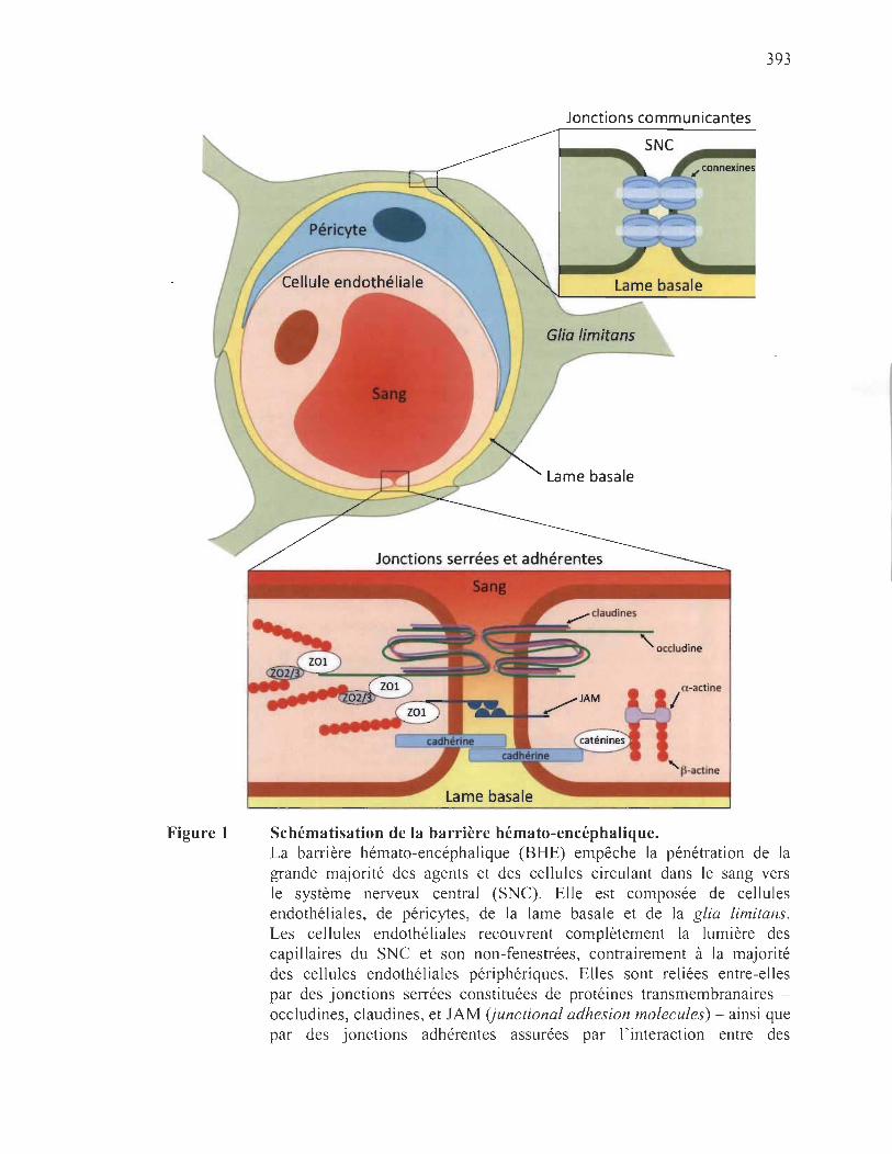
Figure 1
393
Jonctions communicantes
Lame basale
Schématisation de la barrière hémato-encéphalique. La barrière hémato-encéphalique (BHE) empêche la pénétration de la grande majorité des agents et des cellules circulant dans le sang vers le système nerveux central (SNC). Elle est composée de cellules endothéliales, de péricytes, de la lame basale et de la glia limitans. Les cellules endothéliales recouvrent complètement la lumière des capillaires du SNC et son non-fenestrées, contrairement à la majorité des cellules endothéliales périphériques. Elles sont reliées entre-elles par des jonctions serrées constituées de protéines transmembranaires -occludines, claudines, et JAM (junctional adhesion molecules) - ainsi que par des jonctions adhérentes assurées par l'interaction entre des
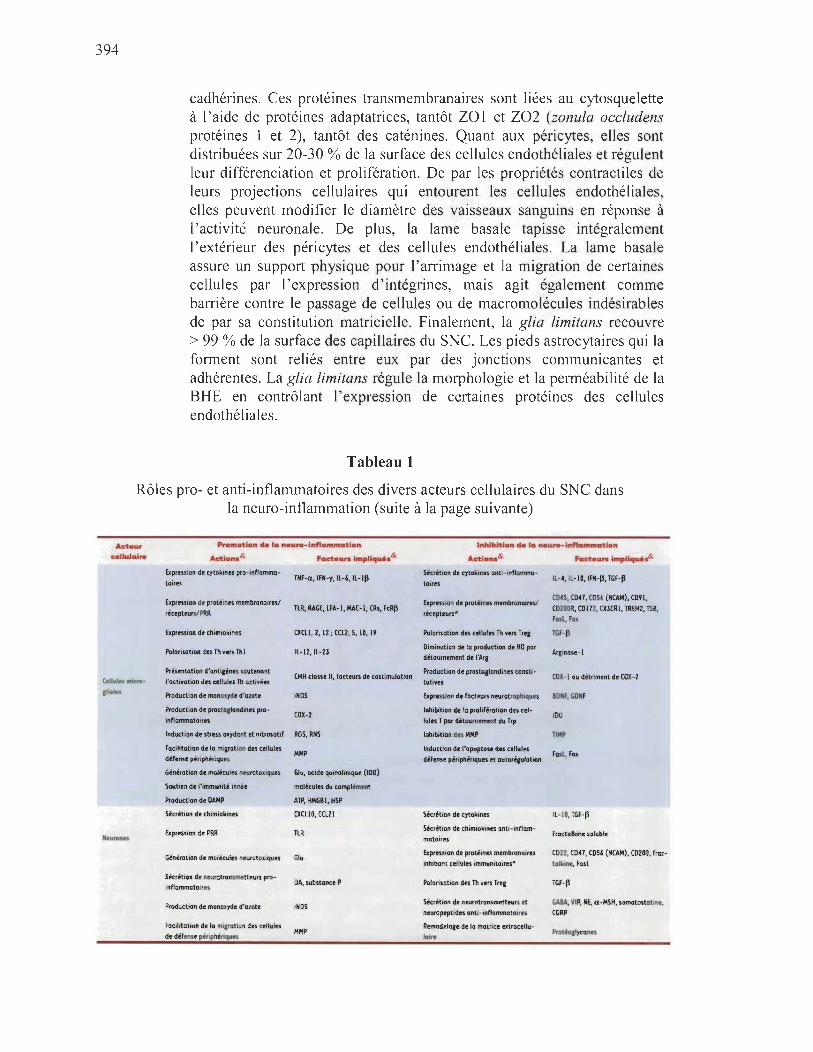
394
cadhérines_ Ces protéines transmembranaires sont liées au cytosquelette à l'aide de protéines adaptatrices, tantôt ZOI et Z02 (zonula occludens protéines 1 et 2), tantôt des caténines_ Quant aux péricytes, elles sont distribuées sur 20-30 % de la surface des cellules endothéliales et régulent leur différenciation et prolifération, De par les propriétés contractiles de leurs projections cellulaires qui entourent les cellules endothéliales, elles peuvent modifier le diamètre des vaisseaux sanguins en réponse à l'activité neuronale. De plus, la lame basale tapisse intégralement l'extérieur des péricytes et des cellules endothéliales. La lame basale assure un support physique pour l' arrimage et la migration de certaines cellules par l' expression d ' intégrines, mais agit également comme barrière contre le passage de cellules ou de macromolécules indésirables de par sa constitution matricielle. Finalement, la glia limitans recouvre > 99 % de la surface des capillaires du SNe. Les pieds astrocytaires qui la forment sont reliés entre eux par des jonctions communicantes et adhérentes. La glia limitans régule la morphologie et la perméabilité de la BHE en contrôlant l'expression de certaines protéines des cellules endothéliales,
Tableau 1
Rôles pro- et anti-inflammatoires des divers acteurs cellulaires du SNC dans la neuro-inflammation (suite à la page suivante)
Arteur cellulaire
Ctll""s MicroIlia ••
Neurones
Promotion de 'ca ,..uro ... inftCUftmotlon
Adlon.& Fade"",lmpllqu.&
uprenlon de cytokines pro+mflammo+ toires
Expression de protéines membranolresl rétepteun/PRR
ExpreSSion de chimloltlnes
Polarisation des Th yers Thl
Présentation d'antieènes soutenant ,'activation d!$ cellul!$ Th activées
Production de monoxyde d'azote
Production de prostollandlne$ proinflammatOi res
Induct ion de $trtU oxydont et mtrasatlf
Facllitatton de la mllrahon d!$ c!lIule$ défense pérlphénques
TNF-<l, IFN-r, Il-6, Il- Ill
TlR, RAGE, lU- l , MAC- l, CRs, FcRjl
CXClI , 2, 12 ; cm, s, 10, 19
Il-12,ll- 23
CMH classe Il, facteurs de coUlmulatlon
iNOS
COX-2
ROS, RNS
MMP
Génération de molKules neurotollques Glu, acide quinohnique (100)
SoutJen de l'immumté innée molécules du complément
ProductIon de OAMP ATP, HMGa l, H5P
Sécrétion de chlmiolcines
Ex pression de PRR
Génération de molécules neurotoxlqulS
Skrétion de neurotransmetteurs proinflammatoires
Production de monoxyde d'olote
Focilitatton de la mlJfabon des cellules dt défense périphériques
CXClIO, CCl21
TLR
Glu
DA, substance P
iN05
MHP
Inhibition de Ica neW"O-lnfla"'ftlClt'on
Adlon.& fod_"'" impliqu'&
Sécrétion de cyto&clntS anti · mflommo·
toires
ExpreSSion de protéines membronolresl
récepteurs·
Polarisation des cellules Th vers Tree
Diminution de la production de NO por
détournement de "Ara
Production de Pfostoe'andlnes tons t,· tutins
Exprtulon de facteurs neurotrophlqufS
Inhibition de la prolifération des cellules T par détournement du Trp
Inhibition des AAP
Induction de l'opoptose des cellules défense périphériques et autoréplatlon
Sécrétion de cytollines
Sécrétion de chimiokines anti- inflammatoires
Expreuion de protéines membranaires Inhibant cellules immunitaires"
Polorisation des Th vers Tre.
Sécrétion de neurotransmetteurs et
neuropeptides anti- inflammatoires
Remodela,t de la matfl te. extracellulaire
1l-4, ll- IO,IFN-Il, TGF-Il
C045, C041, CO 56 (NCA.I4), C091, C0200R, C0112, CX3CRI , TREM2, TSa, Fosl, Fos
TGF-Il
COX-I ou détriment de COX-2
aONF, GONF
100
TIHP
Fost, Fos
Il-IO, TGF-p
froctalkine soluble
C022, COH, C056 (NCA.I4), C0200, froctollune, Fosl
TGF-p
GABA, VIP, NE, a -MSH, somatostotlRe, CGRP
Protéo&1ycones

Acte"r c.n.&loJ ..
N.urones
Astrocytls
Olipdtndroqtes
Promotion cie la neuro-fnftommation
Adlono& Fact • ..,. Impllqu4io &
Production de DAMP
ExpreSSion de cytolnnes pro
Inflammatoires
expression de prot~lnes membranairesl ricept.urs/PRR
ATP, HMGBI, HSP, protéin .. mol rephées (protélnopothllS neurodéeénérotlvH)
THF-a,IFN-y,ll-11I
cxm, 2, 12; ecu, 5, 10, 19
TLR, RAGE,ICAMI, CR
Gin~ratlon de molécules n.urotoxlques Glu (diminution dt son intffnalisatlon)
Présentation d'antilines soutenant "actrvation des c.llules Th adivNs ?
Facilitation dl la mllrollon des cellules
de déf .... p'''ph.''qu ..
Production de monOlyde d'azote
Soutien de l'immuniti InnH
Production de DAMP
Dédifférenciation tn rod,o/-,Ua-lilte ctlls
Expression de PRR
CMH classe Il, fad.urs d. costimulotton
MMP
INOS
Molécules du comp~ment
ATP, HMGBI, HSI'
cm
TlR
395
Inhibition de la neuro-Inflammation
Adlono & FOd •• ". impllquh&
upression de fadeurs neurotrophiques
Induction de "opoptose des microllies et cellules défense périphériques
Absence d'QcUvotion des cellules cyto
toxiques coa' Inhibition des cellules HK par activation
du KIR
Sécrétion de cytolunes ontt-Inflammatoires
uprtsSlon de prot~ntS membranaires Inhibant cellules Immunitaires
Polarisation des lh nrS lItt
Remodelact d.la matrlc, atlacellulaire
upression de fad.urs neurotrophiques
Inhibition des MMP
InductlOll de l'apoptoH des microl'ies et cellules défense plnphtriques
Oécilfférenclotlon en radlal-I"a-/d,e (fils
Sécrétion de cytolcines
Expression de protéines membranai"s inhibant cellules immunitaires
HGf, BONf, NTl, GOHF, CHTF
Fosl
Abs.nce de HLA A, BouC (CMH cl .... 10)
HLA G (CMH cl .... lb)
IH, Il- ID, IFN-II, TGF-II
Inhibiteurs du complément, CTlA-4
TGF-II, atm
BONF, GONf, HGF, CTHF, FGf
TIMP
f.sl, PO-LI (chez coll.lu T octi .... .. primont PO-I)
FGF2,IGFI, SOFl, VEGF
TGF-II
(0200, fractalkine
& Arg, argmme; ATP, adénosine triphosphate; BDNF, brain-derived neurotrophic factor; CCL, chemokine [C-C motif] ligand; CD, cluster of difJerentiation; CD200R, cluster of difJerentiation 200 receptor; CMH, complex majeur d' histocompatibil ité; CNTF; ciliary neurotrophic factor; COX-2, cyclooxygénase-2; CR, complement receptor; CTLA-4, cytotoxic T-lymphocyte-associated protein 4; CX3CR, chemokine [C-X3-C motif] receptor; CXCL, chemokine [C-X-C motif] ligand; DAMP, damage-associated molecular pattern; Fas,flrst apoptosis signal receptor; FasL, flrst apoptosis signal receptor ligand ou ligand Fas; FcR~, fragment crystallizable region (Fe) receptor beta subunit; FGF, flbrob last growth factor; GDNF, glial flbrillary acidic protein; Glu, glutamate; HLA, human leucocyte antigen; HMGB l, high mobility group box J; HSP, heat shock protein; ICAMl, intercellular adhesion molecule; IDO, indoleamine 2,3-dioxygenase; IFN, interferon; IGF, insulin-like growth factor; IL, interleukine; iNOS, inducible nitric oxide synthase; KIR, !..:iller-cell immunoglobulin-like receptor; LF A, lymphocyte fitnction-associated antigen-1; MAC-l, macrophage-J antigen; MMP, matrix metalloproteinases; NCAM, neural cell adhesion molecule; NGF, nerve growth factor; NK, natural killer; NO, oxyde nitrique; Nn, neurotrophin-3; PD- l , programmed cell death protein J; PD-LI, programmed death-ligand 1; PRR, pattern-recognition receptors, RAGE, receptor for advanced glycation end-products; RNS, reactive nitrogen species; ROS, reactive oxygen species; SDF l , stromal cell-derivedfactor 1; TGF-~, transforming growthfactor-beta; Th, T helper cell subtype; TIMP, tissue inhibitor of metalloproteinase; TLR, toll-like receptor; TNF-a, tumor necrosis factor-alpha; Treg, regulatory T cell; TREM2, triggering receptor expressed on myeloid cells 2; TSB, thrombospondin; VEGF, vascular endothelial growthfactor.

396
C microgliales activées classiquement
• t
C de défense
C microgliale au repos
CT cytotoxique
légende
~ molécules du CMH de classe 1
.: PAMPs/DAMPs
~ molécules du CMH de classe Il
\" antigènes présentés
récepteurs de C T
~. cytokines pro-inflammatoires
~. cytokines anti-inflammatoires
y PRRs
..... effet anti-inflammatoire ..... effet pro-inflammatoire
.11 couples membranaires d'immunorégulation
.. transformation phénotypique
Figure 2 Dialogues entre les cellules neuronales dans un contexte de neuroinflammation. Dans un contexte de neuro-inflammation, des molécules de danger nommées P AMPs (pathogen-associated molecular patterns) ou DAMPs activent les cellules munies de récepteurs PRRs, principalement les cellules microgliales_ L'activation de la microglie mène à leur multiplication et à leur transfonnation vers le phénotype proinflammatoire, caractérisé par une fonne amiboïde et l'expression accrue de cytokines et de molécules du CMH de classe IL Suite à leur activation par les P AMPs ou DAMPs, les astrocytes subissent également des

La microglie
397
changements phénotypiques tendant vers l'expression accrue de cytokines, la présentation d'antigènes et la prolifération. Les cellules du SNC, en particulier les cellules microgliales activées classiquement, produisent des cytokines capables de perméabiliser la BHE et sécrètent des chimiokines, permettant la transvasation de cellules de défense périphériques - lymphocytes, granulocytes, macrophages, etc. - des capillaires vers le parenchyme. Ces cellules peuvent également reconnaître des PAMPs et des DAMPs dans le parenchyme, ce qui soutient leur activation et leur prolifération accompagnées de l'expression de cytokines pro-inflammatoires. Néanmoins, plusieurs acteurs expriment constitutivement des cytokines anti-inflammatoires ou des molécules membranaires donc la fonction immunomodulatrice permet de contenir la réaction inflammatoire, par la polarisation des cellules Th en types anti-inflammatoires et par la transformation de la microglie en un état réparateur. L'exposition à long terme aux cytokines pro-inflammatoires endommage les neurones et les oligodendrocytes, les types cellulaires les plus sensibles à une neuro-inflammation chronique dû à leur faible potentiel régénérateur.
La microglie constitue en moyenne 10 % de l'ensemble des cellules gliales.
Originant du sac vitellin à partir d'un précurseur érythro-myéloïde CSF1R+ (coiony
stimuiating factor 1 receptor) commun aux macrophages résidents [7] , les cellules
microgliales colonisent très tôt le neuroépithélium où elles poursuivent leur
différenciation selon une voie originale dans l'environnement particulier du SNe.
Elles conservent tout au long de la vie un certain pouvoir de régénération qui leur permet
à la fois de maintenir leur pool et de répondre à des demandes ponctuelles [8]. Après la
naissance, une sous-population de cellules microgliales semblerait également provenir
de source hématopoïétique par l'infiltration de monocytes dans le parenchyme [9] .
Toujours à explorer leur environnement à travers les mouvements rapides de leurs
extensions cytoplasmiques qui se chevauchent très peu, dotées d 'un très vaste potentiel
d'activités, elles se métamorphosent en réponse aux influences qu 'elles subissent.
Essentielle à la structuration du SNC et à l' exercice des fonctions nerveuses supérieures,
un rôle dont on commence à peine à percevoir l' importance [10] , c'est cependant pour
son rôle en situation traumatique que la microglie est le mieux connue. Exprimant tous
les PRRs identifiés à ce jour, les cellules microgliales sont particulièrement bien
398
équipées pour percevoir les perturbations de leur environnement, fussent-elle causées
par la concentration anormale d'une molécule, son format inhabituel ou par l'apparition
d'un composé inusité [11]. La reconnaissance induit le passage de l'état de veille à
celui d'activation, l'objectif étant, dans un premier temps, d'éliminer la source de la
perturbation et, dans un second temps, de favoriser le remodelage et la régénération.
En présence d'un signal de danger, les cellules microgliales adoptent un profil
amiboïde qui facilite à la fois leur division et leur déplacement. Elles sont recrutées soit
directement par les molécules de danger elles-mêmes, soit par des chimiokines libérées
par les autres cellules neuronales alertées du danger. Elles forment autour de la région
lésée un bouclier protecteur qui vise à en limiter l'étendue. Les conséquences précises de
l'activation dépendent du contexte parti cul ier dans lequel se fait la rencontre du stimulus
initiateur, les cellules microgliales oscillant entre deux états extrêmes: un état
d'activation classique et un état d'activation alternatif à vocation réparatrice [12].
Dans son état d'activation classique, la microglie exhibe un profil pro
inflammatoire (Tableau 1) caractérisé par 1) la sécrétion d'un large éventail de cytokines
pro-inflammatoires et de polarisation Th1 (T helper cell subtype 1); 2) l'expression de
chimiokines; 3) l'expression d'iNOS (inducible nitric oxide synthase) et de COX-2
(cyclooxygénase-2); 4) la production de dérivés toxiques de l'oxygène et de l'azote;
5) l'augmentation de l'expression des molécules du CMH de classe l et II et des facteurs
de costimulation; 6) la sécrétion de métalloprotéinases matricielles; et 7) un pouvoir
phagocytique accru. Si dans un état pro-inflammatoire la microglie possède l'arsenal
nécessaire pour altérer l'étanchéité de la BHE, attirer les leucocytes - principalement les
lymphocytes, monocytes et granulocytes - , faciliter leur déplacement dans la matrice
extracellulaire, présenter des antigènes, soutenir l'activation de l' immunité spécifique et
mettre KO les agents perturbateurs, elle a aussi le pouvoir de causer des dommages
collatéraux souvent irréversibles au SNC. Ce potentiel auto-destructeur de la microglie
activée classiquement est tempéré par les propriétés immunosuppressives du milieu, en
grande partie attribuables aux neurones.

399
Dans son état réparateur, dicté par le contexte du milieu, la microglie adopte plutôt
1) un métabolisme aérobie; 2) une expression accrue des récepteurs éboueurs;
3) la synthèse de COX-1 au détriment de COX-2; 4) la synthèse d'arginase-1 ; ainsi que
5) la sécrétion de divers facteurs neurotrophiques et d'éléments matriciels. Ainsi, la
microglie détient également le potentiel de participer à la réparation des tissus lésés et le
retour à l'homéostasie (Tableau 1), favorisant l'angiogenèse et stimulant la génération
de nouveaux oligodendrocytes, astrocytes, voire même neurones, à partir de cellules
souches toujours présentes dans le parenchyme nerveux adulte.
Les états d'activation classique et réparateur ne constituent que les deux pôles
d'un continuum d'états entre lesquelles les cellules microgliales oscillent en fonction des
particularités activatrices du milieu dans lequel elles baignent [13]. Avec l'âge, pour
des raisons qu 'on s' explique encore mal, l'induction du profil réparateur se fait plus
difficile, une situation à mettre en lien avec l'augmentation des maladies
neurodégénératives associée au vieillissement [14].
Les neurones
Les neurones jouent un rôle primordial dans l'établissement des privilèges
immunitaires du SNC en agissant tant en amont qu ' en aval de la réponse [15].
Ils expriment quelques PRRs par lesquels ils perçoivent les signaux de danger présents
dans leur environnement et expriment plusieurs récepteurs pour les cytokines qui leur
permettent d'ajuster leur contribution à l' immunosuppression en situation d'agression.
Les neurones expriment notamment le récepteur TLR3 (toll-like receptor 3) dont le
P AMP déclencheur est l' ARN double brin. La dimérisation de TLR3 mène à la
production d'interférons de type 1 dont le rôle est crucial dans la défense innée contre les
virus, par exemple lors d'une infection par le virus neurotropique de la rage [16].
La microglie avec son imposant arsenal pro-inflammatoire est une des
cibles préférentielles de l'attention neuronale qui se veut généralement atténuante.
La régulation de la microglie implique à la fois des contacts cellule-cellule et des
400
facteurs solubles contribuant à freiner l'inflammation (Tableau 1). La majorité de ces
protéines sont exprimées de façon constitutive et créent donc d'entrée de jeu un mil ieu
immunosuppresseur capable de contenir les débordements de la microglie. Ces protéines
membranaires agissent principalement en interférant avec les voies de signalisation,
inhibant des kinases essentielles telle la famille des MAPK (mitogen-activated protein
kinase) et les Pi3K (phosphoinositide 3-kinases), réduisant l'expression de facteurs de
transcription tds c-jun ou c-myc ou inhibant leur translocation nucléaire comme c'est le
cas pour Nrf2 (nuclear factor erythroid 2-related factor 2) ou NF-KB (nuclear factor
kappa-light-chain-enhancer of activated B ceUs). Elles permettent, en conséquence,
de diminuer la production de cytokines inflammatoires et de dérivés toxiques de
l'oxygène ou de l'azote dès l'engagement des PRRs de la microglie. Les neurones
expriment en plus et de façon constitutive le FasL (ligand Fas ou CD95L) par lequel
ils contrôlent l'apoptose des cellules microgliales activées. Outre ces facteurs
membranaires, les neurones libèrent nombre de facteurs solubles qui contribuent aussi à
contraindre l'activation de la microglie. Il yale TGF-~ (transforming growth factor
beta) et la fractalkine soluble que les neurones expriment de façon constitutive et dont la
production peut être amplifiée en situation traumatique, tandis que l 'interleukine-l 0
(IL-I0) est plutôt sécrétée en situation de crise. À ces facteurs immunorégulateurs
viennent s'ajouter l'activité neuronale dont l'action anti-inflammatoire est arbitrée
par un bon nombre de neurotransmetteurs et plusieurs neurotrophines (Tableau 1).
Sous cette influence, les cellules microgliales produisent moins de cytokines
inflammatoires, réduisent leur stress oxydant, diminuent leur expression des molécules
du CMH et, ainsi, soutiennent moins efficacement l'activation des cellules de défense
périphériques ayant gagné accès au parenchyme nerveux. Quant à leur pouvoir
phagocytaire, il peut être selon les particularités du contexte soit amplifié, soit diminué.
Le caractère immunosuppresseur des neurones ne contient pas que les ardeurs de
la microglie au moment de son activation; il tempère aussi les conséquences de son
activation en s'exerçant sur les cellules de la défense périphérique qui, répondant à
l'appel, ont gagné ou tentent de s'introduire dans le parenchyme. Ici encore, l'effet
s'exerce tantôt par des contacts cellule-cellule impliquant entre autres des cadhérines,

401
des molécules du CMH, et le FasL, tantôt par l' intermédiaire de facteurs solubles
dont le TGF-~ et les sémaphorines 3 et 7. Les interactions aboutissent à l'immobilisation
des macrophages, la neutralisation des cellules cytotoxiques NK (natural killer) et
CD8+, la polarisation des Th vers des phénotypes plus immunorégulateurs que pro
inflammatoires tels Th2 (T helper cell subtype 2) ou Treg (regulatory T cel!) et la mort
par apoptose des cellules activées.
Les astrocytes
D'origine neuroectodermique, les astrocytes sont les cellules les plus abondantes
du SNC, comprenant jusqu'à 70 % de la névroglie. Ce sont des cellules étoilées qui,
avec leurs extensions cytoplasmiques, participent à la formation de la glia limitans de
la BHE (Figure 1), enrobent les synapses, communiquent entre elles par le biais de
jonctions communicantes et contribuent au drainage glymphatique du liquide interstitiel
parenchymateux vers le liquide céphalo-rachidien (LCR) [17]. Ils délimitent ainsi un
territoire sur lequel ils peuvent exercer leur influence grâce aux récepteurs, canaux
ioniques, transporteurs et enzymes de toutes sortes dont ils sont pourvus [18, 19].
En plus de leur rôle clé dans l'homéostasie du SNC, le métabolisme neuronal
et la modulation dynamique de la transmission synaptique, les astrocytes sont
particulièrement bien outillés pour appuyer la microglie dans sa fonction de sentinelle et
de défense [20, 21]. Ils expriment le TLR3 de façon constitutive et peuvent être induits
à exprimer plusieurs des PRRs. Tout comme les cellules microgliales, ils sont donc aptes
à reconnaître et à réagir à un large éventail de situations dangereuses pour le SNe.
En réponse à l'activation, les astrocytes deviennent hypertrophiques, prolifèrent et
altèrent leur expression génique. Ils sécrètent nombre de facteurs pro-inflammatoires
(Tableau 1) servant à attirer les cellules de défense périphériques et faciliter leur
migration dans le parenchyme, expriment des récepteurs éboueurs par lesquels ils
contribuent à la phagocytose, et produisent les molécules du CMH de classe II leur
permettant de soutenir l'activation des cellules Th activées, bien que leur capacité à
présenter des antigènes, comme le fait la microglie, est controversée. L'activation les

402
amène même à diminuer leur internalisation de glutamate et à perturber le réseau
astrocytaire en diminuant la production de connexine 43 , ce qui contribue de surcroît au
caractère neurotoxique de la réaction inflammatoire.
S'ils participent foncièrement aux processus pro-inflammatoires lorsqu'activés,
Is répondent néanmoins à l'IL-l~ par la sécrétion de TGF-~ et par la libération de
différents facteurs neurotrophiques (Tableau 1) qui soutiennent la réparation de la BHE,
la remyélinisation, le remodelage de la matrice ainsi que la survie des neurones et des
oligodendrocytes. Ils contrôlent l'activation des lymphocytes T par leur expression du
récepteur immunorégulateur CTLA-4 (cytotoxic T-lymphocyte-associated protein 4 ou
CD152) et peuvent déclencher l'apoptose des cellules de défense activées par
l'expression constitutive du FasL. En situation d'inflammation chronique, leur capacité
d'activer les Th est compensée par une interaction qui biaise la réponse en faveur des
Th2, un profil plutôt immunorégulateur que le profil Thl pour le SNC [22]. De plus,
certains signaux d'agression permettent aux astrocytes de se dédifférencier en « radial
glia-like ceUs» capables d'exprimer d'autres facteurs neurotrophiques (Tableau 1) qui
ensemble promeuvent la multiplication des cellules souches neuronales et leur migration
vers le site de lésion [23] . Enfin, les astrocytes sont particulièrement reconnus pour leur
rôle dans la formation de la cicatrice gliale. En effet, lorsqu'activés ils se multiplient
abondamment, circonscrivent le site d'inflammation, occupent les espaces créés par la
mort ou la phagocytose de cellules neuronales et produisent des éléments de la matrice
extracellulaire tels que de l'acide hyaluronique. Cependant, cette réaction de défense qui
a pour but d'inhiber la propagation de l'inflammation et d'offrir un effet restructurant a
aussi comme conséquence néfaste d'inhiber la migration et la différentiation des cellules
souches neuronales [23].
Les oligodendrocytes
Les oligodendrocytes partagent la même ongme neuroectodermique que les
astrocytes et les neurones. Derniers à entrer en scène, ils établissent avec les neurones
une relation symbiotique. Les oligodendrocytes sont tout aussi essentiels au

403
développement et à la survie des axones que les neurones le sont à la myélinisation
[24-26].
Comme les neurones, les oligodendrocytes contribuent au caractère
Immunosuppresseur du parenchyme nerveux par leur production constitutive de
TGF-~ et par leur expression du CD200 et de fractalkine. À l'instar des autres cellules
du SNC, ils expriment des PRRs, notamment TLR2 et TLR3 , qui leur permettent de
réagir à certains signaux de danger. Bien que les conséquences de l' engagement de ces
récepteurs demeurent pour le moment mal connues, elles devraient se répercuter sur
l'ensemble du réseau d'influence des oligodendrocytes. Ce réseau, relativement vaste,
implique tout autant les astrocytes avec lesquels les oligodendrocytes sont en lien par le
biais de jonctions communicantes que les neurones auxquels ils sont associés par la
gaine de myéline, un oligodendrocyte pouvant enrober jusqu'à une cinquantaine
d'axones distincts.
Si les oligodendrocytes peuvent participer activement à la mise en place d'une
immunité innée par l'intermédiaire de leurs TLRs, ils peuvent aussi être eux-mêmes
générateurs de danger. Très sensibles au stress oxydant tout comme ils le sont à la
toxicité du glutamate ou à celle de l'A TP, les oligodendrocytes peuvent causer des
dommages sérieux à leur environnement et entretenir l'inflammation sans être
nécessairement les cellules initialement visées par l'agression.
Les cellules de défense périphériques
Au niveau du SNC sain, rares sont les leucocytes qui arrivent à s' échapper de la
vasculature. Leur présence, bien que limitée, permet tout de même d'assurer une
immunosurveillance dont l'importance nous apparaît indéniable lorsqu'on mesure les
dommages encourus par la suppression de la patrouille des cellules de défense
périphériques [pour revue, voir 27]. Dans le cas du JC polyomavirus, un virus
ubiquitaire chez l'humain, l'arrêt del'immunosurveillance secondaire à des thérapies
404
immunosuppressives entraîne la leucoencéphalopathie multifocale progressIve,
une maladie du SNC généralement mortelle.
Les leucocytes qUI parviennent à pénétrer le SNC, uniquement des
mononucléaires, prennent avantage à la fois de leur expression de certaines protéines
d'adhésion et de l'existence de zones plus permissives de la BHE, telles que la glande
pituitaire, l'éminence médiane, l'aire postrema, l'aire préoptique, la paraphyse,
la glande pinéale et l'endothélium des plexus choroïdes. Les monocytes utilisent la
voie leptoméningée pour venir peupler les espaces de Virchow-Robin en bordure des
artérioles et en continuité avec l'espace sous-arachnoïdien. Quant aux lymphocytes,
et uniquement les T activés, ils s'affranchissent de la barrière sang-LCR (BSLCR) en
empruntant la voie du plexus choroïdien qui exprime constitutivement des molécules
nécessaires à leur admission, telles que le CCL20 (chemokine [C-C motif] ligand 20)
indispensable au recrutement des cellules CCR6+ (C-C chemokine receptor type 6) [28] .
Une fois traversés, ils sont entraînés par le LCR, patrouillent les espaces et quittent soit
vers le sang par le biais des villosités arachnoïdiennes, soit vers les ganglions cervicaux
profonds par le biais de la plaque cribriforme de l'os ethmoïde ou par le biais d'un
système lymphatique propre au SNC récemment mis en évidence [29, 30]. On constate
donc qu'en situation normale, des cellules de défense d'origine médullaire occupent des
niches stratégiques du SNC sans jamais pénétrer le parenchyme, qu'elles exercent
patrouille et immunosurveillance comme elles le font en périphérie et qu'elles possèdent
tous les attributs qui leur permettent de réagir efficacement à l'agression [27, 31-33].
En réponse à l'agression, les cellules du parenchyme s'activent. Elles libèrent dans
le milieu des cytokines pro-inflammatoires qui modifient l'étanchéité de la BHE
ainsi que des chimiokines (Tableau 1) qui guident les cellules de défense, y compris les
granulocytes, et les autorisent à pénétrer au sein même du parenchyme [34].
Les macrophages périvasculaires participent au recrutement par la production de
facteurs de croissance (Tableau 1), la modification des cellules endothéliales et par la
sécrétion de métalloprotéinases qui facilitent les déplacements. Bien que nécessaire à
la résolution des problèmes, l'afflux de cellules sur le pied de guerre dans le parenchyme
405
nerveux comporte des risques importants pour un tissu aussi fragile. Pour contrer les
risques encourus, les cellules du parenchyme limitent le temps de vie des cellules de
défense en provenance de l'extérieur ou les réorientent vers des phénotypes plus
immunorégulateurs, notamment par l'expression constitutive de FasL ou de TGF-~ de
l'environnement nerveux.
Conclusion
La neuro-inflammation est un phénomène hautement complexe par lequel le SNC
s'allie au système immunitaire pour assurer sa protection et maintenir son homéostasie
[35] (Figure 2). La complexité du phénomène, qui le rend difficilement abordable
dans le contexte fragmentaire des études in vitro ou des systèmes modèles, tient tout
autant à la multiplicité des intervenants cellulaires qu'à l'enchevêtrement des voies de
signalisation et aux effets tantôt synergiques, tantôt antagonistes, tantôt neurotoxiques,
tantôt neurotrophiques des signaux émis. Les conséquences en sont difficilement
prévisibles car elles dépendent à la fois de la nature, de l'intensité, de la durée que de
l'historique de l'agression [36] . Il semble cependant que si elle est source du mal,
la neuro-inflammation soit aussi source de la solution [37, 38]. Il nous faut donc
apprendre à décoder son langage et à identifier, pour chaque problématique, le maillon
faible du réseau afin de mettre au point des stratégies d'intervention ciblées qui
permettent de contrer la neurodégénération [39].
Remerciements
JR est récipiendaire d'une bourse doctorale Vanier-Canada. MGM est
récipiendaire d' une subvention à la Découverte-CRSNG (Canada).
406
Références
1. Galic MA, Riazi K, Pittman QJ. Cytokines and brain excitability. Front Neuroendocrinol2012; 33: 116-25.
2. Galea l, Bechmann l, Perry VH. What lS Immune privilege (not)? Trends Immunol 2007; 28: 12-8.
3. Amor S, Puentes F, Baker D, van der Valk P. Inflammation in neurodegenerative diseases. Immuno1201O; 129: 154-69.
4. Lee H, Lee S, Cho IH, Lee SJ. Toll-like receptors : sensor molecules for detecting damage to the nervous system. Curr Protein Pept Sci 2013; 14: 33-42.
5. Tang D, Kang R, Coyne CB, Zeh HJ, Lotze MT. PAMPs and DAMPs: signal Os that spur autophagy and immunity. Immunol Rev 2012; 249: 158-75.
6. Cunningham C. Microglia and neurodegeneration: The role of systemic inflammation. Glia 2013; 61: 71-90.
7. Gomez Perdiguero E, Klapproth K, Schulz C, Busch K, Azzoni E, Crozet L, Garner H, Trouillet C, de Bruijn MF, Geissmann F, Rodewald HR. Tissueresident macrophages ongmate from yolk-sac-derived erythro-myeloid progenitors. Nature 2015; 518: 547-51.
8. Ajami B, Bennett JL, Krieger C, Tetzlaff W, Rossi FM. Local self-renewal can sustain CNS microglia maintenance and function throughout adult life. Nat Neurosci 2007; 10: 1538-43.
9. Chen SK, Tvrdik P, Peden E, Cho S, Wu S, Spangrude G, Capecchi MR. Hematopoietic origin of pathological grooming in Hoxb8 mutant mice. Cell 2010; 141: 775-85.
10. Tremblay M-E, Stevens B, Sierra A, Wake H, Bessis A, Nimmerjanh A. The role ofmicroglia in the healthy brain. J Neurosci 2011; 31: 16064-9.
11. Hanke ML, Kielian T. Toll-like receptors in health and disease in the brain: mechanisms and therapeutic potential. Clin Sci (Lond) 2011; 12: 367-87.
12. Boche D, Perry VH, Nicoll JAR. Review : Activation patterns of microglia and their identification in the human brain. Neuropathol Appl Neurobiol 2013; 39: 3-18.

407
13 . Eggen BJL, Raj D, Hanisch U-K, Boddeke HWGM. Microglial phenotype and adaptation. J Neuroimmunol Pharmacol 2013 ; 8: 807-23 .
14. Norden DM, Godbout JP. Review: microglia of the aged brain: primed to be activated and resistant to regulation. Neuropathol Appl Neurobiol 2013 ; 39: 19-34.
15. Suzumura A. Neuron-microglia interaction in neuroinflammation. CUIT Prot Pept Sci 2013 ; 14: 16-20.
16. Ménager P, Roux P, Mégret F, Bourgeois JP, Le Sourd AM, Danckaert A, Lafage M, Préhaud C, Lafon M. Toll-like receptor 3 (TLR3) plays a major role in the formation ofrabies virus Negri Bodies. PLoS Pathog 2009; 5: e1000315.
17. Iliff JJ, Wang M, Liao Y, Plogg BA, Peng W, Gundersen GA, Benveniste H, Vates GE, Deane R, Goldrnan SA, Nagelhus EA, Nedergaard M. A paravascular pathway facilitates CSF flow through the brain parenchyma and the clearance of interstitial solutes, including amyloid~ . Sci Trans Med 2012; 4: 147rall1.
18. Bélanger M, Magistretti PJ. The role of astroglia in neuroprotection. Dialogues Clin Neurosci 2009; Il : 281-295.
19. Nedergaard M, Verkhratsky A. Artifact versus reality-how astrocytes contribute to synaptic events. Glia 2012; 60: 1013-23.
20. Farina C, Aloisi F, Meinl E. Astrocytes are active players in cerebral innate immunity. Trends Immuno12007; 28: 138-45.
21. Giemsa U, Mitchison NA, Brunner-Weinzierl MC. Immune privilege as an intrinsic CNS property : astrocytes protect the CNS against T -cell-mediated neuroinflammation. Mediators Inflamm 2013 ; 2013: 320519.
22 . Jager A, Dardalhon V, Sobel RA, Bettelli E, Kuchroo. Th1 , Th17, and Th9 effector cells induce experimental auto immune encephalomyelitis with different pathological phenotypes. J Immunol 2009; 183: 7169-77.
23. Müller FJ, Snyder EY, Loring JF. Gene therapy: can neural stem cells deliver? Nat Rev Neurosci. 2006; 1: 75-84.
24. Baumann N, Pham-Dinh D. Biology of oligodendrocyte and myelin ln the mammalian central nervous system. Physiol Rev 2001 ; 81: 871-927.
408
25. Wake H, Lee PR, Fields RD. Control of local prote in synthesis and initial events in myelination by action potentials. Science 2011; 333: 1647-51.
26. Frühbeis C, Fr6hlich D, Kuo WP, Amphornrat J, Thilemann S, Saab AS, Kirchhoff F, M6bius W, Goebbels S, Nave KA, Schneider A, Simons M, Klugmann M, Trotter J, Kramer-Albers EM. Neurotransmitter-triggered transfer of exosomes mediates oligodendrocyte-neuron communication. PLoS Biol 2013; 11: e1001604.
27. Ousman SS, Kubes P. Immune surveillance ln the central nervous system. Nat Neurosci 2013 ; 15: 1096-101.
28. Reboldi A, Coisne C, Baumjohann D, Benvenuto F, Bottinelli D, Lira S, Uccelli A, Lanzavecchia A, Engelhardt B, Sallusto F. C-C chemokine receptor 6-regulated entry of TH-17 cells into the CNS through the choroid plexus is required for the initiation ofEAE. Nat Immuno12009; 10: 514-23 .
29. Louveau A, Smimov I, Keyes TJ, Eccles JD, Rouhani SJ, Peske JD, Derecki NC, Castle D, Mandell JW, Lee KS, Harris TH, Kipnis J. Structural and functional features of central nervous system lymphatic vessels. Nature 2015.
30. Aspelund A, Antila S, Proulx ST, Karlsen TV, Karaman S, Detmar M, Wiig H, Alitalo K. A durai lymphatic vascular system that drains brain interstitial fluid and macromolecules. J Exp Med 2015; 212: 991-9.
31 . Bechmann I, Galea I, Perry VH. What is the blood-brain barrier (not)? Trends ImmunoI2007; 28 : 5-11 .
32. Ransohoff RM, Engelhardt B. The anatomical and cellular basis of immune surveillance in the central nervous system. Nat Rev Immuno12012 ; 12: 623-35.
33 . Carare RO, Hawkes CA, Weller RO. Afferent and efferent immunological pathways of the brain. Anatomy, function and failure. Brain Behav Immun 2013 ; 59: 11-4.
34. Takeshita Y, Ransohoff RM. Inflammatory cell-trafficking across the blood-brain barrier: chemokine regulation and in vitro models. Immunol Rev 2012; 248: 228-39.
35. Lampron A, Elali A, Rivest S. Innate immunity in the CNS: redefining the relationship between the CNS and its environment. Neuron 2013; 78: 214-32.

409
36. Nagajima K, Kohsaka S. Microglia : Neuroprotective and neurotrophic cells in the central nervous system. CUIT Drug Targets Cardiovasc Haematol Disord 2004; 4: 65-84.
37. Rawji KS, Yong VW. The benefits and detriments of microphages/microglia in models of multiple sclerosis. Clin Dev Immuno12013; 2013: 948976.
38. Hanamsagar R, Hanke ML, Kielian T. Toll-like receptor (TLR) and inflammasome action in the central nervous system. Trends Immunol 2012; 33: 333-42.
39. McPherson RC, Anderton MC. Adaptive immune responses in CNS autoimmune disease: mechanisms and therapeutic opportunities. J Neuroimmune Pharmacol 2013; 8: 774-90.


APPENDIXB
OLD MOLECULES, NEW INSIGHTS: THERAPEUTIC CONSIDERATIONS FOR THE USE OF POLYPHENOLS IN NEURODEGENERATIVE DISEASES
Renaud, J., and Martinoli, M. G. (in preparation).
The content of this appendix will be submitted in Fall 2018 to the peer-reviewed
journal Nutrients.
Author contributions
Justine Renaud wrote 95% of the manuscript. Maria-Grazia Martinoli,
Justine Renaud ' s research supervisor, was the guarantor of the work and provided
supervision, preparation and editing of the manuscript.

412
Full review article in English: Old molecules, new insights: therapeutic considerations for the use of polyphenols in neurodegenerative diseases
Abstract
Over the last two decades, neurodegenerative diseases have received increasing
attention due to the rapid rate at which the population is aging and the consequent rise
in the incidence of such illnesses, itself entailing a major social and economic
burden. Today, an increasingly large body of literature lends functional foods and
their biofunctional molecules potential neuroprotective capacities. Among the most
prominently studied dietary molecules, polyphenols stand in a class of their own on
account of their multiple and often overlapping modes of action. However, ambiguity
exists as to the significance of their influence on human health. This review discusses
the many characteristics and functions of polyphenols that shape their possible
therapeutic applications in neurodegenerative diseases. Knowledge gaps that remain
under-explored will be highlighted.
1. Introduction
It has long been acknowledged that dietary habits play a key role in the occurrence
and progression of non-communicable diseases . A hoard of epidemiological evidence
shows that a diet rich in fruit and vegetables reduces the incidence of cardiovascular
diseases [1-4], type 2 diabetes [5,6] , stroke [7,8] and nurnerous cancers [9-11].
Others find inverse associations between the consurnption of green tea and cognitive
decline [12, 13]. These observed health benefits are thought to be at least partly
attributable to a class of non-essential nutrients named polyphenols, abundantly found in
fruit, vegetables and other edible plants [14, 15].
Alongside cancer and cardiovascular diseases, neurodegenerative disorders
constitute a popular field of application for the benefits of polyphenols [see for review
refs 16, 17] . This is the case for Parkinson's and Alzheimer's diseases, conditions that
are often ill defined and lack a clear etiopathogenetic origin besides that they seem to

413
anse from the interaction between agmg, the environment and genetic risk factors.
The etiology of these diseases is further convoluted by a number of proposed causative
mechanisms, such as oxidative stress, neuroinflammation, protein aggregation,
iron toxicity and mitochondrial dysfunction. As such, the number of proposed actions of
polyphenols, both in terms of the numerous disease states they appear to improve and
the manifold cellular mechanisms the y are reported to modulate, makes their use in
complex neurodegenerative disorders compelling. In this review, the factors that
impinge on the biofunctionality and bioavailability of dietary polyphenols in the central
nervous system (eNS) are discussed with a particular focus on therapeutic applications
and limitations.
2. Chemico-structural characteristics
2.1 Classification
Plant polyphenols were originally c1assified in early literature as "vegetable
tannins" owing to their tanning action on animal skins [18]. The first comprehensive
description, referred to as the WBSSH definition, recommended that the term
polyphenol be exclusively ascribed to water-soluble phenolic compounds having a
molecular mass ranging between 500 to 4000 Da, possessing at least 12 phenolic
hydroxyl groups, and 5 to 7 aromatic rings per 1000 Da [19]. A less restrictive
interpretation was proposed by Quideau, offering a broader view of the WBSSH
definition - that was hitherto limited to the structural characteristics common to aIl
phenolics endowed with tanning properties (vegetable tannins) - to include simpler
phenolic compounds with potential biological activities others than tanning [20]:
The term "polyphenol" should be used ta define compounds exclusively
derived from the shikimate/phenylpropanoid and/or the polyketide pathway,
featuring more than one phenolic unit and deprived of nitrogen-based
fitnctions . This definition lets out ail monophenolic structures as weil as ail
their naturally occurring derivatives such as phenyl esters, methyl phenyl
ethers and O-phenyl glycosides.
414
A majority of plant polyphenols originate from phenylalanine that is deaminated to
cinnamic acid, which then enters the phenylpropanoid pathway [21]. Plant metabolism
utilizes the phenylpropanoid unit C6-C3, a phenol ring with a 3-carbon side chain,
as a building block to construct polyphenols. Classification of the resulting molecules is
dictated by the number of phenol rings (C6) they contain and the structural elements
binding these rings to one another. The main subclasses, varying in complexity, are
phenolic acids (C6-C3 and C6-Cl), flavonoids (C6-C3-C6), stilbenes (C6-C2-C6) and
lignans (C6-C3-C3-C6). Within these subclasses, hydroxylations and O-glycosylations
at various positions as weIl as cis-trans isomerization give rise to the thousands of
polyphenols (estimated to > 8000) known to date, offering a tremendously intricate bank
of molecules with prospective pharmacological value to explore. These polyphenols
alongside their content in various food products are available on databases such as
Phenol-Explorer managed by the Institut National de la Recherche Agronomique
(www.phenol-explorer.eu).
2.2 Structure versus biofunctionality in neuroprotection
The particular chemico-structural properties shared by polyphenols are pertinent
to their therapeutic application, especially in the field of neuroprotection. Indeed, the
presence of phenol rings, variable hydroxylation patterns and conjugated double bonds
grants polyphenols metal-chelating, fibril-destabilizing, estrogen-like, enzyme-binding
and antioxidative properties. These numerous modes of action afford polyphenols the
ability to tackle the multifarious pathophysiological aspects of several neurodegenerative
diseases, namely oxidative stress, neuroinflammation, protein aggregation, Iron toxicity
and mitochondrial dysfunction.
The redox properties of divalent metals, such as copper, zinc and iron, are essential
for cellular homeostasis. When in excess, however, these metals contribute to generate
surplus reactive oxygen species. Polyphenols that comprise at least one gallo yi or
catechol group (hydroxyl groups in the ortho-position) are powerful bidentate chelators
of divalent metals [22] , whereas ones having only a phenol substitution (one hydroxyl

415
function) or possessing a resorcinol group (meta-position hydroxyl pair) are less potent
monodentate chelators [23 , 24]. For chelation to occur, a deprotonation step of the
phenolic group is necessary and has been shown to be possible at physiological pH [23].
Self-assembly of amyloidogenic fibrils involves interactions between aromatic
residues [25], as in neuropathologically relevant proteins including tau, beta amyloid
(AB) and a-synuclein, for example. Using the same kind of aromatic interactions, phenol
moieties in polyphenols can Interfere with fibril assembly [26], possibly by weakening
cross-B structures. This Interference seerns to arise from hydrophobic and 7r stacking
interactions [27], although the formation of covalent bonds through Schiff base reactions
has also been proposed for the green tea polyphenol epigallocatechin-3-gallate (EGCG)
[28, 29]. Upon analyzing binding energies between polyphenols and prote in fibrils ,
favorable entropic and enthalpic dynamics were discovered that also suggest the
stabilization of H-bonds [30].
Sorne polyphenols, also referred to as phytoestrogens, have the ability to bind
estrogen receptors (ERs), usually with a greater affinity for ERB [31, 32]. Depending on
structure, dose, cell type and estrogen response element (ERE) sequence, sorne
polyphenols have a weak or strong antagonistic or agonistic effect on ERs, affording a
very wide spectrum of activities in cells [33-36]. As a requirement to bind ERs,
the structure should be composed of a phenolic ring with a configuration resernbling that
of estradiol, as found in flavonoid isoflavones or the stilbene resveratrol, for instance.
Indeed, a specific hydroxylation pattern and an adequate distance between substituted
hydroxyl groups are necessary to bind ERs.
Sorne polyphenols also share structural similarities with endogenous ligands, such
as cyclic AMP (cAMP) or nucleoside triphosphates, endowing thern with the aptitude to
activate or inhibit key enzymes [37, 38]. To date, modulatory effects on enzymes have
been confirmed in cellular or animal models for several polyphenols, su ch as resveratrol
on cAMP phosphodiesterases [39] , theaflavins on the ATP synthase and respiratory
chain [40], and curcumin on gl yoxalase 1 [ 41], to name a few. The presence of
appropriately spaced ketone and hydroxyl groups in a planar configuration, bestowing

416
sorne polyphenols su ch as curcurnin with the ability to mimic an enediolate intennediate
in physiological conditions [42], is a remarkable example of structural elernents that
make enzyme binding possible.
Notwithstanding the aforementioned functions emergmg from their umque
chernical structures, the most vastly studied characteristic of polyphenols is their
antioxidative action. Polyphenols are thought to exert their antioxidative action directly,
by scavenging free radical species firsthand, and/or indirectly, by activating endogenous
antioxidative pathways. Direct antioxidative effects usually occur through H-atom
transfer from polyphenols' (ArOH) hydroxyl (OH) groups to the free radicals (R-):
ArOH + R- ~ ArO- + RH (1)
The existence of multiple conjugated double bonds in polyphenols allows for the
unpaired electron to be delocalized over the aromatic ring, yielding a much more stable,
hence mu ch less reactive, polyphenolic radical (ArO-) (Eq. 1). Sorne polyphenols
also exert indirect antioxidative effects through the Kelch-like ECH-associated
protein lInuclear factor erythroid 2-related factor 2/antioxidant response elements
(Keap l/Nrf2/ ARE) regulatory pathway made possible by the presence of electrophilic
functions (a,~-unsaturated carbonyl group, 1,2- and l,4-quinones, or other groups) that
alkylate thiol sensors in the cysteine pocket of Keap 1 [43, 44]. Others, like stilbenes,
engage their resorcinol hydroxyl functions in hydrogen bonds with the Kelch pocket of
Keap 1 [45]. Both these events lead to the disruption of the Keap l/Nrf2 complex,
allowing Nrf2 to translocate to the nucleus where it can trigger the expression of
antioxidant proteins like herne oxygenase-l via binding of AREs. This cysteine
modifying function of polyphenols may also bear other implications in various enzymes
[44].
3. Factors influencing pharmacokinetics and bioavailability
To be effective in the prevention or amelioration of neurodegenerative diseases,
polyphenols must be bioavailable in the CNS. Extensive reports on the bioavailability of

417
the most common dietary polyphenols can be found elsewhere [46-48]. Within the scope
of this review, we will first discuss the principal obstacles that hinder polyphenol
bioavailability and later address CNS permeability.
3.1 Food matrix or vehicle
Oral administration is by far the most practical of pharmacological routes,
but often raises daunting challenges in terms of bioavailability. Particular factors to
consider wh en developing an oral drug are the molecule's interaction with the vehicle,
the transformations it undergoes by digestive and microbial enzymes, and its absorption
in the gastrointestinal tract [49] .
Acting as the vehicle of administration, food matrices are central to the bioefficacy
of polyphenols [50]. Of the few studies conducted, inconsistent results have been
obtained, demonstrating either a negligible [51 , 52] or a significant [53-56] contribution
of the food matrix in polyphenol absorption. For instance, bioavailability differs for
quercetin among dietary sources, largely depending on the glycoside form they exist in.
Indeed, onions, which contain quercetin conjugated to glucosyl functions, are better
sources of bioavailable quercetin th an are apples and tea, which contain different
quercetin glycosides [57, 58] . It is also unclear whether ethanol bears a role in
polyphenol absorption in light of studies showing improved bioavailability of quercetin
in rats when administered in a 30% solution of ethanol, an alcohol content that is
physiologically improbable in an every day diet [59]. In fact, confounding results were
obtained in humans administered red wine or dealcoholized red wine that showed no
differences in catechin plasma levels and also demonstrated increased catechin excretion
likely due to a diuretic effect [60].
3.2 Gastrointestinal transformations and absorption
Once in transit, sorne polyphenols remain stable though most are converted with
galloyl [61] or methyl [62] groups in the duodenum by digestive enzymes. As a rule,

418
the best-absorbed polyphenols in hurnans are isoflavones (in soybean-derived products)
and gallic acid (in sumac and green tea), followed by catechins (in green tea), flavanones
(in citrus), and quercetin glucosides (in onions), whereas the least absorbed are
proanthocyanidins and anthocyanins (in berries), galloylated tea catechins, and stilbenes.
Whereas aglycones are normally weIl absorbed by the small intestine, nutritional
polyphenols are more commonly detected in the form of glycosides, esters and
polymers, which cannot be efficiently assimilated in the upper portion of the gut.
Molecules not absorbed in the upper gastrointestinal tract continue to the colon to
become substrates to the gut microbiota, responsible for a very wide array of reactions,
sorne of which yield monomers from polymers or aglycones from glycosylated
polyphenols [see for review ref 63]. SmaIler, better-absorbed phenolic acids may also be
produced by the gut microbiota. For example, quercetin microbiotic degradation mainly
generates 3,4-dihydroxyphenylacetic, 3-methoxy-4-hydroxyphenylacetic (homovanillic
acid), and 3-hydroxyphenylacetic acids [64]. Among volunteers challenged with 75 mg
of rutin, a quercetin glycoside, the total urinary excretion of microbial metabolites
accounted for as much as 50% of the ingested dose [65]. Importantly, the sum of these
gastrointestinal transformations and food matrix interactions can either mcrease or
decrease the absorption of the resulting metabolites in the bloodstream.
3.3 Plasma bioavailability, transformations and cellular uptake
Once in the blood, drug-metabolizing enzymes mainly found in the liver and
kidneys further modify polyphenols into various conjugated forms, a process that serves
to detoxify potentially harmful substances. Molecules are rendered more hydrophilic in
order to facilitate their urinary elimination, which usually lowers bioavailability [66, 67].
To date, the green tea polyphenol EGCG remains the only known molecule
to abundantly persist in a free form in the human plasma (up to 90%) [68].
While metabolites usually constitute the greatest fraction of circulating polyphenolic
species, sorne forms undergo enterohepatic recirculation via biliary secretion, followed
by deconjugation into free polyphenols by the gut microbiota and reabsorption in the

419
colon [69, 70] . Additional hepatic reactions may also occur thus reverting circulating
metabolites back to their free form [71-73], as is the case for the conversion of
resveratrol sulphates to bioactive resveratrol by human-expressed sulphatases [73].
Moreover, glucuronide and sulphate metabolites were found to retain sorne of their
beneficial effects in vitro [74, 75]. Chronic administration of polyphenols may be an
efficient strategy to increase plasma bioavailability in humans, as reported for EGCG
[76].
As the finality of drug administration consists in cellular uptake, the notion of
bioavailability encompasses the accessibility of polyphenols to target cells, which
depends not only on their aforementioned metabolism but also on their complexation
with plasma molecules such as proteins, fatty acids and lipoproteins [77]. In fact, the
bioefficacy of therapeutic agents heavily relies on their capacity to bind such serum
transporters [78]. Taking resveratrol for example, its lipophilic nature requires it to be
conjugated into a more hydrophilic form, by sulphation or glucuronidation, or to be
bound to proteins in order to circulate in high concentrations, making its passive
unbound transport in the plasma rather arduous [79]. Complexation of resveratrol to
transporter proteins, principally albumin [80-82] and lipoproteins [83-86], occurs at the
expense of its aptitude to be taken up by cells [79]. Fatty acids are also known to
improve the ability of resveratrol to bind transporter proteins [87].
While the binding of polyphenols by transporter proteins may diminish the
former's availability in the free form , it is thought to provide a reservoir, playing a focal
role in the systemic distribution of bound species [77]. Sorne studies have even proposed
that these complexes could be retained at the cell membrane by albumin and lipoprotein
receptors, offering a carrier-mediated mechanism by which polyphenols may gain entry
to cells [77] aside from passive diffusion [79]. There remains the possibility that
polyphenols need not enter cells to have an effect, as is the case when free resveratrol
binds integrin aV~3 [88] to fulfill part of its angiosuppressive effects and its ability to
trigger p53-dependent apoptosis ofbreast cancer cells [89].
420
3.4 Accumulation in the brain parenchyma
Drugs targeted to the brain must ultimately be able to accumulate in the
parenchyma, both in a biologically active form and in sufficient concentrations.
Three important obstacles stand in the way of brain penetration: the blood-brain barrier
(BBB), efflux transporters, and multidrug resistance-associated proteins [90, 91].
y oudim and colleagues were the first to demonstrate polyphenols crossing through the
BBB in an in vitro model, describing superior permeation of lipophilic molecules
(methylated conjugates) in comparison to hydrophilic ones (sulphated or glucuronidated)
[92, 93]. Another team identified a stereoselective process in the passage of flavonoid
catechins across the BBB [94] . Yet, elucidation of the exact mechanisms used by
polyphenols to penetrate the brain in vivo, either by simple diffusion or by transporters
at the BBB, remains to be achieved.
Although information on brain permeation 1S limited compared to plasma
availability, an increasing number of studies have directly detected polyphenols and
their metabolites in the brains of rodents and even pigs [95], as reviewed elsewhere
[90, 96, 97]. CNS penetration of the most commonly studied polyphenols has been
recurrently confirmed in vivo, as is the case for resveratrol [67, 98-101], EGCG
[102, 103] and quercetin [93, 104-106].
For example, tritiated resveratrol was administered orally to rats (50 mg/kg b.w.)
and levels after 2 ho urs were found to reach 1.7% of the ingested dose in the plasma
and below 0.1% in the brain [67]. Interestingly, 18 hours following administration,
the CNS retained 43% of the concentration measured at 2 h, mainly in the form of free
resveratrol. Despite its persistence in the brai n, resveratrol nonetheless permeates the
brain lowest compared to liver, kidney, testicle and 1ung tissues as confirmed by high
performance liquid chromatography (HPLC) [99]. Despite evidence of entry into the
brain, another study was unable to detect the polyphenol or its metabolites in the
brains of rats fed a 0.2% resveratrol diet for 45 days using a HPLC assay with
a detection limit of 0.5 pmol/ml/mg of tissue [107]. Other teams also used
chromatographic quantification in rat brains using different protocols. In one study,

421
15 mg/kg b.w. ofresveratrol were administered intravenously (i.v.) - which by this route
constitutes a high dose - and brain tissue concentrations reached -0.17 nmol/g
90 minutes after injections [99]. Another team administered escalating oral doses of
resveratrol (100-400 mg/kg b.w.) for 3 days allowing the detection of -l.7 nmol/g of
brain tissue by liquid chromatography-mass spectrometry [100]. Evidently, the detection
ofpolypheno1s in the brain is sensitive to methodo10gical disparities and caution must be
exercised when drawing conclusions from these studies.
Sorne polypheno1s are extensively transformed before they reach the brain,
which possibly dampens their bioavailability, as discussed above. As an example,
curcumin is highly lipophilic and, in theory, should easily gain entry to the brain [108].
However, before reaching the BBB, the free form of curcumin is very rapidly
conjugated, rende ring it only sparingly bioavailable in the CNS [109]. Conversely,
catechins efficiently cross the BBB after oral administration, but are found in
glucuronidated and 3' -O-methyl glucuronidated forms in the brain tissue [102, 11 0].
To date, it remains unclear whether conjugation occurs before or after entry into the
brain. Nevertheless, strategies exist to boost CNS concentrations of the aglycone form,
for example by continuous administration aimed at promoting tissue accumulation [103].
Following 24 hours of continuous intragastric administration, EGCG levels in the
CNS reached 5-10% of concentrations measured in the plasma [103]. These results
imply, however, that a very high plasma concentration is needed for EGCG to
accumulate in therapeutically reasonable concentrations in the brain. The necessity of
maintaining high circulating concentrations may raise questions regarding the safety and
tolerability of polyphenols.
3.5 Synergistic effects
Sorne polyphenols interact beneficially wh en administered in combination.
Synergistic pharmacokinetics are at the basis of emerging multi-drug therapies
[111-113] developed to surmount shortcomings such as low efficacy, acquired resistance
and undesirable side effects in standalone treatments. Polyphenols exert synergy by

422
multiple means, extensively reviewed elsewhere [114-116]. Although synergistic
chemosensitization properties of polyphenols are well appreciated, for example with
respect to EGCG-induced downregulation of the endoplasmic reticulum stress response
elements that renders temozolomide treatments synergistically more efficient in a mouse
model of glioma [117] , what follows will concentrate solely on neuroprotective
mechanisms.
Perhaps at the basis of herb and extract efficacies, different polyphenols may
concurrently regulate identical or separate targets in cells, resulting in a concerted
agonistic effect. For instance, combinations of resveratrol and quercetin [118, 119] or
epicatechin and quercetin [120] afford synergistic protection against amyloid-like
aggregation, oxidative stress and oxygen-glucose deprivation in vitro. An earlier report
of synergy between polyphenols showed that treatment of neuronal PC12 cells with
subliminal doses of resveratrol in combination with catechin conferred greater protection
against A~ toxicity than the sum of their individual actions [121]. However, when
measuring their direct free radical scavenging capacity, the authors found their
combined antioxidative effect to be merely additive, suggesting that their synergistic
neuroprotective competences at combined subliminal doses must depend on other
cellular mechanisms [121]. Very few studies have addressed neuroprotective synergy in
vivo though a combination of polyphenols was found to synergistically rescue
photoreceptors in an animal model ofretinal degeneration [122].
Synergy can also occur between polyphenols and drugs. Many in vitro reports
support this, as is the case for the potentiation of neurite outgrowth by a subeffective
dose of brain-derived neurotrophic factor (BDNF) in conjunction with green tea
catechins [123 , 124], as weIl as for the protection of primary neurons and astrocytes by
a cocktail of subliminal doses of resveratrol and melatonin via upregulation of heme
ox ygenase-l [125]. One of the first reports of pol yphenol-drug synergy in rodents
showed EGCG to favorably interact with rasagiline, an irreversible inhibitor of
dopamine-metabolizing monoamme oxidase B (MAO-B) undergoing clinical
investigations for the treatment of Parkinson's disease [126, 127]. When administered
423
alone in subliminal doses, neither EGCG nor rasagiline were capable of rescuing
nigrostriatal neurons in a 1,2,3,6-tetrahydropyridine (MPTP)-injured mouse model of
Parkinson's disease [128]. However, a combination of these agents at the se subeffective
doses promoted the survival of the dopaminergic nigrostriatal pathway, demonstrating
their synergistic effect. Interestingly, rasagiline's ability to promote the expression of
BDNF in concert with EGCG-induced augmentations of prote in kinase C at the
membrane appeared to produce a sum agonistic effect converging at their downstream
effector Akt/protein kinase B, thought to account for their neuroprotective action.
Other polyphenol-drug synergies exist for valproate and resveratrol in ischemic stroke
[129] as weil as for glatiramer acetate and EGCG in experimental autoimmune
encephalomyelitis [130], among others.
Many polyphenols readily regulate absorption in the gastrointestinal tract,
clearance at the level of the kidneys, and detoxification in the liver by modulating the
activity of transport proteins or drug-metabolizing enzymes, which may improve their
own oral bioavailability or that of other agents. In this respect, the flavonoid EGCG
bears promising potential for use in Parkinson's disease owing to its ability to minimize
levodopa methylation in the liver by inhibiting hum an catechol-O-methyl transferase
(COMT), thereby enhancing bioavailability of the drug [131] . Flavonoids in general are
also known to be potent inhibitors of cytochrome P450 (CYP) drug-metabolizing
enzymes [132, 133] whose aforementioned activity reduces polyphenol bioavailability.
This potential to enhance bioavailability of metabolism-sensitive drugs constitutes a
clear example of polyphenol synergy that may be possible to harness in humans.
4. Safety and tolerability
Beyond favorable pharrnacokinetics, polyphenols must be safe and well-tolerated
when employed in humans in the proposed conditions and posology. Several
investigations have already addressed safety and tolerability issues in humans [see for
review refs 134-137]. What follows will summarize these findings .
424
4.1 Side effects from dosage and chronicity
Virtually ail human investigations perfonued with a wide array of polyphenol
preparations demonstrate that they are safe and tolerable on the short [138, 139],
medium [46, 140] and long tenu [141-143]. Generally, side effects are uncommon, mi Id
and transient, expressing themselves as minor gastrointestinal troubles and, more rarely,
in the fonu of headaches, dizziness and rashes. In a phase II trial , 24 Alzheimer's
patients were administered 2 or 4 g of curcuminoids daily for 48 weeks and 3 withdrew
due to minor gastrointestinal issues [143]. A study using a single 5 g/70 kg b.w. intake
of resveratrol, representing 1/40 of the nephrotoxic dose and 1/4 of the highest dose
reported to be safe in rats [144], did not uncover any serious adverse effects [138].
A great number of investigations have also addressed the safety of specific diets
enriched in polyphenol-rich functional foods. Of particular interest, black cohosh, soy,
and red clover regimens aimed at reducing menopausal symptoms in women have
proven to be safe, with occasional manifestations of mi Id gastrointestinal issues,
musculoskeletal and connective tissue troubles, as weil as weight gain [see for review
ref 134].
4.2 Adverse pharmacological interactions
While a consensus has been reached for the safety and tolerability of polyphenols
in most individuals, certain contexts preclude their use. The case of grapefruit juice is a
notorious example of the possible noxious effects of polyphenols in specific settings.
In 1998, Bailey et al. demonstrated that grapefruit juice potently inhibits drug
detoxifying enzymes, explicitlY members of the CYP family responsible for the
metabolism of several prescribed drugs [145, 146]. Apigenin, naringenin, nobiletin and
hesperetin contained in citrus fruits are thought to be the principal culprits [132, 147,
148]. Interestingly, enzymatic inhibition is apparently irreversible following the
ingestion of 200-300 mL of juice, leading to increased drug bioavailability and toxicity
for up to 24 hours after intake. With this knowledge, medical professionals are now
mindful of the risks of consuming grapefruit juice in individuals already taking
antidepressants such as buspirone (Buspar) and sertraline (Zoloft), beta-blockers,
425
anti-cancer agents, fexofenadine (Allegra), or certain statins (atorvastatin) among many
other drugs [149-152] . Several other adverse interactions exist between polyphenols and
drugs [153,154] and have been extensively discussed elsewhere [136].
4.3 Turnorigenicity
As previously discussed, certain polyphenols, termed phytoestrogens, owe part
of their biofunctionality to their resemblance to steroid hormones. Members of the
flavonoid and stilbene subclasses indeed possess the capacity to bind ERs [155] and
testosterone receptors [156] , albeit with much lower affinities than endogenous ligands.
Many studies find phytoestrogens to be safe with respect to incidences of cancers
[157, 158], and support their role in inhibiting aberrant cell proliferation [159-165].
Nevertheless, a few publications draw attention to the possible carcinogenic actions of
sorne phytoestrogens that should not be disregarded [166]. In particular, soy genistein
and daidzein (0.001-10 )lM) may stimulate the growth of malignant breast tumors, both
in vitro and in vivo [166, 167].
In the case of the stilbene resveratrol, studies confirmed its ability to bind both
ERs equally [168] , however with 7000 times less affinity than estradiol [33].
Interestingly, its effects are ostensible for select EREs regulated by ERu, but not for
EREs dependent on ER~ activation. Unlike other ERu agonists, resveratrol does not
appear to provoke mammary or uterine tissue proliferation in rats [169] and even
promotes neuronal differentiation in vitro [170]. In light of this, resveratrol's favorable
effects may in fact partially hinge on tissue-specific expression profiles of ERu and ER~
[171] . More recently, an eloquent study delineated the discriminatory ability of
resveratrol to impede inflammation without promoting cell proliferation through
pathway-selective ERu activation [172]. Crystallographic renderings of the ligand
binding domain revealed resveratrol to bind in an opposite orientation compared to
estradiol, which may be at the core of its pathway selectivity and its proven safety in
humans [135] , especially concerning risks of carcinogenesis.

426
5. A portrait of clinicat progress
The therapeutic potential of polyphenols is hardly contestable when taking into
account the overwhelming body of lite rature supporting their beneficial effects in
countless preclinical disease settings [see for review refs 16, 17]. N otwithstanding the
weight of epidemiological, anecdotal and fundamental evidence, translation from
bench-to-bedside has proven extremely challenging despite relentless efforts to test
polyphenols in clinical trials [see for review ref 90] . As of yet, a single trial addressing
polyphenols in neurodegenerative diseases has reached phase III clinic,al testing [173].
In this prospective, randomized, double-blind, placebo-controlled parallel group study,
disease progression will be assessed after 48 weeks of daily oral EGCG treatments in
multiple system atrophy patients. Results are still pending as of July 201 8.
The case of standardized Ginkgo biloba extracts, rich in flavonoids, yielded
particularly disappointing results in light of nurnerous failed phase l trials with respect to
the high hopes he Id by many [106, 174-176]. These studies addressed dementia
prevention in large cohorts of healthy or mildly cognitively impaired elderly individuals
administered oral Ginkgo biloba twice daily for several years [177], but no reduction in
the incidence of cognitive decline or Alzheimer's disease was found between groups
[178-182]. Other phase l and II clinical attempts have also been unsuccessful in
confirming the putative positive effects of curcumin in Alzheimer's disease patients
[143 , 183]. The reasons behind these discrepant results are not understood: have
preclinical models failed to fulfill their predictive purpose or are the clinical trials simply
incapable of detecting the beneficial effects of polyphenols due to a flawed approach?
What is important to keep in mind is that successful clinical trials are not common, on
account of the Inherent difficulty of translating drug applications between rodent
paradigms and humans. Poor choice of participants, administration strategies or clinical
endpoints will inexorably undermine the outcomes of these studies. As such, lack of
positive results may not in themselves imply invalid preclinical evidence.
In that respect, the required enrolment profile for testing Ginkgo biloba extracts
was re-evaluated, yielding positive results in a new round of clinical trials, this time
427
performed in full-blown Alzheimer's disease and vascular dementia. These trials
successfully uncovered the benefits of several months of a daily Ginkgo biloba treatment
on cognition and neuropsychiatric symptoms [141, 142]. Changing the endpoints and
focusing on prefrontal dopaminergic functions in elderly humans with self-reported mild
cognitive decline was another fruitful strategy to expose the beneficial effects of Ginkgo
biloba [184]. Nevertheless, the cholinesterase inhibitor rivastigmine, commercially
known as Exelon, has been shown to be more efficient than Ginkgo biloba in treating
Alzheimer's disease and remains the drug of choice to ameliorate cognitive impairment
in mi Id to moderate forms of the disease [185].
Regarding other phase l trials, several have been successful in confirming discrete
positive effects in healthy individuals. A variety of polyphenols, including resveratrol,
were found to increase cerebral blood flow without, however, improving cognitive
performances in young adults, whether administered in a single dose [186-188] or
chronically over 28 days [189]. However, other groups found that longer chronic
interventions in elderly humans using either cocoa flavanols or resveratrol enhanced
dentate gyrus-related cognitive functions [190] or hippocampal-related memory
functions [191], respectively. In Alzheimer's disease patients, resveratrol reached phase
II investigations on the basis of its modulatory role on neuroinflammation, cognitive
decline, and cerebrospinal fluid (CSF) levels of A~40 [192, 193]. Following a twice
daily oral regime for one year, resveratrol and its metabolites were discovered in the
CSF, validating its ability to cross the BBB in humans [192]. Despite its relatively low
bioavailability, resveratrol maintains the hopes of the scientific community towards its
potential use in human neurodegenerative diseases.
6. Future strategies for pharmaceutical development
Polyphenols hold interesting properties that justify the intense scientific efforts
devoted to translating their purported neuroprotective effects in humans. However,
their questionable bioavailability, their mode st effects in humans and the impossibility
of applying exclusive patent protection on natural molecules strips dietary polyphenols

428
of their appeal for pharmaceutical expansion. Nevertheless, several strategies espoused
by drug developing teams in recent years have tackled these issues.
6.1 Alternative formulations
Engineering of novel structural analogues inspired by existing polyphenols or
formulating specific preparations of polyphenols, such as the well-defined Ginkgo
Biloba extract 761 , may be patentable options. Among the latest innovations, chemical
engineering of pro-drug polyphenolic structures has shown promising results.
For instance, acetylation of EGCG or resveratrol via esterification of their hydroxyl
moieties yields stable pro-drugs in vivo whose acetyl groups can be hydrolyzed
intracellularly by esterases to release the free polyphenol within the cell [194-196].
This strategy minimizes polyphenol auto-oxidation and allows better lipophilicity
dependent cellular uptake [197-199]. Production of conjugates with improved
bioefficacy has also been a good experimental approach to promote polyphenol
absorption and activity. For example, the glutamoyl diester of curcumin is a more pote nt
neuroprotective agent than is curcurnin alone [200]. Similar efforts have been deployed
for resveratrol [201-203].
6.2 Alternative drug delivery systems
One of the more hopeful avenues employed by several teams is the development
of novel encapsulation technologies. Progress in vehicle formulation made by
pharmaceutical companies have allowed the preparation of polyphenols captured in lipid
nanocapsules [204-206] , nanoparticles [206, 207], exosomes [208], nanocomposites
[209], emulsified formulations [206,210,211] or in gel form [212]. Promisingly, several
reports demonstrate increased bioavailability in rodents administered encapsulated
polyphenols [see for review ref 213] . Another unusual packaging approach consists in
the administration of biologically compatible carbon nanotubes [214] grafted with
pol yphenols, such as gallic acid [215]. This method was shown to enhance the
antioxidative properties of grafted agents [215] and to improve their ability to traverse

429
biological barriers [214, 216], although the application of such conjugates lS still
not common and the outcome of using them has not been sufficiently addressed.
Possible health concerns arising from the peculiar physicochemical properties of carbon
nanotubes also warrant further investigations [214, 217]. Another simple tactic consists
in improving a polyphenol's solubility in circulation, such as for the lipophilic
resveratrol [218], via complexation with cyclodextrins whose capacity to form inclusion
complexes has already been exploited in other drug delivery strategies [219]. On the
who le, each of these methods possesses its own advantages and disadvantages, but brain
accessibility is generally augrnented owing to improved BBB infiltration by lipophilic
vehicles, brain targeting by functionalized capsules, and efficient evasion of metabolism
[220].
6.3 Alternative administration routes
In order to target the human brain with more efficiency, the administration route is
another variable that can be altered, as long as it remains easy to market. Likely the most
interesting of these alternative options is intranasal administration, usually paired
with one of the previously mentioned encapsulation technologies, which has produced
successful results with brain-bound drugs in humans, at least when considering
bioavailability and the capacity to avoid peripheral side effects as c1inical endpoints
[221 , 222]. Notable examples are the administration of insulin for the treatment of
Alzheimer' s disease [223] and apomorphine for the treatment of Parkinson' s disease
[224]. The me chanis ms by which drugs are delivered to the brain parenchyma have only
begun to be elucidated. It would appear that drugs administered nasally either enter the
brain through retrograde axonal transport at the level of the olfactory sensory cells, or by
their penetration into the CSF across the nasal epithelium [225]. Although studies with
polyphenols are scarce in preclinical models [226-228], intranasal curcumin
administration has gained much attention [see for review ref 229] due to its very poor
oral bioavailability [230] yet promising neuroprotective actions. Curcumin, which is
highly lipophilic, may easily cross the BBB [108] if it is efficiently incorporated in the
bloodstream and preserved from enzymatic modifications [231]. While it is recognized
430
as a very safe route, intranasal administration sometimes leads to minor adverse effects,
principally nasal irritation, constituting a potential roadblock in the development of
intranasal polyphenol administration [224, 232]. Less discussed administration systems
verified preclinically with polyphenols include rectal suppositories for efficient systemic
distribution, bone-marrow administration to improve immunomodulatory effects in situ
and controlled-release implant strategies for direct targeting of tumors, although
intrathecal administration for direct distribution in the CSF remains the only other
interesting yet invasive option for brain targeting [see for review ref 229].
7. On the topic of dose-response
The previous sections have highlighted studies focused on improving polyphenol
bioavailability for their therapeutic use. While bioavai lability remains a core issue
regarding their therapeutic potential, little is known about the actual dosage required for
polyphenols to wield beneficial effects in humans. This constant struggle to prove that
polyphenols can accumulate in high-enough concentrations in target tissues is likely
deeply rooted in a history of earlier reports that lent these molecules potent antioxidative
properties in vitro [233, 234].
More recently, the physiological significance of polyphenols' direct antioxidative
actions is met with much skepticism, particularly with regard to the brain, due to their
modest gastrointestinal absorption, propensity to undergo heavy biotransformations and
rapid excretion by the kidneys [97,235]. On the one hand, H-atom transfer must always
occur faster th an at least one of the reactions of free-radical-production cascades
(e.g., the limiting propagation step in lipid peroxidation), and this is improbable [236] .
On the other, polyphenol concentrations, which rarely exceed micromolar concentrations
in plasma or tissues at any given time [237], are substantially inferior to those of
endogenous antioxidants such as ascorbate (30-100 !-lM) and urate (140-200 !-lM) [238].
Consequently, it is argued that their contribution to plasma's total antioxidative capacity
never exceeds 2% and may therefore be irrelevant in a physiological context [235, 239].
In fact, direct antioxidative effects of polyphenols have not been measured in the brain

431
and sorne recommend assuming the "absence of evidence as evidence of absence" [97].
Studies demonstrating the anti-inflammatory properties of polyphenol analogues devoid
of direct antioxidative capacities challenges the idea by which their health virtues stem
from their ability to hamper oxidative stress [201 , 202].
Nowadays, it is acknowledged that high circulating concentrations of polyphenols
may not be required to achieve certain clinical endpoints. Indeed, by interacting with
various enzymatic targets, for instance Keapl , very small doses of polyphenols may
bene fit from the cascades of events that ensue in cells. Despite this, efforts continue to
foc us on enhancing bioavailability rather than on identifying an adequate dose-response
framework that could predict the behavior of this class of molecules. This oversight may
partly account for the apparent difficulty of translating preclinical findings into actual
positive outcomes in humans. Where disappointingly mode st clinical benefits have been
shown, is increasing the dose always a judicious strategy? The answer may not be as
obvious as once thought.
Sensible explanations have been proposed to explain the bioefficacy of
polyphenols at very low doses. One of these is that polyphenols exert their biological
effects in a non-linear fashion, more precisely by abiding to the kinetics of a biphasic
dose-response profile. One such model predicts dose-response curves in the shape of a
J or an inverted U depending on the endpoint quantified [240, 241]. The biphasic theory
stipulates low-dose stimulatory and high-dose inhibitory effects [242, 243]. It describes
an agent's direct stimulatory effects at low concentrations followed by the organism 's
overcompensation riposte at higher doses [244]. In neuroprotection, hormesis predicts
very low doses to be beneficial and higher doses to be potentially harmful.
The application of this theory is thus intimately linked with whether polyphenols are
indeed stressors that induce a defense response in cells. This has yet to be confirmed.
Today, the biphasic hypothesis to explain polyphenols ' bioefficacy at very low
doses is gaining momentum, resveratrol constituting the best example. A wealth of
reports lends resveratrol hormetic properties in various fields of application, ranging
432
from cancer to neuroscience research, extensively reviewed elsewhere [245]. In sorne
instances, resveratrol stimulates cancer cell proliferation at very low doses, but inhibits
carcinogenesis in higher concentrations [246]. Other reports show resveratrol
inducing atherosclerotic les ions at high doses, while it remains cardioprotective at
lower concentrations [247]. In neurons, resveratrol promotes survival at very low
concentrations, but is neurotoxic at higher doses [121 , 248]. One study performed in
mice and primary cortical neurons proposed a mechanism possibly underlying the
biphasic response of energy-depleted neurons to resveratrol, showing protection at low
doses and toxicity at higher doses [249] . The authors explained resveratrol' s bimodal
effects via its stirnulatory action on silent mating type information regulation 2 homo log
1 (SIRT1), whose low-grade activity can suppress oxidative stress [250] . However,
wh en stimulated by greater doses of resveratrol, SIRT1 expends too much reduced
nicotinamide adenine dinucleotide (N AD+) where neurons are already energeticall y
depleted, causing energy failure. During an ischemic event, resveratrol administration
could either be beneficial or detrimental, depending on dosage and timing to which the
bioenergetic status of neurons is sensitive.
For now, these studies are more often than not performed in pre-clinical models
and do not necessarily reflect what could occur in humans. The best-documented
evidence of biphasic dose-responses in humans pertain to radiation events, for instance
in cancer treatments or in atomic bomb survivors [251, 252]. However, reservations
dwell on the significance of such a dose-response relationship in polyphenols applied to
the hum an brain, seeing as it is highly unlikely that treatments could ever afford to
increase their bioavailability in the parenchyma beyond very low concentrations.
This could mean that the observed bioefficacy of polyphenols may already be optimal
where mode st benefits are found in trials. Indeed, one distinct feature of the
biphasic hypothesis provides that low dose beneficial effects stem from cellular
overcompensation mechanisms in response to the stress charged by polyphenols [253].
Beyond the optimal concentration at which maximal benefits are yielded, this
compensation reaction is slowly overwhelmed by the increasing stress polyphenols
directly exert on the cell. Even at the optimal concentration, these beneficial effects are

433
thus thought to be at best partial. If this theory holds true, this could explain the
mitigated results clinical trials have harvested to date, even upon increasing dosages.
8. Concluding remarks
In theory, polyphenols hold a very attractive therapeutic potential. Their structure
confers them metal-chelating, fibril-destabilizing, estrogen-like, enzyme-binding and
indirect antioxidative competences supporting their usefulness in neurodegenerative
diseases. Epidemiological evidence shows a strong association between their
consumption and a reduced occurrence of various neurodegenerative diseases.
Preclinical models lend them neuroprotective properties. Sorne clinical trials have even
been successful in revealing small but noticeable improvements in human health and
have confirmed their safety in various settings. Nevertheless, the limited bioavailability
of polyphenols faced with their apparent bioefficacy remains an under-explored
dimension of their employment In diseases. To achieve clinical dissemination,
investigators must demonstrate that polyphenols exert significant health benefits,
fulfilling fixed endpoints. However, in neurodegenerative diseases, polyphenol trials
consistently faU through in early clinical testing phases. To overcome this, researchers
must optimize the design of their trials, with respect to the subjects (disease stage,
participant profile, cohort age, medical history), the administration paradigm
(polyphenol formulation, route, dosage, frequency, duration) and endpoints (motor
symptoms, cognitive decline, neuroinflammation, neuron integrity, CNS vascular health,
etc.). This should logically require a clear notion of the relationship between doses and
outcomes in very specific human disease settings, as well as the extent to which
beneficial effects should be expected to manifest themselves. Indeed, as reviewed here,
polyphenols are sensitive to a great number of physiological conditions that impinge on
their bioavailability and biofunctionality, which may account for the markedly high
interindividual variation observed in clinical investigations. Even biphasic dose-response
theories cannot explain this variability.

434
Despite having collected a large amount of information from many pre-clinical
models and applications, the scientific community still cannot agree on a working
theoretical framework that could aid in predicting outcomes in humans. Until then,
optimization of clinical trials remains an exercise of futile guessing. A priority would
consist in determining the maximal health benefits that could be achieved from
polyphenol monotherapies as they most usually stand in trials. Can we really expect
standalone treatments to fui fi Il hard-to-reach clinical endpoints? If epidemiological
evidence is strong for the protective effects of consuming complex mixtures of
polyphenols in food, it may be precarious to expect single molecules to be as effective.
Perhaps concentrating on the concerted effects between polyphenols with each other or
with other drugs that show partial benefits, such as the MAO-B inhibitor rasagiline [127]
or levodopa [131], ma y overcome the as of yet modest effects in humans. Evaluating
polyphenols in preventive clinical paradigms may also constitute a more realistic
strategy. Future research should cease to avoid addressing the limitations of polyphenols
employed in neurodegenerative diseases; it should rather clearly define and hamess
these frontiers in emitting educated projections of their true therapeutic potential in
human health.
Acknowledgments
This research was funded by a grant from the N atural Sciences and Engineering
Research Council (NSERC) of Canada no. 04321 to M.G.M. J.R. is recipient of a
Vanier Graduate Scholarship from the NSERC and a Doctoral Training Scholarship
from the Fonds de recherche en santé du Québec. The authors would like to thank
Dr Lynda M. Williams (University of Aberdeen, Scotland, UK) for revising the
manuscript and helpful suggestions.

435
References
1. Estruch, R., Ros, E. , Salas-Salvado, J. , Covas, M. L, Corella, D. , Aros, F., Gomez-Gracia, E. , Ruiz-Gutiérrez, V., Fiol, M. , Lapetra, 1. , Lamuela-Raventos, R. M. , Serra-Majem L. , Pinto, x., Basora, J., Mufioz, M. A., SorH, J. V., Martinez, J. A. , Martinez-Gonzalez, M. A. , PREDIMED Study Investigators . (2013). Primary prevention of cardiovascular disease with a Mediterranean diet. N. Engl. J Med. , 368, 1279-1290.
2. Hertog, M. G. , Feskens, E. J ., Hollman, P. C., Katan, M. B., Krornhout, D. (1993). Dietary antioxidant flavonoids and risk of coronary heart disease: The Zutphen Elderly Study. Lancet, 342, 1007-10 Il.
3. Joshipura, K. J., Hu, F. B., Manson, J. E., Stampfer, M. J. , Rimm, E. B. , Speizer, F. E. , Colditz, G., Ascherio, A. , Rosner, B., Spiegelman, D. , Willett, W. C. (2001). The effect of fruit and vegetable intake on risk for coronary heart disease. Ann. Intern. Med. , 134, 1106-1114.
4. von Ruesten, A. , Feller, S. , Bergmann, M. M. , Boeing, H. (2013). Diet and risk of chronic diseases: results from the first 8 years of follow-up in the EPIC-Potsdam study. Eur. J Clin. Nutr. , 67, 412-419.
5. Carter, P., Gray, L. J. , Troughton, J., Khunti , K. , Davies, M. J . (2010) . Fruit and vegetable intake and incidence of type 2 diabetes mellitus: systematic review and meta-analysis. BMJ, 341 , c4229.
6. Sargeant, L. A., Khaw, K. T. , Bingham, S. , Day, N. E. , Luben, R. N ., Oakes, S. , Welch, A. , Wareham, N. J. (2001). Fruit and vegetable intake and population glycosylated haemoglobin levels: the EPIC-Norfolk Study. Eur. J Clin. Nutr. , 55, 342-348.
7. He, F. J. , Nowson, C. A., MacGregor, G. A. (2006). Fruit and vegetable consumption and stroke: meta-analysis of cohort studies. Lancet, 367, 320-326.
8. Ness, A. R., Powles, J. W. (1997). Fruit and vegetables and cardiovascular disease: a review. Int. J Epidemiol. , 26, 1-13.
9. Lunet, N. , Lacerda-Vieira, A., Barros, H. (2005). Fruit and vegetables consumption and gastric cancer: a systematic review and meta-analysis of cohort studies. Nutr. Cancer, 53, 1-10.

436
10. Masala, G., Assedi, M., BendineUi, B. , Ermini, L, Sieri, S., Grioni, S., Sacerdote, C., Ricceri, F., Panico, S. , MattieUo, A., Tumino, R., GiurdaneUa, M. C., Berrino, F., Saieva, C., PaUi, D. (2012). Fruit and vegetables consumption and breast cancer risk: the EPIC Italy study. Breast Cancer Res. Treat., 132, 1127-1136.
Il. Steinmetz, K. A., Potter, J. D. (1996). Vegetables, fruit, and cancer prevention: a review. J Am. Diet. Assac., 96, 1027-1039.
12. Feng, L. , Gwee, x., Kua, E.H., Ng, T.P. (2010). Cognitive function and tea consumption in community dweUing older Chinese in Singapore. J Nutr. Health Aging., 14,433-438.
13 . Kuriyama, S., Hozawa, A., Ohmori, K. , Shimazu, T., Ebihara, S., Awata, S., Nagatomi, R., Arai, H., Tsuji, 1. (2006). Green tea consumption and cognitive function: a cross-sectional study from the Tsurugaya Project 1. Am. J Clin. Nutr., 83,355-361.
14. Manach, C., Scalbert, A. , Morand, C., Rémésy, C. , Jiménez, L. (2004). Polyphenols: food sources and bioavailability. Am. J Clin. Nutr., 79, 727-747.
15. Scalbert, A., WiUiamson, G. (2000). Dietary intake and bioavailability of polyphenols. J Nutr., 130, 2073S-85S.
16. Bhullar, K. S., Rupasinghe, H. P. (2013). Polyphenols: multipotent therapeutic agents in neurodegenerative diseases. Oxid. Med. Cel!. Langev., 2013, 891748 .
17. Ebrahimi, A. , Schluesener, H. (2012). Natural polyphenols against neurodegenerative disorders: potentials and pitfalls. Ageing Res. Rev. , 11, 329-345.
18. Bate-Smith, E. C. , Swain, T. (1962). Comparative Biochemistry Volume 3A. Mason and Florkin, eds. Academic Press. pp. 705-809.
19. Haslam, E., Cai, y. (1994). Plant polyphenols (vegetable tannins) : gaUic acid metabolism. Nat. Prad. Rep. , 11, 41-66.
20. Quideau, S., 20 Il. "Why Bother with Polyphenols?" Groupe Polyphenols. Web. 5 Oct. 2014. <http://www.groupepolyphenols.comlthe-society/why-botherwith-polyphenols/>
437
21. Parr, A. 1., Bolwell, G. P. (2000). Phenols in the Plant and in Man. The Potential for Possible Nutritional Enhancement of the Diet by Modifying the Phenols Content or Profile. J Sei. Food Agric., 80, 985-1012.
22. Petry, N. , Egli, L, Zeder, C., Walczyk, T. , Rurrel, R. (2010). Polyphenols and phytic acid contribute to the low iron bioavailability from common beans In
young women. J Nutr., 140, 1977-1982.
23. Rider, R. C. , Liu, Z. D. , Khodr, H. H. (2001). Metal chelation of polyphenols. Methods Enzymol. , 335, 190-203.
24. Purawatt, S. , Siripinyanond, A. , Shiowatana, J. (2007). Flow field-flow fractionation-inductively coupled optical emission spectrometrie investigation of the size-based distribution of iron complexed to phytic and tannic acids in food suspension: implications for iron availability. Anal. Bioanal. Chem., 289, 733-742.
25. Gazit, E. (2002). A possible role for 1t-stacking In self-assembly of amyloid fibrils . FASEB J , 16, 77-83.
26. Porat, Y. , Abramowitz, A. , Gazit, E. (2006). Inhibition of amyloid fibril formation by polyphenols: structural similarity and aromatic interactions as a common inhibition mechanism. Chem. Biol. Drug Des., 6 7, 27-37.
27. Bieschke, J. (2013). Natural compounds may open new routes to treatment of amyloid diseases. Neurotherapeutics, 10, 429-439.
28. Ishii, T. , Ichikawa, T. , Minoda, K. , Kusaka, K. , Ito, S. , Suzuki, Y. , Akagawa, M. , Mochizuki, K. , Goda, T. , Nakayama. , T. (2011) . Ruman serum albumin as an antioxidant in the oxidation of (-)-epigallocatechin gallate: participation of reversible covalent binding for interaction and stabilization. Biosci. Biotechnol. Biochem., 75, 100-106.
29. Palhano, F. L. , Lee, J. , Grimster, N. P. , Kelly, J. W. (2013). Toward the molecular mechanism(s) by which EGCG treatment remodels mature amyloid fibrils. J Am. Chem. Soc., 135, 7503-7510.
30. Maiti, T. K. , Ghosh, K. S. , Dasgupta, S. (2006). Interaction of (-)epigallocatechin-3-gallate with hum an serum albumin: fluorescence, fourier transform infrared, circular dichroism, and docking studies. Proteins, 64, 355-362.
31. Yildiz, F., (2005). Phytoestrogens in Functional Foods. Taylor & Francis Ltd., pp. 3-5 , 210-211.
438
32. Turner, J. V. , Agatonovic-Kustrin, S., Glass, B. D. (2007). Molecular aspects of phytoestrogen selective binding at estrogen receptors. Journal of Pharmaceutical Sciences, 96, 1879-1885.
33. Bowers, J. L. , Tyulmenkov, V. V. , Jemigan, S. C. , Klinge, C. M. (2000). Resveratrol acts as a mixed agonist/antagonist for estrogen receptors alpha and beta. Endocrinology, 141, 3657-3667.
34. Gagné, B., Gélinas, S. , Bureau, G., Lagacé, B., Ramassamy, C., Chiasson, K., Valastro, B., Martinoli, M. G. (2003). Effects of estradiol, phytoestrogens, and Ginkgo biloba ex tracts against I-methyl-4-phenyl-pyridine-induced oxidative stress. Endocrine, 21, 89-95.
35. Cossette, L. J., Gaumond, L, Martinoli, M. G. (2002). Combined effect of xenoestrogens and growth factors in two estrogen-responsive cell lines. Endocrine, 18, 303-308.
36. Gélinas, S., Martinoli, M. G. (2002). Neuroprotective effect of estradiol and phytoestrogens on MPP+-induced cytotoxicity in neuronal PC12 cells. J Neurosci. Res. , 70, 90-96.
37. Ferrel, J. E., Chang Sing, P. D. G., Loew, G., King, R., Mamsour, J. M., Mansour, T. E. (1979). Structure/activity studies of flavonoids as inhibitors of cyclic AMP phosphodiesterase and relationship to quantum chemical indices. Mol. Pharmacol. , 16, 556-568.
38. Cochet, c., Feige, J. J., Pirollet, F., Keramidas, M., Chambaz, E. M. (1982). Selective inhibition of a cyclic nucleotide independent protein kinase (G type case in kinase) by quercetin and related polyphenols. Biochem. Pharmacol., 31, 1357-1361.
39. Park, S. J., Ahmad, F. , Philp, A. , Baar, K. , Williams, T., Luo, H., Ke, H., Rehmann, H. , Taussig, R., Brown, A. L., Kim, M. K., Beaven, M. A., Burgin, A. B. , Manganiello, V., Chung, J. H. (2012). Resveratrol ameliorates aging-related metabolic phenotypes by inhibiting cAMP phosphodiesterases. CeU, 148,421-433.
40. Li, B., Vik, S. B., Tu, Y. (2011). Theaflavins inhibit the ATP synthase and the respiratory chain without increasing superoxide production. J Nutr. Biochem., 23, 953-960.

439
41. Sante l, T. , Pflug, G. , Hemdan, N. Y., Schafer, A. , Hollenbach, M., Hintersdorf, A. , Lindner, L, Otto, A. , Bigl, M., Oerlecke, L, Hutschenreuther, A. , Sack, u., Huse, K. , Groth, M., Birkemeyer, C., Schellenberger, W., Gebhardt, R. , Platzer, M. , Weiss, T. , Vijayalakshmi, M. A. , Krüger, M. , Birkemeier, G. (2008). Curcumin inhibits glyoxalase 1: a possible link to its anti-inflammatory and antitumor activity. PLoS One, 3, e3508.
42. Takasawa, R., Takahashi, S., Saeki, K., Sunaga, S. , Yoshimori, A., Tanurna, S. (2008). Structure-activity relationship of hurnan GLO 1 inhibitory natural flavonoids and their growth inhibitory effects. Bioorg. Med. Chem., 16, 3969-3975.
43 . Foresti, R. , Bains, S. K. , Pitchumony, T. S., de, L. E., Drago, F. , Dubois-Randé, 1. L. , Bucolo, C., Motterlini, R. (2013). Small molecule activators of the Nrf2-HO-1 antioxidant axis modulate heme metabolism and inflammation in BV2 microglia cells. Pharmacol. Res., 76, 132-148.
44. Ishii, T. , Ishikawa, M. , Miyoshi, N. , Yasunaga, M. , Akagawa, M. , Uchida, K. , Nakamura, Y. , 2009. Catechol type polyphenol is a potential modifier of prote in sulfhydryls: development and application of a new probe for understanding the dietary polyphenol actions. Chem. Res. Toxicol. , 22, 1689-1698.
45. Bhakkiyalakshmi, E., Dineshkumar, K. , Karthik, S. , Sireesh, D. , Hopper, W. , Paulmurugan, R. , Ramkumar, K. M. (2016) . Pterostilbene-mediated Nrf2 activation: Mechanistic insights on Keap 1 :Nrf2 interface. Bioorg. Med. Chem. , 24, 3378-3386.
46. Chow, H. H. , Cai, Y. , Hakim, 1. A. , Crowell, J . A., Shahi, F., Brooks, C. A. , DOIT, R. T. , Hara, Y., Alberts, D. S. (2003). Pharmacokinetics and safety of green tea polyphenols after multiple-dose administration of epigallocatechin gallate and polyphenon E in healthy individuals . Clin. Cancer Res., 9, 3312-3319.
47. Graefe, E. u., Wittig, J., Mueller, S., Riethling, A. K. , Uehleke, B. , Drewelow, B. , Pforte, H. , Jacobasch, G. , Derendorf, H., Veit, M . (2001). Pharmacokinetics and bioavailability of quercetin glycosides in humans. J Clin. Pharmacol. 41 , 492-499.
48. Manach, C. , Williamson, G. , Morand, c., Scalbert, A., Rémésy, C. (2005). Bioavailability and bioefficacy of polyphenols in hurnans. 1. Review of 97 bioavailability studies. Am. J Clin. Nutr. 81 , 230S-242S.
49. Huang, W., Lee, S. L. , Lawrence, X. y. (2009). Mechanistic approaches to predicting oral drug absorption. AAPS J , Il , 217-224.

440
50. Scholz, S. , Williamson, G. (2007). Interactions affecting the bioavailability of dietary polyphenols in vivo. Int. J Vitam. Nutr. Res. , 77, 224-235.
51. van het Hof, K. H., Kivits, G. A., Weststrate, J. A., Tijburg, L. B. (1998). Bioavailability of catechins from tea: the effect of milk. Eur. J Clin. Nutr., 52, 356-359.
52. Hollman, P. C. , van Het Hof, K. H., Tijburg, L. B. , Katan, M. B. (2001). Addition of milk does not affect the absorption of flavonols from tea in man. Free Radie. Res. , 34, 297-300.
53 . Yamashita S, Sakane T, Harada M, Sugiura, N. , Koda, H., Kiso, Y. , Sezaki, H. (2002). Absorption and metabolism of antioxidative polyphenolic compounds in red wine. Ann. N. Y Aead. Sei. , 95 7, 325-328.
54. Hollman, P. c., van Trijp, J. M., Buysman, M. N., van der Gaag, M. S., Mengelers, M. J. , de Vries, J. H ., Katan, M. B. (1997). Relative bioavailability of the antioxidant flavonoid quercetin from various foods in man. FEBS LeU., 41 8, 152-156.
55. Olthof, M. R. , Hollman, P. C. H. , Vree, T. B. , Katan, M. B. (2000). Bioavailabilities of quercetin-3-glucoside and quercetin-4'-glucoside do not differ in humans. J Nutr. , 130, 1200-1203 .
56. Manach, c., Morand, C., Crespy, V., Demingné, C., Texier, O., Régérat, F., Rémésy, C. (1998). Quercetin is recovered in human plasma as conjugated derivatives which retain antioxidant properties. FEBS LeU., 426, 331-336.
57. Day, A. 1. , Mellon, F., Barron, D. , Sarrazin, G. , Morgan, M. R. , Williamson, G. (2001). Human metabolism of dietary flavonoids : identification of plasma metabolites of quercetin. Free Radie. Res., 35, 941-952.
58. Sesink, A. L. , O'Leary, K. A. , Hollman, P. C. (2001). Quercetin glucuronides but not glucosides are present in human plasma after consumption of quercetin-3-glucoside or quercetin-4'-glucoside. J Nutr. 131, 1938-1941.
59. Azuma, K., Ippoushi, K. , Ito, H., Higashio, H., Terao, J. (2002). Combination of lipids and emulsifiers enhances the absorption of orally administered quercetin in rats. J Agrie. Food Chem., 50, 1706-1712.
60. Donovan, J. L. , Kasim-Karakas, S., German, J. B., Waterhouse, A. L. (2002). Urinary excretion of catechin metabolites by human subjects after red wine consumption. Br. J Nutr. , 87, 31-37.
441
61. Van Amelsvoort, J. M. , Van Hof, K. H., Mathot, J. N., Mulder, T. P., Wiers ma, A., Tijburg, L. B. (2001). Plasma concentrations of individual tea catechins after a single oral dose in humans. Xenobiotiea. 31, 891-901.
62. Meng, X. , Sang, S. , Zhu, N. , Lu, H. , Sheng, S. , Lee, M. J ., Ho, C. T., Yang, C. S. (2002). Identification and characterization of methylated and ring-fission metabolites of tea catechins formed in humans, mice, and rats. Chem. Res. Toxieol. , 15, 1042-1050.
63 . Shortt, C., Hasselwander, O. , Meynier, A. , Nauta, A., Femandez, E. N., Putz, P., Rowland, 1. , Swann, J ., Türk, J. , Vermeiren, J. , Antoine, J. M. (2018). Systematic review of the effects of the intestinal microbiota on selected nutrients and non-nutrients. Eur. J Nutr., 5 7, 25-49.
64. Aura, A. M. , O'Leary, K. A., Williamson, G. , Ojala, M. , Bailey, M., PuupponenPimiâ, R. , Nuutila, A. M., Oksman-Caldentey, K. M. , Poutanen, K. (2002). Quercetin derivatives are deconjugated and converted to hydroxyphenylacetic acids but not methylated by human fecal fIora in vitro. J Agrie. Food Chem., 50, 1725-1730.
65 . Sawai, Y. , Kohsaka, K. , Nishiyama, Y. , Ando, K. (1987). Serum concentrations of rutoside metabolites after oral administration of a rutoside formulation to humans. Arzneimittelforsehung, 3 7, 729-732.
66. Walle, T. (2011). Bioavailability ofresveratro!. Ann. N Y Aead. Sei., 1215, 9-15.
67. Abd EI-Mohsen, M. , Bayele, H. , Kuhnle, G., Gibson, G. , Debnam, E. , Kaila Srai, S. , Rice-Evans C. , Spencer, J. P. (2006). Distribution of [3H]transresveratrol in rat tissues following oral administration. Br. J Nutr. , 96, 62-70.
68. Ullmann, u., Haller, J. , Decourt, J. D. , Girault, J. , Spitzer, V., Weber, P. (2004) . Plasma-kinetic characteristics of purified and isolated green tea catechin epigallocatechin gallate (EGCG) after 10 days repeated dosing in healthy volunteers. Int. J Vitam . Nutr. Res., 74, 269-278.
69. Maier-Salamon, A. , B6hmdorfer, M. , Riha, J. , Thalhammer, T. , Szekeres, T. , Jaeger, W. (2013). Interplay between metabolism and transport of resveratro!. Ann. N. Y Aead. Sei., 1290, 98-106.
70. Marier, J. F. , Vachon, P. , Gritsas, A. , Zhang, J. , Moreau, J. P. , Ducharme, M. P. (2002). Metabolism and disposition of resveratrol in rats: extent of absorption, glucuronidation, and enterohepatic recirculation evidenced by a linked-rat mode!. J Pharmaeol. Exp. Ther., 302, 369-373 .
442
71. Wenzel, E., Somoza, V. (2005). Metabolism and bioavailability of transresveratrol. Mol. Nutr. Food Res., 49, 472-481.
72. Vitrac, X., Desmoulière, A., Brouillaud, B., Krisa, S., Deffieux, G., Barthe, N ., Rosenbaum, J. , Mérillon, J. M. (2003). Distribution of [14C]-trans-resveratrol, a cancer chemopreventive polyphenol, in mouse tissues after oral administration. Life Sei., 72,2219-2233.
73. Andreadi, C., Britton, R.G. , Patel, K.R., Brown, K. (2014). Resveratrol sulfates provide an intracellular reservoir for generation of parent resveratrol, which induces autophagy in cancer cells. Autophagy, 10, 524-525 . .
74. Marchiani, A., Mammi, S., Siligardi, G., Hussain, R. , Tessari, L, Bubbaco, L., Delogu, G. , Fabbri, D., Dettori, M. A., Sanna, D., Dedola, S., Serra, P. A., Ruzza, P. (2013). Small molecules interacting with a-synuclein: antiaggregating and cytoprotective properties. Amino Aeids, 45, 327-338.
75. Zunino, S. J., Storms, D. H. (2017). Resveratrol-3-0-g1ucuronide and resveratrol-4'-O-glucuronide reduce DNA strand breakage but not apoptosis in Jurkat T ceUs treated with camptothecin. Oneol. LeU. , 14, 2517-2522.
76. Henning, S. M. , Niu, Y., Liu, Y., Lee, N. H., Hara, Y. , Thames, G. D. , Minutti, R. R. , Carpenter, C. L. , Wang, H. , Heber, D. (2005). Bioavailability and antioxidant effect of epigallocatechin gallate administered in purified form versus as green tea extract in healthy individuals. J Nutr. Bioehem., 16, 610-616.
77. Delmas, D., Aires, V., Limagne, E., Dutarte, P ., Mazué, F., Ghiringhelli, F., Latruffe, N. (20 Il). Transport, stability, and biological activity of RESV. Ann. N. y Aead. Sei., 1215, 48-59.
78. Khan, M. A., Muzammil, S. , Musarrat, 1. (2002). DifferentiaI binding of tetracyclines with serum albumin and induced structural alterations in drug-bound protein. Int. J Biol. Maeromol. , 30, 243-249.
79. Lancon, A., Delmas, D. , Osman, H., Thénot, J. P., Jannin, B., Latruffe, N. (2004). Human hepatic cell uptake of resveratrol: involvement of both passive diffusion and carrier-mediated process. Bioehem. Biophys. Res. Commun. , 316, 1132-1137.
80. Bourassa, P. , Kanakis, C. D. , Tarantilis, P., Pollissiou, M. G., Tajmir-Riahi, H. A. (2010). Resveratrol, genistein, and curcumin bind bovine serum albumin. J Phys. Chem. B. , 114, 3348-3354.

443
81. Lu, Z. , Zhang, Y. , Liu, H. , Yuan, J., Zheng, Z., Zou, G. (2007). Transport of a cancer chemopreventive polyphenol, resveratrol: interaction with serum albumin and hemoglobin. J Flu0 resc. , 17, 580-587.
82. Yang, F. , Bian, C., Zhu, L. , Zhao, G., Huang, Z., Huang, M. (2007). Effect of human serum albumin on drug metabolism: structural evidence of esterase activity ofhuman serum albumin. J Struct. Biol., 157, 348-355.
83 . Blache, D., Rustan, L, Durand, P., Lesgards, G., Loreau, N. (1997). Gas chromatographie analysis of resveratrol in plasma, lipoproteins and ce Ils after in vitro incubations. J Chromatogr. B Biomed. Sei. Appl. , 702, 103-110.
84. Belguendouz, L. , Fremont, L. , Gozzelino, M. T. (1998). Interaction of transresveratrol with plasma lipoproteins. Biochem. Pharmacol., 55, 811-816.
85. Burkon, A., Somoza, V. (2008). Quantification of free and protein-bound transresveratrol metabolites and identification of trans-resveratrol-C/O-conjugated diglucuronides - two novel resveratrol metabolites in human plasma. Mol. Nutr. Food Res., 52, 549-557.
86. Urpi-Sarda, M., Zamora-Ros, R., Lamuela-Raventos, R. , Cherubini, A. , Jauregui, O., de la Torre, R. , Covas, M. L, Estruch. R. , Jaeger, W., Andres-Lacueva, C. (2007). HPLC-tandem mass spectrometrie method to characterize resveratrol metabolism in humans. Clin Chem., 53, 292-299.
87. Jannin, B. , Menzel, M., Berlot, J. P., Delmas, D. , Lançon, A. , Latruffe, N. (2004). Transport of resveratrol, a cancer chemopreventive agent, to cellular targets: plasmatic prote in binding and cell uptake. Biochem. Pharmacol. , 68, 1113-1118.
88. Lin, H. Y., Lansing, L. , Merillon, 1. M., Davis, F. B. , Thang, H. Y. , Shih, A. , Vitrac, X., Krisa, S. , Keating, T. , Cao, H. J. , Bergh, K. , Quackenbush, S. , Davis, P. J. (2006). Integrin aiphaVbeta3 contains a receptor site for resveratrol. Faseb. J , 20, 1742-1744.
89. Lin, H. Y. , Thang, H. Y. , Keating, T,. Wu, Y. H. , Shih, A., Harnrnond, D., Sun, M. , Hercbergs, A. , Davis, F. B. , Davis, P. 1. (2008). Resveratrol is proapoptotic and thyroid hormone is anti-apoptotic in glioma cells: both actions are integrin and ERK mediated. Careinogenesis, 29, 62-69.
90. Figueira, L, Meneses, R. , Macedo, D. , Costa, L, Dos Santos, C. N. (2017). Polyphenols Beyond Barriers: A Glimpse into the Brain. Curr. Neuropharmacol. , 15, 562-594.
444
91. Liu, Z., Hu, M. (2007). Natural polyphenol disposition via coupled metabolic pathways. Expert Opin. Drug Metab. Toxieol. , 3, 389-406.
92. Youdim, K. A., Dobbie, M. S., Kuhnle, G., Proteggente, A. R. , Abbott, N. 1., Rice-Evans, C. (2003). Interaction between flavonoids and the blood-brain barrier: in vitro studies. J Neuroehem., 85, 180-192.
93. Youdim, K. A., Qaiser, M. Z., Begley, D. J., Rice-Evans, C. A., Abbott, N. J. (2004). Flavonoid permeability across an in situ model of the blood-brain barrier. Free Radie. Biol. Med., 36, 592-604.
94. Faria, A., Pestana, D., Teixeira, D, Couraud, P. O., Romero, L, Weksler, B., de Freitas, V., Mateus, N., Calhau, C. (2011). Insights into the putative catechin and epicatechin transport across blood-brain barrier. Food Funct., 2, 39-44.
95 . Milbury, P. E., Kalt, W. (2010). Xenobiotic metabolism and berry flavonoid transport across the blood-brain barrier. J Agrie. Food Chem. , 58, 3950-3956.
96. Campos-Bedolla, P. , Walter, F. R., Veszelka, S., Deli, M. A. (2014). Role of the blood-brain barrier in the nutrition of the central nervous system. Arch. Med. Res., 45,610-638.
97. Schaffer, S., Halliwell, B. (2012) Do polyphenols enter the brain and does it matter? Sorne theoretical and practical considerations. Genes Nutr., 7, 99-109.
98. Asensi, M., Medina, L, Ortega, A., Carretero, J., Bano, M. C., Obrador, E., Estrela, J. M. (2002). Inhibition of cancer growth by resveratrol is related to its low bioavailability. Free Radie. Biol. Med., 33, 387-398.
99. Juan, M. E., Maijo, M., Planas, J. M. (2010). Quantification of trans-resveratrol and its metabolites in rat plasma and tissues by HPLC. J Pharmaceut. Biomed. Ana!., 51, 391-398.
100. Vingtdeux, V., Giliberto, L., Zhao, H., Chandakkar, P., Wu, Q., Simon, 1. E., Janle, E. M., Lobo, J., Ferruzzi, M. G., Davies, P. , Marambaud, P. (2010). AMP-activated prote in kinase signalling activation by resveratrol modulates amyloid-beta peptide metabolism. J Biol. Chem. , 285, 9100-9113.
101. Wang, Q., Xu, J., Rottinghaus, G. E., Simonyi, A., Lubahn, D., Sun, G. Y., Sun, A. Y. (2002). Resveratrol protects against global cerebral ischemic injury in gerbils. Brain Res. 958, 439-447.
445
102. Abd EI-Mohsen, M. M. , Kuhnle, G., Rechner, A. R. , Schroeter, H. , Rose, S. , Jenner, P., Rice-Evans, C. A. (2002). Uptake and metabolism of epicatechin and its access to the brain after oral ingestion. Free Radic. Bio/. Med., 33, 1693-1702.
103. Suganuma, M. , Okabe, S. , Oniyama, M. , Tada, Y. , Ito, H. , Fujiki, H. (1998) Wide distribution of [3H](-)-epigallocatechin gallate, a cancer preventive tea polyphenol, in mouse tissue. Careinogenesis, 19, 1771-1776.
104. Dajas, F., Rivera, F., Blasina, F. , Arredondo, F. , Echeverry, C. , Lafon, L. , Morquio, A. , Heinzen, H. (2003). Cell culture protection and ln VIVO
neuroprotective capacity offlavonoids. Neurotox. Res. , 5, 425-432.
105. Mitsunaga, Y. , Takanaga, H. , Matsuo, H. , Naito, M. , Tsuruo, T. , Ohtani, H., Sawada, y. (2000). Effect of bioflavonoids on vincristine transport across bloodbrain barrier. Eur. J Pharmacol. , 395, 193-201.
106. Watanabe, C. M., Wolffram, S. , Ader, P. , Rimbach, G. , Packer, L. , Maguire, J. 1., Schultz, P. G. , Gohil, K. (2001). The in vivo neuromodulatory effects of the herbaI medicine ginkgo biloba. Proc. Nat/. Acad. Sei. U.S. A., 98, 6577-6580.
107. Karuppagounder, S. S. , Pinto, J. T. , Xu, H. , Chen, H. L. , BeaI, M. F., Gibson, G. E. (2009). Dietary supplementation with resveratrol reduces plaque pathology in a transgenic model of Alzheimer' s disease. Neurochem. Int., 54, 111-118.
108. Yang, F. , Lim, G. P. , Begum, A. N. , Ubeda, O. 1. , Simmons, M. R. , Ambegaokar, S. S., Chen, P. P., Kayed, R., Glabe, C. G. , Frautschy, S. A. , Cole, G. M. (2005) Curcumin inhibits formation of amyloid beta oligomers and fibrils , binds plaques, and reduces amyloid in vivo. J Bio/. Chem., 280, 5892-5901.
109. Ke Il off, G. J. , Boone, C. W., Crowell, J. A. , Steele, V. E. , Lubet, R. A. , Doody, L. A. , Malone, W. F. , Hawk, E . T., Sigman, C. C. (1996) New agents for cancer chemoprevention. J Cel!. Biochem. Suppl., 26, 1-28.
110. Mandel, S. , Weinreb, O., Reznichenko, L. , Kalfon, L. , Amit, T. (2006). Green tea catechins as brain-permeable, non toxic iron chelators to "iron out iron" from the brain. J Neural Transm. Suppl. , 2006, 249-257.
111. Degenhardt, L. , Mathers, B. , Vickerman, P. , Rhodes, T. , Latkin, C. , Hickman, M. (2010). Prevention of HIV infection for people who inject drugs: why individual, structural, and combination approaches are needed. Lancet, 3 76, 285-301.
446
112. Kerbel, R. S. , Yu, 1. , Tran, J., Man, S. , Viloria-Petit, A. , Klement, G., Coomber, B. L., Rak, J . (2001). Possible mechanisms of acquired resistance to anti-angiogenic drugs: implications for the use of combination therapy approaches. Cancer Metastasis Rev., 20, 79-86.
11 3. Rodriguez, A. , Mendia, A. , Sirvent, J. M. , BarcenilIa, F. , de la Torre-Prados, M. V., Solé-Violan, 1. , RelIo, J. , CAPUCI Study Group. (2007) . Combination antibiotic therapy improves survival in patients with community-acquired pneumonia and shock. Cril. Care Med., 35, 1493-1498.
114. Yang, Y. , Zhang, Z., Li, S. , Ye, X. , Li, x., He, K. (2014) . Synergy effects ofherb extracts : pharrnacokinetics and pharmacodynamic basis. Filoterapia. 92, 133-147.
115. Wagner, H. , Ulrich-Merzenich, G. (2009). Synergy research: approaching a new generation of phytopharrnaceuticals. Phytomedicine, 16, 97-110.
116. Ulrich-Merzenich, G. , Panek, D. , Zeitler, H. , Vetter, H ., Wagner, H. (2010). Drug development from natural products: exploiting synergistic effects. lndian J Exp . Bio!. , 48, 208-219.
117. Chen, T. C. , Wang, W. , Golden, E. B., Thomas, S., Sivakumar, W., Hofman, F. M. , Louie, S. G. , Sch6nthal, A. H. (2011). Green tea epigallocatechin gallate enhances therapeutic efficacy of temozolomide in orthotopic mouse glioblastoma models. Cancer LeU., 302, 100-108.
118. Gazova, Z., Siposova, K. , Kurin, E. , Mucaji, P. , Nagy, M. (2013). Amyloid aggregation of lysozyme: the synergy study of red wine polyphenols. Proteins, 81, 994-1004.
119. Kurin, E., Mucaji , P., P. , Nagy, M. (2012). In vitro antioxidant activities of three red wine polyphenols and their mixtures: an interaction study. Molecules, 1 7, 14336-14348.
120. Nichols, M. , Zhang, 1. , Poister, B. M., Elustondo, P. A., Thirumaran, A., Pavlov, E. V. , Robertson, G. S. (2015). Synergistic neuroprotection by epicatechin and quercetin: Activation of convergent mitochondrial signaling pathways. Neuroscience, 308, 75-94.
121. Conte, A. , Pellegrini, S. , Tagliazucchi, D. (2003a). Synergistic protection of PC12 cells from beta-amyloid toxicity by resveratrol and catechin. Brain Res. Bull. , 62, 29-38.

447
122. Sanz, M. M., Johnson, L. E., Ahuja, S. , Ekstrom, P. A. , Romero, J. , van Veen, T. (2007) . Significant photoreceptor rescue by treatment with a combination of antioxidants in an animal model for retinal degeneration. Neuroscience, 145, 1120-1129.
123. Gundimeda, u., McNeill , T. H. , Barseghian, B. A., Tzeng, W. S. , Rayudu, D. V. , Cadenas, E., Gopalakrishna, R. (2015). Polyphenols from green tea prevent antineuritogenic action of Nogo-A via 67-kDa laminin receptor and hydrogen peroxide. J Neuroehem., 132, 70-84.
124. Gundimeda, u., McNeill , T. H. , Fan, T. K. , Deng, R. , Rayudu, D. , Chen, Z. , Cadenas, E. , Gopalakrishna, R. (2014). Green tea catechins potentiate the neuritogenic action of brain-derived neurotrophic factor: role of 67 -kDa laminin receptor and hydrogen peroxide. Bioehem. Biophys. Res. Commun. , 445, 218-224.
125. Kwon, K. J. , Kim, J. N., Kim, M. K. , Lee, J. , Ignarro, L. J. , Kim, H. J., Shin, C. Y. , Han, S. H. (2011). Melatonin synergistically increases resveratrolinduced heme oxygenase-1 expression through the inhibition of ubiquitindependent proteasome pathway: a possible role in neuroprotection. J Pineal. Res., 50, 110-123.
126. Olanow, C. W., Rascol, O. , Hauser, R. , Feigin, P. D. , Jankovic, J., Lang, A. , Langston, W. , Melamed, E. , Poewe, W. , Stocchi, F. , Tolosa, E. (2009). A doubleblind, delayed-start trial of rasagiline in Parkinson's disease. N. Engl. J Med. , 361, 1268-1278.
127. Masellis, M. , Collinson, S., Freeman, N., Tampakeras, M., Levy, J. , Tchelet, A. , Eyal, E. , Berkovich, E., Eliaz, R. E., Abler, V. , Grossman, 1. , Fitzer-Attas, C. , Tiwari, A. , Hayden, M. R. , Kennedy, J. L. , Lang, A. E. , Knight, J. , ADAGIO investigators. (2016). D2 receptor gene variants and response to rasagiline in early Parkinson's disease: a pharmacogenetic study. Brain, 139, 2050-2002.
128. Reznichenko, L. , Kalfon, L. , Amit, T., Youdim, M.B., Mandel, S.A. (2010). Low dosage of rasagiline and epigallocatechin gallate synergistically restored the nigrostriatal axis in MPTP-induced parkinsonism. Neurodegener. Dis., 7, 219-231.
129. Faggi, L. , Pignataro, G., Parrella, E. , Porrini, V., Vinciguerra, A. , Cepparulo, P. , Cuomo, O., Lanzillotta, A. , Mota, M. , Benarese, M., Tonin, P., Annunziato, L. , Spano, P., Pizzi, M. (2018). Association of Valproate and Resveratrol Reduces Brain Injury in Ischemie Stroke. Int. J Mol. Sei., 19, pii: E 172.

448
130. Herges, K. , Millward, J. M., Hentschel, N. , Infante-Duarte, C. , Aktas, O., Zipp, F. (2011). Neuroprotective effect of combination therapy of glatiramer acetate and epigallocatechin-3-gallate in neuroinflammation. PLoS One, 6, e25456.
131. Kang, K. S. , Wen, Y. , Yamabe, N., Fukui , M., Bishop, S. C., Zhu, B. T. (2010). Dual beneficial effects of (-)-epigallocatechin-3-gallate on levodopa methylation and hippocampal neurodegeneration: In vitro and in vivo studies. PLoS One, 5, el1951.
132. Doostdar, H., Burke, M. D. , Mayer, R. T. (2000). Bioflavonoids: selective substrates and inhibitors for cytochrome P450CYP 1 A and CYP 1 B 1. Toxieology, 144, 31-38.
133. Chang, T. K. , Chen, J. , Yeung, E. Y. (2006). Effect of Ginkgo biloba extract on procarcinogen-bioactivating human CYP1 enzymes: identification of isorhamnetin, kaempferol, and quercetin as potent inhibitors of CYP lB 1. Toxieol. Appl. Pharmaeol. , 213, 18-26.
134. Czuczwar, P., Paszkowski, T. , Lisiecki, M., Wozniak, S., Sttrpniak, A. (2017). The safety and tolerance of phytotherapies in menopausal medicine - a review of the literature. Prz, Men opauza lny, 16, 8-11 .
135. Cottart, C. H., Nivet-Antoine, V. , Laguillier-Morizot, C. , Beaudeux, J. L. (201 0). Resveratrol bioavailability and toxicity in humans. Mol. Nutr. Food Res. 54, 7- 16.
136. Margina, D., !lie, M., Gradinaru, D., Androutsopoulos, V. P. , Kouretas, D. , Tsatsakis, A. M. (2015). Natural products-friends or foes? Toxieol. LeU., 236, 154-167.
137. Galati, G. , O'Brien, P. J. (2004). Potential toxicity offlavonoids and other dietary phenolics: significance for their chemopreventive and anticancer properties . Free Radie. Biol. Med. , 3 7, 287-303 .
138. Boocock, D. J. , Faust, G. E. , Patel, K. R., Schinas, A. M. , Brown, V. A., Ducharme, M. P., Booth, T. D. , Crowell, J. A., Perloff, M. , Gescher, A. J., Stewart, W. P. , Brenner, D. E. (2007). Phase l dose escalation pharmacokinetic study in healthy volunteers of resveratrol, a potential cancer chemopreventive agent. Cancer Epidemiol. Biomarkers Prev., 16, 1246-1252.
139. Vaz-da-Silva, M. , Loureiro, A. L, Falcao, A., Nunes, T., Rocha, J. F. , FemandesLopes, C., Soares, E. , Wright, L., Almeida, L. , Soares-da-Silva, P. (2008). Effect of food on the pharmacokinetic profile of transresveratrol. Int. J Clin. Pharmaeol. Ther., 46, 564-570.

449
140. Almeida, L. , Vaz-da-Silva, M. , Falcao, A. , Soares, E., Costa, R., Loureiro, A. I. , Fernandes-Lopes, c., Rocha, J. F. , Nunes, T. , Wright, L. , Soares-da-Silva, P. (2009) . Pharmacokinetic and safety profile of trans-resveratrol in a nsmg multiple-dose study in healthy volunteers. Mol. Nutr. Food Res. , 53, 7-15.
141. Herrschaft, H., Nacu, A. , Likhachev, S., Sholomov, I. , Hoerr, R. , Schlaefke, S. (2012). Ginkgo biloba extract EGb 761 ® in dementia with neuropsychiatric features: a randomised, placebo-controlled trial to confirm the efficacy and safety ofa daily dose of240 mg. J Psychiatr. Res., 46, 716-723 .
142. Thl, R. , Tribanek, M. , Bachinskaya, N. , GOTADAY Study Group. (2012) . Efficacy and tolerability of a once daily formulation of Ginkgo biloba extract EGb 761 ® in Alzheimer's disease and vascular dementia: results from a randomised controlled trial. Pharmacopsychiatry, 45, 41-46.
143. Ringman, J. M. , Frautschy, S. A. , Teng, E., Begum, A. N. , Bardens, J. , Beigi, M. , Gylys, K. H. , Badmaev, V. , Heath, D. D., Apostolova, L. G. , Porter, V. , Vanek, Z. , Marshall, G. A., Hellemann, G. , Sugar, C. , Masterman, D. L. , Montine, T. J. , Cummings, J. L. , Cole, G. M. (2012). Oral curcumin for Alzheimer's disease: tolerability and efficacy in a 24-week randomized, double blind, placebo-controlled study. Alzheimers Res. Ther., 4, 43.
144. Crowell, J. A. , Korytko , P. J. , Morrissey, R. L. , Booth, T. D. , Levine, B. S. (2004). Resveratrol-associated renal toxicity. Toxicol. Sei. , 82, 614-619.
145. Bailey, D. G., Malcolm, J. , Arnold, O. , Spence, J. D. (1998). Grapefruit juice drug interactions. Br. J Clin. Pharmacol. , 46, 101-110.
146. Hodek, P ., Trefil, P. , Stiborova, M. (2002). Flavonoids-potent and versatile biologically active compounds interacting with cytochromes P450 Chem. Biol. Interact., 139, 1-21.
147. Uno, T. , Yasui-Furukori, N. (2006). Effect of grapefruit in relation to hum an pharmacokinetic study Curr. Clin. Pharmacol., l , 157-161.
148. Koga, N., Ohta, c., Kato, Y. , Haragushi, K. , Endo, T. , Ogawa, K. , Ohta, H. , Yano, H. (2011). ln vitro metabolism of nobiletin, a polymethoxy-flavonoid, by human liver microsomes and cytochrome P450. Xenobiotica, 41, 927-933 .
149. Ofer, M. , Wolffram, S. , Koggel, A. , Spahn-Langguth, H. , Langguth, P. (2005). Modulation of drug transport by selected flavonoids: lnvolvement of P-gp and OCT? Eur. J Pharm. Sei. , 25, 263-271.
450
150. Bailey, D. G. (2010). Fruit juice inhibition of uptake transport: a new type of food-drug interaction. Br. J Clin. Pharmacol. , 70, 645-655.
151. Owira, P.M. , Ojewole, 1. A. (2010). The grapefruit: an old wine in a new glass? Metabolic and cardiovascular perspectives Cardiovasc. J Afr., 21, 280-285 .
152. Jauregui-Garrido, B. , Jauregui-Lobera, 1. (2012). Interactions between antihypertensive drugs and food. Nutr. Hosp. , 2 7, 1866-1875.
153. Eaton, E. A. , Walle, U. K. , Lewis, A. J. , Hudson, T. , Wilson, A. A., Walle, T. (1996) . Flavonoids, potent inhibitors of the human P-form phenolsulfotransferase: potential role in drug metabolism and chemoprevention. Drug Metab. Dispos., 24, 232-237.
154. Von Moltke, L. L. , Weemhoff, J. L. , Bedir, E. , Khan, 1. A. , Harmatz, 1. S. , Goldman, P. , Green1att, D. J. (2004). Inhibition of human cytochrome P450 by components of Ginkgo biloba. J Pharm. Pharmacol. , 56, 1039-1044.
155. Martin, P. M. , Horwitz, K. B. , Ryan, D. S. , McGuire, W. L. (1978). Phytoestrogen interaction with estrogen receptors in hum an breast cancer cells. Endocrinology, 103, 1860-1867.
156. Nifli, A. P. , Bosson-Kouame, A., Papadopoulou, N., Kogia, C., Kampa, M., Castagnino, C. , Stournaras, C., Vercauteren, J. , Castanas, E. (2005). Monomeric and oligomeric flavanols are agonists of membrane androgen receptors. Exp . Cel!. Res. , 309, 329-339.
157. Stubert, 1. , Gerber, B. (2009) . Isoflavones - Mechanism of Action and Impact on Breast Cancer Risk. Breast Care (Basel), 4, 22-29.
158. Zhang, G. Q. , Chen, J. L. , Liu, Q., Zhang, Y. , Zeng, H. , Zhao, Y. (2015). Soy Intake Is Associated With Lower Endometrial Cancer Risk: A Systematic Review and Meta-Analysis of Observational Studies. Medicine (Baltimore), 94, e2281.
159. Mgbonyebi, O. P. , Russo, J. , Russo, 1. H. (1998). Antiproliferative effect of synthetic resveratro1 on human breast epithelial cells. Int. J Oncol. , 12, 865-869.
160. Della Ragione, F. , Cuccio1a, V. , Borriello, A., Della Pietra, V. , Racioppi, L. , Soldati, G., Manna, C. , Galletti, P. , Zappia, V. (1998). Resveratrol arrests the cell division cycle at S/G2 phase transition. Biochem. Biophys. Res. Commun., 250, 53-58.
451
161. Hsieh, T. C., Burfeind, P., Laud, K. , Backer, J. M. , Traganos, F. , Darzynkiewicz, Z. , Wu, J. M. (1999). Cell cycle effects and control of gene expression by resveratrol in human breast carcinoma cell lines with different metastatic potentials. lnt. J Oneol. , 15, 245-252.
162. Kaushik, S. , Shyam, H. , Sharma, R , Balapure, A. K. (2016). Genistein synergizes centchroman action in hum an breast cancer cells. lndian J Pharmaeol. , 48, 637-642.
163. Choi, E. J. , Jung, J. Y. , Kim, G. H. (2014). Genistein inhibits the proliferation and differentiation of MCF-7 and 3T3-Ll cells via the regulation of ERa expression and induction of apoptosis. Exp. Ther. Med. , 8, 454-458.
164. Gehm, B. D. , McAndrews, J. M. , Chien, P. Y. , Jameson, J. L. (1997). Resveratrol, a polyphenolic compound found in grapes and wine, is an agonist for the estrogen receptor. Proe. Natl. Aead. Sei. US A., 94, 14138-14143.
165. Lu, R., Serrero, G. (1999). Resveratrol , a natural product derived from grape, exhibits antiestrogenic activity and inhibits the growth of human breast cancer cells . J Celt. Physiol., 179, 297-304.
166. de Lemos, M. L. (2001) . Effects of soy phytoestrogens genistein and daidzein on breast cancer growth. Ann. Pharmaeother., 35, 1118-1121.
167. Shike, M. , Doane, A. S. , Russo, L. , Cabal, R , Reis-Filho, J. S., Gerald, W., Cody, H. , Khanin, R. , Bromberg, J., Norton, L. (2014). The effects of soy supplementation on gene expression in breast cancer: a randomized placebocontrolled study. J Nat!. Cancer lnst., 106, 1-12.
168. Saleh, M. C., Connell, B. J. , Saleh, T. M. (2013). Resveratrol induced neuroprotection is mediated via both estrogen receptor subtypes, ER(a) and ERW). Neurosci. LeU., 548, 217-221.
169. Turner, R. T. , Evans, G. L. , Zhang, M., Maran, A., Sibonga, J. D. (1999). Is resveratrol an estrogen agonist in growing rats? Endoerinology, 140, 50-54.
170. Lecomte, S., Lelong, M., Bourgine, G., Efstathiou, T., Saligaut, C., Pakdel, F. (2017). Assessment of the potential activity of major dietary compounds as selective estrogen receptormodulators in two distinct cell models for proliferation and differentiation. Toxieol. Appl. Pharmacol. , 325, 61-70.
17l. Bookout, A. L. , Jeong, Y. , Downes, M. , Yu, R. T. , Evans, R M. , Mangelsdorf, D. J. (2006). Anatomical profiling of nuclear receptor expression reveals a hierarchical transcriptional network. Celt, 126, 789-799.

452
172. Nwachukwu, J. C., Srinivasan, S., Bruno, N. E., Parent, A. A. , Hughes, T. S., Pollock, 1. A., Gjyshi, O., Cavett, V., Nowak, J., Garcia-Ordonez, R. D., Houtman, R., Griffin, P. R., Kojetin, D. 1., Katzenellenbogen, J. A. , Conkright, M. D., Nettles, K W. (2014) . Resveratrol modulates the inflammatory response via an estrogen receptor-signal integration network. Elife. 3, :e02057.
173. Levin, J., Maaf3, S., Schuberth, M., Respondek, G., Paul, F., Mansmann, u., Oertel, W. H. , Lorenzl, S. , Krismer, F., Seppi, K, Poewe, W., Wenning, G., PROMESA study group, Giese, A. , B6tzel. K , H6glinger, G. (2016). The PROMESA-protocol: progression rate of multiple system atrophy under EGCG supplementation as anti-aggregation-approach. J Neural Transm. (Vien na), 123, 439-445.
174. Maurer, K., Ihl, R., Dierks, T. , Fr6lich, L. (1997). Clinical efficacy of Ginkgo biloba special extract EGb 761 in dementia of the Alzheimer type. J Psychiatr. Res., 31, 645-655.
175. Montes, P., Ruiz-Sanchez, E., Rojas, c., Rojas, P. (2015). Ginkgo biloba Extract 761: A Review of Basic Studies and Potential Clinical Use in Psychiatric Disorders. CNS Neural. Disord. Drug Targets, 14, 132-149.
176. Schulz, V. (2003). Ginkgo extract or cholinesterase inhibitors in patients with dementia: what clinical trials and guidelines fail to consider. Phytomedicine, 10,74-79.
177. Andrieu, S., Ousset, P. J., Coley, N., Ouzid, M., Mathiex-Fortunet, H., Vellas, B. GuidAge study. (2008) . GuidAge study: a 5-year double blind, randomised trial of EGb 761 for the prevention of Alzheimer's disease in elderly subjects with memory complaints. i. rationale, design and baseline data. Curr. Alzheimer Res., 5, 406-415 .
178. Vellas, B., Coley, N. , Ousset, P. J., Berrut, G., Dartigues, J. F., Dubois, B. , Grandjean, H. , Pasquier, F., Piette, F., Robert, P., Touchon, J., Garnier, P. , Mathiex-Fortunet, H., Andrieu, S., GuidAge Study. (2012). Long-term use of standardised Ginkgo biloba extract for the prevention of Alzheimer's disease (GuidAge): a randomised placebo-controlled trial. Lancet Neural. , Il, 851-859.
179. DeKosky, S. T., Williamson, 1. D., Fitzpatrick, A. L. , Kronrnal, R. A. , Ives, D. G., Saxton, J. A., Lopez, O. L., Burke, G., Carlson, M. c., Fried, L. P., Kuller, L. H. , Robbins, 1. A., Tracy, R. P., Woolard, N. F., Dunn, L., Snitz, B. E. , Nahin, R. L., Furberg, C. D., Ginkgo Evaluation of Memory (GEM) Study Investigators. (2008). Ginkgo biloba for prevention of dementia: a randomized controlled trial. JAMA, 300, 2253-2262.

453
180. Dodge, H. H ., Zitzelberger, T. , Oken, B. S. , Howieson, D. , Kaye, 1. (2008) . A randomized placebo-controlled trial of Ginkgo biloba for the prevention of cognitive decline. Neur%gy, 70, 1809-1817.
181. Snitz, B. E. , O'Meara, E. S., Carlson, M. c., Arnold, A. M. , Ives, D. G., Rapp, S. R. , Saxton, J., Lopez, O. L. , Dunn, L. O., Sink, K. M ., DeKosky, S. T., Ginkgo Evaluation of Memory (GEM) Study Investigators. (2009). Ginkgo biloba for preventing cognitive decline in older adults: a randomized trial. JAMA, 302, 2663-2670.
182. Scherrer, B. , Andrieu, S. , Ousset, P. J., Berrut, G. , Dartigues, J. F. , Dubois, B. , Pasquier, F. , Piette, F. , Robert, P ., Touchon, J., Garnier, P. , Mathiex-Fortunet, H ., Vellas, B. (2015). Analysing Time to Event Data in Dementia Prevention Trials: The Example of the GuidAge Study of EGb761. J Nutr. Health Aging, 19, 1009-1011.
183 . Baum, L. , Lam, C.W. , Cheung, S. K. (2008). Six-month randomized, placebocontrolled, double-blind, pilot clinical trial of curcumin in patients with Alzheimer disease. J Clin. Psychopharmacol. , 28, 110-113.
184. Beek, S. M ., Ruge, H. , Schindler, C. , Burkart, M. , Miller, R. , Kirschbaum, C. , Goschke, T. (2016). Effects of Ginkgo biloba extract EGb 761 ® on cognitive control functions , mental activity of the prefrontal cortex and stress reactivity in elderly adults with subjective memory impairment - a randomized double-blind placebo-controlled trial. Hum. Psychopharmacol. , 31, 227-242.
185 . Nasab, N. M. , Bahrammi, M. A. , Nikpour, M. R., Rahim, F. , Naghibis, S. N. (2012) . Efficacy of rivastigmine in comparison to ginkgo for treating Alzheimer's dementia. J Pak. Med. Assoc., 62, 677-680.
186. Kennedy, D . O. , Wightman, E. L. , Reay, J. L. , Lietz, G., Okello, E. 1., Wilde, A. , Haskell, C. F. (2010). Am Effects of resveratrol on cerebral blood flow variables and cognitive performance in humans: a double-blind, placebo-controlled, crossover investigation. J Clin. Nutr., 91, 1590-1597.
187. Wightman, E. L. , Haskell, C. F., Forster, J. S. , Veasey, R. C. , Kennedy, D. O. (2012) . Epigallocatechin gallate, cerebral blood flow parameters, cognitive performance and mood in healthy humans: a double-blind, placebo-controlled, crossover investigation. Hum. Psychopharmacol. , 27, 177-186.

454
188. Wightman, E. L. , Reay, J. L., Haskell, C. F., Williamson, G., Dew, T. P., Kennedy, D. O. (2014). Effects of resveratrol alone or in combination with piperine on cerebral blood flow parameters and cognitive performance in human subjects: a randomised, double-blind, placebo-controlled, cross-over investigation. Br. J Nutr., 112, 203-213.
189. Wightman, E. L., Haskell-Ramsay, C. F. , Reay, J. L., Williamson, G., Dew, T., Zhang, W., Kennedy, D. O. (2015). The effects of chronic trans-resveratrol supplementation on aspects of cognitive function, mood, sleep, health and cerebral blood flow in healthy, young humans. Br. J Nutr., 114, 1427-1437.
190. Brickman, A. M. , Khan, U. A., Provenzano, F. A., Yeung, L. K. , Suzuki, W., Schroeter, H. , Wall, M., Sloan, R. P. , Small, S. A. (2014). Enhancing dentate gyrus function with dietary flavanols improves cognition in older adults. Nat. Neurosei., 17, 1798-1803.
191. Witte, A. V., Kerti, L. , Margulies, D. S., Floel, A. (2014). Effects of resveratrol on memory performance, hippocampal functional connectivity, and glucose metabolism in healthy older adults. J Neurosci. , 34, 7862-7870.
192. Moussa, C., Hebron, M., Huang, X., Ahn, J., Rissman, R. A., Aisen, P. S., Turner, R. S. (20 17). Resveratrol regulates neuro-inflammation and induces adaptive immunity in Alzheimer's disease . J Neuroinjlammation, 14, 1.
193. Turner, R. S. , Thomas, R. G., Craft, S., van Dyck, C. H. , Mintzer, J. , Reynolds, B. A., Brewer, J. B., Rissman, R. A., Raman, R., Aisen, P. S. (2015). A randomized, double-blind, placebo-controlled trial of resveratrol for Alzheimer disease. Alzheimer's Disease Cooperative Study. Neurology, 85, 1383-1391.
194. Riva, S. , Monti , D. , Luisetti, M., Danieli , B. (1998). Enzyrnatic modification of natural compounds with pharmacological properties. Ann. N. Y Acad. Sei. , 864, 70-80.
195. Colin, D. , Gimazane, A., Lizard, G., Izard, J. C., Solary, E. , Latruffe, N., Delmas, D. (2009) . Effects of resveratrol analogs on cell cycle progression, cell cycle associated proteins and 5fluoro-uracil sensitivity in human derived colon cancer cells. Int. J Cancer, 124, 2780-2788.
196. Marel, A. K., Lizard, G. , Izard, J. C., Latruffe, N., Delmas, D. (2008). Inhibitory effects of trans-resveratrol analogs molecules on the proliferation and the cell cycle progression of human colon tumoral cells. Mol. Nutr. Food Res., 52, 538-548.

455
197. Chao, 1. , Li, H. , Cheng, K. W., Yu, M. S. , Chang, R. C., Wang, M. (2010) Protective effects of pinostilbene, a resveratrol methylated derivative, against 6-hydroxydopamine-induced neurotoxicity in SH-SY5Y cells. J Nutr. Biochem., 21 , 482-489.
198. Huo, C. , Wan, S. B. , Lam, W. H., Li, L. , Wang, Z., Landis-Piwowar, K. R., Chen, D., Dou, Q. P ., Chan, T. H. (2008) The challenge of developing green tea polyphenols as therapeutic agents. Injlammopharmacology, 16, 248-252.
199. Lambert, M. , Olsen, L. , Jaroszewski, J. W. (2006). Stereoelectronic effects on IH nuclear magnetic resonance chemical shifts in methoxybenzenes. J Org. Chem., 71, 9449-9457.
200. Mythri, R. B. , Harish, G. , Dubey, S. K. , Misra, K. , Bharath, M. M. (2011). Glutamoyl diester of the dietary polyphenol curcumin offers improved protection against peroxynitrite-mediated nitrosative stress and damage of brain mitochondria in vitro: implications for Parkinson's disease. Mol. CeU. Biochem., 347, 135-143.
201. Solberg, N. O. , Chamberlin, R. , Vigil , J. R. , Deck, L. M., Heidrich, 1. E. , Brown, D. C. , Brady, C. L, Van der, T. A. , Garwood, M. , Bisoffi, M. , Sevems, V. , Vander Jagt, D. L. , Sillerud, L. O. (2014). Optical and SPION-Enhanced MR Imaging Shows that trans-Stilbene Inhibitors of NF-KR Concomitantly Lower Alzheimer's Disease Plaque Formation and Microglial Activation in A~PPIPS-l Transgenic Mouse Brain. J Alzheimers Dis., 40, 191-212.
202. Heynekamp, J.J. , Weber, W. M. , Hunsaker, L. A. , Gonzales, A. M. , Orlando, R. A. , Deck, L. M. , Jagt, D. L. (2006). Substituted trans-stilbenes, including analogues of the natural product resveratrol, inhibit the human tumor necrosis factor alpha-induced activation of transcription factor nuclear factor kappaB. J Med. Chem., 49, 7182-7189.
203 . Hauss, F. , Liu, J., Michelucci, A. , Coowar, D. , Morga, E. , Heuschling, P. , Luu, B. (2007) Dual bioactivity of resveratrol fatty alcohols: differentiation of neural stem cells and modulation of neuroinflarnrnation. Bioorg. Med. Chem. LeU., 17, 4218-4222.
204. Frozza, R. L. , Bemardi, A. , Hoppe, 1. B. , Meneghetti, A. B. , Battastini, A. M. , Pohlmann, A. R . .' Guterres, S. S., Salbego, C. (2013). Lipid-core nanocapsules improve the effects of resveratrol against Abeta-induced neuroinflammation. J Biomed. Nanotechnol., 9, 2086-2104.

456
205. Neves, A. R. , Lucio, M., Martins, S. , Lima, J. L., Reis, S. (20 13). Novel resveratrol nanodelivery systems based on lipid nanopartic1es to enhance its oral bioavailability. Int. J Nanomedieine, 8, 177-187.
206. Souto, E. B. , Severino, P. , Basso, R., Santana, M. H. (2013). Encapsulation of antioxidants in gastrointestinal-resistant nanoparticulate carriers. Methods Mol. Biol., 1028,37-46.
207. Augustin, M. A. , Sanguansri, L., Lockett, T. (2013). Nano- and microencapsulated systems for enhancing the delivery of resveratrol. Ann. N. Y Acad. Sei. 1290, 107-112.
208. Tsilioni, L, Panagiotidou, S., Theoharides, T. C. (2014). Exosomes in neurologie and psychiatrie disorders. Clin. Ther. 36, 882-888.
209. Siddique, y. H., Khan, W., Singh, B. R. , Naqvi, A. H. (2013). Synthesis of alginate-curcumin nanocomposite and its protective role in transgenic Drosophila model ofParkinson's disease. ISRN Pharmacol., 2013, 794582.
210. Amiot, M. J., Romier, B. , Anh Dao, T. M. , Fanciullino, R., Ciccolini, J., Burcelin, R. , Pechere, L., Emond, c., Savouret, J. F., Seree, E. (2013). Optimization of trans-resveratrol bioavailability for human therapy. Biochimie, pU: S0300-9084, 00011-4.
21l. Xiao, Y., Chen, X., Yang, L. , Zhu, X., Zou, L., Meng, F., Ping, Q. (2013). Preparation and oral bioavailability study of curcuminoid-Ioaded microemulsion. J Agric. Food Chem., 61 , 3654-3660.
212. Ahirrao, M., Shrotriya, S. (2017) . In vitro and in vivo evaluation of cubosomal in situ nasal gel containing resveratrol for brain targeting. Drug Dev. Ind. Pharm., 43, 1686-1693.
213. Fang, Z., Bhandari, B. (2010). Encapsulation of polyphenols - a reVlew. Trends Food Sei. Technol. , 21,510-523.
214. Cirillo, G., Hampel, S., Spizzirri, U. G., Parisi, O. L, Picci, N., Iemma, F. (2014). Carbon nanotubes hybrid hydrogels in drug delivery: a perspective review. Biomed. Res. Int. , 2014,825017.
215. Cirillo, G. , Hampel, S., Klingeler, R. , Puoci, F. , Iemma, F. , Curcio, M. , Parisi, O. L, Spizzirri, U. G., Picci, N. , Leonhardt, A., Ritschel, M., Büchner, B. (2011). Antioxidant multi-walled carbon nanotubes by free radical grafting of gallic acid: new materials for biomedical applications. Journal of Pharmacy and Pharmacology, 63, 179-188.

457
216. Kostarelos, K. , Lacerda, L. , Pastorin, G. , Wu, W. , Wieckowski, S. , Luandsilivay, 1. , Godefroy, S. , Pantarotto, D. , Briand, J. P. , Muller, S., Prato, M., Bianco, A. (2007). Cellular uptake of functionalized carbon nanotubes 1S
independent of functional group and ceU type. Nat. Nanotechnol., 2, 108-113.
217. Colvin, V. L. (2003). The potential environrnental impact of engineered nanomaterials. Nat. Biotechnol., 21, 1166-1170.
218. Lopez-Nicolas, J . M. , Nufiez-Delicado, E. , Pérez-L6pez, A. J. , Barrachina, A. C., Cuadra-Crespo, P. (2006). Determination of stoichiometric coefficients and apparent formation constants for beta-cyclodextrin complexes of trans-resveratrol using reversed-phase liquid chromatography. J Chromatogr. A., 1135, 158-165.
219. Laza-Knoerr, A. L. , Gref, R., Couvreur, P. (2010.) Cyclodextrins "for drug delivery. J Drug Target, 18, 645-656.
220. Tapeinos, c., Battaglini, M., Ciofani, G. (2017). Advances in the design of solid lipid nanoparticles and nanostructured lipid carriers for targeting brain diseases. J Control Release, 264, 306-332.
22l. Hanson, L. R. , Frey, W. H. 2nd. (2008). Intranasal delivery bypasses the bloodbrain barrier to target therapeutic agents to the central nervous system and treat neurodegenerative disease. BMC Neurosci., 9, 5.
222. Chapman, C. D. , Frey, W. H. 2nd, Craft, S. , Danielyan, L. , Hallschmid, M. , Schiôth, H. B., Benedict, C. (2013). Intranasal treatment of central nervous system dysfunction in humans. Pharm. Res., 30, 2475-2484.
223. Rosenbloom, M. H. , Barclay, T. R. , Pyle, M. , Owens, B. L. , Cagan, A. B., Anderson, C. P. , Frey, W. H. 2nd
, Hanson, L. R. (2014) . A single-dose pilot trial of intranasal rapid-acting insulin in apolipoprotein E4 carriers with mild-moderate Alzheimer's disease. CNS Drugs, 28, 1185-1189.
224. Dewey, R. B. Jr., Maraganore, D. M., Ahlskog, J. E. , Matsumoto, J. Y. (1998). A double-blind, placebo-controlled study of intranasal apomorphine spray as a rescue agent for off-states in Parkinson's disease. Mov. Disord., 13, 782-787.
225. Crowe, T. P. , Greenlee, M. H. W. , Kanthasamy, A. G., Hsu, W. H. (2018). Mechanism of intranasal drug delivery directly to the brain. Life Sei., 195, 44-52.
226. Desai, P. P. , Patravale, V. B. (2018). Curcumin Cocrystal MiceUesMultifunctional Nanocomposites for Management of Neurodegenerative Ailments. J. Pharm. Sei. , 107, 1143-1156.
458
227. Lungare, S., Hallam, K., Badhan, R. K. (2016). Phytochemical-loaded mesoporous silica nanoparticles for nose-to-brain olfactory drug de li very. Int. J Pharm. , 513, 280-293.
228. Nasr, M. (2016). Development of an optimized hyaluronic acid-based lipidic nanoemulsion co-encapsulating two polyphenols for no se to brain delivery. Drug Deliv., 23, 1444-1452.
229. Helson, L. (2013). Curcumin (diferuloylmethane) delivery methods: a reVlew. Biofaetors, 39, 21-26.
230. Sharma, R. A. , McLelland, H. R., Hill, K. A., lreson, C. R. , Euden, S. A., Manson, M. M. , Pirmohamed, M., Marnett, L. J ., Gescher, A. J., Steward, W. P. (2001). Pharmacodynamic and pharmacokinetic study of oral Curcuma extract in patients with colorectal cancer. Clin. Cancer Res. , 7, 1894-1900.
231. DiMauro, T. M. (2006). Intranasally administering curcumin to the brain to treat Alzheimer's disease. Patent US20080075671A1.
232. Dhuria, S. V., Hanson, L. R., Frey, W. H. 2nd. (2010). Intranasal delivery to the central nervous system: mechanisms and experimental considerations. J Pharm. Sei., 99, 1654-1673.
233. Rice-Evans, C. A. , Miller, N. J. , Bolwell , P. G. , Bramley, P. M., Pridham, J. B. (1995). The relative antioxidant activities of plant-derived polyphenolic flavonoids. Free Radie. Res., 22,375-383.
234. Pannala, A. S., Rice-Evans, C. A., Halliwell, B. , Singh, S. (1997). Inhibition of peroxynitrite-mediated tyrosine nitration by catechin polyphenols. Bioehem. Biophys. Res. Commun., 232, 164-168.
235. Hollman, P. C. , Cassidy, A., Comte, B. , Heinonen, M. , Richelle, M., Richling, E., Serafini, M., Scalbert, A., Sies, H., Vidry, S. (2011). The biological relevance of direct antioxidant effects of polyphenols for cardiovascular health in humans is not established. J Nutr., 141, 989S-1009S.
236. Di Meo, F., Lemaur, V., Cornil, 1., Lazzaroni , R., Duroux, J. L., Olivier, Y, Trouillas, P. (2013). Free radical scavenging by natural polyphenols: atom versus electron transfer. J Phys. Chem. A., 117, 2082-2092.
237. Del Rio, D. , Rodriguez-Mateos, A. , Spencer, J. P., Tognolini, M., Borges, G., Crozier, A. (2013). Dietary (poly)phenolics in human health: structures, bioavailability, and evidence of protective effects against chronic diseases. A ntioxid. Redox Signal., 18, 1818-1892.
459
238. Hollman, P. C. (2014). Unravelling of the health effects of polyphenols is a complex puzzle complicated by metabolism. Arch. Biochem. Biophys., 559, 100-105.
239. Benzie, 1. F., Szeto, y. T. , Strain, J. 1. , Tomlinson, B. (1999). Consumption of green tea causes rapid increase in plasma antioxidant power in humans. Nutr. Cancer. , 34, 83-87.
240. Calabrese, E. J. (2008) . Neuroscience and hormesis: overVIeW and general findings. CrU. Rev. Toxicol. , 38, 249-252.
241. Calabrese, E. J. , Baldwin, L A. (2001) The frequency ofU-shaped dose responses in the toxicologicalliterature. Toxicol. Sei., 62, 330-338.
242. Calabrese, E. 1. , Baldwin, L. A. (2003). The hormetic dose-response model is more cornrnon than the threshold model in toxicology. Toxicol. Sei. , 71, 246-250.
243 . Calabrese, E. J ., Baldwin, L. A. (2003) Toxicology rethinks its central belief. Nature, 421 , 691-692 .
244. Peper, A. (2009) . Aspects of the relationship between drug dose and drug effect. Dose Response, 7, 172-192.
245 . Calabrese, E. J. , Mattson, M. P. , Calabrese, V. (2010). Resveratrol cornrnonly displays hormesis: occurrence and biomedical significance. Hum. Exp. Toxicol. , 29, 980-1015.
246. Szende, B. , Tyihak, E. , Kiraly-Véghely, Z. (2000). Dose-dependent effect of resveratrol on proliferation and apoptosis in endothelial and tumor cell cultures. Exp. Mol. Med. , 32, 88-92.
247. Dudley, J. , Das, S. , Mukherjee, S. , Das, D. K. (2009) . Resveratrol, a unique phytoalexin present in red wine, delivers either survival signal or death signal to the ischemic myocardium depending on dose. J Nutr. Biochem., 20, 443-452.
248. Conte, A., Pellegrini, S. , Tagliazucchi, D. (2003). Effect of resveratrol and catechin on PC12 tyrosine kinase activities and their synergistic protection from beta-amyloid toxicity. Drugs Exp. Clin. Res., 29, 243-255.
249. Liu, D., Gharavi, R. , Pitta, M. , Gleichmann, M., Mattson, M. P. (2009). Nicotinamide prevents NAD+ depletion and protects neurons against excitotoxicity and cerebral ischemia: NAD+ consumption by SIRT1 may endanger energetically compromised neurons. Neuromolecular Med. , 11, 28-42.
460
250. Brunet, A. , Sweeney, L. B., Sturgill, J. F., Chua, K. F. , Greer, P. L., Lin, Y., Tran, H. , Ross, S. E., Mostoslavsky, R., Cohen, H. Y. , Hu, L. S., Cheng, H. L., Jedrychowski, M. P. , Gygi, S. P., Sinclair, D. A., AIt, F. W. , Greenberg, M. E. (2004) . Stress-dependent regulation of FOXO transcription factors by the SIRTI deacetylase. Science, 303,2011-2015.
25l. Doss, M. (2012). Evidence supporting radiation horrnesis ln atomic bomb survivor cancer mortality data. Dose Response, 10, 584-592 .
252. Tang, F. R., Loke, W. K. (2015). Molecular mechanisms of low dose ionizing radiation-induced horrnesis, adaptive responses, radioresistance, bystander effects, and genomic instability. [nt. J Radiat. Biol., 91, 13-27.
253. Calabrese, E. J. (2013). Biphasic dose responses in biology, toxicology and medicine: accounting for their generalizability and quantitative features. Environ. Pollut., 182, 452-460.
APPENDIXC
PREVENTION OF NEUROINFLAMMATION BY RESVERATROL: FOCUS ON EXPERIMENTAL MODELS AND MOLECULAR MECHANISMS
Renaud, 1. , and Martinoli, M. G. (2017).
In Farooqui, T. , Farooqui, A. (eds), Neuroprotective EjJects of Phytochemicals
in Neurological Disorders (1 st ed). John Wiley and Sons, Inc. pp. 377-394.
The content of this appendix was published in March 2017 as a chapter in
the peer-reviewed book Neuroprotective EjJects of Phytochemicals in Neurological
Disorders.
Author contributions
Justine Renaud wrote 90% of the manuscript, and prepared and edited the figure.
Maria-Grazia Martino li, Justine Renaud's research supervisor, was the guarantor of the
work and provided supervision, preparation and editing of the manuscript.

462
Full chapter in English: Prevention of neuroinflammation by resveratrol: focus on experimental models and molecular mechanisms
Abstract
The central nervous system (CNS) is normally protected by the blood brain barrier
(BBB) from systemic inflammation, and the role of immunity is fulfilled by its resident
cells, mainly microglia. Resveratrol, trans-3,5,4'-trihydroxystilbene, has long been
acknowledged as a dietary polyphenol bearing potent anti-oxidative and neuroprotective
properties in experimental paradigms of neurodegenerative diseases with a component
of neuroinflammation, such as Parkinson's disease (PD) and Alzheimer's disease (AD).
Nowadays, a growing number of studies indicate that resveratrol is implicated in
slowing or altering the pathological progression of neurodegeneration by modulating
specifie cellular pathways and parameters of neuroinflammation. Here, we present a
global portrait of this polyphenol's role in the neural environ ment with a specifie
emphasis on the experimental paradigms demonstrating resveratrol's molecular and
cellular action on neuroinflammation.
1. Introduction: neuroinflammation
For a long time, the central nervous system (CNS) was considered an immune
privileged region of the organism. This apparent state of retreat was attributed to the
presence of tight junctions at the boundary between the endothelial membrane and nerve
cells named the blood-brain barrier (BBB), and the relatively low expression of major
histocompatibility complex (MHC) molecules. Today, we know this paradigm to be
inaccurate as major evidence demonstrates support dynamic crosstalk between central
and peripheral immunity as established by cytokine-mediated coordination of the
inflammatory reaction (1), active immune surveillance by intruding cells of the
peripheral immunity (2-4), and even the newfound demonstration of a lymphatic
drainage system (5,6).
463
The CNS remams immunologically specialized inasmuch as resident cells
accomplish the bulk of surveillance, defense, and reparation tasks. The principal
antigen-presenting immune cells of the CNS are microglia that, in their resting ramified
state, monitor their environrnent for disruptions of the fragile homeostasis required for
the proper functioning of nerve cells (7-9). Vpon activation, microglia partake in
neuroinflarnrnation by secreting inflarnrnatory mediators, producing toxic reactive
species, and by executing reparation tasks through phagocytosis of debris (10,11).
Astrocytes, which constitute up to 70% of glial cells, also actively participate during the
inflammatory reaction by becoming hypertrophic, proliferating (astrogliosis), presenting
antigens, expressing both pro- and anti-inflarnrnatory molecules, producing neurotrophic
factors, and confining the lesion through the production of a glial scar (12,13) . Just as
important is the presence of neurons that constitutive express transforming growth
factor-beta (TGF-~), soluble fractalkine, and fas ligand (FasL), thus helping to contain
inflammation (14,15). As such, all resident cells of the CNS are capable of fulfilling pro
and anti-inflammatory tasks depending on their expression phenotype that greatly
depends on surrounding soluble and cellular signaIs.
Sustained and uncontrolled neuroinflarnrnation can easily take on auto-destructive
proportions as observed in several neurodegenerative diseases such as Alzheimer's
disease (AD) or Parkinson's disease (PD) (16,17). As such, strategies aiming to
modulate neuroinflammation are of great interest in the development of neuroprotective
therapies for various neurological pathologies.
2. Resveratrol in neuroinflammation
With respect to their proven anti-inflarnrnatory properties, an expanding number of
studies have addressed the potential neuroprotective effects of dietary polyphenols in the
CNS, especially regarding neurodegenerative diseases (18-20). Among the most
promising anti-inflammatory polyphenols, resveratrol (trans-3 ,5,4'-trihydroxystilbene),
a stilbenoid found in important quantities in the roots of Polygonum cuspidatum
(Fallopiajaponica) and to a lesser extent in red wine, grapes, peanuts, and berries (21),

464
has attracted much attention over the past twenty years since its anticancer properties
were first brought to light (22). Resveratrol has also been shown to possess broad
anti-oxidative (23), cardioprotective (24), and anti-inflammatory (25,26) effects.
Its neuroprotective actions in a wealth of in vitro and in vivo models (recently reviewed
in 27 and 28), together with its sufficient bioavailability, oral safety, and ability to cross
the BBB (29,30-36), earn resveratrol a prime role for its potential to counteract
neuroinflammation in a pathological context. The following sections review the advance
made in the characterization of resveratrol's neuroprotective properties pertaining to its
anti-inflammatory actions, with a particular focus on experimental models and the latest
proposed molecular mechanisms.
2.1 Resveratrol and neuroinflammation: In vitro models
2.1.1 Microglia monocultures
Resveratrol's anti-inflammatory effects have been tested repeatedly ln
monocultures of microglial cell lines or primary cultures treated with pro-inflammatory
molecules such as the endotoxin lipopolysaccharide (LPS), a component of gram
negative bacteria outer membranes. LPS elicits a potent inflammatory response by
binding toll-like receptor 4 (TLR4), which leads to receptor homodimerization and
activation that ultimately enhance transcription of pro-inflammatory proteins.
Using BV2 microglial cells treated with LPS, resveratrol attenuated the release of
pro-inflammatory cytokines such as interleukin- l ~ (IL-l~) and tumor necrosis factor-a
(TNF-a) (37), as weil as it lowered levels (mRNA and protein) of the pro-inflammatory
enzymes inducible nitric oxide (iNOS) and cyclooxygenase-2 (COX-2) alongside
their products, nitric oxide (NO) and prostaglandin E2 (PGE2), respectively (38).
Both mRNA and protein levels of the pro-inflammatory transcription factor K-light
chain-enhancer of activated B cells subunit RelA (NF-KB/RelA) were reduced as a result
of resveratrol's inhibitory effect on upstream regulators, in particular the prevention of
mitogen-activated prote in kinases (MAPKs) phosphorylation (38). Of importance, these
anti-inflammatory effects were fully or partially impaired by a treatment with

465
rapamycin, meaning that the mechanistic target of rapamycin (mTOR) was required to
fulfill resveratrol ' s beneficial effects (38). Wh en used as a co-treatment in the
LPS-activated N9 micro glial celIs, resveratrol was also shown to be anti-inflammatory
by reducing iNOS expression and TNF-a release by acting through inhibition of
p38 MAPK phosphorylation (39). More recently, comparable anti-inflammatory effects
were observed in the N13 microglial celI line treated with LPS wherein resveratrol
upregulated suppressor of cytokine signaling-l (SOCS-l), a regulator of inflammation
that drives a negative feedback loop to temper cytokine production (40). Remarkably,
resveratrol was found to inhibit aIl three pathways downstream of TLR4 activation,
namely the MAPK, NF-KB pathway, and janus kinase (JAK)/signal transducer and
activator of transcription (ST AT) axes, leading to hampered cytokine production.
Noteworthy, silencing the expression of SOCS-l impaired resveratrol's pleiotropic anti
inflammatory actions, suggesting a pivotaI role for this regulatory switch in mediating
this polyphenol ' s beneficial effects.
In pnmary rat microglia treated with LPS, resveratrol reported to prevent
microglial activation not only by inhibiting the activity of cyclooxygenases (COXs),
but also through the downregulation of the transcription and expression of the inducible
microsomal PGE synthase-l (mPGES-l) known to be importantly involved in
LPS-activation (41). This observation was not accompanied by altered COX expression
nor modulated by COX-l inhibitors, suggesting that resveratrol's newfound inhibitory
effect on mPGES-1 expression is independent of its action on COX-l.
The use of A~ in mediating glial activation has become an increasingly popular
tool in mimicking neuroinflammation in light of discoveries that demonstrate a clear-cut
link between AD and inflammation (42). One study compared the potential of
resveratrol to antagonize pro-inflammatory events in BV2 microglial celIs activated by
either A~ or LPS (43). Resveratrol inhibited the NF-KB pathway in both models,
and apparently interfered with TLR4 oligodimerization upon activation by LPS as
shown by co-immunoprecipitation experiments in homogenates from murine bone
marrow-derived pro-B Ba/F3 celIs coexpressing two differently tagged versions of

466
TLR4. Although there is no doubt that resveratrol blocked downstream TLR4 pathways,
direct interference with TLR4 oligomerization is unlikely given that this polyphenol
does not possess the structural motif to perform Michael addition on the receptor (44).
Very recently, another group employed oligomeric A~ instead of A~ monomers to
activate BV2 microglial ceIls and found that resveratrol successfully mitigated
microgliosis and production of cytokines, NO, and reactive oxygen species (ROS) (45).
In addition, resveratrol was shown to reduce mRNA and prote in levels of nicotinamide
adenine dinucleotide phosphate oxidase (NADPH oxidase) subunits p47phox and
gp91 phox, thereby explaining the decrease in ROS formation.
2.1.2 Astrocyte monocultures
As astrocytes are the most abundant type of nerve ceIl in the CNS, their regulatory
role in neuroinflammation cannot be ignored. In the C6 astroglial cell strain derived
from a rat glial tumor, resveratrol revoked activation of the NF-KB pathway and
impeded the expression of downstream target genes COX-2 and iNOS, foIlowing LPS
activation (47). Similarly, in primary murine astrocyte cultures activated by LPS,
resveratrol was found to reduce levels of several pro-inflammatory cytokines including
regulators of T cell polarization, IL-12 subunit ~ (IL-12p40) and IL-23, and the
chemokine monocyte chemotactic protein-l (MCP-I) (48). Moreover, another study
used pharmacological inhibitors of the inducible heme oxygenase-l (HO-I), ERKl/2,
and p38, in primary rat astrocytes, to show that HO-I was necessary to mediate
resveratrol's beneficial effects (49). Of note, HO-I is a known inhibitor of NF-KB
nuclear translocation and of iNOS activity (50). Comparable results were obtained in
primary rat astrocyte cultures activated by A~, sustaining a role for resveratrol ln
diminishing COX-2 and iNOS protein levels as weIl as in impeding astrogliosis (51).
In a comparative study using primary murine microglia and astrocytes as weIl as
the N9 microglial ceIl, resveratrol precluded the activation of NF-KB in ail cell types,
but it only successfully inhibited activator protein 1 (AP-I), another key pro
inflammatory transcription factor, in microglia thereby offering a reasonable explanation
467
for resveratrol's less potent anti-inflammatory response in astrocytes (52). Interestingly,
resveratrol had no effect on the phosphorylation levels of ail three members of the
MAPK family, contrary to other data collected albeit from different experimental
models (38-40,53-55). Another comparative study using primary hippocampal astrocyte
cultures from newbom, adult, and aged Wistar rats, revealed that pro-inflammatory
cytokine production was slightly though significantly more elevated in astrocyte cultures
derived from older rats, while expression levels of the anti-oxidant enzyme glutathione
synthase and its product, glutathione, were consistently lower (56). Remarkably,
resveratrol was able to decrease levels of inflammatory markers and ameliorate the
glutathione anti-oxidant response in each one of these unchallenged astrocyte cultures.
2.1.3 Glia-neuron co-cultures
As inflammatory reactions rely heavily on crosstalk between the multiple actors of
immunity, co-cultures have emerged as very useful tools to study these interactions.
Using LPS-challenged mixed primary cortical cultures, one group showed that
resveratrol can reduce microglial activation as demonstrated by a decrease in levels of
ionized calcium-binding adapter molecule 1 (Thal), a marker of activated microglia, and
concomitant drops in TNF-a, IL-lB, and NO secretion (57). Similar results were found
in mesencephalic mixed primary cultures where resveratrol protected dopaminergic
neurons against LPS toxicity (58). The same group further revealed that resveratrol
could impair NADPH oxidase activation, an enzyme that like MPO is responsible for
respiratory burst, thereby preventing the production of ROS. This was likely due to the
inhibition of MAPK phosphorylation, in particular extracellular signal-regulated kinase
(ERKI /2), a known activator of NADPH oxidase. Similar NADPH oxidase-inhibition
effects for resveratrol had also been previously reported in AB-chaUenged BV2
microglia (45). Moreover, resveratrol could not protect the dopaminergic neurons
in mixed cultures from NADPH oxidase-deficient mice, implying that its anti
inflammatory properties may require the inhibition ofthis ROS-producing enzyme.
468
It should be noted, however, that mixed cultures cannot distinctively detect
interactions that are solely based on paracrine signaling in the absence of cell-cell
contact, as is the case for sorne cellular relationships seen in inflammation. To palliate
this problem, the use of conditioned medium was developed as a method for the
compartmentalization of cellular populations. Wang and colleagues (59) demonstrated in
a conditioned medium experiment that secreted soluble factors from LPS-activated
primary microglia reduce the viability of primary hippocampal neurons while resveratrol
co-treatment afforded anti-inflammatory effects, leading to decreased secretion of
cytokines, lower expression of iNOS, and repression of the NF-tcB pathway. Of interest,
resveratrol preserved the physiology of the primary neurons treated with conditioned
medium on the basis of numbers of growth cones, dendrites, and spines. Ye and
colleagues also administered cytokine-rich conditioned medium from LPS-activated
BV2 microglia monocultures as a toxic treatment on separate native PC12 cells (60).
Resveratrol treatrnents were performed either on the BV2 microglial or PC12 cell
population, resulting in protection against LPS-induced PC12 apoptosis, decreased
microglial cytokine release, and all-around increased expression of silent mating type
information regulation 2 homologue 1 (SIRTl), a deacetylase with pleiotropic beneficial
effects.
While the use of conditioned medium does solve the compartmentalization
problems that arise from employing mixeq cultures, this method still bears important
drawbacks. Indeed, in conditioned medium, the communication occurs unidirectionally
instead of bidirectionally, secreted factors become diluted as they diffuse over time,
and short-lived molecules, such as ROS that often go hand in hand with inflammatory
reactions, decay before the conditioned medium is even transferred to the second
population of cells. Currently, insert co-culture systems solve several of these problems.
Our group tested the neuroinflammatory paradigm using LPS-activated N9 microglia
grown in cell culture inserts overlaid in wells containing nerve growth factor
differentiated dopaminergic PC12 neurons (61). Secreted cytokines diffused through the
micropores of the inserts and triggered the apoptotic cascade of the neuronal PC 12
neurons underneath, events that were counteracted by a resveratrol pre-treatment.

469
Similar results were found in the same co-culture neuroinflammation system using the
parkinsonian neurotoxin I-methyl-4-phenylpyridiniurn (MPP+) to activate the microglial
cellline (62).
One group used an array of co-culture techniques to demonstrate the link between
resveratrol neuroprotection and the expression of myeloperoxidase (MPO), a peroxidase
enzyme necessary during micro glial respiratory burst (46). Resveratrol succeeded to
prevent rotenone-induced increases in MPO activity and expression while other anti
inflammatory drugs could not. Remarkably, in a mixed culture of MPO-deficient glial
cells, resveratrol greatly ameliorated the impaired inflammatory response to rotenone
induced production of nitrite and transcriptional up-regulation pro-inflammatory
markers. In the same study, the authors reported that neurons cultured without microglia,
were much more susceptible to rotenone toxicity despite being treated with resveratrol,
suggesting that this polyphenol's mode of action may rely at least partly on MPO
inhibition. Similar results were also obtained by Zhang et al. showing that solely mixed
primary cultures containing microglia were protected against MPP+ by resveratrol
administration (58) .
One of the earliest studies employing A~ as a pro-inflammatory insult showed that
resveratrol could prevent the activation of the NF-KB axis in a mixed culture of cortical
neurons and glia (63) . Concomitantly, expression of the deacetylase SIRTl , a known
inhibitor ofNF-KB activation, was significantly increased. Either overexpressing SIRTI
or treating the mixed culture with resveratrol could suppress increased levels of
cathepsin Band iNOS, two effectors of neuronal injury in AD, suggesting a role for this
deacetylase in resveratrol 's anti-inflammatory actions.
2.1.4 Organotypic slice cultures
Organotypic slice cultures offer interesting advantages over other in vitro
platforms such as preservation of tissue architecture, maintenance of intact local
synaptic circuitry, and pharmacological accessibility (64). In that respect, resveratrol's

470
anti-inflammatory potential was investigated in rat hippocampal slice cultures stimulated
by A~ (65). Resveratrol administered both in a free form or loaded in lipid-core
nanocapsules was found to diminish cytokine release and ROS formation, thereby
protecting the slice cultures against cell death. Remarkably, resveratrol-Ioaded lipid-core
nanocapsules were further able to increase the release of IL-lO, an anti-inflammatory
cytokine. On the other hand, free form resveratrol was less effective than lipid-core
nanocapsules in mediating all-around anti-inflammatory effects and was unable to
stimulate IL-10 release, which opens the way for the improvement of administration
routes for this polyphenol in pre-clinical experimental models.
Another set of interesting studies employing organotypic rat hippocampal slice
cultures · have shown the noxious effects of the neurotransmitter glutamate and the
neurotoxic viral protein Tat, derived from the human immunodeficiency virus-l (HIV -1)
(54,55). Tat is actively secreted by HIV -infected immune cells and is known to be
elevated in the CNS of patients suffering from HIV -associated dementia (HAD) (66),
whereas glutamate plays a role in several neurological diseases with a component of
excitotoxicity such as AD but also in HAD (66,67). Both toxic treatments increased the
expression of pro-inflammatory MCP-l, however they acted through different
mechanisms. Whereas glutamate required ERK1/2-induced IL-l ~ production to
mediate its pro-inflammatory action (54), Tat rather necessitated activation of both
ERK1I2 and tyrosine kinase (TK) to promote TNF-a release (55). In these paradigms,
resveratrol exerted anti-inflammatory protective effects through preventing ERK1I2
phosphorylation and subsequently mitigating cytokine production and ultimately led
to decreased expression of MCP-l, as shown previously (48,52). Noteworthy,
pharmacological manipulations ruled out the implication of any of the two other
MAPKs, which reiterates the pivotai function that ERK1I2 holds in mediating
resveratrol's anti-inflammatory effects in these models and others (38-40,53).
471
2.2 Resveratrol and neuroinflammation: In vivo models
2.2.1 Neurodegenerative disease models
The most common neurodegenerative diseases and brain injuries are attended by
inflammatory events that participate either in their ons et or progression. In the two most
common neurodegenerative disease, AD and PD, neuroinflammation has been broadly
characterized ever since seminal work by McGeer and colleagues exposed the existence
of unusually elevated levels of MHC class II molecules on the surface of activated
microglia in post-mortem brains of PD patients (68).
Experiments perforrned in rodents challenged with LPS have substantiated
resveratrol ' s general potential to prevent neuroinflammation by improving CNS levels
of oxidative markers and pro-inflammatory cytokines, as weIl as ameliorating locomotor
and cognitive behaviors (37,69). Further experimenting using the well-known MPTP
mouse model of PD revealed that orally-administered resveratrol could reduce glial
activation and dopaminergic neuronal death in the substantia nigra pars compacta,
the key region targeted by neurodegeneration in PD. (70). In particular, resveratrol
upregulated the transcription and expression of SOCS-l , reduced cytokines release and
lowed transcription levels of their respective receptors. In 6-hydroxydopamine
(6-0HDA)-treated rats, another rodent model of PD, resveratrol administered per os
showed similar anti-inflammatory effects alongside improving dopaminergic neuronal
morphology, such as chromatin condensation, mitochondrial tumefaction, and
vacuolization (71). Noteworthy, resveratrol was able to attenuate the characteristic
apomorphine-induced tuming behavior observed in these rats.
Resveratrol's anti-inflammatory properties have further been supported in models
of AD. Indeed, resveratrol was shown to diminish plaque area in correlation with
impeded microglial activation in the hippocampus of APP/PS 1 transgenic mice, a well
documented model of early onset AD (43). Resveratrol mixed in the diet and fed to the
mice for a period of one year was found to significantly reduce A~ plaque density in the
cortex, caudate-putamen, and hippocampus (72), and concurrent micro glial activation
472
was hampered in these brain regions. In rats infused with A~ intracerebroventricularly,
resveratrol loaded in lipid-core nanocapsules was shown to improve hippocampal
synaptic integrity and gliosis (73) by inhibiting c-Jun N-terminal kinases (JNK) MAPK
and the ~-catenin pathway through rescuing glycogen synthase kinase 3~ (GSK-3 ~)
phosphorylation. In addition, improvement of spatial working memory and long-term
recognition memory was observed following treatment with resveratrol-Ioaded lipid
core nanocapsules (65). Zaky and colleagues reported that resveratrol could reduce
NF-KB activation and cytokine production and plaque formation in a rodent model
administered with alup1inum chloride (74). Besides, resveratrol was able to increase the
levels of A~40 peptide, a potentially protective form of A~40 and rescued the expression
of apurinic/apyramidic endonuclease 1/redox factor-I (APE 1/Ref-I), an important
player in DNA repair and a redox regulator of transcription factors su ch as NF-KB and
AP-l.
2.2.2 Brain in jury models
Traumatic brain injuries (TBI) represent the importance of controlled and
well-steered neuroinflammation intended to effectively repair CNS lesions. TBI implies
glial activation, cytokine production, debris clearance, and even the recruitment of
a substantial cast of blood-derived inflammatory effectors due to loss of BBB integrity
(75-77). Thus, it is of prime importance to tackle the inflammatory component of TBI to
limit neuronallesions and dysfunction.
Resveratrol administered post-in jury m a munne model of mild TB l, indeed
displayed anti-inflammatory actions by attenuating microgliosis in the corpus callosum,
cerebral cortex, and dentate gyms (78), and elevated levels of IL-6 and IL-I2 observed
in the hippocampus following the injury were rescued by resveratrol administration.
As for TB l, patients with spinal cord injuries (SCI) sustain ongoing inflammatory
damage long after the traumatic event (75). In a rat model of acute SCI, resveratrol
administered post-in jury improved the extent of damages in the CNS by reducing
necrosis, hemorrhage, oxidative stress, and edema, as well as by rescuing neuronal
morphology and apoptosis (79). Alongside the se observations, pro-inflammatory

473
cytokine production and MPO expression were hampered. On the other side, resveratrol
also reduced levels of anti-inflammatory IL-l 0, suggesting an all-around buffering effect
of inflammatory signaling. Remarkably, rats treated with resveratrol displayed improved
locomotor activity.
Following an ischemic event, reperfusion is a key contributor to the up rai se in
robust neuroinflammation (76). A pre-treatrnent of resveratrol in rats challenged with
global cerebral ischemia/reperfusion remarkably attenuated glial activation particularly
in the CAl hippocampal region (80). In parallel, NF-KB and JNK activation were
hampered, leading to decreased production of COX-2 and iNOS.
Status epilepticus (SE), a state of persistent seizure and a known cause of CNS
injury, may implicate neuroinflammation (77). Resveratrol ' s anti-inflammatory potential
has been tested in a rat model of SE, particularly regarding its possible effect on
mTOR (81). Indeed, resveratrol could downregulate the activity of mTOR shown to be
increased in SE rats, leading to the impediment of the NF-KB pathway and reduction of
the classical downstream gene targets. Similar results were obtained with the mTOR
inhibitor rapamycin. Remarkably, resveratrol enhanced the levels of mTOR antagonists
5' adenosine monophosphate-activated protein kinase (AMPK) and SIRTI. However,
further experiments showed that pharmacological inhibition of AMPK but not SIRTI
could reverse resveratrol's beneficial effects, implying a role for AMPK activation in
this polyphenol's mTOR-inhibitory and anti-inflammatory actions. This is in opposition
to results obtained in vitro where resveratrol rather activated mTOR (38).
3. Molecular mechanisms of resveratrol action in neuroinflammation
It is undeniable that resveratrol possesses potent anti-inflammatory properties in
light of the compelling in vitro and in vivo evidence that has pointed out its potential to
downregulate distinct pathways implicated in inflammation, in particular the NF -KB,
AMPKlSIRTl , and MAPKs axes, as weil as pivotai actors such as MPO, NADPH
oxidase, and HO-l, to name a few. Figure 1 presents an overview of the potential key
molecular axes underlying resveratrol's anti-inflammatory benefits.

474
resveratrol activating mechanism inhibitory mechanism indirect mechanism ~ other targets? direct targets of resveratrol r OH ::'---- -
l i S'AMP
cAMP
1
H '" 1
~ I: ~ OH
i NAD+ NADH
~
................ / ! :
pro-inflammatory pro-inflammatory v ,. factors , cytokines and microglial
, enzymes respiratory burst " 1 ~ ~~
" J, I~~~ ~~~ ~ ...... V' neuroinflammation
Figure 1 Possible mechanisms by which resveratrol affords its antiinflammatory actions, particularly in microglia activated by LPS. Firstly, resveratrol is suggested to directly inhibit phosphodiesterases (PDEs) or to activate si lent mating type information regulation (Sir) 2 homolog 1 (SIRT 1). Both targets are responsible for triggering multiple intracellular cascades, sorne of which depicted herein, leading to the activation of adenosine monophosphate kinase (AMPK). Thereafter, AMPK is capable of inhibiting nuclear factor kappa-light-chain-enhancer of activated B cells (NF-K13) activation. Similarly, SIRTI deacetylates both NF-teE and activator proteinl (AP-l), thereby preventing their activation. Moreover, resveratrol has been demonstrated to directly activate heme oxygenase-l (HO-l), an inhibitor of NF-K13, and to

475
stabilize KH-type splicing regulator protein (KSRP), a protein responsible for the degradation of pro-inflammatory mRNAs. As weil, resveratrol is a known inhibitor of apurinic/apyramidic endonuclease lIredox factor-l (APElIRef-1), which potentiates NF-KB- and AP-l-mediated transcription, and of pro-inflammatory enzymes such as cyclooxygenases (COXs) and leukotriene A4 hydrolase (LTA4H), which respectively synthesize the pro-inflammatory mediators prostaglandin E2 (POE2) and leukotriene B4. Finally, resveratrol's role in directly modulating estrogen receptors (ERs) has also been highlighted as a potential anti-inflammatory pathway. This aU-inclusive schematic demonstrates a convergence in the multiple pathways activated or inhibited by resveratrol that ultimately lead to the offset of pro-inflammatory processes in microglia. EPAC, exchange factor directly activated by cyclic adenosine monophosphate 1; LKB 1, liver kinase BI ; LPS, lipopolysaccharide; MAPK, mitogenactivated prote in kinase; MPO, myeloperoxidase; NADPH, nicotinamide adenine dinucleotide phosphate; TLR4, toll-like receptor 4.
3.1 Pro-inflammatory enzymes
COXs are convincingly sorne of the most probable direct targets of resveratrol ' s
attention. Both COX -1 and COX -2 catalyze the conversion of arachidonic acid to pro
inflammatory prostaglandins. Pezzuto ' s group proved resveratrol 's inhibitory effect on
both COX isoforms, with regard to its anti-cancer potential (22), and these observations
have been reinstated in recent days (82). Another inflammation-related protein,
leukotriene 4A hydrolase (LTA4H), has recently been shown to be directly inhibited by
resveratrol (83). LT A4H catalyzes the final step of the neutrophil chemoattractant
leukotriene B4 synthesis pathway. Nonetheless, thwarting results showing LT A4H to
de grade another neutrophil chernoattractant, proline glycine proline peptide (POP), make
it difficult to predict the SUffi anti-inflammatory effect of inhibiting this hydrolase
(84,85).
3.2 Pro-inflammatory mRNAs
More recently, resveratrol has also emerged as a potent disruptor of pro
inflammatory mRNAs. lndeed, using a target-fishing method, resveratrol was found to
bind KH-type splicing regulator prote in (KSRP) whose main activity in the cell consists
in binding inherently instable mRNAs, such as the ones encoding pro-inflammatory

476
proteins IL-8, iNOS, and TNF-a, and recruiting decay machinery (86). Accordingly,
resveratrol was able to increase KSRP stability and to concurrently reduce the life of
pro-inflammatory mRNAs. In primary cells from mice in which KSRP was inactivated,
resveratrol displayed significantly lower anti-inflammatory aptitudes. Interestingly, these
effects were observed independently of SIRTI involvement.
3.3 APElIRefl
Resveratrol's anti-inflammatory actions could furthermore operate by inhibiting
APElIRef-l (74,87-89), a DNA repair prote in that also comprises a redox sensitive
catalytic activity, the Ref-l domain, known to bind to pro-inflammatory factors AP-l
and NF-KB and to activate their transcriptional activity. It would appear that resveratrol
not only acts through modulating the cell's redox environment, but also by directly
binding pockets located in the redox domain (90). Interestingly, APElIRef-l has also
been linked to neurodegenerative diseases and holds a special place among the most
recent emerging therapeutic targets (91,92). Conversely, previous work has rather shown
that resveratrol may be responsible for increasing APE lIRef-l levels, though evidence
of direct binding was not provided (74) .
3.4 ERs
Another indirect mechanism by which resveratro1 could mediate its anti
inflammatory effects in the CNS is through activation of estrogen receptors (ERs),
which it is weil known to directly bind (93,94). More recently, resveratrol was
successfully crystalized in a complex with ERa, and was found to bind the receptor in a
unique orientation that would explain why it do es not behave like estrogen in several
cell types (95). In fact, this "flipped" binding orientation explains why resveratrol acts as
a pathway-selective ERa activator, since it perturbs the receptor's coactivator-binding
surfaces. In the same published data, resveratrol was proven to require ERa to inhibit
IL-6 expression.

477
3.5 AMPKlSIRTl
The most reputable aXIs renowned to be activated by resveratrol is the
AMPKlSIRTl pathway. AMPK is a metabolic power player that is thought to afford
several of resveratrol's pleiotropic effects, not only pertaining to inflammation. AMPK
integrates information on the intracellular energy status, thus acting as an A TP:AMP
sensor. Upon ATP depletion, AMPK is activated and increases the NAD+/NADH ratio,
itself leading to SIRTI activation. SIRTI is also in itself an indirect activator of AMPK
as it deacetylates liver kinase B 1 (LKB 1) that in tum phosphorylates and activates
the kinase. Whether resveratrol binds and activates SIRTI or whether it inhibits
phosphodiesterases (PDEs), which eventually leading to AMPK activation, is still a
debated topic (see 27 for a detailed review on this topic), as findings in different
models and at different concentrations have yielded confounding results (96-103).
The activation of the AMPKlSIRTI axis may well explain resveratrol's anti
inflammatory effects through 1) deacetylation of AP-l and NF-KB (104) and 2) NADPH
oxidase inactivation by AMPK (105-107).
4. Summary and perspectives
Resveratrol remains today the center of much scientific attention in the global
effort to develop therapeutic options to improve or cure neurological diseases, most of
which comprise a neuroinflammatory facet. As it has been demonstrated in countless
models, resveratrol possesses a very large number of possible modes of action that
explain its promiscuity in exerting such a wide range of physiological effects.
This chapter has reviewed in details the experimental paradigms that contribute to
improve our knowledge of the possible molecular mechanisms of resveratrol action on
neuroinflammation. Even if the key direct interactions between resveratrol and its
possible molecular target are still under investigation (reviewed recently in 109),
resveratrol has been demonstrated as a promising therapeutic strategy in ameliorating
neuroinflammation and other pathological expressions with a component of
inflammation.

478
To temper our fervor, it should be reiterated that ail the promlsmg anti
inflammatory pathways elicited by resveratrol in nerve cells and reported in this article,
derive from cell culture or small animal model systems with no report on resveratrol
action on hurnan health or alternate animal models. Indeed, translation of resveratrol
body of work from basic sciences to the clinics is still difficult, and the scarce data
obtained in controlled studies are disappointing and controversial, especially so with
respect to the human response to different doses of resveratrol (reported in 110).
It appears capital to us that the next efforts of the resveratrol community should
accurately study the molecular target of resveratrol as weil as improve the application of
pre-clinical advances in large animal models in order to proper understand the
unambiguous anti-inflammatory effects, safety and efficacy of resveratrol and any more
potential benefits it may have in humans.
Acknowledgments
This work was funded by a Natural Sciences and Engineering Research Council
(NSERC) Canada grant to MGM. JR is a NSERC-Vanier student fellow.
479
References
1. Galic MA, Riazi K, Pittman QJ. Cytokines and brain excitability. Front Neuroendocrino12012 ; 33 : 116-25.
2. Ousman SS, Kubes P. Immune surveillance In the central nervous system. Nat Neurosci 2013 ; 15: 1096-101.
3. Reboldi A, Coisne C, Baurnjohann D, Benvenuto F, Bottinelli D, Lira S, Uccelli A, Lanzavecchia A, Engelhardt B, Sallusto F. C-C chemokine receptor 6-regulated entry of TH-17 cells into the CNS through the choroid plexus is required for the initiation ofEAE. Nat Immuno12009; 10: 514-23.
4. Ransohoff RM, Engelhardt B. The anatomical and cellular basis of immune surveillance in the central nervous system. Nat Rev Immuno12012 ; 12: 623-35.
5. Louveau A, Smimov l , Keyes TJ, Eccles ID, Rouhani SJ, Peske JD, Derecki NC, Castle D, Mandell JW, Lee KS, Harris TH, Kipnis 1. Structural and functional features of central nervous system lymphatic vessels. Nature 2015.
6. Aspelund A, Antila S, Proulx ST, Karlsen TV, Karaman S, Detmar M, Wiig H, Alitalo K. A duraI lymphatic vascular system that drains brain interstitial fluid and macromolecules . J Exp Med 2015; 212 : 991-9.
7. Kreutzberg GW. Microglia: a sens or for pathological events in the CNS. Trends Neurosci 1996; 19: 312-18.
8. Hanisch UK, Kettenmann H. Microglia: active sensor and versatile effector cells in the normal and pathologic brain. Nat Neurosci 2007; 10: 1387-94.
9. Davalos D, Grutzendler J, Yang G, Kim JV, Zuo Y, Jung S, Littman DR, Dustin ML, Gan WB. A TP mediates rapid micro glial response to local brain injury in vivo. Nat Neurosci 2005 ; 8: 752-8.
10. Tremblay M-E, Stevens B, Sierra A, Wake H, Bessis A, Nimmerjanh A. The role of microglia in the healthy brain. J Neurosci 20 Il ; 31 : 16064-9.
Il. Block ML, Zecca L, Hong JS. Microglia-mediated neurotoxicity: uncovering the molecular mechanisms. Nat Rev Neurosci 2007; 8: 57-69.
12. Farina C, Aloisi F, Meinl E. Astrocytes are active players in cerebral innate immunity. Trends Immuno12007; 28: 138-45.
480
13. Jager A, Dardalhon Y, Sobel RA, Bettelli E, Kuchroo. Thl, Thl7, and Th9 effector cells induce experimental auto immune encephalomyelitis with different pathological phenotypes. J Immunol 2009; 183: 7169-77.
14. Giemsa U, Mitchison NA, Brunner-Weinzierl MC. Immune privilege as an intrinsic CNS property: astrocytes protect the CNS against T -cell-mediated neuroinflammation. Mediators Inflamm 2013; 2013: 320519.
15. Boche D, Cunningham C, Docagne F, Scott H, Perry YH. TGFbetal regulates the inflammatory response during chronic neurodegeneration. Neurobiol Dis 2006; 22: 638-50.
16. Amor S, Puentes F, Baker D, van der Yalk P. Inflammation in neurodegenerative diseases. Immuno12010; 129: 154-69.
17. Wyss-Coray T, Mucke L. Inflammation in neurodegenerative disease-a doubleedged sword. Neuron 2002; 35 : 419-32.
18. Bhullar KS, Rupasinghe HP. Polyphenols: multipotent therapeutic agents m neurodegenerative diseases. Oxid Med Cell Longev 2013; 891748.
19. Reglodi D, Renaud J, Tamas A, Tizabi Y, Sodas B, Del-Bel E, Raisman-Yozari R. Novel tactics for neuroprotection in Parkinson's disease: Role of antibiotics, polyphenols and neuropeptides. Prog Neurobiol 2015; pii: S0301-0082( 15)00 128-8.
20. Rubio-Perez JM, Morillas-Ruiz JM. A review: inflammatory process m Alzheimer's disease, role of cytokines. Scientific World Journal 2012: 756357.
21. Manach C, Scalbert A, Morand C, Rémésy C, Jiménez L. Polyphenols: food sources and bioavailability. Am J Clin Nutr 2004; 79: 727-47.
22. Jang M, Cai L, Udeani GO, Slowing KY, Thomas CF, Beecher CW, Fong HH, Farnsworth NR, Kinghorn AD, Mehta RG, Moon RC, Pezzuto JM. Cancer chemopreventive activity of resveratrol, a natural product derived from grapes. Science 1997; 275: 218-20.
23 . Leonard SS, Xia C, Jiang BH, Stinefelt B, Klandorf H, Harris GK, Shi X. Resveratrol scavenges reactive oxygen species and effects radical-induced cellular responses. Biochem Biophys Res Commun 2003; 309: 1017-26.

481
24. Wu JM, Hsieh TC, Wang Z. Cardioprotection by resveratrol: a review of effects/targets in cultured cells and animal tissues. Am J Cardiovasc Dis 20 Il ; 1: 38-47.
25. Svajger U, Jeras M. Anti-inflammatory effects of resveratrol and its potential use in therapy of immune-mediated diseases. Int Rev Immuno12012 ; 31 : 202-22.
26. Das S, Das DK. Anti-inflammatory responses of resveratrol. Inflamm Allergy Drug Targets 2007; 6: 168-73.
27. Renaud J, Martinoli MG. Resveratrol as a protective molecule for neuroinflammation: a reVlew of mechanisms. Curr Pharrn Biotechnol 2014; 15: 318-29.
28. Bastianetto S, Menard C, Quirion R. Neuroprotective actions of resveratrol. Biochim Biophys Acta 2015; 1852: 1195-1201.
29. Cottart CH, Nivet-Antoine V, Laguillier-Morizot C, Beaudeux JL. Resveratrol bioavailability and toxicity in hurnans. Mol Nutr Food Res 2010; 54: 7-16.
30. Wang Q, Xu J, Rottinghaus GE, Simonyi A, Lubahn D, Sun GY, Sun A Y: Resveratrol protects against global cerebral ischemic injury in gerbils. Brain Res 2002; 958: 439-47.
31. Vingtdeux V, Giliberto L, Zhao H, Chandakkar P, Wu Q, Simon JE, Janle EM, Lobo J, Ferruzzi MG, Davies P, Marambaud P.AMP-activated prote in kinase signaling activation by resveratrol modulates amyloid-beta peptide metabolism. J Biol Chem 2010; 285: 9100-13.
32. Karuppagounder SS, Pinto JT, Xu H, Chen HL, Beai MF, Gibson GE. Dietary supplementation with resveratrol reduces plaque pathology in a transgenicmodel of Alzheimer's disease. Neurochem Int 2009; 54: 111-8.
33 . Mokni M, Elkahoui S, Limam F, Amri M, Aouani E: Effect of resveratrol on antioxidant enzyme activities in the brain of healthy rat. Neurochem Res 2007; 32: 981-987.
34. Asensi M, Medina l, Ortega A, Carretero J, Bano MC, Obrador E, Estrela JM. Inhibition of cancer growth by resveratrol is related to its low bioavailability. Free Radic Biol Med 2002; 33 : 387-98.

482
35. Abd El-Mohsen M, Bayele H, Kuhnle G, Gibson G, Debnam E, Kaila Srai S, Rice-Evans C, Spencer JP. Distribution of [3H]trans-resveratrol in rat tissues following oral administration. Br J Nutr 2006; 96: 62-70.
36. Juan ME, Maijo M, Planas JM. Quantification of trans-resveratrol and its metabolites in rat plasma and tissues by HPLC. J Pharmaceut Biomed Anal 201 0; 51: 391-98.
37. Abraham J, Johnson RW. Consuming a di et supplemented with resveratrol reduced infection-related neuroinflammation and deficits in working memory in aged mice. Rejuvenation Res 2009; 12: 445-43.
38. Zhong LM, Zong Y, Sun L, Guo JZ, Zhang W, He Y, Song R, Wang WM, Xiao CJ, Lu D. Resveratro1 inhibits inflammatory responses via the mammalian target of rapamycin signaling pathway in cultured LPS-stimulated microglial cells. PLoS One 2012; 7: e32195 .
39. Bi XL, Yang JY, Dong YX, Wang JM, Cui YH, Ikeshima T, Zhao YQ, Wu CF. Resveratrol inhibits nitric oxide and TNP-alpha production by lipopolysaccharide-activated microglia. Int. Immunopharmacol 2005; 5: 185-93.
40. Dragone T, Cianciulli A, Calvello R, Porro C, Trotta T, Panaro MA. Resveratrol counteracts lipopolysaccharide-mediated micro glial inflammation by modulating a SOCS-l dependent signaling pathway. Toxicol In Vitro 2014; 28: 1126-35.
41. Candelario-Jalil E, de Oliveira AC, Graf S, Bhatia HS, Hüll M, Mufioz E, Fiebich BL. Resveratrol potently reduces prostaglandin E2 production and free radical formation in lipopolysaccharide-activated primary rat microglia. J Neuroinflammation 2007; 10: 4-25.
42. McGeer PL, McGeer EG. The amyloid cascade-inflammatory hypothesis of Alzheimer disease: implications for therapy. Acta Neuropathol 2013; 126: 479-97.
43. Capiralla H, Vingtdeux V, Zhao H, Sankowski R, Al-Abed Y, Davies P, Marambaud P. Resveratrol mitigates lipopolysaccharide- and A~-mediated
microglial inflammation by inhibiting theTLR4/NF-ill/STAT signaling cascade. JNeurochem 2012; 120: 461-72.
44. Zhao L, Lee JY, Hwang DH. Inhibition of pattern recognition receptor-mediated inflammation by bioactive phytochemicals. Nutr Rev 20 Il ; 69: 310-20.

483
45 . Yao Y, Li J, Niu Y, Yu JQ, Yan L, Miao ZH, Zhao XX, Li YJ, Yao WX, Zheng P, Li WQ. Resveratrol inhibits oligomeric A~-induced microglial activation via NADPH oxidase. Mol Med Rep 2015; 12: 6133-9.
46. Chang CY, Choi DK, Lee DK, Hong YJ, Park El Resveratrol confers protection against rotenone-induced neurotoxicity by modulating myeloperoxidase levels in glial cells. PLoS One 2013; 8: e60654.
47. Kim Y A, Kim GY, Park KY, Choi YH. Resveratrol inhibits nitric oxide and prostaglandin E2 production by lipopolysaccharide-activated C6 microglia. J Med. Food 2007; 10: 218-24.
48. Wight RD, Tull CA, Deel MW, Stroope BL, Eubanks AG, Chavis JA, Drew PD, Hensley LL. Resveratrol effects on astrocyte function: relevance to neurodegenerative diseases. Biochem Biophys Res Commun 2012; 426: 112-5.
49. Bellaver B, Souza DG, Bobermin LD, Souza DO, Gonçalves CA, QuincozesSantos A. Resveratrol Protects Hippocampal Astrocytes Against LPS-Induced Neurotoxicity Through HO-l , p38 and ERK Pathways. Neurochem Res 2015; 40: 1600-8.
50. Wakabayashi N, Slocum SL, Skoko JJ, Shin S, Kensler TW. When NRF2 talks, who's listening? Antioxid Redox Signal 2010; 13: 1649-63.
51. Scuderi C, Stecca C, Bronzuoli MR, Rotili D, Valente S, Mai A, Steardo L. Sirtuin modulators control reactive gliosis in an in vitro model of Alzheimer's disease. Front Pharmacol 2014; 5: 89.
52. Lu X, Ma L, Ruan L, Kong Y, Mou H, Zhang Z, Wang Z, Wang JM, Le Y. Resveratrol differentially modulates inflarnrnatory responses of microglia and astrocytes. J Neuroinflarnrnation 2010; 7: 46.
53. Zhang F, Shi JS, Zhou H, Wilson B, Hong JS, Gao HM. Resveratrol protects dopamine neurons against lipopolysaccharide-induced neurotoxicity through its anti-inflarnrnatory actions. Mol Pharmacol 2010; 78: 466-77.
54. Lee EO, Park HJ, Kang JL, Kim HS, Chong YH. Resveratrol reduces glutamatemediated monocyte chemotactic protein-1 expression via inhibition of extracellular signal-regulated kinase 1/2 pathway in rat hippocampal slice cultures. J Neurochem 2010; 112: 1477-87.

484
55. Lee EO, Kim SE, Park HK, Kang JL, Chong YH. Extracellular HIV-1 Tat upregulates TNF-a dependent MCP-lICCL2 production via activation ofERK1I2 pathway in rat hippocampal slice cultures: inhibition by resveratrol, a polyphenolic phytostilbene. Exp Neurol 20 Il; 229: 399-408.
56. Bellaver B, Souza DG, Souza DO, Quincozes-Santos A. Resveratrol increases antioxidant defenses and decreases proinflammatory cytokines in hippocampal astrocyte cultures from newbom, adult and aged Wistar rats. Toxicol ln Vitro 2014; 28: 479-84.
57. Zhang F, Wang H, Wu Q, Lu Y, Nie J, Xie X, Shi 1. Resveratrol protects cortical neurons against microglia-mediated neuroinflammation. Phytother Res 2013; 27: 344-9.
58. Zhang F, Shi JS, Zhou H, Wilson B, Hong JS, Gao HM. Resveratrol protects dopamine neurons against lipopolysaccharide-induced neurotoxicity through its a!lti-inflammatory actions. Mol Pharmacol2010; 78: 466-77.
59. Wang F, Cui N, Yang L, Shi L, Li Q, Zhang G, Wu J, Zheng J, Jiao B. Resveratrol Rescues the Impairments of Hippocampal Neurons Stimulated by Microglial Over-Activation ln Vitro. Cell Mol Neurobiol2015; 35: 1003-15 .
60. Ye J, Liu Z, Wei J, Lu L, Huang Y, Luo L, Xie H. Protective effect ofSIRTl on toxicity of microglial-derived factors induced by LPS to PC12 cells via the p53-caspase-3-dependent apoptotic pathway. Neurosci Lett 2013 ; 553: 72-7.
6l. Bureau G, Longpré F, Martinoli MG. Resveratrol and quercetin, two natural polyphenols, reduce apoptotic neuronal cell death induced by neuroinflammation. J Neurosci Res 2008; 86: 403-10.
62. Boumival J, Plouffe M, Renaud J, Provencher C, Martinoli MG. Quercetin and sesamin prote ct dopaminergic cells from MPP+-induced neuroinflammation in a microglial (N9)-neuronal (PC12) coculture system. Oxid Med Cell Longev 2012; 2012: 921941.
63. Chen J, Zhou Y, Mueller-Steiner S, Chen LF, Kwon H, Yi S, Mucke L, Gan L. SIRT1 protects against microglia-dependent amyloid-beta toxicity through inhibiting NF-kappaB signaling. J Biol Chem 2005; 280: 40364-74.
64. Cho S, Wood A, Bowlby MR. Brain slices as models for neurodegenerative disease and screening platforms to identify novel therapeutics. CUIT Neuropharmacol2007; 5: 19-33 .

485
65. Frozza RL, Bernardi A, Hoppe JB, Meneghetti AB, Battastini AM, Pohlmann AR, Guterres SS, Salbego C. Lipid-core nanocapsules improve the effects of resveratrol against Abeta-induced neuroinflammation. J Biomed NanotechnoI2013 ; 9 : 2086-104.
66. Hudson L, Liu J, Nath A, Jones M, Raghavan R, Narayan 0 , Male D, Everall I. Detection of the human immunodeficiency virus regulatory prote in tat in CNS tissues. J Neurovirol2000; 6: 145-55.
67. Bellizzi MJ, Lu SM, Gelbard HA. Protecting the synapse: evidence for a rational strategy to treat HIV-l associated neurologic disease. J Neuroimmune Pharmacol 2006; 1: 20-31.
68. McGeer PL, Itagaki S, Boyes BE, McGeer EG. Reactive microglia are positive for HLA-DR in the substantia nigra of Parkinson's and Alzheimer's disease brains. Neurology 1988; 38: 1285-91.
69. Sebai H, Gadacha W, Sani M, Aouani E, Ghanem-Boughanmi N, Ben-Attia M. Protective effect of resveratrol against lipopolysaccharide-induced oxidative stress in rat brain. Brain Inj 2009; 23: 1089-94.
70. Lofrurnento DD, Nicolardi G, Cianciulli A, De Nuccio F, La Pesa V, Carofiglio V, Dragone T, Calvello R, Panaro MA. Neuroprotective effects of resveratrol in an MPTP mouse model ofParkinson's-like disease: Possible role of SOCS-l in reducing pro-inflammatory responses. Innate Immun 2013 ; 20: 249-60.
71. Jin F, Wu Q, Lu YF, Gong QH, Shi JS. Neuroprotective effect of resveratrol on 6-0HDA-induced Parkinson's disease in rats. Eur J Pharmacol2008; 600: 78-82.
72. Solberg NO, Chamberlin R, Vigil JR, Deck LM, Heidrich JE, Brown DC, Brady CI, Vander Jagt TA, Garwood M, Bisoffi M, Severns V, Vander Jagt DL, Sillerud LO. Optical and SPION-Enhanced MR Imaging Shows that transStilbene Inhibitors of NF-KB Concomitantly Lower Alzheimer's Disease Plaque Formation and Microglial Activation in ABPP/PS-l Transgenic Mouse Brain. J Alzheimers Dis 2014; 40: 191-212.
73. Frozza RL, Bernardi A, Hoppe JB, Meneghetti AB, Matté A, Battastini AM, Pohlmann AR, Guterres SS, Salbego C. Neuroprotective effects of resveratrol against AB administration in rats are improved by lipid-core nanocapsules. Mol Neurobiol2013 ; 47: 1066-80.

486
74. Zaky A, Mohammad B, Moftah M, Kandeel KM, Bassiouny AR. Apurinic/apyrimidinic endonuclease 1 is a key modulator of aluminum-induced neuroinflammation. BMC Neurosci 2013; 14: 26.
75. David S, L6pez-Vales R, Wee Yong V. Harmful and beneficial effects of inflammation after spinal cord injury: potential therapeutic implications. Handb Clin Neuro12012; 109: 485-502.
76. Amantea D, Nappi G, Bemardi G, Bagetta G, Corasaniti MT. Post-ischemic brain damage: pathophysiology and role of inflammatory mediators. FEBS J 2009; 276: 13-26.
77. Janigro D, Iffland PH 2nd, Marchi N, Granata T. A role for inflammation in status epilepticus is revealed by a review of CUITent therapeutic approaches. Epilepsia 2013 ; 54: 30-2.
78. Gatson JW, Liu MM, Abdelfattah K, Wigginton JG, Smith S, Wolf S, Minei JP. Resveratrol decreases inflammation in the brain of mice with mild traumatic brain injury. J Trauma Acute Care Surg 2013 ; 74: 470-4; discussion 474-5.
79. Liu C, Shi Z, Fan L, Zhang C, Wang K, Wang B. Resveratrol irnproves neuron protection and functional recovery in rat model of spinal cord injury. Brain Res 2011; 1374: 100-9.
80. Simao FI , Matté A, Pagnussat AS, Netto CA, Salbego CG. Resveratrol preconditioning modulates inflammatory response in the rat hippocampus following global cerebral ischemia. Neurochem Int 2012; 61 : 659-65.
81. Wang SJ, Bo QY, Zhao XH, Yang X, Chi ZF, Liu XW. Resveratrol pre-treatment reduces early inflammatory responses induced by status epilepticus via mTOR signaling. Brain Res 2013; 1492: 122-9.
82. Zykova TA, Zhu F, Zhai X, Ma WY, Ermakova SP, Lee KW, Bode AM, Dong Z. Resveratrol directly targets COX-2 to inhibit carcinogenesis. Mol Carcinog 2008; 47: 797-805.
83 . Oi N, Jeong CH, Nadas J, Cho YY, Pugliese A, Bode AM, Dong Z. Resveratrol, a red wine polyphenol, suppresses pancreatic cancer by inhibiting leukotriene A(4) hydrolase. Cancer Res 2010; 70: 9755-64.
84. Snelgrove RJ, Jackson PL, Hardison MT, Noerager BD, Kinloch A, Gaggar A, Shastry S, Rowe SM, Shim YM, Hussell T, Blalock JE. A critical role for LTA( 4)H in limiting chronic pulmonary neutrophilic inflammation. Science 2010; 330: 90-4.
487
85. Davies DR, Mamat B, Magnusson OT, Christensen J, Haraldsson MH, Mishra R, Pease B, Hansen E, Singh J, Zembower D, Kim H, KiseIyov AS, Burgin AB, Gurney ME, Stewart LJ. Discovery of leukotriene A4 hydrolase inhibitors using metabolomics biased fragment crystallography. J Med Chem 2009; 52: 4694-715 .
86. Bollmann F, Art J, Henke J, Schrick K, Besche V, Bros M, Li H, Siuda D, Handler N, Bauer F, Erker T, Behnke F, M6nch B, HardIe L, Hoffmann M, Chen CY, F6rstermann U, Dirsch VM, Werz 0 , Kleinert H, Pautz A. Resveratrol posttranscriptionally regulates pro-inflammatory gene expression via regulation of KSRP RNA binding activity. Nucleic Acids Res 2014; 42: 12555-69.
87. Yang S, Irani K, Heffron SE, Jurnak F, Meyskens FL Jr. Alterations in the expression of the apurinic/apyrimidinic endonuclease-l/redox factor-l (APE/Ref-l) in human melanoma and identification of the therapeutic potential of resveratrol as an APE/Ref-l inhibitor. Mol Cancer Ther 2005; 4: 1923-35.
88. Raffoul 11, Heydari AR, Hillman GG. DNA Repair and Cancer Therapy: Targeting APEl/Ref-1 Using Dietary Agents. J Onco12012; 2012: 370481.
89. Thakur S, Sarkar B, Cholia RP, Gautam N, Dhiman M, Mantha AK. APEI /Ref-i as an emerging therapeutic target for various human diseases: phytochemical modulation ofits functions . Exp Mol Med 2014; 46: el06.
90. Yang S, Irani K, Heffron SE, Jurnak F, Meyskens FL Jr. Alterations in the expression of the apurinic/apyrimidinic endonuclease-l/redox factor-l (APE/Ref-l) in human melanoma and identification of the therapeutic potential of resveratrol as an APE/Ref-l inhibitor. Mol Cancer Ther 2005; 4: 1923-35.
91. Shaikh A Y, Martin LJ. DNA base-excision repair enzyme apurinic/apyrimidinic endonuclease/redox factor-l is increased and competent in the brain and spinal cord of individuals with amyotrophic lateral sclerosis. Neuromolecular Med 2002; 2: 47-60.
92. Tan Z, Shi L, Schreiber SS. DifferentiaI expression of redox factor-l associated with beta-amyloid-mediated neurotoxicity. Open Neurosci J 2009; 3: 26-34.
93 . Gehm BD, McAndrews JM, Chien PY, Jameson JL. Resveratrol, a polyphenolic compound found in grapes and wine, is an agonist for the estrogen receptor. Proc Nat Acad Sci USA 1997; 94: 14138-43.
94. Bowers JL, Tyulmenkov VV, Jernigan SC, Klinge CM. Resveratroi acts as a mixed agonistlantagonist for estrogen receptors alpha and beta. Endocrinology 2000; 141: 3657-67.

488
95. Nwachukwu JC, Srinivasan S, Bruno NE, Parent AA, Hughes TS, Pollock JA, Gjyshi 0 , Cavett V, Nowak J, Garcia-Ordonez RD, Houtman R, Griffin PR, Kojetin DJ, Katzenellenbogen JA, Conkright MD, Nettles KW. Resveratrol modulates the inflammatory response via an estrogen receptor-signal integration network. Elife 2014; 25: e02057.
96. Howitz KT, Bitterman KJ, Cohen HY, Lamming DW, Lavu S, Wood JG, Zipkin RE, Chung P, Kisielewski A, Zhang LL, Scherer B, Sinclair DA. Small molecule activators of sirtuins extend Saccharomyces cerevisiae lifespan. Nature 2003; 425: 191-6.
97. Dai H, Kustigian L, Camey D, Case A, Considine T, Hubbard BP, Pemi RB, Riera TV, Szczepankiewicz B, Vlasuk GP, Stein RL. SIRTI activation by small molecules: kinetic and biophysical evidence for direct interaction of enzyme and activator. J Biol Chem 2010; 285: 32695-703.
98. Beher D, Wu J, Cumine S, Kim KW, Lu SC, Atangan L, Wang M. Resveratrol is not a direct activator of SIRT1 enzyme activity. Chem Biol Drug Des 2009; 74: 619-24.
99. Lakshminarasimhan M, Rauh D, Schutkowski M, Steegbom C. Sirt1 activation by resveratrol is substrate sequence-selective. Aging (Albany NY) 2013; 5: 151-4.
100. Park SJ, Ahmad F, Philp A, Baar K, Williams T, Luo H, Ke H, Rehmann H, Taussig R, Brown AL, Kim MK, Beaven MA, Burgin AB, Manganiello V, Chung JH. Resveratrol ameliorates aging-related metabolic phenotypes by inhibiting cAMP phosphodiesterases. Ce1l2012; 148: 421-33.
101. Canto C, Gerhart-Hines Z, Feige JN, Lagouge M, Noriega L, Milne JC, Elliott PJ, Puigserver P, Auwerx 1. AMPK regulates energy expenditure by modulating NAD+ metabolism and SIRT1 activity. Nature 2009; 458: 1056-60.
102. Price NL, Gomes AP, Ling AJ, Duarte FV, Martin-Montalvo A, North BJ, Agarwal B, Ye L, Ramadori G, Teodoro JS , Hubbard BP, Vare\a AT, Davis JG, Varamini B, Hafner A, Moaddel R, Rolo AP, Coppari R, Palmeira CM, de Cabo R, Baur JA, Sinclair DA. SIRTI is required for AMPK activation and the beneficial effects of resveratrol on mitochondrial function. Cell Metab 2012; 15: 675-90.
103. Pacholec M, Bleasdale JE, Chrunyk B, Cunningham D, Flynn D, Garofalo RS, Griffith D, Griffor M, Loulakis P, Pabst B, Qiu X, Stockman B, Thanabal V,Varghese A, Ward J, Withka J, Ahn K. SRTl720, SRT2183 , SRT1460, and resveratrol are not direct activators ofSIRTI. J Biol Chem 2010; 285 : 8340-51.

489
104. Yeung F, Hoberg JE, Ramsey CS, Keller MD, Jones DR, Frye RA, Mayo MW. Modulation ofNF-kappaB-dependent transcription and cell survival by the SIRTI deacetylase. EMBO J 2004; 23 : 2369-80.
105. McCarty MF, Barroso-Aranda J, Contreras F. AMP-activated kinase may suppress NADPH oxidase activation in vascular tissues. Med Hypotheses 2009; 72: 468-70.
106. Song P, Zou MH. Regulation ofNAD(P)H oxidases by AMPK in cardiovascular systems. Free Radic Biol Med 2012; 52: 1607-19.
107. Tang H, Pan CS, Mao XW, Liu YY, Yan L, Zhou CM, Fan JY, Zhang SV, Han JY. Role of NAD PH Oxidase in Total Salvianolic Acid Injection Attenuating Ischemia-Reperfusion Impaired Cerebral Microcirculation and Neurons: Implication of AMPKlAkt/PKC. Microcirculation 2014; 21: 615-27.
108. Yamamori T, DeRicco J, Naqvi A, Hoffman TA, Mattagajasingh I, Kasuno K, Jung SB, Kim CS, !rani K. SIRTI deacetylates APEI and regulates cellular base excision repair. Nucleic Acids Res 2010; 38: 832-45.
109. Britton RG, Kovoor C, Brown K. Direct molecular targets of resveratrol : identifying key interactions to unlock complex mechanisms. Ann N Y Acad Sci 2015; 1348: 124-33.
110. Tang PC-TT, Ng Y-F, Ho S, Gyda M, Chan S-W,. Resveratrol and cardiovascular health- promising therapeutic or hopeless illusion? Pharmacol Res 2014; 90: 88-115.

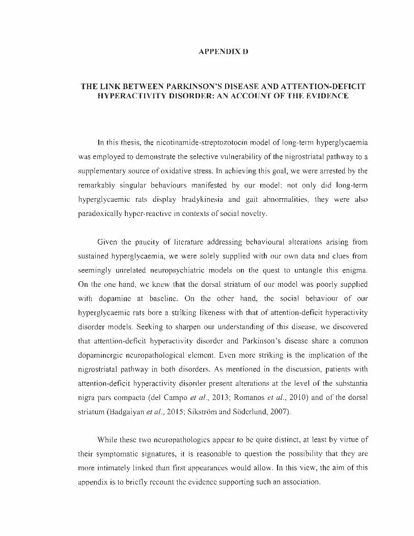
APPENDIXD
THE LINKBETWEEN PARKINSON'S DISEASE AND ATTENTION-DEFICIT HYPERACTIVITY DISORDER: AN ACCOUNT OF THE EVIDENCE
In this thesis, the nicotinamide-streptozotocin model of long-term hyperglycaemia
was employed to demonstrate the selective vulnerability of the nigrostriatal pathway to a
supplementary source of oxidative stress. In achieving this goal, we were arrested by the
remarkably singular behaviours manifested by our model: not only did long-term
hyperglycaemic rats display bradykinesia and gait abnormalities, they were also
paradoxically hyper-reactive in contexts of social novelty.
Given the paucity of literature addressing behavioural alterations ansmg from
sustained hyperglycaemia, we were solely supplied with our own data and cIues from
seemingly unrelated neuropsychiatric models on the quest to untangle this enigma.
On the one hand, we knew that the dorsal striatum of our model was poorly supplied
with dopamine at baseline. On the other hand, the social behaviour of our
hyperglycaemic rats bore a striking likeness with that of attention-deficit hyperactivity
disorder models. Seeking to sharpen our understanding of this disease, we discovered
that attention-deficit hyperactivity disorder and Parkinson ' s disease share a common
dopaminergic neuropathological element. Even more striking is the implication of the
nigrostriatal pathway in both disorders. As mentioned in the discussion, patients with
attention-deficit hyperactivity disorder present alterations at the level of the substantia
nigra pars compacta (deI Campo et al. , 2013 ; Romanos et al. , 2010) and of the dorsal
striatum (Badgaiyan et al., 2015 ; Sikstrom and Soderlund, 2007).
While these two neuropathologies appear to be quite distinct, at least by virtue of
their symptomatic signatures, it is reasonable to question the possibility that they are
more intimately linked than first appearances would allow. In this view, the aim of this
appendix is to briefly recount the evidence supporting such an association.
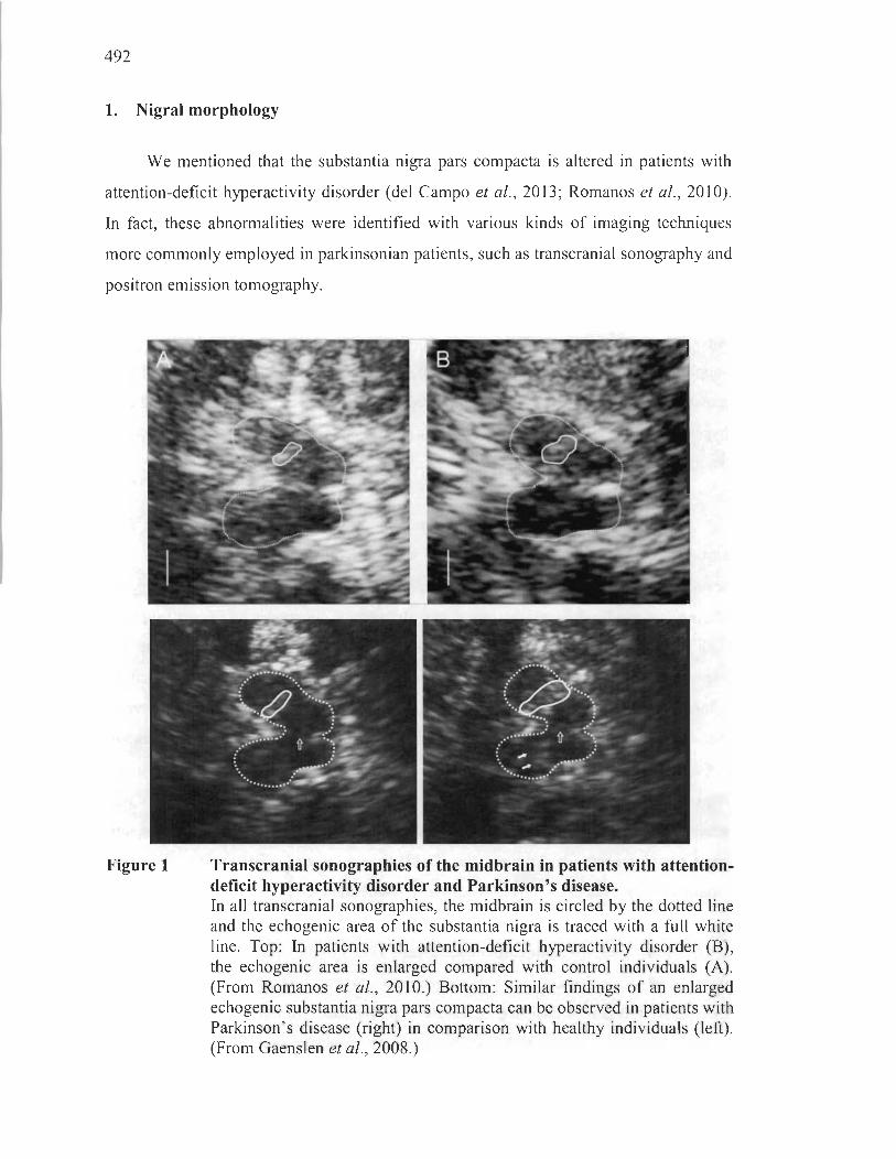
492
1. Nigral morphology
We mentioned that the substantia nigra pars compacta is altered in patients with
attention-deficit hyperactivity disorder (deI Campo et al. , 2013; Romanos et al. , 2010).
In fact, these abnormalities were identified with various kinds of imaging techniques
more commonly employed in parkinsonian patients, such as transcranial sonography and
positron emission tomography.
Figure 1 Transcranial sonographies of the midbrain in patients with attentiondeficit hyperactivity disorder and Parkinson 's disease. In aIl transcranial sono graphies, the midbrain is circled by the dotted line and the echogenic area of the substantia nigra is traced with a full white line. Top: In patients with attention-deficit hyperactivity disorder (B), the echogenic area is enlarged compared with control individuals (A). (From Romanos et al. , 2010.) Bottom: Similar findings of an enlarged echogenic substantia nigra pars compacta can be observed in patients with Parkinson's disease (right) in comparison with healthy individuals (Ieft) . (From Gaenslen et al., 2008.)

493
ln particular, evidence of an enhanced echogenicity was gathered in children
with attention-deficit hyperactivity disorder (Romanos et al. , 2010), a pathological sign
linked to increased iron content paired with a susceptibility for nigrostriatal dysfunction
(Becker et al., 1995; Berg et al. , 2002; Gaenslen et al. , 2008). Remarkably, the same
pattern of echogenicity can be observed in parkinsonian patients (Figure 1).
These important findings launched investigations aimed at uncovering the role of
oxidative stress, perhaps linked to nigral iron contents, in this neuropsychiatric condition
(Hess et al., 2017). It is unc1ear how iron contents are linked to neuropathogenicity in
both these diseases.
2. Animal models
Looking into animal models also provides sorne indications of a possible link
between the se pathologies. Quite teIling is the existence of a longstanding model of
attention-deficit hyperactivity disorder, used for sorne 40 years now, that employs
6-0HDA. ln neonate rats, this parkinsonian toxin destroys the near totality of
nigrostriatal neurons, sparing the ventral tegmental area, and causes a compensatory
serotoninergic hyperinnervation of the striatum that is thought to underlie behavioural
aberrations (Breese et al. , 2005; Kostrzewa et al. , 2004, 2006). The exact underpinnings
of the hyper-reactive phenotype remains ill understood. Noteworthy, total depletion of
dorsostriatal dopamine is likely not physiologicaIly relevant in patients with attention
deficit hyperactivity disorder. The hypotonic and hyperphasic aspects thought to
underlie this illness are therefore not represented therein (Badgaiyan et al., 2015;
Sikstrom and Soderlund, 2007).
More recently, in an attempt to model Parkinson's disease, one group uncovered
a hyper-reactive phenotype resulting from subeffective treatments. lndeed, primates
treated with varying chronic doses of the parkinsonian toxin MPTP displayed early
premotor social alterations, inc1uding enhanced aggressive and affiliative behaviours,
highly correlated with dorsostriatal denervation. ln the later phase of the pathology
wherein aIl evaluated dopamine-related cortical and subcortical structures were
494
fully degenerated, these hyper-reactive behaviours were drastically decreased and
conspicuous motor symptoms emerged. This study actually sheds light on a possible
continuum of nigrostriatal dopaminergic manifestations, ranging from hyper-reactivity at
low levels of neurodegeneration to apathy and motor impairments at high degrees of
denervation (Durand et al. , 2015). In relation to our own findings, we reiterate the
possibility that long-term hyperglycaemic rats examined between 3 and 6 months
display mixed moderate behaviours representative of both ends of the continuum. It is
tempting to propose that sorne forms of attention-deficit hyperactivity disorder and
Parkinson's disease may overlap.
3. Epidemiological evidence
Last, we can address existing epidemiological data suggesting a link between
attention-deficit hyperactivity disorder and Parkinson's disease or related pathologies.
Indeed, a growing body of literature has addressed the relationship between early-life
attention-deficit hyperactivity disorder or its symptoms and late-life parkinsonism
(Golimstok et al., 2011; Kim et al., 2013; Walitza et al., 2007). Interestingly,
Parkinson's disease and attention-deficit hyperactivity disorder may share certain
genetic underpinnings. Specifically, PARK2, a causative gene in Parkinson's disease
encoding parkin, was found to be associated with attention-deficit hyperactivity disorder
(Jarick et al., 2014). For sorne researchers, this invites the interrogation ofwhether both
diseases are in fact the early- and late-life continuum of a single dopaminergic pathology
(Golimstok et al., 2011). We wish to stress, however, that in this setting it is
unclear whether treatments addressing attention-deficit hyperactivity disorder, like
methylphenidate, may in fact contribute to overt nigrostriatal neurodegeneration found
in Parkinson's disease (Sadasivan et al., 2012).
Although any conclusions based on this scanty yet evocative evidence would be
deemed premature, the relationship between attention-deficit hyperactivity disorder and
Parkinson's disease does merit to be addressed by serious investigations aiming to
expose any possible overlapping neurobiological mechanisms. It remains that no study

495
has appropriately accounted for the history of hyperactivity, aggression or impulsivity in
large cohorts of parkinsonian patients.
These findings do, however, provide novel insight into often overlooked
non-motor behavioural alterations that may arise from nigrostriatal dopaminergic
neurodegeneration. Indeed, the nigrostriatal pathway is just leaving the shadow of the
mesocorticolimbic circuit as a regulator of social interactions (Burke et al. , 2010;
King-Casas et al. , 2005; Lamichhane et al. , 2014; Ong et al. , 2011 ; Palme ri et al., 2017;
Plavén-Sigray et al. , 2014; Stoeckel et al., 2014). Impaired social aptitudes recognized
to dim the quality of life of parkinsonian patients - even besides the well-appreciated
impulse control disorders that ensue from dopaminergic treatments (Cilia, 2012) - are
gaining attention in the scientific community (Schrag et al., 2000; Yoshimura et al.,
2005; PeU et al. , 2006). Granted this era of neuroscientists is shining a powerful
limelight on non-motor symptoms in Parkinson's disease, there is hope that these
interrogations will soon be addressed.
496
References
Badgaiyan, R. D., Sinha, S. , Sajjad, M., Wack, D. S. (2015). Attenuated Tonic and Enhanced Phasic Release of Dopamine in Attention Deficit Hyperactivity Disorder. PLoS One, la, e0137326.
Becker, G., Seufert, J. , Bogdahn, U. , Reichmann, H. , Reiners, K. (1995). Degeneration of substantia nigra in chronic Parkinson's disease visualized by transcranial colorcoded real-time sonography. Neurology, 45, 182-184.
Berg, D'., Roggendorf, W., Schrôder, u., Klein, R. , Tatschner, T. , Benz, P., Tucha, O., Preier, M. , Lange, K. W., Reiners, K. , Gerlach, M. , Becker, G. (2002). Echogenicity of the substantia nigra: association with increased iron content and marker for susceptibility to nigrostriatal injury. Arch. Neurol. , 59, 999-1005.
Breese, G. R. , Knapp, D. J. , Criswell, H. E., Moy, S. S. , Papadeas, S. T. , Blake, B. L. (2005). The neonate-6-hydroxydopamine-Iesioned rat: a model for clinical neuroscience and neurobiological principles. Brain Res. Rev. , 48, 57-73 .
Burke, C. J. , Tobler, P. N. , Schultz, W. , Baddeley, M. (2010). Striatal BOLD response reflects the impact of herd information on financial decisions. Front. Hum. Neurosci., 4, 48 .
Cilia, R. (2012). How neurodegeneration, dopamine and maladaptive behavioral leaming interact to pro duce impulse control disorders in Parkinson ' s disease. Basal Ganglia, 2, 195-199.
deI Campo, N., Fryer, T. D. , Hong, Y. T., Smith, R., Brichard, L. , Acosta-Cabronero, J. , Chamberlain, S. R. , Tait, R. , Izquierdo, D. , Regenthal, R., Dowson, J., Suckling, J. , Baron, J. C. , Aigbirhio, F. L, Robbins, T. W., Sahakian, B. J., Müller, U, (2013). A positron emission tomography study of nigrostriatal dopaminergic mechanisms underlying attention: implications for ADHD and its treatrnent. Brain, 136, 3252-3270.
Durand, E. , Petit, O. , Tremblay, L. , Zimmer, C., Sgambato-Faure, V. , Chassain, C., Laurent, M. , Pereira, B., Silberberg, C. , Durif, F. (2015) . Social behavioral changes in MPTP-treated monkey model of Parkinson's disease. Front. Behav. Neurosci., 9, 42.
Gaenslen, A. , Unmuth, B. , Godau, J. , Liepelt, L, Di Santo, A. , Schweitzer, K. J., Gasser, T., Machulla, H. J. , Reimold, M., Marek, K. , Berg, D. (2008). The specificity and sensitivity of transcranial ultrasound in the differential diagnosis of Parkinson's disease: a prospective blinded study. Lancet Neurol. , 7, 417-424.

497
Golimstok, A. , Rojas, 1. I., Romano, M., Zurru, M. C., Doctorovich, D. , Cristiano, E. (20 Il). Previous adult attention-deficit and hyperactivity disorder symptoms and risk of dementia with Lewy bodies: a case-control study. Eur. J Neural. , 18, 78-84.
Hess, 1. L. , Akutagava-Martins, G. C. , Patak, 1. D., Glatt, S. J. , Faraone, S. V. (2017). Why is there selective subcortical vulnerability in ADHD? CIues from postmortem brain gene expression data. Mol. Psychiatry., 23, 1787-1793.
Jarick, L, Volckmar. A. L. , Pütter, C. , Pechlivanis, S. , Nguyen, T. T. , Dauvermann, M. R., Beck, S., Albayrak, Ù. , Scherag, S., Gilsbach, S. , Cichon, S. , Hoffmann, P., Degenhardt, F., Nothen, M. M. , Schreiber. S., Wichmann, H. E. , Jockel, K. H., Heinrich, J., Tiesler, C. M. , Faraone, S. V., Walitza, S. , Sinzig, 1., Freitag, c., Meyer, J. , Herpertz-Dahlmann, B. , Lehmkuhl, G. , Renner, T. J. , Warnke, A. , Romanos, M. , Lesch, K. P. , Reif, A., Schimmelmann, B. G. , Hebebrand, J. , Scherag, A. , Hinney, A. (2014). Genome-wide analysis of rare copy number variations reveals PARK2 as a candidate gene for attention-deficitlhyperactivity disorder. Mol. Psychiatry, 16, 115-121.
Kim, J. S. , Youn, J., Shin, H. , Cho, J. W. (2013). Nonmotor symptoms in drug-induced parkinsonism and drug-naïve Parkinson disease. Cano J Neural. Sei. , 40, 36-41.
King-Casas, B. , Tomlin, D. , Anen, C., Camerer, C. F., Quartz, S. R. , Montague, P. R. (2005). Getting to know you: reputation and trust in a two-person economic exchange. Science, 308, 78-83.
Kostrzewa, R. M. , Brus, R, Ka1bfleisch, 1. H., Perry, K. W. , Fuller, R. W. (1994) . Proposed animal mode1 of attention deficit hyperactivity disorder. Brain Res. Bull. 34, 161-167.
Kostrzewa, R. M. , Kostrzewa, J. P. , Brus, R. , Kostrzewa, R A. , Nowak, P. (2006). Proposed animal model of severe Parkinson's disease: neonatal 6-hydroxydopamine les ion of dopaminergic innervation of striatum. J Neural Transm. Suppl. , 70, 277-279.
Lamichhane, B. , Adhikari, B. M., Brosnan, S. F. , Dhamala, M. (2014). The neural basis of perceived unfaimess in economic exchanges. Brain Connect., 4, 619-630.
Ong, L. K. , Bobrovskaya, L. , Walker, F. R , Day, T. A. , Dickson, P. W. , Dunk1ey, P. R. (20 Il). The effect of social defeat on tyrosine hydroxylase phosphorylation in the rat brain and adrenal gland. Neurochem. Res., 36, 27-33 .

498
Palmeri, R. , Lo Buono, V., Corallo, F. , Foti, M., Di Lorenzo, G. , Bramanti, P., Marino, S. (2017). Nonmotor Symptoms in Parkinson Disease: A Descriptive Review on Social Cognition Abi1ity. J Geriatr. Psychiatry Neurol. , 30, 109-12l.
Pell, M. D. , Cheang, H. S., Leonard, C. L. (2006). The impact of Parkinson's disease on vocal-prosodic communication from the perspective of listeners . Brain Lang., 97, 123-134.
Plavén-Sigray, P., Gustavsson, P., Farde, L., Borg, J., Stenkrona, P., Nyberg, L., Backman, L., Cervenka, S. (2014). Dopamine Dl receptor availability is related to social behavior: a positron emission tomography study. Neuroimage, 102, 590-595.
Romanos, M. , Weise, D. , Schliesser, M., Scheck1mann, M., L6ffler, 1., Warnke, A. , Gerlach, M. , Classem, J., Mehler-Wex, C. (2010). Structural abnormality of the substantia nigra in children with attention-deficit hyperactivity disorder. J Psychiatry Neurosci. , 35, 55-58.
Sadasivan, S., Pond, B. B. , Pani, A. K., Qu, C., Jiao, Y., Smeyne, R. 1. (2012). Methylphenidate exposure induces dopamine neuron 10ss and activation of microglia in the basal ganglia ofmice. PL oS One, 7, e33693.
Schrag, A., Jahanshahi, M., Quinn, N. (2000). How does Parkinson's disease affect quality of life? A comparison with quality of life in the general population. Mov. Disord. , 15, 1112-1118.
Sikstr6m, S., S6derlund, G. (2007). Stimulus-dependent dopamine release in attentiondeficit/hyperactivity disorder. Psycho!. Rev. , 114, 1047-1075.
Stoeckel, L. E., Palley, L. S. , Gollub, R. L., Niemi, S. M., Evins, A. E. (2014). Patterns of brain activation when mothers view their own chi Id and dog: an fMRI study. PLoS One, 9, e107205.
Walitza, S., Melfsen, S. , Herhaus, G., Scheuerpflug, P., Warnke, A. , Müller, T., Lange, K. W. , Gerlach, M. (2007). Association of Parkinson's disease with symptoms of attention deficit hyperactivity disorder in childhood. J Neural Transm. Suppl., 2007, 311-315.
Yoshimura, N., Kawamura, M., Masaoka, Y., Homma, 1. (2005). The amygda1a of patients with Parkinson's disease is silent in response to fearful facial expressions. Neuroscience, 131, 523-534.