role of microbiota-gut-brain axis in regulating dopaminergic
TRANSCRIPT

Biomedicines 2022, 10, 436. https://doi.org/10.3390/biomedicines10020436 www.mdpi.com/journal/biomedicines
Review
Role of Microbiota-Gut-Brain Axis in Regulating Dopaminergic Signaling Sevag Hamamah 1, Armin Aghazarian 1, Anthony Nazaryan 1, Andras Hajnal 2 and Mihai Covasa 1,3,*
1 Department of Basic Medical Sciences, College of Osteopathic Medicine, Western University of Health Sciences, Pomona, CA 91766, USA; [email protected] (S.H.); [email protected] (A.A.); [email protected] (A.N.)
2 Department of Neural and Behavioral Sciences, College of Medicine, The Pennsylvania State University, Hershey, PA 17033, USA; [email protected]
3 Department of Biomedical Sciences, College of Medicine and Biological Science, University of Suceava, 7200229 Suceava, Romania
* Correspondence: [email protected]
Abstract: Dopamine is a neurotransmitter that plays a critical role both peripherally and centrally in vital functions such as cognition, reward, satiety, voluntary motor movements, pleasure, and motivation. Optimal dopamine bioavailability is essential for normal brain functioning and protec-tion against the development of neurological diseases. Emerging evidence shows that gut microbi-ota have significant roles in maintaining adequate concentrations of dopamine via intricate, bidirec-tional communication known as the microbiota-gut-brain axis. The vagus nerve, immune system, hypothalamus–pituitary–adrenal axis, and microbial metabolites serve as important mediators of the reciprocal microbiota-gut-brain signaling. Furthermore, gut microbiota contain intrinsic enzy-matic activity that is highly involved in dopamine metabolism, facilitating dopamine synthesis as well as its metabolite breakdown. This review examines the relationship between key genera of gut microbiota such as Prevotella, Bacteroides, Lactobacillus, Bifidobacterium, Clostridium, Enterococcus, and Ruminococcus and their effects on dopamine. The effects of gut dysbiosis on dopamine bioavailabil-ity and the subsequent impact on dopamine-related pathological conditions such as Parkinson’s disease are also discussed. Understanding the role of gut microbiota in modulating dopamine ac-tivity and bioavailability both in the periphery and in the central nervous system can help identify new therapeutic targets as well as optimize available methods to prevent, delay, or restore dopa-minergic deficits in neurologic and metabolic disorders.
Keywords: dopamine; Prevotella; Bacteroides Lactobacillus; Bifidobacterium Clostridium; Enterococcus; Ruminococcus; Parkinson’s disease
1. Introduction The human gastrointestinal (GI) tract is host to trillions of commensal microbes,
which play an integral role in the maintenance of the host immune and homeostatic mech-anisms. Collectively, the genomic composition of the microbes contained within the hu-man gut is referred to as the microbiome, while the collection of bacteria, archaea, and eukarya that reside there is termed microbiota [1,2]. Of the six main phyla of bacteria pre-dominating the mammalian gut, Bacteroidetes and Firmicutes comprise approximately 90% of the total microbial population [3]. For the past decade, an abundance of studies have examined the symbiotic relationship between the mammalian host and the gut mi-crobiota as it pertains to microbiota’s role in immunity, nutrient and drug metabolism, bone formation, growth maintenance of mucosal barriers, and protection against patho-gens, to name a few [4,5]. More specifically, gut microbes have been implicated in endo-
Citation: Hamamah, S.; Aghazarian,
A.; Nazaryan, A.; Hajnal, A.; Covasa,
M. Role of Microbiota-Gut-Brain
Axis in Regulating Dopaminergic
Signaling. Biomedicines 2022, 10, 436.
https://doi.org/10.3390/
biomedicines10020436
Academic Editor: Marc Ekker
Received: 16 January 2022
Accepted: 11 February 2022
Published: 13 February 2022
Publisher’s Note: MDPI stays neu-
tral with regard to jurisdictional
claims in published maps and institu-
tional affiliations.
Copyright: © 2022 by the authors. Li-
censee MDPI, Basel, Switzerland.
This article is an open access article
distributed under the terms and con-
ditions of the Creative Commons At-
tribution (CC BY) license (https://cre-
ativecommons.org/licenses/by/4.0/).
Biomedicines 2022, 10, 436 2 of 24
crine functions via improvements in insulin sensitivity [6], reduction of intestinal inflam-mation [7], and facilitation of digestion through production of short-chain fatty acids (SCFAs) from complex carbohydrates [8–10]. The importance of the gut microbiota has been further supported by studies primarily demonstrating associations between the de-gree of microbial abundance, richness, and diversity, and some of the most prevalent dis-eases, such as obesity [11], type 2 diabetes mellitus, and metabolic syndrome [12,13], heart failure [14], and even cancers [15]. Therefore, it is not surprising that significant research efforts have been directed toward better understanding the effects that various microbes may have on both normal and pathophysiological processes in humans and their potential therapeutic implications. The regulation of these intricate processes involves a complex and dynamic bidirectional communication between the gut and the brain via the so-called gut–brain axis [16–18]. For example, several studies have shown that changes in the gut microbiome composition through manipulation techniques such as probiotic administra-tion or fecal microbiota transplant can cause changes in brain activity and cognitive be-havior by modulating neurotransmitter activity [19,20]. Dopamine, in particular, has drawn significant interest due to its contribution to pathological conditions in both the GI tract and the central nervous system [21–23]. The dopaminergic pathway contains neu-rons that reside in the ventral tegmental area (VTA) and substantia nigra pars compacta (SNpc) of the midbrain, which project to the nucleus accumbens via the mesocortical or mesolimbic pathways and dorsal striatum via the nigrostriatal pathway, respectively [24]. The dopamine transporter (DAT) and dopamine receptors (D1–D5) are essential in carry-ing out the numerous functions of dopamine and have roles that include, but are not lim-ited to, cognition, reward, satiety, voluntary motor movements, intestinal motility, and secretions [25–27]. For example, D3 receptor deletion increases GABA-induced inhibition in the nucleus accumbens, subsequently reducing reward-mediated voluntary alcohol in-take [28]. It has also been shown that increased D2 receptor postsynaptic signaling inter-feres with the firing patterns of dopaminergic neurons in the VTA [29]. In turn, this re-duced cortical dopaminergic bioavailability leads to cognitive deficits. Additionally, res-cuing deficiencies in fear extinction has been associated with enhanced gene expression of D1/D2 receptors in the prefrontal cortex (PFC), indicating the role of dopamine recep-tors in the onset of stress-related pathologies [30,31]. Further, changes in DAT and D1/D2 receptor expression in the striatum have been associated with alterations in the gut mi-crobiome [20,32,33]. More specifically, certain microbes belonging to genera such as Prevotella, Bacteroides, Lactobacillus, Bifidobacterium, Clostridium, Enterococcus, and Rumino-coccus have been shown to modulate receptors, transporters, and specific targets of the dopaminergic pathway, either in a positive or negative manner [20,34–36]. Although sev-eral studies have demonstrated important links between microbes and dopaminergic pathways both within the central nervous system (CNS) and peripherally in the GI system that result in behavioral changes [37], the underlying mechanisms are yet to be fully elu-cidated.
In this review, we present emerging evidence demonstrating the relationship be-tween gut microbiota and dopaminergic pathways that impact behavior. As such, we briefly describe the mechanisms of reciprocal communication between peripheral and central nervous systems via the gut–brain axis and the role of gut microbiota in this pro-cess. Then, we present evidence showing the ability of gut microbes belonging to Prevotella, Bacteroides, Lactobacillus, Bifidobacterium, Clostridium, Enterococcus, and Rumino-coccus to modulate dopaminergic activity and how subsequent alterations contribute to the pathogenesis of dopamine-related disorders. Overall, this review highlights the link between the gut microbiota and the dopaminergic system by presenting current evidence while identifying gaps and proposing future research directions to elucidate the potential contribution of gut microbiota to neurodegenerative disorders affecting dopaminergic neurons.
Biomedicines 2022, 10, 436 3 of 24
2. Microbiota-Gut-Brain Axis The influence of gut microbiota is not limited to the gut but is a major player in the
bidirectional communication between the gut and the brain. For example, disturbance of the healthy intestinal flora has been associated with numerous pathological conditions including neurological disorders such as Parkinson’s disease, ADHD, depression, anxiety, and autism [38]. It is not clear yet whether changes in the gut microbiota are the results of faulty brain signaling or whether they can actually drive brain disorders. It is known, however, that several intestinal microbes are involved in neurotransmitter synthesis such as glutamate, serotonin, noradrenaline, dopamine, and γ-aminobutyric acid (GABA) and their functions and bioavailability both in the CNS and in the periphery [39,40]. Changes in some of these neurotransmitters such as decreased dopamine concentrations have been linked with the etiology of neurodegenerative disorders. These neurotransmitters activate nerve ganglia in the myenteric and submucosal plexuses of the enteric nervous system (ENS) and are important mediators in the interface between the intestinal tract and the brain through the gut–brain axis. For example, enteroendocrine cells activate vagal affer-ents by using glutamate and serotonin as neurotransmitters, transducing luminal stimuli through the vagus nerve to the CNS [41].
In addition to an indirect effect mediated through the vagus nerve, microbes can in-teract directly with the CNS by direct actions in the GI tract, for example, via serotonin secretion into the lamina propria [42] or translocation of metabolites and endotoxins from the intestinal lumen to the main circulation [43–45]. Vagal afferents are also activated by numerous other signals, including hormones, nutrients, and peptides that are produced by intestinal microbes. Therefore, alterations in gut microbial species and the metabolites they produce can modulate afferent vagal nerve activity by multiple mechanisms. Indeed, SCFA-producing microbes were shown to activate and directly act on intestinal vagal ter-minals [46], and the introduction of Lactobacillus rhamnosus into the small intestine in-creased vagal afferent firing frequency [47]. Similarly, Lactobacillus johnsonii was shown to induce activation of vagal sensory neurons innervating the GI tract [48] and ablation of the vagus blocked anxiolytic and gene expression effects of Lactobacillus rhamnosus [49]. Together, these findings provide strong support for the role of the vagus nerve in medi-ating the effects of gut microbes on brain structures receiving direct viscero-sensory input. It should be noted, however, that the reported effects of gut microbes on the brain or whether the vagus nerve is involved in this effect is strain-dependent. For example, unlike Lactobacillus rhamnosus (JB-1), the effects of Lactobacillus salivarius on the enteric nervous system was not neurally dependent [50]. It is not clear, however, whether the vagus nerve is directly activated by microbes or rather indirectly via its soluble metabolites. Nonethe-less, compelling evidence exists demonstrating how specific microbes alter brain neuro-chemistry and subsequent behavior relevant to the dopaminergic system. Indeed, the va-gus nerve serves as a primary mediator in the gut–brain crosstalk to influence central and peripheral dopamine concentrations. For example, vagal nerve stimulation has been shown to induce dopaminergic activation and influence dopamine concentrations in the CNS [51]. The importance of vagal communication between the ENS and CNS has been well documented mainly through rodent models that undergo vagotomy. Vagotomized mice exhibit hampered neurogenesis regulation and decreased levels of brain-derived neurotrophic factor (BDNF) mRNA expression throughout the hippocampus [52]. BDNF has been shown to exert neuroprotective effects against dopamine neurons degeneration [53]. In addition to vagal-mediated pathways, the bidirectional communication between gut microbes and the CNS is facilitated through SCFAs, the immune system, and the hy-pothalamic–pituitary–adrenal (HPA) axis. Together, these modes of interaction of the mi-crobiota-gut-brain axis link cognitive, emotional, and reward centers in the brain with visceral signals from the gut.
Biomedicines 2022, 10, 436 4 of 24
2.1. Short-Chain Fatty Acids and Dopamine Gut microbiota metabolize complex, indigestible carbohydrates in the large intestine
through anaerobic fermentation of dietary fibers and resistant starch to yield short-chain fatty acids SCFA The three well-known, main SCFAs produced in the colon as the end products of soluble fiber fermentation are butyrate (C4), propionate (C3), and acetate (C2) [54]. Their concentrations vary among individuals as a function of the abundance of spe-cific microbes and have been shown to control and protect against systemic inflammation and maintain intestinal barrier integrity through gut–brain communication [55–57]. For example, inoculation of germ-free mice (GF) with Clostridium butyricum and Bacteroides thetaiotaomicron reduced blood–brain barrier (BBB) permeability by upregulating expres-sion of brain tight junction proteins, occluding zonulin and claudin-5 through the neuro-active potential of SCFAs [58]. Likewise, significantly elevated claudin-5 serum levels have been found in individuals diagnosed with obsessive compulsive disorder, while de-creased tight junction protein expression can lead to neuroinflammation [59], suggesting an important role of SCFAs in optimizing tight junction protein levels in endothelial tis-sues of the brain.
Butyrate and Dopamine Butyrate has significant associations with gastrointestinal health, neurotransmitter
concentrations, and gut–brain communication. Colonic enterocytes utilize butyrate pro-duced by intestinal microbes from dietary fiber as their major energy source [57]. Firmic-utes are major producers of butyrate with Clostridium, Rumminococcus, Eubacterium, and Faecalbacterium being notable genera largely contributing to its synthesis [60]. Butyrate’s intrinsic histone deacetylase (HDAC) inhibitor activity affects neurotransmitter levels and is therefore a key component of gut–brain signaling. For example, the HDAC inhibitory behavior of sodium butyrate has been shown to exert beneficial effects in neurotoxicity-induced rats by improving locomotor symptoms and increasing striatal dopamine [61]. It was shown that H3 histone acetylation was simultaneously increased when beneficial ef-fects on locomotor symptoms and striatal dopamine levels became noticeable. HDACs remove acetyl groups from histones, further condensing DNA into a heterochromatin form. This makes DNA less accessible to transcription factors and other gene regulatory proteins. In addition, butyrate is a potent inhibitor of heterochromatin formation and functions to relax chromatin, leading to significant roles in epigenetic modulation. The HDAC inhibitory activity of sodium butyrate has been shown to reduce oxidative stress, exert beneficial effects on α-synuclein damage, and rescue dopaminergic cells in Parkin-son’s disease (PD) models [62]. Recently, HDAC inhibitors have also been used as neuro-pharmacological agents to attenuate symptoms of glioblastomas [63]. Furthermore, so-dium butyrate infusion attenuated neuronal apoptosis in mice with middle cerebral artery occlusions [64]. Together, these studies indicate possible therapeutic implications of bu-tyrate’s intrinsic HDAC inhibitory activity on neurological disorders and neurotransmit-ter concentrations.
SCFAs can also incorporate colonic dopamine and produce serotonin through G-pro-tein-coupled receptor (GPCR)-mediated pathways [42,65]. Two GPCRs that are expressed on enteroendocrine cells, including enteric and sympathetic neurons, are GPR41, also known as FA3R, and GPR43, also known as FA2R [66]. FA3R and FA2R are preferentially activated by butyrate and acetate, respectively. Study findings show protective effects of butyrate through FA3R signaling against salsolinol (SALS)-induced toxicity, a selective neurotoxin affecting dopaminergic neurons. When challenged with a selective FA3R an-tagonist, the protective effects of butyrate were significantly reduced [67], supporting the importance of FA3R/butyrate signaling in attenuating the progression of neurodegenera-tive disorders. Antagonism of FA3R diminished the positive effects of restoring the via-bility of neuroblastoma-derived dopaminergic cells via sodium butyrate administration in an ethanol-induced toxicity model [68]. Overall, these findings show that butyrogenic

Biomedicines 2022, 10, 436 5 of 24
microbes play a major role in maintaining adequate dopamine concentrations by protect-ing against dopaminergic neuronal loss.
Additionally, sodium butyrate reduced degeneration of dopaminergic neurons, im-proved locomotor impairment, and elevated dopamine levels in a Drosophila transgenic model of PD [69]. A recent study supports these findings and shows that sodium butyrate administration in rodent models of PD reduces motor deficits while elevating dopamine levels and increasing histone acetylation [70]. Although much of the literature supports the beneficial effects of butyrate on the dopaminergic pathways, conflicting studies still exist. Qiao et al. concluded that sodium butyrate exacerbated motor symptoms, acceler-ated the loss of dopamine levels, and reduced tyrosine-hydroxylase-positive dopaminer-gic cells [71]. This study evaluated the effects of sodium butyrate after 1 week of treatment in neurotoxicity-induced mice, while other studies showing beneficial and contradictory findings examined the effects after 2 to 3 weeks of treatment [61,70,72]. This suggests that butyrate either can take time to exert its beneficial effects or that dopamine levels can be recovered after neurotoxicity induction.
Furthermore, butyrate has been shown to stimulate the production of gut hormones via enteroendocrine cells such as glucagon-like peptide 1 (GLP-1) and peptide YY (PYY), while also increasing GLP-1 receptor (GLP-1R) expression in the brain [72]. The increase in GLP-1R expression mitigated behavioral impairments in rodents. GLP-1 analogs and GLP-1R agonists have been shown to enhance synaptic membrane expression of DAT in the forebrain lateral septum [73]. More recently, GLP-1 analogs that cross the BBB and bind brain GLP-1R such as liratuglide have been found to improve cognition and enhance long-term potentiation [74]. Liratuglide-treated groups also showed significantly in-creased concentrations of neurotransmitters including dopamine in hippocampal tissue. Similar beneficial results were obtained with exenatide, a GLP-1R agonist, showing en-hanced dopamine midbrain function and behavioral improvement [75]. Several butyro-genic microbial species can naturally enhance GLP-1 levels and brain GLP-1R expression to positively affect dopamine concentration and improve neurobehavioral deficits. Recent work on the potential use of synthetic GLP-1 agonists as a treatment for drug addiction [76,77] further highlights the importance of microbiota in the regulation of dopamine functions in health and disease via the GLP-1 system.
When oral butyrate tablets were administered, there were notable changes in con-centrations of key microbes such as Bacteroides uniformis and Prevotella copri that affected DAT binding [20]. Heart rate variance was also observed exclusively in the butyrate-treated group and not in controls, indicating that the vagus nerve is stimulated by neuro-active butyrate. Furthermore, several studies showed the importance of maintaining ade-quate butyrate concentrations in the gut as it relates to neurological disorders that affect behavior. In children with autism spectrum disorder, levels of butyric acid, along with acetic and propionic acid, were significantly decreased and dopamine metabolism disor-der, characterized by decreased homovanillic acid (HVA) was present [78]. Probiotic in-troduction containing SFCA-producing gut microbes increased HVA and improved the dopamine metabolism deficit. Overall, gut microbiota that produce butyrate have a myr-iad of effects on dopamine and contribute significantly to attenuating deficits seen in neu-rodegenerative disorders.
2.2. Cytokines, Gut Microbiota, and Dopamine Cytokines appear to play an important role in the proposed regulatory axis between
gut microbes, the nervous system, and the immune system. Regulation of dopamine has been shown to be associated with cytokine release and vice versa. For example, dopamine released in the gut due to microbial processing can transcriptionally regulate the produc-tion of certain cytokines such as ILF-4 and IFN-gamma [37]. In dysbiosis and dopamine deficiency, ILF-4 and IFN-gamma levels increase, potentially activating immune response through natural killer cells (NKT) [37]. Mice treated with an anti-bacterial cocktail showed markedly reduced levels of tyrosine hydroxylase both at the mRNA and protein levels

Biomedicines 2022, 10, 436 6 of 24
within the small intestines. Subsequent restoration of gut microbiota following the anti-bacterial cocktail resulted in the recovery of intestinal tyrosine hydroxylase. The mecha-nism by which peripheral dopamine acts to decrease cytokine levels has been attributed to the activation of D1-like receptors on hepatic invariant NKT cells that play a role in regulating liver immunity. Additionally, the introduction of IFN-α for 4–6 weeks changes presynaptic dopamine function and decreases dopamine synthesis/release in the basal ganglia of patients with chronic hepatitis C virus [79]. These findings were associated with behavioral changes including depression and fatigue, indicating the widespread conse-quences of prolonged cytokine exposure in the body. It may also suggest that gut microbes promoting cytokine-mediated inflammation can alter striatal dopamine function by re-ducing dopamine production or release.
2.3. Hypothalamic–Pituitary–Adrenal Axis and Gut Microbiota The hypothalamic–pituitary–adrenal (HPA) axis plays a dynamic role in the stress
response and gut–brain communication [80]. Studies from GF rodents indicate that lack of gut microbiota is associated with a hyperreactive HPA axis in response to stress. This effect can be partially normalized by colonizing fecal matter with the normal flora of healthy rodents, indicating the importance of microbiota in maintaining homeostasis in the HPA axis [81]. Similarly, intestinal microbes have been shown to normalize the stress response by affecting HPA axis gene expression in conditions of chronic stress. For exam-ple, gut microbiota downregulate the FK506 binding protein 5 (Fkbp5) gene, which en-codes a protein that regulates the negative feedback loop by reducing cortisol affinity for glucocorticoid receptors [82]. Thus, in the absence of gut microbiota, negative feedback becomes dysregulated, and an exaggerated HPA response ensues. Although gut microbi-ota function to regulate stress response, stress-inducing events confer detrimental effects to the normal intestinal flora. As such, hyperactivation of the HPA axis leads to gut mi-crobiota dysbiosis through production of several proinflammatory cytokines such as IL-1β, IL-6, and TNF [80]. This can result in neurotransmitter dysregulations, leading to un-favorable behaviors via gut brain crosstalk, some of which can be restored through the administration of beneficial gut microbes. For example, administration of Lactobacillus plantarum PS128 to GF mice significantly increased dopamine levels and improved anxi-ety-like behaviors [83], demonstrating how bidirectional gut–brain communication through the HPA-axis affects neurotransmitter release and activity.
3. Gut Microbes Effects on Dopamine Gut microbiota are major contributors to dopamine bioavailability in the enteric and
central nervous systems. The following subsections depict how key gut microbes such as Prevotella, Bacteroides, Lactobacillus, Bifidobacterium, Clostridium, Enterococcus, and Rumino-coccus impact dopaminergic pathways.
3.1. Prevotella, Bacteroides, and Dopaminergic System Prevotella and Bacteroides are microbial genera belonging to the phylum Bacteroidetes
that have been shown to improve glucose metabolism, degrade a variety of plant polysac-charides, and produce favorable neuroactive SCFAs and vitamins that are necessary to promote gut health [84,85]. In addition to these symbiotic effects, metabolites produced by these microbes have been associated with dopamine functioning through modulation of dopaminergic synaptic cleft activity. In a recent study, Hartstra et al. showed that ad-ministration of Bacteroides uniformis through fecal microbiota transplant increased striatal DAT binding, while Prevotella copri was inversely correlated with transporter binding [20]. DAT, a presynaptic membrane protein present in dopaminergic terminals to regulate syn-aptic and extracellular dopamine is a critical modulator of dopaminergic tone within the CNS. Striatal dopamine binding to DAT within the synaptic cleft of dopaminergic neurons allows for recycling and storage of dopamine within vesicles in the presynaptic terminals

Biomedicines 2022, 10, 436 7 of 24
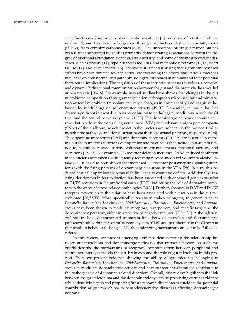
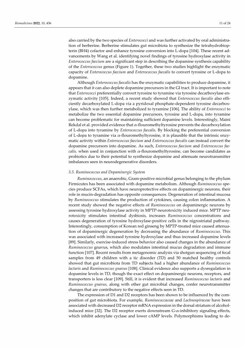
for subsequent release. Although the mechanisms by which Bacteroides uniformis and Prevotella copri alter striatal binding are not clear, neuroactive metabolites are obvious can-didates for modulating striatal DAT expression via activation of vagal afferents projecting to the nigrostriatal pathway within the synaptic cleft of dopaminergic neurons (Figure.1).
Furthermore, it was found that an abundance of Prevotella led to increased plasma concentrations of ghrelin, a gut-peptide orexigenic hormone that regulates satiety by act-ing on the VTA [86]. The VTA is a convergence point between ghrelin and dopamine, both of which are important in mediating reward pathways. As such, ghrelin was shown to recruit dopaminergic neurons by either direct activation or through indirect modulation of GABA neurons [87]. Systemically injected ghrelin has also been shown to stimulate motor activity and increase dopamine levels in rodent models. This suggests that Prevotella may indirectly recruit dopaminergic neurons by increasing ghrelin concentra-tions. Together, these findings indicate that the presence of Prevotella and Bacteroides in the intestinal tract does affect dopaminergic bioavailability either through direct actions on DAT binding or indirectly through alteration of gut-peptide hormone levels.
Figure 1. Effects of gut microbiota on the dopaminergic synaptic cleft and dopamine metabolism. (1) Bacteroides uniformis upregulates DAT/Dopamine binding efficiency. Prevotella copri downregu-lates DAT binding efficiency [20]. (2) Enterococcus faecalis and Enterococcus faecium demonstrate ty-rosine hydroxylase and aromatic L-amino acid decarboxylase activity. Oral berberine synthesizes cofactor BH4, allowing enhanced tyrosine to dopamine conversion [35]. (3) Lactobacillus plantarum

Biomedicines 2022, 10, 436 8 of 24
PS128 increases DAT expression [88]. (4) Ruminococcacae downregulates D2R expression [32]. (5) Ruminococcus is correlated with decreased tyrosine hydroxylase activity [89]. Abbreviations: DAT, dopamine transporter; BH4, tetrahydrobiopterin; D1R, dopamine 1 receptor; D2R, dopamine 2 re-ceptor.
3.2. Lactobacillus, Bifidobacterium, and Dopaminergic System Lactobacillus and Bifidobacterium are two microbial genera commonly used as probi-
otics that belong to the phyla Firmicutes and Actinobacteria, respectively. Administration of Lactobacillus has been shown to reduce depression- and anxiety-like behaviors in mice whereas oral administration of Bifidobacterium reduced exaggerated HPA stress response [90]. Likewise, patients with psychiatric conditions display a lower abundance of Bifidobacterium and Lactobacillus when compared to healthy controls, suggesting that these probiotic genera modulate neurotransmission in the dopaminergic pathways [91]. For ex-ample, oral administration of Lactobacillus plantarum PS128 for four weeks alleviated ele-vations in corticosterone, nigrostriatal dopaminergic neuronal death, and striatal dopa-mine reduction in 1-methyl-4-phenyl-1,2,3,6-tetrathydropyridine (MPTP)-induced PD in mouse models. MPTP is a neurotoxin that stimulates dopaminergic cell death in substan-tia nigra resulting in permanent manifestations of clinical symptoms in PD. Furthermore, PS128 administration was found to diminish MPTP-induced oxidative stress and neuroin-flammation in the nigrostriatal pathway, supporting its function in dopamine modulation [92]. Additionally, Liao et al. further demonstrated that the administration of psychobiot-ics containing PS128 can improve dopamine-related neurological disorders. In this study, PS128 was administered to ameliorate tic-like behaviors by using 5-HT2A and 5-HT2C receptor agonist 2,5-Dimethoxy-4-iodoamphetamine (DOI). Rats were administered PS128 orally for two weeks followed by two daily DOI injections. Analysis of the brain tissues showed that PS128 administration improved dopamine metabolism, increased DAT, and increased norepinephrine (Figures 1 and 2) [88]. Taken together, these studies provide strong evidence for the positive effects of Lactobacillus plantarum PS128 on dopa-mine metabolism and function.
In a clinical randomized, double-blind, placebo-controlled trial, administration of Lactobacillus plantarum DR7 for 12 weeks lowered stress and anxiety in stressed adults and also reduced plasma pro-inflammatory and increased plasma anti-inflammatory cyto-kines compared to the placebo group. Additionally, Lactobacillus plantarum DR7 lowered plasma cortisol, dopamine β-hydroxylase and tyrosine hydroxylase, two important en-zymes in dopamine metabolism. It has also been documented that dysregulation of dopa-mine β-hydroxylase can stimulate the stress response [93]. Therefore, administration of Lactobacillus plantarum DR7 can help reduce the stress response by lowering dopamine β-hydroxylase and tyrosine hydroxylase levels (Figure 2). Similarly, the effects of Bifidobac-terium infantis were studied in a rat maternal separation (MS) model by evaluating moti-vational state, cytokine concentrations, brain monoamine levels, and central and periph-eral HPA responses in MS rats subjected to a forced swim test (FST). Control MS rats dis-played immobility in the FST, decreased brain noradrenaline, elevated IL-6, and enhanced amygdala corticotropin-releasing factor mRNA levels. However, probiotic treatment nor-malized the immune response and noradrenaline concentrations and reversed behavioral deficits of MS rats [94]. This study further supports the idea that Bifidobacterium infantis has an impact on neural function and neurotransmitter activity.
Biomedicines 2022, 10, 436 9 of 24
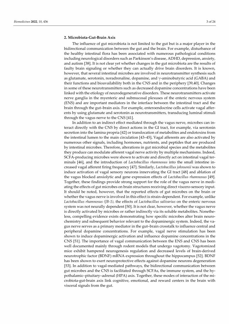
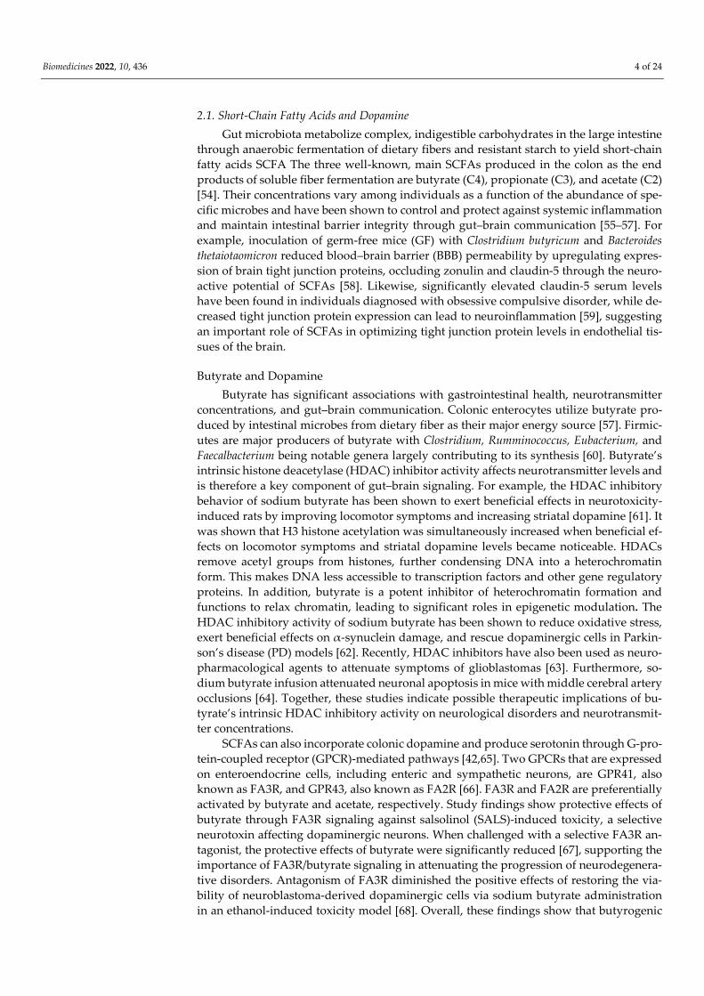
Figure 2. Effects of gut microbiota on metabolic byproducts of dopamine. (1) Coprococcus comes and Coprococcus catus are strongly associated with DOPAC synthesis potential [95]. (2) Clostridium tetani has degradative effects on dopamine, promoting degradation into HVA via the DOPAC intermedi-ate [96]. (3) Lactobacillus rhamnsosus downregulates MAO [97]. (4) Lactobacillus plantarum DR7 down-regulates Dopamine β-hydroxylase [93]. (5) Clostridia species shown to downregulate Dopamine β-hydroxylase [98]. (6) Lactobacillus plantarum PS128 administration improves dopamine metabolism and increases norepinephrine levels [92]. Abbreviations: MAO, monoamine oxidase; DOPAC, 3,4-Dihydroxyphenylacetic acid; HVA, homovanillic acid.
3.3. Clostridium and Dopaminergic System Clostridium is a Gram-positive, anaerobic gut microbe that comprises roughly 180
species, making it one of the larger genera in microbial taxonomic classification to date. The Clostridium genus belongs to the phylum Firmicutes and contains species that have been shown to be the causative agents of pathogenic processes in humans [99]. Addition-ally, certain species in the Clostridium genus, such as Clostridium tetani, have been de-scribed to have degradative effects on dopamine. For example, when biogenic amines were placed in a medium containing Clostridium tetani, dopamine was metabolized into significant amounts of HVA via the DOPAC intermediate (Figure 2) [96]. HVA is an end product of enzymatic degradation of dopamine and coincides with decreased dopamine levels. Additionally, metabolites produced by Clostridia inhibit dopamine β-hydroxylase, thus blocking the conversion of dopamine to norepinephrine [98]. This causes a significant increase in dopamine levels while indirectly decreasing norepinephrine. The excess do-pamine metabolites lead to toxic accumulation in the cytoplasm causing oxidative damage due to the depletion of glutathione in the brain. In turn, the oxidative stress produces a variety of detrimental effects, including apoptosis of dopaminergic neurons and a build-up of products such as α-synuclein protofibrils that are contributory to the pathogenesis

Biomedicines 2022, 10, 436 10 of 24
of neurodegenerative disorders. These findings demonstrate how neuropathogenic spe-cies within the Clostridium genus can exert unfavorable effects on the enzymes involved in dopamine metabolism.
Notwithstanding the above findings, several studies have been shown to have bene-ficial probiotic-like effects of Clostridium species on dopamine concentration through their endospore-forming capacity [100]. Clostridium butyricum forms spores that germinate in the intestinal tract and lead to the production of considerable amounts of SCFAs, particu-larly butyrate [101]. These beneficial outcomes were demonstrated in a recent study, where administration of Clostridium butyricum to piglets exhibiting weaning stress was significantly associated with increased hypothalamic concentrations of dopamine [36]. This was attributed to high butyrate concentrations observed in weaning piglets supple-mented with Clostridium butyricum. As noted above, butyrate can cross the BBB to influ-ence activity in the brain and affect microglial function and development. Thus, sodium butyrate decreases neuroinflammation and enhances neuroprotective function on cortical neurons [102], including direct effects on hypothalamic dopamine levels.
In addition to Clostridium butyricum, other species such as Clostridium coccoides and Clostridium leptum have been shown to have β-glucuronidase enzymatic activity that as-sists in the production of free gut catecholamines [18]. When comparing GF mice with select-pathogen-free (SPF) mice, SPF mice were shown to have considerably higher amounts of dopamine, epinephrine, and norepinephrine levels compared to GF mice. Pe-ripheral catecholamines are commonly found to be conjugated with either glucuronide or sulfate and when conjugated are considered biologically inactive. Most dopamine was identified in the “free” form in the cecum and colon along with substantial amounts found in the ileum. In SPF mice, gut catecholamines, including dopamine, were found in the biologically active free form, while in GF mice, the gut catecholamines were in the biolog-ically inactive, conjugated form. This indicates that Clostridium coccoides and Clostridium leptum contained β-glucuronidase enzymatic activity to convert peripheral dopamine into its active form. Thus, Clostridium plays important roles in increasing the available, func-tional dopamine in the periphery as well.
More recently, a study by Chang et al. showed that decarboxylated neurotransmitter substrates, including dopamine, can be conjugated with fatty acids to produce fatty acid amides. Clostridia produce these fatty acid amides in the gut lumen either endogenously or by using exogenous substrates that are common in the human diet. They also act as ligands binding GPCRs, leading to downstream signaling effects that drive physiological processes [65]. Thus, Clostridia’s ability to conjugate dopamine-like substrates into fatty acid amides to stimulate GPCR signaling warrants further studies into its pathogenic and homeostatic effects. Nevertheless, it is apparent that the genus Clostridium contains spe-cies that exhibit differing effects on dopamine metabolism. These widespread findings merit further exploration into the potential role of Clostridium to modulate systemic effects through the incorporation of dopamine substrates and metabolites.
3.4. Enterococcus and Dopaminergic System Enterococcus is another key genus of microbiota that is a part of the phylum Firmicu-
tes, which contains species that have been shown to participate in dopamine production. Although the genus is part of the normal intestinal flora, it has been identified as an op-portunistic pathogen. Its two major species, Enterococcus faecalis and Enterococcus faecium, have been shown to cause urinary tract infections, endocarditis, and other hospital-ac-quired infections [103]. Notwithstanding, significant research has been conducted to fur-ther understand their functions and impact on dopaminergic pathways. For example, sev-eral studies have demonstrated that supplementation of L-dopa enables Enterococcus fae-cium to convert newly introduced L-dopa into dopamine in the GI tract [35]. Additionally, transplantation of both Enterococcus faecalis and Enterococcus faecium into a mouse model of PD was shown to drastically increase the amount of striatal dopamine, as confirmed by neuroimaging. More specifically, this study found that tyrosine hydroxylase activity was
Biomedicines 2022, 10, 436 11 of 24
also carried by the two species of Enterococci and was further activated by oral administra-tion of berberine. Berberine stimulates gut microbiota to synthesize the tetrahydrobiop-terin (BH4) cofactor and enhance tyrosine conversion into L-dopa [104]. These recent ad-vancements by Wang et al. identifying novel findings of tyrosine hydroxylase activity in Enterococcus faecium are a significant step in describing the dopamine synthesis capability of the Enterococcus genus (Figure 1). Together, these two studies highlight the enzymatic capacity of Enterococcus faecium and Enterococcus faecalis to convert tyrosine or L-dopa to dopamine.
Although Enterococcus faecalis has the enzymatic capabilities to produce dopamine, it appears that it can also deplete dopamine precursors in the GI tract. It is important to note that Enterococci preferentially convert tyrosine to tyramine via tyrosine decarboxylase en-zymatic activity [105]. Indeed, a recent study showed that Enterococcus faecalis also effi-ciently decarboxylated L-dopa via a pyridoxal phosphate-dependent tyrosine decarbox-ylase, which was then further metabolized to tyramine [106]. The ability of Enterococci to metabolize the two essential dopamine precursors, tyrosine and L-dopa, into tyramine can become problematic for maintaining sufficient dopamine levels. Interestingly, Maini Rekdal et al. provided evidence that α-flouromethyltyrosine prevents the decarboxylation of L-dopa into tyramine by Enterococcus faecalis. By blocking the preferential conversion of L-dopa to tyramine via α-flouromethyltyrosine, it is plausible that the intrinsic enzy-matic activity within Enterococcus faecium and Enterococcus faecalis can instead convert the dopamine precursors into dopamine. As such, Enterococcus faecium and Enterococcus fae-calis, when used in conjunction with α-flouromethyltyrosine, can become candidates as probiotics due to their potential to synthesize dopamine and attenuate neurotransmitter imbalances seen in neurodegenerative disorders.
3.5. Ruminococcus and Dopaminergic System Ruminococcus, an anaerobic, Gram-positive microbial genus belonging to the phylum
Firmicutes has been associated with dopamine metabolism. Although Ruminococcus spe-cies produce SCFAs, which have neuroprotective effects on dopaminergic neurons, their role in mucin-degradation has opposite consequences. Degeneration of intestinal mucosa by Ruminococcus stimulates the production of cytokines, causing colon inflammation. A recent study showed the negative effects of Ruminococcus on dopaminergic neurons by assessing tyrosine hydroxylase activity in MPTP-neurotoxicity induced mice. MPTP neu-rotoxicity stimulates intestinal dysbiosis, increases Ruminococcus concentrations and causes degeneration of tyrosine hydroxylase-positive cells in the nigrostriatal pathway. Interestingly, consumption of Korean red ginseng by MPTP-treated mice caused attenua-tion of dopaminergic degeneration by decreasing the abundance of Ruminococcus. This was associated with increased tyrosine hydroxylase and thus increased dopamine levels [89]. Similarly, exercise-induced stress behavior also caused changes in the abundance of Ruminococcus gnavus, which also modulates intestinal mucus degradation and immune function [107]. Recent results from metagenomic analysis via shotgun sequencing of fecal samples from 49 children with a tic disorder (TD) and 50 matched healthy controls showed that gut microbiota from TD subjects had a higher abundance of Ruminococcus lactaris and Ruminococcus gnavus [108]. Clinical evidence also supports a dysregulation in dopamine levels in TD, though the exact effect on dopaminergic neurons, receptors, and transporters is less clear [109]. Still, it is evident that increased Ruminococcus lactaris and Ruminococcus gnavus, along with other gut microbial changes, confer neurotransmitter changes that are contributory to the negative effects seen in TD.
The expression of D1 and D2 receptors has been shown to be influenced by the com-position of gut microbiota. For example, Ruminococcaceae and Lachnospiraceae have been associated with decreased D2 receptor mRNA expression in the dorsal striatum of alcohol-induced mice [32]. The D2 receptor exerts downstream G-α-inhibitory signaling effects, which inhibit adenylate cyclase and lower cAMP levels. Polymorphisms leading to de-

Biomedicines 2022, 10, 436 12 of 24
creased dopamine binding to D2 receptor haplotypes have been associated with dyskine-sia [110,111], one of the classical symptoms of PD. Thus, it can be hypothesized that the abundance of Ruminococcacae seen in patients with PD causes downregulation of D2 re-ceptor expression, manifesting in motor symptoms such as bradykinesia. This is an intri-guing proposition that can be investigated in future studies.
4. Microbiota, Dopamine, and Parkinson’s Disease Dopamine bioavailability in humans is significantly intertwined with psychiatric
conditions [112]. Several studies associate changes in gut microbes and dopamine with the pathogenesis and clinical presentation of patients with PD that is caused by neuro-degeneration of substantia nigra and midbrain nuclei dopaminergic neurons [113,114]. The pathophysiology of PD can be largely attributed to the accumulation of α-synucleins, which interfere with synaptic transmission in dopaminergic neurons resulting in reduced dopamine bioavailability [115]. Dopamine loss in the periphery manifests as GI malfunc-tions, including delayed gastric emptying and intestinal dysmotility. Additionally, the neurodegeneration of dopaminergic neurons in the nigrostriatal pathway leads to motor symptoms, including bradykinesia, resting tremors, and postural instability. These motor symptoms, termed parkinsonism, have been directly associated with changes in gut mi-crobial composition. For example, elevated Enterobacteriaceae has been correlated with postural instability and gait abnormalities [85].
Some gut microbes have been shown to exert neuroprotective effects on dopaminer-gic neurons to attenuate dopamine loss. On the other hand, other microbes can exert neg-ative effects by stimulating inflammatory responses through endotoxins to further deplete dopamine concentrations. Overall, beneficial microflora tend to be reduced in the parkin-sonian gut, while levels of microbes that induce pathological processes are elevated. Trends that reflect the gut microbial composition of patients with PD have been identified in the literature. Although discrepancies do exist between studies, extensive microbial genotyping of the parkinsonian gut has been conducted and many analyses display com-parable results. All six major bacteria phyla are affected in some way or another by PD and changes in gut microbial species may serve as prodromal biomarkers that may indi-cate the early onset of the disease [85]. Altered taxa of PD patients include, but is not lim-ited to, an increase in Akkermansia muciniphilia, Enterobacteriaceae, Eggerthella, Oscillibacter, Catabacter, Escherichia, Shigella, Megomanus, Lachnospiraceae, and Streptococcus and de-creases in Roseburia, Coprococcus, Faecalibacterium, and Eubacterium biforme [116–119].
Of the genera described extensively in this review, Enterococcus, Bifidobacterium, and Rumminococcus levels were found to be elevated while Prevotella, Bacteroides, and Clostrid-ium concentrations were decreased in PD [120–122]. Findings regarding the effects of Lac-tobacillus appear to be less certain within the literature, with some reporting increases while others claim decreases in this microbial genus [121,123,124]. Together, these studies show that changes in these gut microbial genera contribute either positively or negatively to the pathophysiology of PD (Table 1). The possible mechanisms by which intestinal mi-crobial dysbiosis and its resulting effect on dopamine bioavailability may lead to the path-ogenesis of PD are highlighted in the following sections (Figure 3).
Table 1. Microbes’ effects in Parkinson’s Disease.
Phylum Genus Change Effects on Parkinson’s Patho-
physiology References
Bacteroidetes Prevotella Reduced
Reduced secretion of neuropro-tective hydrogen sulfide into the gut lumen and decreased
intestinal motility
[117,118,121,125]
Bacteroides Reduced [119,126]

Biomedicines 2022, 10, 436 13 of 24
Firmicutes
Lactobacillus Reduced
Less neuroprotective effects, unable to rescue dopaminergic
neuron loss, Inability to downregulate
MAO-B, Reduced BDNF and tyrosine
hydroxylase expression
[34,97,119,121]
Lactobacillus Elevated Deconjugates neuroprotective bile acids, TDCA and UDCA
[118,124,127,128]
Clostridium Reduced Increased α-synuclein accumu-
lation [117,124,128,129]
Enterococcus Elevated [122]
Ruminococcus Elevated
Ruminococcus Albus reduces ROS species, increases BDNF effects, and has associations
with decreased neuroprotective bile acids
[120,121,127,130]
Actinobacteria Bifidobacterium Elevated Confers neuroprotective effects on dopaminergic neuron loss
[97,121]
Verrucomicrobia Akkermansia Elevated Mucin-degrading genus, in-
creases LPS and microglial ac-tivity
[116–118,131]
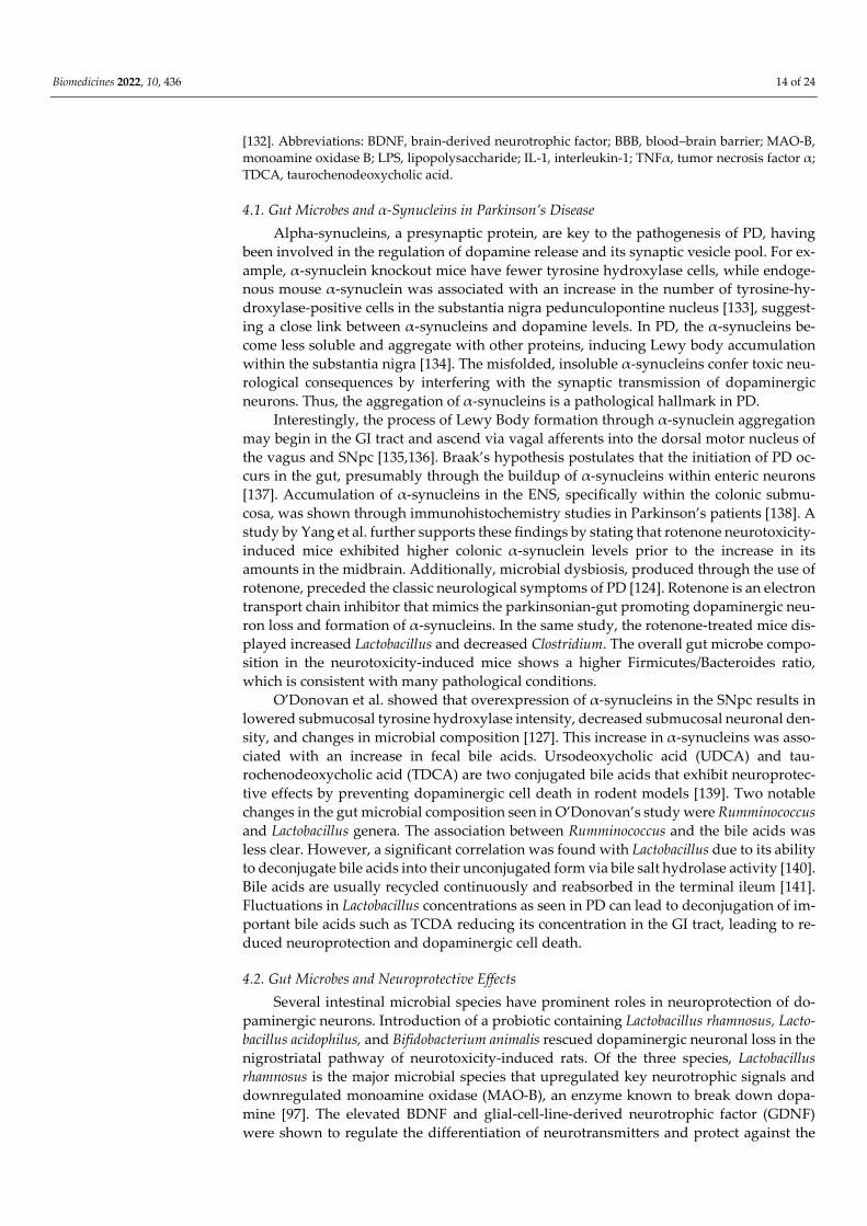
Figure 3. Gut microbial dysbiosis in Parkinson’s Disorder pathophysiology in the brain and GI tract. Patients with PD exhibit increased microglial activity [131], BBB permeability [58], α-synuclein ag-gregation [128], MAO-B expression [97], LPS secretion [131], intestinal permeability, intestinal layer mucin degradation, and pro-inflammatory cytokine release [131]. There is a decrease in BDNF ex-pression and tyrosine hydroxylase expression [34], butyrate synthesis, and TDCA concentration
Biomedicines 2022, 10, 436 14 of 24
[132]. Abbreviations: BDNF, brain-derived neurotrophic factor; BBB, blood–brain barrier; MAO-B, monoamine oxidase B; LPS, lipopolysaccharide; IL-1, interleukin-1; TNFα, tumor necrosis factor α; TDCA, taurochenodeoxycholic acid.
4.1. Gut Microbes and α-Synucleins in Parkinson’s Disease Alpha-synucleins, a presynaptic protein, are key to the pathogenesis of PD, having
been involved in the regulation of dopamine release and its synaptic vesicle pool. For ex-ample, α-synuclein knockout mice have fewer tyrosine hydroxylase cells, while endoge-nous mouse α-synuclein was associated with an increase in the number of tyrosine-hy-droxylase-positive cells in the substantia nigra pedunculopontine nucleus [133], suggest-ing a close link between α-synucleins and dopamine levels. In PD, the α-synucleins be-come less soluble and aggregate with other proteins, inducing Lewy body accumulation within the substantia nigra [134]. The misfolded, insoluble α-synucleins confer toxic neu-rological consequences by interfering with the synaptic transmission of dopaminergic neurons. Thus, the aggregation of α-synucleins is a pathological hallmark in PD.
Interestingly, the process of Lewy Body formation through α-synuclein aggregation may begin in the GI tract and ascend via vagal afferents into the dorsal motor nucleus of the vagus and SNpc [135,136]. Braak’s hypothesis postulates that the initiation of PD oc-curs in the gut, presumably through the buildup of α-synucleins within enteric neurons [137]. Accumulation of α-synucleins in the ENS, specifically within the colonic submu-cosa, was shown through immunohistochemistry studies in Parkinson’s patients [138]. A study by Yang et al. further supports these findings by stating that rotenone neurotoxicity-induced mice exhibited higher colonic α-synuclein levels prior to the increase in its amounts in the midbrain. Additionally, microbial dysbiosis, produced through the use of rotenone, preceded the classic neurological symptoms of PD [124]. Rotenone is an electron transport chain inhibitor that mimics the parkinsonian-gut promoting dopaminergic neu-ron loss and formation of α-synucleins. In the same study, the rotenone-treated mice dis-played increased Lactobacillus and decreased Clostridium. The overall gut microbe compo-sition in the neurotoxicity-induced mice shows a higher Firmicutes/Bacteroides ratio, which is consistent with many pathological conditions.
O’Donovan et al. showed that overexpression of α-synucleins in the SNpc results in lowered submucosal tyrosine hydroxylase intensity, decreased submucosal neuronal den-sity, and changes in microbial composition [127]. This increase in α-synucleins was asso-ciated with an increase in fecal bile acids. Ursodeoxycholic acid (UDCA) and tau-rochenodeoxycholic acid (TDCA) are two conjugated bile acids that exhibit neuroprotec-tive effects by preventing dopaminergic cell death in rodent models [139]. Two notable changes in the gut microbial composition seen in O’Donovan’s study were Rumminococcus and Lactobacillus genera. The association between Rumminococcus and the bile acids was less clear. However, a significant correlation was found with Lactobacillus due to its ability to deconjugate bile acids into their unconjugated form via bile salt hydrolase activity [140]. Bile acids are usually recycled continuously and reabsorbed in the terminal ileum [141]. Fluctuations in Lactobacillus concentrations as seen in PD can lead to deconjugation of im-portant bile acids such as TCDA reducing its concentration in the GI tract, leading to re-duced neuroprotection and dopaminergic cell death.
4.2. Gut Microbes and Neuroprotective Effects Several intestinal microbial species have prominent roles in neuroprotection of do-
paminergic neurons. Introduction of a probiotic containing Lactobacillus rhamnosus, Lacto-bacillus acidophilus, and Bifidobacterium animalis rescued dopaminergic neuronal loss in the nigrostriatal pathway of neurotoxicity-induced rats. Of the three species, Lactobacillus rhamnosus is the major microbial species that upregulated key neurotrophic signals and downregulated monoamine oxidase (MAO-B), an enzyme known to break down dopa-mine [97]. The elevated BDNF and glial-cell-line-derived neurotrophic factor (GDNF) were shown to regulate the differentiation of neurotransmitters and protect against the

Biomedicines 2022, 10, 436 15 of 24
degradation of dopaminergic neurons [53]. Reduced BDNF mRNA expression in the sub-stantia nigra has also been shown in PD [142]. Probiotic administration resulted in in-creased brain butyrate concentrations in neurotoxicity-induced rats providing strong ev-idence that butyrate influences attenuation of the dopaminergic neuron loss.
Liu et al. offered another intriguing explanation behind butyrate’s neuroprotective mechanism against PD, suggesting that sodium butyrate upregulates tight junction pro-teins to attenuate BBB disruption in neurotoxicity-induced mice. In the same study, in-creased expression of colonic and brain GLP-1 receptors was also present [72]. GLP-1 an-alogs and GLP-1R agonists have been shown to enhance synaptic membrane expression of DAT in the forebrain lateral septum [73]. This would, in turn, increase transport capac-ity, recycling, and vesicle storage of dopamine for later release. As a result, it is possible that the protective effects against dopaminergic degeneration seen in Liu et al.’s study can be attributed to increased GLP-1 and GLP-1R expression. Likewise, a recent study showed that gut-microbiota-derived propionic acid could mediate the neuroprotective effect of osteocalcin in a 6-hydroxydopamine-induced mouse model of PD. Considering the pro-tective role of propionic acid, it is likely that the reported low levels of propionic acid in the feces of PD patients may play a role in the pathogenesis of PD [143].
Lactobacillus plantarum also was shown to exert similar neuroprotective effects in con-ditions of oxidative stress. Cheon et al. created an environment of oxidative stress known to exacerbate neurodegenerative diseases before injecting the heat-killed Lactobacillus plantarum strain into a conditioned media. There was an increased mRNA expression of BDNF and tyrosine hydroxylase [34], indicating that the microbe stimulates attenuation of dopaminergic neuronal loss. This experiment followed a similar model of an earlier study that investigated the neuroprotective effects of Lactobacillus buchneri [144] and Rum-minococcus albus in conditions of oxidative stress [130]. The results were comparable, showing that heat-killed Lactobacillus buchneri and Rumminococcus albus increased BDNF expression and reduced reactive oxygen species (ROS) to exert neuroprotective effects on neurons. However, tyrosine hydroxylase expression was not elevated in Rumminococcus albus and Lactobacillus buchneri. It is also important to note that Lactobacillus plantarum, Rumminococcus albus, and Lactobacillus buchneri diminished the Bax/Bcl-2 ratio induced by the oxidative environment. Increased Bax/Bcl-2 stimulates cellular apoptosis [145]. As such, it appears that the protective mechanisms of these microbes in PD are present at the cellular and neuronal levels.
4.3. Lipopolysaccharides Liposaccharides (LPSs) are endotoxins naturally found in the outer membrane of cell
walls in Gram-negative bacteria. LPSs promote inflammation by binding Toll-like recep-tor 4 (TLR-4), leading to transcriptional upregulation of pro-inflammatory cytokines. In the short-term, systemic inflammation through LPS-mediated pathways has been found to elevate dopamine concentrations in the dorsal striatum [146]. These findings are con-sistent with studies showing that IFN-α administration in a primate model for two weeks enhances dopamine release. However, chronic exposure causes an opposite effect, reduc-ing striatal dopamine release as well as D2 receptor binding [147]. Furthermore, LPS ad-ministration can influence brain activity and is correlated with the development of neu-rodegenerative pathologies. LPS produces ROS through NADPH oxidase activity, result-ing in microglial activation and dopaminergic neuronal loss [148]. It is also documented that tyrosine hydroxylase activity is reduced in the substantia nigra, leading to noticeable motor dysfunction. This is in line with studies suggesting that LPS administration in-creases intestinal permeability, allowing endotoxins to travel towards the midbrain, caus-ing neuroinflammatory effects [131], increased mid brain TLR-4 reactivity, activation of microglia, and dopamine loss in rotenone-induced mice. These findings were associated with increases in the abundance of Akkermansia, a Gram-negative, mucin-degrading mi-crobe shown to be elevated in the parkinsonian gut [116], leading to endotoxemia-induced chronic inflammation.

Biomedicines 2022, 10, 436 16 of 24
As mentioned, α-synucleins aggregation has been associated with bacterial endo-toxin [149]. The mucin-degrading capacity of Akkermansia beneficially assists in maintain-ing intestinal barrier integrity at moderate to low levels. When elevated, reduced tight junction protein expression leads to intestinal leakiness. LPS can escape through the more permeable intestines into enteric neurons and systemic circulation [150]. As a result, LPS can then exert widespread effects that promote α-synuclein aggregation and dopaminer-gic neuronal loss. Additionally, LPS induces amyloidogenesis through the stabilization of α-helical intermediates. LPS and α-synucleins examined via microscopic imaging were shown to undergo a series of rapid nucleation events, leading to the formation of α-synu-clein/LPS fibrils [151] that can worsen PD pathogenesis. Taken together, these findings suggest that increased LPS activity exerts detrimental effects on dopamine bioavailability by producing ROS, increasing intestinal permeability, activating microglia, and promot-ing the formation of α-synucleins leading to dopaminergic neuronal loss.
4.4. Gut Microbes, Dopamine, and Intestinal Dysmotility The literature suggests that clinical GI manifestations often precede motor com-
plaints in PD. For example, the prodromal phase of PD includes symptoms of delayed gastric emptying and constipation that are correlated with dopaminergic neuronal loss and α-synucleins accumulation in the periphery [152]. These findings can be an important sign in diagnosing the early onset of PD. In general, higher Firmicutes/Bacteroides ratios have been correlated with intestinal dysmotility. More specifically, the Firmicutes sub-genera, Clostridium, and subfamily, Rumminococcacae, were shown to be elevated in pa-tients with functional constipation, while Prevotella levels were decreased [153]. Prevotella has been shown to secrete hydrogen sulfide into the gut lumen. In rodents, hydrogen sul-fide exerts protective properties against degeneration of tyrosine hydroxylase-containing dopaminergic neurons [154], which is correlated with manifestations of intestinal dys-motility in PD [155]. Thus, some studies have proposed that decreased concentrations of the hydrogen-sulfide secreting Prevotella contribute to GI symptoms such as constipation secondary to lower peripheral dopamine levels [125]. Although dopamine is known to inhibit upper GI motility and gastric emptying, it also plays a stimulatory role in lower GI motility, primarily in the colon, ameliorating the discomfort from constipation [156]. Therefore, lesser concentrations of peripheral dopamine would reduce lower intestinal motility, causing constipation. As such, reduced Prevotella concentrations present in the parkinsonian gut and associated reduction of hydrogen-sulfide secretion would be a fac-tor in motility-related symptoms seen in PD. These conclusions are further supported by an analysis that identifies Prevotella as the only taxon that was significantly reduced in PD patients and was correlated with ROME III constipation scores [126]. Furthermore, recent findings indicate that Prevotella-enriched enterotypes, as opposed to Firmicute-enriched enterotypes, lead to much lower constipation severity in PD [157]. This concurs with data suggesting that Prevotella remains an important microbial genus for the prevention of common GI symptoms in neurodegenerative disorders.
Although most studies support the idea that reduced Prevotella plays an important role in the parkinsonian gut, conflicting studies exist. An analysis of a transgenic rodent model with similar pathophysiological components to PD showed a transient increase in Prevotella at 20–24 weeks as compared to age-matched controls. At similar time points, there was a decrease in dopamine production both in the midbrain and in the gut [158]. This was correlated to an early onset of GI symptoms associated with PD. Moreover, an-other study claimed that Unified Parkinson’s Disease Rating Scores (UPDRS) were not associated with Prevotella, in contrast with previous work [159]. Notwithstanding, the pre-ponderance of studies show that Prevotella richness is commonly reduced in the parkin-sonian gut.
Furthermore, species within the Lactobacillus and Bifidobacterium genera that are com-monly used in probiotics to treat intestinal dysmotility were unchanged in constipated

Biomedicines 2022, 10, 436 17 of 24
patients [153]. However, introducing Lactobacillus and Bifidobacterium as a probiotic im-proved stool frequency and reduced the average gut transit time from 135 h to 77 h in PD patients [160]. These effects have been attributed to SCFAs that increase GI motility through the regulation of enteric neurons. Since SCFAs increase α-synuclein deposition in enteric neurons [122], increased abundance of SCFA-producing microbial species atten-uates constipation symptoms over time by reducing α-synuclein aggregation. Addition-ally, Lactobacillus deconjugates bile acids such as TDCA in the GI tract, which has been shown to inhibit intestinal motility [132] in addition to its neuroprotective effects. Thus, it is possible that the introduction of Lactobacillus can restore GI motility by deconjugating TDCA and removing its inhibitory effect. Overall, intestinal dysbiosis resulting in de-creased Prevotella, Lactobacillus, and Bifidobacterium concentrations is a major factor in the intestinal dysmotility seen in patients with PD.
5. Conclusions and Perspectives Substantial evidence supports the involvement of microbiota-gut-brain signaling in
dopamine release, synthesis, and bioavailability. In this review, we focused on key micro-bial genera, Prevotella, Bacteroides, Lactobacillus, Bifidobacterium, Clostridium, Enterococcus, and Ruminococcus, that are intricately intertwined with dopaminergic pathways via myr-iad effects on dopamine precursors, enzymes, receptors, transporters, and metabolites. States of intestinal dysbiosis involving these key genera disrupt microbiota-gut-brain sig-naling, leading to dopaminergic deficits that manifest in neuropathological conditions like PD. Overall, the literature continues to support the notion that pathophysiological effects in PD begin within the GI system. Notable pathological markers in the GI tract that are attributed to gut microbial changes include increased intestinal inflammation, mucin-layer degradation, LPS secretion, and α-synuclein accumulation and decreased butyrate synthesis and neuroprotective bile acid production. Vagus-mediated pathways communi-cate these pathological manifestations to the CNS, leading to neurodegenerative effects on dopaminergic neurons. Consequently, expression of BDNF, MAO-B, and tyrosine hy-droxylase is negatively affected by decreased BBB integrity and increased neuroinflam-mation through microglial activation and ROS production. Although significant advances have been made to elucidate the associations between gut microbiota, dopamine, and re-lated pathophysiology, there remains much to be learned. Many studies have been con-ducted in animal models that have been developed to mimic a parkinsonian patient through MPTP or rotenone-induced neurotoxicity. PD etiology is a progressive neuro-degenerative disease that involves contributory lifestyle, genetic, and socioeconomic fac-tors which are difficult to replicate in animal studies. Food consumption and environmen-tal variability are associated with differing gut microbiomes worldwide [161]. For exam-ple, consumption of the Western diet has been associated with specific microbial entero-types that can predispose individuals to various pathologies. Elevated Ruminococcus re-sulting from a Western diet results in pro-inflammatory behavior underlying the patho-physiology seen in neurodegenerative disorders such as PD [162]. Metagenomic sequenc-ing of gut microbiota following Western diets also reveals a limited prevalence of Prevotella that is associated with decreased DAT binding affinity. Further, the results from twin studies demonstrate how gut microbiome composition is largely connected to non-genetic factors [163,164]. Thus, it is plausible that individuals from different communities are more susceptible to developing dopamine-related pathologies based on their gut mi-crobial makeup shaped by diet and environment. Controlling for these variables in future studies is critically important and will help in our understanding of gut microbiota in-volvement in neurodegenerative etiology. Furthermore, some animal studies did not con-sider the different time points and progression of the parkinsonian state when measuring dopaminergic changes. Early PD and late PD have different clinical manifestations and may reflect the various effects on the dopaminergic pathways and other pathological changes detailed in this review. Nevertheless, the progress thus far establishing the link

Biomedicines 2022, 10, 436 18 of 24
between gut microbiota and neurodegenerative disorders is a step forward in our under-standing of the complex mechanisms underlying these pathologies.
Author Contributions: Conceptualization, S.H., A.A., A.N., and M.C.; methodology S.H., A.A., A.N., and M.C.; investigation, S.H., A.H., and M.C.; writing—original draft preparation, S.H., A.A., A.N., A.H. and M.C.; writing—review and editing, S.H., A.H. and M.C.; visualization, S.H.; super-vision, M.C. All authors have read and agreed to the published version of the manuscript.Funding: This research received no external funding.
Institutional Review Board Statement: Not applicable.
Informed Consent Statement: Not applicable.
Data Availability Statement: Not applicable.
Conflicts of Interest: The authors declare no conflicts of interest.
References 1. Bäckhed, F.; Roswall, J.; Peng, Y.; Feng, Q.; Jia, H.; Kovatcheva-Datchary, P.; Li, Y.; Xia, Y.; Xie, H.; Zhong, H.; et al. Dynamics
and Stabilization of the Human Gut Microbiome during the First Year of Life. Cell Host Microbe. 2015, 17, 690–703. 2. Turnbaugh, P.J.; Ley, R.E.; Hamady, M.; Fraser-Liggett, C.M.; Knight, R.; Gordon, J.I. The human microbiome project. Nature
2007, 449, 804–810. 3. Eckburg, P.B.; Bik, E.M.; Bernstein, C.N.; Purdom, E.; Dethlefsen, L.; Sargent, M.; Gill, S.R.; Nelson, K.E.; Relman, D.A. Diversity
of the human intestinal microbial flora. Science 2005, 308, 1635–1638. 4. Wu, B.B.; Yang, Y.; Xu, X.; Wang, W.P. Effects of Bifidobacterium supplementation on intestinal microbiota composition and
the immune response in healthy infants. World, J. Pediatr. 2016, 12, 177–182. 5. Yan, J.; Herzog, J.W.; Tsang, K.; Brennan, C.A.; Bower, M.A.; Garrett, W.S.; Sartor, B.R.; Aliprantis, A.O.; Charles, J.F. Gut mi-
crobiota induce IGF-1 and promote bone formation and growth. Proc. Natl. Acad. Sci. USA 2016, 113, E7554–E7563. 6. Chambers, E.S.; Byrne, C.S.; Morrison, D.J.; Murphy, K.G.; Preston, T.; Tedford, C.; Garcia-Perez, I.; Fountana, S.; Serrano-
Contreras, J.I.; Holmes. E.; et al. Dietary supplementation with inulin-propionate ester or inulin improves insulin sensitivity in adults with overweight and obesity with distinct effects on the gut microbiota, plasma metabolome and systemic inflammatory responses: A randomised cross-over trial. Gut 2019, 68, 1430–1438.
7. Ooi, C.Y.; Syed, S.A.; Rossi, L.; Garg, M.; Needham, B.; Avolio, J.; Young, K.; Surette, M.G.; Gonska, T. Impact of CFTR modu-lation with Ivacaftor on Gut Microbiota and Intestinal Inflammation. Sci Rep. 2018, 8, 17834.
8. Dao, M.C.; Everard, A.; Aron-Wisnewsky, J.; Sokolovska, N.; Prifti, E.; Verger, E.O.; Kayser, B.D.; Levenez, F.; Chilloux, J.; Hoyles, L.; et al. Akkermansia muciniphila and improved metabolic health during a dietary intervention in obesity: Relation-ship with gut microbiome richness and ecology. Gut 2016, 65, 426–436.
9. Kim, H.K.; Rutten, N.B.; Besseling-van der Vaart, I.; Niers, L.E.; Choi, Y.H.; Rijkers, G.T.; et al. Probiotic supplementation influ-ences faecal short chain fatty acids in infants at high risk for eczema. Benef. Microbes 2015, 6, 783–790.
10. Topping, D.L.; Clifton, P.M. Short-chain fatty acids and human colonic function: Roles of resistant starch and nonstarch poly-saccharides. Physiol. Rev. 2001, 81, 1031–1064.
11. Torres-Fuentes, C.; Schellekens, H.; Dinan, T.G.; Cryan, J.F. The microbiota-gut-brain axis in obesity. Lancet Gastroenterol. Hepa-tol. 2017, 2, 747–756.
12. Frost, F.; Storck, L.J.; Kacprowski, T.; Gärtner, S.; Rühlemann, M.; Bang, C.; Franke, A.; Völker, U.; Aghdassi, A.A.; Steveling, A.; et al. A structured weight loss program increases gut microbiota phylogenetic diversity and reduces levels of Collinsella in obese type 2 diabetics: A pilot study. PLoS ONE 2019, 14, e0219489.
13. Guevara-Cruz, M.; Flores-López, A.G.; Aguilar-López, M.; Sánchez-Tapia, M.; Medina-Vera, I.; Díaz, D.; et al. Improvement of Lipoprotein Profile and Metabolic Endotoxemia by a Lifestyle Intervention That Modifies the Gut Microbiota in Subjects with Metabolic Syndrome. J. Am. Heart Assoc. 2019, 8, e012401.
14. Hayashi, T.; Yamashita, T.; Watanabe, H.; Kami, K.; Yoshida, N.; Tabata, T.; Emoto, T.; Sasaki. N.; Mizoguchi, T.; Irino, Y.; et al. Gut Microbiome and Plasma Microbiome-Related Metabolites in Patients with Decompensated and Compensated Heart Failure Circ. J. 2018, 83, 182–92.
15. Xuan, C.; Shamonki, J.M.; Chung, A.; Dinome, M.L.; Chung, M.; Sieling, P.A.; Lee, D.J. Microbial dysbiosis is associated with human breast cancer. PLoS ONE 2014, 9, e83744.
16. Collins, S.M.; Bercik, P. The relationship between intestinal microbiota and the central nervous system in normal gastrointesti-nal function and disease. Gastroenterology 2009, 136, 2003–2014.
17. Rhee, S.H.; Pothoulakis, C.; Mayer, E.A. Principles and clinical implications of the brain-gut-enteric microbiota axis. Nat. Rev Gastroenterol. Hepatol. 2009, 6, 306–314.
18. Asano, Y.; Hiramoto, T.; Nishino, R.; Aiba, Y.; Kimura, T.; Yoshihara, K.; Koga, Y.; Sudo, N. Critical role of gut microbiota in the production of biologically active, free catecholamines in the gut lumen of mice. Am. J. Physiol. Gastrointest. Liver Physiol. 2012, 303, G1288–G1295.

Biomedicines 2022, 10, 436 19 of 24
19. Bagga, D.; Reichert, J.L.; Koschutnig, K.; Aigner, C.S.; Holzer, P.; Koskinen, K.; Moissl-Eichinger, C.; Schöpf, V. Probiotics drive gut microbiome triggering emotional brain signatures. Gut Microbes 2018, 9, 486–496.
20. Hartstra, A.V.; Schüppel, V.; Imangaliyev, S.; Schrantee, A.; Prodan, A.; Collard, D.; Levin, E.; Dallinga-Thie, G.; Ackermans, M.T.; Winkelmeijer, M.; et al. Infusion of donor feces affects the gut-brain axis in humans with metabolic syndrome. Mol. Metab. 2020, 42, 101076.
21. Aarts, E.; Ederveen, T.H.A.; Naaijen, J.; Zwiers, M.P.; Boekhorst, J.; Timmerman, H.M.; Smeekens, S.P.; Netea, M.G.; Buitelaar, J.K.;Franke, B.; et al. Gut microbiome in ADHD and its relation to neural reward anticipation. PLoS ONE 2017, 12, e0183509.
22. Georgescu, D.; Ancusa, O.E.; Georgescu, L.A.; Ionita, I.; Reisz, D. Nonmotor gastrointestinal disorders in older patients with Parkinson's disease: Is there hope? Clin. Interv. Aging. 2016, 11, 1601–1608.
23. Santocchi, E.; Guiducci, L.; Fulceri, F.; Billeci, L.; Buzzigoli, E.; Apicella, F.; Calderoni, S.; Grossi, E.; Morales, M.A.; Muratori, F. Gut to brain interaction in Autism Spectrum Disorders: A randomized controlled trial on the role of probiotics on clinical, biochemical and neurophysiological parameters. BMC Psychiatry 2016, 16, 183.
24. Luo, S.X.; Huang, E.J. Dopaminergic Neurons and Brain Reward Pathways: From Neurogenesis to Circuit Assembly. Am. J. Pathol. 2016, 186, 478–488.
25. Dubol, M.; Trichard, C.; Leroy, C.; Sandu, A.L.; Rahim, M.; Granger, B.; Tzavara, E.T.; Karila, L.; Martinot, J.L.; Artiges, E. Dopamine Transporter and Reward Anticipation in a Dimensional Perspective: A Multimodal Brain Imaging Study. Neuropsy-chopharmacology 2018, 43, 820–827.
26. Leddy, J.J.; Waxmonsky, J.G.; Salis, R.J.; Paluch, R.A.; Gnagy, E.M.; Mahaney, P.; Erbe. R.; Pelham. W.E.; Epstein. L.H. Dopa-mine-related genotypes and the dose-response effect of methylphenidate on eating in attention-deficit/hyperactivity disorder youths. J. Child. Adolesc. Psychopharmacol. 2009, 19, 127–136.
27. Uhl, G.R. Dopamine transporter: Basic science and human variation of a key molecule for dopaminergic function, locomotion, and parkinsonism. Mov. Disord. 2003, 18 (Suppl. 7), S71–S80.
28. Leggio, G.M.; Di Marco, R.; Gulisano, W.; D'Ascenzo, M.; Torrisi, S.A.; Geraci, F.; Lavanco, G.; Dahl, K.; Giurdanella, G.; Castorina, A.; et al. Dopaminergic-GABAergic interplay and alcohol binge drinking. Pharmacol. Res. 2019, 141, 384–391.
29. Torrisi, S.A.; Laudani, S.; Contarini, G.; De Luca, A.; Geraci, F.; Managò, F.; Papaleo, F.; Salomone, S.; Drago, F.; Leggio, G.M. Dopamine, Cognitive Impairments and Second-Generation Antipsychotics: From Mechanistic Advances to More Personalized Treatments. Pharmaceuticals 2020, 13, 365.
30. Torrisi, S.A.; Leggio, G.M.; Drago, F.; Salomone, S. Therapeutic Challenges of Post-traumatic Stress Disorder: Focus on the Dopaminergic System. Front. Pharmacol. 2019, 10, 404.
31. Whittle, N.; Maurer, V.; Murphy, C.; Rainer, J.; Bindreither, D.; Hauschild, M.; Hauschild, M.; Scharinger, A.; Oberhauser, M.; Keil, T.; Brehm, C.; et al. Enhancing dopaminergic signaling and histone acetylation promotes long-term rescue of deficient fear extinction. Transl. Psychiatry 2016, 6, e974.
32. Jadhav, K.S.; Peterson, V.L.; Halfon, O.; Ahern, G.; Fouhy, F.; Stanton, C.; Dinan, T.G.; Cryan, J.F.; Boutrel, B. Gut microbiome correlates with altered striatal dopamine receptor expression in a model of compulsive alcohol seeking. Neuropharmacology 2018, 141, 249–259.
33. Nettleton, J.E.; Klancic, T.; Schick, A.; Choo, A.C.; Shearer, J.; Borgland, S.L.; Chleilat, F.; Mayengbam, S.; Reimer, R.A. Low-Dose Stevia (Rebaudioside A) Consumption Perturbs Gut Microbiota and the Mesolimbic Dopamine Reward System. Nutrients 2019, 11, 1248.
34. Cheon, M.J.; Lee, N.K.; Paik, H.D. Neuroprotective Effects of Heat-Killed Lactobacillus plantarum 200655 Isolated from Kimchi Against Oxidative Stress. Probiotics Antimicrob. Proteins 2021, 13, 788–795.
35. Villageliú, D.; Lyte, M. Dopamine production in Enterococcus faecium: A microbial endocrinology-based mechanism for the selection of probiotics based on neurochemical-producing potential. PLoS ONE 2018, 13, e0207038.
36. Cao, G.; Tao, F.; Hu, Y.; Li, Z.; Zhang, Y.; Deng, B.; Zhan, X. Positive effects of a Clostridium butyricum-based compound probiotic on growth performance, immune responses, intestinal morphology, hypothalamic neurotransmitters, and colonic mi-crobiota in weaned piglets. Food Funct. 2019, 10, 2926–2934.
37. Xue, R.; Zhang, H.; Pan, J.; Du, Z.; Zhou, W.; Zhang, Z.; Tian, Z.; Zhoum R.; Baim L. Peripheral Dopamine Controlled by Gut Microbes Inhibits Invariant Natural Killer T Cell-Mediated Hepatitis. Front. Immunol. 2018, 9, 2398.
38. Weiss, G.A.; Hennet, T. Mechanisms and consequences of intestinal dysbiosis. Cell Mol. Life Sci. 2017, 74, 2959–2977. 39. Jang, S.-H.; Woo, Y.S.; Lee, S.-Y.; Bahk, W.-M. The Brain–Gut–Microbiome Axis in Psychiatry. Int. J. Mol. Sci. 2020, 21, 7122. 40. Strandwitz, P. Neurotransmitter modulation by the gut microbiota. Brain Res. 2018, 1693, 128–133. 41. Kaelberer, M.M.; Buchanan, K.L.; Klein, M.E.; Barth, B.B.; Montoya, M.M.; Shen, X.; Bohórquez, D.V. A gut-brain neural circuit
for nutrient sensory transduction. Science 2018, 361, 6408. 42. Reigstad, C.S.; Salmonson, C.E.; Rainey, J.F.; Szurszewski, J.H.; 3rd.; Linden, D.R.; Sonnenburg, J.L.; Farrugia, G.; Kashyap, P.C.
Gut microbes promote colonic serotonin production through an effect of short-chain fatty acids on enterochromaffin cells. Faseb, J. 2015, 29, 1395–1403.
43. Borre, Y.E.; O'Keeffe, G.W.; Clarke, G.; Stanton, C.; Dinan, T.G.; Cryan, J.F. Microbiota and neurodevelopmental windows: Im-plications for brain disorders. Trends Mol. Med. 2014, 20, 509–518.
44. Fung, T.C.; Olson, C.A.; Hsiao, E.Y. Interactions between the microbiota, immune and nervous systems in health and disease. Nat. Neurosci. 2017, 20, 145–155.

Biomedicines 2022, 10, 436 20 of 24
45. Dalile, B.; Van Oudenhove, L.; Vervliet, B.; Verbeke, K. The role of short-chain fatty acids in microbiota-gut-brain communica-tion. Nat. Rev. Gastroenterol. Hepatol. 2019, 16, 461–478.
46. Lal, S.; Kirkup, A.J.; Brunsden, A.M.; Thompson, D.G.; Grundy, D. Vagal afferent responses to fatty acids of different chain length in the rat. Am. J. Physiol. Gastrointest. Liver Physiol. 2001, 281, G907–G915.
47. Perez-Burgos, A.; Wang, B.; Mao, Y.K.; Mistry, B.; McVey Neufeld, K.A.; Bienenstock, J.; Kunze, W. Psychoactive bacteria Lac-tobacillus rhamnosus (JB-1) elicits rapid frequency facilitation in vagal afferents. Am. J. Physiol. Gastrointest. Liver Physiol. 2013, 304, G211–G220.
48. Tanida, M.; Yamano, T.; Maeda, K.; Okumura, N.; Fukushima, Y.; Nagai, K. Effects of intraduodenal injection of Lactobacillus johnsonii La1 on renal sympathetic nerve activity and blood pressure in urethane-anesthetized rats. Neurosci. Lett. 2005, 389, 109–114.
49. Bravo, J.A.; Forsythe, P.; Chew, M.V.; Escaravage, E.; Savignac, H.M.; Dinan, T.G.; Bienenstock, J.; Cryan, J.F. Ingestion of Lac-tobacillus strain regulates emotional behavior and central GABA receptor expression in a mouse via the vagus nerve. Proc. Natl. Acad. Sci. USA 2011, 108, 16050–16055.
50. Wang, B.; Mao, Y.K.; Diorio, C.; Pasyk, M.; Wu, R.Y.; Bienenstock, J.; Kunze, W.A. Luminal administration ex vivo of a live Lactobacillus species moderates mouse jejunal motility within minutes. Faseb, J. 2010, 24, 4078–4088.
51. Breit, S.; Kupferberg, A.; Rogler, G.; Hasler, G. Vagus Nerve as Modulator of the Brain-Gut Axis in Psychiatric and Inflammatory Disorders. Front. Psychiatry 2018, 9, 44.
52. O’Leary, O.F.; Ogbonnaya, E.S.; Felice, D.; Levone, B.R.; LCC.; Fitzgerald, P.; Bienenstock, J.; Dinan, T.G.; et al. The vagus nerve modulates BDNF expression and neurogenesis in the hippocampus. Eur. Neuropsychopharmacol. 2018, 28, 307–316.
53. Popova, N.K.; Ilchibaeva, T.V.; Naumenko, V.S. Neurotrophic Factors (BDNF and GDNF) and the Serotonergic System of the Brain. Biochemistry 2017, 82, 308–317.
54. Puertollano, E.; Kolida, S.; Yaqoob, P. Biological significance of short-chain fatty acid metabolism by the intestinal microbiome. Curr. Opin. Clin. Nutr. Metab. Care 2014, 17, 139–144.
55. Vinolo, M.A.; Rodrigues, H.G.; Nachbar, R.T.; Curi, R. Regulation of inflammation by short chain fatty acids. Nutrients 2011, 3, 858–876.
56. Trompette, A.; Gollwitzer, E.S.; Yadava, K.; Sichelstiel, A.K.; Sprenger, N.; Ngom-Bru, C.; Blanchard, C.; Junt, T.; Nicod, L.P.; Harris, N.L.; et al. Gut microbiota metabolism of dietary fiber influences allergic airway disease and hematopoiesis. Nat. Med. 2014, 20, 159–166.
57. Rivera-Chávez, F.; Zhang, L.F.; Faber, F.; Lopez, C.A.; Byndloss, M.X.; Olsan, E.E.; Xu, G.; Velazquez, E.M.; Lebrilla, C.B.; Winter ,S.E.; et al. Depletion of Butyrate-Producing Clostridia from the Gut Microbiota Drives an Aerobic Luminal Expansion of Salmonella. Cell Host. Microbe 2016, 19, 443–454.
58. Braniste, V.; Al-Asmakh, M.; Kowal, C.; Anuar, F.; Abbaspour, A.; Tóth, M.; Korecka, A.; Bakocevic, N.; Ng, L.G.; Kundu, P.; et al. The gut microbiota influences blood-brain barrier permeability in mice. Sci. Transl. Med. 2014, 6, 263ra158.
59. Asbjornsdottir, B.; Snorradottir, H.; Andresdottir, E.; Fasano, A.; Lauth, B.; Gudmundsson, L.S.; Gottfredsson, M.; Halldorsson, T.I.; Birgisdottir, B.E. Zonulin-Dependent Intestinal Permeability in Children Diagnosed with Mental Disorders: A Systematic Review and Meta-Analysis. Nutrients 2020, 12, 1982.
60. Mokhtari, Z.; Gibson, D.L.; Hekmatdoost, A. Nonalcoholic Fatty Liver Disease, the Gut Microbiome, and Diet. Adv. Nutr. 2017, 8, 240–252.
61. Sharma, S.; Taliyan, R.; Singh, S. Beneficial effects of sodium butyrate in 6-OHDA induced neurotoxicity and behavioral abnor-malities: Modulation of histone deacetylase activity. Behav. Brain Res. 2015, 291, 306–314.
62. Paiva, I.; Pinho, R.; Pavlou, M.A.; Hennion, M.; Wales, P.; Schütz, A.L.; Rajput, A.; Szego, É.M.; Kerimoglu, C.; Gerhardt, E.; et al. Sodium butyrate rescues dopaminergic cells from alpha-synuclein-induced transcriptional deregulation and DNA damage. Hum. Mol. Genet. 2017, 26, 2231–2246.
63. Gurbani, S.S.; Yoon, Y.; Weinberg, B.D.; Salgado, E.; Press, R.H.; Cordova, J.S.; Ramesh, K.K.; Liang, Z.; Vega, J.V.; Voloschin, A.; et al. Assessing Treatment Response of Glioblastoma to an HDAC Inhibitor Using Whole-Brain Spectroscopic MRI. Tomog-raphy 2019, 5, 53–60.
64. Zhou, Z.; Xu, N.; Matei, N.; McBride, D.W.; Ding, Y.; Liang, H.; Tang, J.; Zhang, J.H. Sodium butyrate attenuated neuronal apoptosis via GPR41/Gβγ/PI3K/Akt pathway after MCAO in rats. J. Cereb. Blood Flow Metab. 2021, 41, 267–281.
65. Chang, F.Y.; Siuti, P.; Laurent, S.; Williams, T.; Glassey, E.; Sailer, A.W.; Gordon, D.B.; Hemmerle, H.; Voigt, C.A. Gut-inhabiting Clostridia build human GPCR ligands by conjugating neurotransmitters with diet- and human-derived fatty acids. Nat. Micro-biol. 2021, 6, 792–805.
66. Nøhr, M.K.; Pedersen, M.H.; Gille, A.; Egerod, K.L.; Engelstoft, M.S.; Husted, A.S.; Sichlau, R.M.; Grunddal, K.V.; Poulsen, S.S.; Han, S.; et al. GPR41/FFAR3 and GPR43/FFAR2 as cosensors for short-chain fatty acids in enteroendocrine cells vs FFAR3 in enteric neurons and FFAR2 in enteric leukocytes. Endocrinology 2013, 154, 3552–3564.
67. Getachew, B.; Csoka, A.B.; Bhatti, A.; Copeland, R.L.; Tizabi, Y. Butyrate Protects Against Salsolinol-Induced Toxicity in SH-SY5Y Cells: Implication for Parkinson’s Disease. Neurotox. Res. 2020, 38, 596–602.
68. Getachew, B.; Csoka, A.B.; Garden, A.R.; Copeland, R.L.; Tizabi, Y. Sodium Butyrate Protects Against Ethanol-Induced Toxicity in SH-SY5Y Cell Line. Neurotox. Res. 2021, 39, 1–8.
69. St Laurent, R.; O'Brien, L.M.; Ahmad, S.T. Sodium butyrate improves locomotor impairment and early mortality in a rotenone-induced Drosophila model of Parkinson’s disease. Neuroscience 2013, 246, 382–390.

Biomedicines 2022, 10, 436 21 of 24
70. Kakoty, V.K.C.S.; Dubey, S.K.; Yang, C.H.; Taliyan, R. Neuroprotective Effects of Trehalose and Sodium Butyrate on Preformed Fibrillar Form of α-Synuclein-Induced Rat Model of Parkinson’s Disease. ACS Chem. Neurosci. 2021, 12, 2643–36660.
71. Qiao, C.M.; Sun, M.F.; Jia, X.B.; Li, Y.; Zhang, B.P.; Zhao, L.P.; Shi, Y.; Zhou, Z.L.; Zhu, Y.L.; Cui, C.; et al. Sodium Butyrate Exacerbates Parkinson’s Disease by Aggravating Neuroinflammation and Colonic Inflammation in MPTP-Induced Mice Model. Neurochem. Res. 2020, 45, 2128–2142.
72. Liu, J.; Wang, F.; Liu, S.; Du, J.; Hu, X.; Xiong, J.; Fang, R.; Chen, W.; Sun, J. Sodium butyrate exerts protective effect against Parkinson’s disease in mice via stimulation of glucagon like peptide-1. J. Neurol. Sci. 2017, 381, 176–181.
73. Reddy, I.A.; Pino, J.A.; Weikop, P.; Osses, N.; Sørensen, G.; Bering, T.; Valle, C.; Bluett, R.J.; Erreger, K.; Wortwein, G.; et al. Glucagon-like peptide 1 receptor activation regulates cocaine actions and dopamine homeostasis in the lateral septum by de-creasing arachidonic acid levels. Transl. Psychiatry 2016, 6, e809.
74. Abd El-Rady, N.M.; Ahmed, A.; Abdel-Rady, M.M.; Ismail, O.I. Glucagon-like peptide-1 analog improves neuronal and behav-ioral impairment and promotes neuroprotection in a rat model of aluminum-induced dementia. Physiol. Rep. 2021, 8, e14651.
75. Wang, V.; Kuo, T.T.; Huang, E.Y.; Ma, K.H.; Chou, Y.C.; Fu, Z.Y.; Lai, L.W.; Jung, J.; Choi, H.I.; Choi, D.S.; et al. Sustained Release GLP-1 Agonist PT320 Delays Disease Progression in a Mouse Model of Parkinson’s Disease. ACS Pharmacol. Transl. Sci. 2021, 4, 858–869.
76. Klausen, M.K.; Thomsen, M.; Wortwein, G.; Fink-Jensen, A. The role of glucagon-like peptide 1 (GLP-1) in addictive disorders. Br. J. Pharmacol. 2021, 179, 625–641.
77. Douton, J.E.; Horvath, N.; Mills-Huffnagle, S.; Nyland, J.E.; Hajnal, A.; Grigson, P.S. Glucagon-like peptide-1 receptor agonist, liraglutide, reduces heroin self-administration and drug-induced reinstatement of heroin-seeking behaviour in rats. Addict. Biol. 2021, e13117.
78. Wang, Y.; Li, N.; Yang, J.J.; Zhao, D.M.; Chen, B.; Zhang, G.Q.; Chen, S.; Cao, R.F.; Yu, H.; Zhao, C.Y.; et al. Probiotics and fructo-oligosaccharide intervention modulate the microbiota-gut brain axis to improve autism spectrum reducing also the hyper-ser-otonergic state and the dopamine metabolism disorder. Pharmacol. Res. 2020, 157, 104784.
79. Capuron, L.; Pagnoni, G.; Drake, D.F.; Woolwine, B.J.; Spivey, J.R.; Crowe, R.J.; Votaw, J.R.; Goodman, M.M.; Miller, A.H. Do-paminergic mechanisms of reduced basal ganglia responses to hedonic reward during interferon alfa administration. Arch. Gen. Psychiatry 2012, 69, 1044–1053.
80. Palma-Gudiel, H.; Prather, A.A.; Lin, J.; Oxendine, J.D.; Guintivano, J.; Xia, K.; Rubinow, D.R.; Wolkowitz, O.; Epel, E.S.; Zannas, A.S. HPA axis regulation and epigenetic programming of immune-related genes in chronically stressed and non-stressed mid-life women. Brain Behav. Immun. 2021, 92, 49–56.
81. Rosin, S.; Xia, K.; Azcarate-Peril, M.A.; Carlson, A.L.; Propper, C.B.; Thompson, A.L.; Grewen, K.; Knickmeyer, R.C. A prelimi-nary study of gut microbiome variation and HPA axis reactivity in healthy infants. Psychoneuroendocrinology 2021, 124, 105046.
82. Vodička, M.; Ergang, P.; Hrnčíř, T.; Mikulecká, A.; Kvapilová, P.; Vagnerová, K.; Šestáková B, Fajstová A, Hermanová, P.; Hudcovic, T.; et al. Microbiota affects the expression of genes involved in HPA axis regulation and local metabolism of gluco-corticoids in chronic psychosocial stress. Brain Behav. Immun. 2018, 73, 615–624.
83. Liu, W.H.; Chuang, H.L.; Huang, Y.T.; Wu, C.C.; Chou, G.T.; Wang, S.; Tsai, Y.C. Alteration of behavior and monoamine levels attributable to Lactobacillus plantarum PS128 in germ-free mice. Behav. Brain Res. 2016, 298, 202–209.
84. Kovatcheva-Datchary, P.; Nilsson, A.; Akrami, R.; Lee, Y.S.; De Vadder, F.; Arora, T.; Hallen. A.; Martens, E.; Björck, I.; Bäckhed, F. Dietary Fiber-Induced Improvement in Glucose Metabolism Is Associated with Increased Abundance of Prevotella. Cell Metab. 2015, 22, 971–982.
85. Scheperjans, F.; Aho, V.; Pereira, P.A.; Koskinen, K.; Paulin, L.; Pekkonen, E.; Haapaniemi, E.; Kaakkola, S.; Eerola-Rautio, J.; Pohja, M.; et al. Gut microbiota are related to Parkinson’s disease and clinical phenotype. Mov. Disord. 2015, 30, 350–358.
86. Kang, C.; Zhang, Y.; Zhu, X.; Liu, K.; Wang, X.; Chen, M.; Wang, J.; Chen, H.; Hui, S.; Huang, L.; et al. Healthy Subjects Differ-entially Respond to Dietary Capsaicin Correlating with Specific Gut Enterotypes. J. Clin. Endocrinol. Metab. 2016, 101, 4681–4689.
87. Cornejo, M.P.; Barrile, F.; De Francesco, P.N.; Portiansky, E.L.; Reynaldo, M.; Perello, M. Ghrelin Recruits Specific Subsets of Dopamine and GABA Neurons of Different Ventral Tegmental Area Sub-nuclei. Neuroscience 2018, 392, 107–120.
88. Liao, J.F.; Cheng, Y.F.; Li, S.W.; Lee, W.T.; Hsu, C.C.; Wu, C.C.; Jeng OJ, Wang, S.; Tsai, Y.C. Lactobacillus plantarum PS128 ameliorates 2,5-Dimethoxy-4-iodoamphetamine-induced tic-like behaviors via its influences on the microbiota-gut-brain-axis. Brain Res. Bull. 2019, 153, 59–73.
89. Jeon, H.; Bae, C.H.; Lee, Y.; Kim, H.Y.; Kim, S. Korean red ginseng suppresses 1-methyl-4-phenyl-1,2,3,6-tetrahydropyridine-induced inflammation in the substantia nigra and colon. Brain Behav. Immun. 2021, 94, 410–423.
90. Sudo, N.; Chida, Y.; Aiba, Y.; Sonoda, J.; Oyama, N.; Yu, X.N.; Kubo, C.; Koga, Y. Postnatal microbial colonization programs the hypothalamic-pituitary-adrenal system for stress response in mice. J. Physiol. 2004, 558 Part 1, 263–275.
91. Aizawa E, Tsuji H, Asahara T, Takahashi T, Teraishi T, Yoshida, S.; Ota, M.; Koga, N.; Hattori, K.; Kunugi, H. Possible associa-tion of Bifidobacterium and Lactobacillus in the gut microbiota of patients with major depressive disorder. J. Affect. Disord. 2016, 202, 254–257.
92. Liao, J.F.; Cheng, Y.F.; You, S.T.; Kuo, W.C.; Huang, C.W.; Chiou, J.J.; Hsu, C.C.; Hsieh-Li, H.M.; Wang, S.; Tsai, Y.C. Lactoba-cillus plantarum PS128 alleviates neurodegenerative progression in 1-methyl-4-phenyl-1,2,3,6-tetrahydropyridine-induced mouse models of Parkinson’s disease. Brain Behav. Immun. 2020, 90, 26–46.
93. Liu, G.; Chong, H.X.; Chung, F.Y.; Li, Y.; Liong, M.T. Lactobacillus plantarum DR7 Modulated Bowel Movement and Gut Mi-crobiota Associated with Dopamine and Serotonin Pathways in Stressed Adults. Int. J. Mol. Sci. 2020, 21, 4608.

Biomedicines 2022, 10, 436 22 of 24
94. Desbonnet, L.; Garrett, L.; Clarke, G.; Kiely, B.; Cryan, J.F.; Dinan, T.G. Effects of the probiotic Bifidobacterium infantis in the maternal separation model of depression. Neuroscience 2010, 170, 1179–1188.
95. Valles-Colomer, M.; Falony, G.; Darzi, Y.; Tigchelaar, E.F.; Wang, J.; Tito, R.Y.; Schiweck, C.; Kurilshikov, A.; Joossens, M.; Wijmenga, C.; et al. The neuroactive potential of the human gut microbiota in quality of life and depression. Nat. Microbiol. 2019, 4, 623–632.
96. Taj, A.; Jamil, N. Bioconversion of Tyrosine and Tryptophan Derived Biogenic Amines by Neuropathogenic Bacteria. Biomole-cules 2018, 8, 10.
97. Srivastav, S.; Neupane, S.; Bhurtel, S.; Katila, N.; Maharjan, S.; Choi, H.; Hong, J.T.; Choi, D.Y. Probiotics mixture increases butyrate, and subsequently rescues the nigral dopaminergic neurons from MPTP and rotenone-induced neurotoxicity. J. Nutr. Biochem. 2019, 69, 73–86.
98. Shaw, W. Elevated Urinary Glyphosate and Clostridia Metabolites with Altered Dopamine Metabolism in Triplets with Autistic Spectrum Disorder or Suspected Seizure Disorder: A Case Study. Integr. Med. 2017, 16, 50–57.
99. Kiu, R.; Caim, S.; Alcon-Giner, C.; Belteki, G.; Clarke, P.; Pickard, D.; Dougan, G.; Hall, L.J. Preterm Infant-Associated Clostrid-ium tertium, Clostridium cadaveris, and Clostridium paraputrificum Strains: Genomic and Evolutionary Insights. Genome Biol. Evol. 2017, 9, 2707–2714.
100. Dürre, P.; Eichenberger, P.; Driks, A. Physiology and Sporulation in Clostridium. Microbiol. Spectrum 2014, 2, 2–4. 101. Sato, R.; Tanaka, M. Intestinal distribution and intraluminal localization of orally administered Clostridium butyricum in rats.
Microbiol. Immunol. 1997, 41, 665–671. 102. Patnala, R.; Arumugam, T.V.; Gupta, N.; Dheen, S.T. HDAC Inhibitor Sodium Butyrate-Mediated Epigenetic Regulation En-
hances Neuroprotective Function of Microglia During Ischemic Stroke. Mol. Neurobiol. 2017, 54, 6391–6411. 103. Arias, C.A.; Murray, B.E. The rise of the Enterococcus: Beyond vancomycin resistance. Nat. Rev. Microbiol. 2012, 10, 266–278. 104. Wang, Y.; Tong, Q.; Ma, S.R.; Zhao, Z.X.; Pan, L.B.; Cong, L.; Han. P.; Peng, R.; Yu, H.; Lin, Y.; et al. Oral berberine improves
brain dopa/dopamine levels to ameliorate Parkinson’s disease by regulating gut microbiota. Signal. Transduct. Target Ther. 2021, 6, 77.
105. Linares, D.M.; Fernández, M.; Martín, M.C.; Alvarez, M.A. Tyramine biosynthesis in Enterococcus durans is transcriptionally regulated by the extracellular pH and tyrosine concentration. Microb Biotechnol. 2009;2(6):625-33.
106. Maini Rekdal, V.; Bess, E.N.; Bisanz, J.E.; Turnbaugh, P.J.; Balskus, E.P. Discovery and inhibition of an interspecies gut bacterial pathway for Levodopa metabolism. Science 2019, 364, 6445.
107. Clark, A.; Mach, N. Exercise-induced stress behavior, gut-microbiota-brain axis and diet: A systematic review for athletes. J. Int. Soc. Sports Nutr. 2016, 13, 43.
108. Xi, W.; Gao, X.; Zhao, H.; Luo, X.; Li, J.; Tan, X.; Wang L, Zhao JB, Wang J, Yang, G.; et al. Depicting the composition of gut microbiota in children with tic disorders: An exploratory study. J. Child Psychol. Psychiatry 2021, 62, 1246–1254.
109. Maia, T.V.; Conceição, V.A. Dopaminergic Disturbances in Tourette Syndrome: An Integrative Account. Biol. Psychiatry 2018, 84, 332–344.
110. Rieck, M.; Schumacher-Schuh, A.F.; Altmann, V.; Francisconi, C.L.; Fagundes, P.T.; Monte, T.L.; Callegari-Jacques, S.M.; Rieder, C.R.; Hutz, M.H. DRD2 haplotype is associated with dyskinesia induced by levodopa therapy in Parkinson’s disease patients. Pharmacogenomics 2012, 13, 1701–1710.
111. Zappia, M.; Annesi, G.; Nicoletti, G.; Arabia, G.; Annesi, F.; Messina, D.; Pugliese, P.; Spadafora, P.; Tarantino, P.; Carrideo, S.; et al. Sex differences in clinical and genetic determinants of levodopa peak-dose dyskinesias in Parkinson disease: An explora-tory study. Arch. Neurol. 2005, 62, 601–605.
112. Grace, A.A. Dysregulation of the dopamine system in the pathophysiology of schizophrenia and depression. Nat. Rev. Neurosci. 2016, 17, 524–532.
113. Burokas, A.; Moloney, R.D.; Dinan, T.G.; Cryan, J.F. Microbiota regulation of the Mammalian gut-brain axis. Adv. Appl. Micro-biol. 2015, 91, 1–62.
114. Vizcarra, J.A.; Wilson-Perez, H.E.; Fasano, A.; Espay, A.J. Small intestinal bacterial overgrowth in Parkinson’s disease: Tribula-tions of a trial. Parkinsonism. Relat. Disord. 2018, 54, 110–112.
115. Rocha, E.M.; De Miranda, B.; Sanders, L.H. Alpha-synuclein: Pathology, mitochondrial dysfunction and neuroinflammation in Parkinson’s disease. Neurobiol. Dis. 2018, 109, 249–257.
116. Nishiwaki, H.; Ito, M.; Ishida, T.; Hamaguchi, T.; Maeda, T.; Kashihara, K.; Tsuboi, Y.; Ueyama, J.; Shimamura, T.; Mori, H.; et al. Meta-Analysis of Gut Dysbiosis in Parkinson’s Disease. Mov. Disord. 2020, 35, 1626–1635.
117. Bedarf, J.R.; Hildebrand, F.; Coelho, L.P.; Sunagawa, S.; Bahram, M.; Goeser, F.; Bork, P.; Wüllner, U. Functional implications of microbial and viral gut metagenome changes in early stage L-DOPA-naïve Parkinson’s disease patients. Genome Med. 2017, 9, 39.
118. Gerhardt, S.; Mohajeri, M.H. Changes of Colonic Bacterial Composition in Parkinson’s Disease and Other Neurodegenerative Diseases. Nutrients 2018, 10, 708.
119. Unger, M.M.; Spiegel, J.; Dillmann, K.-U.; Grundmann, D.; Philippeit, H.; Bürmann, J.; Faßbender, K.; Schwiertz, A.; Schäfer, K.-H. Short chain fatty acids and gut microbiota differ between patients with Parkinson’s disease and age-matched controls. Parkinsonism. Related Disorders 2016, 32, 66–72.

Biomedicines 2022, 10, 436 23 of 24
120. Hegelmaier, T.; Lebbing, M.; Duscha, A.; Tomaske, L.; Tönges, L.; Holm, J.B.; Bjørn, N.H.; Gatermann, S.G.; Przuntek, H.; Haghikia, A. Interventional Influence of the Intestinal Microbiome Through Dietary Intervention and Bowel Cleansing Might Improve Motor Symptoms in Parkinson’s Disease. Cells 2020, 9, 376.
121. Jin, M.; Li, J.; Liu, F.; Lyu, N.; Wang, K.; Wang, L.; Liang, S.; Tao, H.; Zhu, B.; Alkasir, R. Analysis of the Gut Microflora in Patients with Parkinson’s Disease. Front. Neurosci. 2019, 13, 1184.
122. Li, W.; Wu, X.; Hu, X.; Wang, T.; Liang, S.; Duan, Y.; Jin, F.; Qin, B. Structural changes of gut microbiota in Parkinson’s disease and its correlation with clinical features. Sci. China Life Sci. 2017, 60, 1223–1233.
123. Hill-Burns, E.M.; Debelius, J.W.; Morton, J.T.; Wissemann, W.T.; Lewis, M.R.; Wallen, Z.D.; Peddada, S.D.; Factor, S.A.; Molho, E.; Zabetian, C.P.; et al. Parkinson’s disease and Parkinson’s disease medications have distinct signatures of the gut microbiome. Mov. Disord. 2017, 32, 739–749.
124. Yang, X.; Qian, Y.; Xu, S.; Song, Y.; Xiao, Q. Longitudinal Analysis of Fecal Microbiome and Pathologic Processes in a Rotenone Induced Mice Model of Parkinson’s Disease. Front. Aging Neurosci. 2018, 9, 441.
125. Cakmak, Y.O. Provotella-derived hydrogen sulfide, constipation, and neuroprotection in Parkinson’s disease. Mov. Disord. 2015, 30, 1151.
126. Aho, V.T.E.; Pereira, P.A.B.; Voutilainen, S.; Paulin, L.; Pekkonen, E.; Auvinen, P.; Scheperjans, F. Gut microbiota in Parkinson’s disease: Temporal stability and relations to disease progression. EBioMedicine 2019, 44, 691–707.
127. O’Donovan, S.M.; Crowley, E.K.; Brown, J.R.; O’Sullivan, O.; O’Leary, O.F.; Timmons, S.; Nolan, Y.M.; Clarke, D.J.; Hyland, N.P.; Joyce, S.A.; et al. Nigral overexpression of α-synuclein in a rat Parkinson’s disease model indicates alterations in the enteric nervous system and the gut microbiome. Neurogastroenterol. Motil. 2020, 32, e13726.
128. Hasegawa, S.; Goto, S.; Tsuji, H.; Okuno, T.; Asahara, T.; Nomoto, K.; Shibata A, Fujisawa Y, Minato, T.; Okamoto, A.; et al. Intestinal Dysbiosis and Lowered Serum Lipopolysaccharide-Binding Protein in Parkinson’s Disease. PLoS ONE 2015, 10, e0142164.
129. Vascellari, S.; Melis, M.; Cossu, G.; Melis, M.; Serra, A.; Palmas, V.; Perra, D.; Oppo, V.; Fiorini, M.; Cusano, R.; et al. Genetic variants of TAS2R38 bitter taste receptor associate with distinct gut microbiota traits in Parkinson’s disease: A pilot study. Int. J. Biol. Macromol. 2020, 165, 665–674.
130. Park, J.; Lee, J.; Yeom, Z.; Heo, D.; Lim, Y.H. Neuroprotective effect of Ruminococcus albus on oxidatively stressed SH-SY5Y cells and animals. Sci. Rep. 2017, 7, 14520.
131. Dodiya, H.B.; Forsyth, C.B.; Voigt, R.M.; Engen, P.A.; Patel, J.; Shaikh, M.; Green, S.J.; Naqib, A.; Roy, A.; Kordower, J.H.; et al. Chronic stress-induced gut dysfunction exacerbates Parkinson’s disease phenotype and pathology in a rotenone-induced mouse model of Parkinson’s disease. Neurobiol. Dis. 2020, 135, 104352.
132. Abdu, F.; Albaik, M. Effect of Conjugated Bile Salt Taurodeoxycholic Acid (TDCA) on Mice Colonic Motor Activity. Periodicum Biologorum. 2016, 118.
133. Garcia-Reitboeck, P.; Anichtchik, O.; Dalley, J.W.; Ninkina, N.; Tofaris, G.K.; Buchman, V.L.; Spillantini, M.G. Endogenous al-pha-synuclein influences the number of dopaminergic neurons in mouse substantia nigra. Exp. Neurol. 2013, 248, 541–545.
134. Bridi, J.C.; Hirth, F. Mechanisms of α-Synuclein Induced Synaptopathy in Parkinson’s Disease. Front. Neurosci. 2018, 12, 80. 135. Chiang, H.L.; Lin, C.H. Altered Gut Microbiome and Intestinal Pathology in Parkinson’s Disease. J. Mov. Disord. 2019, 12, 67–
83. 136. Travagli, R.A.; Browning, K.N.; Camilleri, M. Parkinson disease and the gut: New insights into pathogenesis and clinical rele-
vance. Nat. Rev. Gastroenterol. Hepatol. 2020, 17, 673–685. 137. Braak, H.; Del Tredici, K.; Rüb, U.; de Vos, R.A.; Jansen Steur, E.N.; Braak, E. Staging of brain pathology related to sporadic
Parkinson’s disease. Neurobiol. Aging 2003, 24, 197–211. 138. Shannon, K.M.; Keshavarzian, A.; Mutlu, E.; Dodiya, H.B.; Daian, D.; Jaglin, J.A.; Kordower JH. Alpha-synuclein in colonic
submucosa in early untreated Parkinson’s disease. Mov. Disord. 2012, 27, 709–715. 139. Castro-Caldas, M.; Carvalho, A.N.; Rodrigues, E.; Henderson, C.J.; Wolf, C.R.; Rodrigues, C.M.; Gama, M.J. Tauroursodeoxy-
cholic acid prevents MPTP-induced dopaminergic cell death in a mouse model of Parkinson’s disease. Mol. Neurobiol. 2012, 46, 475–486.
140. Long, S.L.; Gahan, C.G.M.; Joyce, S.A. Interactions between gut bacteria and bile in health and disease. Mol. Aspects Med. 2017, 56, 54–65.
141. Chiang, J.Y. Bile acid metabolism and signaling. Compr. Physiol. 2013, 3, 1191–1212. 142. Howells, D.W.; Porritt, M.J.; Wong, J.Y.; Batchelor, P.E.; Kalnins, R.; Hughes, A.J.; Donnan, G.A. Reduced BDNF mRNA expres-
sion in the Parkinson’s disease substantia nigra. Exp. Neurol. 2000, 166, 127–135. 143. Shan, J.; Qu, Y.; Wang, S.; Wei, Y.; Chang, L.; Ma, L.; Hashimoto, K. Regulation of neurotoxicity in the striatum and colon of
MPTP-induced Parkinson’s disease mice by gut microbiome. Brain Res. Bull. 2021, 177, 103–110. 144. Cheon, M.J.; Lim, S.M.; Lee, N.K.; Paik, H.D. Probiotic Properties and Neuroprotective Effects of Lactobacillus buchneri
KU200793 Isolated from Korean Fermented Foods. Int. J. Mol. Sci. 2020, 21, 1227. 145. Luo, X.; O’Neill, K.L.; Huang, K. The third model of Bax/Bak activation: A Bcl-2 family feud finally resolved? F1000Research
2020, 9. 146. Petrulli, J.R.; Kalish, B.; Nabulsi, N.B.; Huang, Y.; Hannestad, J.; Morris, E.D. Systemic inflammation enhances stimulant-in-
duced striatal dopamine elevation. Transl. Psychiatry 2017, 7, e1076.

Biomedicines 2022, 10, 436 24 of 24
147. Felger, J.C.; Mun, J.; Kimmel, H.L.; Nye, J.A.; Drake, D.F.; Hernandez, C.R.; Freeman, A.A.; Rye, D.B.; Goodman, M.M.; Howell, L.L.; et al. Chronic Interferon-α Decreases Dopamine 2 Receptor Binding and Striatal Dopamine Release in Association with Anhedonia-Like Behavior in Nonhuman Primates. Neuropsychopharmacology 2013, 38, 2179–2187.
148. Qin, L.; Liu, Y.; Hong, J.S.; Crews, F.T. NADPH oxidase and aging drive microglial activation, oxidative stress, and dopamin-ergic neurodegeneration following systemic LPS administration. Glia 2013, 61, 85568.
149. Kelly, L.P.; Carvey, P.M.; Keshavarzian, A.; Shannon, K.M.; Shaikh, M.; Bakay, R.A.; Kordower, J.H. Progression of intestinal permeability changes and alpha-synuclein expression in a mouse model of Parkinson’s disease. Mov. Disord. 2014, 29, 999–1009.
150. Gorecki, A.M.; Preskey, L.; Bakeberg, M.C.; Kenna, J.E.; Gildenhuys, C.; MacDougall, G.; Dunlop, S.A.; Mastaglia, F.L.; Akkari, P.A.; Koengten, F.; et al. Altered Gut Microbiome in Parkinson’s Disease and the Influence of Lipopolysaccharide in a Human α-Synuclein Over-Expressing Mouse Model. Front. Neurosci. 2019, 13, 839.
151. Bhattacharyya, D.; Mohite, G.M.; Krishnamoorthy, J.; Gayen, N.; Mehra, S.; Navalkar, A.; Kotler, S.A.; Ratha, B.N.; Ghosh, A.; Kumar, R.; et al. Lipopolysaccharide from Gut Microbiota Modulates α-Synuclein Aggregation and Alters Its Biological Func-tion. ACS Chem. Neurosci. 2019, 10, 2229–2236.
152. Schapira, A.H.V.; Chaudhuri, K.R.; Jenner, P. Non-motor features of Parkinson disease. Nat. Rev. Neurosci. 2017, 18, 435–450. 153. Zhu, L.; Liu, W.; Alkhouri, R.; Baker, R.D.; Bard, J.E.; Quigley, E.M.; Baker, S.S. Structural changes in the gut microbiome of
constipated patients. Physiol. Genomics 2014, 46, 679–686. 154. Kida, K.; Yamada, M.; Tokuda, K.; Marutani, E.; Kakinohana, M.; Kaneki, M.; Ichinose, F. Inhaled hydrogen sulfide prevents
neurodegeneration and movement disorder in a mouse model of Parkinson’s disease. Antioxid. Redox. Signal. 2011, 15, 343–352. 155. Chaumette, T.; Lebouvier, T.; Aubert, P.; Lardeux, B.; Qin, C.; Li, Q.; Accary, D.; Bézard, E.; Bruley des Varannes, S.; Derkinderen;
P.; et al. Neurochemical plasticity in the enteric nervous system of a primate animal model of experimental Parkinsonism. Neu-rogastroenterol. Motil. 2009, 21, 215–222.
156. Chen, Z.; Li, G.; Liu, J. Autonomic dysfunction in Parkinson’s disease: Implications for pathophysiology, diagnosis, and treat-ment. Neurobiol. Dis. 2020, 134, 104700.
157. Heinzel, S.; Aho, V.T.E.; Suenkel, U.; von Thaler, A.K.; Schulte, C.; Deuschle, C.; Paulin, L.; Hantunen, S.; Brockmann, K.; Eschweiler, G.W.; et al. Gut Microbiome Signatures of Risk and Prodromal Markers of Parkinson Disease. Ann. Neurol. 2020, 88, 320–331.
158. Ghaisas, S.; Langley, M.R.; Palanisamy, B.N.; Dutta, S.; Narayanaswamy, K.; Plummer, P.J.; Sarkar, S.; Ay, M.; Jin, H.; Anantharam, V.; et al. MitoPark transgenic mouse model recapitulates the gastrointestinal dysfunction and gut-microbiome changes of Parkinson’s disease. Neurotoxicology 2019, 75, 186–99.
159. Keshavarzian, A.; Green, S.J.; Engen, P.A.; Voigt, R.M.; Naqib, A.; Forsyth, C.B.; Mutlu, E.; Shannon, K.M. Colonic bacterial composition in Parkinson’s disease. Mov. Disord. 2015, 30, 1351–1360.
160. Ibrahim, A.; Ali, R.A.R.; Manaf, M.R.A.; Ahmad, N.; Tajurruddin, F.W.; Qin, W.Z.; Desa, S.H.M.; Ibrahim, N.M. Multi-strain probiotics (Hexbio) containing MCP BCMC strains improved constipation and gut motility in Parkinson’s disease: A random-ised controlled trial. PLoS ONE 2020, 15, e0244680.
161. Gentile, C.L.; Weir, T.L. The gut microbiota at the intersection of diet and human health. Science 2018, 362, 776–780. 162. Rajilić-Stojanović, M.; Jonkers, D.M.; Salonen, A.; Hanevik, K.; Raes, J.; Jalanka, J.; de Vos, W.M.; Manichanh, C.; Golic, N.; Enck,
P.; et al. Intestinal microbiota and diet in IBS: Causes, consequences, or epiphenomena? Am. J. Gastroenterol. 2015, 110, 278–287. 163. Asnicar, F.; Berry, S.E.; Valdes, A.M.; Nguyen, L.H.; Piccinno, G.; Drew, D.A.; Leeming, E.; Gibson, R.; Le Roy, C.; Khatib, H.A.;
et al. Microbiome connections with host metabolism and habitual diet from 1098 deeply phenotyped individuals. Nat. Med. 2021, 27, 321–332.
164. Xie, H.; Guo, R.; Zhong, H.; Feng, Q.; Lan, Z.; Qin, B.; Ward KJ, Jackson MA, Xia, Y.; Chen, X.; et al. Shotgun Metagenomics of 250 Adult Twins Reveals Genetic and Environmental Impacts on the Gut Microbiome. Cell Syst. 2016, 3, 572–584.