the role of host glycobiology and gut microbiota in ... - mdpi
TRANSCRIPT
International Journal of
Molecular Sciences
Review
The Role of Host Glycobiology and Gut Microbiota inRotavirus and Norovirus Infection, an Update
Nazaret Peña-Gil 1,†, Cristina Santiso-Bellón 1,† , Roberto Gozalbo-Rovira 1 , Javier Buesa 1 ,Vicente Monedero 2 and Jesús Rodríguez-Díaz 1,*
�����������������
Citation: Peña-Gil, N.; Santiso-Bellón,
C.; Gozalbo-Rovira, R.; Buesa, J.;
Monedero, V.; Rodríguez-Díaz, J. The
Role of Host Glycobiology and Gut
Microbiota in Rotavirus and
Norovirus Infection, an Update. Int. J.
Mol. Sci. 2021, 22, 13473. https://
doi.org/10.3390/ijms222413473
Academic Editors: Maria
Grazia Romanelli and Greta Forlani
Received: 2 December 2021
Accepted: 13 December 2021
Published: 15 December 2021
Publisher’s Note: MDPI stays neutral
with regard to jurisdictional claims in
published maps and institutional affil-
iations.
Copyright: © 2021 by the authors.
Licensee MDPI, Basel, Switzerland.
This article is an open access article
distributed under the terms and
conditions of the Creative Commons
Attribution (CC BY) license (https://
creativecommons.org/licenses/by/
4.0/).
1 Department of Microbiology, School of Medicine, University of Valencia, Avda. Blasco Ibáñez 17,46010 Valencia, Spain; [email protected] (N.P.-G.); [email protected] (C.S.-B.); [email protected] (R.G.-R.);[email protected] (J.B.)
2 Department of Biotechnology, Institute of Agrochemistry and Food Technology (IATA-CSIC),46980 Paterna, Spain; [email protected]
* Correspondence: [email protected]; Tel.: +34-963-864-903; Fax: +34-963-864-960† These two authors contributed equally.
Abstract: Rotavirus (RV) and norovirus (NoV) are the leading causes of acute gastroenteritis (AGE)worldwide. Several studies have demonstrated that histo-blood group antigens (HBGAs) have arole in NoV and RV infections since their presence on the gut epithelial surfaces is essential for thesusceptibility to many NoV and RV genotypes. Polymorphisms in genes that code for enzymesrequired for HBGAs synthesis lead to secretor or non-secretor and Lewis positive or Lewis negativeindividuals. While secretor individuals appear to be more susceptible to RV infections, regardingNoVs infections, there are too many discrepancies that prevent the ability to draw conclusions. Asecond factor that influences enteric viral infections is the gut microbiota of the host. In vitro andanimal studies have determined that the gut microbiota limits, but in some cases enhances entericviral infection. The ways that microbiota can enhance NoV or RV infection include virion stabilizationand promotion of virus attachment to host cells, whereas experiments with microbiota-depleted andgerm-free animals point to immunoregulation as the mechanism by which the microbiota restrictinfection. Human trials with live, attenuated RV vaccines and analysis of the microbiota in responderand non-responder individuals also allowed the identification of bacterial taxa linked to vaccineefficacy. As more information is gained on the complex relationships that are established betweenthe host (glycobiology and immune system), the gut microbiota and intestinal viruses, new avenueswill open for the development of novel anti-NoV and anti-RV therapies.
Keywords: rotavirus; norovirus; gut microbiota; HBGAs
1. Enteric Viruses and Their Impact on Human Health
Diarrheal disease was one of the top 10 global causes of death in 2016, being the secondmost common in low-income countries, as reported by the World Health Organization(WHO) [1]. Acute gastroenteritis (AGE) caused by viral infections is the most common typeof diarrheal disease. Enteric viruses such as human noroviruses (NoVs) and rotaviruses(RVs) are one of the most important causes of AGE and are known to cause diarrhea,dehydration, or vomiting among other symptoms, leading to the death of patients in theworst cases. These infections have been associated with the consumption of contaminatedfood or water, person-to-person transmission via direct contact, exposure to aerosols, orthe fecal–oral route [2].
RVs caused the death of 528,000 (465,000–591,000) children less than five years old in2000 worldwide. This number decreased to 215,000 (197,000–233,000) in 2013 thanks to theintroduction of vaccines [3] and as of October 2018, 98 countries have included them intheir vaccination programs [4]. Currently, there are four different anti-RV vaccines: Rotarix,Rotateq, Rotasiil, and Rotavac [5]. Although they have lower efficacy in low-income
Int. J. Mol. Sci. 2021, 22, 13473. https://doi.org/10.3390/ijms222413473 https://www.mdpi.com/journal/ijms
Int. J. Mol. Sci. 2021, 22, 13473 2 of 19
countries, a greater reduction in the absolute numbers of AGE and related deaths has beenlinked to RV vaccination [6].
RV is a member of the Reoviridae family and its genome is fragmented into 11 segmentsof double-stranded RNA. Each segment encodes one protein, except for segment 11 whichencodes two of them. Its genome codes for six structural proteins (VPs, from viral proteins)and six non-structural proteins (NSPs). The virion consists of a core layer made of VP2,an intermediate layer made of VP6, and an outer shell made of glycoprotein VP7 andprotease-sensitive protein VP4, which extends from the VP7 shell and elicits neutralizationantibodies [7]. RVs are classified into ten species or groups (A-J) based on the geneticdiversity of protein VP6 [8]. Groups A, B, and C are the most common species that infectanimals, including humans, with group A being the most prevalent. This group is furtherclassified into G and P genotypes depending on the variability of the genes encoding theouter capsid proteins VP7 and VP4, respectively [1]. Globally, the most commonly reportedstrains are G1P[8], G2P[4], G3P[8], G9P[8], G4P[8], and G12P[8], with G1P[8] being themost prevalent [9].
Countries that have introduced RV vaccination have experienced dramatic decreases inRV infections and transmission, so NoV is now the leading cause of viral AGE. According tothe CDC (Centre for Disease Control and Prevention; Atlanta, GA, USA), NoV is responsiblefor one out of every five cases of AGE that leads to diarrhea and vomiting and causes thedeath of 50,000 children every year in the USA [10]. Contrary to RV, there are no NoVvaccines available, although some candidates are under development [11].
NoVs belong to the family Caliciviridae and its single-stranded, positive-sense RNAgenome of 7.7 kb contains three open reading frames (ORF). ORF1 encodes a polyproteinwhich is cleaved into seven non-structural mature proteins (NS1 to NS7), essential for viralreplication. ORF2 encodes the major structural capsid protein VP1 and ORF3 encodes theminor capsid structural protein VP2. VP1 protein is subdivided into two domains, theprotruding (P) and shell (S) domains. The P-domain is further subdivided into P1 and P2domains, the second one having a highly variable sequence. Since it is also located on thesurface of the capsid, the P2 domain is believed to be critical for host immune and receptorinteraction. Meanwhile, the S domain acts as a scaffold for the RNA [12,13].
NoVs are classified into 10 genogroups (GI-GX) according to the VP1 amino acidsequence [14], with GI, GII, and GIV being the ones able to infect humans. Among thesethree genogroups, GI and GII are responsible for the majority of cases. Genogroupsare further divided into genotypes, with GII.4 being the most frequent cause of NoVoutbreaks [12].
2. Glycobiology Mediates Enteric Virus/Host Interactions
Carbohydrate binding is a common method many viruses and other microorganismsuse to attach to their host cells. As for RV and NoV, several studies demonstrate that histo-blood group antigens (HBGAs) act as their receptors [15,16]. These complex carbohydratesare linked to proteins or lipids on the surface of red blood cells and mucosal epithelia ofthe respiratory, genitourinary, and digestive tracts, or as free oligosaccharides in biologicalfluids such as saliva [17]. HBGAs are synthesised from precursors by stepwise addition ofmonosaccharides, catalyzed by a set of glycosyltransferases coded by three major HBGAgene families: secretor, Lewis, and ABO [17]. The secretor gene codes for an α-1,2 fucosyl-transferase (FUT2), the Lewis gene codes for an α-1,3 or α-1,4 fucosyltransferase (FUT3),while the ABO family codes for two glycosyltransferases (A and B enzymes) [18].
The type-1 (galactose-β-1→3-N-acetyl-glucosamine, lacto-N-biose) and the type-2(galactose-β-1→4-N-acetyl-glucosamine, N-acetyl-lactosamine) precursors act as a sub-strate of the FUT2 enzyme, which modifies them by the addition of an L-fucose on thegalactose moiety through an α-1→2 linkage, generating type-1 and type-2 H antigens,respectively. However, if it is the FUT3 enzyme that modifies the precursors, Lea (type-1precursor) and Lex (type-2 precursor) antigens are generated. This modification consists ofthe addition of an L-fucose to N-acetyl-glucosamine with an α-1→4 linkage in the case of
Int. J. Mol. Sci. 2021, 22, 13473 3 of 19
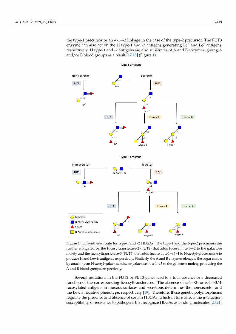
the type-1 precursor or an α-1→3 linkage in the case of the type-2 precursor. The FUT3enzyme can also act on the H type-1 and -2 antigens generating Leb and Ley antigens,respectively. H type-1 and -2 antigens are also substrates of A and B enzymes, giving Aand/or B blood groups as a result [17,18] (Figure 1).
Int. J. Mol. Sci. 2021, 22, x FOR PEER REVIEW 3 of 19
galactose moiety through an α‐1→2 linkage, generating type‐1 and type‐2 H antigens, re‐
spectively. However, if it is the FUT3 enzyme that modifies the precursors, Lea (type‐1
precursor) and Lex (type‐2 precursor) antigens are generated. This modification consists
of the addition of an L‐fucose to N‐acetyl‐glucosamine with an α‐1→4 linkage in the case
of the type‐1 precursor or an α‐1→3 linkage in the case of the type‐2 precursor. The FUT3
enzyme can also act on the H type‐1 and ‐2 antigens generating Leb and Ley antigens,
respectively. H type‐1 and ‐2 antigens are also substrates of A and B enzymes, giving A
and/or B blood groups as a result [17,18] (Figure 1).
Figure 1. Biosynthesis route for type‐1 and ‐2 HBGAs. The type‐1 and the type‐2 precursors are
further elongated by the fucosyltransferase‐2 (FUT2) that adds fucose in α‐1→2 to the galactose
moiety and the fucosyltransferase‐3 (FUT3) that adds fucose in α‐1→3/4 to N‐acetyl‐glucosamine to
produce H and Lewis antigens, respectively. Similarly, the A and B enzymes elongate the sugar
chains by attaching an N‐acetyl‐galactosamine or galactose in α‐1→3 to the galactose moiety, pro‐
ducing the A and B blood groups, respectively.
Figure 1. Biosynthesis route for type-1 and -2 HBGAs. The type-1 and the type-2 precursors arefurther elongated by the fucosyltransferase-2 (FUT2) that adds fucose in α-1→2 to the galactosemoiety and the fucosyltransferase-3 (FUT3) that adds fucose in α-1→3/4 to N-acetyl-glucosamine toproduce H and Lewis antigens, respectively. Similarly, the A and B enzymes elongate the sugar chainsby attaching an N-acetyl-galactosamine or galactose in α-1→3 to the galactose moiety, producing theA and B blood groups, respectively.
Several mutations in the FUT2 or FUT3 genes lead to a total absence or a decreasedfunction of the corresponding fucosyltransferases. The absence of α-1→2- or α-1→3/4-fucosylated antigens in mucous surfaces and secretions determines the non-secretor andthe Lewis negative phenotype, respectively [19]. Therefore, these genetic polymorphismsregulate the presence and absence of certain HBGAs, which in turn affects the interaction,susceptibility, or resistance to pathogens that recognize HBGAs as binding molecules [20,21].
Int. J. Mol. Sci. 2021, 22, 13473 4 of 19
2.1. HBGAs and RV
Despite the fact that a substantial amount of research has been carried out in the lastfew years on the RV mechanisms for host cell attachment, the process is still far from beingunderstood. It is known that the VP4 spike protein (determining the viral P genotype)participates in the process and early studies with some P genotypes from animal originrevealed its interaction with sialic acid, whereas other animal and the human RV were sialicacid-independent and interacted with HBGAs [22,23]. VP4 from RV is post-translationallycleaved into VP8* (glycan-binding domain) and VP5* polypeptides, the VP8* portion beingthe one responsible for cellular attachment and entry, as well as HBGA binding [24]. TheP genotypes would thus determine the pattern of genetic susceptibility. VP8* from P[8],P[4], P[6], P[14], P[11], and P[19] genotypes recognize the secretor HBGAs. RegardingP[8] and P[4] genotypes, there were controversial studies since some determined thatthey bind the Leb and H type-1 [25–28], while others report no Leb binding for thesegenotypes [29,30]. P[6] binds the H1 antigen but was reported not to bind Leb [31], whereasP[19] binds mucin core glycans with the GlcNAc-β-1→6-GalNAc motif and the type-1HBGA precursor [32]. Other studies also documented that P[9], P[14], and P[25] strainsinteract with the A antigen [24,33], whereas P[11] interacts with single and repeated N-acetyl-lactosamine, the type-2 precursor glycan [34]. The HBGAs interactions with VP8*have been investigated by X-ray crystallography in some cases, identifying sugar bindingpockets which are different from the sialic acid binding site identified in animal RV [30].The binding site for N-acetyl-lactosamine and A-antigen in P[11] and P[14] genotypes,respectively, is situated in a cleft between two twisted β-sheets of the typical galectin foldin VP8* [33,34]. A second pocket was identified in P[4], P[6], and P[19] genotypes forlacto-N-fucopentaose I (Fuc-α-1→2-Gal-β-1→3-GlcNAc-β-1→3-Gal-β-1→4-Glc) [30,35]and in P[8] for lacto-N-biose and H type-1 antigen binding [29], which is formed by oneof the β-sheets and a C-terminal α-helix. This pocket is not able to accommodate the Leb
antigen, which contains an extra α-1→4-linked L-fucose. A second glycan-binding sitefor Leb formed by the edges of two β-sheets in the VP8* structure has been identifiedin P[4] and P[8] genotypes and validated by crystallography and NMR techniques [27].This provides evidence that classical techniques to identify the interactions between VP8*and HBGAs (e.g., glycan-binding assays in ELISA-like format) do not always give reliableresults. Therefore, these two viral genotypes possess two glycan-binding sites which mayreflect an adaptation to different host HBGAs polymorphisms.
Several observational studies have investigated the association between the secretorstatus and susceptibility to RV infection in vivo [36–40]. Although some discrepancies havebeen found, most reports have shown that positive secretor status was strongly associatedwith susceptibility to P[8] and P[4] genotypes [36,41,42]. As for serological studies, higherRV-specific immunoglobulin G (IgG) titres in serum and IgA titres in saliva have beenreported in secretors compared to non-secretors [43–46]. Higher anti-RV antibody titresreflect a larger number of previous infections, making it an indirect marker of susceptibility.A study conducted by Sharma et al. showed that human intestinal enteroids isolatedfrom secretor individuals were more susceptible to RV infection as compared with humanintestinal enteroids of a non-secretor individual [8]. Moreover, in experiments performedwith porcine enteroids, it was shown that infection with a P[8] human RV strain wasenhanced by the presence of H and A antigens [47]. It has been determined that the L-fucose moiety of H type-1 glycan in position α-1→2 does not make contact with VP8* andthat the unfucosylated precursor (lacto-N-biose) also binds the P[8] genotype at the samebinding pocket [29]. The lack of protein interactions of the HBGAs L-fucose moiety wasalso reported when analysing the binding of P[9] and P[14] genotypes to the A-antigenby NMR and minimal contacts of L-fucose have been reported for P[4] and P[6] duringlacto-N-fucopentaose I binding [35]. However, the presence of L-fucose increases two-foldthe affinity of P[8] VP8* to the glycan as measured by surface plasmon resonance [29].Interestingly, this increase in affinity mediated by fucose residues that do not interact withthe protein has been also observed in human galectin-3 binding to HBGAs [48]. Whether

Int. J. Mol. Sci. 2021, 22, 13473 5 of 19
this increase in the in vitro affinity might provide an explanation as to why secretor positiveindividuals have a higher susceptibility to RV infections deserves further research. In thisregard, it has to be pointed out that although infection with the P[8) genotype RV takesplace in FUT2-/- individuals, it occurs at lower levels, as determined by measuring specificantibody titres [45], and that the soluble unfucosylated H type-1 antigen precursor (lacto-N-biose) has inhibitory properties against P[8] RV infection in vitro [29].
The lack of minor interaction of L-fucose with VP8* in particular cases has led someauthors to the conclusion that the secretory L-fucose does not play a relevant role in infec-tivity, at least for some P genotypes, which would agree with the epidemiological data [49].Thus, a study by MacDonald et al. suggested that there was no association between secretorstatus and susceptibility to P[6] RV infection since similar proportions amongst secretors(53%) and non-secretors (47%) was observed [50]. Therefore, the controversy about the roleof HBGAs on susceptibility still exists and it is intensified by the fact that, as already men-tioned, different techniques employed to determine the interaction of VP8* from differentP genotypes to HBGAs (i.e., glycan-binding assays, crystallography, or NMR) have usuallyrendered distinct or contradictory results.
The initial steps of RV attachment to cells and cell entry also include protein-proteininteractions, as well as the fusion of membranes mediated by the VP5* portion of VP4. Acomprehensive revision of the steps of RV entry and the triggered signalling pathways hasbeen recently published [22].
2.2. HBGAs and NoV
The P2 subdomain found in the P-domain of the VP1 protein from NoVs interactswith HBGAs. Several studies have been made in order to elucidate the recognition patternof NoVs. Some of them are based on the expression of the P-domain in vitro, which resultsin dimerization (P dimer) and the formation of P particles that retain HBGA-bindingfunction, while others used virus-like particles (VLPs). These studies have utilized ELISAor haemagglutination-based assays using saliva, human milk, red blood cells, or syntheticoligosaccharides as HBGAs sources. The prototype Norwalk virus (GI.1) recognizes thetype A and H secretors, but does not interact with type B secretors and non-secretors;Va387 (GII.4) binds to A, B, and O secretors; MOH (GII.5) and Hiro (GII.12) bind to A andB secretors; and Va207 (GII.9) recognizes Lewis positive secretors and non-secretors (Lex
and Ley) [18,51]. As for the GII.4 strains Den Haag_2006b and Sydney_2012, Carmona et al.demonstrated that they did not recognize any HBGAs [52]. By contrast, these strains mayrecognize heparan sulphate or citrate since they are all capable of binding human NoV andmay potentially play a role in NoV pathogenesis as cellular receptors/co-factors [53]. GI.3NoV VLPs show strong binding to blood type A salivary HBGAs, slightly lower bindingto blood type O salivary HBGAs, and weakly binding or none to blood type B and ABsalivary HBGAs [54].
However, whether the secretor status mediates resistance to NoV infection is yet to besolved. As early as 1977, Parrino et al. observed that some individuals were repeatedlysusceptible to Norwalk virus (GI.1) infection, whereas a second group was repeatedlyresistant. They postulated that a genetic factor might be responsible for susceptibility toinfection [55]. While most studies have shown that non-secretors are protected againstGII.4 infection and disease, exceptions have been found since there is some evidence ofboth asymptomatic and symptomatic infections among non-secretors [56–59]. The reasonsfor this are unknown, but they could be related to several causes, including microbiotadiversity, which would also comprise HBGA-expressing bacteria, differences betweenGII.4 variants, general health status, weak-secretor phenotype, or other unidentified hostfactors. Interestingly, Lin et al. described that secretor patients have prolonged diarrhea,more frequent vomiting, more severe disease, and greater infection transmissibility thannon-secretors [60].
Int. J. Mol. Sci. 2021, 22, 13473 6 of 19
3. The Role of Bacteria in RVs and NoVs Infection: Studies with Cultured Cells andAnimal Models
A large and diverse population of commensal microbes consisting of bacteria, viruses,fungi, and parasites inhabit the gastrointestinal tract. NoV and RV, being enteric pathogens,interact with them, resulting in outcomes either beneficial or detrimental to the host [61–63].The coevolution of the commensal microbiota and their host has resulted in a mutuallybeneficial condition in which the host can benefit from physiological, metabolic, andimmunological regulations provided by the microbiota, while the commensal microbiotadepends absolutely on the host for nutrient acquisition and propagation sites. In theregulation of viral infection, commensal microbiota can promote inhibitory effects or viralinfectivity through diverse mechanisms [64,65].
3.1. Bacteria against Enteric Viral Infections
Several studies demonstrate the beneficial effect of probiotic bacteria against entericvirus infections and many other diseases [66–69]. Probiotics protect the host from viralinfection by modulating gut microbiota composition, enhancing intestinal barrier function,and promoting mucosal immunity [70]. Additionally, they interfere with the binding of thevirus to their target cells by competitive exclusion by blocking viral receptors and bindingviruses on the surface to promote their elimination in faeces [71].
The presence of Bifidobacterium adolescentis inhibits the attachment of human NoV(hNoV) GI.1 VLPs to epithelial cells in vitro [72]. Similarly, Lacticaseibacillus casei and Es-cherichia coli Nissle 1917 impaired the attachment of GI.1 P-particles to HT-29 cells [73].In another study, gnotobiotic pigs colonized with Lacticaseibacillus rhamnosus GG and Es-cherichia coli Nissle 1917 were infected with human norovirus from the GII.3 and GII.4genotypes, and a virus faecal shedding below the limit of detection was observed, indicat-ing significant inhibition on hNoV infection by the colonization of such bacteria [74].
As for RV, Escherichia coli Nissle 1917 seemed to reduce diarrhea in gnotobiotic pigsby modulating an immune response [75–78]. Bacterial flagellin is efficient against RVinfection since it induces the production of IL-22 and IL-18 [79]. It has also been provedthat Ruminococcus gauvreauii, a bacterium that has been isolated from human bile and istherefore likely present at the site of RV infection (the small intestine) can bind RV [80].This binding might be mediated by HGBA-like substances that are present on the bacterialsurfaces. Further experiments employing Caco-2 cells demonstrated that R. gauvreauiiinterferes with RV infection in vitro since a threefold decrease in viral infectivity was foundin its presence, demonstrating the anti-RV effect of this bacterium [80].
3.2. Microbiota and Promotion of Enteric Viral Infections
Despite the significant evidence available about the role of intestinal-derived bacteriain the inhibition of viral infections, several investigations argued for a role of micro-biota in promoting virus infection [81]. This was first demonstrated by Kuss et al. [82]and Kane et al. [83] when using poliovirus, reovirus, and mouse mammary tumour virus(MMTV) for infecting germ-free or antibiotic-treated mice. In these cases, it was ob-served that a substantial attenuation of infection occurred when compared to infection ofmicrobially-colonized mice. Reconstitution of intestinal microorganisms into antibiotic-treated mice was enough to restore poliovirus pathogenesis [82]. Moreover, intestinaltitres of reovirus were substantially reduced in antibiotic-treated, compared with controlmice [83]. Similar findings were reported with RV and NoV when antibiotic-treated orgerm-free mice were used [84,85], suggesting that microbiota enhances the pathogenesis ofmultiple families of enteric viruses.
The intestinal microbiota can directly facilitate enteric virus replication by severalmechanisms, including stabilization of virions and promotion of virus attachment to hostcells. Indirectly, it enhances the infection of enteric viruses by altering the antiviral immuneresponse [86]. Several enteric viruses that benefit from the microbiota bind bacterialsurface polysaccharides, resulting in enhanced viral infectivity and pathogenesis. When
Int. J. Mol. Sci. 2021, 22, 13473 7 of 19
poliovirus and other members of the Picornaviridae family bind to lipopolysaccharide(LPS), a component of the Gram-negative bacterial wall, an increase in thermostability andresistance to inactivation at elevated temperatures and in the presence of dilute chlorinebleach can be observed [87]. LPS can also promote poliovirus attachment to the surface oftarget cells by facilitating viral binding to its receptor [88]. MMTV can bind LPS as well,which stimulates TLR4, initiating a signalling pathway that results in the production of theimmunosuppressive cytokine IL-10, generating a tolerogenic environment that allows viralpersistence [89].
There is also evidence that suggests that RV and NoV infection is facilitated by micro-biota. Interactions of NoV with members of the intestinal microbiota have been demon-strated, including Enterobacter cloacae, Escherichia coli, and Helicobacter pylori [85,90,91].These interactions are mediated via HBGA-like carbohydrates expressed on the surface ofthese bacteria [92,93], although NoV has been reported to bind additional carbohydrateresidues widely expressed on microbiota [53]. In vitro, human NoV is able to infect Bcells in the presence of HBGA-coated bacteria, and a reduction of viral replication wasobserved in this model if bacteria were not present. Infection of B cells is restored if cellsare incubated with Enterobacter cloacae, suggesting that the binding of viral particles toHBGA-coated bacteria enables uptake of the virus into the host cells [85]. It was alsoobserved that HBGA-expressing bacteria, such as some E. coli strains, protect NoV VLPsduring heat treatment, such as the one accomplished during food processing, facilitatingtheir transmission [93]. Experiments performed in gnotobiotic pigs with transplantedhuman intestinal microbiota showed that replication of the human NoV GII.4/2006b strainwas stimulated by the microbiota, which increased shedding titres and duration [94]; ex-periments in antibiotic-treated mice demonstrated that microbiota eradication prevents thepersistent infection of the murine NoV CR6 strain [95]. It was suggested that the intestinalmicrobiota limits the IFNλ-dependent innate immunity, allowing NoV persistence. Theeffects of antibiotics were restricted to the intestine, because when CR6 was administeredintraperitoneally or in mice lacking IFNα and IFNβ receptors, viral levels in mesentericlymph nodes and spleen did not change with respect to control mice [95].
3.3. Microbiota and Restriction of Enteric Viral Infections
Recently, it has been shown that microbiota ablation with antibiotics in mice allowsfor infection with the human RV strain Wa (G1P[8]), which replicates very inefficiently inanimals with normal microbiota [96]. These results are in conflict with earlier experimentswhich demonstrated that microbiota eradication by antibiotics results in reduced infectionof murine RV (EC strain), as shown by lower viral shedding in adult mice and diminisheddiarrhea incidence in mice pups [84]. Nevertheless, viral clearance lasted longer in thismodel. Furthermore, recent experiments with the murine EDIM strain confirmed thatantibiotic treatment and the consequent decrease in intestinal bacterial loads or the use ofgerm-free mice results in increased RV infection [97], which argues against a positive effectof the microbiota in RV infection. In the experiments with the Wa strain, animals with ab-lated microbiota and subsequent subjection to self-transplantation of intestinal microbiotapartially recovered the resistance to infection, which allowed the identification of bacterialtaxa that likely participate indirectly or directly in the restriction of Wa infection in mice [96].Thus, bacteria belonging to lactobacilli, Mucispirillum, Oscillospira, and Bilophila generawere negatively linked to RV infection in mice. Faecal material transplantation with infantsas donors did not restrict infectivity in this model, suggesting that the microbiota from thedonors was not able to control RV infection in this model and that mice autochthonousbacteria were needed for the process [96]. Although differences depending on the hostand viral strains cannot be excluded, all these data point to the microbiota as a majorfactor limiting RV replication. In this sense, other studies have also determined the roleof specific bacterial taxa from the intestinal microbiota in viral replication. Thus, Shi et al.discovered that the elevated RV resistance of certain colonies of Rag1-KO mice (lacking Band T lymphocytes), which usually tend to develop chronic RV infection, to the murine EC
Int. J. Mol. Sci. 2021, 22, 13473 8 of 19
strain was due to elevated levels of colonization by Candidatus Arthromitus. This bacteriumis a member of the segmented filamentous bacteria (SFB), which are typical in mice but canreach elevated numbers in the immunocompromised Rag1-KO strain [98]. SFB participatedin the exclusion of RV from mice by processes that involve direct RV/bacteria contact andother mechanisms that are probably based on an increased enterocyte turnover triggeredby SFB.
Data about the negative effects of the microbiota on NoV infectivity are scarcer. Epi-demiological studies suggest that supplements of vitamin A had an anti-NoV effect. Studieswith murine NoV showed an increased population of intestinal Lactobacillus sp. in miceafter vitamin A supplementation, and it was postulated that the antiviral effects of thesebacteria, which were demonstrated in vitro on RAW264.7 cells, account for reduced NoVinfection [99]. However, as mentioned in the last section, the use of other animal modelsand NoV points to a positive effect of the microbiota in the replication of this viral group.However, it has to be noted that more recent and detailed analysis with antibiotic-treatedmice shows that, while the microbiota enhances murine NoV strain MNV-1 infection indistal regions of the intestine, it restricts infectivity in the proximal small intestine [100].This regionalisation of the effects is mediated by a distinct abundance of bile acid receptorsdepending on the intestinal location, which are involved in triggering an anti-NoV IFNλ
response that was enhanced by bacteria in the proximal intestine. Microbial modificationof bile acids in the small intestine thus has an effect on murine NoV infection at differentintestine locations. Inoculation of antibiotic-treated animals with Clostridium scindens, abacterium known to transform primary bile acids into secondary bile acids (that were low-ered in antibiotic-treated animals), restored viral inhibition in the proximal small intestine,although it did not enhance infection in the distal gut [100].
It appears that specific members of the microbiota possess restrictive traits to entericviral infection, at least for RV, whereas evidence for NoV is weaker. Immune regulationis emerging as the mechanism underlying this phenomenon and it is extended to virusesthat do not target the gut. Specific bacterial taxa have also been described in the preventionof viral diseases such as encephalomyocarditis virus (EMCV) systemic infection in mice,in which antibiotic treatment also exacerbated infection [101]. Contrarily to other intesti-nal bacteria tested, Blautia coccoides (a former member of the Ruminococcus genus) wasidentified among members of the gut microbiota as a bacterium able to restrict systemicEMCV replication in monocolonized mice. In animals carrying B. coccoides, the capacity ofmacrophages for inducing IFNβ, which protects against EMCV, was restored [101]. Type IIFN (IFNβ) plays a pivotal role in the response against viral pathogens. Lack of bacterialeads to a weaker innate immune response which is characterized by low expression ofIFNβ, which hampers an effective macrophage antiviral response [102]. Several bacte-rial components or derived metabolites are involved in priming host innate immunityagainst respiratory viruses through IFNβ production, such as the lipo-oligosaccharidesfrom Bacteroides in influenza infection [103] or acetate (and propionate or butyrate) fromgut microbial metabolism, which is per se able to induce an IFNβ response in the lungs ofmice and protect them from the respiratory syncytial virus when supplemented in drinkingwater [103].
The above-presented studies confirm the influence that the microbiota has over entericviral infections, as well as over other non-enteric viruses. However, the mechanisms bywhich this takes place are not well-understood yet for RV and NoV, and new studies areneeded in order to gain knowledge about them. Similar to NoV, few examples about thedisclosure of in vivo mechanisms of microbiota restriction of infectivity are known in RV.TLR4, recognizing bacterial flagellins, and the NLR-C4 component of the inflammasomeare crucial for the production of IL-22 and IL-18, which are important in protection againstRV [79]. In the studies conducted by Schnepf et al., a pivotal role for IL-22 induced bythe microbiota in its limiting effects against RV infection was also established [97]. It wasshown that microbiota depletion resulted in reduced IL-22 production, and that protectionagainst RV can be achieved by IL-22 administration. In this case, IL-22-mediated protection
Int. J. Mol. Sci. 2021, 22, 13473 9 of 19
did not involve IFN, because it was also found in mice lacking the transcriptional factorSTAT1, which increases the expression of interferon-stimulated genes [97]. Mice treatedwith antibiotics in which the human RV Wa strain was able to replicate also presentedalterations in the expression of genes related to the immune and inflammatory response,such as IL-1β and CXCL15, and the FUT2 enzyme [96], but the relevance of these facts inRV infection has to be evaluated.
Another plausible mechanism that the microbiota could use to restrict infection impliessecretory immunoglobulin A (sIgA). The sIgA molecules are secreted into the intestinallumen, where they attach antigens and act as the first barrier of mucosal defence. Thegut microbiota has been shown to be able to regulate IgA production, and the level ofIgA in the gut is considerably decreased in germ-free mice [104]. Other ways by whichmicrobiota members could mediate RV protection include direct attachment of RV particlesto bacteria, as hypothesized for SFB [46], or have been described for some probiotics. Theseinteractions may be promoted by HBGA-like molecules that can be presented on bacterialsurfaces [92]. The consequences derived from this interaction may differ between diverseviral groups and while some enteric viruses, such as NoV or poliovirus, may benefit fromthem via enhancement of virion stability or target cell attachment [82,85,93], they can alsomediate virus sequestration on the bacterial surface and/or competition with the viralbinding molecules present at the surface of host cells.
4. Microbiota and Enteric Viruses, Studies in Humans
Very few studies have addressed the relationships between gut microbiota and theinfection of enteric viruses in humans. Most of the results linking different bacterial taxato viral infection are derived from vaccination trials, in which the intestinal microbiotaanalyses have been linked to vaccine outcomes (i.e., RV vaccines (RVVs)). Microbiotacomposition varies depending on the population [105] since it is affected by many factorsincluding nutrition [106], sex, age, genetics, and health status [107], and these vary greatlybetween low-income and high-income countries. Such differences could be some of thereasons why RVVs have significantly lower efficacy in low-income countries [108,109].
However, another important reason that could explain such differences is relatedto the prevalence of Lewis negative individuals in Asia, Latin America, and Africancountries [58,110], where P[6] is the prevalent genogroup [41] since it recognizes Lewisnegative antigens [31]. Moreover, RVVs do not include the P[6] genotype, so this couldexplain the lower RVVs efficacy in those areas.
RVVs have dramatically reduced the morbidity and mortality of AGE caused byRV infection [3]. Understanding the mechanisms implicated in the reduced efficacy ofRRVs is relevant since even small improvements in vaccine efficacy might increase thenumber of children’s lives saved by hundreds of thousands during the coming years [111].Although the reasons for variations in efficacy are not fully understood, they are thoughtto be differences in co-infections with other enteropathogens at the time of vaccination,gut microbiota composition, and HBGAs genotype [107]. Studies in Africa and Asia usingRotarix and Rotateq vaccines have been conducted in order to elucidate gut microbiotadifferences (by means of 16S rDNA sequencing) between RVVs responders and non-responders. Rotarix consists of a human attenuated single strain (G1P[8]) and Rotateqincludes five bovine-human reassortant strains (G1, G2, G3, G4, and P[8]) [6].
Harris et al., using the Rotarix vaccine, demonstrated that intestinal microbiota differssignificantly between RVVs responders and non-responders in Ghana [112]. Responderswere considered the ones that had anti-RV IgA antibodies ≥20 IU/mL after vaccination.The study found that children that responded to RVVs had abundant counts of bacteriafrom the Bacilli phylum, especially Streptococcus bovis, while the non-responders presentedabundant numbers of Bacteroidetes phylum, specifically Bacteroides and Prevotella species.Moreover, the study showed that the Enterobacteria/Bacteroidetes ratio was significantlyhigher in vaccine responders as compared to non-responders. In addition, responders hadmore microbiota similarities with Dutch children (assumed to be RVVs responders, in line
Int. J. Mol. Sci. 2021, 22, 13473 10 of 19
with clinical trial data demonstrating a >90% RVVs seroconversion rate in northern Euro-pean countries) than with non-responders [112]. This group conducted similar researchwith Pakistani infants, also concluding that microbiota varies significantly between RVVsresponders and non-responders. They determined that the relative abundance of Gram-negative bacteria such as Serratia spp. and Escherichia coli correlated positively with RVVsresponse as compared to non-responders [113]. Researches hypothesised that differencesin RVVs efficacy are due to Bacteroides, present in more abundance in non-responders sincethey have LPS that differs from that present in Enterobacteriacae. LPS from Bacteroides specieshave been demonstrated to inhibit the stimulation of inflammatory cytokines in vitro usingthe LPS from Enterobacteriacae as a reference [111]. In a similar way, a relative abundanceof flagellin-producing bacteria may enhance the innate and subsequent adaptive immuneresponses to RVVs [79]. In opposition to this, in a study carried with children that hadreceived Rotarix in Zimbabwe and where a very low percentage of vaccine take was ob-served, faecal microbiota analyses showed that Bacteroides thetaiotaomicron was the onlybacterium that correlated with high specific IgA titres (responders) [114]. Another hypothe-sis, given that vaccines contain a live attenuated virus, is that bacteria present in respondersmight be expressing HBGAs or glycans needed for RV replication [24]. However, otherstudies conducted with Indian infants showed no significant differences in microbiotacomposition between responders and non-responders [115]. The researchers hypothesizedthat the discrepancies in both studies regarding differences in microbiota compositionbetween responders and non-responders could be due to differences in methodology (next-generation sequencing versus microarray) or baseline microbiota composition. As for theRotateq vaccine, a study carried out with Nicaraguan children determined no statisticallysignificant differences in the microbiome composition between RVVs responders and non-responders [28]. However, the sample size of these studies is small, so further research isadvisable in order to have more reliable results.
A recent study evaluated whether microbiota modification by the use of broad- andnarrow-spectrum antibiotics had an effect on immunization with Rotarix in adults [116].Although the experimental groups did not differ in terms of total IgA produced, the narrowspectrum group showed a boost in IgA at day seven post-vaccination (basal levels of anti-RV IgA were high in the vaccination group) and the viral shedding was increased in bothgroups treated with antibiotics. Differences in the microbiota composition in faeces wereevident between the groups and correlations between enrichment in Bacteroides populationsat the boost at day seven were observed and several taxa (Prevotellaceae, Cloacibacilluseverynsis, and Proteobacteria members such as Escherichia and Shigella) were associatedwith increased viral shedding. Antibiotic treatment had no effect on the immunogenicityof other systemic vaccines applied (pneumococcal and tetanus vaccine) [116]. These resultshighlight the fact that targeting the microbiota could be an alternative strategy to enhanceRVVs efficacy, although the effectiveness in children still needs further investigation.
None of the above discussed studies considered the secretor status in the vaccineefficacy. Other studies determined that anti-RV IgA seroconversion rates after Rotarixvaccination differed significantly depending on salivary HBGA phenotype, having thelowest rate of seroconversion (non-responders) infants who were non-secretors [117–119].This finding is consistent with in vitro data, which demonstrated that P[8] strains inter-acted with H type 1 antigens [29] (and Leb depending on the author [26,27]; these two arecarbohydrates expressed only in individuals with functional FUT2 alleles). Thus, differ-ences in HBGA expression may be responsible for some of the discrepancies in the level ofprotection detected for RVVs in low-income and high-income countries. Other studies haveapplied 16S rDNA sequencing to analyse the intestinal microbiota of groups of volunteers,examining the susceptibility to RV and NoV measured as the level of salivary NoV andRV-specific IgA, and performing FUT2 genotyping [45]. The results showed that all threefactors (gut microbiota, FUT2 genotype, and susceptibility to RV and NoV) are intercon-nected. It was also found that certain bacterial genera, such as Ruminococcus, correlatednegatively with the susceptibility to RV and NoV, while Akkermansia, an intestinal mucin

Int. J. Mol. Sci. 2021, 22, 13473 11 of 19
degrader, correlated positively with RV IgA titres [45]. In mice pups infected with RV,a shift in the ileal microbial populations was observed, with increased levels of mucindegraders such as Akkermansia and Bacteroides [120]. It was postulated that the observed re-lease of mucin during infection may favour this species, whose glycan-degrading activitieson mucin create in turn a glycan environment more favourable for RV infection [120].
An ex vivo study analysed the bacterial groups that were interacting with RV instool samples from children suffering RV (G1P[8]) diarrhea by flow cytometry followed by16S rDNA sequencing [80]. This study also allowed the identification of Ruminococcus asRV-interacting bacteria. As already mentioned, a species of this group (R. gauvreauii) wasshown to inhibit RV infection in vitro [80]. This, together with the correlation Ruminococcus-anti-RV IgA in humans and the fact that higher Ruminococcus numbers are found in healthychildren compared to children with RV diarrhea [121], postulates these bacteria as likelyplayers in the cross-talk bacteria-virus-host. Similar studies conducted with individualssuffering AGE caused by NoV will certainly aid in identifying bacterial taxons that interactwith these viruses in stools. However, whether this interaction has some relevance in theinfection process needs further investigation.
All these findings may help to improve RVVs performance in such a way that theyhave higher efficacy in low-income countries, preventing tens of thousands of RV-relateddeaths per year. However, differences in the conclusions drawn from the microbiotaanalyses are evident, and standardized and controlled methods (e.g., sampling and DNAextraction techniques, bacterial 16S rDNA sequencing platforms, microbial compositionanalysis methods, etc.) are needed to get a clearer picture.
Regarding vaccines, a different situation is found for NoV. Since up to now there is noNoV vaccine available, differences in vaccine efficacy depending on differences in micro-biota composition cannot be studied. Although no NoV vaccine is commercially available,a few of them are in clinical trials [11]. The candidate furthest along in the developmentpipeline is developed by Takeda Pharmaceuticals. It is a bivalent (genotypes GI.1/GII.4),intramuscular VLP vaccine, currently in phase IIb. The vaccine failed to significantlyprevent acute gastroenteritis. However, it reduced severe diarrhea and vomiting [122].This bivalent vaccine was well tolerated and immunogenic, and the antibodies generatedelicited HBGA-binding blocking activity [122]. As for secretor status, secretor and non-secretor individuals responded similarly to the first dose of vaccine [123]. Such a geneticdifference in the small intestine is unlikely to have a large impact on vaccine immuno-genicity, since most VLP-based vaccines are designed for parenteral administration, thusavoiding the mucosa.
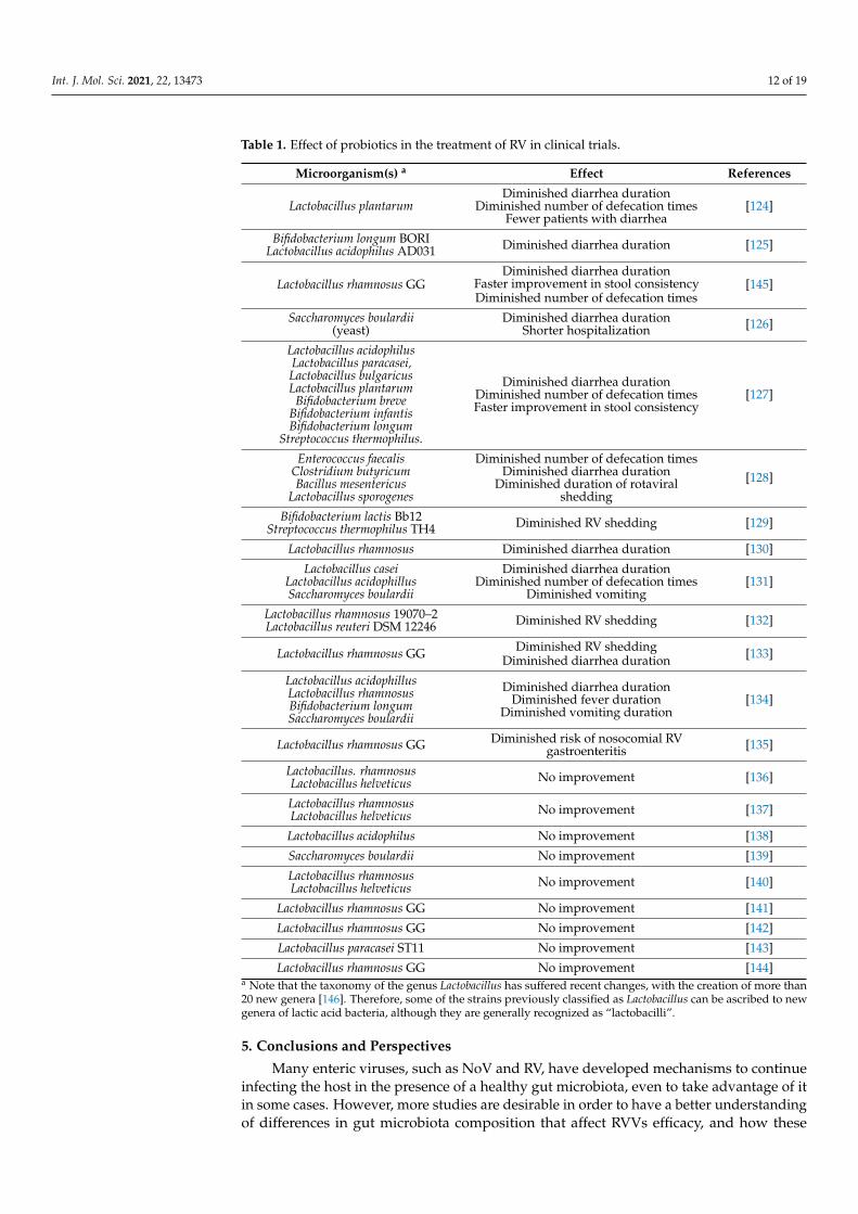
Some clinical trials have studied the relationship between probiotic bacteria and theirinfluence on enteric virus infections. While in vitro assays and studies in animal modelshave helped to determine probiotic strains with antiviral activity that can be useful in thetreatment of RV infections, there is a large controversy in terms of its beneficial effects inhumans. Few clinical trials have studied the influence of probiotics in RV infections andmany differences can be found. Some of them determined that probiotic treatment forpatients with RV-related diarrhea produces shorter diarrhea duration, less RV shedding,faster improvement in stool consistency, and fewer defecation times [124–133], whileonly two of them found vomiting reduction [130,134]. One of them even concludes thatprobiotics reduce the risk of nosocomial RV gastroenteritis [135]. Contrarily, almost halfof the analysed clinical trials determined that the intake of probiotics does not produceany improvement in RV-related diarrhea symptoms [136–144] (Table 1). The number ofsubjects enrolled in these clinical trials, the probiotic used, application methods, doses,and the way in which the effects are measured are possible factors affecting the results, forwhich, again, more standardized and controlled trial conditions are required to assess theefficacy of probiotics in viral AGE.
Int. J. Mol. Sci. 2021, 22, 13473 12 of 19
Table 1. Effect of probiotics in the treatment of RV in clinical trials.
Microorganism(s) a Effect References
Lactobacillus plantarumDiminished diarrhea duration
Diminished number of defecation timesFewer patients with diarrhea
[124]
Bifidobacterium longum BORILactobacillus acidophilus AD031 Diminished diarrhea duration [125]
Lactobacillus rhamnosus GGDiminished diarrhea duration
Faster improvement in stool consistencyDiminished number of defecation times
[145]
Saccharomyces boulardii(yeast)
Diminished diarrhea durationShorter hospitalization [126]
Lactobacillus acidophilusLactobacillus paracasei,Lactobacillus bulgaricusLactobacillus plantarum
Bifidobacterium breveBifidobacterium infantisBifidobacterium longum
Streptococcus thermophilus.
Diminished diarrhea durationDiminished number of defecation timesFaster improvement in stool consistency
[127]
Enterococcus faecalisClostridium butyricumBacillus mesentericus
Lactobacillus sporogenes
Diminished number of defecation timesDiminished diarrhea duration
Diminished duration of rotaviralshedding
[128]
Bifidobacterium lactis Bb12Streptococcus thermophilus TH4 Diminished RV shedding [129]
Lactobacillus rhamnosus Diminished diarrhea duration [130]
Lactobacillus caseiLactobacillus acidophillusSaccharomyces boulardii
Diminished diarrhea durationDiminished number of defecation times
Diminished vomiting[131]
Lactobacillus rhamnosus 19070–2Lactobacillus reuteri DSM 12246 Diminished RV shedding [132]
Lactobacillus rhamnosus GG Diminished RV sheddingDiminished diarrhea duration [133]
Lactobacillus acidophillusLactobacillus rhamnosusBifidobacterium longumSaccharomyces boulardii
Diminished diarrhea durationDiminished fever duration
Diminished vomiting duration[134]
Lactobacillus rhamnosus GG Diminished risk of nosocomial RVgastroenteritis [135]
Lactobacillus. rhamnosusLactobacillus helveticus No improvement [136]
Lactobacillus rhamnosusLactobacillus helveticus No improvement [137]
Lactobacillus acidophilus No improvement [138]
Saccharomyces boulardii No improvement [139]
Lactobacillus rhamnosusLactobacillus helveticus No improvement [140]
Lactobacillus rhamnosus GG No improvement [141]
Lactobacillus rhamnosus GG No improvement [142]
Lactobacillus paracasei ST11 No improvement [143]
Lactobacillus rhamnosus GG No improvement [144]a Note that the taxonomy of the genus Lactobacillus has suffered recent changes, with the creation of more than20 new genera [146]. Therefore, some of the strains previously classified as Lactobacillus can be ascribed to newgenera of lactic acid bacteria, although they are generally recognized as “lactobacilli”.
5. Conclusions and Perspectives
Many enteric viruses, such as NoV and RV, have developed mechanisms to continueinfecting the host in the presence of a healthy gut microbiota, even to take advantage of itin some cases. However, more studies are desirable in order to have a better understandingof differences in gut microbiota composition that affect RVVs efficacy, and how these
Int. J. Mol. Sci. 2021, 22, 13473 13 of 19
differences impact possible anti- and pro-viral mechanisms. Therefore, identification of keybacteria that correlate with RVVs efficacy could be important for designing future vaccinesin countries where RVVs have less effectivity [147]. Such bacteria could be also used asbiomarkers for vaccine efficacy and interventions that modify the microbiota compositionin order to increase it could be envisaged [147].
As for NoV, there is still controversy regarding the role of secretor status in NoVinfection. New experiments based on human enteroid models that mimic the humanintestinal epithelium could be performed. Therefore, libraries of enteroids generated fromindividuals with different FUT2, FUT3, and ABO polymorphisms may provide importantinformation on how secretor, Lewis status, and other HBGAs affect NoV infection. There isalso a great need for the development of the NoV vaccine. The one being developed byTakeda Pharmaceuticals is currently in phase IIb, and it is based on VLPs. If attenuatedNoV vaccines are developed, testing whether their efficacy varies depending on hostglycobiology and microbiota will be necessary.
It can be concluded that this is a fast-evolving research field where the complexinteractions between the enteric pathogens RV and NoV with the host glycobiology andthe gut microbiota are starting to be elucidated. The knowledge earned about theseinteractions will allow the scientific community to improve the prevention strategiesagainst these viruses as well as to design novel therapeutic approaches for the managementof infected patients.
Author Contributions: Conceptualization, V.M., J.B. and J.R.-D.; writing—original draft preparation,N.P.-G., C.S.-B., V.M. and R.G.-R.; writing—review and editing, N.P.-G., C.S.-B., R.G.-R., J.B., V.M. andJ.R.-D.; supervision, V.M. and J.R.-D.; project administration, V.M. and J.R.-D.; funding acquisition,J.B. and J.R.-D. All authors have read and agreed to the published version of the manuscript.
Funding: This research was supported by Spanish Ministry of Science and Innovation (MICIN)/SpanishState Research Agency (AEI)/10.13039/ 501100011033, and by “ERDF A way of making Europe”grants AGL2017-84165-C2-2-R and PID2020-115403RB-C22 to J.R.-D., C.S.-B. is the recipient of a pre-doctoral grant RE2018-083315 funded by MCIN/AEI/ 10.13039/501100011033 and by “ESF Investingin your future”. This work was also supported by Valencian Government grant IDIFEDER/2018/056.N.P.-G. is the recipient of a predoctoral grant from the Valencian Government ACIF/2020/085. R.G.-R.is the recipient of a postdoctoral grant from the Valencian Government APOST/2017/037.
Conflicts of Interest: The authors declare no conflict of interest.
References1. World Health Organization (WHO). The Top 10 Causes of Death. Available online: http://www.who.int/en/news-room/fact-
sheets/detail/the-top-10-causes-of-death (accessed on 10 September 2021).2. Donaldson, E.F.; Lindesmith, L.C.; Lobue, A.D.; Baric, R.S. Norovirus pathogenesis: Mechanisms of persistence and immune
evasion in human populations. Immunol. Rev. 2008, 225, 190–211. [CrossRef]3. Tate, J.E.; Burton, A.H.; Boschi-Pinto, C.; Parashar, U.D.; Agocs, M.; Serhan, F.; De Oliveira, L.; Mwenda, J.M.; Mihigo, R.;
Ranjan Wijesinghe, P.; et al. Global, Regional, and National Estimates of Rotavirus Mortality in Children <5 Years of Age,2000–2013. Clin. Infect. Dis. 2016, 62, S96–S105. [CrossRef]
4. Rota Council. Global Introduction Status. Available online: http://rotacouncil.org/vaccine-introduction/global-introduction-status/ (accessed on 10 September 2021).
5. Rota Council. Available Rotavirus Vaccine Products. Available online: https://preventrotavirus.org/vaccine-evidence/available-rotavirus-vaccine-products/ (accessed on 10 September 2021).
6. Cárcamo-Calvo, R.; Muñoz, C.; Buesa, J.; Rodríguez-Díaz, J.; Gozalbo-Rovira, R. The rotavirus vaccine landscape, an update.Pathogens 2021, 10, 520. [CrossRef] [PubMed]
7. Ramani, S.; Hu, L.; Venkataram Prasad, B.V.; Estes, M.K. Diversity in Rotavirus-Host Glycan Interactions: A “Sweet” Spectrum.Cell. Mol. Gastroenterol. Hepatol. 2016, 2, 263–273. [CrossRef]
8. Sharma, S.; Hagbom, M.; Svensson, L.; Nordgren, J. The Impact of Human Genetic Polymorphisms on Rotavirus Susceptibility,Epidemiology, and Vaccine Take. Viruses 2020, 12, 324. [CrossRef] [PubMed]
9. Dóró, R.; László, B.; Martella, V.; Leshem, E.; Gentsch, J.; Parashar, U.; Bányai, K. Review of global rotavirus strain prevalencedata from six years post vaccine licensure surveillance: Is there evidence of strain selection from vaccine pressure? Infect. Genet.Evol. 2014, 28, 446–461. [CrossRef] [PubMed]
Int. J. Mol. Sci. 2021, 22, 13473 14 of 19
10. CDC. Norovirus Worldwide Global Trends. Available online: https://www.cdc.gov/norovirus/trends-outbreaks/index.html(accessed on 22 November 2021).
11. Lopman, B. Global Burden of Norovirus and Prospects for Vaccine Development. Available online: https://www.cdc.gov/norovirus/downloads/global-burden-report.pdf (accessed on 18 November 2021).
12. Vinjé, J. Advances in Laboratory Methods for Detection and Typing of Norovirus. J. Clin. Microbiol. 2015, 53, 373–381. [CrossRef][PubMed]
13. Doerflinger, S.Y.; Weichert, S.; Koromyslova, A.; Chan, M.; Schwerk, C.; Adam, R.; Jennewein, S.; Hansman, G.S.; Schroten, H.Human Norovirus Evolution in a Chronically Infected Host. mSphere 2017, 2, e00352-16. [CrossRef]
14. Chhabra, P.; de Graaf, M.; Parra, G.I.; Chan, M.C.-W.; Green, K.; Martella, V.; Wang, Q.; White, P.A.; Katayama, K.; Vennema, H.;et al. Updated classification of norovirus genogroups and genotypes. J. Gen. Virol. 2019, 100, 1393–1406. [CrossRef]
15. Jiang, X.; Liu, Y.; Tan, M. Histo-blood group antigens as receptors for rotavirus, new understanding on rotavirus epidemiologyand vaccine strategy. Emerg. Microbes Infect. 2017, 6, e22. [CrossRef]
16. Shanker, S.; Czakó, R.; Sapparapu, G.; Alvarado, G.; Viskovska, M.; Sankaran, B.; Atmar, R.L.; Crowe, J.E.; Estes, M.K.;Prasad, B.V.V. Structural basis for norovirus neutralization by an HBGA blocking human IgA antibody. Proc. Natl. Acad. Sci. USA2016, 113, E5830–E5837. [CrossRef]
17. Marionneau, S.; Cailleau-Thomas, A.; Rocher, J.; Le Moullac-Vaidye, B.; Ruvoën, N.; Clément, M.; Le Pendu, J. ABH and Lewishisto-blood group antigens, a model for the meaning of oligosaccharide diversity in the face of a changing world. Biochimie 2001,83, 565–573. [CrossRef]
18. Tan, M.; Jiang, X. Histo-blood group antigens: A common niche for norovirus and rotavirus. Expert. Rev. Mol. Med. 2014, 16, e5.[CrossRef] [PubMed]
19. Barbé, L.; Le Pendu, J.; Echasserieau, K.; Ruvoën-Clouet, N.; Bernardeau, K.; Le Moullac-Vaidye, B.; Bovin, N.; Nordgren, J.;Carton, T.; Svensson, L. Histo-blood group antigen-binding specificities of human rotaviruses are associated with gastroenteritisbut not with in vitro infection. Sci. Rep. 2018, 8, 12961. [CrossRef]
20. Marionneau, S.; Ruvoën, N.; Le MoullacVaidye, B.; Clement, M.; CailleauThomas, A.; RuizPalacois, G.; Huang, P.; Jiang, X.;Le Pendu, J. Norwalk Virus binds to histo-blood group antigens present on gastroduodenal epithelial cells of secretor individuals.Gastroenterology 2002, 122, 1967–1977. [CrossRef]
21. Ayouni, S.; Sdiri-Loulizi, K.; de Rougemont, A.; Estienney, M.; Ambert-Balay, K.; Aho, S.; Hamami, S.; Aouni, M.;Neji-Guediche, M.; Pothier, P.; et al. Rotavirus P[8] infections in persons with secretor and nonsecretor phenotypes, Tunisia.Emerg. Infect. Dis. 2015, 21, 2055–2058. [CrossRef]
22. Arias, C.F.; López, S. Rotavirus cell entry: Not so simple after all. Curr. Opin. Virol. 2021, 48, 42–48. [CrossRef] [PubMed]23. Böhm, R.; Fleming, F.E.; Maggioni, A.; Dang, V.T.; Holloway, G.; Coulson, B.S.; Von Itzstein, M.; Haselhorst, T. Revisiting the role
of histo-blood group antigens in rotavirus host-cell invasion. Nat. Commun. 2015, 6, 5907. [CrossRef]24. Liu, Y.Y.; Huang, P.; Tan, M.; Biesiada, J.; Meller, J.; Castello, A.A.; Jiang, B.; Jiang, X. Rotavirus VP8*: Phylogeny, host range, and
interaction with histo-blood group antigens. J. Virol. 2012, 86, 9899–9910. [CrossRef]25. Xu, S.; McGinnis, K.R.; Liu, Y.; Huang, P.; Tan, M.; Stuckert, M.R.; Burnside, R.E.; Jacob, E.G.; Ni, S.; Jiang, X.; et al. Structural
basis of P[II] rotavirus evolution and host ranges under selection of histo-blood group antigens. Proc. Natl. Acad. Sci. USA 2021,118, e2107963118. [CrossRef]
26. Huang, P.; Xia, M.; Tan, M.; Zhong, W.; Wei, C.; Wang, L.; Morrow, A.; Jiang, X. Spike protein VP8* of human rotavirus recognizeshisto-blood group antigens in a type-specific manner. J. Virol. 2012, 86, 4833–4843. [CrossRef]
27. Xu, S.; Ahmed, L.U.; Stuckert, M.R.; McGinnis, K.R.; Liu, Y.; Tan, M.; Huang, P.; Zhong, W.; Zhao, D.; Jiang, X.; et al. Molecularbasis of P[II] major human rotavirus VP8* domain recognition of histo-blood group antigens. PLoS Pathog. 2020, 16, e1008386.[CrossRef]
28. Fix, J.; Chandrashekhar, K.; Perez, J.; Bucardo, F.; Hudgens, M.G.; Yuan, L.; Twitchell, E.; Azcarate-Peril, M.A.; Vilchez, S.;Becker-Dreps, S. Association between Gut Microbiome Composition and Rotavirus Vaccine Response among Nicaraguan Infants.Am. J. Trop. Med. Hyg. 2020, 102, 213–219. [CrossRef] [PubMed]
29. Gozalbo-Rovira, R.; Ciges-Tomas, J.R.; Vila-Vicent, S.; Buesa, J.; Santiso-Bellón, C.; Monedero, V.; Yebra, M.J.; Marina, A.;Rodríguez-Díaz, J. Unraveling the role of the secretor antigen in human rotavirus attachment to histo-blood group antigens. PLoSPathog. 2019, 15, e1007865. [CrossRef] [PubMed]
30. Liu, Y.; Xu, S.; Woodruff, A.L.; Xia, M.; Tan, M.; Kennedy, M.A.; Jiang, X. Structural basis of glycan specificity of P[19] VP8*:Implications for rotavirus zoonosis and evolution. PLoS Pathog. 2017, 13, e1006707. [CrossRef] [PubMed]
31. Lee, S.-K.; Oh, S.J.; Choi, S.; Choi, S.H.; Shin, S.-H.; Lee, E.J.; Cho, E.-J.; Hyun, J.; Kim, H.S. Relationship Between RotavirusP[6] Infection in Korean Neonates and Histo-Blood Group Antigen: A Single-Center Study. Ann. Lab. Med. 2021, 41, 181–189.[CrossRef] [PubMed]
32. Liu, Y.; Ramelot, T.A.; Huang, P.; Liu, Y.; Li, Z.; Feizi, T.; Zhong, W.; Wu, F.-T.; Tan, M.; Kennedy, M.A.; et al. Glycan Specificity ofP[19] Rotavirus and Comparison with Those of Related P Genotypes. J. Virol. 2016, 90, 9983–9996. [CrossRef] [PubMed]
33. Hu, L.; Crawford, S.E.; Czako, R.; Cortes-Penfield, N.W.; Smith, D.F.; Le Pendu, J.; Estes, M.K.; Prasad, B.V.V. Cell attachmentprotein VP8* of a human rotavirus specifically interacts with A-type histo-blood group antigen. Nature 2012, 485, 256–259.[CrossRef]
Int. J. Mol. Sci. 2021, 22, 13473 15 of 19
34. Liu, Y.; Huang, P.; Jiang, B.; Tan, M.; Morrow, A.L.; Jiang, X. Poly-LacNAc as an Age-Specific Ligand for Rotavirus P[11] inNeonates and Infants. PLoS ONE 2013, 8, e78113. [CrossRef]
35. Hu, L.; Sankaran, B.; Laucirica, D.R.; Patil, K.; Salmen, W.; Ferreon, A.C.M.; Tsoi, P.S.; Lasanajak, Y.; Smith, D.F.; Ramani, S.; et al.Glycan recognition in globally dominant human rotaviruses. Nat. Commun. 2018, 9, 2631. [CrossRef]
36. Pérez-Ortín, R.; Vila-Vicent, S.; Carmona-Vicente, N.; Santiso-Bellón, C.; Rodríguez-Díaz, J.; Buesa, J. Histo-Blood Group Antigensin Children with Symptomatic Rotavirus Infection. Viruses 2019, 11, 339. [CrossRef]
37. Rossouw, E.; Brauer, M.; Meyer, P.; du Plessis, N.M.; Avenant, T.; Mans, J. Virus Etiology, Diversity and Clinical Characteristics inSouth African Children Hospitalised with Gastroenteritis. Viruses 2021, 13, 215. [CrossRef] [PubMed]
38. Farahmand, M.; Jalilvand, S.; Arashkia, A.; Shahmahmoodi, S.; Afchangi, A.; Mollaei-Kandelous, Y.; Shoja, Z. Associationbetween circulating rotavirus genotypes and histo-blood group antigens in the children hospitalized with acute gastroenteritis inIran. J. Med. Virol. 2021, 93, 4817–4823. [CrossRef]
39. Loureiro Tonini, M.A.; Pires Gonçalves Barreira, D.M.; Bueno de Freitas Santolin, L.; Bondi Volpini, L.P.; Gagliardi Leite, J.P.;Le Moullac-Vaidye, B.; Le Pendu, J.; Cruz Spano, L. FUT2, Secretor Status and FUT3 Polymorphisms of Children with AcuteDiarrhea Infected with Rotavirus and Norovirus in Brazil. Viruses 2020, 12, 1084. [CrossRef]
40. Guo, L.-A.; Zhang, M.; Hou, Y.; Hu, H.; Fang, L.; Tan, M.; Huang, Q.; Li, H.; Sun, L.-M.; Jiang, X.; et al. Epidemiology andHBGA-susceptibility investigation of a G9P[8] rotavirus outbreak in a school in Lechang, China. Arch. Virol. 2020, 165, 1311–1320.[CrossRef] [PubMed]
41. Nordgren, J.; Sharma, S.; Bucardo, F.; Nasir, W.; Günaydin, G.; Ouermi, D.; Nitiema, L.W.; Becker-Dreps, S.; Simpore, J.;Hammarström, L.; et al. Both Lewis and secretor status mediate susceptibility to rotavirus infections in a rotavirus genotype-dependent manner. Clin. Infect. Dis. 2014, 59, 1567–1573. [CrossRef]
42. Zhang, X.-F.; Long, Y.; Tan, M.; Zhang, T.; Huang, Q.; Jiang, X.; Tan, W.-F.; Li, J.-D.; Hu, G.-F.; Tang, S.; et al. P[8] and P[4]Rotavirus Infection Associated with Secretor Phenotypes Among Children in South China. Sci. Rep. 2016, 6, 34591. [CrossRef]
43. Vila-Vicent, S.; Gozalbo-Rovira, R.; Rubio-Del-Campo, A.; Santiso-Bellón, C.; Navarro-Lleó, N.; Muñoz, C.; Buesa, J.; Rodríguez-Díaz, J. Sero-epidemiological study of the rotavirus VP8* protein from different P genotypes in Valencia, Spain. Sci. Rep. 2020,10, 7753. [CrossRef] [PubMed]
44. Günaydın, G.; Nordgren, J.; Sharma, S.; Hammarström, L. Association of elevated rotavirus-specific antibody titers with HBGAsecretor status in Swedish individuals: The FUT2 gene as a putative susceptibility determinant for infection. Virus Res. 2016, 211,64–68. [CrossRef]
45. Rodriguez-Diaz, J.; Garcia-Mantrana, I.; Vila-Vicent, S.; Gozalbo-Rovira, R.; Buesa, J.; Monedero, V.; Collado, M.C. Relevance ofsecretor status genotype and microbiota composition in susceptibility to rotavirus and norovirus infections in humans. Sci. Rep.2017, 7, 45559. [CrossRef]
46. Wang, J.-X.; Chen, L.-N.; Zhang, C.-J.; Zhou, H.-L.; Zhang, Y.-H.; Zhang, X.-J.; Hao, Z.-Y.; Qiu, C.; Ma, J.-C.; Zhao, Y.-L.; et al.Genetic susceptibility to rotavirus infection in Chinese children: A population-based case–control study. Hum. Vaccin. Immunother.2021, 17, 1803–1810. [CrossRef] [PubMed]
47. Guo, Y.; Candelero-Rueda, R.A.; Saif, L.J.; Vlasova, A.N. Infection of porcine small intestinal enteroids with human and pigrotavirus A strains reveals contrasting roles for histo-blood group antigens and terminal sialic acids. PLoS Pathog. 2021,17, e1009237. [CrossRef]
48. Gimeno, A.; Delgado, S.; Valverde, P.; Bertuzzi, S.; Berbís, M.A.; Echavarren, J.; Lacetera, A.; Martín-Santamaría, S.; Surolia, A.;Cañada, F.J.; et al. Minimizing the Entropy Penalty for Ligand Binding: Lessons from the Molecular Recognition of the HistoBlood-Group Antigens by Human Galectin-3. Angew. Chemie Int. Ed. 2019, 58, 7268–7272. [CrossRef]
49. Kambhampati, A.; Payne, D.C.; Costantini, V.; Lopman, B.A. Host Genetic Susceptibility to Enteric Viruses: A Systematic Reviewand Metaanalysis. Clin. Infect. Dis. 2016, 62, 11–18. [CrossRef] [PubMed]
50. MacDonald, J.; Groome, M.J.; Mans, J.; Page, N. FUT2 Secretor Status Influences Susceptibility to VP4 Strain-Specific RotavirusInfections in South African Children. Pathogens 2020, 9, 795. [CrossRef] [PubMed]
51. Huang, P.; Farkas, T.; Zhong, W.; Tan, M.; Thornton, S.; Morrow, A.L.; Jiang, X. Norovirus and Histo-Blood Group Antigens:Demonstration of a Wide Spectrum of Strain Specificities and Classification of Two Major Binding Groups among MultipleBinding Patterns. J. Virol. 2005, 79, 6714–6722. [CrossRef]
52. Carmona-Vicente, N.; Vila-Vicent, S.; Allen, D.; Gozalbo-Rovira, R.; Iturriza-Gómara, M.; Buesa, J.; Rodríguez-Díaz, J. Charac-terization of a Novel Conformational GII.4 Norovirus Epitope: Implications for Norovirus-Host Interactions. J. Virol. 2016, 90,7703–7714. [CrossRef]
53. Almand, E.A.; Moore, M.D.; Jaykus, L.-A. Norovirus Binding to Ligands Beyond Histo-Blood Group Antigens. Front. Microbiol.2017, 8, 2549. [CrossRef]
54. Zheng, L.; Zhang, H.; Ma, J.; Liu, J.; Ma, S.; Wang, M.; Huo, Y. Phylogenetic and biological characterizations of a GI.3 norovirus.Infect. Genet. Evol. 2020, 85, 104554. [CrossRef]
55. Parrino, T.A.; Schreiber, D.S.; Trier, J.S.; Kapikian, A.Z.; Blacklow, N.R. Clinical Immunity in Acute Gastroenteritis Caused byNorwalk Agent. N. Engl. J. Med. 1977, 297, 86–89. [CrossRef]
56. Frenck, R.; Bernstein, D.I.; Xia, M.; Huang, P.; Zhong, W.; Parker, S.; Dickey, M.; McNeal, M.; Jiang, X. Predicting Susceptibility toNorovirus GII.4 by Use of a Challenge Model Involving Humans. J. Infect. Dis. 2012, 206, 1386–1393. [CrossRef] [PubMed]

Int. J. Mol. Sci. 2021, 22, 13473 16 of 19
57. Liu, P.; Wang, X.; Lee, J.-C.; Teunis, P.; Hu, S.; Paradise, H.T.; Moe, C. Genetic Susceptibility to Norovirus GII.3 and GII.4 Infectionsin Chinese Pediatric Diarrheal Disease. Pediatr. Infect. Dis. J. 2014, 33, e305–e309. [CrossRef]
58. Nordgren, J.; Nitiema, L.W.; Ouermi, D.; Simpore, J.; Svensson, L. Host Genetic Factors Affect Susceptibility to NorovirusInfections in Burkina Faso. PLoS ONE 2013, 8, e69557. [CrossRef]
59. Carlsson, B.; Kindberg, E.; Buesa, J.; Rydell, G.E.; Lidon, M.F.; Montava, R.; Abu Mallouh, R.; Grahn, A.; Rodriguez-Diaz, J.;Bellido, J.; et al. The G428A Nonsense Mutation in FUT2 Provides Strong but Not Absolute Protection against Symptomatic GII.4Norovirus Infection. PLoS ONE 2009, 4, e5593. [CrossRef] [PubMed]
60. Lin, H.-Y.; Lai, H.-H.; Elaine Chen, Y.F.; Chao, H.-C.; Tsai, C.-N.; Chang, Y.-J.; Chen, S.-Y. Clinical significance of the fucosyltrans-ferase 2 (FUT2) secretor status in children hospitalized with acute gastroenteritis in Taiwan. J. Formos. Med. Assoc. 2021, 120,212–216. [CrossRef]
61. Monedero, V.; Buesa, J.; Rodríguez-Díaz, J. The Interactions between Host Glycobiology, Bacterial Microbiota, and Viruses in theGut. Viruses 2018, 10, 96. [CrossRef] [PubMed]
62. Desselberger, U. Significance of the Gut Microbiome for Viral Diarrheal and Extra-Intestinal Diseases. Viruses 2021, 13, 1601.[CrossRef] [PubMed]
63. Sullender, M.E.; Baldridge, M.T. Norovirus interactions with the commensal microbiota. PLoS Pathog. 2018, 14, e1007183.[CrossRef] [PubMed]
64. Valdes, A.M.; Walter, J.; Segal, E.; Spector, T.D. Role of the gut microbiota in nutrition and health. BMJ 2018, 361, 36–44. [CrossRef]65. Li, N.; Ma, W.T.; Pang, M.; Fan, Q.L.; Hua, J.L. The commensal microbiota and viral infection: A comprehensive review. Front.
Immunol. 2019, 10, 1551. [CrossRef]66. Hao, Q.; Lu, Z.; Dong, B.R.; Huang, C.Q.; Wu, T. Probiotics for preventing acute upper respiratory tract infections. Cochrane
Database Syst. Rev. 2015, 2, CD006895. [CrossRef] [PubMed]67. Ahmadi, E.; Alizadeh-Navaei, R.; Rezai, M.S. Efficacy of probiotic use in acute rotavirus diarrhea in children: A systematic review
and meta-analysis. Casp. J. Intern. Med. 2015, 6, 187–195.68. Wu, Y.; Zhang, Q.; Ren, Y.; Ruan, Z. Effect of probiotic Lactobacillus on lipid profile: A systematic review and meta-analysis of
randomized, controlled trials. PLoS ONE 2017, 12, e0178868. [CrossRef] [PubMed]69. Rees, C.M.; Hall, N.J.; Fleming, P.; Eaton, S. Probiotics for the prevention of surgical necrotising enterocolitis: Systematic review
and meta-analysis. BMJ Paediatr. Open 2017, 1, e000066. [CrossRef]70. Lei, S.; Twitchell, E.; Yuan, L. Pathogenesis, Immunity and the Role of Microbiome/Probiotics in Enteric Virus Infections in
Humans and Animal Models. In Mechanisms Underlying Host-Microbiome Interactions in Pathophysiology of Human Diseases; Springer:Boston, MA, USA, 2018; pp. 55–78.
71. Monedero, V.; Rodríguez-Díaz, J. Intestinal microbiota and susceptibility to viral infections: Role of probiotics. Probiotics PrebioticsSynbiotics 2016, 813–826. [CrossRef]
72. Li, D.; Breiman, A.; le Pendu, J.; Uyttendaele, M. Anti-viral Effect of Bifidobacterium adolescentis against Noroviruses. Front.Microbiol. 2016, 7, 864. [CrossRef]
73. Rubio-del-Campo, A.; Coll-Marqués, J.M.; Yebra, M.J.; Buesa, J.; Pérez-Martínez, G.; Monedero, V.; Rodríguez-Díaz, J. Noroviralp-particles as an in vitro model to assess the interactions of noroviruses with probiotics. PLoS ONE 2014, 9, e89586. [CrossRef][PubMed]
74. Lei, S.; Ramesh, A.; Twitchell, E.; Wen, K.; Bui, T.; Weiss, M.; Yang, X.; Kocher, J.; Li, G.; Giri-Rachman, E.; et al. High ProtectiveEfficacy of Probiotics and Rice Bran against Human Norovirus Infection and Diarrhea in Gnotobiotic Pigs. Front. Microbiol. 2016,7, 1699. [CrossRef]
75. Vlasova, A.N.; Shao, L.; Kandasamy, S.; Fischer, D.D.; Rauf, A.; Langel, S.N.; Chattha, K.S.; Kumar, A.; Huang, H.-C.;Rajashekara, G.; et al. Escherichia coli Nissle 1917 protects gnotobiotic pigs against human rotavirus by modulating pDC andNK-cell responses. Eur. J. Immunol. 2016, 46, 2426–2437. [CrossRef]
76. Kandasamy, S.; Vlasova, A.N.; Fischer, D.; Kumar, A.; Chattha, K.S.; Rauf, A.; Shao, L.; Langel, S.N.; Rajashekara, G.; Saif, L.J.Differential Effects of Escherichia coli Nissle and Lactobacillus rhamnosus Strain GG on Human Rotavirus Binding, Infection, and BCell Immunity. J. Immunol. 2016, 196, 1780–1789. [CrossRef]
77. Paim, F.C.; Langel, S.N.; Fischer, D.D.; Kandasamy, S.; Shao, L.; Alhamo, M.A.; Huang, H.-C.; Kumar, A.; Rajashekara, G.; Saif, L.J.;et al. Effects of Escherichia coli Nissle 1917 and Ciprofloxacin on small intestinal epithelial cell mRNA expression in the neonatalpiglet model of human rotavirus infection. Gut Pathog. 2016, 8, 66. [CrossRef]
78. Michael, H.; Paim, F.C.; Langel, S.N.; Miyazaki, A.; Fischer, D.D.; Chepngeno, J.; Amimo, J.; Deblais, L.; Rajashekara, G.; Saif, L.J.;et al. Escherichia coli Nissle 1917 Enhances Innate and Adaptive Immune Responses in a Ciprofloxacin-Treated Defined-MicrobiotaPiglet Model of Human Rotavirus Infection. mSphere 2021, 6, e00074-21. [CrossRef]
79. Zhang, B.; Chassaing, B.; Shi, Z.; Uchiyama, R.; Zhang, Z.; Denning, T.L.; Crawford, S.E.; Pruijssers, A.J.; Iskarpatyoti, J.A.;Estes, M.K.; et al. Prevention and cure of rotavirus infection via TLR5/NLRC4-mediated production of IL-22 and IL-18. Science2014, 346, 861–865. [CrossRef]
80. Gozalbo-Rovira, R.; Rubio-Del-campo, A.; Santiso-Bellón, C.; Vila-Vicent, S.; Buesa, J.; Delgado, S.; Molinero, N.; Margolles, A.;Yebra, M.J.; Collado, M.C.; et al. Interaction of intestinal bacteria with human rotavirus during infection in children. Int. J. Mol.Sci. 2021, 22, 1010. [CrossRef] [PubMed]

Int. J. Mol. Sci. 2021, 22, 13473 17 of 19
81. Domínguez-Díaz, C.; García-Orozco, A.; Riera-Leal, A.; Padilla-Arellano, J.R.; Fafutis-Morris, M. Microbiota and Its Role on ViralEvasion: Is It with Us or Against Us? Front. Cell. Infect. Microbiol. 2019, 9, 256. [CrossRef] [PubMed]
82. Kuss, S.K.; Best, G.T.; Etheredge, C.A.; Pruijssers, A.J.; Frierson, J.M.; Hooper, L.V.; Dermody, T.S.; Pfeiffer, J.K. Intestinalmicrobiota promote enteric virus replication and systemic pathogenesis. Science 2011, 334, 249–252. [CrossRef] [PubMed]
83. Kane, M.; Case, L.K.; Kopaskie, K.; Kozlova, A.; MacDearmid, C.; Chervonsky, A.V.; Golovkina, T.V. Successful Transmission of aRetrovirus Depends on the Commensal Microbiota. Science 2011, 334, 245–249. [CrossRef] [PubMed]
84. Uchiyama, R.; Chassaing, B.; Zhang, B.; Gewirtz, A.T. Antibiotic treatment suppresses rotavirus infection and enhances specifichumoral immunity. J. Infect. Dis. 2014, 210, 171–182. [CrossRef] [PubMed]
85. Jones, M.K.; Watanabe, M.; Zhu, S.; Graves, C.L.; Keyes, L.R.; Grau, K.R.; Gonzalez-Hernandez, M.B.; Iovine, N.M.; Wobus, C.E.;Vinje, J.; et al. Enteric bacteria promote human and mouse norovirus infection of B cells. Science 2014, 346, 755–759. [CrossRef][PubMed]
86. Karst, S.M. The influence of commensal bacteria on infection with enteric viruses. Nat. Rev. Microbiol. 2016, 14, 197–204.[CrossRef]
87. Aguilera, E.R.; Nguyen, Y.; Sasaki, J.; Pfeiffer, J.K. Bacterial Stabilization of a Panel of Picornaviruses. mSphere 2019, 4, e00183-19.[CrossRef] [PubMed]
88. Robinson, C.M.; Jesudhasan, P.R.; Pfeiffer, J.K. Bacterial Lipopolysaccharide Binding Enhances Virion Stability and PromotesEnvironmental Fitness of an Enteric Virus. Cell Host Microbe 2014, 15, 36–46. [CrossRef] [PubMed]
89. Wilks, J.; Lien, E.; Jacobson, A.N.; Fischbach, M.A.; Qureshi, N.; Chervonsky, A.V.; Golovkina, T.V. Mammalian LipopolysaccharideReceptors Incorporated into the Retroviral Envelope Augment Virus Transmission. Cell Host Microbe 2015, 18, 456–462. [CrossRef]
90. Yi, W.; Shao, J.; Zhu, L.; Li, M.; Singh, M.; Lu, Y.; Lin, S.; Li, H.; Ryu, K.; Shen, J.; et al. Escherichia coli O86 O-Antigen BiosyntheticGene Cluster and Stepwise Enzymatic Synthesis of Human Blood Group B Antigen Tetrasaccharide. J. Am. Chem. Soc. 2005, 127,2040–2041. [CrossRef]
91. Rasko, D.A.; Wang, G.; Monteiro, M.A.; Palcic, M.M. Synthesis of mono- and di-fucosylated type I Lewis blood group antigens byHelicobacter pylori. Eur. J. Biochem. 2000, 267, 6059–6066. [CrossRef]
92. Miura, T.; Sano, D.; Suenaga, A.; Yoshimura, T.; Fuzawa, M.; Nakagomi, T.; Nakagomi, O.; Okabe, S. Histo-Blood GroupAntigen-Like Substances of Human Enteric Bacteria as Specific Adsorbents for Human Noroviruses. J. Virol. 2013, 87, 9441–9451.[CrossRef]
93. Li, D.; Breiman, A.; le Pendu, J.; Uyttendaele, M. Binding to histo-blood group antigen-expressing bacteria protects humannorovirus from acute heat stress. Front. Microbiol. 2015, 6, 659. [CrossRef] [PubMed]
94. Lei, S.; Twitchell, E.L.; Ramesh, A.K.; Bui, T.; Majette, E.; Tin, C.M.; Avery, R.; Arango-Argoty, G.; Zhang, L.; Becker-Dreps, S.; et al.Enhanced GII.4 human norovirus infection in gnotobiotic pigs transplanted with a human gut microbiota. J. Gen. Virol. 2019, 100,1530–1540. [CrossRef] [PubMed]
95. Baldridge, M.T.; Nice, T.J.; McCune, B.T.; Yokoyama, C.C.; Kambal, A.; Wheadon, M.; Diamond, M.S.; Ivanova, Y.; Artyomov, M.;Virgin, H.W. Commensal microbes and interferon-determine persistence of enteric murine norovirus infection. Science 2015, 347,266–269. [CrossRef]
96. Gozalbo-Rovira, R.; Santiso-Bellón, C.; Buesa, J.; del Campo, A.R.; Vila-Vicent, S.; Muñoz, C.; Yebra, M.J.; Monedero, V.;Rodríguez-Díaz, J. Microbiota Depletion Promotes Human Rotavirus Replication in an Adult Mouse Model. Biomedicines 2021,9, 846. [CrossRef] [PubMed]
97. Schnepf, D.; Hernandez, P.; Mahlakõiv, T.; Crotta, S.; Sullender, M.E.; Peterson, S.T.; Ohnemus, A.; Michiels, C.; Gentle, I.;Dumoutier, L.; et al. Rotavirus susceptibility of antibiotic-treated mice ascribed to diminished expression of interleukin-22. PLoSONE 2021, 16, e0247738.
98. Shi, Z.; Zou, J.; Zhang, Z.; Zhao, X.; Noriega, J.; Zhang, B.; Zhao, C.; Ingle, H.; Bittinger, K.; Mattei, L.M.; et al. SegmentedFilamentous Bacteria Prevent and Cure Rotavirus Infection. Cell 2019, 179, 644–658.e13. [CrossRef]
99. Lee, H.; Ko, G. Antiviral effect of vitamin A on norovirus infection via modulation of the gut microbiome. Sci. Rep. 2016, 6, 25835.[CrossRef] [PubMed]
100. Grau, K.R.; Zhu, S.; Peterson, S.T.; Helm, E.W.; Philip, D.; Phillips, M.; Hernandez, A.; Turula, H.; Frasse, P.; Graziano, V.R.; et al.The intestinal regionalization of acute norovirus infection is regulated by the microbiota via bile acid-mediated priming of typeIII interferon. Nat. Microbiol. 2020, 5, 84–92. [CrossRef] [PubMed]
101. Yang, X.-L.; Wang, G.; Xie, J.-Y.; Li, H.; Chen, S.-X.; Liu, W.; Zhu, S.J. The Intestinal Microbiome Primes Host Innate Immunityagainst Enteric Virus Systemic Infection through Type I Interferon. MBio 2021, 12, e00366-21. [CrossRef]
102. Stefan, K.L.; Kim, M.V.; Iwasaki, A.; Kasper, D.L. Commensal Microbiota Modulation of Natural Resistance to Virus Infection.Cell 2020, 183, 1312–1324.e10. [CrossRef] [PubMed]
103. Antunes, K.H.; Fachi, J.L.; de Paula, R.; da Silva, E.F.; Pral, L.P.; dos Santos, A.Á.; Dias, G.B.M.; Vargas, J.E.; Puga, R.;Mayer, F.Q.; et al. Microbiota-derived acetate protects against respiratory syncytial virus infection through a GPR43-type 1interferon response. Nat. Commun. 2019, 10, 3273. [CrossRef] [PubMed]
104. Fagarasan, S.; Kawamoto, S.; Kanagawa, O.; Suzuki, K. Adaptive Immune Regulation in the Gut: T Cell–Dependent and TCell–Independent IgA Synthesis. Annu. Rev. Immunol. 2010, 28, 243–273. [CrossRef]
105. Gupta, V.K.; Paul, S.; Dutta, C. Geography, Ethnicity or Subsistence-Specific Variations in Human Microbiome Composition andDiversity. Front. Microbiol. 2017, 8, 1162. [CrossRef]
Int. J. Mol. Sci. 2021, 22, 13473 18 of 19
106. Srivastava, V.; Deblais, L.; Huang, H.-C.; Miyazaki, A.; Kandasamy, S.; Langel, S.N.; Paim, F.C.; Chepngeno, J.; Kathayat, D.;Vlasova, A.N.; et al. Reduced rotavirus vaccine efficacy in protein malnourished human-faecal-microbiota-transplanted gnotobi-otic pig model is in part attributed to the gut microbiota. Benef. Microbes 2020, 11, 733–751. [CrossRef]
107. Desselberger, U. Differences of Rotavirus Vaccine Effectiveness by Country: Likely Causes and Contributing Factors. Pathogens2017, 6, 65. [CrossRef] [PubMed]
108. Armah, G.E.; Sow, S.O.; Breiman, R.F.; Dallas, M.J.; Tapia, M.D.; Feikin, D.R.; Binka, F.N.; Steele, A.D.; Laserson, K.F.; Ansah, N.A.;et al. Efficacy of pentavalent rotavirus vaccine against severe rotavirus gastroenteritis in infants in developing countries insub-Saharan Africa: A randomised, double-blind, placebo-controlled trial. Lancet 2010, 376, 606–614. [CrossRef]
109. Madhi, S.A.; Cunliffe, N.A.; Steele, D.; Witte, D.; Kirsten, M.; Louw, C.; Ngwira, B.; Victor, J.C.; Gillard, P.H.; Cheuvart, B.B.;et al. Effect of Human Rotavirus Vaccine on Severe Diarrhea in African Infants. N. Engl. J. Med. 2010, 362, 289–298. [CrossRef][PubMed]
110. Larsson, M.M.; Rydell, G.E.P.; Rodriguez-Diaz, J.; Akerlind, B.; Hutson, A.M.; Estes, M.K.; Larson, G.; Svensson, L. Antibodyprevalence and titer to norovirus (genogroup II) correlate with secretor (FUT2) but not with ABO phenotype or Lewis (FUT3)genotype. J. Infect. Dis. 2006, 1422–1427. [CrossRef]
111. Vatanen, T.; Kostic, A.D.; D’Hennezel, E.; Siljander, H.; Franzosa, E.A.; Yassour, M.; Kolde, R.; Vlamakis, H.; Arthur, T.D.;Hämäläinen, A.-M.; et al. Variation in Microbiome LPS Immunogenicity Contributes to Autoimmunity in Humans. Cell 2016, 165,842–853. [CrossRef]
112. Harris, V.C.; Armah, G.; Fuentes, S.; Korpela, K.E.; Parashar, U.; Victor, J.C.; Tate, J.; de Weerth, C.; Giaquinto, C.; Wiersinga, W.J.;et al. Significant Correlation Between the Infant Gut Microbiome and Rotavirus Vaccine Response in Rural Ghana. J. Infect. Dis.2017, 215, 34–41. [CrossRef] [PubMed]
113. Harris, V.; Ali, A.; Fuentes, S.; Korpela, K.; Kazi, M.; Tate, J.; Parashar, U.; Wiersinga, W.J.; Giaquinto, C.; de Weerth, C.; et al.Rotavirus vaccine response correlates with the infant gut microbiota composition in Pakistan. Gut Microbes 2018, 9, 93–101.[CrossRef] [PubMed]
114. Robertson, R.C.; Church, J.A.; Edens, T.J.; Mutasa, K.; Min Geum, H.; Baharmand, I.; Gill, S.K.; Ntozini, R.; Chasekwa, B.; Carr, L.;et al. The fecal microbiome and rotavirus vaccine immunogenicity in rural Zimbabwean infants. Vaccine 2021, 39, 5391–5400.[CrossRef] [PubMed]
115. Parker, E.P.K.; Praharaj, I.; Zekavati, A.; Lazarus, R.P.; Giri, S.; Operario, D.J.; Liu, J.; Houpt, E.; Iturriza-Gómara, M.;Kampmann, B.; et al. Influence of the intestinal microbiota on the immunogenicity of oral rotavirus vaccine given to infants insouth India. Vaccine 2018, 36, 264–272. [CrossRef] [PubMed]
116. Harris, V.C.; Haak, B.W.; Handley, S.A.; Jiang, B.; Velasquez, D.E.; Hykes, B.L.; Droit, L.; Berbers, G.A.M.; Kemper, E.M.;van Leeuwen, E.M.M.; et al. Effect of Antibiotic-Mediated Microbiome Modulation on Rotavirus Vaccine Immunogenicity: AHuman, Randomized-Control Proof-of-Concept Trial. Cell Host Microbe 2018, 24, 197–207.e4. [CrossRef]
117. Kazi, A.M.; Cortese, M.M.; Yu, Y.; Lopman, B.; Morrow, A.L.; Fleming, J.A.; McNeal, M.M.; Steele, A.D.; Parashar, U.D.;Zaidi, A.K.M.; et al. Secretor and Salivary ABO Blood Group Antigen Status Predict Rotavirus Vaccine Take in Infants. J. Infect.Dis. 2017, 215, 786–789. [CrossRef] [PubMed]
118. Armah, G.E.; Cortese, M.M.; Dennis, F.E.; Yu, Y.; Morrow, A.L.; McNeal, M.M.; Lewis, K.D.C.; Awuni, D.A.; Armachie, J.;Parashar, U.D. Rotavirus Vaccine Take in Infants Is Associated with Secretor Status. J. Infect. Dis. 2019, 219, 746–749. [CrossRef][PubMed]
119. Bucardo, F.; Nordgren, J.; Reyes, Y.; Gonzalez, F.; Sharma, S.; Svensson, L. The Lewis A phenotype is a restriction factor forRotateq and Rotarix vaccine-take in Nicaraguan children. Sci. Rep. 2018, 8, 1502. [CrossRef]
120. Engevik, M.A.; Banks, L.D.; Engevik, K.A.; Chang-Graham, A.L.; Perry, J.L.; Hutchinson, D.S.; Ajami, N.J.; Petrosino, J.F.;Hyser, J.M. Rotavirus infection induces glycan availability to promote ileum-specific changes in the microbiome aiding rotavirusvirulence. Gut Microbes 2020, 11, 1324–1347. [CrossRef] [PubMed]
121. Dinleyici, E.C.; Martínez-Martínez, D.; Kara, A.; Karbuz, A.; Dalgic, N.; Metin, O.; Yazar, A.S.; Guven, S.; Kurugol, Z.; Turel, O.;et al. Time Series Analysis of the Microbiota of Children Suffering from Acute Infectious Diarrhea and Their Recovery AfterTreatment. Front. Microbiol. 2018, 9, 1230. [CrossRef]
122. Bernstein, D.I.; Atmar, R.L.; Lyon, G.M.; Treanor, J.J.; Chen, W.H.; Jiang, X.; Vinje, J.; Gregoricus, N.; Frenck, R.W.; Moe, C.L.; et al.Norovirus Vaccine Against Experimental Human GII.4 Virus Illness: A Challenge Study in Healthy Adults. J. Infect. Dis. 2015,211, 870–878. [CrossRef]
123. Treanor, J.J.; Atmar, R.L.; Frey, S.E.; Gormley, R.; Chen, W.H.; Ferreira, J.; Goodwin, R.; Borkowski, A.; Clemens, R.;Mendelman, P.M. A Novel Intramuscular Bivalent Norovirus Virus-Like Particle Vaccine Candidate—Reactogenicity, Safety, andImmunogenicity in a Phase 1 Trial in Healthy Adults. J. Infect. Dis. 2014, 210, 1763–1771. [CrossRef]
124. Shin, D.Y.; Yi, D.Y.; Jo, S.; Lee, Y.M.; Kim, J.-H.; Kim, W.; Park, M.R.; Yoon, S.m.; Kim, Y.; Yang, S.; et al. Effect of a new Lactobacillusplantarum product, LRCC5310, on clinical symptoms and virus reduction in children with rotaviral enteritis. Medicine 2020,99, e22192. [CrossRef]
125. Park, M.; Kwon, B.; Ku, S.; Ji, G. The Efficacy of Bifidobacterium longum BORI and Lactobacillus acidophilus AD031 ProbioticTreatment in Infants with Rotavirus Infection. Nutrients 2017, 9, 887. [CrossRef] [PubMed]
126. Das, S.; Gupta, P.K.; Das, R.R. Efficacy and Safety of Saccharomyces boulardii in Acute Rotavirus Diarrhea: Double BlindRandomized Controlled Trial from a Developing Country. J. Trop. Pediatr. 2016, 62, fmw032. [CrossRef]
Int. J. Mol. Sci. 2021, 22, 13473 19 of 19
127. Dubey, A.P.; Rajeshwari, K.; Chakravarty, A.; Famularo, G. Use of VSL]3 in the Treatment of Rotavirus Diarrhea in Children. J.Clin. Gastroenterol. 2008, 42, S126–S129. [CrossRef]
128. Narayanappa, D. Randomized double blinded controlled trial to evaluate the efficacy and safety of Bifilac in patients with acuteviral diarrhea. Indian J. Pediatr. 2008, 75, 709–713. [CrossRef] [PubMed]
129. Mao, M.; Yu, T.; Xiong, Y.; Wang, Z.; Liu, H.; Gotteland, M.; Brunser, O. Effect of a lactose-free milk formula supplemented withbifidobacteria and streptococci on the recovery from acute diarrhoea. Asia Pac. J. Clin. Nutr. 2008, 17, 30–34. [PubMed]
130. Szymanski, H.; Pejcz, J.; Jawien, M.; Chmielarczyk, A.; Strus, M.; Heczko, P.B. Treatment of acute infectious diarrhoea in infantsand children with a mixture of three Lactobacillus rhamnosus strains—A randomized, double-blind, placebo-controlled trial.Aliment. Pharmacol. Ther. 2006, 23, 247–253. [CrossRef] [PubMed]
131. Gaón, D.; García, H.; Winter, L.; Rodríguez, N.; Quintás, R.; González, S.N.; Oliver, G. Effect of Lactobacillus strains andSaccharomyces boulardii on persistent diarrhea in children. Medicina 2003, 63, 293–298.
132. Rosenfeldt, V.; Michaelsen, K.F.; Jakobsen, M.; Larsen, C.N.; Møller, P.L.; Pedersen, P.; Tvede, M.; Weyrehter, H.; Valerius, N.H.;Paerregaard, A. Effect of probiotic Lactobacillus strains in young children hospitalized with acute diarrhea. Pediatr. Infect. Dis. J.2002, 21, 411–416. [CrossRef]
133. Guarino, A.; Canani, R.B.; Spagnuolo, M.I.; Albano, F.; Di Benedetto, L. Oral Bacterial Therapy Reduces the Duration of Symptomsand of Viral Excretion in Children with Mild Diarrhea. J. Pediatr. Gastroenterol. Nutr. 1997, 25, 516–519. [CrossRef]
134. Grandy, G.; Medina, M.; Soria, R.; Terán, C.G.; Araya, M. Probiotics in the treatment of acute rotavirus diarrhoea. A randomized,double-blind, controlled trial using two different probiotic preparations in Bolivian children. BMC Infect. Dis. 2010, 10, 253.[CrossRef] [PubMed]
135. Szajewska, H.; Kotowska, M.; Mrukowicz, J.Z.; Arma′nska, M.; Mikolajczyk, W. Efficacy of Lactobacillus GG in prevention ofnosocomial diarrhea in infants. J. Pediatr. 2001, 138, 361–365. [CrossRef] [PubMed]
136. Freedman, S.B.; Xie, J.; Nettel-Aguirre, A.; Pang, X.-L.; Chui, L.; Williamson-Urquhart, S.; Schnadower, D.; Schuh, S.;Sherman, P.M.; Lee, B.E.; et al. A randomized trial evaluating virus-specific effects of a combination probiotic in children withacute gastroenteritis. Nat. Commun. 2020, 11, 2533. [CrossRef]
137. Freedman, S.B.; Williamson-Urquhart, S.; Farion, K.J.; Gouin, S.; Willan, A.R.; Poonai, N.; Hurley, K.; Sherman, P.M.; Finkelstein, Y.;Lee, B.E.; et al. Multicenter Trial of a Combination Probiotic for Children with Gastroenteritis. N. Engl. J. Med. 2018, 379, 2015–2026.[CrossRef] [PubMed]
138. Hong Chau, T.T.; Minh Chau, N.N.; Hoang Le, N.T.; Chung The, H.; Voong Vinh, P.; Nguyen To, N.T.; Ngoc, N.M.; Tuan, H.M.;Le Chau Ngoc, T.; Kolader, M.-E.; et al. A Double-blind, Randomized, Placebo-controlled Trial of Lactobacillus acidophilus for theTreatment of Acute Watery Diarrhea in Vietnamese Children. Pediatr. Infect. Dis. J. 2018, 37, 35–42. [CrossRef] [PubMed]
139. Dalgic, N.; Sancar, M.; Bayraktar, B.; Pullu, M.; Hasim, O. Probiotic, zinc and lactose-free formula in children with rotavirusdiarrhea: Are they effective? Pediatr. Int. 2011, 53, 677–682. [CrossRef] [PubMed]
140. Dutta, P.; Mitra, U.; Dutta, S.; Rajendran, K.; Saha, T.K.; Chatterjee, M.K. Randomised controlled clinical trial of Lactobacillussporogenes (Bacillus coagulans), used as probiotic in clinical practice, on acute watery diarrhoea in children. Trop. Med. Int. Health2011, 16, 555–561. [CrossRef] [PubMed]
141. Ritchie, B.K.; Brewster, D.R.; Tran, C.D.; Davidson, G.P.; McNeil, Y.; Butler, R.N. Efficacy of Lactobacillus GG in Aboriginal Childrenwith Acute Diarrhoeal Disease: A Randomised Clinical Trial. J. Pediatr. Gastroenterol. Nutr. 2010, 50, 619–624. [CrossRef]
142. Basu, S.; Chatterjee, M.; Ganguly, S.; Chandra, P.K. Efficacy of Lactobacillus rhamnosus GG in acute watery diarrhoea of Indianchildren: A randomised controlled trial. J. Paediatr. Child Health 2007, 43, 837–842. [CrossRef]
143. Sarker, S.A. Lactobacillus paracasei Strain ST11 Has No Effect on Rotavirus but Ameliorates the Outcome of Nonrotavirus Diarrheain Children from Bangladesh. Pediatrics 2005, 116, e221–e228. [CrossRef]
144. Mastretta, E.; Longo, P.; Laccisaglia, A.; Balbo, L.; Russo, R.; Mazzaccara, A.; Gianino, P. Effect of Lactobacillus GG and Breast-feeding in the Prevention of Rotavirus Nosocomial Infection. J. Pediatr. Gastroenterol. Nutr. 2002, 35, 527–531. [CrossRef][PubMed]
145. Aggarwal, S.; Upadhyay, A.; Shah, D.; Teotia, N.; Agarwal, A.; Jaiswal, V. Lactobacillus GG for treatment of acute childhooddiarrhoea: An open labelled, randomized controlled trial. Indian J. Med. Res. 2014, 139, 379–385.
146. Zheng, J.; Wittouck, S.; Salvetti, E.; Franz, C.M.A.P.; Harris, H.M.B.; Mattarelli, P.; O’Toole, P.W.; Pot, B.; Vandamme, P.; Walter, J.;et al. A taxonomic note on the genus Lactobacillus: Description of 23 novel genera, emended description of the genus LactobacillusBeijerinck 1901, and union of Lactobacillaceae and Leuconostocaceae. Int. J. Syst. Evol. Microbiol. 2020, 70, 2782–2858. [CrossRef]
147. Monedero, V.; Collado, M.C.; Rodríguez-Díaz, J. Therapeutic Opportunities in Intestinal Microbiota–Virus Interactions. TrendsBiotechnol. 2018, 36, 645–648. [CrossRef] [PubMed]