regulation of gut microbiota and metabolic endotoxemia with
TRANSCRIPT
Regulation of Gut Microbiota and MetabolicEndotoxemia with Dietary Factors
著者 Fuke Nobuo, Nagata Naoto, Suganuma Hiroyuki,Ota Tsuguhito
著者別表示 長田 直人, 太田 嗣人journal orpublication title
Nutrients
volume 11number 10page range 38p.year 2019-09-23URL http://doi.org/10.24517/00055660
doi: 10.3390/nu11102277
Creative Commons : 表示 - 非営利 - 改変禁止http://creativecommons.org/licenses/by-nc-nd/3.0/deed.ja
nutrients
Review
Regulation of Gut Microbiota and MetabolicEndotoxemia with Dietary Factors
Nobuo Fuke 1 , Naoto Nagata 2 , Hiroyuki Suganuma 1 and Tsuguhito Ota 3,*1 Department of Nature & Wellness Research, Innovation Division, KAGOME CO., LTD.,
Nasushiobara 329-2762, Japan; [email protected] (N.F.); [email protected] (H.S.)2 Department of Cellular and Molecular Function Analysis, Kanazawa University Graduate School of
Medical Science, Kanazawa 920-8640, Japan; [email protected] Division of Metabolism and Biosystemic Science, Department of Medicine, Asahikawa Medical University,
Asahikawa 078-8510, Japan* Correspondence: [email protected]; Tel.: +81-166-68-2450
Received: 22 August 2019; Accepted: 18 September 2019; Published: 23 September 2019�����������������
Abstract: Metabolic endotoxemia is a condition in which blood lipopolysaccharide (LPS) levels areelevated, regardless of the presence of obvious infection. It has been suggested to lead to chronicinflammation-related diseases such as obesity, type 2 diabetes mellitus, non-alcoholic fatty liverdisease (NAFLD), pancreatitis, amyotrophic lateral sclerosis, and Alzheimer’s disease. In addition,it has attracted attention as a target for the prevention and treatment of these chronic diseases. Asmetabolic endotoxemia was first reported in mice that were fed a high-fat diet, research regardingits relationship with diets has been actively conducted in humans and animals. In this review, wesummarize the relationship between fat intake and induction of metabolic endotoxemia, focusingon gut dysbiosis and the influx, kinetics, and metabolism of LPS. We also summarize the recentfindings about dietary factors that attenuate metabolic endotoxemia, focusing on the regulation ofgut microbiota. We hope that in the future, control of metabolic endotoxemia using dietary factorswill help maintain human health.
Keywords: metabolic endotoxemia; lipopolysaccharide; gut microbiota; dietary factors
1. Introduction
Lipopolysaccharide (LPS) is a component of the outer membrane of gram-negative bacteria and isknown to induce a variety of inflammatory reactions through Toll-like receptor 4 (TLR4). Injection ofLPS into human blood elicits an inflammatory response [1,2], but it was thought that LPS is rarelydetected in human blood, except under pathological conditions such as infection and colitis. However,in 2007, Cani et al. showed that mice fed with a high-fat diet had higher blood LPS levels than normalchow-fed mice, resulting in inflammation of the liver and adipose tissue, which led to the developmentof NAFLD and insulin resistance, and the authors defined this condition as metabolic endotoxemia [3].Since then, studies on metabolic endotoxemia have been conducted for a variety of diseases. It has beenreported that blood LPS levels are higher in humans with obesity [4], type 2 diabetes [5], NAFLD [6],pancreatitis [7], amyotrophic lateral sclerosis [8], and Alzheimer’s disease [8] than those in healthyindividuals. Although the causal relationship between metabolic endotoxemia and disease onset isunclear, it is expected to be an interesting target in the future from the viewpoint of disease preventionand treatment. In recent years, the association between metabolic endotoxemia and dietary factors,and the mechanism by which fat intake induces metabolic endotoxemia have been actively studied. Incontrast, dietary factors that suppress metabolic endotoxemia have also been explored. Here, we reviewthe relationship between fat intake and induction of metabolic endotoxemia, focusing on gut dysbiosis
Nutrients 2019, 11, 2277; doi:10.3390/nu11102277 www.mdpi.com/journal/nutrients
Nutrients 2019, 11, 2277 2 of 38
and the influx, kinetics, and metabolism of LPS. We also summarize the recent findings in humansand animals about dietary factors that attenuate metabolic endotoxemia, focusing on regulation ofgut microbiota.
2. Fat Intake and Metabolic Endotoxemia
2.1. Dysbiosis
As Cani et al. reported an increase in blood LPS levels due to a high-fat diet in mice, the mechanismof LPS influx by fat ingestion has been investigated. LPS content both in cecal contents and bloodwas concomitantly increased by fat ingestion [9], and this increase of LPS was suppressed with oraladministration of intestinal alkaline phosphatase, a LPS inactivating enzyme [9]. Oral administrationof ampicillin and neomycin, broad-spectrum antibiotics that are poorly absorbed, also suppressed theincrease in blood LPS concentration induced by a high-fat diet [10]. These reports suggest that intestinalbacteria are an important source of LPS. In particular, Cani et al. demonstrated changes in intestinalflora (reduction in Bacteroides, Bifidobacterium, and Eubacterium) due to a high-fat diet. Thus, dysbiosisof the intestinal flora due to a high-fat diet has attracted attention as a possible cause for metabolicendotoxemia. Changes in the intestinal bacteria due to ingestion of a high-fat diet have been studied inanimals and humans and have been summarized in a review by Netto Candido et al. [11]. In animals,it has been reported that a high-fat diet increases the proportion of Firmicutes, Proteobacteria, and theratio of Firmicutes to Bacteroidetes. On the other hand, in humans, it has been reported that high-fatdietary intake increases the proportion of Bacteroidetes and decreases the proportion of Firmicutes andProteobacteria. One possible cause of the different changes in the gut microbiota at the phylum level(e.g., Firmicutes, Bacteroidetes, Proteobacteria) in human and animal studies is the difference in the typeof fat consumed. The high-fat diet used in animal experiments (e.g., Research Diets Inc., catalog#D12451) contains lard, while human studies assess fat intake in daily diets. Devkota et al. evaluatedthe gut microbiota in C57BL/6 mice fed a low-fat diet, a high-fat diet with lard, or a high-fat diet withmilk fat for 21 days [12]. In this experiment, both high-fat diets were isocaloric, rich in saturated fattyacids, and 37% of the ingested kcal were from fat. As a result, the proportion of Firmicutes increasedand that of Bacteroidetes decreased in the gut microbiota of mice fed a high-fat diet containing lard,compared to mice fed a low-fat diet. In contrast, in mice fed a high-fat diet containing milk fat, theproportion of Firmicutes decreased and that of Bacteroidetes increased compared to the low-fat diet fedmice. Interestingly, Devkota et al. also identified specific bacteria that increased only by ingestionof a high-fat diet containing milk fat [12]. Compared to mice fed a low-fat diet, or a high-fat dietcontaining lard, mice fed with a high-fat diet containing milk fat had increased proportions of Bilophilawadsworthia, a sulfite-reducing bacterium, in gut microbiota. They also elucidated the mechanismunderlying this increase; intake of milk fat increased the level of taurocholic acid in bile. Bilophilawadsworthia populations increased by utilizing sulfur components in taurocholic acid, causing intestinalinflammation in mice. An increase in total fecal bile acid and a concomitant increase in Bilophilawadsworthia in the gut microbiota was also reported in humans upon dietary intake of animal fat [13].Natividad et al. also showed that increased Bilophila wadsworthia in mice fed a high-fat diet contributedto increased blood LPS levels (they measured soluble CD14 as a surrogate marker), increased fastingblood glucose levels, and the development of a fatty liver [14]. As Helicobacter pylori was discovered asa pathogen in gastric cancer, some pathobionts may also exist for induction of metabolic endotoxemia(however, this cannot be detected by evaluating changes of the gut flora at the phylum levels). Wefurther discuss the bacterial genera that are thought to be associated with metabolic endotoxemia inSection 4. It is also necessary to consider dietary LPS as a source of LPS. For example, milk has beenreported to contain high concentrations of LPS in some commercial products [15]. Multiple animalstudies have reported that ingested LPS may contribute to increased blood LPS levels. Specifically,Kaliannan et al. measured blood LPS levels 45 min after ingestion of LPS alone or corn oil and LPS inmice [9]. It showed that blood LPS levels were elevated when corn oil and LPS were co-administered.
Nutrients 2019, 11, 2277 3 of 38
Lindenberg et al. reported that LPS concentrations in the blood were higher in mice fed a high-fat dietcontaining LPS than in mice fed a high-fat diet without LPS [16]. However, the effect of LPS levels infood on blood LPS levels has not been adequately studied in humans and further studies are needed.
2.2. Mechanisms of the Influx of LPS into the Bloodstream
The gut is protected by a barrier consisting of a mucin layer and epithelial cells. Thus, even if thenumber of gram-negative bacteria that produce LPS increases in the gut, it is unlikely that the bacteriumitself will invade the body. The limulus amebocyte lysate assay used to measure LPS recognizeslipid A, a glycolipid moiety of LPS [17], but because lipid A is embedded in the outer membrane ofgram-negative bacteria [18], elevated blood LPS levels suggest that LPS released from gram-negativebacteria is flowing into the blood. In an in vitro study with Escherichia coli, the concentration of free LPSin the culture medium increased with bacterial growth, but the addition of antibiotics stimulated furtherLPS release [19]. In addition, Jin et al. suggested that treatment with penicillin and erythromycinkilled the gram-negative bacteria, Bacteroides and γ-Proteobacteria, leading to increased blood LPS levelsin mice [20]. Radilla-Vázquez et al. conducted a correlation analysis of blood LPS levels with fecalEscherichia coli, Prevotella, and Bacteroides fragilis counts in humans and reported that the lower thenumber of gram-negative bacteria Escherichia coli, the higher the risk of increased blood LPS levels [21].These reports suggest that LPS release by lysis as well as the increase in gram-negative bacteria may beimportant factors in increasing blood LPS levels, which may contribute to the inconsistent relationshipbetween changes in intestinal flora and blood LPS levels described above.
With respect to the influx of free LPS, Laugerette et al. reported that in an in vitro assay systemusing the intestinal epithelial cell line caco-2, LPS permeability to the basal side was increased in thepresence of oleic acid, 2-oleoylglycerol, soybean lecithin, cholesterol, and sodium taurocholate [22].In addition, Clement-Postigo et al. reported a positive correlation between increased LPS levels inthe chylomicron fraction and increased triglyceride concentration in serum up to 3 h after a high-fatmeal [23]. LPS uptake in chylomicrons has been observed by immunoelectron microscopy [22]. Theseresults suggest that released-LPS in the intestine is taken up into micelles during lipid absorption,and then LPS is absorbed from the intestine together with lipids. In mice, ingestion of a high-fatdiet has been reported to increase intestinal permeability by inhibiting the mRNA expression of tightjunction-related factors, zonula occludens-1 (ZO-1) and occludin in intestinal epithelial cells [10]. Thisincrease in intestinal permeability is markedly inhibited by antibiotic administration [10], suggestingthat it is not the direct effect of lipids but rather a change in intestinal flora. Indeed, secondary bileacids metabolized by enteric bacteria are known to inhibit expression of intestinal tight junctionproteins [24,25]. Increased intestinal LPS has been reported to destroy the tight junction of intestinalepithelial cells through TLR4 [26]. Although ingestion of a high-fat diet broadly enhances intestinaland colonic permeability [27], permeability in the colon is closely related to increased blood LPSlevels [28,29]. Therefore, disruption of the barrier function by a high-fat diet may have also contributedto the LPS inflow, and the colon may be important as a site of the absorption. The transit time ofcolonic contents is also probably important. In mice, Anitha et al. suggested that saturated fatty acidsinduced apoptosis of neurons in the large intestine, reduced peristalsis, induced constipation, andincreased blood LPS levels [30]. On the other hand, Reichardt et al. similarly evaluated peristalsis ofthe large intestine by ingestion of a high-fat diet, but did not observe a clear decrease in peristalsis andan increase in blood LPS levels [31]. Anitha et al. and Reichardt et al. used high-fat diets where either60% or 30%, respectively of ingested kcal came from fat. Although the ratio of fat to energy intakevaried, it has been reported that blood LPS levels increased by consumption of a high-fat diet with30% of kcal ingested being from fat [32,33]. Therefore, the reason for the lack of increase in blood LPSlevels in the study of Reichardt et al. is not considered to be a difference in the fat content of the diet.Ingestion of a high-fat diet does not simply increase blood LPS levels, and retention time of coloniccontents due to constipation may also contribute to absorption of LPS.
Nutrients 2019, 11, 2277 4 of 38
2.3. Kinetics and Activity of LPS
The LPS concentration in the portal blood is approximately 10 times higher than the LPSconcentration in the peripheral blood [34], suggesting that a part of the LPS released in the intestinaltract is flowing from the portal vein. On the other hand, LPS which is concomitantly absorbed withlipids binds to lipoproteins in chylomicrons via LPS-binding protein (LBP) [35], and is thought topass through the lymphatic system, flow into the blood stream from the left subclavian vein, andthen circulate throughout the body. It is reported that blood LPS is bound to various lipoproteins,with plasma LPS concentrations of 31%, 30%, 29%, and 10% for the very low-density lipoprotein(VLDL) fraction, low-density lipoprotein (LDL) fraction, high-density lipoprotein (HDL) fraction, andfree LPS, respectively [36]. In addition, LPS bound to lipoproteins of HDL has been reported to betransferred to VLDL and LDL by LBP and phospholipid transfer protein [37], suggesting that the LPSconcentration of each lipoprotein fraction changes actively. There are several reports that bioactivity ofLPS bound to lipoprotein varies with the type of lipoprotein. First, Vreugdenhil et al. evaluated theeffect of chylomicrons, HDL, LDL, and VLDL on the production of tumor necrosis factor-α (TNF-α)from human peripheral blood mononuclear cells on LPS stimulation and showed that chylomicronsinhibited TNF-α production the most [35]. Emansipator et al. reported that a mix of LPS with LDLor HDL decreased the spike recovery of LPS activity in the limulus amebocyte lysate test, and thatincubation of LPS with apo A1 decreased the febrile response of rabbits when injected compared tothose without apo A1 [38]. In a study using human mononuclear cells [39] and the mouse macrophagecell line Raw 264.7 [40], it was reported that LPS bound to HDL showed reduced interleukin-6 (IL-6)and TNF-α production. VLDL has also been reported to inhibit LPS-induced activation of nuclearfactor κB (NF-κB) [41]. On the other hand, oxidized LDL has been shown to promote NFκB activationwith LPS in macrophages [42], suggesting that binding to lipoproteins not only decreases LPS activitybut also may promote inflammatory responses.
Increased LPS content has been reported in the livers of mice fed a high-fat diet [43], suggestingthat the liver is an important site for LPS clearance. Ninety percent of the free LPS that entered thebloodstream is captured by liver resident macrophages (i.e., Kupffer cells) within 1 h [44]. LPS boundto HDL attaches primarily to sinusoidal epithelial cells of the liver [40,44], but it shows slower bloodkinetics than free LPS, with 50% present in plasma even 1 h after administration and the amountaccumulated in the liver accounted for only 15% of the dose [44]. LPS bound to HDL on the otherhand is distributed widely to organs other than the liver, such as the kidney and adipose tissue [44].LPS accumulated in the liver is inactivated by acyloxyacyl hydroxylase produced by Kupffer cellsregardless of free or HDL-bound form [44]. Previously, in a mouse model of high-fat diet plusstreptozotocin-induced non-alcoholic steatohepatitis-hepatocellular carcinoma, fecal LPS levels werecontinuously elevated from six weeks, while liver LPS levels were transiently elevated at eight weeks,followed by increased plasma LPS levels [45]. This report suggests that the liver acts as the first barrieragainst LPS entering from the intestinal tract and that liver dysfunction leads to elevated blood LPSlevels. Interestingly, LPS administration in mice increased the expression of apolipoprotein AIV in theliver via TLR4, suggesting that the liver has a mechanism to increase HDL production and protectitself against LPS stimulation [46].
3. Dietary Factors that Decrease Blood LPS Levels
Previous reports investigating the effects of dietary factors on blood LPS levels are summarized inTable 1 (human interventional studies), Table 2 (human epidemiological studies) and Table 3 (animalstudies). The findings about representative food categories are reviewed in the following sections.
Nutrients 2019, 11, 2277 5 of 38
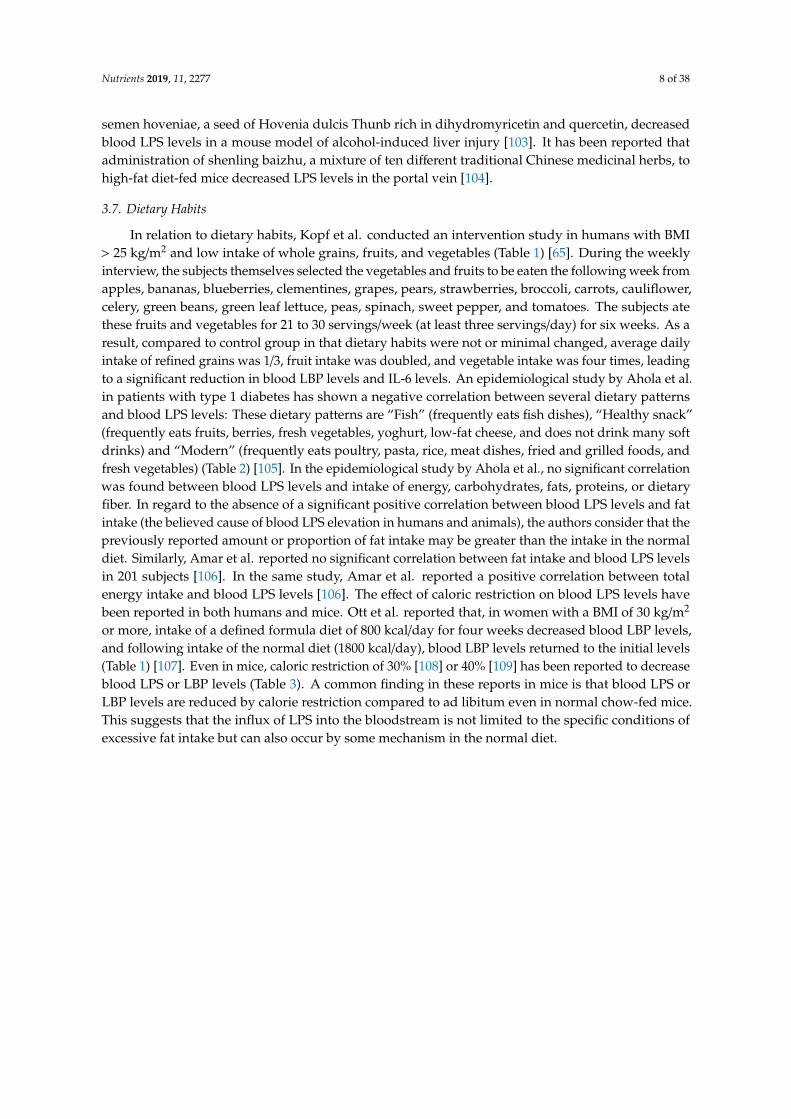
3.1. Probiotics
Probiotics were defined by Fuller in 1989 as “A live microbial feed supplement which beneficiallyaffects the host animal by improving its intestinal microbial balance” [47], and has been studiedmainly for lactic acid bacteria and bifidobacteria. Since metabolic endotoxemia has been implicatedin gut dysbiosis, the effects of probiotics have been investigated. However, the results in humansare unfavorable (Table 1). Lever et al. administered 195 mL of Yakult light (containing 2 × 10¹⁰
colony-forming unit (CFU) of Lactobacillus casei Shirota) for three months to individuals with metabolicsyndrome. The absence of detectable blood LPS in this study led to an assessment of the surrogateLBP level, which was significantly higher in the Yakult light-fed group than in the non-fed group [48].Pei et al. conducted a nine-week study in which low-fat yogurt was ingested in healthy or obeseindividuals, however no significant decrease in blood LPS or LBP levels was observed [49]. In addition,Pei et al. studied whether low-fat yogurt could be administered before a meal to suppress the increasein blood LPS after a meal [50] and found no efficacy. On the other hand, there have been several reportsof the efficacy of probiotics in animal studies (Table 3) [43,51–55]. Lactobacillus rhamnosus, Lactobacillussakei, Lactobacillus acidophilus, Lactobacillus plantarum, Bifidobacterium longum, Bifidobacterium infantis, andBacillus cereus are used as species, and the dosage ranges from 107 to 1010 CFU/day for four to twelveweeks. These animal studies used a high-fat diet, a high-fat high-sucrose diet, or a Zucker-Lepfa/fa
obesity model. In addition to a significant decrease in blood LPS or LBP levels, improvement ofobesity, glucose metabolism, and dyslipidemia was also observed. Since the effects of probiotics arestrain-specific, it is expected that the effects of strains that have been effective in animal studies will beverified in humans.
3.2. Prebiotics
Prebiotics was defined by Gibson and Roberfroid in 1995 as “nondigestible food ingredientsthat beneficially affect the host by selectively stimulating the growth and/or activity of one or alimited number of bacterial species already resident in the colon, and thus attempt to improve hosthealth” [56], and among the food components, dietary fiber and oligosaccharides are known astypical prebiotics. To date, human intervention studies have been conducted with oligofructose [57],inulin [58,59], galacto-oligosaccharides [60–62], resistant dextrin [63], insoluble dietary fiber [64], andwhole grains (Table 1) [65]. Oligofructose is an oligosaccharide containing one molecule of glucoseand several molecules of fructose and is found in many fruits and vegetables. Inulin is a type offructose-polymerized polysaccharide that is abundant in vegetables such as burdock and onion. Inintervention studies with oligofructose [57] and inulin [58,59], subjects with obesity, overweight subjects,and subjects with type 2 diabetes consumed 10–21 g of test substances for 8–12 weeks. Two of the threestudies showed a significant decrease in blood LPS levels [57,58]. One study also showed a decreasein plasminogen activator inhibitor-1 (PAI-1), a risk indicator for thrombosis [57], and the other studyshowed an improvement in glucose metabolism [58]. Galacto-oligosaccharides are oligosaccharides inwhich multiple molecules of galactose are attached to one molecule of glucose. Similar to oligofructose,there have been three reports of interventional trials for galacto-oligosaccharide in obese, overweight,and type 2 diabetic patients. One study showed that galacto-oligosaccharides reduced blood LPS levels,and improved obesity by suppressing appetite [62]. In mice, chronic administration of LPS has beenreported to induce hyperphagia by decreasing leptin sensitivity of afferent vagal nerves [66], and thereduced blood LPS levels and appetite suppression seen with galacto-oligosaccharide administrationare of interest in supporting an association between LPS and appetite.
Nutrients 2019, 11, 2277 6 of 38
3.3. Polyphenols
Polyphenols are secondary metabolites found in plants and are responsible for protection againstoxidative stress, UV damage, and pathogenic microorganisms [67]. Polyphenols are found in awide range of foods, including vegetables, fruits, tea, beans, and spices, and their consumption hasbeen reported to improve metabolic syndrome (decreased body weight, decreased blood pressure,improved glucose metabolism, and improved lipid metabolism) [68]. However, up to 27% of ingestedpolyphenols are detected in urine [69], suggesting that many of them are not absorbed and reachthe large intestine [70]. Since polyphenols reaching the large intestine have been reported to alterthe proportions of microbiota [71], it is expected that the effect of polyphenols against metabolicsyndrome is mediated through the improvement of dysbiosis and of the accompanying metabolicendotoxemia. There are two human intervention studies investigating the relationship betweenpolyphenol intake and blood LPS, both of which evaluated the inhibitory effect on postprandialelevation of blood LPS levels (Table 1) [72,73]. In the study performed by Ghanim et al., healthyindividuals ingested capsules containing 100 mg of resveratrol and 75 mg of polyphenol 10 min beforea 930-kcal high-fat, high-carbohydrate meal. Blood LBP levels up to 5 h after a meal were evaluatedand showed increased blood LBP levels in the placebo group but not in the capsule group [72]. On theother hand, Clemente-Postigo et al. administered 272 mL of red wine to humans simultaneously withexcessive fat and found no effect on either blood LPS or LBP levels [73]. The efficacy of polyphenols hasbeen also reported in animal studies. The effects of grape seed proanthocyanin [29,33], resveratrol [74],apple-derived polymeric procyanidins [75], genistein [76], isoflavone [77], and syringarecinol [78] onblood LPS levels in animal models have been reported (Table 3). In particular, L’openz et al. reportedthat six-month administration of genistein to high-fat diet-fed mice reduced their blood LPS levels andimproved their spatial memory ability [76]. Cho et al. administered syringalesinol to 40-week-old micefor 10 weeks and showed that the decrease in blood LBP levels was accompanied with suppression ofchanges in immune cells due to aging (decreased naive T cells and decreased T-cell proliferation) [78].It has also been reported that adoption of a high-fat diet results in abnormal differentiation of bonemarrow hematopoietic stem cells due to increased blood LPS levels [79], suggesting that the effect ofsyringalecinol on immunoaging might be also exerted in other models of metabolic endotoxemia.
3.4. Sulfated Polysaccharide
Sulfated polysaccharides are widely present in animal tissues and seaweed and are used industriallyas anticoagulants, pharmaceuticals, and gelling agents for foods. The effect of sulfated polysaccharideson metabolic endotoxemia has been studied only in animals (Table 3). Intervention studies withsea cucumber-derived sulfated polysaccharides [80,81], acaudina molpadioides-derived fucosylatedchondroitin sulfate [82], chicken-derived chondroitin sulfate [83] or fucoidan [84] have been performed.Of these studies, two showed that administration of sulfated polysaccharides to high-fat diet-fed miceincreased the amount of short-chain fatty acids in the intestinal tract, decreased the blood LPS orLBP concentration and attenuated weight gain [80,82]. Zhu et al. also reported the same effect ofsulfated polysaccharides in chow-fed lean mice [81]. Liu et al. demonstrated that exhaustive exercisewith a treadmill significantly impaired kidney function, decreased fecal butyrate levels, changedintestinal morphology, and induced metabolic endotoxemia [83]. Their study is interesting in showingthat exercise stress also increased blood LPS levels, and that dietary factors are also effective in themodel mice.
Nutrients 2019, 11, 2277 7 of 38
3.5. Other Dietary Components/Extracts/Foods
In the study by Abboud et al., obese or over weight subjects ingested 30 g of glutamine per dayfor eight weeks (Table 1) [85]. As a result, their blood LPS levels and waist circumference decreased. Inan epidemiological study conducted with healthy subjects, 25-hydroxy vitamin D was reported tonegatively correlate to blood LPS levels (Table 2) [86]. The protective effect of vitamin D is supportedby animal studies in which vitamin D-deficient mice, exposed to a bacterial pathogen, exhibited lowerLPS detoxification activity of the intestine and greater endotoxin translocation [87]. The effect of otherdietary components, including tetrahydro iso-alpha acid [88], rhein [89], phlorizin [90], capsaicin [91],rutin [92], and lycopene [93] on blood LPS levels in animals has also been reported (Table 3). Amongthem, administration of tetrahydro iso-alpha acid [88], phlorizin [90], or rutin [92] to high-fat diet-fedmice or db/db mice improved metabolic impairment. Administration of rhein [89], or lycopene [93]to high-fat diet-fed mice showed a unique effect; they not only reduced blood LPS levels but alsoprevented high-fat diet-induced memory impairment. Kang et al. showed that administration ofantibiotics to mice given capsaicin abolished the effect of capsaicin on blood LPS levels [91]. Theyalso showed that capsaicin-induced protection against high-fat diet-induced blood LPS increase istransferrable by fecal microbiota transplantation.
It has also been reported that intervention with crude food extracts or the food itself can lowerblood LPS levels in animals (Table 3). We studied the effect of broccoli sprout extract, enriched infunctional glucosinolate “glucoraphanin” (details are described in Section 4) [94]. Anhê et al. examinedthe effects of extracts from cranberry [95] or camu camu [96]. Camu camu is an Amazonian fruitthat contains an abundance of vitamin C and flavonoids such as ellagic acid, ellagitannins, andproanthocyanidins. Administration of camu camu extracts to high-fat/high-sucrose diet-fed micereduced plasma bile acid pool size, altered gut microbiota composition, and reduced blood LPS levels.Dey et al. reported that administration of green tea extract to high-fat diet-fed mice suppressedinflammation and gut permeability especially in the ileum and colon, and reduced LPS influx from theportal vein [34]. The reduction of blood LPS levels by feeding with Tartary buckwheat protein wasreported by Zhou et al. [32]. This study is valuable in that it elucidates one of the underlying mechanismsby which plant protein intake leads to improvement of metabolic abnormalities. Intervention studieswith cocoa [97], nopal [98], and steamed fish meat [99] have been performed. Among these, Zhang et al.performed unique experiments [99]. They divided mice into four groups, and fed them ad libitum withnormal chow, steamed fish, pork or beef at 9:00 and 18:00 daily for eight weeks. As a result, only micegroup fed with steamed fish showed decreased blood LBP levels compared to the other three groups.
3.6. Chinese Medicines
The effect of the Chinese medicines; geniposide + chlorogenic acid [100], potentilla discolorbunge water extract [101], ganoderma lucidum mycelium water extract [102], semen hoveniaeextract [103], and shenling baizhu powder [104] on blood LPS levels have been reported in animals(Table 3). The combination of geniposide and chlorogenic acid is included in a traditional Chinesemedicine, Qushi Huayu Decoction. Peng et al. indicated that administration of geniposide andchlorogenic acid to high-fat diet-fed mice restored colonic tight junctions by inhibiting down-regulationof RhoA/Rho-associated kinase signaling, and reduced blood LPS levels and hepatic LBP proteinlevels [100]. Han et al. examined the effect of potentilla discolor bunge water extract in type 2 diabeticmice induced by high-fat diet feeding and streptozotocin injection [101]. The results showed that fecalLPS levels in the type 2 diabetic model mice were significantly increased compared to the controlnormal mice. The administration of potentilla discolor bunge water extract to mice reduced fecalLPS levels, decreased blood LPS levels and increased the expression levels of tight junction proteins(Claudin-3, ZO-1, and Occludin) in the colon. Chang et al. studied the effect of ganoderma lucidummycelium, a Basidiomycete fungus [102]. They showed the dose-dependent effect of ganoderma lucidummycelium water extract on blood LPS reduction, suggesting that high molecular weight polysaccharides(>300 kDa) isolated from the extract is an effective component. Ping et al. reported that the extract of
Nutrients 2019, 11, 2277 8 of 38
semen hoveniae, a seed of Hovenia dulcis Thunb rich in dihydromyricetin and quercetin, decreasedblood LPS levels in a mouse model of alcohol-induced liver injury [103]. It has been reported thatadministration of shenling baizhu, a mixture of ten different traditional Chinese medicinal herbs, tohigh-fat diet-fed mice decreased LPS levels in the portal vein [104].
3.7. Dietary Habits
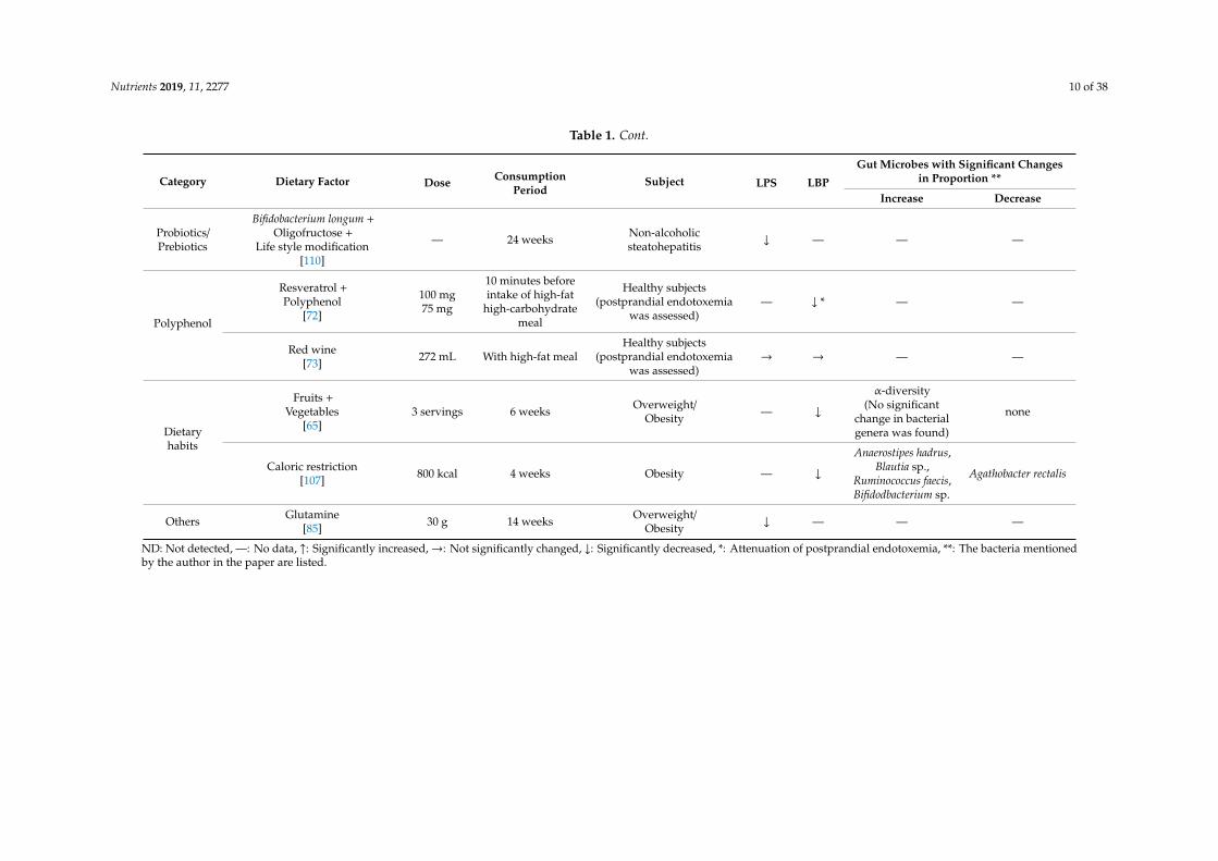
In relation to dietary habits, Kopf et al. conducted an intervention study in humans with BMI> 25 kg/m2 and low intake of whole grains, fruits, and vegetables (Table 1) [65]. During the weeklyinterview, the subjects themselves selected the vegetables and fruits to be eaten the following week fromapples, bananas, blueberries, clementines, grapes, pears, strawberries, broccoli, carrots, cauliflower,celery, green beans, green leaf lettuce, peas, spinach, sweet pepper, and tomatoes. The subjects atethese fruits and vegetables for 21 to 30 servings/week (at least three servings/day) for six weeks. As aresult, compared to control group in that dietary habits were not or minimal changed, average dailyintake of refined grains was 1/3, fruit intake was doubled, and vegetable intake was four times, leadingto a significant reduction in blood LBP levels and IL-6 levels. An epidemiological study by Ahola et al.in patients with type 1 diabetes has shown a negative correlation between several dietary patternsand blood LPS levels: These dietary patterns are “Fish” (frequently eats fish dishes), “Healthy snack”(frequently eats fruits, berries, fresh vegetables, yoghurt, low-fat cheese, and does not drink many softdrinks) and “Modern” (frequently eats poultry, pasta, rice, meat dishes, fried and grilled foods, andfresh vegetables) (Table 2) [105]. In the epidemiological study by Ahola et al., no significant correlationwas found between blood LPS levels and intake of energy, carbohydrates, fats, proteins, or dietaryfiber. In regard to the absence of a significant positive correlation between blood LPS levels and fatintake (the believed cause of blood LPS elevation in humans and animals), the authors consider that thepreviously reported amount or proportion of fat intake may be greater than the intake in the normaldiet. Similarly, Amar et al. reported no significant correlation between fat intake and blood LPS levelsin 201 subjects [106]. In the same study, Amar et al. reported a positive correlation between totalenergy intake and blood LPS levels [106]. The effect of caloric restriction on blood LPS levels havebeen reported in both humans and mice. Ott et al. reported that, in women with a BMI of 30 kg/m2
or more, intake of a defined formula diet of 800 kcal/day for four weeks decreased blood LBP levels,and following intake of the normal diet (1800 kcal/day), blood LBP levels returned to the initial levels(Table 1) [107]. Even in mice, caloric restriction of 30% [108] or 40% [109] has been reported to decreaseblood LPS or LBP levels (Table 3). A common finding in these reports in mice is that blood LPS orLBP levels are reduced by calorie restriction compared to ad libitum even in normal chow-fed mice.This suggests that the influx of LPS into the bloodstream is not limited to the specific conditions ofexcessive fat intake but can also occur by some mechanism in the normal diet.
Nutrients 2019, 11, 2277 9 of 38
Table 1. Dietary factors that have been evaluated for efficacy on blood lipopolysaccharide (LPS) levels in human interventional studies.
Category Dietary Factor Dose ConsumptionPeriod
Subject LPS LBP
Gut Microbes with Significant Changesin Proportion **
Increase Decrease
Probiotics/Prebiotics
Yakult light(Lactobacillus casei Shirota 1 × 10⁸
CFU/mL) [48]195 mL 3 months Metabolic syndrome ND ↑ — —
Low-fat yogurt[49] 339 g 9 weeks Healthy subject or Obesity → → — —
Low-fat yogurt[50] 226 g Premeal
Healthy subject or Obesity(postprandial endotoxemia
was assessed)→ → — —
Oligofructose[57] 21 g 12 weeks Overweight/
Obesity ↓ — — —
Oligofructose-enriched inulin
[58]10 g 8 weeks Type 2 diabetes ↓ — — —
Inulin +Oligofructose
[59]
8 g8 g 3 months Obesity → —
Bifidobacterium,Faecalibacterium
prausnitzii
Bacteroidesintestinalis,
Bacteroides vulgatus,Propionibacterium
Galacto-oligosaccharide
[60]5.5 g 12 weeks Type 2 diabetes → → none none
Galacto-oligosaccharide
[61]15 g 12 weeks Overweight/
Obesity — → Bifidobacterium spp. none
α-Galacto-oligosaccharide
[62]6–18 g 14 days Overweight ↓ — Bifidobacteria none
Resistant dextrin[63] 10 g 8 weeks Type 2 diabetes ↓ — — —
Insoluble dietary fiber[from Fiber One Original cereal
(General mills)][64]
30 g With high-fat,high-calorie meal
Healthy subject(postprandial endotoxemia
was assessed)↓* — — —
Whole grains[65] 3 servings 6 weeks Overweight/
Obesity — ↓ none none
Nutrients 2019, 11, 2277 10 of 38
Table 1. Cont.
Category Dietary Factor Dose ConsumptionPeriod
Subject LPS LBP
Gut Microbes with Significant Changesin Proportion **
Increase Decrease
Probiotics/Prebiotics
Bifidobacterium longum +Oligofructose +
Life style modification[110]
— 24 weeks Non-alcoholicsteatohepatitis ↓ — — —
Polyphenol
Resveratrol +Polyphenol
[72]
100 mg75 mg
10 minutes beforeintake of high-fat
high-carbohydratemeal
Healthy subjects(postprandial endotoxemia
was assessed)— ↓ * — —
Red wine[73] 272 mL With high-fat meal
Healthy subjects(postprandial endotoxemia
was assessed)→ → — —
Dietaryhabits
Fruits +Vegetables
[65]3 servings 6 weeks Overweight/
Obesity — ↓
α-diversity(No significant
change in bacterialgenera was found)
none
Caloric restriction[107] 800 kcal 4 weeks Obesity — ↓
Anaerostipes hadrus,Blautia sp.,
Ruminococcus faecis,Bifidodbacterium sp.
Agathobacter rectalis
Others Glutamine[85] 30 g 14 weeks Overweight/
Obesity ↓ — — —
ND: Not detected, —: No data, ↑: Significantly increased,→: Not significantly changed, ↓: Significantly decreased, *: Attenuation of postprandial endotoxemia, **: The bacteria mentionedby the author in the paper are listed.
Nutrients 2019, 11, 2277 11 of 38
Table 2. Correlation of dietary factors, gut microbes, and blood LPS levels in human epidemiological studies.
Subject Number ofSubject
Correlation ofDietary Factor and Gut Microbe *
Correlation ofBlood LPS and Gut Microbe
Correlation ofBlood LPS and Dietary Factor
Over-weight
pregnantwomen
[111]
88P
Dietary fibervs.
diversity, richness,Firmicutes in
unidentified family oforder Clostridiales,
Barnciellaceae familybelonging to the
phylum Bacteroidetes
P none P none
Vitamin A,β-Carotene
vs.Firmicutes
N Fat vs. diversity, richness,Barnsiellaceae N none N none
Healthysubjects
[86]150 N
25-Hydroxyvitamin D
vs.
Coprococcus,Bifdobacterium N LPS vs. Faecalibacterium N LPS
vs. 25-Hydroxy vitamin D
Type 1diabetes
[105]668 — — — — — — N LPS
vs.
Dietary pattern;“Fish”(frequently eat fishdishes), “Healthy snack”
(frequently eat fruits, berries,fresh vegetable, yoghurt,
low-fat cheese, and do notdrink much soft drinks),
“Modern”(frequently eatpoultry, pasta, rice, meat
dishes, fried and grilled foods,and fresh vegetables)
—: No data, P: Positive correlation, N: Negative correlation, LPS: lipopolysaccharide, *: The bacteria mentioned by the author in the paper are listed.
Nutrients 2019, 11, 2277 12 of 38
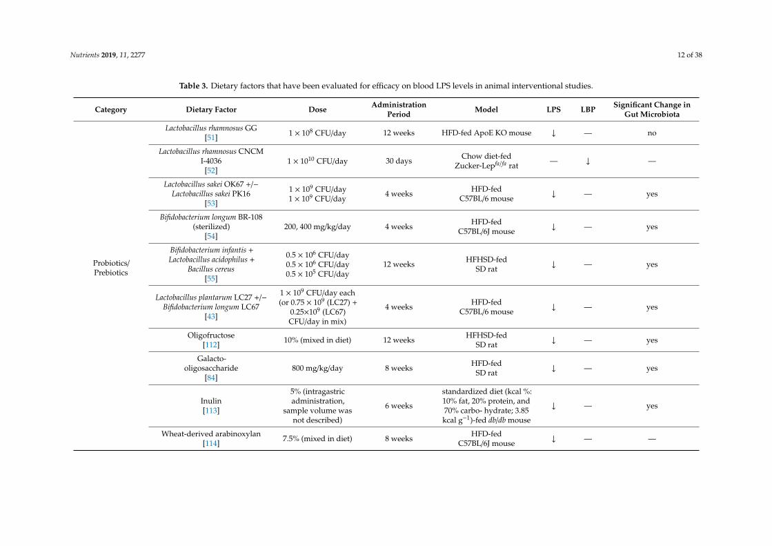
Table 3. Dietary factors that have been evaluated for efficacy on blood LPS levels in animal interventional studies.
Category Dietary Factor Dose AdministrationPeriod Model LPS LBP Significant Change in
Gut Microbiota
Probiotics/Prebiotics
Lactobacillus rhamnosus GG[51] 1 × 10⁸ CFU/day 12 weeks HFD-fed ApoE KO mouse ↓ — no
Lactobacillus rhamnosus CNCMI-4036
[52]1 × 10¹⁰ CFU/day 30 days Chow diet-fed
Zucker-Lepfa/fa rat — ↓ —
Lactobacillus sakei OK67 +/−Lactobacillus sakei PK16
[53]
1 × 10⁹ CFU/day1 × 109 CFU/day
4 weeks HFD-fedC57BL/6 mouse ↓ — yes
Bifidobacterium longum BR-108(sterilized)
[54]200, 400 mg/kg/day 4 weeks HFD-fed
C57BL/6J mouse ↓ — yes
Bifidobacterium infantis +Lactobacillus acidophilus +
Bacillus cereus[55]
0.5 × 10⁶ CFU/day0.5 × 106 CFU/day0.5 × 105 CFU/day
12 weeks HFHSD-fedSD rat ↓ — yes
Lactobacillus plantarum LC27 +/−Bifidobacterium longum LC67
[43]
1 × 10⁹ CFU/day each(or 0.75 × 10⁹ (LC27) +
0.25×10⁹ (LC67)CFU/day in mix)
4 weeks HFD-fedC57BL/6 mouse ↓ — yes
Oligofructose[112] 10% (mixed in diet) 12 weeks HFHSD-fed
SD rat ↓ — yes
Galacto-oligosaccharide
[84]800 mg/kg/day 8 weeks HFD-fed
SD rat ↓ — yes
Inulin[113]
5% (intragastricadministration,
sample volume wasnot described)
6 weeks
standardized diet (kcal %:10% fat, 20% protein, and70% carbo- hydrate; 3.85kcal g−1)-fed db/db mouse
↓ — yes
Wheat-derived arabinoxylan[114] 7.5% (mixed in diet) 8 weeks HFD-fed
C57BL/6J mouse ↓ — —
Nutrients 2019, 11, 2277 13 of 38
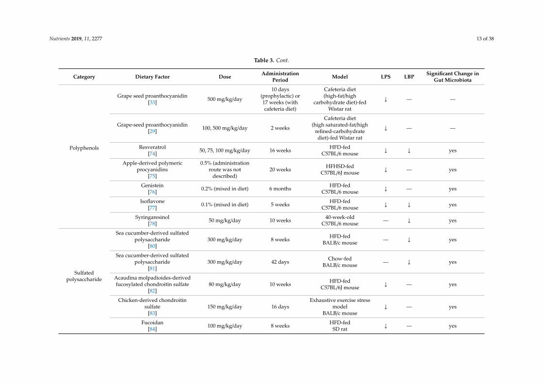
Table 3. Cont.
Category Dietary Factor Dose AdministrationPeriod Model LPS LBP Significant Change in
Gut Microbiota
Polyphenols
Grape seed proanthocyanidin[33] 500 mg/kg/day
10 days(prophylactic) or17 weeks (withcafeteria diet)
Cafeteria diet(high-fat/high
carbohydrate diet)-fedWistar rat
↓ — —
Grape-seed proanthocyanidin[29] 100, 500 mg/kg/day 2 weeks
Cafeteria diet(high saturated-fat/high
refined-carbohydratediet)-fed Wistar rat
↓ — —
Resveratrol[74] 50, 75, 100 mg/kg/day 16 weeks HFD-fed
C57BL/6 mouse ↓ ↓ yes
Apple-derived polymericprocyanidins
[75]
0.5% (administrationroute was not
described)20 weeks HFHSD-fed
C57BL/6J mouse ↓ — yes
Genistein[76] 0.2% (mixed in diet) 6 months HFD-fed
C57BL/6 mouse ↓ — yes
Isoflavone[77] 0.1% (mixed in diet) 5 weeks HFD-fed
C57BL/6 mouse ↓ ↓ yes
Syringaresinol[78] 50 mg/kg/day 10 weeks 40-week-old
C57BL/6 mouse — ↓ yes
Sulfatedpolysaccharide
Sea cucumber-derived sulfatedpolysaccharide
[80]300 mg/kg/day 8 weeks HFD-fed
BALB/c mouse — ↓ yes
Sea cucumber-derived sulfatedpolysaccharide
[81]300 mg/kg/day 42 days Chow-fed
BALB/c mouse — ↓ yes
Acaudina molpadioides-derivedfucosylated chondroitin sulfate
[82]80 mg/kg/day 10 weeks HFD-fed
C57BL/6J mouse ↓ — yes
Chicken-derived chondroitinsulfate
[83]150 mg/kg/day 16 days
Exhaustive exercise stressmodel
BALB/c mouse↓ — yes
Fucoidan[84] 100 mg/kg/day 8 weeks HFD-fed
SD rat ↓ — yes
Nutrients 2019, 11, 2277 14 of 38
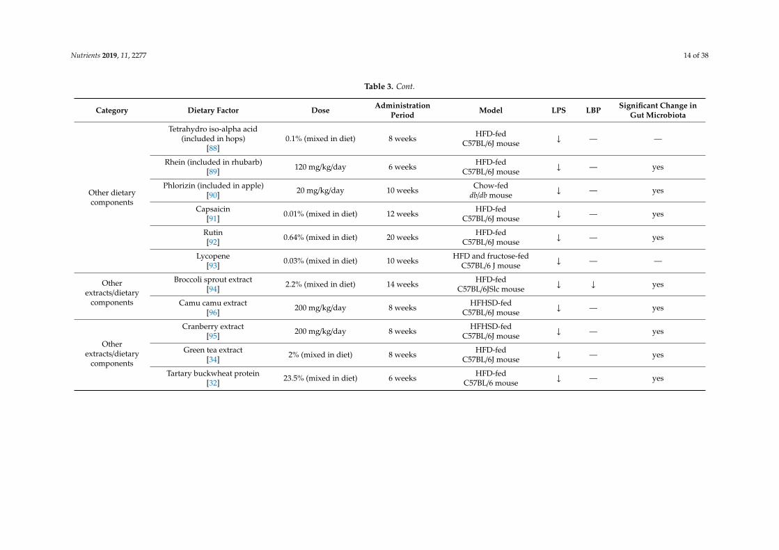
Table 3. Cont.
Category Dietary Factor Dose AdministrationPeriod Model LPS LBP Significant Change in
Gut Microbiota
Other dietarycomponents
Tetrahydro iso-alpha acid(included in hops)
[88]0.1% (mixed in diet) 8 weeks HFD-fed
C57BL/6J mouse ↓ — —
Rhein (included in rhubarb)[89] 120 mg/kg/day 6 weeks HFD-fed
C57BL/6J mouse ↓ — yes
Phlorizin (included in apple)[90] 20 mg/kg/day 10 weeks Chow-fed
db/db mouse ↓ — yes
Capsaicin[91] 0.01% (mixed in diet) 12 weeks HFD-fed
C57BL/6J mouse ↓ — yes
Rutin[92] 0.64% (mixed in diet) 20 weeks HFD-fed
C57BL/6J mouse ↓ — yes
Lycopene[93] 0.03% (mixed in diet) 10 weeks HFD and fructose-fed
C57BL/6 J mouse ↓ — —
Otherextracts/dietary
components
Broccoli sprout extract[94] 2.2% (mixed in diet) 14 weeks HFD-fed
C57BL/6JSlc mouse ↓ ↓ yes
Camu camu extract[96] 200 mg/kg/day 8 weeks HFHSD-fed
C57BL/6J mouse ↓ — yes
Otherextracts/dietary
components
Cranberry extract[95] 200 mg/kg/day 8 weeks HFHSD-fed
C57BL/6J mouse ↓ — yes
Green tea extract[34] 2% (mixed in diet) 8 weeks HFD-fed
C57BL/6J mouse ↓ — yes
Tartary buckwheat protein[32] 23.5% (mixed in diet) 6 weeks HFD-fed
C57BL/6 mouse ↓ — yes
Nutrients 2019, 11, 2277 15 of 38
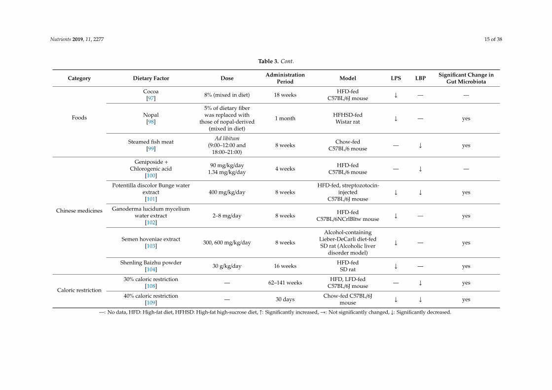
Table 3. Cont.
Category Dietary Factor Dose AdministrationPeriod Model LPS LBP Significant Change in
Gut Microbiota
Foods
Cocoa[97] 8% (mixed in diet) 18 weeks HFD-fed
C57BL/6J mouse ↓ — —
Nopal[98]
5% of dietary fiberwas replaced with
those of nopal-derived(mixed in diet)
1 month HFHSD-fedWistar rat ↓ — yes
Steamed fish meat[99]
Ad libitum(9:00–12:00 and
18:00–21:00)8 weeks Chow-fed
C57BL/6 mouse — ↓ yes
Chinese medicines
Geniposide +Chlorogenic acid
[100]
90 mg/kg/day1.34 mg/kg/day 4 weeks HFD-fed
C57BL/6 mouse — ↓ —
Potentilla discolor Bunge waterextract[101]
400 mg/kg/day 8 weeksHFD-fed, streptozotocin-
injectedC57BL/6J mouse
↓ ↓ yes
Ganoderma lucidum myceliumwater extract
[102]2–8 mg/day 8 weeks HFD-fed
C57BL/6NCrlBltw mouse ↓ — yes
Semen hoveniae extract[103] 300, 600 mg/kg/day 8 weeks
Alcohol-containingLieber-DeCarli diet-fedSD rat (Alcoholic liver
disorder model)
↓ — yes
Shenling Baizhu powder[104] 30 g/kg/day 16 weeks HFD-fed
SD rat ↓ — yes
Caloric restriction
30% caloric restriction[108] — 62–141 weeks HFD, LFD-fed
C57BL/6J mouse — ↓ yes
40% caloric restriction[109] — 30 days Chow-fed C57BL/6J
mouse ↓ ↓ yes
—: No data, HFD: High-fat diet, HFHSD: High-fat high-sucrose diet, ↑: Significantly increased,→: Not significantly changed, ↓: Significantly decreased.
Nutrients 2019, 11, 2277 16 of 38
4. Association of Dietary Factor-Induced Reduction of Blood LPS and Modulation ofGut Microbiota
Although few studies have evaluated the relationship between the effect of dietary factors on bloodLPS and intestinal flora in humans, several studies have evaluated intestinal flora in oligosaccharideintervention studies (Table 1). A common finding in these reports is an increase in Bifidobacterium.Bifidobacterium has been reported to enhance the intestinal tight junction by preserving claudin 4 andoccludin localization at tight junctions, and inhibit permeability in mice with colitis [115]. Similarly,in human colonic epithelial cell line T84, the addition of culture supernatant of Bifidobacterium hasbeen reported to enhance barrier function through increased expression of tight junction protein,suggesting that some humoral factors contribute to improved intestinal barrier function [116]. Increasedexpression of tight junction protein in Bifidobacterium-treated mice has been reported to be associatedwith increased short-chain fatty acids (acetic acid, butyric acid, and propionic acid) in the intestinaltract [117]. These short-chain fatty acids have been reported in the human colonic epithelial cell linecaco-2 to act as an energy source for epithelial cells to protect themselves, and also act as a histonedeacetylase inhibitor which inhibit Nod-like receptor P3 inflammasomes to maintain the barrierfunction of epithelial cells [118]. These results suggest that the increase in Bifidobacterium induced byoligosaccharide intake decreases blood LPS levels through the improvement of the barrier functionof the intestinal tract. In addition, dietary factors that increase Bifidobacterium are expected to reduceblood LPS levels.
Changes in intestinal flora by the dietary factors listed in Table 4 was greatly dependent on thestudy. However, all of the dietary factors commonly lowered blood LPS or LBP levels in animals,as described in Table 3. In other words, by finding bacteria that have decreased or increased inmany dietary factor intervention studies, we can find specific bacteria that contribute to the increaseor decrease in blood LPS levels. To this end, we have organized the number of reports that showincreases or decreases of each bacterial genus (Figure 1). We selected eight of these genera (Lactobacillus,Bacteroides, Akkermansia, Clostridium, Escherichia, Roseburia, Prevotella and Desulfovibrio) as bacteriaincluded in a sufficient number (five or more) of reports, and a biased number of reports (Bifidobacteriumwas excluded because it was discussed above. Faecalibacterium was also excluded because there isalmost no bias in the number of reports).
Lactobacillus, Bacteroides, Akkermansia, Roseburia, and Prevotella are possible bacterial genera thatmay contribute to the reduction of blood LPS levels. Lactobacillus is a gram-positive bacterium thatproduces large amounts of lactic acid during carbohydrate fermentation. The probiotic contributionof Lactobacillus to the regulation of metabolic endotoxemia is studied (Table 3). Administration ofLactobacillus rhamnosus CNCM I-4036 to obese Zucker-Leprfa/fa rats decreased the mRNA expressionlevels of endothelin receptor type B (Ednrb) in the intestinal mucosa, and reduced the blood LBPlevel [52]. Reduction of Ednrb decreases the density of negative charge of the colonic mucin layer,leading to an increase in the ability of the mucin layer to adsorb microparticles and bacteria, therebyinhibiting their penetration through the colonic mucosa [119]. Lactobacillus sakei OK67 and PK16 arereported to suppress high-fat diet-induced colitis, and to reduce the fecal Proteobacteria populationand fecal LPS levels in mice [53]. In addition to the previous reports described in Table 3, it hasbeen reported that oral administration of Lactobacillus reuteri ZJ617 suppresses LPS-induced apoptosisof intestinal epithelial cells and maintains the intestinal barrier function [120]. We have describedin Section 2.2 that LPS is absorbed from the intestinal tract during lipid absorption. Interestingly,oral ingestion of Lactobacillus acidophilus ATCC 4356 in mice has been reported to reduce the mRNAlevels of Niemann-Pick C1-like 1, which is involved in lipid absorption in the intestine, and inthe suppression of cholesterol absorption [121]. Taken together, this suggests that Lactobacilluscontributes to a decrease in blood LPS levels through strengthening the intestinal barrier, reducing theamount of LPS in feces, and suppressing lipid absorption. As described in Table 4, Bifidobacterium,oligofructose, galacto-oligosaccharide, syringaresinol, acaudina molpadioides-derived fucosylatedchondroitin sulfate, green tea extract, Tartary buckwheat protein, nopal, semen hoveniae extract,

Nutrients 2019, 11, 2277 17 of 38
and 30% caloric restriction are dietary factors that increase the proportion of Lactobacillus in the gutmicrobiota. Among them, the amylolytic Bifidobacterium strain is reported to stimulate the growth of anonamylolytic Lactobacillus probably by producing intermediate metabolites of starch metabolism [122].Oligosaccharides (oligofructose and galacto-oligosaccharide) were reported to support the growthof Lactobacillus as prebiotics [123]. Green tea extract [124] and buckwheat-resistant starch [125] werereported to promote the growth of Lactobacillus in a fermentation assay. On the other hand, inan in vitro fermentation assay using gut microbiota, it was reported that fucosylated chondroitinsulfate promotes the growth of Bacteroides, Bifidobacterium, and Clostridium, while the number ofLactobacillus decreases [126]. Thus, the mechanism by which Lactobacillus increased in mice fed withfucosylated chondroitin sulfate needs to be further studied. The mechanism by which the proportion ofLactobacillus in gut microbiota increases due to calorie restriction also remains unknown. As it has beenreported that the bacteria adapted to the nutritional environment can grow predominantly in the gutmicrobiota consortium [127], Lactobacillus might be able to grow even under malnutrition. The effect ofsyringaresinol, nolpal, and semen hoveniae on the growth of Lactobacillus has not been revealed.
Bacteroides is a gram-negative obligate anaerobe. Hooper et al. reported that Bacteroidesthetaiotaomicron, a prominent component of the normal mouse and human intestinal microflora,modulates expression of genes involved in mucosal barrier fortification [128]. The administration ofBacteroides fragilis HCK-B3 and Bacteroides ovatus ELH-B2 to mice attenuated LPS-induced intestinalinflammation, by either modulating cytokine production or restoring the Treg/Th-17 balance [129]. Onthe other hand, in a state in which no dietary fiber is ingested, it has been suggested that Bacteroidesdegrades the mucin layer of the intestinal tract, decreases the barrier function of mucus, and inducesinflammation [130]. Therefore, it should be noted that depending on the diet of the host, Bacteroidescan act as either probiotics or pathobionts. As described in Table 4, an increase in Bacteroides wasreported in four out of five intervention studies with sulfated polysaccharides. Bacteroides is aunique bacterium among gut flora that has degrading enzymes corresponding to various sulfatedpolysaccharides [131] and is able to utilize sulfated polysaccharides such as heparin [131], heparansulfate [131], and chondroitin sulfate [132] as energy sources. It is therefore thought that intakeof sulfate polysaccharide preferentially nourishes Bacteroides in gut flora and suppresses metabolicendotoxemia via its anti-inflammatory and barrier function-enhancing effects.
Akkermansia is a mucin-adherent intestinal bacterium [133], which grows by degrading mucin [134],and produces propionic acid, a short-chain fatty acid [135]. In addition, Akkermansia promotes butyrateproduction, by supporting the growth of Anaerostipes caccae through mucin degradation [136]. As notedabove, these short-chain fatty acids are known to enhance intestinal barrier function. In addition, it hasbeen reported that Akkermansia-derived extracellular vesicles administered in mice are localized to thelarge intestine, and directly enhance intestinal barrier function by increasing epithelial cell expression oftight junction proteins [137]. Furthermore, oral administration of Akkermansia to mice inhibited high-fatdiet-induced thinning of the mucin layer, reduced blood LPS concentration, and inhibited obesityand abnormal glucose metabolism [138]. Akkermansia has been reported to be negatively correlatedwith obesity (waist-to-hip ratio and subcutaneous adipocyte diameter) and diabetes mellitus (glucoseintolerance states), and is attracting attention as a next-generation probiotic [139]. Among the dietaryfactors that increase the proportion of Akkermansia in the gut flora, polyphenols are intriguing becausemost of intervention studies with polyphenols (apple-derived polymeric procyanidins, genistein, andisoflavone) or polyphenol-rich food extracts (camu camu extract, cranberry extract, and green teaextract) consistently reported an increase of Akkermansia (Table 4). Anhê et al. reported that cranberryextract administration to mice increased colonic Kruppel-like factor 4 (a marker of goblet cells) andMuc2 mRNA expression, suggesting that polyphenols enhance mucin production and support thegrowth of Akkermansia [95]. On the other hand, direct prebiotic action of polyphenols to Akkermansia hasbeen reported in a study using the Simulator of Human Intestinal Microbial Ecosystem (SHIME®) [140].

Nutrients 2019, 11, 2277 18 of 38
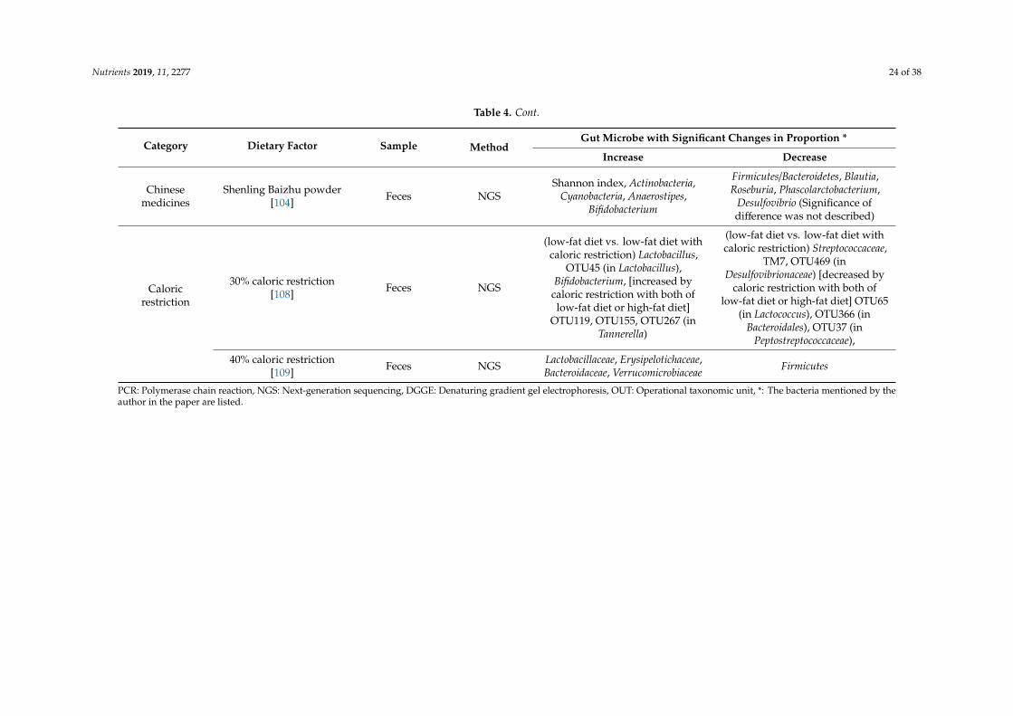
Table 4. Changes of gut microbiota induced by dietary factor intervention in animal experiments.
Category Dietary Factor Sample MethodGut Microbe with Significant Changes in Proportion *
Increase Decrease
Probiotics/Prebiotics
Lactobacillus sakei OK67+/−
Lactobacillus sakei PK16[53]
Feces PCR, NGS OTU (O67), Ace (O67), Chao1(O67), Shanon (O67)
Simpson (O67), Proteobacteria,Firmicutes, Firmicutes/Bacteroidetes,
Proteobacteria/Bacteroidetes
Bifidobacterium longumBR-108
(sterilized)[54]
Cecal contents PCR Bifidobacterium spp.,Lactobacillus spp. Firmicutes
Bifidobacterium infantis +Lactobacillus acidophilus +
Bacillus cereus[55]
Feces PCRBifidobacteria, Lactobacillus,
Bacteroides,Bifidobacteria/Escherichia coli
Escherichia coli, Enterococcus
Lactobacillus plantarumLC27 +/−
Bifidobacterium longumLC67[43]
Feces PCR Actinobacteria (LC67, LC27 + LC67)
Firmicutes, Bacteroidetes,δ/γ-Proteobacteria, Deferribacteres
(LC67, LC27+LC67),Firmicutes/Bacteroidetes,
Proteobacteria/Bacteroidetes
Oligofructose[112] Cecal contents PCR
Bacteroides/Prevotella,Bifidobacterium, Lactobacillus,
Roseburia
Clostridium leptum (cluster IV),Clostridium cluster I, Clostridium
cluster XI, Methanobrevibacter,Akkemansia muciniphila,
Faecalibacterium prausnitzii
Galacto-oligosaccharide[84] Cecal contents NGS
Verrucomicrobia, Akkermansia,Ruminococcus, Blautia, Bacteroidetes,
Proteobacteria, Adlercreutzia,Staphylococcus, Prevotella,Oscillospira, Lactobacillus,
Desulfovibrio
Firmicutes, Actinobacteria,Clostridium, Bacillus
Inulin[113] Feces NGS Bacteroidetes, Cyanobacteria,
Bacteroides
Firmicutes, Deferribacteres,Tenericutes, Ruminiclostridium_6,
Mucispirillum
Nutrients 2019, 11, 2277 19 of 38
Table 4. Cont.
Category Dietary Factor Sample MethodGut Microbe with Significant Changes in Proportion *
Increase Decrease
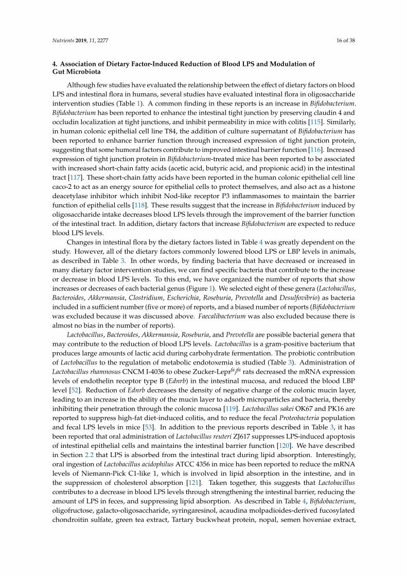
Polyphenols
Resveratrol[74] Cecal contents NGS Deferribacteraceae
none (In this study, population ofDesulfovibrionaceae in the high-fatdiet + intervention group was atthe same level with normal chow
group, but there was no significantreduction from high-fat diet group.)
Apple-derived polymericprocyanidins
[75]Cecal contents NGS
Bacteroidetes, Verrucomicrobia,Adlerceitzia, Roseburia, S24-7,Bacteroids, Anaerovorax, rc4-4,
Akkermansia
Firmicutes, Firmicutes/Bacteroidetes,Clostridium, Lachnospiraceae,
Bifidobacterium
Polyphenols
Genistein[76] Feces NGS
Firmicutes, Verrucomicrobia,Prevotellaceae, Verrucomicrobia,
Prevotella, Akkermansia,Faecalibacterium, Prevotella copri,Prevotella stercorea, Akkermansia
muciniphila
Bacteroidetes, Bacteroidaceae,Bacteroides, Bacteroides acidifaciens,
Bacteroides uniformis
Isoflavone[77] Feces NGS
α-diversity, Actinobacteria,Verrucomicrobia,
Bifidobacterium/Enterobacteriaceae,Akkermansia
Proteobacteria
Syringaresinol[78] Cecal contents NGS
Firmicutes/Bacteroidetes, Firmicutes,Lactobacillus, Lactobacillus animalis,Lactobacillus johnsonii, Lactobacillus
reuteri, Lactobacillus intestinalis,Bifidobacterium pseudolongum
Shannon diversity indices,Jeotgalicoccus nanhaiensis,
Staphylococcus lentus, Bacteroidaceae(EF098405_s), Bacteroides vulgatus,
Akkermansia muciniphila

Nutrients 2019, 11, 2277 20 of 38
Table 4. Cont.
Category Dietary Factor Sample MethodGut Microbe with Significant Changes in Proportion *
Increase Decrease
Sulfatedpolysaccharide
Sea cucumber-derivedsulfated polysaccharide
[80]Feces NGS
bacterial diversity, Verrucomicrobia(depolymerized sulfated
polysaccharide), Bacteroides,Alloprevotella, Ruminiclostridium_9,
Butyricicoccus, Akkermansia
Proteobacteria, Escherichia-Shigella(polymerized sulfated polysaccharide),Pseudomonas (depolymerized sulfated
polysaccharide), Yersinia(depolymerized sulfated
polysaccharide), (In this study,decrease of Desulfovibrio with the
intervention of sulfated polysaccharideto high-fat diet-fed mouse was shown
as heatmap, but significance ofdifference was not described.)
Sea cucumber-derivedsulfated polysaccharide
[81]Feces NGS
Proteobacteria (polymerized sulfatedpolysaccharide), Bacteroides
(polymerized sulfatedpolysaccharide), Allobaculum
(depolymerized sulfatedpolysaccharide), Alloprevotella,
Roseburia, Turicibacter, Desulfovibrio
Enterococcus, Streptococcus,Escherichia-Shigella, Lactobacillus
Acaudinamolpadioides-derived
fucosylated chondroitinsulfate
[82]
Feces PCR, NGS
Bacteroidetes, Lactobacillus,Actinobacteria, Faecalibacterium
prausnitzii, Deferribacteres,Bacteroidales, Bifidobacteriales,
Lachnospiraceae NK4A136 group,Bacteroides, Bacteroides acidifaciens,
Bifidobacterium choerinum
Firmicutes, Escherichia coli, Clostridiales,Bacilli, Lactobacillales, Clostridia
Clostridiales, Firmicutes Clostridiales,Lactococcus, Clostridium ruminantium
Chicken-derivedchondroitin sulfate
[83]Feces NGS
Bacteroidetes, Bacteroides acidifaciens,family S24-7, Lysinibacillus
boronitoleransFirmicutes, β-Proteobacteria
Fucoidan[84] Cecal contents NGS
Proteobacteria, Verrucomicrobia,Enterobacter, Bacteroidetes, Bacillus,
Ruminococcus, Adlercreutzia,Prevotella, Oscillospira, Desulfovibrio,
Firmicutes, Actinobacteria, Clostridium,Corynebacterium, Staphylococcus,
Lactobacillus, Aerococcus
Nutrients 2019, 11, 2277 21 of 38
Table 4. Cont.
Category Dietary Factor Sample MethodGut Microbe with Significant Changes in Proportion *
Increase Decrease
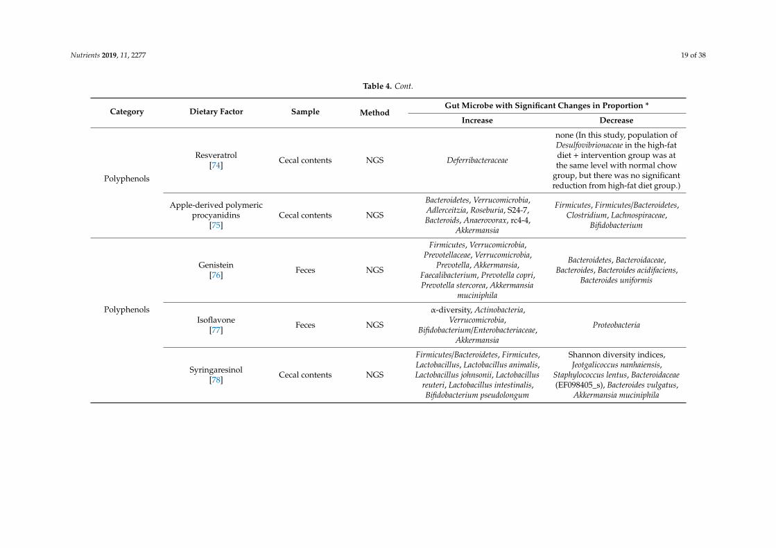
Other dietarycomponents
Rhein (included inrhubarb)
[89]Cecal contents PCR Bacteroides/Prevotella, Desulfovibrio Bifidobacterium, Lactobacillus
Phlorizin (included inapple)
[90]Feces PCR, DGGE Akkermansia muciniphila, Prevotella none
Capsaicin[91] Cecal contents NGS Ruminococcaceae, Lachnospiraceae family S24_7
Rutin[92]
Small intestinalcontents NGS
Bacteroidales_S24-7 group,Bacteroidaceae, Porphyromonadaceae,
Rikenellaceae, Desulfovibrionaceae
Firmicutes, Firmicutes/Bacteroidetes,Deferribacteraceae, Lachnospiraceae

Nutrients 2019, 11, 2277 22 of 38
Table 4. Cont.
Category Dietary Factor Sample MethodGut Microbe with Significant Changes in Proportion *
Increase Decrease
Otherextracts/dietary
components
Broccoli sprout extract[94] Cecal contents NGS none Proteobacteria, Desulfovibrionaceae
Camu camu extract[96] Feces NGS
microbial richness, Bifidobacterium,Barnesiella, Barnesiella spp.,
Turicibacter spp., Akkermansiamuciniphila, Delftia, Roseburia,
Anaerostipes, unclassified generawithin the families
Christensenellaceae, unclassifiedgenera within the families
Erysipelotrichaceae
Firmicutes/Bacteroidetes, Lactobacillus,Anaerotruncus, Parabacteroides
Cranberry extract[95] Feces PCR, NGS Akkermansia none
Green tea extract[34] Cecal contents NGS
Shannon index, Chao1 richness,Bacteroidetes, Actinobacteria,
Verrucomicrobia, Bacteroidales,Bifidobacteriales, Verrucomicrobiales,
Turicibacterales. RF39,Coriobacteriales, Bifidobacterium,
Blautia, Dorea, Lactobacillus,Ruminococcus, Akkermansia,Butyrivibrio, Akkermansia
muciniphila, Ruminococcus gnavus,Bifidobacterium pseudolongum,
Bifidobacterium adolescentis
Firmicutes, Firmicutes/Bacteroidetes,Clostridiales, SMB53
Tartary buckwheatprotein
[32]Feces PCR Bifidobacterium, Lactobacillus,
Enterococcus, Clostridium Escherichia coli, Bacaeroides
Nutrients 2019, 11, 2277 23 of 38
Table 4. Cont.
Category Dietary Factor Sample MethodGut Microbe with Significant Changes in Proportion *
Increase Decrease
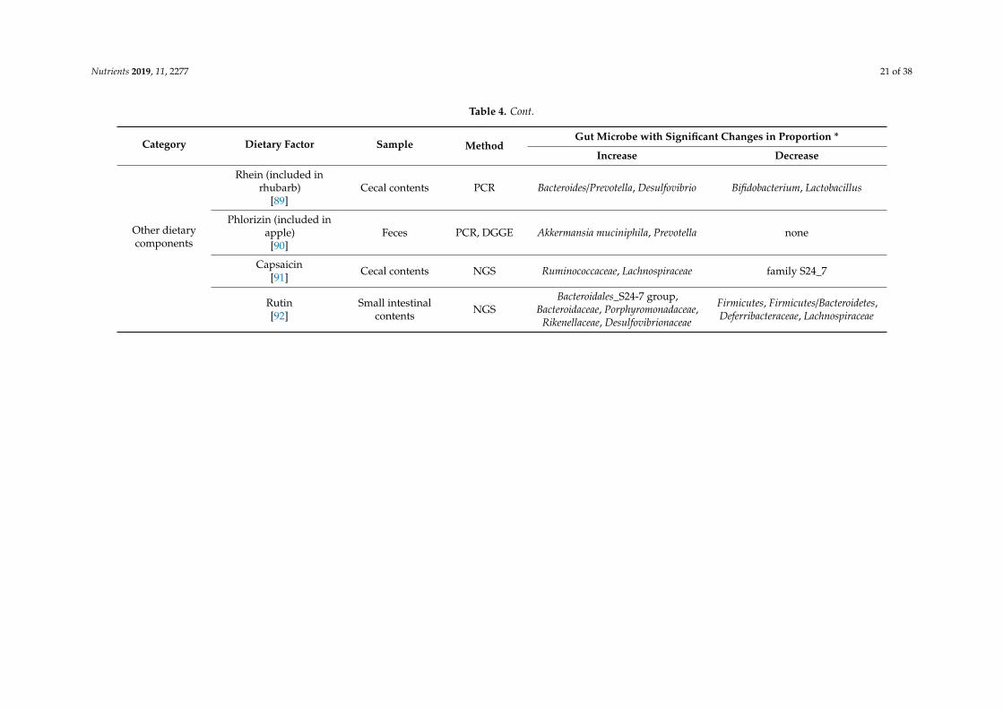
FoodsNopal
[98] Feces NGS
α-diversity, Anaeroplasma, Prevotella,Ruminucoccus, Bacteroides fragilis,
Ruminococcus bromii, Rumminococcusflavefaciens, Lactobacillus reuteri,
Akkermansia muciniphila
Firmicutes/Bacteroidetes,Faecalibacterium, Clostridium,
Butyricicoccus, Bacteroidesacidifaciens, Blautia producta,Faecalibacterium prausnitzii,Butyricicoccus pullicaecorum,
Clostridium citroniae
Steamed fish meat[99] Feces NGS
Proteobacteria, Firmicutes,Ruminococcaceae, Oscillospira,
Clostridium, EscherichiaShannon index, Bacteroidetes, S24-7
Chinesemedicines
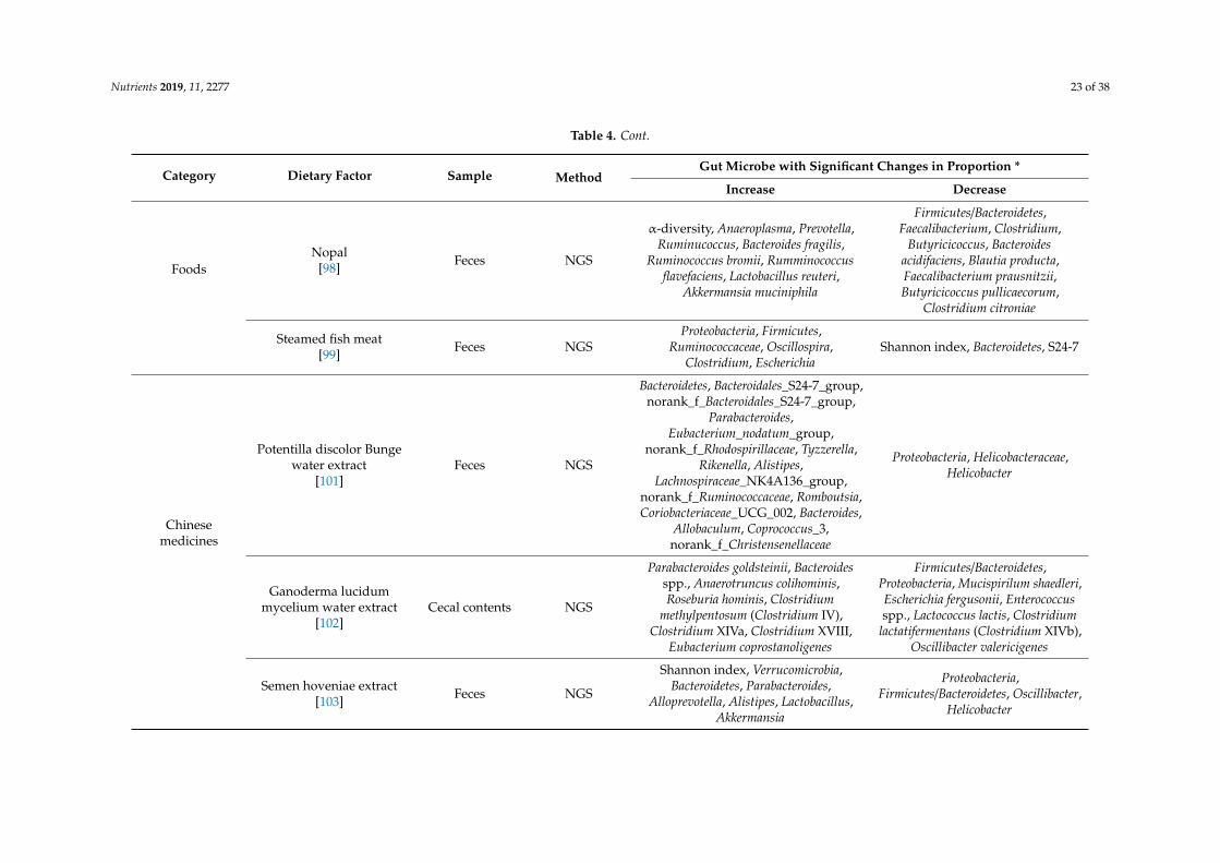
Potentilla discolor Bungewater extract
[101]Feces NGS
Bacteroidetes, Bacteroidales_S24-7_group,norank_f_Bacteroidales_S24-7_group,
Parabacteroides,Eubacterium_nodatum_group,
norank_f_Rhodospirillaceae, Tyzzerella,Rikenella, Alistipes,
Lachnospiraceae_NK4A136_group,norank_f_Ruminococcaceae, Romboutsia,Coriobacteriaceae_UCG_002, Bacteroides,
Allobaculum, Coprococcus_3,norank_f_Christensenellaceae
Proteobacteria, Helicobacteraceae,Helicobacter
Ganoderma lucidummycelium water extract
[102]Cecal contents NGS
Parabacteroides goldsteinii, Bacteroidesspp., Anaerotruncus colihominis,Roseburia hominis, Clostridium
methylpentosum (Clostridium IV),Clostridium XIVa, Clostridium XVIII,
Eubacterium coprostanoligenes
Firmicutes/Bacteroidetes,Proteobacteria, Mucispirilum shaedleri,Escherichia fergusonii, Enterococcusspp., Lactococcus lactis, Clostridium
lactatifermentans (Clostridium XIVb),Oscillibacter valericigenes
Semen hoveniae extract[103] Feces NGS
Shannon index, Verrucomicrobia,Bacteroidetes, Parabacteroides,
Alloprevotella, Alistipes, Lactobacillus,Akkermansia
Proteobacteria,Firmicutes/Bacteroidetes, Oscillibacter,
Helicobacter
Nutrients 2019, 11, 2277 24 of 38
Table 4. Cont.
Category Dietary Factor Sample MethodGut Microbe with Significant Changes in Proportion *
Increase Decrease
Chinesemedicines
Shenling Baizhu powder[104] Feces NGS
Shannon index, Actinobacteria,Cyanobacteria, Anaerostipes,
Bifidobacterium
Firmicutes/Bacteroidetes, Blautia,Roseburia, Phascolarctobacterium,
Desulfovibrio (Significance ofdifference was not described)
Caloricrestriction
30% caloric restriction[108] Feces NGS
(low-fat diet vs. low-fat diet withcaloric restriction) Lactobacillus,
OTU45 (in Lactobacillus),Bifidobacterium, [increased by
caloric restriction with both oflow-fat diet or high-fat diet]
OTU119, OTU155, OTU267 (inTannerella)
(low-fat diet vs. low-fat diet withcaloric restriction) Streptococcaceae,
TM7, OTU469 (inDesulfovibrionaceae) [decreased by
caloric restriction with both oflow-fat diet or high-fat diet] OTU65
(in Lactococcus), OTU366 (inBacteroidales), OTU37 (in
Peptostreptococcaceae),
40% caloric restriction[109] Feces NGS Lactobacillaceae, Erysipelotichaceae,
Bacteroidaceae, Verrucomicrobiaceae Firmicutes
PCR: Polymerase chain reaction, NGS: Next-generation sequencing, DGGE: Denaturing gradient gel electrophoresis, OUT: Operational taxonomic unit, *: The bacteria mentioned by theauthor in the paper are listed.

Nutrients 2019, 11, 2277 25 of 38Nutrients 2019, 8, x FOR PEER REVIEW 16 of 34
Figure 1. The number of reported changes of intestinal bacterial genera in dietary factor intervention studies in animals.
Lactobacillus, Bacteroides, Akkermansia, Roseburia, and Prevotella are possible bacterial genera that may contribute to the reduction of blood LPS levels. Lactobacillus is a gram-positive bacterium that produces large amounts of lactic acid during carbohydrate fermentation. The probiotic contribution of Lactobacillus to the regulation of metabolic endotoxemia is studied (Table 3). Administration of Lactobacillus rhamnosus CNCM I-4036 to obese Zucker-Leprfa/fa rats decreased the mRNA expression levels of endothelin receptor type B (Ednrb) in the intestinal mucosa, and reduced the blood LBP level [52]. Reduction of Ednrb decreases the density of negative charge of the colonic mucin layer, leading to an increase in the ability of the mucin layer to adsorb microparticles and bacteria, thereby
8 6 4 2 0 2 4 6 8 10 12
LactobacillusBacteroides
AkkermansiaBifidobacterium
ClostridiumEscherichia
RoseburiaPrevotella
DesulfovibrioFaecalibacterium
RuminococcusParabacteroides
BlautiaEnterococcusOscillospira
AlloprevotellaStaphylococcus
LactococcusAdlercreutziaAllobaculumAnaerostipesEubacterium
AlistipesBacillus
RuminiclostridiumButyricicoccus
HelicobacterOscillibacterAnaerovoraxTuricibacter
LysinibacillusBarnesiella
TuricibacterDelftiaDorea
ButyrivibrioAnaeroplasma
TyzzerellaRikenella
RomboutsiaCoprococcus
AnaerotruncusTannerella
MethanobrevibacterMucispirillumJeotgalicoccusPseudomonas
YersiniaStreptococcus
CorynebacteriumAerococcus
AnaerotruncusMucispirilum
Phascolarctobacterium
Number of reports
Increase
Decrease
Figure 1. The number of reported changes of intestinal bacterial genera in dietary factor interventionstudies in animals.
Roseburia [141] is an enteric bacterium that utilizes dietary fiber and may enhance intestinalbarriers by producing butyric acid. It has been reported that administration of Roseburia to miceenhanced differentiation of regulatory T cells in the intestinal lamina propria and suppressed intestinal

Nutrients 2019, 11, 2277 26 of 38
inflammation [142]. As described in Table 4, oligofructose, apple-derived polymeric procyanidins, seacucumber-derived sulfated polysaccharide, camu camu extract, and ganoderma lucidum myceliumwater extract were reported to increase the proportion of Roseburia in the gut flora. Roseburia metabolizesoligofructose into fructose, which is used for growth, but for this process, acetic acid that is producedby Bifidobacterium is required [143]. Therefore, in order to grow Roseburia by oligofructose intake, it isnecessary to pay attention to the symbiotic relationship with other intestinal bacteria and the amountof short-chain fatty acids in the intestine. Other dietary factors, procyanidins, sea cucumber-derivedsulfated polysaccharide, camu camu extract, and ganoderma lucidum mycelium, have not been studiedfor their prebiotic function for Roseburia.
It has been suggested that LPS from Prevotella has fewer phosphate and acyl moieties contributingto endotoxin activity, resulting in a lower TLR4 stimulatory capacity than LPS from Salmonella [144].Therefore, by increasing the population of Prevotella in the intestinal flora, endotoxin activity inthe intestinal contents and damage to intestinal epithelial cells might be decreased, leading to thereduction of blood LPS levels. On the other hand, Prevotella produces succinate as a metabolite of sugarmetabolism [145]. It has also been reported that succinate from intestinal bacteria is utilized by andpromotes growth of Salmonella serovar Typhimurium [146] and Clostridium difficile [147], which arethe pathogens of pseudomembranous colitis. Succinate has also been reported to induce colitis viasuccinate receptors and to promote colonic fibrosis [148]. In addition, proportion of Prevotella in thegut flora has been reported to be positively correlated with blood LPS levels in patients with type 2diabetes [149]. Thus, an increase in the proportion of Prevotella does not necessarily have a positiveeffect on intestinal health. It is necessary to carefully investigate the contribution of Prevotella to bloodLPS levels.
Clostridium, Escherichia, and Desulfovibrio are bacterial genera that may contribute to the increaseof blood LPS levels. Many pathogenic bacteria (such as enterohemorrhagic Escherichia coli, Clostridiumbotulinum, Clostridium tetani, and Clostridium perfringens), which produce effector proteins or enterotoxinsthat disrupt epithelial tight junction belong to these genera [150]. In addition, the endotoxin activity ofLPS in non-pathogenic Escherichia is also higher than in Bacteroides, and an increased proportion ofthese Escherichia in enteric flora aggravate colitis [151]. Clostridium species catabolize cholic acid todeoxycholic acid for their growth [152]. It is reported that, in mice, deoxycholic acid increases intestinalpermeability through the reduction of goblet cell number, suppression of mucin production, inductionof low-grade inflammation, and suppression of tight junction protein (ZO-1) expression [153]. In termsof dietary factors that reduce Escherichia, there are many reports of sulfated polysaccharides (Table 4).We could not find any reports that suggested the direct inhibitory effect of sulfated polysaccharideon growth of Escherichia. On the other hand, it is suggested that Bacteroides, that can be preferentiallygrown in sulfated polysaccharide feeding, compete with Escherichia in the co-culture assay [127].In order to elucidate the mechanism by which sulfated polysaccharides reduce the proportion ofEscherichia, it is hoped to study focusing on the interaction between gut microbes. Among the dietaryfactors that reduce Clostridium, procyanidin is reported to decrease the growth of Clostridium in fecalbatch culture [154]. The bactericidal activity of methanol extract of nopal against Clostridium has alsobeen reported [155].
Desulfovibrio is a gram-negative, obligate anaerobe, sulfate-reducing bacterium. Desulfovibrioutilizes electrons supplied by the oxidation of lactic acid in the electron transport system of therespiratory chain, uses sulfuric acid as the final electron acceptor, and produces hydrogen sulfide asa metabolite [156]. Desulfovibrio is ubiquitous in the intestines of humans and mice. Of the studiesthat showed significant changes in the proportion of Desulfovibrio, most studies reported that theproportion was increased associated to the reduction of blood LPS levels (Table 4). However, it is alsoreported that proportions of Desulfovibrio increased in the colons of patients with ulcerative colitis [157]and has attracted attention as a pathogen of colitis. In addition, Xie et al. reported in mice that theincrease of Desulfovibrio in feces was positively correlated with the increase of LPS levels in feces,liver, and blood [45]. Qui et al. reported that ingestion of a high-fat diet in mice increased fecal
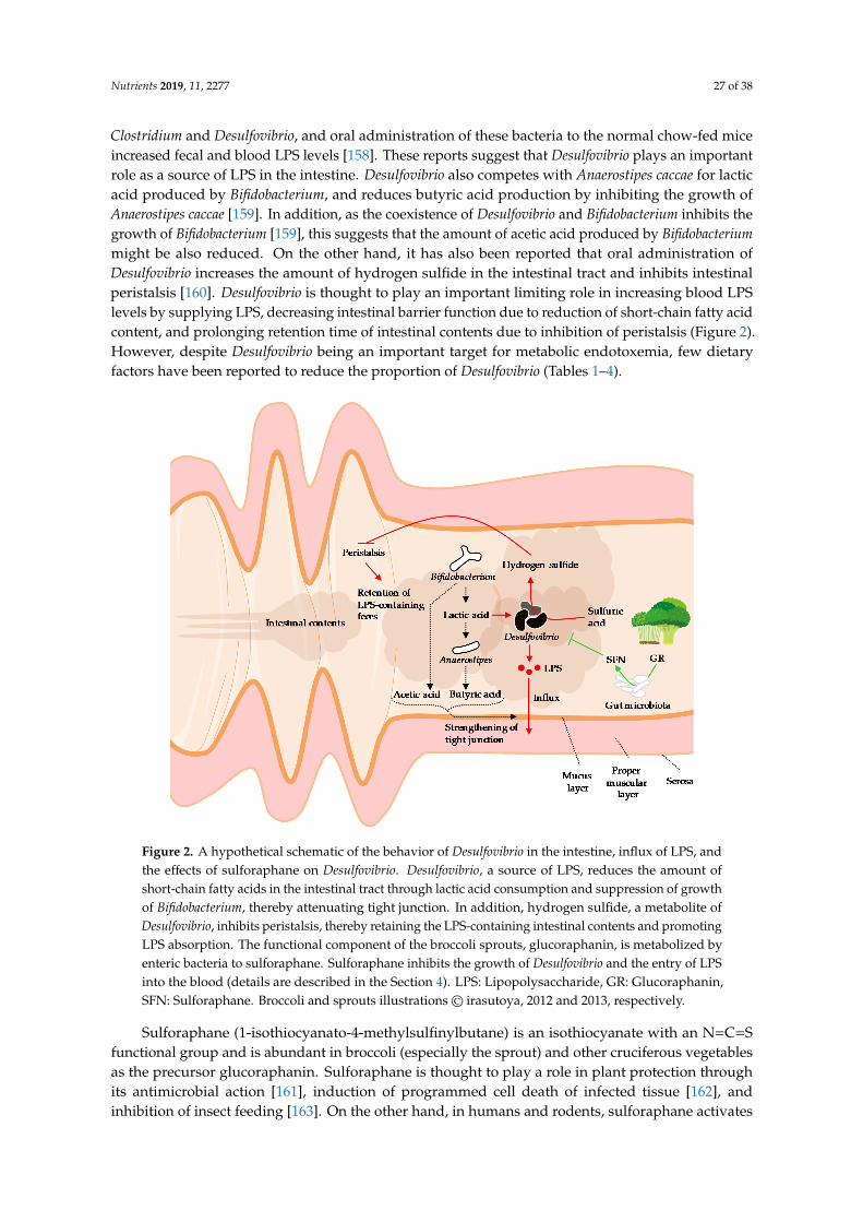
Nutrients 2019, 11, 2277 27 of 38
Clostridium and Desulfovibrio, and oral administration of these bacteria to the normal chow-fed miceincreased fecal and blood LPS levels [158]. These reports suggest that Desulfovibrio plays an importantrole as a source of LPS in the intestine. Desulfovibrio also competes with Anaerostipes caccae for lacticacid produced by Bifidobacterium, and reduces butyric acid production by inhibiting the growth ofAnaerostipes caccae [159]. In addition, as the coexistence of Desulfovibrio and Bifidobacterium inhibits thegrowth of Bifidobacterium [159], this suggests that the amount of acetic acid produced by Bifidobacteriummight be also reduced. On the other hand, it has also been reported that oral administration ofDesulfovibrio increases the amount of hydrogen sulfide in the intestinal tract and inhibits intestinalperistalsis [160]. Desulfovibrio is thought to play an important limiting role in increasing blood LPSlevels by supplying LPS, decreasing intestinal barrier function due to reduction of short-chain fatty acidcontent, and prolonging retention time of intestinal contents due to inhibition of peristalsis (Figure 2).However, despite Desulfovibrio being an important target for metabolic endotoxemia, few dietaryfactors have been reported to reduce the proportion of Desulfovibrio (Tables 1–4).
Nutrients 2019, 8, x FOR PEER REVIEW 19 of 34
reported to decrease the growth of Clostridium in fecal batch culture [154]. The bactericidal activity of methanol extract of nopal against Clostridium has also been reported [155].
Desulfovibrio is a gram-negative, obligate anaerobe, sulfate-reducing bacterium. Desulfovibrio utilizes electrons supplied by the oxidation of lactic acid in the electron transport system of the respiratory chain, uses sulfuric acid as the final electron acceptor, and produces hydrogen sulfide as a metabolite [156]. Desulfovibrio is ubiquitous in the intestines of humans and mice. Of the studies that showed significant changes in the proportion of Desulfovibrio, most studies reported that the proportion was increased associated to the reduction of blood LPS levels (Table 4). However, it is also reported that proportions of Desulfovibrio increased in the colons of patients with ulcerative colitis [157] and has attracted attention as a pathogen of colitis. In addition, Xie et al. reported in mice that the increase of Desulfovibrio in feces was positively correlated with the increase of LPS levels in feces, liver, and blood [45]. Qui et al. reported that ingestion of a high-fat diet in mice increased fecal Clostridium and Desulfovibrio, and oral administration of these bacteria to the normal chow-fed mice increased fecal and blood LPS levels [158]. These reports suggest that Desulfovibrio plays an important role as a source of LPS in the intestine. Desulfovibrio also competes with Anaerostipes caccae for lactic acid produced by Bifidobacterium, and reduces butyric acid production by inhibiting the growth of Anaerostipes caccae [159]. In addition, as the coexistence of Desulfovibrio and Bifidobacterium inhibits the growth of Bifidobacterium [159], this suggests that the amount of acetic acid produced by Bifidobacterium might be also reduced. On the other hand, it has also been reported that oral administration of Desulfovibrio increases the amount of hydrogen sulfide in the intestinal tract and inhibits intestinal peristalsis [160]. Desulfovibrio is thought to play an important limiting role in increasing blood LPS levels by supplying LPS, decreasing intestinal barrier function due to reduction of short-chain fatty acid content, and prolonging retention time of intestinal contents due to inhibition of peristalsis (Figure 2). However, despite Desulfovibrio being an important target for metabolic endotoxemia, few dietary factors have been reported to reduce the proportion of Desulfovibrio (Tables 1–4).
Figure 2. A hypothetical schematic of the behavior of Desulfovibrio in the intestine, influx of LPS, and the effects of sulforaphane on Desulfovibrio. Desulfovibrio, a source of LPS, reduces the amount of short-chain fatty acids in the intestinal tract through lactic acid consumption and suppression of growth of Bifidobacterium, thereby attenuating tight junction. In addition, hydrogen sulfide, a metabolite of
Figure 2. A hypothetical schematic of the behavior of Desulfovibrio in the intestine, influx of LPS, andthe effects of sulforaphane on Desulfovibrio. Desulfovibrio, a source of LPS, reduces the amount ofshort-chain fatty acids in the intestinal tract through lactic acid consumption and suppression of growthof Bifidobacterium, thereby attenuating tight junction. In addition, hydrogen sulfide, a metabolite ofDesulfovibrio, inhibits peristalsis, thereby retaining the LPS-containing intestinal contents and promotingLPS absorption. The functional component of the broccoli sprouts, glucoraphanin, is metabolized byenteric bacteria to sulforaphane. Sulforaphane inhibits the growth of Desulfovibrio and the entry of LPSinto the blood (details are described in the Section 4). LPS: Lipopolysaccharide, GR: Glucoraphanin,SFN: Sulforaphane. Broccoli and sprouts illustrations© irasutoya, 2012 and 2013, respectively.
Sulforaphane (1-isothiocyanato-4-methylsulfinylbutane) is an isothiocyanate with an N=C=Sfunctional group and is abundant in broccoli (especially the sprout) and other cruciferous vegetablesas the precursor glucoraphanin. Sulforaphane is thought to play a role in plant protection throughits antimicrobial action [161], induction of programmed cell death of infected tissue [162], andinhibition of insect feeding [163]. On the other hand, in humans and rodents, sulforaphane activates
Nutrients 2019, 11, 2277 28 of 38
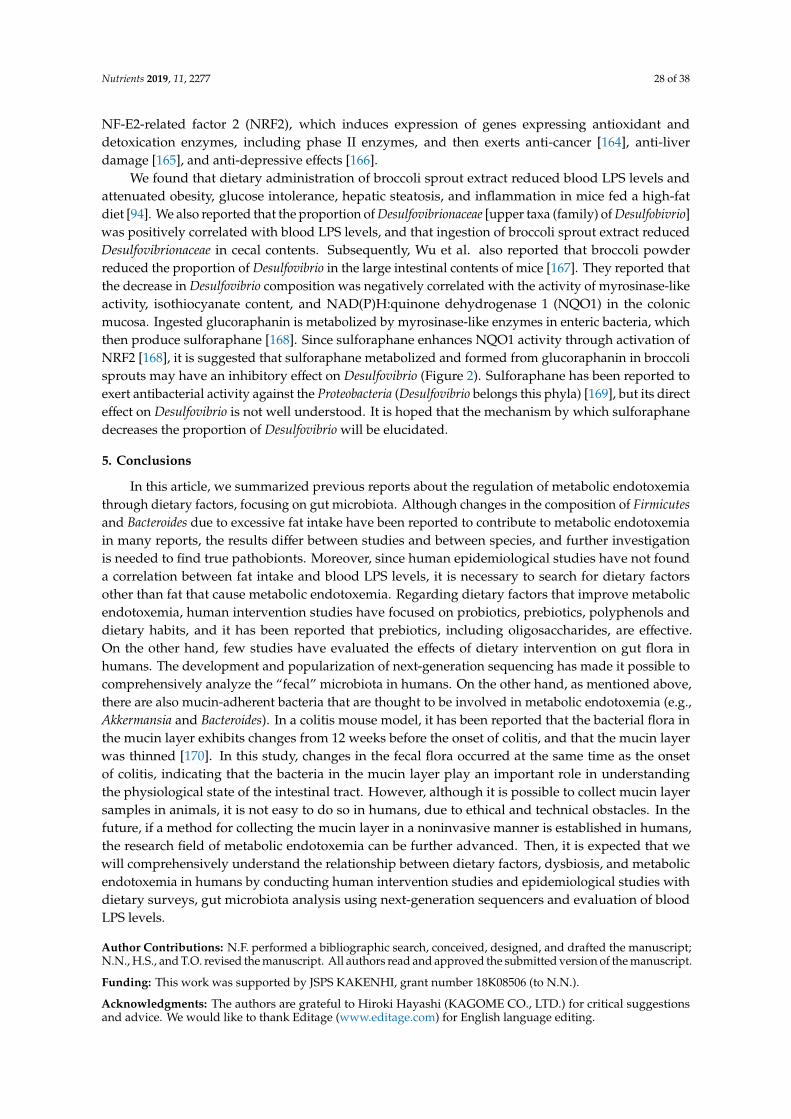
NF-E2-related factor 2 (NRF2), which induces expression of genes expressing antioxidant anddetoxication enzymes, including phase II enzymes, and then exerts anti-cancer [164], anti-liverdamage [165], and anti-depressive effects [166].
We found that dietary administration of broccoli sprout extract reduced blood LPS levels andattenuated obesity, glucose intolerance, hepatic steatosis, and inflammation in mice fed a high-fatdiet [94]. We also reported that the proportion of Desulfovibrionaceae [upper taxa (family) of Desulfobivrio]was positively correlated with blood LPS levels, and that ingestion of broccoli sprout extract reducedDesulfovibrionaceae in cecal contents. Subsequently, Wu et al. also reported that broccoli powderreduced the proportion of Desulfovibrio in the large intestinal contents of mice [167]. They reported thatthe decrease in Desulfovibrio composition was negatively correlated with the activity of myrosinase-likeactivity, isothiocyanate content, and NAD(P)H:quinone dehydrogenase 1 (NQO1) in the colonicmucosa. Ingested glucoraphanin is metabolized by myrosinase-like enzymes in enteric bacteria, whichthen produce sulforaphane [168]. Since sulforaphane enhances NQO1 activity through activation ofNRF2 [168], it is suggested that sulforaphane metabolized and formed from glucoraphanin in broccolisprouts may have an inhibitory effect on Desulfovibrio (Figure 2). Sulforaphane has been reported toexert antibacterial activity against the Proteobacteria (Desulfovibrio belongs this phyla) [169], but its directeffect on Desulfovibrio is not well understood. It is hoped that the mechanism by which sulforaphanedecreases the proportion of Desulfovibrio will be elucidated.
5. Conclusions
In this article, we summarized previous reports about the regulation of metabolic endotoxemiathrough dietary factors, focusing on gut microbiota. Although changes in the composition of Firmicutesand Bacteroides due to excessive fat intake have been reported to contribute to metabolic endotoxemiain many reports, the results differ between studies and between species, and further investigationis needed to find true pathobionts. Moreover, since human epidemiological studies have not founda correlation between fat intake and blood LPS levels, it is necessary to search for dietary factorsother than fat that cause metabolic endotoxemia. Regarding dietary factors that improve metabolicendotoxemia, human intervention studies have focused on probiotics, prebiotics, polyphenols anddietary habits, and it has been reported that prebiotics, including oligosaccharides, are effective.On the other hand, few studies have evaluated the effects of dietary intervention on gut flora inhumans. The development and popularization of next-generation sequencing has made it possible tocomprehensively analyze the “fecal” microbiota in humans. On the other hand, as mentioned above,there are also mucin-adherent bacteria that are thought to be involved in metabolic endotoxemia (e.g.,Akkermansia and Bacteroides). In a colitis mouse model, it has been reported that the bacterial flora inthe mucin layer exhibits changes from 12 weeks before the onset of colitis, and that the mucin layerwas thinned [170]. In this study, changes in the fecal flora occurred at the same time as the onsetof colitis, indicating that the bacteria in the mucin layer play an important role in understandingthe physiological state of the intestinal tract. However, although it is possible to collect mucin layersamples in animals, it is not easy to do so in humans, due to ethical and technical obstacles. In thefuture, if a method for collecting the mucin layer in a noninvasive manner is established in humans,the research field of metabolic endotoxemia can be further advanced. Then, it is expected that wewill comprehensively understand the relationship between dietary factors, dysbiosis, and metabolicendotoxemia in humans by conducting human intervention studies and epidemiological studies withdietary surveys, gut microbiota analysis using next-generation sequencers and evaluation of bloodLPS levels.
Author Contributions: N.F. performed a bibliographic search, conceived, designed, and drafted the manuscript;N.N., H.S., and T.O. revised the manuscript. All authors read and approved the submitted version of the manuscript.
Funding: This work was supported by JSPS KAKENHI, grant number 18K08506 (to N.N.).
Acknowledgments: The authors are grateful to Hiroki Hayashi (KAGOME CO., LTD.) for critical suggestionsand advice. We would like to thank Editage (www.editage.com) for English language editing.
Nutrients 2019, 11, 2277 29 of 38
Conflicts of Interest: The authors, N.F. and H.S., are employees of KAGOME CO., LTD., which has a product anda patent for broccoli sprout extract. N.F. holds stock in KAGOME CO., LTD.
References
1. Kiers, D.; Leijte, G.P.; Gerretsen, J.; Zwaag, J.; Kox, M.; Pickkers, P. Comparison of different lots of endotoxinand evaluation of in vivo potency over time in the experimental human endotoxemia model. Innate Immun.2019, 25, 34–45. [CrossRef]
2. Benson, S.; Engler, H.; Wegner, A.; Rebernik, L.; Spreitzer, I.; Schedlowski, M.; Elsenbruch, S. What makesyou feel sick after inflammation? Predictors of acute and persisting physical sickness symptoms induced byexperimental endotoxemia. Clin. Pharmacol. Ther. 2017, 102, 141–151. [CrossRef]
3. Cani, P.D.; Amar, J.; Iglesias, M.A.; Poggi, M.; Knauf, C.; Bastelica, D.; Neyrinck, A.M.; Fava, F.; Tuohy, K.M.;Chabo, C.; et al. Metabolic endotoxemia initiates obesity and insulin resistance. Diabetes 2007, 56, 1761–1772.[CrossRef]
4. Liang, H.; Hussey, S.E.; Sanchez-Avila, A.; Tantiwong, P.; Musi, N. Effect of lipopolysaccharide oninflammation and insulin action in human muscle. PLoS ONE 2013, 8, e63983. [CrossRef]
5. Pussinen, P.J.; Havulinna, A.S.; Lehto, M.; Sundvall, J.; Salomaa, V. Endotoxemia is associated with anincreased risk of incident diabetes. Diabetes Care 2011, 34, 392–397. [CrossRef]
6. Jin, R.; Willment, A.; Patel, S.S.; Sun, X.; Song, M.; Mannery, Y.O.; Kosters, A.; McClain, C.J.; Vos, M.B.Fructose induced endotoxemia in pediatric nonalcoholic fatty liver disease. Int. J. Hepatol. 2014, 2014, 560620.[CrossRef]
7. Jandhyala, S.M.; Madhulika, A.; Deepika, G.; Rao, G.V.; Reddy, D.N.; Subramanyam, C.; Sasikala, M.;Talukdar, R. Altered intestinal microbiota in patients with chronic pancreatitis: Implications in diabetes andmetabolic abnormalities. Sci. Rep. 2017, 7, 43640. [CrossRef]
8. Zhang, R.; Miller, R.G.; Gascon, R.; Champion, S.; Katz, J.; Lancero, M.; Narvaez, A.; Honrada, R.;Ruvalcaba, D.; McGrath, M.S. Circulating endotoxin and systemic immune activation in sporadic amyotrophiclateral sclerosis (sALS). J. Neuroimmunol. 2009, 206, 121–124. [CrossRef]
9. Kaliannan, K.; Hamarneh, S.R.; Economopoulos, K.P.; Nasrin Alam, S.; Moaven, O.; Patel, P.; Malo, N.S.;Ray, M.; Abtahi, S.M.; Muhammad, N.; et al. Intestinal alkaline phosphatase prevents metabolic syndrome inmice. Proc. Natl. Acad. Sci. USA 2013, 110, 7003–7008. [CrossRef]
10. Cani, P.D.; Bibiloni, R.; Knauf, C.; Waget, A.; Neyrinck, A.M.; Delzenne, N.M.; Burcelin, R. Changes ingut microbiota control metabolic endotoxemia-induced inflammation in high-fat diet-induced obesity anddiabetes in mice. Diabetes 2008, 57, 1470–1481. [CrossRef]
11. Candido, T.L.N.; Bressan, J.; Alfenas, R.d.C.G. Dysbiosis and metabolic endotoxemia induced by high-fatdiet. Nutr. Hosp. 2018, 35, 1432–1440.
12. Devkota, S.; Wang, Y.; Musch, M.W.; Leone, V.; Fehlner-Peach, H.; Nadimpalli, A.; Antonopoulos, D.A.;Jabri, B.; Chang, E.B. Dietary-fat-induced taurocholic acid promotes pathobiont expansion and colitis inIl10-/- mice. Nature 2012, 487, 104–108. [CrossRef]
13. David, L.A.; Maurice, C.F.; Carmody, R.N.; Gootenberg, D.B.; Button, J.E.; Wolfe, B.E.; Ling, A.V.; Devlin, A.S.;Varma, Y.; Fischbach, M.A.; et al. Diet rapidly and reproducibly alters the human gut microbiome. Nature2014, 505, 559–563. [CrossRef]
14. Natividad, J.M.; Lamas, B.; Pham, H.P.; Michel, M.-L.; Rainteau, D.; Bridonneau, C.; da Costa, G.; vanHylckama Vlieg, J.; Sovran, B.; Chamignon, C.; et al. Bilophila wadsworthia aggravates high fat diet inducedmetabolic dysfunctions in mice. Nat. Commun. 2018, 9, 2802. [CrossRef]
15. Gehring, U.; Spithoven, J.; Schmid, S.; Bitter, S.; Braun-Fahrländer, C.; Dalphin, J.-C.; Hyvärinen, A.;Pekkanen, J.; Riedler, J.; Weiland, S.K.; et al. Endotoxin levels in cow’s milk samples from farming andnon-farming families - the PASTURE study. Environ. Int. 2008, 34, 1132–1136. [CrossRef]
16. Lindenberg, F.C.B.; Ellekilde, M.; Thörn, A.C.; Kihl, P.; Larsen, C.S.; Hansen, C.H.F.; Metzdorff, S.B.;Aalbæk, B.; Hansen, A.K. Dietary LPS traces influences disease expression of the diet-induced obese mouse.Res. Vet. Sci. 2019, 123, 195–203. [CrossRef]
17. Gutsmann, T.; Howe, J.; Zähringer, U.; Garidel, P.; Schromm, A.B.; Koch, M.H.J.; Fujimoto, Y.; Fukase, K.;Moriyon, I.; Martínez-de-Tejada, G.; et al. Structural prerequisites for endotoxic activity in the limulus test ascompared to cytokine production in mononuclear cells. Innate Immun. 2010, 16, 39–47. [CrossRef]
Nutrients 2019, 11, 2277 30 of 38
18. Bishop, R.E. Structural biology of membrane-intrinsic beta-barrel enzymes: Sentinels of the bacterial outermembrane. Biochim. Biophys. Acta 2008, 1778, 1881–1896. [CrossRef]
19. Van Den Berg, C.; de Neeling, A.J.; Schot, C.S.; Hustinx, W.N.M.; Wemer, J.; de Wildt, D.J. Delayedantibiotic-induced lysis of escherichia coli in vitro is correlated with enhancement of LPS release. Scand. J.Infect. Dis. 1992, 24, 619–627. [CrossRef]
20. Jin, Y.; Wu, Y.; Zeng, Z.; Jin, C.; Wu, S.; Wang, Y.; Fu, Z. From the cover: Exposure to oral antibiotics inducesgut microbiota dysbiosis associated with lipid metabolism dysfunction and low-grade inflammation in mice.Toxicol. Sci. 2016, 154, 140–152. [CrossRef]
21. Radilla-Vázquez, R.B.; Parra-Rojas, I.; Martínez-Hernández, N.E.; Márquez-Sandoval, Y.F.; Illades-Aguiar, B.;Castro-Alarcón, N. Gut microbiota and metabolic endotoxemia in young obese mexican subjects. Obes. Facts2016, 9, 1–11. [CrossRef]
22. Laugerette, F.; Vors, C.; Géloën, A.; Chauvin, M.; Soulage, C.; Lambert-Porcheron, S.; Peretti, N.; Alligier, M.;Burcelin, R.; Laville, M.; et al. Emulsified lipids increase endotoxemia: Possible role in early postprandiallow-grade inflammation. J. Nutr. Biochem. 2011, 22, 53–59. [CrossRef]
23. Clemente-Postigo, M.; Queipo-Ortuño, M.I.; Murri, M.; Boto-Ordoñez, M.; Perez-Martinez, P.;Andres-Lacueva, C.; Cardona, F.; Tinahones, F.J. Endotoxin increase after fat overload is related to postprandialhypertriglyceridemia in morbidly obese patients. J. Lipid Res. 2012, 53, 973–978. [CrossRef]
24. Murakami, Y.; Tanabe, S.; Suzuki, T. High-fat diet-induced intestinal hyperpermeability is associated withincreased bile acids in the large intestine of mice. J. Food Sci. 2016, 81, H216–H222. [CrossRef]
25. Ahmad, R.; Rah, B.; Bastola, D.; Dhawan, P.; Singh, A.B. Obesity-induces organ and tissue specific tightjunction restructuring and barrier deregulation by claudin switching. Sci. Rep. 2017, 7, 5125. [CrossRef]
26. Guo, S.; Nighot, M.; Al-Sadi, R.; Alhmoud, T.; Nighot, P.; Ma, T.Y. Lipopolysaccharide regulation of intestinaltight junction permeability is mediated by TLR4 signal transduction pathway activation of FAK and MyD88.J. Immunol. 2015, 195, 4999–5010. [CrossRef]
27. Fang, W.; Xue, H.; Chen, X.; Chen, K.; Ling, W. Supplementation with sodium butyrate modulates thecomposition of the gut microbiota and ameliorates high-fat diet-induced obesity in mice. J. Nutr. 2019, 149,747–754. [CrossRef]
28. Blanchard, C.; Moreau, F.; Chevalier, J.; Ayer, A.; Garcon, D.; Arnaud, L.; Pais de Barros, J.-P.; Gautier, T.;Neunlist, M.; Cariou, B.; et al. Sleeve gastrectomy alters intestinal permeability in diet-induced obese mice.Obes. Surg. 2017, 27, 2590–2598. [CrossRef]
29. González-Quilen, C.; Gil-Cardoso, K.; Ginés, I.; Beltrán-Debón, R.; Pinent, M.; Ardévol, A.; Terra, X.; Blay, M.T.Grape-Seed proanthocyanidins are able to reverse intestinal dysfunction and metabolic endotoxemia inducedby a cafeteria diet in wistar rats. Nutrients 2019, 11, 979. [CrossRef]
30. Anitha, M.; Reichardt, F.; Tabatabavakili, S.; Nezami, B.G.; Chassaing, B.; Mwangi, S.; Vijay-Kumar, M.;Gewirtz, A.; Srinivasan, S. Intestinal dysbiosis contributes to the delayed gastrointestinal transit in high-fatdiet fed mice. Cell. Mol. Gastroenterol. Hepatol. 2016, 2, 328–339. [CrossRef]
31. Reichardt, F.; Chassaing, B.; Nezami, B.G.; Li, G.; Tabatabavakili, S.; Mwangi, S.; Uppal, K.; Liang, B.;Vijay-Kumar, M.; Jones, D.; et al. Western diet induces colonic nitrergic myenteric neuropathy anddysmotility in mice via saturated fatty acid- and lipopolysaccharide-induced TLR4 signalling. J. Physiol.2017, 595, 1831–1846. [CrossRef]
32. Zhou, X.-L.; Yan, B.; Xiao, Y.; Zhou, Y.; Liu, T. Tartary buckwheat protein prevented dyslipidemia in high-fatdiet-fed mice associated with gut microbiota changes. Food Chem. Toxicol. 2018, 119, 296–301. [CrossRef]
33. Gil-Cardoso, K.; Ginés, I.; Pinent, M.; Ardévol, A.; Blay, M.; Terra, X. The co-administration ofproanthocyanidins and an obesogenic diet prevents the increase in intestinal permeability and metabolicendotoxemia derived to the diet. J. Nutr. Biochem. 2018, 62, 35–42. [CrossRef]
34. Dey, P.; Sasaki, G.Y.; Wei, P.; Li, J.; Wang, L.; Zhu, J.; McTigue, D.; Yu, Z.; Bruno, R.S. Green tea extract preventsobesity in male mice by alleviating gut dysbiosis in association with improved intestinal barrier functionthat limits endotoxin translocation and adipose inflammation. J. Nutr. Biochem. 2019, 67, 78–89. [CrossRef]
35. Vreugdenhil, A.C.E.; Rousseau, C.H.; Hartung, T.; Greve, J.W.M.; van ’t Veer, C.; Buurman, W.A.Lipopolysaccharide (LPS)-binding protein mediates LPS detoxification by chylomicrons. J. Immunol.2003, 170, 1399–1405. [CrossRef]
Nutrients 2019, 11, 2277 31 of 38
36. Vergès, B.; Duvillard, L.; Lagrost, L.; Vachoux, C.; Garret, C.; Bouyer, K.; Courtney, M.; Pomié, C.; Burcelin, R.Changes in lipoprotein kinetics associated with type 2 diabetes affect the distribution of lipopolysaccharidesamong lipoproteins. J. Clin. Endocrinol. Metab. 2014, 99, E1245–E1253. [CrossRef]
37. Levels, J.H.M.; Marquart, J.A.; Abraham, P.R.; van den Ende, A.E.; Molhuizen, H.O.F.; van Deventer, S.J.H.;Meijers, J.C.M. Lipopolysaccharide is transferred from high-density to low-density lipoproteins bylipopolysaccharide-binding protein and phospholipid transfer protein. Infect. Immun. 2005, 73, 2321–2326.[CrossRef]
38. Emancipator, K.; Csako, G.; Elin, R.J. In vitro inactivation of bacterial endotoxin by human lipoproteins andapolipoproteins. Infect. Immun. 1992, 60, 596–601.
39. Correa, W.; Brandenburg, K.; Zähringer, U.; Ravuri, K.; Khan, T.; von Wintzingerode, F. Biophysical analysisof lipopolysaccharide formulations for an understanding of the low endotoxin recovery (LER) phenomenon.Int. J. Mol. Sci. 2017, 18, 2737. [CrossRef]
40. Yao, Z.; Mates, J.M.; Cheplowitz, A.M.; Hammer, L.P.; Maiseyeu, A.; Phillips, G.S.; Wewers, M.D.;Rajaram, M.V.S.; Robinson, J.M.; Anderson, C.L.; et al. Blood-Borne lipopolysaccharide is rapidly eliminatedby liver sinusoidal endothelial cells via high-density lipoprotein. J. Immunol. 2016, 197, 2390–2399. [CrossRef]
41. van Bergenhenegouwen, J.; Kraneveld, A.D.; Rutten, L.; Garssen, J.; Vos, A.P.; Hartog, A. Lipoproteinsattenuate TLR2 and TLR4 activation by bacteria and bacterial ligands with differences in affinity and kinetics.BMC Immunol. 2016, 17, 42. [CrossRef]
42. Wiesner, P.; Choi, S.; Almazan, F.; Benner, C.; Huang, W.; Diehl, C.J.; Gonen, A.; Butler, S.; Witztum, J.L.;Glass, C.K.; et al. Low doses of lipopolysaccharide and minimally oxidized low-density lipoproteincooperatively activate macrophages via nuclear factor kappa B and activator protein-1: Possible mechanismfor acceleration of atherosclerosis by subclinical endotoxemia. Circ. Res. 2010, 107, 56–65. [CrossRef]
43. Kim, H.I.; Kim, J.-K.; Kim, J.-Y.; Jang, S.E.; Han, M.J.; Kim, D.-H. Lactobacillus plantarum LC27 andBifidobacterium longum LC67 simultaneously alleviate high-fat diet-induced colitis, endotoxemia, liversteatosis, and obesity in mice. Nutr. Res. 2019, 67, 78–89. [CrossRef]
44. Shao, B.; Munford, R.S.; Kitchens, R.; Varley, A.W. Hepatic uptake and deacylation of the LPS in bloodborneLPS-lipoprotein complexes. Innate Immun. 2012, 18, 825–833. [CrossRef]
45. Xie, G.; Wang, X.; Liu, P.; Wei, R.; Chen, W.; Rajani, C.; Hernandez, B.Y.; Alegado, R.; Dong, B.; Li, D.;et al. Distinctly altered gut microbiota in the progression of liver disease. Oncotarget 2016, 7, 19355–19366.[CrossRef]
46. Dandekar, A.; Qiu, Y.; Kim, H.; Wang, J.; Hou, X.; Zhang, X.; Zheng, Z.; Mendez, R.; Yu, F.-S.; Kumar, A.;et al. Toll-like receptor (TLR) signaling interacts with CREBH to modulate high-density lipoprotein (HDL) inresponse to bacterial endotoxin. J. Biol. Chem. 2016, 291, 23149–23158. [CrossRef]
47. Fuller, R. Probiotics in man and animals. J. Appl. Bacteriol. 1989, 66, 365–378.48. Leber, B.; Tripolt, N.J.; Blattl, D.; Eder, M.; Wascher, T.C.; Pieber, T.R.; Stauber, R.; Sourij, H.; Oettl, K.;
Stadlbauer, V. The influence of probiotic supplementation on gut permeability in patients with metabolicsyndrome: An open label, randomized pilot study. Eur. J. Clin. Nutr. 2012, 66, 1110–1115. [CrossRef]
49. Pei, R.; DiMarco, D.M.; Putt, K.K.; Martin, D.A.; Gu, Q.; Chitchumroonchokchai, C.; White, H.M.; Scarlett, C.O.;Bruno, R.S.; Bolling, B.W. Low-fat yogurt consumption reduces biomarkers of chronic inflammation andinhibits markers of endotoxin exposure in healthy premenopausal women: A randomised controlled trial.Br. J. Nutr. 2017, 118, 1043–1051. [CrossRef]
50. Pei, R.; DiMarco, D.M.; Putt, K.K.; Martin, D.A.; Chitchumroonchokchai, C.; Bruno, R.S.; Bolling, B.W.Premeal low-fat yogurt consumption reduces postprandial inflammation and markers of endotoxin exposurein healthy premenopausal women in a randomized controlled trial. J. Nutr. 2018, 148, 910–916. [CrossRef]
51. Chan, Y.K.; Brar, M.S.; Kirjavainen, P.V.; Chen, Y.; Peng, J.; Li, D.; Leung, F.C.; El-Nezami, H. High fat dietinduced atherosclerosis is accompanied with low colonic bacterial diversity and altered abundances thatcorrelates with plaque size, plasma A-FABP and cholesterol: A pilot study of high fat diet and its interventionwith Lactobacillus rhamno. BMC Microbiol. 2016, 16, 264. [CrossRef]
52. Plaza-Díaz, J.; Robles-Sánchez, C.; Abadía-Molina, F.; Morón-Calvente, V.; Sáez-Lara, M.J.; Ruiz-Bravo, A.;Jiménez-Valera, M.; Gil, Á.; Gómez-Llorente, C.; Fontana, L. Adamdec1, Ednrb and Ptgs1/Cox1, inflammationgenes upregulated in the intestinal mucosa of obese rats, are downregulated by three probiotic strains. Sci.Rep. 2017, 7, 1939. [CrossRef]
Nutrients 2019, 11, 2277 32 of 38
53. Jang, H.-M.; Han, S.-K.; Kim, J.-K.; Oh, S.-J.; Jang, H.-B.; Kim, D.-H. Lactobacillus sakei alleviateshigh-fat-diet-induced obesity and anxiety in mice by inducing AMPK activation and SIRT1 expression andinhibiting gut microbiota-mediated NF-κB activation. Mol. Nutr. Food Res. 2019, 63, e1800978. [CrossRef]
54. Kikuchi, K.; Ben Othman, M.; Sakamoto, K. Sterilized bifidobacteria suppressed fat accumulation and bloodglucose level. Biochem. Biophys. Res. Commun. 2018, 501, 1041–1047. [CrossRef]
55. Xue, L.; He, J.; Gao, N.; Lu, X.; Li, M.; Wu, X.; Liu, Z.; Jin, Y.; Liu, J.; Xu, J.; et al. Probiotics may delay theprogression of nonalcoholic fatty liver disease by restoring the gut microbiota structure and improvingintestinal endotoxemia. Sci. Rep. 2017, 7, 45176. [CrossRef]
56. Gibson, G.R.; Roberfroid, M.B. Dietary modulation of the human colonic microbiota: Introducing the conceptof prebiotics. J. Nutr. 1995, 125, 1401–1412. [CrossRef]
57. Parnell, J.A.; Klancic, T.; Reimer, R.A. Oligofructose decreases serum lipopolysaccharide and plasminogenactivator inhibitor-1 in adults with overweight/obesity. Obesity 2017, 25, 510–513. [CrossRef]
58. Dehghan, P.; Pourghassem Gargari, B.; Asghari Jafar-abadi, M. Oligofructose-enriched inulin improves someinflammatory markers and metabolic endotoxemia in women with type 2 diabetes mellitus: A randomizedcontrolled clinical trial. Nutrition 2014, 30, 418–423. [CrossRef]
59. Dewulf, E.M.; Cani, P.D.; Claus, S.P.; Fuentes, S.; Puylaert, P.G.B.; Neyrinck, A.M.; Bindels, L.B.; de Vos, W.M.;Gibson, G.R.; Thissen, J.; et al. Insight into the prebiotic concept: Lessons from an exploratory, double blindintervention study with inulin-type fructans in obese women. Gut 2013, 62, 1112–1121. [CrossRef]
60. Pedersen, C.; Gallagher, E.; Horton, F.; Ellis, R.J.; Ijaz, U.Z.; Wu, H.; Jaiyeola, E.; Diribe, O.; Duparc, T.;Cani, P.D.; et al. Host-microbiome interactions in human type 2 diabetes following prebiotic fibre(galacto-oligosaccharide) intake. Br. J. Nutr. 2016, 116, 1869–1877. [CrossRef]
61. Canfora, E.E.; van der Beek, C.M.; Hermes, G.D.A.; Goossens, G.H.; Jocken, J.W.E.; Holst, J.J.; van Eijk, H.M.;Venema, K.; Smidt, H.; Zoetendal, E.G.; et al. Supplementation of diet with Galacto-oligosaccharidesincreases bifidobacteria, but not insulin sensitivity, in obese prediabetic individuals. Gastroenterology 2017,153, 87–97. [CrossRef]
62. Morel, F.B.; Dai, Q.; Ni, J.; Thomas, D.; Parnet, P.; Fança-Berthon, P. α-Galacto-oligosaccharidesdose-dependently reduce appetite and decrease inflammation in overweight adults. J. Nutr. 2015, 145,2052–2059. [CrossRef]
63. Farhangi, M.A.; Javid, A.Z.; Sarmadi, B.; Karimi, P.; Dehghan, P. A randomized controlled trial onthe efficacy of resistant dextrin, as functional food, in women with type 2 diabetes: Targeting thehypothalamic-pituitary-adrenal axis and immune system. Clin. Nutr. 2018, 37, 1216–1223. [CrossRef]
64. Ghanim, H.; Batra, M.; Abuaysheh, S.; Green, K.; Makdissi, A.; Kuhadiya, N.D.; Chaudhuri, A.; Dandona, P.Antiinflammatory and ROS suppressive effects of the addition of fiber to a high-fat high-calorie meal. J. Clin.Endocrinol. Metab. 2017, 102, 858–869.
65. Kopf, J.C.; Suhr, M.J.; Clarke, J.; Eyun, S.; Riethoven, J.M.; Ramer-Tait, A.E.; Rose, D.J. Role of whole grainsversus fruits and vegetables in reducing subclinical inflammation and promoting gastrointestinal healthin individuals affected by overweight and obesity: A randomized controlled trial. Nutr. J. 2018, 17, 72.[CrossRef]
66. de La Serre, C.B.; de Lartigue, G.; Raybould, H.E. Chronic exposure to low dose bacterial lipopolysaccharideinhibits leptin signaling in vagal afferent neurons. Physiol. Behav. 2015, 139, 188–194. [CrossRef]
67. Afreen, A.; Ahmed, Z.; Anjum, N. Novel nutraceutical compounds. In Therapeutic, Probiotic, and UnconventionalFoods; Elsevier: Amsterdam, The Netherlands, 2018; pp. 201–226.
68. Chiva-Blanch, G.; Badimon, L. Effects of polyphenol intake on metabolic syndrome: Current evidences fromhuman trials. Oxid. Med. Cell. Longev. 2017, 2017, 5812401. [CrossRef]
69. Scalbert, A.; Williamson, G. Dietary intake and bioavailability of polyphenols. J. Nutr. 2000, 130, 2073S–2085S.[CrossRef]
70. Williamson, G.; Clifford, M.N. Role of the small intestine, colon and microbiota in determining the metabolicfate of polyphenols. Biochem. Pharmacol. 2017, 139, 24–39. [CrossRef]
71. Ozdal, T.; Sela, D.A.; Xiao, J.; Boyacioglu, D.; Chen, F.; Capanoglu, E. The Reciprocal Interactions betweenpolyphenols and gut microbiota and effects on bioaccessibility. Nutrients 2016, 8, 78. [CrossRef]
72. Ghanim, H.; Sia, C.L.; Korzeniewski, K.; Lohano, T.; Abuaysheh, S.; Marumganti, A.; Chaudhuri, A.;Dandona, P. A resveratrol and polyphenol preparation suppresses oxidative and inflammatory stressresponse to a high-fat, high-carbohydrate meal. J. Clin. Endocrinol. Metab. 2011, 96, 1409–1414. [CrossRef]
Nutrients 2019, 11, 2277 33 of 38
73. Clemente-Postigo, M.; Queipo-Ortuño, M.I.; Boto-Ordoñez, M.; Coin-Aragüez, L.; Roca-Rodriguez, M.D.M.;Delgado-Lista, J.; Cardona, F.; Andres-Lacueva, C.; Tinahones, F.J. Effect of acute and chronic red wineconsumption on lipopolysaccharide concentrations. Am. J. Clin. Nutr. 2013, 97, 1053–1061. [CrossRef]
74. Campbell, C.L.; Yu, R.; Li, F.; Zhou, Q.; Chen, D.; Qi, C.; Yin, Y.; Sun, J. Modulation of fat metabolism and gutmicrobiota by resveratrol on high-fat diet-induced obese mice. Diabetes. Metab. Syndr. Obes. 2019, 12, 97–107.[CrossRef]
75. Masumoto, S.; Terao, A.; Yamamoto, Y.; Mukai, T.; Miura, T.; Shoji, T. Non-absorbable apple procyanidinsprevent obesity associated with gut microbial and metabolomic changes. Sci. Rep. 2016, 6, 31208. [CrossRef]
76. López, P.; Sánchez, M.; Perez-Cruz, C.; Velázquez-Villegas, L.A.; Syeda, T.; Aguilar-López, M.;Rocha-Viggiano, A.K.; Del Carmen Silva-Lucero, M.; Torre-Villalvazo, I.; Noriega, L.G.; et al. Long-termgenistein consumption modifies gut microbiota, improving glucose metabolism, metabolic endotoxemia,and cognitive function in mice fed a high-fat diet. Mol. Nutr. Food Res. 2018, 62, e1800313. [CrossRef]
77. Kaliannan, K.; Robertson, R.C.; Murphy, K.; Stanton, C.; Kang, C.; Wang, B.; Hao, L.; Bhan, A.K.; Kang, J.X.Estrogen-mediated gut microbiome alterations influence sexual dimorphism in metabolic syndrome in mice.Microbiome 2018, 6, 205. [CrossRef]
78. Cho, S.-Y.; Kim, J.; Lee, J.H.; Sim, J.H.; Cho, D.; Bae, I.; Lee, H.; Seol, M.A.; Shin, H.M.; Kim, T.-J.; et al.Modulation of gut microbiota and delayed immunosenescence as a result of syringaresinol consumption inmiddle-aged mice. Sci. Rep. 2016, 6, 39026. [CrossRef]
79. Liu, A.; Chen, M.; Kumar, R.; Stefanovic-Racic, M.; O’Doherty, R.M.; Ding, Y.; Jahnen-Dechent, W.;Borghesi, L. Bone marrow lympho-myeloid malfunction in obesity requires precursor cell-autonomous TLR4.Nat. Commun. 2018, 9, 708. [CrossRef]
80. Zhu, Z.; Zhu, B.; Sun, Y.; Ai, C.; Wang, L.; Wen, C.; Yang, J.; Song, S.; Liu, X. Sulfated polysaccharide fromsea cucumber and its depolymerized derivative prevent obesity in association with modification of gutmicrobiota in high-fat diet-fed mice. Mol. Nutr. Food Res. 2018, 62, e1800446. [CrossRef]
81. Zhu, Z.; Zhu, B.; Sun, Y.; Ai, C.; Wu, S.; Wang, L.; Song, S.; Liu, X. Sulfated polysaccharide from sea cucumbermodulates the gut microbiota and its metabolites in normal mice. Int. J. Biol. Macromol. 2018, 120, 502–512.[CrossRef]
82. Hu, S.; Wang, J.; Xu, Y.; Yang, H.; Wang, J.; Xue, C.; Yan, X.; Su, L. Anti-inflammation effects of fucosylatedchondroitin sulphate from acaudina molpadioides by altering gut microbiota in obese mice. Food Funct.2019, 10, 1736–1746. [CrossRef]
83. Liu, F.; Zhang, N.; Li, Z.; Wang, X.; Shi, H.; Xue, C.; Li, R.W.; Tang, Q. Chondroitin sulfate disaccharidesmodified the structure and function of the murine gut microbiome under healthy and stressed conditions.Sci. Rep. 2017, 7, 6783. [CrossRef]
84. Chen, Q.; Liu, M.; Zhang, P.; Fan, S.; Huang, J.; Yu, S.; Zhang, C.; Li, H. Fucoidan and galactooligosaccharidesameliorate high-fat diet-induced dyslipidemia in rats by modulating the gut microbiota and bile acidmetabolism. Nutrition 2019, 65, 50–59. [CrossRef]
85. Abboud, K.Y.; Reis, S.K.; Martelli, M.E.; Zordão, O.P.; Tannihão, F.; de Souza, A.Z.Z.; Assalin, H.B.;Guadagnini, D.; Rocha, G.Z.; Saad, M.J.A.; et al. Oral glutamine supplementation reduces obesity,pro-inflammatory markers, and improves insulin sensitivity in DIO wistar rats and reduces waistcircumference in overweight and obese humans. Nutrients 2019, 11, 536. [CrossRef]
86. Luthold, R.V.; Fernandes, G.R.; Franco-de-Moraes, A.C.; Folchetti, L.G.D.; Ferreira, S.R.G. Gut microbiotainteractions with the immunomodulatory role of vitamin D in normal individuals. Metabolism. 2017, 69,76–86. [CrossRef]
87. Ryz, N.R.; Lochner, A.; Bhullar, K.; Ma, C.; Huang, T.; Bhinder, G.; Bosman, E.; Wu, X.; Innis, S.M.; Jacobson, K.;et al. Dietary vitamin D3 deficiency alters intestinal mucosal defense and increases susceptibility to Citrobacterrodentium-induced colitis. Am. J. Physiol. Gastrointest. Liver Physiol. 2015, 309, G730–G742. [CrossRef]
88. Everard, A.; Geurts, L.; Van Roye, M.; Delzenne, N.M.; Cani, P.D. Tetrahydro iso-alpha acids from hopsimprove glucose homeostasis and reduce body weight gain and metabolic endotoxemia in high-fat diet-fedmice. PLoS ONE 2012, 7, e33858. [CrossRef]
89. Wang, S.; Huang, X.; Zhang, P.; Wang, H.; Zhang, Q.; Yu, S.; Yu, Y. Chronic rhein treatment improvesrecognition memory in high-fat diet-induced obese male mice. J. Nutr. Biochem. 2016, 36, 42–50. [CrossRef]
Nutrients 2019, 11, 2277 34 of 38
90. Mei, X.; Zhang, X.; Wang, Z.; Gao, Z.; Liu, G.; Hu, H.; Zou, L.; Li, X. Insulin sensitivity-enhancing activity ofphlorizin Is associated with lipopolysaccharide decrease and gut microbiota changes in obese and type 2diabetes (db/db) mice. J. Agric. Food Chem. 2016, 64, 7502–7511. [CrossRef]
91. Kang, C.; Wang, B.; Kaliannan, K.; Wang, X.; Lang, H.; Hui, S.; Huang, L.; Zhang, Y.; Zhou, M.; Chen, M.; et al.Gut microbiota mediates the protective effects of dietary capsaicin against chronic low-grade Inflammationand associated obesity induced by high-fat diet. MBio 2017, 8, 1–14. [CrossRef]
92. Guo, X.; Tang, R.; Yang, S.; Lu, Y.; Luo, J.; Liu, Z. Rutin and its combination with Inulin attenuate gutdysbiosis, the inflammatory status and endoplasmic reticulum stress in paneth cells of obese mice inducedby high-fat diet. Front. Microbiol. 2018, 9, 2651. [CrossRef]
93. Wang, J.; Wang, Z.; Li, B.; Qiang, Y.; Yuan, T.; Tan, X.; Wang, Z.; Liu, Z.; Liu, X. Lycopene attenuateswestern-diet-induced cognitive deficits via improving glycolipid metabolism dysfunction and inflammatoryresponses in gut-liver-brain axis. Int. J. Obes. 2018. [CrossRef]
94. Nagata, N.; Xu, L.; Kohno, S.; Ushida, Y.; Aoki, Y.; Umeda, R.; Fuke, N.; Zhuge, F.; Ni, Y.; Nagashimada, M.;et al. Glucoraphanin ameliorates obesity and insulin resistance through adipose tissue browning andreduction of metabolic endotoxemia in mice. Diabetes 2017, 66, 1222–1236. [CrossRef]
95. Anhê, F.F.; Roy, D.; Pilon, G.; Dudonné, S.; Matamoros, S.; Varin, T.V.; Garofalo, C.; Moine, Q.; Desjardins, Y.;Levy, E.; et al. A polyphenol-rich cranberry extract protects from diet-induced obesity, insulin resistance andintestinal inflammation in association with increased Akkermansia spp. population in the gut microbiota ofmice. Gut 2015, 64, 872–883. [CrossRef]
96. Anhê, F.F.; Nachbar, R.T.; Varin, T.V.; Trottier, J.; Dudonné, S.; Le Barz, M.; Feutry, P.; Pilon, G.; Barbier, O.;Desjardins, Y.; et al. Treatment with camu camu (Myrciaria dubia) prevents obesity by altering the gutmicrobiota and increasing energy expenditure in diet-induced obese mice. Gut 2018, 1–12. [CrossRef]
97. Gu, Y.; Yu, S.; Park, J.Y.; Harvatine, K.; Lambert, J.D. Dietary cocoa reduces metabolic endotoxemia andadipose tissue inflammation in high-fat fed mice. J. Nutr. Biochem. 2014, 25, 439–445. [CrossRef]
98. Sánchez-Tapia, M.; Aguilar-López, M.; Pérez-Cruz, C.; Pichardo-Ontiveros, E.; Wang, M.; Donovan, S.M.;Tovar, A.R.; Torres, N. Nopal (Opuntia ficus indica) protects from metabolic endotoxemia by modifying gutmicrobiota in obese rats fed high fat/sucrose diet. Sci. Rep. 2017, 7, 4716. [CrossRef]
99. Zhang, Z.; Li, D.; Tang, R. Changes in mouse gut microbial community in response to the different types ofcommonly consumed meat. Microorganisms 2019, 7, 76. [CrossRef]
100. Peng, J.; Leng, J.; Tian, H.; Yang, T.; Fang, Y.; Feng, Q.; Zhao, Y.; Hu, Y.-Y. Geniposide and chlorogenic acidcombination ameliorates non-alcoholic steatohepatitis involving the protection on the gut barrier function inmouse induced by high-fat diet. Front. Pharmacol. 2018, 9, 1399. [CrossRef]
101. Han, L.; Li, T.; Du, M.; Chang, R.; Zhan, B.; Mao, X. Beneficial effects of potentilla discolor bunge waterextract on inflammatory cytokines release and gut microbiota in high-fat diet and streptozotocin-inducedtype 2 diabetic mice. Nutrients 2019, 11, 670. [CrossRef]
102. Chang, C.; Lin, C.; Lu, C.; Martel, J.; Ko, Y.; Ojcius, D.M.; Tseng, S.; Wu, T.; Chen, Y.M.; Young, J.D.; et al.Ganoderma lucidum reduces obesity in mice by modulating the composition of the gut microbiota. Nat.Commun. 2015, 6, 7489. [CrossRef] [PubMed]
103. Qiu, P.; Dong, Y.; Zhu, T.; Luo, Y.-Y.; Kang, X.-J.; Pang, M.-X.; Li, H.-Z.; Xu, H.; Gu, C.; Pan, S.-H.; et al.Semen hoveniae extract ameliorates alcohol-induced chronic liver damage in rats via modulation of theabnormalities of gut-liver axis. Phytomedicine 2019, 52, 40–50. [CrossRef] [PubMed]
104. Zhang, Y.; Tang, K.; Deng, Y.; Chen, R.; Liang, S.; Xie, H.; He, Y.; Chen, Y.; Yang, Q. Effects of shenling baizhupowder herbal formula on intestinal microbiota in high-fat diet-induced NAFLD rats. Biomed. Pharmacother.2018, 102, 1025–1036. [CrossRef] [PubMed]
105. Ahola, A.J.; Lassenius, M.I.; Forsblom, C.; Harjutsalo, V.; Lehto, M.; Groop, P.-H. Dietary patterns reflectinghealthy food choices are associated with lower serum LPS activity. Sci. Rep. 2017, 7, 6511. [CrossRef][PubMed]
106. Amar, J.; Burcelin, R.; Ruidavets, J.B.; Cani, P.D.; Fauvel, J.; Alessi, M.C.; Chamontin, B.; Ferriéres, J. Energyintake is associated with endotoxemia in apparently healthy men. Am. J. Clin. Nutr. 2008, 87, 1219–1223.[CrossRef] [PubMed]
107. Ott, B.; Skurk, T.; Hastreiter, L.; Lagkouvardos, I.; Fischer, S.; Büttner, J.; Kellerer, T.; Clavel, T.; Rychlik, M.;Haller, D.; et al. Effect of caloric restriction on gut permeability, inflammation markers, and fecal microbiotain obese women. Sci. Rep. 2017, 7, 11955. [CrossRef] [PubMed]
Nutrients 2019, 11, 2277 35 of 38
108. Zhang, C.; Li, S.; Yang, L.; Huang, P.; Li, W.; Wang, S.; Zhao, G.; Zhang, M.; Pang, X.; Yan, Z.; et al. Structuralmodulation of gut microbiota in life-long calorie-restricted mice. Nat. Commun. 2013, 4, 2163. [CrossRef][PubMed]
109. Fabbiano, S.; Suárez-Zamorano, N.; Chevalier, C.; Lazarevic, V.; Kieser, S.; Rigo, D.; Leo, S.; Veyrat-Durebex, C.;Gaïa, N.; Maresca, M.; et al. Functional gut microbiota remodeling contributes to the caloric restriction-inducedmetabolic improvements. Cell Metab. 2018, 28, 907–921. [CrossRef] [PubMed]
110. Malaguarnera, M.; Vacante, M.; Antic, T.; Giordano, M.; Chisari, G.; Acquaviva, R.; Mastrojeni, S.;Malaguarnera, G.; Mistretta, A.; Li Volti, G.; et al. Bifidobacterium longum with fructo-oligosaccharides inpatients with non alcoholic steatohepatitis. Dig. Dis. Sci. 2012, 57, 545–553. [CrossRef] [PubMed]
111. Röytiö, H.; Mokkala, K.; Vahlberg, T.; Laitinen, K. Dietary intake of fat and fibre according to reference valuesrelates to higher gut microbiota richness in overweight pregnant women. Br. J. Nutr. 2017, 118, 343–352.[CrossRef]
112. Rios, J.L.; Bomhof, M.R.; Reimer, R.A.; Hart, D.A.; Collins, K.H.; Herzog, W. Protective effect of prebiotic andexercise intervention on knee health in a rat model of diet-induced obesity. Sci. Rep. 2019, 9, 3893. [CrossRef][PubMed]
113. Li, K.; Zhang, L.; Xue, J.; Yang, X.; Dong, X.; Sha, L.; Lei, H.; Zhang, X.; Zhu, L.; Wang, Z.; et al. Dietary inulinalleviates diverse stages of type 2 diabetes mellitus via anti-inflammation and modulating gut microbiota indb/db mice. Food Funct. 2019, 10, 1915–1927. [CrossRef] [PubMed]
114. Neyrinck, A.M.; Van Hée, V.F.; Piront, N.; De Backer, F.; Toussaint, O.; Cani, P.D.; Delzenne, N.M.Wheat-derived arabinoxylan oligosaccharides with prebiotic effect increase satietogenic gut peptidesand reduce metabolic endotoxemia in diet-induced obese mice. Nutr. Diabetes 2012, 2, e28. [CrossRef][PubMed]
115. Bergmann, K.R.; Liu, S.X.L.; Tian, R.; Kushnir, A.; Turner, J.R.; Li, H.-L.; Chou, P.M.; Weber, C.R.; De Plaen, I.G.Bifidobacteria stabilize claudins at tight junctions and prevent intestinal barrier dysfunction in mousenecrotizing enterocolitis. Am. J. Pathol. 2013, 182, 1595–1606. [CrossRef] [PubMed]
116. Ewaschuk, J.B.; Diaz, H.; Meddings, L.; Diederichs, B.; Dmytrash, A.; Backer, J.; Looijer-van Langen, M.;Madsen, K.L. Secreted bioactive factors from Bifidobacterium infantis enhance epithelial cell barrier function.Am. J. Physiol. Gastrointest. Liver Physiol. 2008, 295, G1025–G1034. [CrossRef]
117. Wang, H.; Zhang, W.; Zuo, L.; Zhu, W.; Wang, B.; Li, Q.; Li, J. Bifidobacteria may be beneficial to intestinalmicrobiota and reduction of bacterial translocation in mice following ischaemia and reperfusion injury. Br. J.Nutr. 2013, 109, 1990–1998. [CrossRef]
118. Feng, Y.; Wang, Y.; Wang, P.; Huang, Y.; Wang, F. Short-Chain fatty acids manifest stimulative and protectiveeffects on intestinal barrier function through the inhibition of NLRP3 inflammasome and autophagy. Cell.Physiol. Biochem. 2018, 49, 190–205. [CrossRef]
119. Yildiz, H.M.; Carlson, T.L.; Goldstein, A.M.; Carrier, R.L. Mucus barriers to microparticles and microbes arealtered in hirschsprung’s disease. Macromol. Biosci. 2015, 15, 712–718. [CrossRef]
120. Cui, Y.; Liu, L.; Dou, X.; Wang, C.; Zhang, W.; Gao, K.; Liu, J.; Wang, H. Lactobacillus reuteri ZJ617maintains intestinal integrity via regulating tight junction, autophagy and apoptosis in mice challenged withlipopolysaccharide. Oncotarget 2017, 8, 77489–77499. [CrossRef]
121. Huang, Y.; Wang, J.; Quan, G.; Wang, X.; Yang, L.; Zhong, L. Lactobacillus acidophilus ATCC 4356 preventsatherosclerosis via inhibition of intestinal cholesterol absorption in apolipoprotein E-knockout mice. Appl.Environ. Microbiol. 2014, 80, 7496–7504. [CrossRef]
122. Ambalam, P.; Kondepudi, K.K.; Balusupati, P.; Nilsson, I.; Wadström, T.; Ljungh, Å. Prebiotic preferences ofhuman lactobacilli strains in co-culture with bifidobacteria and antimicrobial activity against Clostridiumdifficile. J. Appl. Microbiol. 2015, 119, 1672–1682. [CrossRef]
123. Sims, I.M.; Ryan, J.L.J.; Kim, S.H. In vitro fermentation of prebiotic oligosaccharides by Bifidobacteriumlactis HN019 and Lactobacillus spp. Anaerobe 2014, 25, 11–17. [CrossRef] [PubMed]
124. Amirdivani, S.; Baba, A.S.H. Green tea yogurt: Major phenolic compounds and microbial growth. J. Food Sci.Technol. 2015, 52, 4652–4660. [CrossRef] [PubMed]
125. Feng, L.; Xie, Y.; Peng, C.; Liu, Y.; Wang, H. A novel antidiabetic food produced via solid-state fermentationof tartary buckwheat by L. plantarum TK9 and L. paracasei TK1501. Food Technol. Biotechnol. 2018, 56,373–380. [CrossRef] [PubMed]
Nutrients 2019, 11, 2277 36 of 38
126. Wei, C.-Y.; Liao, N.-B.; Zhang, Y.; Ye, X.-Q.; Li, S.; Hu, Y.-Q.; Liu, D.-H.; Linhardt, R.J.; Wang, X.; Chen, S.-G.In vitro fermentation behaviors of fucosylated chondroitin sulfate from pearsonothuria graeffei by humangut microflora. Int. J. Biol. Macromol. 2017, 102, 1195–1201. [CrossRef]
127. Medina, D.A.; Pinto, F.; Ovalle, A.; Thomson, P.; Garrido, D. Prebiotics mediate microbial interactions in aconsortium of the infant gut microbiome. Int. J. Mol. Sci. 2017, 18, 2095. [CrossRef] [PubMed]
128. Hooper, L.V.; Wong, M.H.; Thelin, A.; Hansson, L.; Falk, P.G.; Gordon, J.I. Molecular analysis of commensalhost-microbial relationships in the intestine. Science 2001, 291, 881–884. [CrossRef]
129. Tan, H.; Zhao, J.; Zhang, H.; Zhai, Q.; Chen, W. Novel strains of Bacteroides fragilis and Bacteroides ovatusalleviate the LPS-induced inflammation in mice. Appl. Microbiol. Biotechnol. 2019, 103, 2353–2365. [CrossRef]
130. Desai, M.S.; Seekatz, A.M.; Koropatkin, N.M.; Kamada, N.; Hickey, C.A.; Wolter, M.; Pudlo, N.A.; Kitamoto, S.;Terrapon, N.; Muller, A.; et al. A dietary fiber-deprived gut microbiota degrades the colonic mucus barrierand enhances pathogen susceptibility. Cell 2016, 167, 1339–1353.e21. [CrossRef]
131. Cartmell, A.; Lowe, E.C.; Baslé, A.; Firbank, S.J.; Ndeh, D.A.; Murray, H.; Terrapon, N.; Lombard, V.;Henrissat, B.; Turnbull, J.E.; et al. How members of the human gut microbiota overcome the sulfationproblem posed by glycosaminoglycans. Proc. Natl. Acad. Sci. USA 2017, 114, 7037–7042. [CrossRef]
132. Salyers, A.A.; O’Brien, M.; Kotarski, S.F. Utilization of chondroitin sulfate by Bacteroides thetaiotaomicrongrowing in carbohydrate-limited continuous culture. J. Bacteriol. 1982, 150, 1008–1015. [PubMed]
133. Earley, H.; Lennon, G.; Balfe, A.; Kilcoyne, M.; Clyne, M.; Joshi, L.; Carrington, S.; Martin, S.T.; Coffey, J.C.;Winter, D.C.; et al. A preliminary study examining the binding capacity of Akkermansia muciniphila anddesulfovibrio spp., to colonic mucin in health and ulcerative colitis. PLoS ONE 2015, 10, e0135280. [CrossRef][PubMed]
134. Ottman, N.; Davids, M.; Suarez-Diez, M.; Boeren, S.; Schaap, P.J.; Martins Dos Santos, V.A.P.; Smidt, H.;Belzer, C.; de Vos, W.M. Genome-scale model and omics analysis of metabolic capacities of Akkermansiamuciniphila reveal a preferential mucin-degrading lifestyle. Appl. Environ. Microbiol. 2017, 83, 1–15.[CrossRef] [PubMed]
135. Van Herreweghen, F.; Van den Abbeele, P.; De Mulder, T.; De Weirdt, R.; Geirnaert, A.; Hernandez-Sanabria, E.;Vilchez-Vargas, R.; Jauregui, R.; Pieper, D.H.; Belzer, C.; et al. In vitro colonisation of the distal colon byAkkermansia muciniphila is largely mucin and pH dependent. Benef. Microbes 2017, 8, 81–96. [CrossRef][PubMed]
136. Chia, L.W.; Hornung, B.V.H.; Aalvink, S.; Schaap, P.J.; de Vos, W.M.; Knol, J.; Belzer, C. Deciphering thetrophic interaction between Akkermansia muciniphila and the butyrogenic gut commensal Anaerostipescaccae using a metatranscriptomic approach. Antonie Van Leeuwenhoek 2018, 111, 859–873. [CrossRef][PubMed]
137. Chelakkot, C.; Choi, Y.; Kim, D.-K.; Park, H.T.; Ghim, J.; Kwon, Y.; Jeon, J.; Kim, M.-S.; Jee, Y.-K.; Gho, Y.S.; et al.Akkermansia muciniphila-derived extracellular vesicles influence gut permeability through the regulationof tight junctions. Exp. Mol. Med. 2018, 50, e450. [CrossRef] [PubMed]
138. Everard, A.; Belzer, C.; Geurts, L.; Ouwerkerk, J.P.; Druart, C.; Bindels, L.B.; Guiot, Y.; Derrien, M.;Muccioli, G.G.; Delzenne, N.M.; et al. Cross-talk between Akkermansia muciniphila and intestinal epitheliumcontrols diet-induced obesity. Proc. Natl. Acad. Sci. USA 2013, 110, 9066–9071. [CrossRef]
139. Cani, P.D.; de Vos, W.M. Next-Generation Beneficial Microbes: The case of Akkermansia muciniphila. Front.Microbiol. 2017, 8, 1765. [CrossRef]
140. García-Villalba, R.; Vissenaekens, H.; Pitart, J.; Romo-Vaquero, M.; Espín, J.C.; Grootaert, C.; Selma, M.V.;Raes, K.; Smagghe, G.; Possemiers, S.; et al. Gastrointestinal simulation model TWIN-SHIME showsdifferences between human Urolithin-Metabotypes in gut microbiota composition, Pomegranate PolyphenolMetabolism, and Transport along the Intestinal Tract. J. Agric. Food Chem. 2017, 65, 5480–5493. [CrossRef]
141. Mirande, C.; Kadlecikova, E.; Matulova, M.; Capek, P.; Bernalier-Donadille, A.; Forano, E.; Béra-Maillet, C.Dietary fibre degradation and fermentation by two xylanolytic bacteria Bacteroides xylanisolvens XB1A andRoseburia intestinalis XB6B4 from the human intestine. J. Appl. Microbiol. 2010, 109, 451–460.
142. Patterson, A.M.; Mulder, I.E.; Travis, A.J.; Lan, A.; Cerf-Bensussan, N.; Gaboriau-Routhiau, V.; Garden, K.;Logan, E.; Delday, M.I.; Coutts, A.G.P.; et al. Human gut symbiont roseburia hominis promotes and regulatesinnate immunity. Front. Immunol. 2017, 8, 1166. [CrossRef] [PubMed]
Nutrients 2019, 11, 2277 37 of 38
143. Falony, G.; Vlachou, A.; Verbrugghe, K.; De Vuyst, L. Cross-feeding between Bifidobacterium longum BB536and acetate-converting, butyrate-producing colon bacteria during growth on oligofructose. Appl. Environ.Microbiol. 2006, 72, 7835–7841. [CrossRef] [PubMed]
144. Chilton, P.M.; Hadel, D.M.; To, T.T.; Mitchell, T.C.; Darveau, R.P. Adjuvant activity of naturally occurringmonophosphoryl lipopolysaccharide preparations from mucosa-associated bacteria. Infect. Immun. 2013, 81,3317–3325. [CrossRef] [PubMed]
145. Franke, T.; Deppenmeier, U. Physiology and central carbon metabolism of the gut bacterium Prevotella copri.Mol. Microbiol. 2018, 109, 528–540. [CrossRef] [PubMed]
146. Spiga, L.; Winter, M.G.; Furtado de Carvalho, T.; Zhu, W.; Hughes, E.R.; Gillis, C.C.; Behrendt, C.L.; Kim, J.;Chessa, D.; Andrews-Polymenis, H.L.; et al. An oxidative central metabolism enables salmonella to utilizemicrobiota-derived succinate. Cell Host Microbe 2017, 22, 291–301. [CrossRef]
147. Ferreyra, J.A.; Wu, K.J.; Hryckowian, A.J.; Bouley, D.M.; Weimer, B.C.; Sonnenburg, J.L. Gutmicrobiota-produced succinate promotes C. difficile infection after antibiotic treatment or motility disturbance.Cell Host Microbe 2014, 16, 770–777. [CrossRef]
148. Macias-Ceja, D.C.; Ortiz-Masiá, D.; Salvador, P.; Gisbert-Ferrándiz, L.; Hernández, C.; Hausmann, M.;Rogler, G.; Esplugues, J.V.; Hinojosa, J.; Alós, R.; et al. Succinate receptor mediates intestinal inflammationand fibrosis. Mucosal Immunol. 2019, 12, 178–187. [CrossRef]
149. Leite, A.Z.; Rodrigues, N.d.C.; Gonzaga, M.I.; Paiolo, J.C.C.; de Souza, C.A.; Stefanutto, N.A.V.; Omori, W.P.;Pinheiro, D.G.; Brisotti, J.L.; Matheucci Junior, E.; et al. Detection of increased plasma Interleukin-6 levelsand prevalence of prevotella copri and bacteroides vulgatus in the feces of type 2 diabetes Patients. Front.Immunol. 2017, 8, 1107. [CrossRef]
150. Awad, W.A.; Hess, C.; Hess, M. Enteric pathogens and their toxin-induced disruption of the intestinal barrierthrough alteration of tight junctions in chickens. Toxins 2017, 9, 60. [CrossRef]
151. Gronbach, K.; Flade, I.; Holst, O.; Lindner, B.; Ruscheweyh, H.J.; Wittmann, A.; Menz, S.; Schwiertz, A.;Adam, P.; Stecher, B.; et al. Endotoxicity of lipopolysaccharide as a determinant of T-cell-mediated colitisinduction in mice. Gastroenterology 2014, 146, 765–775. [CrossRef]
152. Marion, S.; Studer, N.; Desharnais, L.; Menin, L.; Escrig, S.; Meibom, A.; Hapfelmeier, S.; Bernier-Latmani, R.In vitro and in vivo characterization of Clostridium scindens bile acid transformations. Gut Microbes 2019,10, 481–503. [CrossRef] [PubMed]
153. Liu, L.; Dong, W.; Wang, S.; Zhang, Y.; Liu, T.; Xie, R.; Wang, B.; Cao, H. Deoxycholic acid disrupts theintestinal mucosal barrier and promotes intestinal tumorigenesis. Food Funct. 2018, 9, 5588–5597. [CrossRef][PubMed]
154. Cueva, C.; Sánchez-Patán, F.; Monagas, M.; Walton, G.E.; Gibson, G.R.; Martín-Álvarez, P.J.; Bartolomé, B.;Moreno-Arribas, M.V. In vitro fermentation of grape seed flavan-3-ol fractions by human faecal microbiota:Changes in microbial groups and phenolic metabolites. FEMS Microbiol. Ecol. 2013, 83, 792–805. [CrossRef][PubMed]
155. Sánchez, E.; Dávila-Aviña, J.; Castillo, S.L.; Heredia, N.; Vázquez-Alvarado, R.; García, S. Antibacterial andantioxidant activities in extracts of fully grown cladodes of 8 cultivars of cactus pear. J. Food Sci. 2014, 79,M659–M664. [CrossRef] [PubMed]
156. Carbonero, F.; Benefiel, A.C.; Gaskins, H.R. Contributions of the microbial hydrogen economy to colonichomeostasis. Nat. Rev. Gastroenterol. Hepatol. 2012, 9, 504–518. [CrossRef] [PubMed]
157. Rowan, F.; Docherty, N.G.; Murphy, M.; Murphy, B.; Calvin Coffey, J.; O’Connell, P.R. Desulfovibrio bacterialspecies are increased in ulcerative colitis. Dis. Colon Rectum 2010, 53, 1530–1536. [CrossRef] [PubMed]
158. Qiu, M.; Huang, K.; Liu, Y.; Yang, Y.; Tang, H.; Liu, X.; Wang, C.; Chen, H.; Xiong, Y.; Zhang, J.; et al.Modulation of intestinal microbiota by glycyrrhizic acid prevents high-fat diet-enhanced pre-metastaticniche formation and metastasis. Mucosal Immunol. 2019, 12, 945–957. [CrossRef]
159. Marquet, P.; Duncan, S.H.; Chassard, C.; Bernalier-Donadille, A.; Flint, H.J. Lactate has the potential topromote hydrogen sulphide formation in the human colon. FEMS Microbiol. Lett. 2009, 299, 128–134.[CrossRef]
160. Ritz, N.L.; Lin, D.M.; Wilson, M.R.; Barton, L.L.; Lin, H.C. Sulfate-reducing bacteria slow intestinal transit ina bismuth-reversible fashion in mice. Neurogastroenterol. Motil. 2017, 29, e12907. [CrossRef]
Nutrients 2019, 11, 2277 38 of 38
161. Tierens, K.F.; Thomma, B.P.; Brouwer, M.; Schmidt, J.; Kistner, K.; Porzel, A.; Mauch-Mani, B.; Cammue, B.P.;Broekaert, W.F. Study of the role of antimicrobial glucosinolate-derived isothiocyanates in resistance ofArabidopsis to microbial pathogens. Plant Physiol. 2001, 125, 1688–1699. [CrossRef]
162. Andersson, M.X.; Nilsson, A.K.; Johansson, O.N.; Boztas, G.; Adolfsson, L.E.; Pinosa, F.; Petit, C.G.;Aronsson, H.; Mackey, D.; Tör, M.; et al. Involvement of the electrophilic isothiocyanate sulforaphane inArabidopsis local defense responses. Plant Physiol. 2015, 167, 251–261. [CrossRef] [PubMed]
163. Hopkins, R.J.; van Dam, N.M.; van Loon, J.J.A. Role of glucosinolates in insect-plant relationships andmultitrophic interactions. Annu. Rev. Entomol. 2009, 54, 57–83. [CrossRef] [PubMed]
164. Kensler, T.W.; Egner, P.A.; Agyeman, A.S.; Visvanathan, K.; Groopman, J.D.; Chen, J.-G.; Chen, T.-Y.;Fahey, J.W.; Talalay, P. Keap1-nrf2 signaling: A target for cancer prevention by sulforaphane. Top. Curr. Chem.2013, 329, 163–177. [PubMed]
165. Yoshida, K.; Ushida, Y.; Ishijima, T.; Suganuma, H.; Inakuma, T.; Yajima, N.; Abe, K.; Nakai, Y. Broccoli sproutextract induces detoxification-related gene expression and attenuates acute liver injury. World J. Gastroenterol.2015, 21, 10091–10103. [CrossRef] [PubMed]
166. Zhang, J.-C.; Yao, W.; Dong, C.; Yang, C.; Ren, Q.; Ma, M.; Han, M.; Wu, J.; Ushida, Y.; Suganuma, H.;et al. Prophylactic effects of sulforaphane on depression-like behavior and dendritic changes in mice afterinflammation. J. Nutr. Biochem. 2017, 39, 134–144. [CrossRef] [PubMed]
167. Wu, Y.; Shen, Y.; Zhu, Y.; Mupunga, J.; Zou, L.; Liu, C.; Liu, S.; Mao, J. Broccoli ingestion increases theglucosinolate hydrolysis activity of microbiota in the mouse gut. Int. J. Food Sci. Nutr. 2019, 70, 585–594.[CrossRef] [PubMed]
168. Thimmulappa, R.K.; Mai, K.H.; Srisuma, S.; Kensler, T.W.; Yamamoto, M.; Biswal, S. Identification ofNrf2-regulated genes induced by the chemopreventive agent sulforaphane by oligonucleotide microarray.Cancer Res. 2002, 62, 5196–5203.
169. Aires, A.; Mota, V.R.; Saavedra, M.J.; Rosa, E.A.S.; Bennett, R.N. The antimicrobial effects of glucosinolatesand their respective enzymatic hydrolysis products on bacteria isolated from the human intestinal tract. J.Appl. Microbiol. 2009, 106, 2086–2095. [CrossRef]
170. Glymenaki, M.; Singh, G.; Brass, A.; Warhurst, G.; McBain, A.J.; Else, K.J.; Cruickshank, S.M. Compositionalchanges in the gut mucus microbiota precede the onset of colitis-induced inflammation. Inflamm. Bowel Dis.2017, 23, 912–922. [CrossRef]
© 2019 by the authors. Licensee MDPI, Basel, Switzerland. This article is an open accessarticle distributed under the terms and conditions of the Creative Commons Attribution(CC BY) license (http://creativecommons.org/licenses/by/4.0/).