micrornas and deregulated gene expression networks in neurodegeneration
TRANSCRIPT
B R A I N R E S E A R C H 1 3 3 8 ( 2 0 1 0 ) 4 8 – 5 7
ava i l ab l e a t www.sc i enced i r ec t . com
www.e l sev i e r . com/ loca te /b ra i n res
Review
MicroRNAs and deregulated gene expression networksin neurodegeneration
Kai-Christian Sonntag⁎
Department of Psychiatry, McLean Hospital, Harvard Medical School, MRC 223, McLean Hospital, 115 Mill Street, Belmont, MA 02478, USA
A R T I C L E I N F O
⁎ Fax: +1 617 855 3793.E-mail address: [email protected]
0006-8993/$ – see front matter © 2010 Elsevidoi:10.1016/j.brainres.2010.03.106
A B S T R A C T
Article history:Accepted 31 March 2010Available online 7 April 2010
Neurodegeneration is characterized by the progressive loss of neuronal cell types in thenervous system. Although the main cause of cell dysfunction and death in manyneurodegenerative diseases is not known, there is increasing evidence that their demise isa result of a combination of genetic and environmental factors which affect key signalingpathways in cell function. This view is supported by recent observations that disease-compromised cells in late-stage neurodegeneration exhibit profound dysregulation of geneexpression. MicroRNAs (miRNAs) introduce a novel concept of regulatory control over geneexpression and there is increasing evidence that they play a profound role in neuronal cellidentity as well as multiple aspects of disease pathogenesis. Here, we review the molecularproperties of brain cells derived from patients with neurodegenerative diseases, anddiscuss how deregulated miRNA/mRNA expression networks could be a mechanism inneurodegeneration. In addition, we emphasize that the dysfunction of these regulatorynetworks might overlap between different cell systems and suggest that miRNA functionsmight be common between neurodegeneration and other disease entities.
© 2010 Elsevier B.V. All rights reserved.
Keywords:miRNAMicroarrayGene expressionNeurodegeneration
Contents
1. Introduction . . . . . . . . . . . . . . . . . . . . . . . . . . . . . . . . . . . . . . . . . . . . . . . . . . . . . . . . . . 482. Gene expression profiling and general concepts of mechanisms in neurodegeneration . . . . . . . . . . . . . . . . . 493. miRNAs in neurodegeneration . . . . . . . . . . . . . . . . . . . . . . . . . . . . . . . . . . . . . . . . . . . . . . . . 514. Regulatory miRNA/mRNA expression networks to develop new conceptual views of neurodegeneration . . . . . . . 525. Concluding remarks . . . . . . . . . . . . . . . . . . . . . . . . . . . . . . . . . . . . . . . . . . . . . . . . . . . . . . 55Acknowledgments . . . . . . . . . . . . . . . . . . . . . . . . . . . . . . . . . . . . . . . . . . . . . . . . . . . . . . . . . 55References. . . . . . . . . . . . . . . . . . . . . . . . . . . . . . . . . . . . . . . . . . . . . . . . . . . . . . . . . . . . . . 55
1. Introduction
The main characteristic of neurodegeneration is the progres-sive dysfunction, deterioration and eventual loss of neurons in
.edu.
er B.V. All rights reserved
the nervous system. This can be caused by a variety of factorssuch as the intrinsic properties of the underlying neurode-generative disorder, ischemia, inflammation, and toxic insult(Bossy-Wetzel et al., 2004; Jellinger, 2009). A pathological
.
49B R A I N R E S E A R C H 1 3 3 8 ( 2 0 1 0 ) 4 8 – 5 7
hallmark of many neurodegenerative diseases is a disturbedcellular homeostasis with accumulation of misfolded proteinsin the form of cellular aggregates and the cytotoxicity ofintermediate products, such as oligomers and protofibrils.Many of the mechanisms in neuronal cell function inphysiology and pathophysiology are currently not wellunderstood and the impact of neurodegeneration on patientsis often devastating, since there are no or only insufficienttherapies available.
Neurodegenerative diseases can be roughly classified intwo forms: The familial (early onset) forms that are associatedwith genetic mutations, such as the poly glutamine (polyQ)disorders (Ataxia and Huntington's disease - HD), and someforms of Parkinson disease (PD), and the sporadic (late onset)forms, for which in many cases the cause is not known, e.g.,sporadic Alzheimer's disease (AD), amyotrophic lateral scle-rosis (ALS), and sporadic PD. Despite this distinction, there isalso mounting evidence that sporadic neurodegeneration is acombination of genetic predisposition and environmentalfactors with overlapping disease mechanisms as seen in thefamilial disease forms (a comprehensive review on the geneticand biochemical classifications of neurodegenerative diseasewas recently published by (Jellinger, 2009)).
Although the understanding of the pathophysiology ofneurodegeneration is still limited, research has progressedover the past years and, in particular, the development of newtechnologies has offered new insights into the molecularproperties of the degenerating neuronal cell. Also, newdiscoveries have changed some of our understanding ofdisease mechanisms and this includes miRNAs, whichintroduce an entirely novel level of regulatory control overgene expression (Ambros, 2004). This new concept seems to bean important part in a diversity of cell systems in develop-ment, function and disease. In the nervous system, miRNAsare essential in developmental timing, cell proliferation, celldeath and patterning as well as function and identity of neuralcell populations (Ambros, 2004; Barbato et al., 2008; Fiore et al.,2008; Kuss and Chen, 2008; Satterlee et al., 2007; Schratt, 2009).In addition, there is also mounting evidence that miRNAsmight play a role in neurodegeneration (Barbato et al., 2009;Bushati and Cohen, 2008; Eacker et al., 2009; Hebert and DeStrooper, 2009; Hebert et al., 2009; Nelson et al., 2008; Singh,2007). However, in contrast to an increasingly vast amount ofknowledge about miRNA function in developmental systemsand some disease entities such as, e.g., cancer (Croce, 2009;Garzon et al., 2009; Krichevsky and Gabriely, 2009), there is todate very little information of how miRNAs function in thepathogenesis of neurodegenerative diseases. This is in partdue to the difficulties of evaluating miRNAs in patientpopulations - which represents a general caveat in studyingneurodegeneration. Because of an often very subtle symp-tomatology at disease onset, the slow disease progression andlong duration as well as the restricted accessibility of nervecells, the availability of patients-derived neural tissue islimited. On the other hand, it is possible to study miRNAs inexperimental in vitro and in vivo model systems for neurode-generative diseases, and some of them have already served togenerate new information about potential functions of thesemolecules in disease mechanisms (e.g., Junn et al., 2009;Schaefer et al., 2007; Wang et al., 2009). However, results from
these studies tend to be limited to isolated disease aspectsoften leading to correlative conclusions that yet have to beconfirmed in the complex biological systems of patients.
Concepts of miRNA function in neurodegenerative dis-eases have been extensively discussed (Barbato et al., 2009;Bushati and Cohen, 2008; Eacker et al., 2009; Hebert andDe Strooper, 2007, 2009; Nelson et al., 2008; Singh, 2007)and are based on information from miRNA function in othersystems as well as on a few studies demonstrating experi-mental evidence of the potential role of miRNAs in patho-genetic mechanisms of neurodegeneration. In this reviewwe will focus on the molecular properties based on data fromgene expression profiles of patients-derived brain cellsaffected with neurodegenerative diseases and discuss howderegulated expression networks could be associated withmiRNAs. In addition, we will extend our discussion to theview that the function of miRNAs in normal and abnormalregulatory networks might be common between neuronsand other cell systems and how this view could influence thegeneral understanding of pathogenetic mechanisms inneurodegeneration.
2. Gene expression profiling and generalconcepts of mechanisms in neurodegeneration
The combined feature of neurodegenerative diseases is theprogressive degeneration, function, and loss of neuronal cells.However, despite this commonality, there are also cleardifferences as there is distinctive and localized neuronal cellloss in the brain. Neurodegeneration appears to be “cell-specific” predominantly affecting entorhinal and neocorticalglutamatergic and nucleus basalis cholinergic neurons in AD,nigrostriatal neurons in PD, Purkinje cells in SpinocerebellarAtaxia (SCA), and spinal motor neurons in ALS. Therefore,despite their pan-neuronal characteristics, the differences inphenotypic identity could be a cause or the consequence ofvulnerability in neurodegenerative disorders. Gene expressionprofiling has revealed insight into the similarities as well asdifferences of the transcriptomes of these neurons and itappears that the differences seem to be more related tovariability in gene expression levels, rather than to a “non-or-all” expression of particular genes or gene groups. This patternis also observed in disease-affected neurons, where typicallygene groups and subfamilies are deregulated that are linked tosignaling pathways relevant in pathogenesis (Altar et al., 2009;Liang et al., 2008; Simunovic et al., 2009, 2010). Interestingly,many of these signaling pathways seem to overlap betweenneurons within one or across multiple disease entities (Altaret al., 2009; Bronner et al., 2009; Liang et al., 2008). Theseobservations suggest a regulatory gene expression networkthat is responsible for phenotype identity and normalhomeostasis and that is deregulated in neurodegeneration.On themolecular level, there are three aspects of this network:genetic predisposition, gene expression, and regulatorymechanisms (Fig. 1). Genetic predisposition is the key aspectin familial forms of neurodegenerative disorders, in whichdefined mutations or enhanced expression levels of key genesare causative for neuronal cell death. Examples are thefamilial forms of PDwithmutations or increased gene dosages
50 B R A I N R E S E A R C H 1 3 3 8 ( 2 0 1 0 ) 4 8 – 5 7
of genes from the PARK family (Schiesling et al., 2008) or thepolyQ diseases, such as HD and ataxia, which are geneticallycharacterized by an extension of a CAG repeat in thehuntingtin or ataxin gene (Bauer and Nukina, 2009), andsome forms of familial-based ALS (Dion et al., 2009). Incontrast, polymorphisms in sporadic neurodegeneration aremore difficult to clearly associate to disease. Although it iscommonly accepted that genetic predisposition is a risk factor,it is less clear how and to what extent it impacts disease onsetand progression (Dion et al., 2009; Sutherland et al., 2009a;Webster et al., 2009).
Gene expression profiling, such as high throughput micro-array and quantitative real-time (qRT)-PCR, has become apowerful methodology to delineate some of the molecularproperties of neural cell populations derived from patients.Most of these studies have been focused on AD and PD. Thesestudies revealed a vast amount of data and have reiterated thecomplexity of neuronal cell function as well as transformedthe general understanding of their dysfunction (Mufson et al.,2006). However, it should be noted that the methodologies ofgene expression profiling are also associated with manycaveats ranging from patient-related parameters (diagnosis,age, disease-type, medication, gender), over technical issues(material collection, preparation, assay platforms, controls) todata analysis and interpretation (computational data trans-
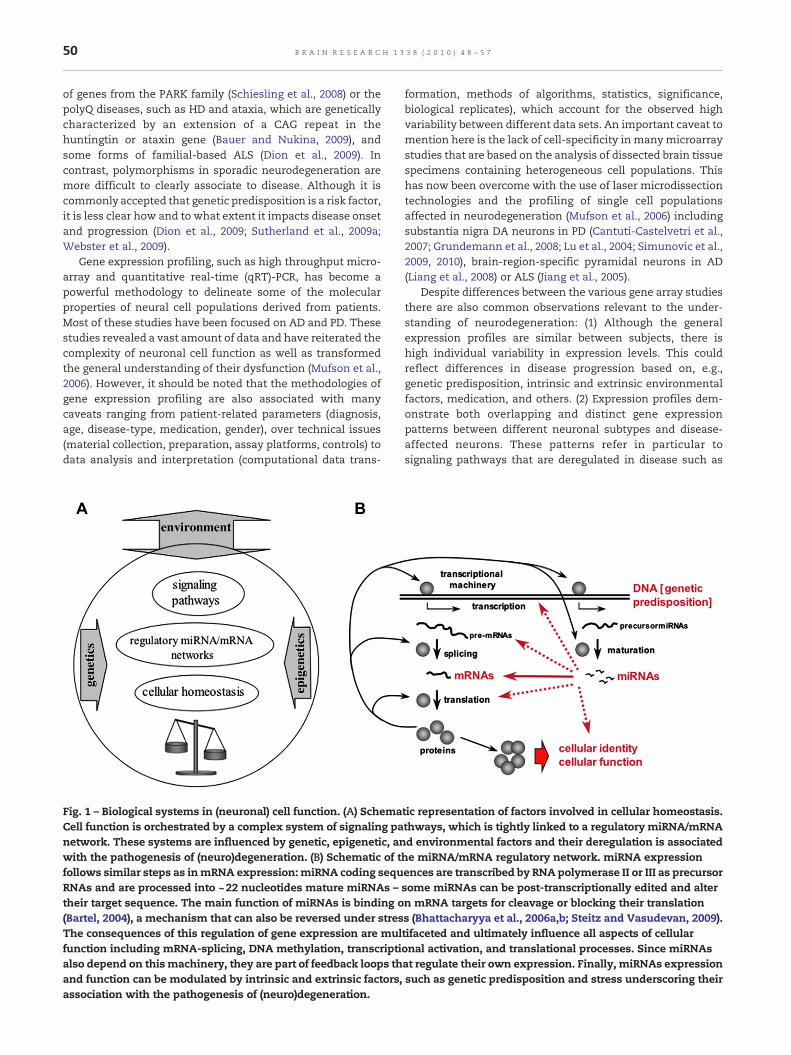
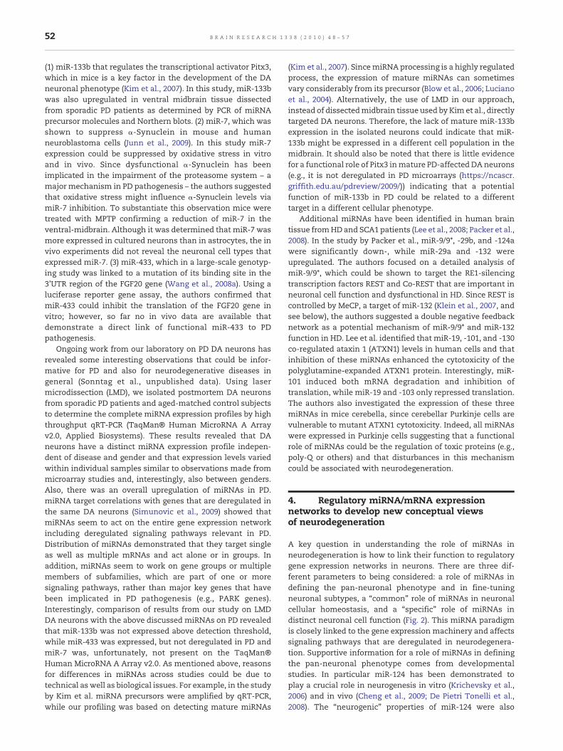
Fig. 1 – Biological systems in (neuronal) cell function. (A) SchemaCell function is orchestrated by a complex system of signaling panetwork. These systems are influenced by genetic, epigenetic, anwith the pathogenesis of (neuro)degeneration. (B) Schematic of tfollows similar steps as inmRNA expression: miRNA coding sequRNAs and are processed into ~22 nucleotides mature miRNAs –their target sequence. The main function of miRNAs is binding o(Bartel, 2004), a mechanism that can also be reversed under stresThe consequences of this regulation of gene expression are mulfunction including mRNA-splicing, DNA methylation, transcriptialso depend on this machinery, they are part of feedback loops thand function can be modulated by intrinsic and extrinsic factors,association with the pathogenesis of (neuro)degeneration.
formation, methods of algorithms, statistics, significance,biological replicates), which account for the observed highvariability between different data sets. An important caveat tomention here is the lack of cell-specificity in manymicroarraystudies that are based on the analysis of dissected brain tissuespecimens containing heterogeneous cell populations. Thishas now been overcome with the use of laser microdissectiontechnologies and the profiling of single cell populationsaffected in neurodegeneration (Mufson et al., 2006) includingsubstantia nigra DA neurons in PD (Cantuti-Castelvetri et al.,2007; Grundemann et al., 2008; Lu et al., 2004; Simunovic et al.,2009, 2010), brain-region-specific pyramidal neurons in AD(Liang et al., 2008) or ALS (Jiang et al., 2005).
Despite differences between the various gene array studiesthere are also common observations relevant to the under-standing of neurodegeneration: (1) Although the generalexpression profiles are similar between subjects, there ishigh individual variability in expression levels. This couldreflect differences in disease progression based on, e.g.,genetic predisposition, intrinsic and extrinsic environmentalfactors, medication, and others. (2) Expression profiles dem-onstrate both overlapping and distinct gene expressionpatterns between different neuronal subtypes and disease-affected neurons. These patterns refer in particular tosignaling pathways that are deregulated in disease such as
tic representation of factors involved in cellular homeostasis.thways, which is tightly linked to a regulatory miRNA/mRNAd environmental factors and their deregulation is associatedhe miRNA/mRNA regulatory network. miRNA expressionences are transcribed by RNA polymerase II or III as precursorsome miRNAs can be post-transcriptionally edited and altern mRNA targets for cleavage or blocking their translations (Bhattacharyya et al., 2006a,b; Steitz and Vasudevan, 2009).tifaceted and ultimately influence all aspects of cellularonal activation, and translational processes. Since miRNAsat regulate their own expression. Finally, miRNAs expressionsuch as genetic predisposition and stress underscoring their
51B R A I N R E S E A R C H 1 3 3 8 ( 2 0 1 0 ) 4 8 – 5 7
oxidative stress, exitotoxic insult, dysfunction of mitochon-dria, the ubiquitin-proteasome system, synapses, and thecytoskeleton, failure of axonal and dendritic transport, and ageneral disintegration of homeostatis (Altar et al., 2009; Jianget al., 2005; Liang et al., 2008; Mufson et al., 2006; Simunovicet al., 2009). These findings support the view that neurode-generation might be in part an accumulation of unifyingevents (Bossy-Wetzel et al., 2004; Jellinger, 2009). (3) Differ-ences in levels of gene expression are often subtle betweenneuronal subtypes and in neurons affected by diseaseimplying that even small changes in gene expression couldhave a profound impact on neuronal cell function andsurvival. In addition, diseased neurons exhibit deregulationwith predominantly downregulation of single (key) genes orgroups of gene families relevant in signaling pathways ofpathogenesis (although this seems to be less evident for ALS(Jiang et al., 2005)). Altogether, conceptually it appears that theentire gene expression networks underlie regulatory mechan-isms that define the pan-neuronal phenotype as well asneuronal subtypes and these mechanisms are disturbed inneurodegeneration (Altar et al., 2009; Jiang et al., 2005; Lianget al., 2008; Mufson et al., 2006; Simunovic et al., 2009, 2010).miRNAs fit well in this concept because of their function inmammalian neurogenesis, their role in determining neuronalphenotypes and function of postmitotic neurons, their distinctdistribution in several brain regions and single neurons andtheir deregulation in neurodegeneration. miRNAs seem toplay a profound role in steering and/or fine-tuning processesof neuronal cell function in both health and disease (Barbatoet al., 2008; Schratt, 2009).
3. miRNAs in neurodegeneration
As discussed above, large-scale gene profiling has revealednew insights into the molecular properties of disease-affectedneurons and an increasing body of data now offers theopportunity to tie together different aspects of normal andabnormal cell function, i.e., the influence of genetic predispo-sition (variability in coding and non-coding sequences), levelsof gene expression, functionality of gene products (pre- andpost-translation), gender, and the role of regulatory mechan-isms, such as the miRNA system. These factors provide acomplex regulatory expression network (Fig. 1), which mightor might not be unique to certain disease entities (see below).
The aspects of gene expression profiles in neurodegenera-tion as described above can be linked together with knownfeatures of miRNAs to develop a conceptual view of how thesemolecules might be involved in neurodegenerative disorders.So far, considerable screening efforts for miRNA expressionhave been performed on postmortem brain material from ADpatients (summarized in Hebert and De Strooper, 2009;Cogswell et al., 2008) and, in one study, also in PD patients(Kim et al., 2007). As discussed above, it should be mentionedthat in these studies dissected tissue samples containingheterogeneous cell populations were analyzed. This is animportant parameter for data interpretation, since it iscurrently not clear how miRNA expression and functionrelates to individual cell types in the human brain and howthey are involved in the complex interplay of neural subtypes.
Nevertheless, in AD, these studies revealed some insight intopossible mechanisms of pathogenesis. For example, themiRNAs miR-106a, -520c, -106b, -17-5p, and 20a could down-regulate amyloid beta precursor protein (APP) gene expressionin vitro (Hebert et al., 2009; Patel et al., 2008), but so far onlymiR-106b expression was found in sporadic AD patients(Hebert et al., 2009). In another study, reduction of miR-29b-1and miR-29a in the temporal cortex of AD patients correlatedwith increased β-site APP cleaving enzyme 1 (BACE1) expres-sion in the same sample population (Hebert et al., 2008; Hebertand De Strooper, 2009). Since BACE1 is a target for bothmiRNAs and the rate-limiting enzyme for Aβ production,these results could imply correlative evidence for a mecha-nism in this disease. Similarly, another study demonstrateddiminished miR-107 expression in cortices from AD patientsand biochemical analyses showed that BACE1 was a target forthis miRNA (Wang et al., 2008b). Supportive information camealso from a mouse model of AD, in which miR-298 and -328could be identified as targeting miRNAs for BACE1 (Boisson-neault et al., 2009). These data imply that critical genes in ADpathogenesis could be regulated by multiple miRNAs. Addi-tional evidence for possible miRNA functions in AD wasrecently provided by data from the same AD mouse model asused by (Boissonneault et al., 2009) – i.e., APPswe/PSΔE9transgenic mice, which contain a mouse-human hybrid ofthe APP gene with the Swedish mutation K594N/M595L andthe PS1ΔE9 transgene that encodes the exon-9-deleted humanpresenilin-1 (Wang et al., 2009). In this study miR-34a wasfunctionally linked to downregulation of bcl2 expression, ananti-apoptotic protein involved in the regulation of theapoptotic proteins caspase 9 and 3. The authors couldexperimentally demonstrate that miR-34a indirectly regulatescaspase 3 by targeting bcl2 and this correlated with anincreased expression of miR-34a and caspase 3 in the corticesof the transgenic mice. These results indicated an involve-ment of miRNAs in the apoptotic signaling pathway, which isrelevant in AD neurodegeneration (LeBlanc, 2005). Interest-ingly, miRNA arrays in the transgenic mice revealed deregu-lated expression of the AD-associated miRNAs miR-20a, -29a,-125b, -128a and -106b, but not others, such asmiR-29b-1, -107,-298, and -328 (Wang et al., 2009). This profile also overlappedpoorly with data from a recent large-scale miRNA screeningusing high throughput miRNA qRT-PCR technology on severalbrain regions and cerebral spinal fluid (CSF) from AD patientsand age-matched controls (Cogswell et al., 2008). In this study,the authors reported a set of multiple miRNAs that wereregionally and stage-specifically deregulated, but this includ-ed neither miR-29a/b-1 and miR-107 nor miR-298, -328, and-34a from the mouse studies. Despite the differences in thebiological systems, little data overlap inmiRNA profiling couldbe related to technical differences and difficulties in conduct-ing the miRNA screening experiments (see Hebert and DeStrooper, 2009, for further discussion), considerable variationswithin patient populations as also seen in microarray studies(discussed, e.g., in (Simunovic et al., 2009, 2010)), and/ordistinct patterns in the biology of miRNA functions (see textbelow).
In contrast to AD, there is very little information of miRNAexpression in PD-affected brains. So far, three miRNAs havebeen linked to the dopaminergic phenotype in relation to PD:
52 B R A I N R E S E A R C H 1 3 3 8 ( 2 0 1 0 ) 4 8 – 5 7
(1) miR-133b that regulates the transcriptional activator Pitx3,which in mice is a key factor in the development of the DAneuronal phenotype (Kim et al., 2007). In this study, miR-133bwas also upregulated in ventral midbrain tissue dissectedfrom sporadic PD patients as determined by PCR of miRNAprecursor molecules and Northern blots. (2) miR-7, which wasshown to suppress α-Synuclein in mouse and humanneuroblastoma cells (Junn et al., 2009). In this study miR-7expression could be suppressed by oxidative stress in vitroand in vivo. Since dysfunctional α-Synuclein has beenimplicated in the impairment of the proteasome system – amajor mechanism in PD pathogenesis – the authors suggestedthat oxidative stress might influence α-Synuclein levels viamiR-7 inhibition. To substantiate this observation mice weretreated with MPTP confirming a reduction of miR-7 in theventral-midbrain. Although it was determined that miR-7 wasmore expressed in cultured neurons than in astrocytes, the invivo experiments did not reveal the neuronal cell types thatexpressed miR-7. (3) miR-433, which in a large-scale genotyp-ing study was linked to a mutation of its binding site in the3'UTR region of the FGF20 gene (Wang et al., 2008a). Using aluciferase reporter gene assay, the authors confirmed thatmiR-433 could inhibit the translation of the FGF20 gene invitro; however, so far no in vivo data are available thatdemonstrate a direct link of functional miR-433 to PDpathogenesis.
Ongoing work from our laboratory on PD DA neurons hasrevealed some interesting observations that could be infor-mative for PD and also for neurodegenerative diseases ingeneral (Sonntag et al., unpublished data). Using lasermicrodissection (LMD), we isolated postmortem DA neuronsfrom sporadic PD patients and aged-matched control subjectsto determine the complete miRNA expression profiles by highthroughput qRT-PCR (TaqMan® Human MicroRNA A Arrayv2.0, Applied Biosystems). These results revealed that DAneurons have a distinct miRNA expression profile indepen-dent of disease and gender and that expression levels variedwithin individual samples similar to observations made frommicroarray studies and, interestingly, also between genders.Also, there was an overall upregulation of miRNAs in PD.miRNA target correlations with genes that are deregulated inthe same DA neurons (Simunovic et al., 2009) showed thatmiRNAs seem to act on the entire gene expression networkincluding deregulated signaling pathways relevant in PD.Distribution of miRNAs demonstrated that they target singleas well as multiple mRNAs and act alone or in groups. Inaddition, miRNAs seem to work on gene groups or multiplemembers of subfamilies, which are part of one or moresignaling pathways, rather than major key genes that havebeen implicated in PD pathogenesis (e.g., PARK genes).Interestingly, comparison of results from our study on LMDDA neurons with the above discussed miRNAs on PD revealedthat miR-133b was not expressed above detection threshold,while miR-433 was expressed, but not deregulated in PD andmiR-7 was, unfortunately, not present on the TaqMan®Human MicroRNA A Array v2.0. As mentioned above, reasonsfor differences in miRNAs across studies could be due totechnical as well as biological issues. For example, in the studyby Kim et al. miRNA precursors were amplified by qRT-PCR,while our profiling was based on detecting mature miRNAs
(Kim et al., 2007). SincemiRNA processing is a highly regulatedprocess, the expression of mature miRNAs can sometimesvary considerably from its precursor (Blow et al., 2006; Lucianoet al., 2004). Alternatively, the use of LMD in our approach,instead of dissectedmidbrain tissue used by Kim et al., directlytargeted DA neurons. Therefore, the lack of mature miR-133bexpression in the isolated neurons could indicate that miR-133b might be expressed in a different cell population in themidbrain. It should also be noted that there is little evidencefor a functional role of Pitx3 inmature PD-affected DAneurons(e.g., it is not deregulated in PD microarrays (https://ncascr.griffith.edu.au/pdreview/2009/)) indicating that a potentialfunction of miR-133b in PD could be related to a differenttarget in a different cellular phenotype.
Additional miRNAs have been identified in human braintissue fromHD and SCA1 patients (Lee et al., 2008; Packer et al.,2008). In the study by Packer et al., miR-9/9*, -29b, and -124awere significantly down-, while miR-29a and -132 wereupregulated. The authors focused on a detailed analysis ofmiR-9/9*, which could be shown to target the RE1-silencingtranscription factors REST and Co-REST that are important inneuronal cell function and dysfunctional in HD. Since REST iscontrolled by MeCP, a target of miR-132 (Klein et al., 2007, andsee below), the authors suggested a double negative feedbacknetwork as a potential mechanism of miR-9/9* and miR-132function in HD. Lee et al. identified that miR-19, -101, and -130co-regulated ataxin 1 (ATXN1) levels in human cells and thatinhibition of these miRNAs enhanced the cytotoxicity of thepolyglutamine-expanded ATXN1 protein. Interestingly, miR-101 induced both mRNA degradation and inhibition oftranslation, while miR-19 and -103 only repressed translation.The authors also investigated the expression of these threemiRNAs in mice cerebella, since cerebellar Purkinje cells arevulnerable to mutant ATXN1 cytotoxicity. Indeed, all miRNAswere expressed in Purkinje cells suggesting that a functionalrole of miRNAs could be the regulation of toxic proteins (e.g.,poly-Q or others) and that disturbances in this mechanismcould be associated with neurodegeneration.
4. Regulatory miRNA/mRNA expressionnetworks to develop new conceptual viewsof neurodegeneration
A key question in understanding the role of miRNAs inneurodegeneration is how to link their function to regulatorygene expression networks in neurons. There are three dif-ferent parameters to being considered: a role of miRNAs indefining the pan-neuronal phenotype and in fine-tuningneuronal subtypes, a “common” role of miRNAs in neuronalcellular homeostasis, and a “specific” role of miRNAs indistinct neuronal cell function (Fig. 2). This miRNA paradigmis closely linked to the gene expression machinery and affectssignaling pathways that are deregulated in neurodegenera-tion. Supportive information for a role of miRNAs in definingthe pan-neuronal phenotype comes from developmentalstudies. In particular miR-124 has been demonstrated toplay a crucial role in neurogenesis in vitro (Krichevsky et al.,2006) and in vivo (Cheng et al., 2009; De Pietri Tonelli et al.,2008). The “neurogenic” properties of miR-124 were also
53B R A I N R E S E A R C H 1 3 3 8 ( 2 0 1 0 ) 4 8 – 5 7
demonstrated in a series of other studies (Cao et al., 2007;Cheng et al., 2009; Makeyev et al., 2007; Visvanathan et al.,2007) including an elegant experiment conducted by Lim et al.,in which overexpression of miR-124 in HeLa cells could inducea neuron-specific expression profile (Lim et al., 2005). ThemiR-124 transfected cells downregulated 174 genes, and fromthese genes ∼20% were also deregulated in PD DA neurons(Simunovic et al., 2009, 2010). Similarly, from a set of 27validated targets after immunoprecipitation assays usingHEK-293S cells transfected with miR-124a (Karginovet al., 2007), eight of the targets were deregulated in PD DAneurons. These results correlated with information from theMIRECORDS database (http://mirecords.umn.edu/), fromwhich 49 (24%) genes out of 202 “validated” targets for miR-124 were deregulated in PD. Since miR-124 is abundantlyexpressed in the adult brain (Nelson et al., 2008; Nelson andWilfred, 2009), one could speculate that this miRNA might bealso involved in the DA neuronal phenotype and in PDpathogenesis or in both. Another example for pan-neuronalphenotype-specific miRNAs is miR-132, whose expression isactivated by the transcriptional activator CREB, and whichseems to form a feedback loop with its target MeCP2 andbrain-derived neurotrophic factor (BDNF) – BDNF increasesmiR-132 expression, which downregulates MeCP2 that in turndecreases BDNF (Klein et al., 2007; Vo et al., 2005). Interest-ingly, BDNF inhibits the function of the spine-specific miRNAmiR-132, which targets Lmtk1 a protein involved in dendriticspine densities (Schratt et al., 2006). These data attest to the
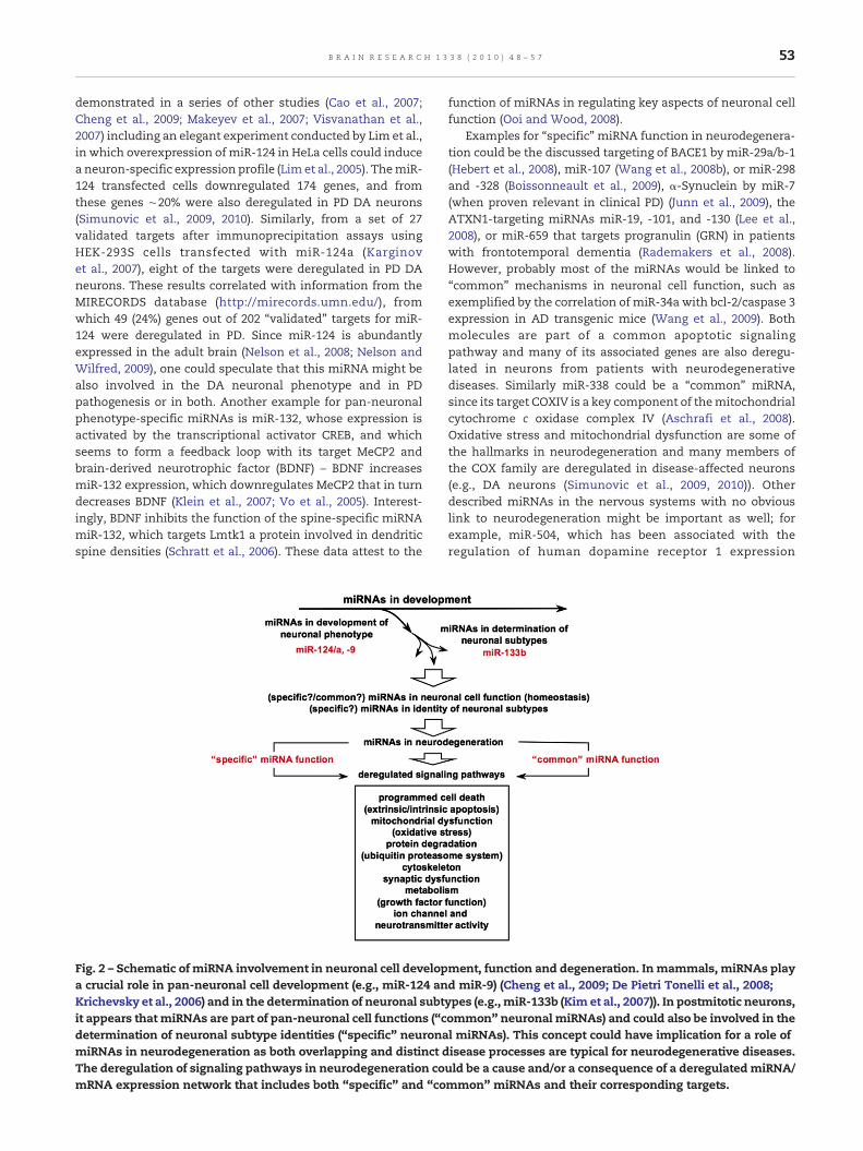
Fig. 2 – Schematic of miRNA involvement in neuronal cell developa crucial role in pan-neuronal cell development (e.g., miR-124 anKrichevsky et al., 2006) and in the determination of neuronal subtit appears that miRNAs are part of pan-neuronal cell functions (“cdetermination of neuronal subtype identities (“specific” neuronamiRNAs in neurodegeneration as both overlapping and distinct dThe deregulation of signaling pathways in neurodegeneration comRNA expression network that includes both “specific” and “co
function of miRNAs in regulating key aspects of neuronal cellfunction (Ooi and Wood, 2008).
Examples for “specific” miRNA function in neurodegenera-tion could be the discussed targeting of BACE1 by miR-29a/b-1(Hebert et al., 2008), miR-107 (Wang et al., 2008b), or miR-298and -328 (Boissonneault et al., 2009), α-Synuclein by miR-7(when proven relevant in clinical PD) (Junn et al., 2009), theATXN1-targeting miRNAs miR-19, -101, and -130 (Lee et al.,2008), or miR-659 that targets progranulin (GRN) in patientswith frontotemporal dementia (Rademakers et al., 2008).However, probably most of the miRNAs would be linked to“common” mechanisms in neuronal cell function, such asexemplified by the correlation of miR-34a with bcl-2/caspase 3expression in AD transgenic mice (Wang et al., 2009). Bothmolecules are part of a common apoptotic signalingpathway and many of its associated genes are also deregu-lated in neurons from patients with neurodegenerativediseases. Similarly miR-338 could be a “common” miRNA,since its target COXIV is a key component of themitochondrialcytochrome c oxidase complex IV (Aschrafi et al., 2008).Oxidative stress and mitochondrial dysfunction are some ofthe hallmarks in neurodegeneration and many members ofthe COX family are deregulated in disease-affected neurons(e.g., DA neurons (Simunovic et al., 2009, 2010)). Otherdescribed miRNAs in the nervous systems with no obviouslink to neurodegeneration might be important as well; forexample, miR-504, which has been associated with theregulation of human dopamine receptor 1 expression
ment, function and degeneration. In mammals, miRNAs playd miR-9) (Cheng et al., 2009; De Pietri Tonelli et al., 2008;ypes (e.g., miR-133b (Kim et al., 2007)). In postmitotic neurons,ommon” neuronal miRNAs) and could also be involved in thel miRNAs). This concept could have implication for a role ofisease processes are typical for neurodegenerative diseases.uld be a cause and/or a consequence of a deregulatedmiRNA/mmon” miRNAs and their corresponding targets.
54 B R A I N R E S E A R C H 1 3 3 8 ( 2 0 1 0 ) 4 8 – 5 7
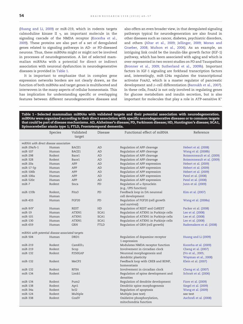
(Huang and Li, 2009) or miR-219, which in rodents targetscalmoduline kinase II γ, an important molecule in thesignaling cascade of the NMDA receptor (Kocerha et al.,2009). These proteins are also part of a set of deregulatedgenes related to signaling pathways in AD- or PD-diseasedneurons. Thus, these miRNAs might or might not be involvedin processes of neurodegeneration. A list of selected mam-malian miRNAs with a potential for direct or indirectassociation with neuronal dysfunction in neurodegenerativediseases is provided in Table 1.
It is important to emphasize that in complex geneexpression networks borders are not clearly drawn, as thefunction of both miRNAs and target genes is multifaceted andinterwoven in the many aspects of cellular homeostasis. Thishas implication for understanding specific or overlappingfeatures between different neurodegenerative diseases and
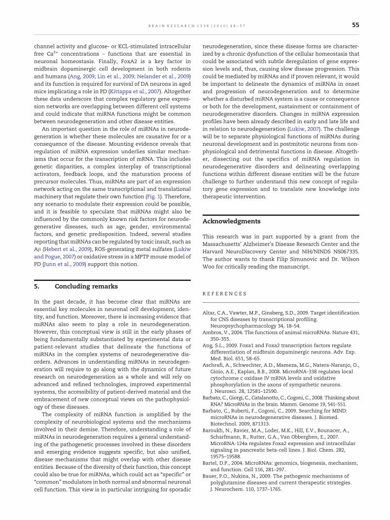
Table 1 – Selected mammalian miRNAs with validated targetmiRNAswere organized according to their direct associationwitthat could be part of diseasemechanisms. AD, Alzheimer's diseSpinocerebellar ataxia type 1; FTLD, Frontotemporal dementia.
Species Validatedtarget
Disease
miRNA with direct disease associationmiR-29a/b-1 Human BACE1 AD RemiR-107 Human BACE1 AD RemiR-298 Rodent Bace1 AD RemiR-328 Rodent Bace1 AD RemiR-20a Human APP AD RemiR-17-5p Human APP AD RemiR-106b Human APP AD RemiR-106a Human APP AD RemiR-520c Human APP AD RemiR-7 Rodent Snca PD Re
(e.miR-133b Rodent,
HumanPitx3 PD Fe
cemiR-433 Human FGF20 PD Re
anmiR-9/9* Human REST HD RemiR-19 Human ATXN1 SCA1 RemiR-101 Human ATXN1 SCA1 RemiR-130 Human ATXN1 SCA1 RemiR-659 Human GRN FTLD Re
miRNA with potential disease-associated targetsmiR-504 Human DRD1 Re
1 emiR-219 Rodent CamKIIγ MomiR-219 Rodent Scop InmiR-132 Rodent P250GAP Ne
demiR-132 Rodent MeCP2 Fe
homiR-132 Rodent RFX4 InmiR-134 Rodent Limk1 Re
demiR-134 Rodent Pum2 RemiR-138 Rodent Apt1 DemiR-34a Rodent bcl2 RemiR-124 Rodent Multiple MumiR-338 Rodent CoxIV Ox
m
also offers an even broader view, in that deregulated signalingpathways typical for neurodegeneration are also found inother diseases such as cancer, diabetes, psychiatric disorders,and others (Altar et al., 2009; Jellinger, 2009; Moran andGraeber, 2008; Mufson et al., 2006). As an example, anintriguing link could be the insulin-like growth factor (IGF-1)pathway, which has been associated with aging and which isover-represented in two recent studies on PD and Tauopathies(Bronner et al., 2009; Sutherland et al., 2009b). Importantfactors in IGF-1 signaling are forkhead transcription factorsand, interestingly, miR-124a regulates the transcriptionalactivator FoxA2, which is a master regulator of pancreaticdevelopment and β-cell differentiation (Baroukh et al., 2007).In these cells, FoxA2 is not only involved in regulating genesfor glucose metabolism and insulin secretion, but is alsoimportant for molecules that play a role in ATP-sensitive K+
s and their potential association with neurodegeneration.h specific neurodegenerative diseases or to common targetsase; PD, Parkinson disease; HD, Huntington's disease; SCA1,
Functional effect of miRNA Reference
gulation of APP cleavage Hebert et al. (2008)gulation of APP cleavage Wang et al. (2008b)gulation of APP cleavage Boissonneault et al. (2009)gulation of APP cleavage Boissonneault et al. (2009)gulation of APP expression Hebert et al. (2009)gulation of APP expression Hebert et al. (2009)gulation of APP expression Hebert et al. (2009)gulation of APP expression Patel et al. (2008)gulation of APP expression Patel et al. (2008)gulation of α-Synucleing., UPS function)
Junn et al. (2009)
edback loop in DA neuronalll development
Kim et al. (2007)
gulation of FGF20 (cell growthd survival)
Wang et al. (2008a)
gulation of REST and CoREST Packer et al. (2008)gulation of ATXN1 in Purkinje cells Lee et al. (2008)gulation of ATXN1 in Purkinje cells Lee et al. (2008)gulation of ATXN1 in Purkinje cells Lee et al. (2008)gulation of GRN (cell growth) Rademakers et al. (2008)
gulation of dopamine receptorxpression
Huang and Li (2009)
dulates NMDA receptor function Kocerha et al. (2009)volvement in circadian clock Cheng et al. (2007)uronal morphogenesis andndritic plasticity
(Vo et al., 2005;Wayman et al., 2008)
edback loop with CREB and BDNF,meostasis
Klein et al. (2007)
volvement in circadian clock Cheng et al. (2007)gulation of spine development andnsities
Schratt et al. (2006)
gulation of dendrite development Fiore et al. (2009)ndritic spine morphogenesis Siegel et al. (2009)gulation of apoptosis Wang et al. (2009)ltiple (see text) see textidative phosphorylation,itochondria function
Aschrafi et al. (2008)
55B R A I N R E S E A R C H 1 3 3 8 ( 2 0 1 0 ) 4 8 – 5 7
channel activity and glucose- or KCL-stimulated intracellularfree Ca2+ concentrations – functions that are essential inneuronal homeostasis. Finally, FoxA2 is a key factor inmidbrain dopaminergic cell development in both rodentsand humans (Ang, 2009; Lin et al., 2009; Nelander et al., 2009)and its function is required for survival of DA neurons in agedmice implicating a role in PD (Kittappa et al., 2007). Altogetherthese data underscore that complex regulatory gene expres-sion networks are overlapping between different cell systemsand could indicate that miRNA functions might be commonbetween neurodegeneration and other disease entities.
An important question in the role of miRNAs in neurode-generation is whether these molecules are causative for or aconsequence of the disease. Mounting evidence reveals thatregulation of miRNA expression underlies similar mechan-isms that occur for the transcription of mRNA. This includesgenetic disparities, a complex interplay of transcriptionalactivators, feedback loops, and the maturation process ofprecursor molecules. Thus, miRNAs are part of an expressionnetwork acting on the same transcriptional and translationalmachinery that regulate their own function (Fig. 1). Therefore,any scenario to modulate their expression could be possible,and it is feasible to speculate that miRNAs might also beinfluenced by the commonly known risk factors for neurode-generative diseases, such as age, gender, environmentalfactors, and genetic predisposition. Indeed, several studiesreporting thatmiRNAs can be regulated by toxic insult, such asAβ (Hebert et al., 2009), ROS-generating metal sulfates (Lukiwand Pogue, 2007) or oxidative stress in a MPTPmousemodel ofPD (Junn et al., 2009) support this notion.
5. Concluding remarks
In the past decade, it has become clear that miRNAs areessential key molecules in neuronal cell development, iden-tity, and function. Moreover, there is increasing evidence thatmiRNAs also seem to play a role in neurodegeneration.However, this conceptual view is still in the early phases ofbeing fundamentally substantiated by experimental data orpatient-relevant studies that delineate the functions ofmiRNAs in the complex systems of neurodegenerative dis-orders. Advances in understanding miRNAs in neurodegen-eration will require to go along with the dynamics of futureresearch on neurodegeneration as a whole and will rely onadvanced and refined technologies, improved experimentalsystems, the accessibility of patient-derived material and theembracement of new conceptual views on the pathophysiol-ogy of these diseases.
The complexity of miRNA function is amplified by thecomplexity of neurobiological systems and the mechanismsinvolved in their demise. Therefore, understanding a role ofmiRNAs in neurodegeneration requires a general understand-ing of the pathogenetic processes involved in these disordersand emerging evidence suggests specific, but also unified,disease mechanisms that might overlap with other diseaseentities. Because of the diversity of their function, this conceptcould also be true for miRNAs, which could act as “specific” or“common”modulators in both normal and abnormal neuronalcell function. This view is in particular intriguing for sporadic
neurodegeneration, since these disease forms are character-ized by a chronic dysfunction of the cellular homeostasis thatcould be associated with subtle deregulation of gene expres-sion levels and, thus, causing slow disease progression. Thiscould be mediated by miRNAs and if proven relevant, it wouldbe important to delineate the dynamics of miRNAs in onsetand progression of neurodegeneration and to determinewhether a disturbedmiRNA system is a cause or consequenceor both for the development, sustainment or containment ofneurodegenerative disorders. Changes in miRNA expressionprofiles have been already described in early and late life andin relation to neurodegeneration (Lukiw, 2007). The challengewill be to separate physiological functions of miRNAs duringneuronal development and in postmitotic neurons from non-physiological and detrimental functions in disease. Altogeth-er, dissecting out the specifics of miRNA regulation inneurodegenerative disorders and delineating overlappingfunctions within different disease entities will be the futurechallenge to further understand this new concept of regula-tory gene expression and to translate new knowledge intotherapeutic intervention.
Acknowledgments
This research was in part supported by a grant from theMassachusetts’ Alzheimer's Disease Research Center and theHarvard NeuroDiscovery Center and NIH/NINDS NS067335.The author wants to thank Filip Simunovic and Dr. WilsonWoo for critically reading the manuscript.
R E F E R E N C E S
Altar, C.A., Vawter, M.P., Ginsberg, S.D., 2009. Target identificationfor CNS diseases by transcriptional profiling.Neuropsychopharmacology 34, 18–54.
Ambros, V., 2004. The functions of animal microRNAs. Nature 431,350–355.
Ang, S.L., 2009. Foxa1 and Foxa2 transcription factors regulatedifferentiation of midbrain dopaminergic neurons. Adv. Exp.Med. Biol. 651, 58–65.
Aschrafi, A., Schwechter, A.D., Mameza, M.G., Natera-Naranjo, O.,Gioio, A.E., Kaplan, B.B., 2008. MicroRNA-338 regulates localcytochrome c oxidase IV mRNA levels and oxidativephosphorylation in the axons of sympathetic neurons.J. Neurosci. 28, 12581–12590.
Barbato, C., Giorgi, C., Catalanotto, C., Cogoni, C., 2008. Thinking aboutRNA? MicroRNAs in the brain. Mamm. Genome 19, 541–551.
Barbato, C., Ruberti, F., Cogoni, C., 2009. Searching for MIND:microRNAs in neurodegenerative diseases. J. Biomed.Biotechnol. 2009, 871313.
Baroukh, N., Ravier, M.A., Loder, M.K., Hill, E.V., Bounacer, A.,Scharfmann, R., Rutter, G.A., Van Obberghen, E., 2007.MicroRNA-124a regulates Foxa2 expression and intracellularsignaling in pancreatic beta-cell lines. J. Biol. Chem. 282,19575–19588.
Bartel, D.P., 2004. MicroRNAs: genomics, biogenesis, mechanism,and function. Cell 116, 281–297.
Bauer, P.O., Nukina, N., 2009. The pathogenic mechanisms ofpolyglutamine diseases and current therapeutic strategies.J. Neurochem. 110, 1737–1765.
56 B R A I N R E S E A R C H 1 3 3 8 ( 2 0 1 0 ) 4 8 – 5 7
Bhattacharyya, S.N., Habermacher, R., Martine, U., Closs, E.I.,Filipowicz, W., 2006a. Relief of microRNA-mediatedtranslational repression in human cells subjected to stress. Cell125, 1111–1124.
Bhattacharyya, S.N., Habermacher, R., Martine, U., Closs, E.I.,Filipowicz, W., 2006b. Stress-induced reversal of microRNArepression and mRNA P-body localization in human cells. ColdSpring Harb. Symp. Quant. Biol. 71, 513–521.
Blow, M.J., Grocock, R.J., van Dongen, S., Enright, A.J., Dicks, E.,Futreal, P.A., Wooster, R., Stratton, M.R., 2006. RNA editing ofhuman microRNAs. Genome Biol. 7, R27.
Boissonneault, V., Plante, I., Rivest, S., Provost, P., 2009.MicroRNA-298 and microRNA-328 regulate expression ofmouse beta-amyloid precursor protein-converting enzyme 1.J. Biol. Chem. 284, 1971–1981.
Bossy-Wetzel, E., Schwarzenbacher, R., Lipton, S.A., 2004.Molecular pathways to neurodegeneration. Nat. Med. 10 Suppl,S2–S9.
Bronner, I.F., Bochdanovits, Z., Rizzu, P., Kamphorst, W., Ravid, R.,van Swieten, J.C., Heutink, P., 2009. Comprehensive mRNAexpression profiling distinguishes tauopathies and identifiesshared molecular pathways. PLoS ONE 4, e6826.
Bushati, N., Cohen, S.M., 2008. MicroRNAs in neurodegeneration.Curr. Opin. Neurobiol. 18, 292–296.
Cantuti-Castelvetri, I., Keller-McGandy, C., Bouzou, B., Asteris, G.,Clark, T.W., Frosch, M.P., Standaert, D.G., 2007. Effects of genderon nigral gene expression and parkinson disease. Neurobiol.Dis. 26, 606–614.
Cao, X., Pfaff, S.L., Gage, F.H., 2007. A functional study ofmiR-124 inthe developing neural tube. Genes Dev. 21, 531–536.
Cheng, H.Y., Papp, J.W., Varlamova, O., Dziema, H., Russell, B.,Curfman, J.P., Nakazawa, T., Shimizu, K., Okamura, H., Impey, S.,Obrietan, K., 2007. microRNA modulation of circadian-clockperiod and entrainment. Neuron 54, 813–829.
Cheng, L.C., Pastrana, E., Tavazoie, M., Doetsch, F., 2009. miR-124regulates adult neurogenesis in the subventricular zone stemcell niche. Nat. Neurosci. 12, 399–408.
Cogswell, J.P., Ward, J., Taylor, I.A., Waters, M., Shi, Y., Cannon, B.,Kelnar, K., Kemppainen, J., Brown, D., Chen, C., Prinjha, R.K.,Richardson, J.C., Saunders, A.M., Roses, A.D., Richards, C.A.,2008. Identification of miRNA changes in Alzheimer's diseasebrain and CSF yields putative biomarkers and insights intodisease pathways. J. Alzheimers Dis. 14, 27–41.
Croce, C.M., 2009. Causes and consequences of microRNAdysregulation in cancer. Nat. Rev. Genet. 10, 704–714.
De Pietri Tonelli, D., Pulvers, J.N., Haffner, C., Murchison, E.P.,Hannon, G.J., Huttner, W.B., 2008. miRNAs are essential forsurvival and differentiation of newborn neurons but not forexpansion of neural progenitors during early neurogenesis inthe mouse embryonic neocortex. Development 135, 3911–3921.
Dion, P.A., Daoud, H., Rouleau, G.A., 2009. Genetics of motorneuron disorders: new insights into pathogenic mechanisms.Nat. Rev. Genet. 10, 769–782.
Eacker, S.M., Dawson, T.M., Dawson, V.L., 2009. UnderstandingmicroRNAs in neurodegeneration. Nat. Rev. Neurosci. 10, 837–841.
Fiore, R., Siegel, G., Schratt, G., 2008. MicroRNA function inneuronal development, plasticity and disease. Biochim.Biophys. Acta 1779, 471–478.
Fiore, R., Khudayberdiev, S., Christensen, M., Siegel, G., Flavell, S.W.,Kim, T.K., Greenberg, M.E., Schratt, G., 2009. Mef2-mediatedtranscription of the miR379-410 cluster regulatesactivity-dependent dendritogenesis by fine-tuning Pumilio2protein levels. Embo J. 28, 697–710.
Garzon, R., Calin, G.A., Croce, C.M., 2009. MicroRNAs in cancer.Annu. Rev. Med. 60, 167–179.
Grundemann, J., Schlaudraff, F., Haeckel, O., Liss, B., 2008. Elevatedalpha-synuclein mRNA levels in individualUV-laser-microdissected dopaminergic substantia nigra neuronsin idiopathic Parkinson's disease. Nucleic Acids Res. 36, e38.
Hebert, S.S., De Strooper, B., 2007. Molecular biology. miRNAs inneurodegeneration. Science 317, 1179–1180.
Hebert, S.S., De Strooper, B., 2009. Alterations of the microRNAnetwork cause neurodegenerative disease. Trends Neurosci.32, 199–206.
Hebert, S.S., Horre, K., Nicolai, L., Papadopoulou, A.S.,Mandemakers,W., Silahtaroglu, A.N., Kauppinen, S., Delacourte, A., De Strooper,B., 2008. Loss of microRNA cluster miR-29a/b-1 in sporadicAlzheimer's disease correlates with increasedBACE1/beta-secretase expression. Proc. Natl. Acad. Sci. U.S.A.105, 6415–6420.
Hebert, S.S., Horre, K., Nicolai, L., Bergmans, B., Papadopoulou, A.S.,Delacourte, A., De Strooper, B., 2009. MicroRNA regulation ofAlzheimer's amyloid precursor protein expression. Neurobiol.Dis. 33, 422–428.
Huang, W., Li, M.D., 2009. Differential allelic expression ofdopamine D1 receptor gene (DRD1) is modulated by microRNAmiR-504. Biol. Psychiatry 65, 702–705.
Jellinger, K.A., 2009. Recent advances in our understanding ofneurodegeneration. J. Neural Transm. 116, 1111–1162.
Jiang, Y.M., Yamamoto, M., Kobayashi, Y., Yoshihara, T., Liang, Y.,Terao, S., Takeuchi, H., Ishigaki, S., Katsuno,M., Adachi, H., Niwa,J., Tanaka, F., Doyu, M., Yoshida, M., Hashizume, Y., Sobue, G.,2005. Gene expression profile of spinal motor neurons insporadic amyotrophic lateral sclerosis. Ann. Neurol. 57, 236–251.
Junn, E., Lee, K.W., Jeong, B.S., Chan, T.W., Im, J.Y., Mouradian, M.M.,2009. Repression of alpha-synuclein expression and toxicity bymicroRNA-7. Proc. Natl. Acad. Sci. U.S.A. 106, 13052–13057.
Karginov, F.V., Conaco, C., Xuan, Z., Schmidt, B.H., Parker, J.S.,Mandel, G., Hannon, G.J., 2007. A biochemical approach toidentifying microRNA targets. Proc. Natl. Acad. Sci. U.S.A. 104,19291–19296.
Kim, J., Inoue, K., Ishii, J., Vanti, W.B., Voronov, S.V., Murchison, E.,Hannon, G., Abeliovich, A., 2007. A MicroRNA feedback circuitin midbrain dopamine neurons. Science 317, 1220–1224.
Kittappa, R., Chang,W.W., Awatramani, R.B., McKay, R.D., 2007. Thefoxa2 gene controls the birth and spontaneous degeneration ofdopamine neurons in old age. PLoS Biol. 5, e325.
Klein, M.E., Lioy, D.T., Ma, L., Impey, S., Mandel, G., Goodman, R.H.,2007. Homeostatic regulation of MeCP2 expression by aCREB-induced microRNA. Nat. Neurosci. 10, 1513–1514.
Kocerha, J., Faghihi, M.A., Lopez-Toledano, M.A., Huang, J., Ramsey,A.J., Caron,M.G., Sales,N.,Willoughby, D., Elmen, J., Hansen,H.F.,Orum, H., Kauppinen, S., Kenny, P.J., Wahlestedt, C., 2009.MicroRNA-219 modulates NMDA receptor-mediatedneurobehavioral dysfunction. Proc. Natl. Acad. Sci. U.S.A. 106,3507–3512.
Krichevsky, A.M., Gabriely, G., 2009. miR-21: a small multi-facetedRNA. J. Cell. Mol. Med. 13, 39–53.
Krichevsky, A.M., Sonntag, K.C., Isacson, O., Kosik, K.S., 2006.Specific microRNAs modulate embryonic stem cell-derivedneurogenesis. Stem Cells 24, 857–864.
Kuss, A.W., Chen, W., 2008. MicroRNAs in brain function anddisease. Curr. Neurol. Neurosci. Rep. 8, 190–197.
LeBlanc, A.C., 2005. The role of apoptotic pathways in Alzheimer'sdisease neurodegeneration and cell death. Curr. AlzheimerRes. 2, 389–402.
Lee, Y., Samaco, R.C., Gatchel, J.R., Thaller, C., Orr, H.T., Zoghbi, H.Y.,2008. miR-19, miR-101 andmiR-130 co-regulate ATXN1 levels topotentially modulate SCA1 pathogenesis. Nat. Neurosci. 11,1137–1139.
Liang, W.S., Dunckley, T., Beach, T.G., Grover, A., Mastroeni, D.,Ramsey, K., Caselli, R.J., Kukull, W.A., McKeel, D., Morris, J.C.,Hulette, C.M., Schmechel, D., Reiman, E.M., Rogers, J., Stephan,D.A., 2008. Altered neuronal gene expression in brain regionsdifferentially affected by Alzheimer's disease: a reference dataset. Physiol. Genomics 33, 240–256.
Lim, L.P., Lau, N.C., Garrett-Engele, P., Grimson, A., Schelter, J.M.,Castle, J., Bartel, D.P., Linsley, P.S., Johnson, J.M., 2005. Microarray
57B R A I N R E S E A R C H 1 3 3 8 ( 2 0 1 0 ) 4 8 – 5 7
analysis shows that somemicroRNAs downregulate largenumbers of target mRNAs. Nature 433, 769–773.
Lin, W., Metzakopian, E., Mavromatakis, Y.E., Gao, N., Balaskas, N.,Sasaki, H., Briscoe, J., Whitsett, J.A., Goulding, M., Kaestner, K.H.,Ang, S.L., 2009. Foxa1 and Foxa2 function both upstream of andcooperatively with Lmx1a and Lmx1b in a feedforward looppromoting mesodiencephalic dopaminergic neurondevelopment. Dev. Biol. 333, 386–396.
Lu, L., Neff, F., Dun, Z., Hemmer, B., Oertel, W.H., Schlegel, J.,Hartmann, A., 2004. Gene expression profiles derived fromsingle cells in human postmortem brain. Brain Res. Brain Res.Protoc. 13, 18–25.
Luciano, D.J., Mirsky, H., Vendetti, N.J., Maas, S., 2004. RNA editingof a miRNA precursor. Rna 10, 1174–1177.
Lukiw, W.J., 2007. Micro-RNA speciation in fetal, adult andAlzheimer's disease hippocampus. NeuroReport 18, 297–300.
Lukiw, W.J., Pogue, A.I., 2007. Induction of specific micro RNA(miRNA) species by ROS-generating metal sulfates in primaryhuman brain cells. J. Inorg. Biochem. 101, 1265–1269.
Makeyev, E.V., Zhang, J., Carrasco, M.A., Maniatis, T., 2007. TheMicroRNA miR-124 promotes neuronal differentiation bytriggering brain-specific alternative pre-mRNA splicing. Mol.Cell 27, 435–448.
Moran, L.B., Graeber, M.B., 2008. Towards a pathway definition ofParkinson's disease: a complex disorder with links to cancer,diabetes and inflammation. Neurogenetics 9, 1–13.
Mufson, E.J., Counts, S.E., Che, S., Ginsberg, S.D., 2006. Neuronalgene expression profiling: uncovering the molecular biology ofneurodegenerative disease. Prog. Brain Res. 158, 197–222.
Nelander, J., Hebsgaard, J.B., Parmar, M., 2009. Organization of thehuman embryonic ventral mesencephalon. Gene Expr.Patterns 9, 555–561.
Nelson, P.T., Wilfred, B.R., 2009. In situ hybridization is a necessaryexperimental complement to microRNA (miRNA) expressionprofiling in the human brain. Neurosci. Lett. 466, 69–72.
Nelson, P.T., Wang, W.X., Rajeev, B.W., 2008. MicroRNAs (miRNAs)in neurodegenerative diseases. Brain Pathol. 18, 130–138.
Ooi, L., Wood, I.C., 2008. Regulation of gene expression in thenervous system. Biochem. J. 414, 327–341.
Packer, A.N., Xing, Y., Harper, S.Q., Jones, L., Davidson, B.L., 2008.The bifunctional microRNA miR-9/miR-9* regulates REST andCoREST and is downregulated in Huntington's disease.J. Neurosci. 28, 14341–14346.
Patel, N., Hoang, D., Miller, N., Ansaloni, S., Huang, Q., Rogers, J.T.,Lee, J.C., Saunders, A.J., 2008. MicroRNAs can regulate humanAPP levels. Mol. Neurodegener. 3, 10.
Rademakers, R., Eriksen, J.L., Baker, M., Robinson, T., Ahmed, Z.,Lincoln, S.J., Finch, N., Rutherford, N.J., Crook, R.J., Josephs,K.A., Boeve, B.F., Knopman, D.S., Petersen, R.C., Parisi, J.E.,Caselli, R.J., Wszolek, Z.K., Uitti, R.J., Feldman, H., Hutton, M.L.,Mackenzie, I.R., Graff-Radford, N.R., Dickson, D.W., 2008.Common variation in the miR-659 binding-site of GRN is amajor risk factor for TDP43-positive frontotemporal dementia.Hum. Mol. Genet. 17, 3631–3642.
Satterlee, J.S., Barbee, S., Jin, P., Krichevsky, A., Salama, S., Schratt,G., Wu, D.Y., 2007. Noncoding RNAs in the brain. J. Neurosci. 27,11856–11859.
Schaefer, A., O'Carroll, D., Tan, C.L., Hillman, D., Sugimori, M.,Llinas, R., Greengard, P., 2007. Cerebellar neurodegeneration inthe absence of microRNAs. J. Exp. Med. 204, 1553–1558.
Schiesling, C., Kieper, N., Seidel, K., Kruger, R., 2008. Review:Familial Parkinson's disease—genetics, clinical phenotype andneuropathology in relation to the common sporadic form ofthe disease. Neuropathol. Appl. Neurobiol. 34, 255–271.
Schratt, G., 2009. Fine-tuning neural gene expression withmicroRNAs. Curr. Opin. Neurobiol. 19, 213–219.
Schratt, G.M., Tuebing, F., Nigh, E.A., Kane, C.G., Sabatini, M.E.,Kiebler, M., Greenberg, M.E., 2006. A brain-specific microRNAregulates dendritic spine development. Nature 439, 283–289.
Siegel, G., Obernosterer, G., Fiore, R., Oehmen, M., Bicker, S.,Christensen, M., Khudayberdiev, S., Leuschner, P.F., Busch,C.J., Kane, C., Hubel, K., Dekker, F., Hedberg, C., Rengarajan,B., Drepper, C., Waldmann, H., Kauppinen, S., Greenberg,M.E., Draguhn, A., Rehmsmeier, M., Martinez, J., Schratt,G.M., 2009. A functional screen implicatesmicroRNA-138-dependent regulation of the depalmitoylationenzyme APT1 in dendritic spine morphogenesis. Nat. Cell Biol.11, 705–716.
Simunovic, F., Yi, M., Wang, Y., Macey, L., Brown, L.T., Krichevsky,A.M., Andersen, S.L., Stephens, R.M., Benes, F.M., Sonntag, K.C.,2009. Gene expression profiling of substantia nigra dopamineneurons: further insights into Parkinson's disease pathology.Brain 132, 1795–1809.
Simunovic, F., Yi, M., Wang, Y., Stephens, R., Sonntag, K.C.,2010. Evidence for gender-specific transcriptional profiles ofnigral dopamine neurons in Parkinson disease. PLoS ONE 5,e8856.
Singh, S.K., 2007. miRNAs: from neurogeneration toneurodegeneration. Pharmacogenomics 8, 971–978.
Steitz, J.A., Vasudevan, S., 2009. miRNPs: versatile regulators ofgene expression in vertebrate cells. Biochem. Soc. Trans. 37,931–935.
Sutherland, G.T., Halliday, G.M., Silburn, P.A., Mastaglia, F.L.,Rowe, D.B., Boyle, R.S., O'Sullivan, J.D., Ly, T., Wilton, S.D.,Mellick, G.D., 2009a. Do polymorphisms in the familialParkinsonism genes contribute to risk for sporadic Parkinson'sdisease? Mov. Disord. 24, 833–838.
Sutherland, G.T., Matigian, N.A., Chalk, A.M., Anderson, M.J.,Silburn, P.A., Mackay-Sim, A., Wells, C.A., Mellick, G.D., 2009b.A cross-study transcriptional analysis of Parkinson's disease.PLoS ONE 4, e4955.
Visvanathan, J., Lee, S., Lee, B., Lee, J.W., Lee, S.K., 2007. ThemicroRNA miR-124 antagonizes the anti-neural REST/SCP1pathway during embryonic CNS development. Genes Dev. 21,744–749.
Vo, N., Klein, M.E., Varlamova, O., Keller, D.M., Yamamoto, T.,Goodman, R.H., Impey, S., 2005. A cAMP-response elementbinding protein-induced microRNA regulates neuronalmorphogenesis. Proc. Natl. Acad. Sci. U.S.A. 102, 16426–16431.
Wang, G., van derWalt, J.M., Mayhew, G., Li, Y.J., Zuchner, S., Scott,W.K., Martin, E.R., Vance, J.M., 2008a. Variation in themiRNA-433 binding site of FGF20 confers risk for Parkinsondisease by overexpression of alpha-synuclein. Am. J. Hum.Genet. 82, 283–289.
Wang,W.X., Rajeev, B.W., Stromberg, A.J., Ren, N., Tang, G., Huang,Q., Rigoutsos, I., Nelson, P.T., 2008b. The expression ofmicroRNA miR-107 decreases early in Alzheimer's disease andmay accelerate disease progression through regulation ofbeta-site amyloid precursor protein-cleaving enzyme 1. J.Neurosci. 28, 1213–1223.
Wang, X., Liu, P., Zhu, H., Xu, Y., Ma, C., Dai, X., Huang, L., Liu, Y.,Zhang, L., Qin, C., 2009. miR-34a, a microRNAup-regulated in a double transgenic mouse model ofAlzheimer's disease, inhibits bcl2 translation. Brain Res. Bull.80, 268–273.
Wayman, G.A., Davare, M., Ando, H., Fortin, D., Varlamova, O.,Cheng, H.Y., Marks, D., Obrietan, K., Soderling, T.R., Goodman,R.H., Impey, S., 2008. An activity-regulated microRNA controlsdendritic plasticity by down-regulating p250GAP. Proc. Natl.Acad. Sci. U.S.A. 105, 9093–9098.
Webster, J.A., Gibbs, J.R., Clarke, J., Ray, M., Zhang,W., Holmans, P.,Rohrer, K., Zhao, A., Marlowe, L., Kaleem, M., McCorquodale III,D.S., Cuello, C., Leung, D., Bryden, L., Nath, P., Zismann, V.L.,Joshipura, K., Huentelman, M.J., Hu-Lince, D., Coon, K.D., Craig,D.W., Pearson, J.V., Heward, C.B., Reiman, E.M., Stephan, D.,Hardy, J., Myers, A.J., 2009. Genetic control of human braintranscript expression in Alzheimer disease. Am. J. Hum. Genet.84, 445–458.