ubiquitin signalling in neurodegeneration: mechanisms and
TRANSCRIPT
Cell Death & Differentiation (2021) 28:570–590https://doi.org/10.1038/s41418-020-00706-7
REVIEW ARTICLE
Ubiquitin signalling in neurodegeneration: mechanismsand therapeutic opportunities
Marlene F. Schmidt1,2 ● Zhong Yan Gan 1,2● David Komander 1,2
● Grant Dewson1,2
Received: 15 November 2020 / Revised: 1 December 2020 / Accepted: 1 December 2020 / Published online: 7 January 2021© The Author(s) 2021. This article is published with open access
AbstractNeurodegenerative diseases are characterised by progressive damage to the nervous system including the selective loss ofvulnerable populations of neurons leading to motor symptoms and cognitive decline. Despite millions of people beingaffected worldwide, there are still no drugs that block the neurodegenerative process to stop or slow disease progression.Neuronal death in these diseases is often linked to the misfolded proteins that aggregate within the brain (proteinopathies)as a result of disease-related gene mutations or abnormal protein homoeostasis. There are two major degradation pathwaysto rid a cell of unwanted or misfolded proteins to prevent their accumulation and to maintain the health of a cell: theubiquitin–proteasome system and the autophagy–lysosomal pathway. Both of these degradative pathways depend on themodification of targets with ubiquitin. Aging is the primary risk factor of most neurodegenerative diseases includingAlzheimer’s disease, Parkinson’s disease and amyotrophic lateral sclerosis. With aging there is a general reduction inproteasomal degradation and autophagy, and a consequent increase of potentially neurotoxic protein aggregates of β-amyloid, tau, α-synuclein, SOD1 and TDP-43. An often over-looked yet major component of these aggregates is ubiquitin,implicating these protein aggregates as either an adaptive response to toxic misfolded proteins or as evidence ofdysregulated ubiquitin-mediated degradation driving toxic aggregation. In addition, non-degradative ubiquitin signalling iscritical for homoeostatic mechanisms fundamental for neuronal function and survival, including mitochondrialhomoeostasis, receptor trafficking and DNA damage responses, whilst also playing a role in inflammatory processes.This review will discuss the current understanding of the role of ubiquitin-dependent processes in the progressive loss ofneurons and the emergence of ubiquitin signalling as a target for the development of much needed new drugs to treatneurodegenerative disease.
Facts
● Genetic mutations and risk alleles found in neurodegen-erative diseases including Alzheimer’s, Parkinson’s,Huntington’s and amyotrophic lateral sclerosis are asso-ciated with proteins involved in the ubiquitin–proteasomesystem (UPS) and the autophagy–lysosomal system.
● Ubiquitin and ubiquitin binding proteins are majorconstituents of the neurotoxic protein aggregates thatcharacterise many neurodegenerative diseases.
● Dysregulated mitochondrial function supported byubiquitin-mediated protein degradation pathways (UPSand mitophagy) are causally linked to neurodegenerativediseases.
● Non-degradative ubiquitin signalling is important forneuronal survival and function.
● Ubiquitin signalling is an emerging new target todiagnose and treat neurodegenerative conditions.
Open questions
● Are disease-associated protein deposits an indicator ofdefective proteasomal degradation or a driver ofproteasomal impairment, or both?
These authors contributed equally: Marlene F Schmidt, Zhong Yan Gan
Edited by G. Melino
* Grant [email protected]
1 The Walter and Eliza Hall Institute of Medical Research, 1G RoyalParade, Melbourne, VIC 3052, Australia
2 Department of Medical Biology, University of Melbourne, RoyalParade, Melbourne, VIC 3052, Australia
1234
5678
90();,:
1234567890();,:
● Is aggregation the cause of neuronal toxicity or anadaptive strategy by which neurons (and other braincells) sequester toxic proteins?
● Why are neurons selectively sensitive to defects inubiquitin signalling?
● What is the interplay between different brain cell typesin the pathogenesis of the neurodegenerative disease?
● Will targeting ubiquitin signalling limitneurodegeneration?
Ubiquitin and proteostasis inneurodegenerative disease
The long-lived nature of neuronal cells and their inability toundergo division predisposes them to the toxic effects ofaccumulated misfolded proteins or damaged organelles.Neurons must therefore rely on quality control mechanisms,and the breakdown of these mechanisms is a hallmark ofaging and negatively impacts neuronal health in neurode-generative disease [1, 2]. Although most cases of neuro-degenerative diseases are idiopathic with no clear geneticbasis, some are caused by mutation in, or dysregulatedexpression of, genes that control protein turnover anddegradation, including ubiquitin itself (Table 1) [3].
Ubiquitin is a highly conserved 76 amino acid proteinthat is conjugated to substrate proteins through linkage viaits C-terminal glycine residue. Modification typically occursat the side chain of lysine residues or the N-terminalmethionine, although recently serine, threonine and cysteineresidues have also been identified as sites for ubiquitination[4]. The process of ubiquitination occurs through an enzy-matic cascade involving the coordinated action of a hier-archy of increasingly specific and numerous enzymes: E1ubiquitin-activating enzymes, E2 ubiquitin-conjugatingenzymes and E3 ubiquitin ligases, and is counteracted bythe deubiquitinases (DUBs) that detach ubiquitin moleculesfrom substrates (Fig. 1A) [5].
Each of ubiquitin’s seven lysine residues, along with itsN-terminal methionine, can be modified with ubiquitin togenerate polyubiquitin chains of different linkage types.The nature of the linkage determines the fate of the mod-ified protein. Although the myriad complexities of theselinkages and their consequence are only starting to beresolved, the majority of ubiquitin signals direct taggedsubstrates for proteasomal degradation [5].
A characteristic of many neurodegenerative diseases ismisfolded protein aggregates in distinct regions in the brain,suggesting severe impairment in cellular protein degrada-tion pathways [6, 7]. Typically, these aggregates consist ofβ-amyloid (Aβ) and hyperphosphorylated tau in Alzhei-mer’s disease (AD), α-synuclein in Parkinson’s disease(PD), huntingtin (htt) in Huntington’s disease (HD), and
super oxide dismutase 1 (SOD1) or TAR DNA bindingprotein-43 (TDP-43) in amyotrophic lateral sclerosis (ALS).However, these aggregates, even Aβ plaques that formextracellularly, also commonly include ubiquitin andenzymes that catalyse its addition to substrates, furthersuggesting that defects in proteostasis underpin diseasepathogenesis [8]. However, many types of non-degradativeubiquitin signals also play an essential role in signallingpathways (Fig. 1D) and are fundamental for neuronalfunctioning and survival including mitochondrial homo-eostasis, membrane receptor trafficking and DNA damageresponses (Fig. 2). Hence, defects in ubiquitin-regulatedpathways are emerging as important drivers of neurode-generative disease.
The ubiquitin–proteasome system (UPS)
The main cellular mechanism for protein turnover is theUPS involving the 26S proteasome (Fig. 1B) [9]. Proteinsmodified with ubiquitin chains are commonly destined forproteasomal degradation as the attached ubiquitin chain iseither recognised directly through the proteasome 19Sregulatory particle [9] or is shuttled to the proteasomethrough association with ubiquitin-binding shuttle factors,including ubiquilin-2 (UBQLN2), a protein that is mutatedin familial ALS and frontotemporal dementia (FTD)(Table 1) [10, 11]. Once bound at the proteasome, sub-strates are deubiquitinated by the DUB Rpn11 to recycle theubiquitin, unfolded, and threaded into the proteolytic 20Score particle to be degraded [9]. The DUBs Ubiquitin-Specific Protease 14 (USP14) and Ubiquitin Carboxyl-terminal Hydrolase L5 (UCHL5/UCH37) associate with theproteasome to deubiquitinate incoming substrates to limittheir degradation [9].
Proteasomal turnover represents a particular challengefor neurons due to their highly connected dendritic trees,long axons (e.g. motoneurons) and complex zones forpresynaptic neurotransmitter release and postsynapticreceptor regulation [12, 13]. Hence, neurons are particularlysensitive to defects in proteasomal turnover and proteos-tasis. To overcome these challenges, neurons can regulateproteasomal activity and recruitment to distant dendriticspines in a synapse activity-dependent manner [14].
Autophagy–lysosomal pathway
The Autophagy–lysosomal pathway involves ubiquitin-dependent degradation of cargo via lysosomes rather thanthe proteasome and is regulated by the coordinated activityof autophagy-related (Atg) genes (Fig. 1C). Autophagy iskey for the degradation of cytoplasmic contents in responseto cellular stresses such as starvation, but is also critical forthe selective capture and degradation of particular
Ubiquitin signalling in neurodegeneration: mechanisms and therapeutic opportunities 571
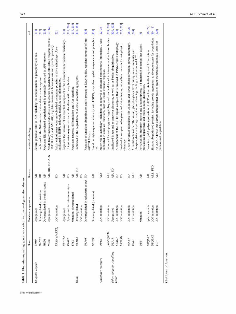
Table1Ubiqu
itin-sign
allin
ggenesassociated
with
neurod
egenerativedisease.
Gene
Mutation,
expression
Disease
Function/pathology
Ref
UbiquitinLigases
CHIP
Upregulated
AD
Several
potentialrolesin
AD,includingtheubiquitin
ationof
phosphorylated
tau.
[211
]
HACE1
Dow
nregulated
instriatum
HD
Implicated
intheNrf2-mediatedantio
xidativ
estress
response.
[212
]
HRD1
Dow
nregulated
incerebral
cortex
AD
Regulates
ER-associateddegradationandispotentially
involved
inAPPturnover.
[213
]
Nedd4
Upregulated
AD,HD,PD,ALS
Nedd4
family
mediatesendocytosisandlysosomaldegradationof
cellsurfacereceptors(suchas
DAT,IG
F-1R
andAMPAR),regulatestransm
itter
homoeostasisandsynaptic
plasticity
.[67,
69]
PRKN
(PARK2)
LOFmutation
PD
Parkinubiquitin
ates
outermito
chondrialmem
braneproteins
todrivemito
phagy.
Also
implicated
inEGFR
endocytic
traffickingandinflam
mation.
[23]
RNF182
Upregulated
AD
Regulates
theturnover
ofan
essentialcomponent
oftheneurotransmitter
releasemachinery.
[214
]
TRAF6
Upregulated
insubstantia
nigra
PD
Ubiquitinatesα-synucleinandco-localises
with
Lew
ybodies.
[215
,216
]
TTC3
Mutation,
downregulated
AD
Regulates
neuronal
differentiatio
nandAkt
signallin
g.[217
,218
]
DUBs
UCHL1
Dow
nregulated
AD,PD
Implicated
inthedegradationof
disease-associated
aggregates.
[178
,181
]
LOFmutation
PD
USP
9XDow
nregulated
insubstantia
nigra
PD
Regulates
α-synucleinubiquitin
ationandispresentin
Lew
ybodies,regulatesturnover
ofpro-
survival
MCL1.
[133
]
USP
9YDow
nregulated
(inmales)
AD
Based
onhigh
sequence
similarity
with
USP9X
,may
also
regulate
α-synucleinandphospho-
tau.
[131
]
Autophagy
receptors
OPTN
LOFmutation
ALS
Major
role
inautophagy,
includingtheremoval
ofdamaged
mito
chondria
(mito
phagy).Also
implicated
inothercellfunctio
ns,such
asinnate
immuneresponse.
[22,
33]
p62/SQ
STM1
LOFmutation
ALS
Importantfor
autophagyandaggrephagy
andcanbe
detected
inintraneuronalinclusionbodies.[219
,220
]
Other
ubiquitin
signallin
ggenes
USP
13Upregulated
AD,PD
Implicated
intauandα-synucleinclearance,
aswellas
inParkinubiquitin
ation.
[130
,132
]
FBXO7
LOFmutation
PD
Acomponent
oftheSCFE3lig
asecomplex
that
isinvolved
inPIN
K1/Parkinmito
phagy.
[221
]
LRSA
M1
LOFmutation
PD
Involved
inreceptor
endocytosisandubiquitin
atingintracellularbacteria
forautophagic
clearance.
[222
,223
]
PINK1
LOFmutation
PD
ASer/Thr
kinase
responsibleforubiquitin
andParkinphosphorylationduring
mito
phagy.
[26,
27]
TBK1
LOFmutation
ALS
AmultifunctionalSer/Thr
kinase,phosphorylates
IRF3during
innate
immunity
and
phosphorylates
autophagyreceptorsto
enhancingbindingto
ubiquitin
andLC3.
[224
]
UBB
Mutation
AD
Polyubiquitinprecursorproteinandatranscriptional+1fram
eshiftmutationthat
causes
proteasomeim
pairmentandmito
chondrialdysfunction.
[3]
UBQLN1
Splicevariants
AD
Promotes
Lys63
polyubiquitin
ationof
APPto
limitits
traffickingandAβsecretion
[76,
77]
UBQLN2
LOFmutation
ALS,FTD
Shuttles
ubiquitin
ated
proteins,including
proteinaggregates,totheproteasomefordegradation.
[10,
11]
VCP
LOFmutation
ALS
AnAAA-A
TPasethat
extractsubiquitin
ated
proteins
from
mem
branes/structures,oftenfor
proteasomal
degradation.
[225
]
LOFLossof
functio
n.
572 M. F. Schmidt et al.
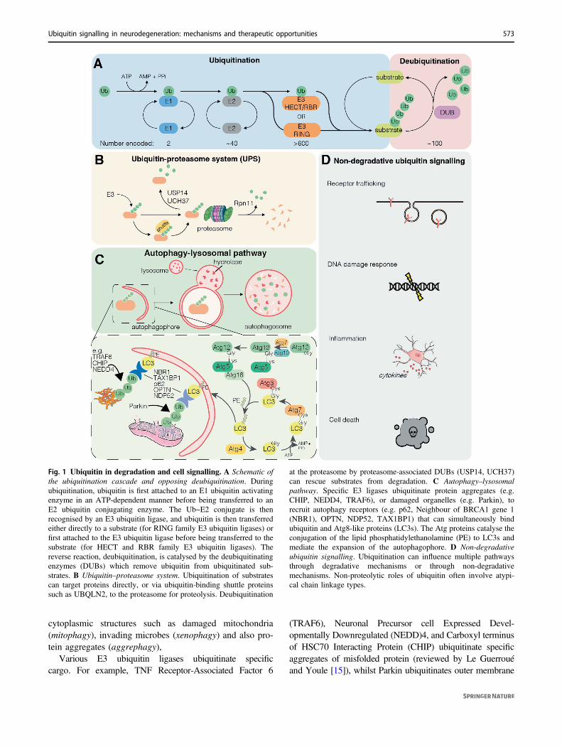
cytoplasmic structures such as damaged mitochondria(mitophagy), invading microbes (xenophagy) and also pro-tein aggregates (aggrephagy),
Various E3 ubiquitin ligases ubiquitinate specificcargo. For example, TNF Receptor-Associated Factor 6
(TRAF6), Neuronal Precursor cell Expressed Devel-opmentally Downregulated (NEDD)4, and Carboxyl terminusof HSC70 Interacting Protein (CHIP) ubiquitinate specificaggregates of misfolded protein (reviewed by Le Guerrouéand Youle [15]), whilst Parkin ubiquitinates outer membrane
Fig. 1 Ubiquitin in degradation and cell signalling. A Schematic ofthe ubiquitination cascade and opposing deubiquitination. Duringubiquitination, ubiquitin is first attached to an E1 ubiquitin activatingenzyme in an ATP-dependent manner before being transferred to anE2 ubiquitin conjugating enzyme. The Ub~E2 conjugate is thenrecognised by an E3 ubiquitin ligase, and ubiquitin is then transferredeither directly to a substrate (for RING family E3 ubiquitin ligases) orfirst attached to the E3 ubiquitin ligase before being transferred to thesubstrate (for HECT and RBR family E3 ubiquitin ligases). Thereverse reaction, deubiquitination, is catalysed by the deubiquitinatingenzymes (DUBs) which remove ubiquitin from ubiquitinated sub-strates. B Ubiquitin–proteasome system. Ubiquitination of substratescan target proteins directly, or via ubiquitin-binding shuttle proteinssuch as UBQLN2, to the proteasome for proteolysis. Deubiquitination
at the proteasome by proteasome-associated DUBs (USP14, UCH37)can rescue substrates from degradation. C Autophagy–lysosomalpathway. Specific E3 ligases ubiquitinate protein aggregates (e.g.CHIP, NEDD4, TRAF6), or damaged organelles (e.g. Parkin), torecruit autophagy receptors (e.g. p62, Neighbour of BRCA1 gene 1(NBR1), OPTN, NDP52, TAX1BP1) that can simultaneously bindubiquitin and Atg8-like proteins (LC3s). The Atg proteins catalyse theconjugation of the lipid phosphatidylethanolamine (PE) to LC3s andmediate the expansion of the autophagophore. D Non-degradativeubiquitin signalling. Ubiquitination can influence multiple pathwaysthrough degradative mechanisms or through non-degradativemechanisms. Non-proteolytic roles of ubiquitin often involve atypi-cal chain linkage types.
Ubiquitin signalling in neurodegeneration: mechanisms and therapeutic opportunities 573
proteins on damaged mitochondria to mediate mitophagy(Fig. 1C). This ubiquitin platform recruits ubiquitin-binding autophagy receptors including p62/sequestosome 1(SQSTM1), Optineurin (OPTN), Nuclear Domain 10 Protein52 (NDP52) and Tax1 Binding Protein 1 (TAX1BP1) [16].Often, autophagy receptors simultaneously bind to the ubi-quitinated cargo via ubiquitin-binding domains, and to Atg8-like proteins: the GABA type A receptor-associated proteins(GABARAPs) and the microtubule-associated protein 1 lightchain 3 (MAP1LC3s), that participate in autophagosomebiogenesis [16]. In doing so, the autophagy receptors tetherubiquitinated cargo to the nascent phagophore, which subse-quently fuses with lysosomes for cargo degradation andrecycling (Fig. 1C).
Autophagy is a major player in removing misfoldedaggregated proteins. This is highlighted by the accumula-tion of ubiquitin positive aggregates and neurodegenerationfollowing the conditional deletion of the essential autop-hagy proteins Atg5 or Atg7 in neuronal cells [17, 18].Disrupted autophagy is observed in AD and HD [19–21],and mutation of various autophagy-related genes drives thepathogenesis of various neurodegenerative diseasesincluding PD and ALS (Table 1). For example, mutations inthe autophagy receptor protein OPTN are linked to familialALS [22], whilst loss-of-function mutations in proteinsinvolved in the clearance of damaged mitochondria,including the E3 ubiquitin ligase Parkin, cause autosomalrecessive juvenile onset forms of PD [23].
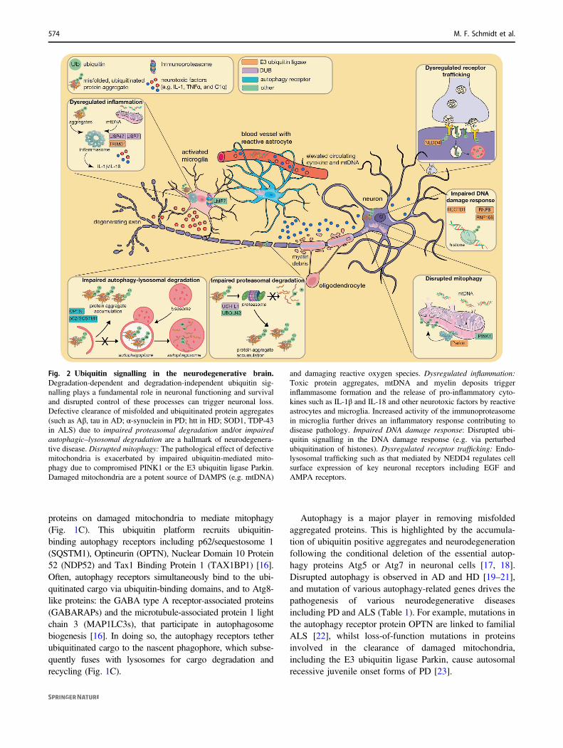
Fig. 2 Ubiquitin signalling in the neurodegenerative brain.Degradation-dependent and degradation-independent ubiquitin sig-nalling plays a fundamental role in neuronal functioning and survivaland disrupted control of these processes can trigger neuronal loss.Defective clearance of misfolded and ubiquitinated protein aggregates(such as Aβ, tau in AD; α-synuclein in PD; htt in HD; SOD1, TDP-43in ALS) due to impaired proteasomal degradation and/or impairedautophagic–lysosomal degradation are a hallmark of neurodegenera-tive disease. Disrupted mitophagy: The pathological effect of defectivemitochondria is exacerbated by impaired ubiquitin-mediated mito-phagy due to compromised PINK1 or the E3 ubiquitin ligase Parkin.Damaged mitochondria are a potent source of DAMPS (e.g. mtDNA)
and damaging reactive oxygen species. Dysregulated inflammation:Toxic protein aggregates, mtDNA and myelin deposits triggerinflammasome formation and the release of pro-inflammatory cyto-kines such as IL-1β and IL-18 and other neurotoxic factors by reactiveastrocytes and microglia. Increased activity of the immunoproteasomein microglia further drives an inflammatory response contributing todisease pathology. Impaired DNA damage response: Disrupted ubi-quitin signalling in the DNA damage response (e.g. via perturbedubiquitination of histones). Dysregulated receptor trafficking: Endo-lysosomal trafficking such as that mediated by NEDD4 regulates cellsurface expression of key neuronal receptors including EGF andAMPA receptors.
574 M. F. Schmidt et al.
Ubiquitin signalling in mitochondrialhomoeostasis
Given that neurons are particularly reliant on mitochondrialoxidative phosphorylation to meet their significant energyrequirements [24], it is not surprising that mitochondrialdefects are emerging as important drivers of neurodegen-erative diseases. Maintaining a healthy mitochondrial poolis particularly important given that mature, terminally dif-ferentiated neurons cannot employ cell division to dilute theimpact of mitochondrial damage or dysfunction. To miti-gate mitochondrial stress, numerous quality controlmechanisms are employed, including a network of chaper-ones and proteases that maintain mitochondrial proteostasisand buffer against proteotoxic stress [25], and the selectivedegradation of damaged mitochondria via mitophagy.
PINK1/Parkin-mediated mitophagy
Mitophagy is intricately coordinated by multiple mechan-isms in a context-dependent fashion. The most well char-acterised pathway is the ubiquitin-dependent clearance ofdamaged mitochondria regulated by the serine/threoninekinase PINK1 and the E3 ubiquitin ligase Parkin (PRKN/PARK2) (Fig. 3). This pathway is implicated in neurode-generation since multiple loss-of-function mutations ineither PINK1 or Parkin are known to cause autosomalrecessive juvenile onset PD (Table 1) [23, 26].
PINK1 acts as a sensor of mitochondrial damage. Inhealthy mitochondria, PINK1 is rapidly imported into themitochondria, cleaved, then degraded via the UPS [27].However, upon mitochondrial damage, it is instead stabi-lised on the outer mitochondrial membrane where itdimerises and autoactivates [28, 29]. PINK1 then phos-phorylates ubiquitin conjugated to mitochondrial outermembrane proteins, which recruits cytosolic Parkin. Parkinis then itself phosphorylated by PINK1 fully licensing itsE3 ubiquitin ligase activity to ubiquitinate numerousmitochondrial proteins with mono and short chain ubiquitinsignals, the latter featuring Lys6 and Lys11 linkages[30, 31]. This in turn provides more ubiquitin substrates forPINK1 to establish a positive feedback loop [32]. Ubiqui-tinated mitochondrial proteins recruit autophagy receptorproteins OPTN and NDP52, which facilitate autophago-phore biogenesis and encapsulation of the mitochondriawithin the autophagosome [33, 34]. The autophagosomesubsequently fuses with a lysosome to instigate mito-chondrial degradation. This ubiquitin-dependent clearanceof mitochondria is negatively regulated by DUBs includingUSP30 [35] and USP15 [36]. In opposing the activity ofParkin, USP30 preferentially cleaves Lys6 ubiquitin lin-kages that are catalysed by Parkin during mitophagy(Fig. 3) [37–39].
PINK1/Parkin-mediated mitophagy in response to mito-chondrial damage can be induced in cultured cells andin vivo by mitochondrial toxins, some of which causeparkinsonism in humans [40, 41], and mitochondrial stressdue to impaired mitochondrial protein homoeostasis andincreased mutational load [42, 43]. However, the physio-logical trigger of mitochondrial damage in the brain ofpatients with familial early onset PD has not been resolved.
Early studies indicated that mice deficient for Parkin orPINK1 do not exhibit overt neuronal degeneration [44, 45].However, a recent study revealed that parkin knockout micedo in fact show degeneration of dopaminergic neurons andconsequent motor deficits when aged more than 2 years[46], supporting the compelling evidence that Parkin defi-ciency leads to parkinsonism in patients. Moreover, sup-porting the notion that Parkin responds to mitochondrialdamage, dopaminergic neuron loss and motor deficits canbe provoked in parkin knockout mice by mitochondrialstress due to accumulated mitochondrial DNA mutationscaused by defects in the proofreading enzyme, DNA poly-merase γ (PolG) [43].
Dopaminergic neurons are particularly susceptible toPINK1/Parkin deficiency in PD. However, Parkin loss maycontribute to the pathogenesis of other neurodegenerativediseases as parkin mRNA expression is controlled by ALS-associated TDP-43 [47], tau impedes Parkin recruitment tomitochondria, and increased mitochondrial Parkin isobserved in astrocytes of Apolipoprotein E4-carriers, amajor genetic risk factor for AD [48–50]. Whether thepathogenic effect of Parkin loss and deficits in respondingto mitochondrial damage is intrinsic to neurons in patientsand animal models, or whether its loss of function in non-neuronal cells drives disease, is unclear.
Ubiquitin signalling in the DNA damageresponse
Post-mitotic mature neurons have to deal with accumulatedDNA damage as a consequence of chronic oxidative stress.DNA adducts and oxidative base damage are observedin nuclear and mtDNA in the aging brain as well as inneurodegenerative diseases, including AD, PD and ALS[51, 52]. E3 ubiquitin ligases play a critical role in baseexcision repair and determining cell survival in response toDNA damage. For example, DNA-damage Binding protein1 (DDB1), through its interaction with the Cullin4 E3ubiquitin ligase complex, and the E3 ligase HECTD1mediate excision repair [53, 54]. Whilst an association withneurodegenerative disease in patients has not been repor-ted, DDB1 mutations are linked to the neurological dis-order, Cockayne’s syndrome, and deletion of HECTD1 orDDB1 in mice promotes neuronal degeneration and
Ubiquitin signalling in neurodegeneration: mechanisms and therapeutic opportunities 575
neurodevelopmental defects [54, 55]. Intriguingly, DDB1has also been shown to interact with Amyloid PrecursorProtein (APP), suggesting a molecular link in AD [56].
The post-mitotic state of neurons reduces the risk ofdouble strand DNA breaks, but also renders neurons parti-cularly sensitive to their impact. The ability to repair such
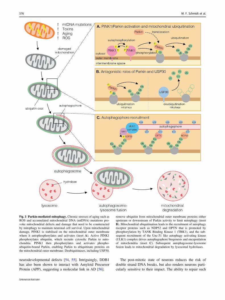
Fig. 3 Parkin-mediated mitophagy. Chronic stresses of aging such asROS and accumulated mitochondrial DNA (mtDNA) mutations pro-voke mitochondrial defects and damage that need to be counteractedby mitophagy to maintain neuronal cell survival. Upon mitochondrialdamage, PINK1 is stabilised on the mitochondrial outer membranewhere it autophosphorylates and activates (inset A). Active PINK1phosphorylates ubiquitin, which recruits cytosolic Parkin to mito-chondria. PINK1 then phosphorylates and activates phospho-ubiquitin-bound Parkin, enabling Parkin to ubiquitinate proteins onthe mitochondrial outer membrane. Deubiquitinases, including USP30,
remove ubiquitin from mitochondrial outer membrane proteins eitherupstream or downstream of Parkin activity to limit mitophagy (insetB). Mitochondrial ubiquitination leads to the recruitment of autophagyreceptor proteins such as NDP52 and OPTN that is promoted byphosphorylation by TANK Binding Kinase 1 (TBK1), and the sub-sequent recruitment of the Unc-51 like autophagy activating kinase(ULK1) complex drives autophagophore biogenesis and encapsulationof mitochondria (inset C). Subsequent autophagosome-lysosomefusion leads to mitochondrial degradation by lysosomal hydrolases.
576 M. F. Schmidt et al.
DNA damage declines with age and defects in the responseare linked to neurodegeneration [57]. E3 ubiquitin ligasesincluding RNF8, RNF168 and RAD18 are critical compo-nents of double-strand break repair by mediating histone 2Aand 2B ubiquitination. A patient with homozygous mutationin the RNF168 ubiquitin binding domain exhibited mildataxia-telangiectasia [58] and mice lacking RNF8 sufferfrom neuronal degeneration [59]. Furthermore, mice lackingRAD6B/UBE2B, the E2 ubiquitin-conjugating enzyme forthe RING E3 ligase RAD18, have overt neurodegenerationdue to a defective response to irradiation-induced DNAdamage [60]. Toxic protein aggregates have been linkedwith DNA damage in neurodegenerative disease, withectopic expression of mutant htt promoting extensive his-tone deubiquitination [61] and aggregates of p62/SQSTM1in ALS and FTD able to inhibit the DNA repair ubiquitinligase RNF168 [62].
If the DNA damage overwhelms the repair mechanisms aneuronal response is to trigger cell cycle re-entry andapoptosis. This control of cell cycle re-entry is potentiallydysregulated in AD, as neurons from a transgenic ADmouse model, or isolated neurons treated with Aβ1–42 pep-tide, exhibited aberrant activation of the E3 ubiquitin ligaseItch, to provoke re-entry into the cell cycle due to Tap73degradation, and consequently cell death [63]. Although themechanisms and whether DNA damage is a driver or aconsequence of neurodegenerative disease are still to beresolved, these data suggest that the ubiquitin-dependentresponse to DNA damage is an under-appreciated player inneurodegenerative diseases that can be compromised byprotein aggregates.
Ubiquitin signalling in endocytosis
Endocytic membrane trafficking of cell surface receptors iscritical for neuronal survival and functioning, includingsynapse development and synapse plasticity [64]. Assynapse loss and dysfunction is a key feature of neurode-generation, defective endolysosomal trafficking is impli-cated in various neurodegenerative diseases including PDand ALS [65]. Receptor endocytosis and degradationthrough the endolysosomal pathway is governed bymonoubiquitination or Lys63-polyubiquitination, with theextent of ubiquitination determining whether the receptor istargeted to the lysosome for degradation or trafficked backto the cell surface [66]. The NEDD4 family of Homologousto E6AP C-terminus (HECT) E3 ubiquitin ligases areupregulated in AD, PD and HD, and ubiquitinate variousneuronal receptors to promote their lysosomal degradation,including the dopamine transporter that mediates uptake ofdopamine into dopaminergic neurons, insulin growth factor-1 receptor that is important for neurotrophic signalling, and
glutamate receptors that control synapse plasticity [67, 68].In addition, Aβ-induced defects in synapse plasticitydue to degradation of α-amino-3-hydroxy-5-methyl-4-iso-xazolepropionate (AMPA) glutamate receptors involvesubiquitination by NEDD4-1 [69]. However, the linkbetween NEDD4 and neurodegeneration is not straightforward as NEDD4-1 is also implicated in the endolyso-somal degradation of α-synuclein in PD [70], suggesting itsactivity may be protective in certain conditions.
Akt signalling through the epidermal growth factorreceptor (EGFR) provides survival cues to dopaminergicneurons. Parkin, in addition to its role in mitophagy, isimplicated to inhibit clathrin-mediated endocytosis ofEGFR through mono-ubiquitination of epidermal growthfactor receptor substrate 15, and in doing so promotes Aktsignalling and neuronal survival [71]. Additionally, Parkinmono-ubiquitinates the PDZ protein PICK1, which reg-ulates trafficking of AMPA receptors thereby potentiallyregulating excitotoxicity [72, 73]. How Parkin is activatedin these contexts, how this activity relates to its role inmitophagy, and the relative contributions of these see-mingly distinct functions to the survival of dopaminergicneurons in PD is not resolved.
As well as regulating receptor signalling, endosomaltrafficking can play an active role in neurodegeneration asextracellular protein aggregates can also be transmittedbetween neurons by endocytic uptake, thereby propagatingthe disease [74]. Endosomes are also the site for the pro-cessing of APP to aggregation-prone Aβ peptides. F-Box/Leucine rich repeat protein 2 (FBL2/FBXL2) is part of theSKP1-Cullin-F-box ubiquitin ligase complex that ubiquiti-nates APP to drive its degradation and also limit its entryinto endosomes, hence Aβ plaques are reduced in FBL2-transgenic mice [75]. Likewise, UBQLN1, the mutation ofwhich has been linked to AD, promotes APP ubiquitinationto limit Aβ secretion [76, 77]. Together, these findingsindicate that ubiquitin-mediated endolysosomal traffickingplays an important yet complex role in the progression ofneurodegenerative disease.
Ubiquitin signalling in glia andneuroinflammation
Although neurodegenerative diseases have long been con-sidered neuron-autonomous disorders, an important role fornon-neuronal glial cells (microglia, astrocytes, oligoden-drocytes) in disease pathogenesis is emerging. Glial cellsmaintain brain homoeostasis and support neuronal survivaland function [78]. However, in the degenerative brain,microglia and astrocytes display impaired phagocytoticclearance of unwanted material such as myelin debris, deadcells and protein aggregates [79], and adopt a reactive
Ubiquitin signalling in neurodegeneration: mechanisms and therapeutic opportunities 577
neurotoxic inflammatory phenotype [80]. Neuroinflamma-tion is an established feature of neurodegenerative diseasesincluding AD and PD, evidenced by elevated pro-inflammatory cytokine levels in the blood and cerebralspinal fluid [81] with mounting evidence suggesting thatinflammation is a driver of disease pathology rather than aconsequence.
mtDNA, myelin debris and disease-associated aggregatescomprising Aβ, α-synuclein and tau, serve as Danger-Associated Molecular Patterns (DAMPs) that elicit a neu-roinflammatory response. These DAMPs are sensed by pat-tern recognition receptors with protein aggregates detected bythe inflammasome sensor protein, NOD-like receptor protein3 (NLRP3) (reviewed in [82]). When activated, NLRP3, andother inflammasome sensors, form multi-protein complexesthat act as platforms for the activation of caspase-1. Caspase-1cleaves the pro-inflammatory cytokines Interleukin-1β (IL-1β)and IL-18 into their mature bioactive fragments and, in par-allel, triggers the inflammatory cell death pathway of pyr-optosis, which facilitates inflammatory cytokine egress (seebelow). Inflammasome assembly and activation is tightlycontrolled by ubiquitination [83]. Pre-clinical evidence sug-gests NLRP3, caspase-1 and IL-1β are promising therapeutictargets in several neurodegenerative diseases [84]. Compel-lingly, pharmacological inhibition of NLRP3 prevented α-synuclein pathology and dopaminergic neurodegeneration inPD mouse models [85], hence NLRP3 inhibitors are nowentering clinical trials for the treatment of PD.
Consistent with the link between inflammatory signallingand neurodegeneration, a genome-wide association studyrevealed common genotypes between PD and autoimmunedisease, including mutation of the E3 ubiquitin ligaseTRIM31, which ubiquitinates NLRP3 to promote itsdegradation and so limit inflammasome activity [86, 87].Conversely, deubiquitination of NLRP3 by USP7 andUSP47 positively regulates inflammasome activation andUSP47 is significantly downregulated in dopaminergicneurons from idiopathic PD patient post-mortem brainsamples [84, 88].
Recently, an E3 ubiquitin ligase COP1/RFWD2 wasrevealed to control pro-inflammatory gene expression inmicroglia by promoting degradation of the transcriptionfactor c/EBPβ [89]. Conditional deletion of COP1 inmicroglia triggered a pro-inflammatory response leading tocomplement C1q-dependent neurotoxicity that exacerbatedtau-driven pathology in mice. Complement protein C3 iscleaved in human AD brain and is required for neurode-generation in mouse models of amyloidosis and tauopathy[90], suggesting that glia-derived complement plays animportant role in AD pathogenesis.
mtDNA released from damaged mitochondria ignitesan inflammatory response via cGAS/STING or NLRP3signalling [91–94]. Loss of Parkin, or its activator PINK1,
promotes inflammatory signalling in response to mito-chondrial stress, and strikingly, blocking inflammatorysignalling was sufficient to rescue dopaminergic neuron lossin the parkin/POLG mouse model of PD [95]. Degenerationof dopaminergic neurons and motor deficits can likewise beinduced in parkin knockout mice by triggering inflamma-tion through the systemic administration of lipopoly-saccharide (LPS) [96], further highlighting the role ofParkin in controlling inflammation. Although the respectiveroles of neurons and glia were not explored in these studies,parkin knockout mice do exhibit damaged mitochondriain mesencephalic glia [97], and Parkin deficiency in primarymicroglia exacerbated NLRP3 signalling upon LPSstimulation [98]. This suggests that disruptions to PINK1/Parkin-mediated mitophagy likely has an important role incontrolling inflammatory signalling that is extrinsic todopaminergic neurons. A recent study revealed that TDP-43can provoke mtDNA release and cGAS/STING-mediated inflammatory responses in ALS models andpatient samples [99], suggesting that mtDNA may act asa DAMP in diverse neurodegenerative diseases. As wellas protecting the mitochondrial network and limitingmtDNA-driven inflammation, Parkin has also been shownto ubiquitinate RIPK1 to directly regulate inflammatorysignalling through its control of nuclear factor-κB (NF-κB)[100, 101]. As in its proposed role in EGFR endocytosis,how Parkin is activated to target RIPK1 is not clear, butthese data suggest that Parkin’s neuroprotective role may bemulti-factorial.
Ubiquitinated protein aggregates are detectable in gliacells, potentially due to phagocytosis of neuronal elementsor uptake of aggregates released from neurons [102].However, glia are less vulnerable than neurons to their toxiceffects, possibly due to higher UPS activity [103]. Fur-thermore, in addition to the 26S proteasome, microglia, asthe predominant immune cell in the brain, also express theimmunoproteasome, which processes peptides for antigenpresentation and regulates inflammatory responses. Immu-noproteasome subunits, such as LMP7, are upregulated inthe brain of PD patients and α-synuclein aggregate-pronemice [104]. Likewise, in an AD mouse model and ADpatient post-mortem tissue, Aβ plaques promoted immu-noproteasome activity in proximal astrocytes and microglia,whilst inhibiting the immunoproteasome in microgliaex vivo decreased inflammatory signalling [105]. This rai-ses the question whether the immunoproteasome might bemore effective in degrading aggregated proteins. However,in the APP/PS1 mouse model of AD, although deficiency ofLMP7 inhibited microglial cytokine response and improvedcognition, it did not alter Aβ pathology [106].
Long thought of as a reaction to dying and dead neurons,dysregulated inflammation is now known to exacerbate, andpossibly even trigger, the neuronal cell death that underpins
578 M. F. Schmidt et al.
disease pathology. As such, glial cells have come to the foreas therapeutic targets, highlighting the need for modelswhere neuron:glia communication is retained.
Ubiquitin signalling in neuronal cell death
Ultimately, the pathology of neurodegenerative diseases isdue to the death of specific neuronal populations. However,it remains unclear why these neurons die as various triggersare implicated, including atypical protein aggregates,mitochondrial dysfunction, neuroinflammatory mediators oroxidative stress. In addition, how neurons die is alsounclear, with different death modalities potentially playinga role.
Apoptosis is considered the main route to neuronal deathin various neurodegenerative diseases [107], evidenced byDNA fragmentation and activity of the executioner caspase-3 in AD, PD and FTD patients [108–111]. X-linked Inhi-bitor of Apoptosis (XIAP) is an E3 ubiquitin ligase thatubiquitinates apoptotic caspases (including caspase-3, -7and -9) to promote their degradation [112]. Nitric Oxide(NO), reported to inhibit XIAP’s ligase activity, is elevatedin neurons with inflammation, and indeed, increased S-nitrosylated XIAP is found in brain tissue of patients withAD, HD and PD [113, 114], suggesting a potential role forXIAP in neurodegeneration. Other E3 ubiquitin ligases,including Parkin, are also S-nitrosylated in PD patientbrains, although the data on the effect of NO on Parkinfunction and thus, a potential impact on cell death, areconflicting [115, 116].
Various ubiquitin-modulating enzymes regulate theintrinsic apoptotic machinery by targeting members of theBCL-2 family of proteins [117], and hence have beenimplicated in dictating neuronal survival and neurodegen-erative diseases. Parkin can directly impair apoptosis byinhibiting the ability of the redundant BCL-2 effector pro-teins BAK and BAX to damage mitochondria, either bypreventing mitochondrial localisation (BAX) or preventingoligomerisation (BAK) [118, 119]. Post-mitotic neurons areproposed to only express a truncated variant of BAK thatlacks intrinsic apoptotic capacity, suggesting that BAX islikely key in apoptosis of mature neurons [120]. Consistentwith this, BAX deletion is sufficient to protect neurons fromdeath induced in vitro by the mitochondrial toxins 1-methyl-4-phenyl-1,2,3,6-tetrahydropyridine (MPTP) and 6-hydroxydopamine (6-OHDA) that promote parkinsonism[121, 122]. Conversely, inhibition of pro-survival proteinMCL1 leads to BAX activation and dopaminergic neurondeath [123] and deletion of Mcl1 in parkin knockout micepromoted dopaminergic neuron death and motor deficits[124], suggesting that pro-survival BCL-2 proteins, andperhaps MCL1 in particular, are important to keep
dopaminergic neurons alive in the face of mitochondrialdefects. Various E3 ubiquitin ligases are reported to targetMCL1 to promote its proteasomal turnover includingHUWE1 [125] and also Parkin [126]. Whilst degradation ofa pro-survival protein by the neuroprotective Parkin appearscounter-intuitive, it might reflect a response to severe orprolonged mitochondrial damage [127]. Two DUBs USP9Xand USP13 deubiquitinate and stabilise MCL1, and hypo-morphic mutations in both have been linked to neurode-velopmental disorders and neurodegenerative disease[128, 129]. However, USP9X and USP13 target multiplesubstrates and are implicated in the clearance of disease-associated protein aggregates in AD [130, 131] and PD[132, 133], hence whether their deubiquitination of MCL1is key to their neuroprotective function is unclear.
Apoptosis is largely considered a non-inflammatorymode of cell death. This potentially conflicts with neu-roinflammation as a feature of certain neurodegenerativediseases and suggests that other more inflammatory modesof cell death may play a role in disease pathology.
Necroptosis is a caspase-independent, lytic form of celldeath induced by a variety of stimuli, including ligation ofcell surface death receptors such as TNF-α receptor 1 andToll-like receptors (TLR). Necroptosis is executed by theoligomerisation of the pseudokinase Mixed Lineage Kinase-Like (MLKL) downstream of Receptor Interacting ProteinKinase 1 (RIPK1) and RIPK3, leading to the disruption ofthe plasma membrane and thus, to cell death and the releaseof pro-inflammatory stimuli and DAMPs [134–136].RIPK1, RIPK3 and MLKL are activated in the brains of ADpatients and their elevated expression correlated with dis-ease progression [137, 138]. Moreover, motor deficits inan AD mouse model were partially rescued by pharmaco-logical inhibition or genetic ablation of MLKL [137].Necroptosis is also linked to PD, as phosphorylated MLKL(a marker of activation) was detected in brains of PDpatients and of 6-OHDA-treated mice [139]. Deletion orinhibition of MLKL or RIPK1 protected neuronal cells,including human induced pluripotent stem cell-deriveddopaminergic neurons, from degeneration driven byMPTP or 6-OHDA [140–142]. RIPK3 ablation likewiseprotected neuronal cells from MPTP-induced toxicity bothin vitro and in vivo, although intriguingly this was due toimpaired apoptosis rather than necroptosis [143]. A role fornecroptosis signalling in ALS has also been proposed aspharmacological inhibition or genetic ablation of RIPK1delayed symptom onset in the SOD1G93A mouse model[144]. However, subsequent studies have questioned therole of necroptosis in ALS as neither a kinase dead mutantof RIPK1 [145], nor deletion of MLKL [146], delayeddisease progression in the same mouse model.
Ubiquitination is a key checkpoint in death receptorsignalling. Upon ligation of cell surface death receptors,
Ubiquitin signalling in neurodegeneration: mechanisms and therapeutic opportunities 579
RIPK1 is ubiquitinated by Cellular Inhibitor of Apoptosis(cIAP) 1 and cIAP2. This not only impairs its kinaseactivity, but also serves to recruit the linear ubiquitinassembly complex (LUBAC), and thereby diverts signallingfrom cell death to survival and pro-inflammatory signallingthrough NF-κB [147]. Interestingly, Parkin represents amolecular link between death receptor signalling and PD, asit is recruited to LUBAC to ubiquitinate the NF-κBEssential Mediator and promote cell survival signalling[148], whilst it also ubiquitinates RIPK1 to limit necroptosissignalling [101, 135].
Pyroptosis, like necroptosis, is a tightly regulated modeof inflammatory cell death. Inflammasome activation inresponse to various Pathogen-Associated Molecular Pat-terns and DAMPs, including aggregated α-synuclein andAβ [149, 150], triggers activation of caspase-1 to cleavepro-inflammatory cytokines, but also the pore-formingprotein gasdermin D, which permeabilises the plasmamembrane leading to lytic cell death [151]. Whilst ubiquitinsignalling regulates inflammasome activity in the initialphase of NLRP3 activation, whether ubiquitination controlsthe effector phase of pyroptosis is unknown. Althoughinflammasome signalling has been reported in neurons,consistent with its key role in innate immunity, pyroptosis ismainly described in microglia to potentially drive neuronaldegeneration indirectly [152–154]. Caspase-1 inhibitionreduced motor deficits in a 6-OHDA-induced rat model ofPD [155], and also ameliorated the cognitive defects in amouse model of AD [156]. NLRP3 and caspase-1 levelsalso correlated with neurons prone to degenerate in a mousemodel of HD [157], and the NLRP3 inflammasome acti-vation has been linked to FTD [158] and ALS [159].However, NLRP3 activation in microglia in PD has beenreported to occur in the absence of significant pyroptoticcell death [85], suggesting that cell death can be separatedfrom IL-1β release. Therefore, it remains to be seen ifpharmacological targeting of pyroptosis per se will be ofbenefit in these diseases.
Ferroptosis occurs as a result of excessive iron-dependent peroxidation of lipids [160]. Iron accumulationis a feature of the neurodegenerative brain and ferroptosis isimplicated in PD, HD and AD (reviewed in [161]). Whilstrelatively unexplored in the context of ferroptosis in neu-rons, ubiquitin signalling is emerging as a potentialcheckpoint. For example, in a non-neuronal setting, induc-tion of ferroptosis with the small molecule erastin is sup-pressed by the ubiquitin ligase NEDD4 [162], whilst theDUBs OTUB1 and USP7 promote ferroptosis by stabilisingthe mediators SLC7A11 and p53, respectively [163, 164].
Understanding the pathway by which neurons die inspecific neurodegenerative disease and how these pathwaysare controlled by ubiquitin signalling will undoubtedly leadto new opportunities for targeted intervention and the
development of disease-modulating therapies that limitneuronal loss.
Targeting ubiquitin signalling to treatneurodegenerative disease
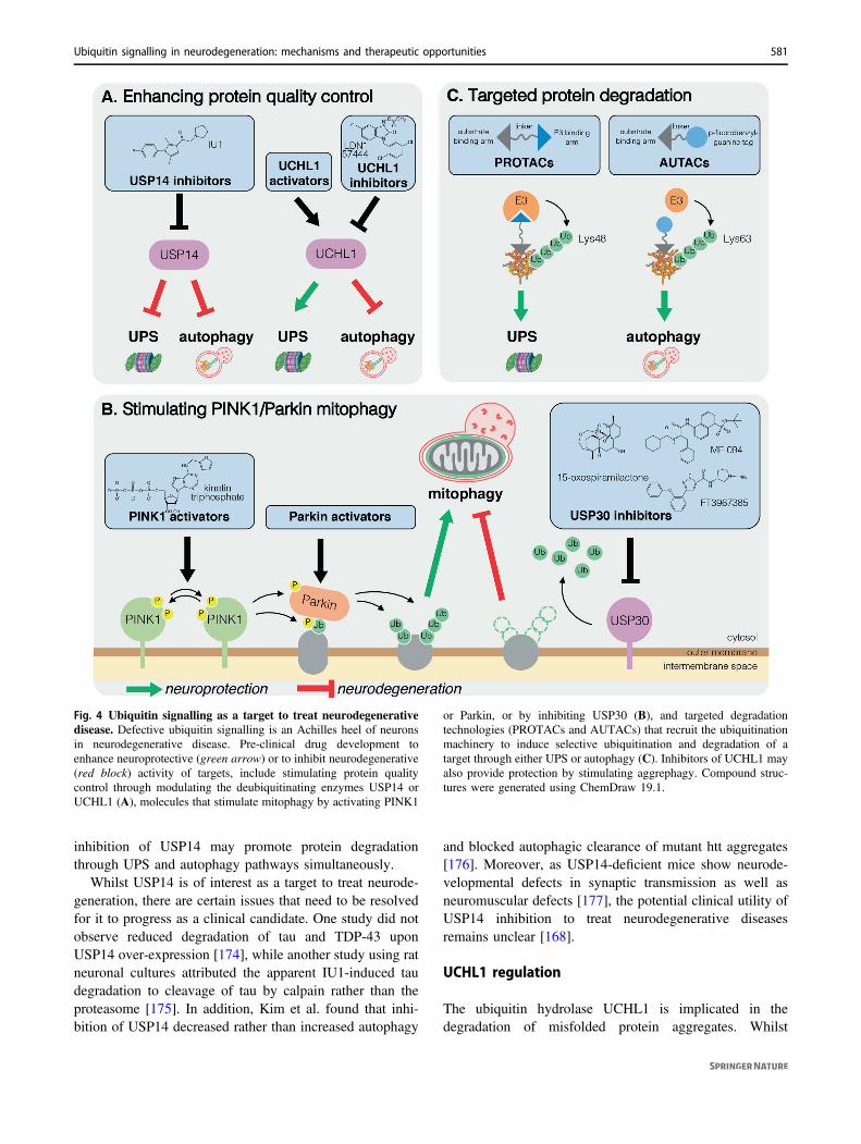
There are currently no treatments that modify the under-lying cause of neurodegeneration to halt or slow diseaseprogression. There is a dire need for new therapies tocombat the global rise in prevalence of neurodegenerativedisease in an aging population. Whilst development ofdrugs targeting the ubiquitin system are in clinical trials aspotential cancer therapies, in neurodegenerative diseasesuch drugs are currently limited to preclinical studies. Thediversity and specificity of the more than 600 E3 enzymesaffords an attractive opportunity for precise targeting ofdisease-relevant pathways [165]. On the other hand,DUBs, of which there are ~100 encoded by the humangenome, also represent promising targets to promote thedegradation of neurotoxic aggregation-prone proteins[165] (Fig. 4).
USP14 inhibition
USP14 is a DUB that has emerged over the last decade as atarget to treat neurodegenerative disorders. USP14 is acti-vated upon binding to the proteasome via its N-terminalubiquitin-like domain and it removes ubiquitin chains tonegatively regulate the degradation of select substrates[166]. Hence, USP14 knockdown enhances proteasomaldegradation [167]. Notably, overexpression of USP14 incultured cells spared ectopically expressed tau and TDP-43from proteasomal degradation [168]. Conversely, a selec-tive inhibitor of USP14, IU1, promoted degradation ofoverexpressed tau, TDP-43 and ataxin-3 [168] (Fig. 4A).Various derivatives of IU1 with increased potency havesince been generated [169, 170], and were shown to beeffective in enhancing the degradation of endogenous tau aswell as exogenous mutant tau in primary neuronal cultures[169]. Although the effect of IU1 in animal models ofneurodegenerative disease has not been tested, IU1 didreduce the severity of brain injury induced by ischaemia/reperfusion in mice [171], supporting its in vivo neuro-protective capacity.
Interestingly, it appears USP14 not only negatively reg-ulates the UPS, but also negatively regulates autophagythrough removing Lys63-linked ubiquitin chains from theautophagy regulator Beclin-1 [172]. Accordingly, pharma-cological inhibition of USP14 enhanced autophagy andmitophagy in cultured cells [169, 172, 173], and USP14knockdown partially reversed the locomotor deficits inPINK1/Parkin-deficient flies [173]. Thus, therapeutic
580 M. F. Schmidt et al.
inhibition of USP14 may promote protein degradationthrough UPS and autophagy pathways simultaneously.
Whilst USP14 is of interest as a target to treat neurode-generation, there are certain issues that need to be resolvedfor it to progress as a clinical candidate. One study did notobserve reduced degradation of tau and TDP-43 uponUSP14 over-expression [174], while another study using ratneuronal cultures attributed the apparent IU1-induced taudegradation to cleavage of tau by calpain rather than theproteasome [175]. In addition, Kim et al. found that inhi-bition of USP14 decreased rather than increased autophagy
and blocked autophagic clearance of mutant htt aggregates[176]. Moreover, as USP14-deficient mice show neurode-velopmental defects in synaptic transmission as well asneuromuscular defects [177], the potential clinical utility ofUSP14 inhibition to treat neurodegenerative diseasesremains unclear [168].
UCHL1 regulation
The ubiquitin hydrolase UCHL1 is implicated in thedegradation of misfolded protein aggregates. Whilst
Fig. 4 Ubiquitin signalling as a target to treat neurodegenerativedisease. Defective ubiquitin signalling is an Achilles heel of neuronsin neurodegenerative disease. Pre-clinical drug development toenhance neuroprotective (green arrow) or to inhibit neurodegenerative(red block) activity of targets, include stimulating protein qualitycontrol through modulating the deubiquitinating enzymes USP14 orUCHL1 (A), molecules that stimulate mitophagy by activating PINK1
or Parkin, or by inhibiting USP30 (B), and targeted degradationtechnologies (PROTACs and AUTACs) that recruit the ubiquitinationmachinery to induce selective ubiquitination and degradation of atarget through either UPS or autophagy (C). Inhibitors of UCHL1 mayalso provide protection by stimulating aggrephagy. Compound struc-tures were generated using ChemDraw 19.1.
Ubiquitin signalling in neurodegeneration: mechanisms and therapeutic opportunities 581

(UCHL1/PARK5) is a PD susceptibility gene [178], its over-expression via intracranial administration of adenovirus alsodelays Aβ-induced neuronal loss in a mouse model of AD[179], suggesting that small molecule agonists may bebroadly neuroprotective. However, somewhat surprisingly,small molecule inhibition of UCHL1 was actually found tolimit α-synuclein aggregation in oligodendrocytes by sti-mulating autophagy [180] (Fig. 4A). Moreover, as patho-genic UCHL1 variants that have gained ubiquitin ligaseactivity have been identified in PD [181], like USP14, itsutility as a target to treat neurodegenerative disease remainsuncertain.
Amplifying mitophagy to treat neurodegeneration
Given that hypomorphic mutations in Parkin or PINK1cause early onset parkinsonism, there is particular interestin activating either protein to treat early onset disease[182]. However, as genetic inhibition of PINK1 enhancedα-synuclein aggregation in cultured cells [183] and exa-cerbated dopaminergic neuron loss in α-synuclein trans-genic mice [184], stimulating PINK1/Parkin-mediatedmitophagy could also be protective in sporadic disease(Fig. 4B). Additionally, mitochondrial dysfunction is acommon feature of neurodegenerative disease, henceamplifying PINK1/Parkin mitophagy may also be bene-ficial in a broad spectrum of diseases including AD andHD [185, 186].
Parkin activation
The structural and mechanistic details of Parkin activationhave been resolved over the last decade [187–193]. As aRing-Between-RING E3 ligase, Parkin comprises multipledomains, including the RING1 and RING2 domains, theIBR (In-Between-RING) domain, the UPD (Unique ParkinDomain; also known as RING0) and an N-terminal ubi-quitin-like (Ubl) domain [194]. Parkin is autoinhibitedunder basal conditions as the catalytic Cys431 in the RING2that receives a ubiquitin molecule during catalysis is buriedin a hydrophobic interface formed between the RING2 andthe UPD [187–189]. Parkin is activated in a stepwisefashion. First, the binding of phospho-ubiquitin leads to therelease of the Ubl domain from the Parkin core [190, 193].Secondly, phosphorylation of the Ubl domain by PINK1enables the phospho-Ubl to bind the UPD, thereby dislod-ging the RING2 domain from its bound state and exposingthe catalytic Cys431 to receive a ubiquitin from a chargedE2~ubiquitin conjugate [191, 192]. The intricate under-standing of this highly coordinated reconfiguration mayenable the development of small molecules that targetspecific functional domains of Parkin to trigger discretesteps in its activation pathway [182, 191, 195]. Targeting
intermediate conformers of Parkin rather than the auto-inhibited form may limit on-target activity in non-targettissues such as heart, whilst also avoiding the potential off-target ubiquitination of cytosolic proteins, including Parkinitself.
PINK1 activation
Compared with Parkin, opportunities to activate PINK1are less well defined as the structure of human PINK1 hasnot yet been solved. However, given that PINK1 autop-hosphorylation in trans is required for PINK1 kinaseactivity [28, 29], promoting dimerisation and/or autop-hosphorylation are potential therapeutic strategies.Despite our incomplete understanding of PINK1 at themolecular level, efforts have been made to target andactivate PINK1 using the ATP analogue kinetin tripho-sphate (KTP) [196, 197] (Fig. 4B). Compared with ATP,KTP enhanced activity of recombinant PINK1 and PINK1in cells, demonstrating its therapeutic potential [196].Indeed, KTP treatment restored locomotor activity andaberrant mitochondrial morphology in PINK1 knockdownflies [198]. Interestingly, the precursor of KTP, kinetin(a plant cytokinin), is metabolised to KTP when addedto cells [196], but whilst long-term oral administrationof kinetin was tolerated in rats, it failed to preventneurodegeneration in response to α-synuclein aggregates[199]. However, it will be important to test whetherkinetin treatment can limit neurodegeneration in modelsof PD involving mitochondrial stress or impairedmitophagy.
USP30 inhibition
As opposed to activating PINK1/Parkin, another strategy toamplify mitophagy would be to inhibit negative regulators.Several candidates have been considered in this regard, withUSP30 currently the most well-studied. USP30 is amitochondria-anchored DUB that preferentially hydrolysesLys6-linked ubiquitin chains on mitochondrial substratesshared with Parkin to set a threshold for mitophagy [35, 37–39, 200]. In a key study, Bingol et al. showed that USP30overexpression limited PINK1/Parkin-mediated mitophagyin cells. Conversely, USP30 knockdown enhanced mito-phagy in rat neuronal cultures and in vivo in PINK1- orParkin-deficient flies [35].
These mechanistic studies demonstrated the therapeuticpotential of small molecule USP30 inhibitors, the first ofwhich, 15-oxospiramilactone, was identified from a phe-notypic screen for compounds that rescue mitochondrialmorphology defects in mitofusin1-deficient cells [201](Fig. 4B). Kluge et al. reported the phenylalanine derivativeMF-094 as a selective inhibitor of USP30, which
582 M. F. Schmidt et al.
accelerated mitophagy in C2C12 myotubes [202]. Recently,Rusilowicz-Jones et al. reported a N-cyano pyrrolidinederivative (FT3967385) inhibited USP30 with an IC50 of1.5 nM and, whilst it showed some off-target inhibition ofUSP6, enhanced mitophagy in cultured neuroblastoma cells[200] (Fig. 4B). USP30 governs mitochondrial proteinimport at the Translocase of the Outer Membrane complex[203], suggesting that USP30 inhibition could have poten-tially toxic effects. That Usp30 knockout mice are viablewith no gross developmental phenotype raises confidencethat USP30 could be targeted pharmacologically [203],however, studies in aged (and challenged) Usp30 knockoutmice will be imperative to understand the long-term con-sequence of USP30 inhibition.
Hijacking ubiquitin signalling to treatneurodegenerative disease
Whilst elements of ubiquitin signalling are targets in theirown right, hijacking ubiquitin-dependent degradation is alsoemerging as a potential therapeutic avenue to treat neuro-degeneration. Targeted protein degradation technologieshave emerged over the last decade as a novel strategy toclear neurotoxic proteins from cells. This technology relieson a heterobifunctional peptide or small molecule thatsimultaneously binds a protein of interest and a componentof the UPS or autophagy machinery, thereby directing theprotein towards degradation (Fig. 4C). The most well-established are the proteolysis targeting chimeras (PRO-TACs), which bind an E3 ubiquitin ligase to induce Lys48polyubiquitination and proteasomal degradation of a targetprotein. Peptide-based PROTACs that degrade tau and α-synuclein aggregates have been developed [204–206]. Thetau-degrading PROTACs fused a tau-binding motif from β-tubulin to peptides that bind VHL or KEAP1, adaptorproteins for Cullin E3 ligase complexes [204, 205]. The α-synuclein PROTAC fused an α-synuclein-binding peptidefrom β-synuclein to a C-terminal RRRG degron to recruitE3 ubiquitin ligases [206]. Both were conjugated to a cellpenetrating peptide to promote cellular uptake [204–206].Chu et al. further showed that their PROTAC couldpromote tau degradation in vivo in an AD mousemodel, although whether it impacted cognition was notassessed [204].
Compared to peptide PROTACs, small molecule PRO-TACs are likely to have better bioavailability and blood-brain barrier permeability. They can also be designed todistinguish neurotoxic misfolded proteins from the physio-logical form, thus retaining the physiological function of theprotein. Tomoshige and colleagues developed a smallmolecule PROTAC that exploited the E3 ubiquitin ligasecIAP1 to degrade htt in HD patient-derived fibroblasts[207, 208]. Based on a tau-binding positron emission
tomography tracer, a small molecule PROTAC, QC-01-175,could recruit the E3 ligase substrate receptor Cereblon tospecifically degrade disease-associated tau thereby limitingthe sensitivity of stressed neurons derived from FTDpatients [209].
Targeted degradation technologies exploiting autop-hagy have also been developed, including autophagytargeting chimeras (AUTACs). AUTACs incorporate aguanine-derived p-fluorobenzylguanine tag to induceLys63-linked ubiquitination and autophagic degradationof target proteins, and also organelles [210]. However, asthe UPS and autophagy machineries are often compro-mised in neurodegenerative disease, the concept ofexploiting these machineries as a therapeutic strategy toremove disease-associated aggregates, or dysfunctionalorganelles such as mitochondria, has not been fullyvalidated.
Chronic neurodegenerative diseases develop over dec-ades. As neurons are terminally differentiated, the neuronalloss in these diseases is usually irreversible. Importantly,there are no treatments that stall neuronal loss to slow orstop disease progression and current therapies are limitedto symptomatic treatments. Given the key role for ubiquitinsignalling in many facets of neuronal function andsurvival, targeting ubiquitin signalling either to enhance theclearance of toxic protein aggregates or to rescue specificvulnerabilities as drivers of neuronal degeneration mayrepresent game-changing treatments that slow or even stopneurodegenerative disease progression. Furthermore, suchpharmacological intervention may augment exciting yetchallenging stem cell transplantation or glia-neurontransdifferentiation strategies that may actually reverse thedisease.
Acknowledgements The authors acknowledge James Vince, ScottAyton and Michael Lazarou for advice and discussions in the pre-paration of the manuscript and Peter Maltezos for assistance in pre-paration of the figures. MFS and ZYG are supported by the AustralianGovernment Research Training Programme. DK is supported by aFellowship from the National Health and Medical Research CouncilAustralia. GD is supported by a Fellowship from the Bodhi EducationFund. The authors acknowledge the funding support from theMichael J Fox Foundation and Shake It Up Foundation Australia andphilanthropic funding from Annette Davis and Leon Davis AO. Thiswork was supported by operational infrastructure grants through theAustralian Government Independent Research Institute InfrastructureSupport Scheme (9000587) and the Victorian State GovernmentOperational Infrastructure Support, Australia.
Compliance with ethical standards
Conflict of interest The authors declare that they have no conflict ofinterest.
Publisher’s note Springer Nature remains neutral with regard tojurisdictional claims in published maps and institutional affiliations.
Ubiquitin signalling in neurodegeneration: mechanisms and therapeutic opportunities 583
Open Access This article is licensed under a Creative CommonsAttribution 4.0 International License, which permits use, sharing,adaptation, distribution and reproduction in any medium or format, aslong as you give appropriate credit to the original author(s) and thesource, provide a link to the Creative Commons license, and indicate ifchanges were made. The images or other third party material in thisarticle are included in the article’s Creative Commons license, unlessindicated otherwise in a credit line to the material. If material is notincluded in the article’s Creative Commons license and your intendeduse is not permitted by statutory regulation or exceeds the permitteduse, you will need to obtain permission directly from the copyrightholder. To view a copy of this license, visit http://creativecommons.org/licenses/by/4.0/.
References
1. Alves-Rodrigues A, Gregori L, Figueiredo-Pereira ME. Ubiqui-tin, cellular inclusions and their role in neurodegeneration.Trends Neurosci. 1998;21:516–20.
2. Lopez-Otin C, Blasco MA, Partridge L, Serrano M, Kroemer G.The hallmarks of aging. Cell. 2013;153:1194–217.
3. van Leeuwen FW, de Kleijn DP, van den Hurk HH, Neubauer A,Sonnemans MA, Sluijs JA, et al. Frameshift mutants of betaamyloid precursor protein and ubiquitin-B in Alzheimer’s andDown patients. Science. 1998;279:242–7.
4. McClellan AJ, Laugesen SH, Ellgaard L. Cellular functions andmolecular mechanisms of non-lysine ubiquitination. Open Biol.2019;9:190147.
5. Komander D, Rape M. The ubiquitin code. Annu Rev Biochem.2012;81:203–29.
6. Ciechanover A, Kwon YT. Degradation of misfolded proteins inneurodegenerative diseases: therapeutic targets and strategies.Exp Mol Med. 2015;47:e147.
7. Ross CA, Poirier MA. Protein aggregation and neurodegenera-tive disease. Nat Med. 2004;10:S10–17.
8. Lowe J, Blanchard A, Morrell K, Lennox G, Reynolds L, BillettM, et al. Ubiquitin is a common factor in intermediate filamentinclusion bodies of diverse type in man, including those ofParkinson’s disease, Pick’s disease, and Alzheimer’s disease, aswell as Rosenthal fibres in cerebellar astrocytomas, cytoplasmicbodies in muscle, and mallory bodies in alcoholic liver disease. JPathol. 1988;155:9–15.
9. Bard JAM, Goodall EA, Greene ER, Jonsson E, Dong KC,Martin A. Structure and function of the 26S proteasome. AnnuRev Biochem. 2018;87:697–724.
10. Hjerpe R, Bett JS, Keuss MJ, Solovyova A, McWilliams TG,Johnson C, et al. UBQLN2 mediates autophagy-independentprotein aggregate clearance by the proteasome. Cell. 2016;166:935–49.
11. Deng HX, Chen W, Hong ST, Boycott KM, Gorrie GH, Siddi-que N, et al. Mutations in UBQLN2 cause dominant X-linkedjuvenile and adult-onset ALS and ALS/dementia. Nature. 2011;477:211–5.
12. Tai HC, Schuman EM. Ubiquitin, the proteasome and proteindegradation in neuronal function and dysfunction. Nat RevNeurosci. 2008;9:826–38.
13. Lee S, Sato Y, Nixon RA. Lysosomal proteolysis inhibitionselectively disrupts axonal transport of degradative organellesand causes an Alzheimer’s-like axonal dystrophy. J Neurosci.2011;31:7817–30.
14. Hamilton AM, Zito K. Breaking it down: the ubiquitin protea-some system in neuronal morphogenesis. Neural Plast. 2013;2013:196848.
15. Le Guerroue F, Youle RJ. Ubiquitin signaling in neurodegen-erative diseases: an autophagy and proteasome perspective. CellDeath Differ. 2020.
16. Dikic I. Proteasomal and autophagic degradation systems. AnnuRev Biochem. 2017;86:193–224.
17. Hara T, Nakamura K, Matsui M, Yamamoto A, Nakahara Y,Suzuki-Migishima R, et al. Suppression of basal autophagy inneural cells causes neurodegenerative disease in mice. Nature.2006;441:885–9.
18. Komatsu M, Waguri S, Chiba T, Murata S, Iwata J, Tanida I,et al. Loss of autophagy in the central nervous system causesneurodegeneration in mice. Nature. 2006;441:880–4.
19. Nixon RA, Wegiel J, Kumar A, Yu WH, Peterhoff C, Cataldo A,et al. Extensive involvement of autophagy in Alzheimer disease: animmuno-electron microscopy study. J Neuropathol Exp Neurol.2005;64:113–22.
20. Lee JH, Yu WH, Kumar A, Lee S, Mohan PS, Peterhoff CM,et al. Lysosomal proteolysis and autophagy require presenilin 1and are disrupted by Alzheimer-related PS1 mutations. Cell.2010;141:1146–58.
21. Martinez-Vicente M, Talloczy Z, Wong E, Tang G, Koga H,Kaushik S, et al. Cargo recognition failure is responsible forinefficient autophagy in Huntington’s disease. Nat Neurosci.2010;13:567–76.
22. Maruyama H, Morino H, Ito H, Izumi Y, Kato H, Watanabe Y,et al. Mutations of optineurin in amyotrophic lateral sclerosis.Nature. 2010;465:223–6.
23. Kitada T, Asakawa S, Hattori N, Matsumine H, Yamamura Y,Minoshima S, et al. Mutations in the parkin gene cause auto-somal recessive juvenile parkinsonism. Nature. 1998;392:605–8.
24. Budd SL, Nicholls DG. A reevaluation of the role of mito-chondria in neuronal Ca2+ homeostasis. J Neurochem. 1996;66:403–11.
25. Ruan L, Wang Y, Zhang X, Tomaszewski A, McNamara JT, LiR. Mitochondria-associated proteostasis. Annu Rev Biophys.2020;49:41–67.
26. Valente EM, Abou-Sleiman PM, Caputo V, Muqit MM, HarveyK, Gispert S, et al. Hereditary early-onset Parkinson’s diseasecaused by mutations in PINK1. Science. 2004;304:1158–60.
27. Pickrell AM, Youle RJ. The roles of PINK1, parkin, and mito-chondrial fidelity in Parkinson’s disease. Neuron. 2015;85:257–73.
28. Okatsu K, Oka T, Iguchi M, Imamura K, Kosako H, Tani N,et al. PINK1 autophosphorylation upon membrane potentialdissipation is essential for Parkin recruitment to damaged mito-chondria. Nat Commun. 2012;3:1016.
29. Okatsu K, Uno M, Koyano F, Go E, Kimura M, Oka T, et al. Adimeric PINK1-containing complex on depolarized mitochondriastimulates Parkin recruitment. J Biol Chem. 2013;288:36372–84.
30. Ordureau A, Paulo JA, Zhang J, An H, Swatek KN, Cannon JR,et al. Global landscape and dynamics of Parkin and USP30-dependent ubiquitylomes in iNeurons during mitophagic sig-naling. Mol Cell. 2020;77:1124–42 e1110.
31. Sarraf SA, Raman M, Guarani-Pereira V, Sowa ME, Huttlin EL,Gygi SP, et al. Landscape of the PARKIN-dependent ubiquity-lome in response to mitochondrial depolarization. Nature. 2013;496:372–6.
32. Ordureau A, Sarraf SA, Duda DM, Heo JM, Jedrychowski MP,Sviderskiy VO, et al. Quantitative proteomics reveal a feedfor-ward mechanism for mitochondrial PARKIN translocation andubiquitin chain synthesis. Mol Cell. 2014;56:360–75.
33. Lazarou M, Sliter DA, Kane LA, Sarraf SA, Wang C, BurmanJL, et al. The ubiquitin kinase PINK1 recruits autophagyreceptors to induce mitophagy. Nature. 2015;524:309–14.
34. Vargas JNS, Wang C, Bunker E, Hao L, Maric D, Schiavo G,et al. Spatiotemporal control of ULK1 activation by NDP52
584 M. F. Schmidt et al.
and TBK1 during selective autophagy. Mol Cell. 2019;74:347–62 e346.
35. Bingol B, Tea JS, Phu L, Reichelt M, Bakalarski CE, Song Q,et al. The mitochondrial deubiquitinase USP30 opposes parkin-mediated mitophagy. Nature. 2014;510:370–5.
36. Cornelissen T, Haddad D, Wauters F, Van Humbeeck C, Man-demakers W, Koentjoro B, et al. The deubiquitinase USP15antagonizes Parkin-mediated mitochondrial ubiquitination andmitophagy. Hum Mol Genet. 2014;23:5227–42.
37. Cunningham CN, Baughman JM, Phu L, Tea JS, Yu C,Coons M, et al. USP30 and parkin homeostatically regulateatypical ubiquitin chains on mitochondria. Nat Cell Biol. 2015;17:160–9.
38. Gersch M, Gladkova C, Schubert AF, Michel MA,Maslen S, Komander D. Mechanism and regulation of theLys6-selective deubiquitinase USP30. Nat Struct Mol Biol.2017;24:920–30.
39. Sato Y, Okatsu K, Saeki Y, Yamano K, Matsuda N, Kaiho A,et al. Structural basis for specific cleavage of Lys6-linkedpolyubiquitin chains by USP30. Nat Struct Mol Biol. 2017;24:911–9.
40. Langston JW, Ballard P, Tetrud JW, Irwin I. Chronic Parkin-sonism in humans due to a product of meperidine-analogsynthesis. Science. 1983;219:979–80.
41. Sandy MS, Di Monte D, Cohen P, Smith MT. Role of active oxygenin paraquat and 1-methyl-4-phenyl-1,2,3,6-tetrahydropyridine(MPTP) cytotoxicity. Basic Life Sci. 1988;49:795–801.
42. Fiesel FC, James ED, Hudec R, Springer W. Mitochondrialtargeted HSP90 inhibitor Gamitrinib-TPP (G-TPP) inducesPINK1/Parkin-dependent mitophagy. Oncotarget. 2017;8:106233–48.
43. Pickrell AM, Huang CH, Kennedy SR, Ordureau A, Sideris DP,Hoekstra JG, et al. Endogenous parkin preserves dopaminergicsubstantia nigral neurons following mitochondrial DNA mutagenicstress. Neuron. 2015;87:371–81.
44. Goldberg MS, Fleming SM, Palacino JJ, Cepeda C, Lam HA,Bhatnagar A, et al. Parkin-deficient mice exhibit nigrostriataldeficits but not loss of dopaminergic neurons. J Biol Chem.2003;278:43628–35.
45. Kitada T, Pisani A, Porter DR, Yamaguchi H, Tscherter A,Martella G, et al. Impaired dopamine release and synaptic plas-ticity in the striatum of PINK1-deficient mice. Proc Natl AcadSci USA. 2007;104:11441–6.
46. Noda S, Sato S, Fukuda T, Tada N, Uchiyama Y, Tanaka K,et al. Loss of Parkin contributes to mitochondrial turnover anddopaminergic neuronal loss in aged mice. Neurobiol Dis.2020;136:104717.
47. Sun X, Duan Y, Qin C, Li JC, Duan G, Deng X, et al. Distinctmultilevel misregulations of Parkin and PINK1 revealed in celland animal models of TDP-43 proteinopathy. Cell Death Dis.2018;9:953.
48. Schmukler E, Solomon S, Simonovitch S, Goldshmit Y, WolfsonE, Michaelson DM, et al. Altered mitochondrial dynamics andfunction in APOE4-expressing astrocytes. Cell Death Dis.2020;11:578.
49. Cummins N, Tweedie A, Zuryn S, Bertran-Gonzalez J, Gotz J.Disease-associated tau impairs mitophagy by inhibiting Parkintranslocation to mitochondria. EMBO J. 2019;38:e99360.
50. Kim J, Basak JM, Holtzman DM. The role of apolipoprotein E inAlzheimer’s disease. Neuron. 2009;63:287–303.
51. Madabhushi R, Pan L, Tsai LH. DNA damage and its links toneurodegeneration. Neuron. 2014;83:266–82.
52. Maynard S, Fang EF, Scheibye-Knudsen M, Croteau DL, BohrVA. DNA damage, DNA repair, aging, and neurodegeneration.Cold Spring Harb Perspect Med. 2015;5:a025130.
53. Bennett L, Madders E, Parsons JL. HECTD1 promotes baseexcision repair in nucleosomes through chromatin remodelling.Nucleic Acids Res. 2020;48:1301–13.
54. Cang Y, Zhang J, Nicholas SA, Bastien J, Li B, Zhou P, et al.Deletion of DDB1 in mouse brain and lens leads to p53-dependent elimination of proliferating cells. Cell. 2006;127:929–40.
55. Zohn IE, Anderson KV, Niswander L. The HECTD1 ubiquitinligase is required for development of the head mesenchyme andneural tube closure. Dev Biol. 2007;306:208–21.
56. Watanabe T, Sukegawa J, Sukegawa I, Tomita S, Iijima K,Oguchi S, et al. A 127-kDa protein (UV-DDB) binds to thecytoplasmic domain of the Alzheimer’s amyloid precursor pro-tein. J Neurochem. 1999;72:549–56.
57. Lee Y, McKinnon PJ. Responding to DNA double strand breaksin the nervous system. Neuroscience. 2007;145:1365–74.
58. Devgan SS, Sanal O, Doil C, Nakamura K, Nahas SA, PettijohnK, et al. Homozygous deficiency of ubiquitin-ligase ring-fingerprotein RNF168 mimics the radiosensitivity syndrome of ataxia-telangiectasia. Cell Death Differ. 2011;18:1500–6.
59. Ouyang S, Song Y, Tian Y, Chen Y, Yu X, Wang D. RNF8deficiency results in neurodegeneration in mice. NeurobiolAging. 2015;36:2850–60.
60. Guo Z, Tian Y, Guo Y, Li B, Liu X, Xie K, et al. RAD6B plays acritical role in neuronal dna damage response to resist neurode-generation. Front Cell Neurosci. 2019;13:392.
61. Ben Yehuda A, Risheq M, Novoplansky O, Bersuker K, KopitoRR, Goldberg M, et al. Ubiquitin accumulation on diseaseassociated protein aggregates is correlated with nuclear ubiquitindepletion, histone de-ubiquitination and impaired DNA damageresponse. PLoS ONE. 2017;12:e0169054.
62. Wang Y, Zhang N, Zhang L, Li R, Fu W, Ma K, et al. Autop-hagy regulates chromatin ubiquitination in DNA damageresponse through elimination of SQSTM1/p62. Mol Cell. 2016;63:34–48.
63. Chauhan M, Modi PK, Sharma P. Aberrant activation of neu-ronal cell cycle caused by dysregulation of ubiquitin ligase Itchresults in neurodegeneration. Cell Death Dis. 2020;11:441.
64. Schwarz LA, Patrick GN. Ubiquitin-dependent endocytosis,trafficking and turnover of neuronal membrane proteins. MolCell Neurosci. 2012;49:387–93.
65. Schreij AM, Fon EA, McPherson PS. Endocytic membranetrafficking and neurodegenerative disease. Cell Mol Life Sci.2016;73:1529–45.
66. Haglund K, Di Fiore PP, Dikic I. Distinct monoubiquitin signalsin receptor endocytosis. Trends Biochem Sci. 2003;28:598–603.
67. Kwak YD, Wang B, Li JJ, Wang R, Deng Q, Diao S, et al.Upregulation of the E3 ligase NEDD4-1 by oxidative stressdegrades IGF-1 receptor protein in neurodegeneration. J Neurosci.2012;32:10971–81.
68. Sorkina T, Miranda M, Dionne KR, Hoover BR, Zahniser NR,Sorkin A. RNA interference screen reveals an essential role ofNedd4-2 in dopamine transporter ubiquitination and endocytosis.J Neurosci. 2006;26:8195–205.
69. Rodrigues EM, Scudder SL, Goo MS, Patrick GN. A beta-inducedsynaptic alterations require the E3 ubiquitin ligase Nedd4-1.J Neurosci. 2016;36:1590–5.
70. Tofaris GK, Kim HT, Hourez R, Jung JW, Kim KP, GoldbergAL. Ubiquitin ligase Nedd4 promotes alpha-synuclein degrada-tion by the endosomal-lysosomal pathway. Proc Natl Acad SciUSA. 2011;108:17004–9.
71. Fallon L, Belanger CM, Corera AT, Kontogiannea M, Regan-Klapisz E, Moreau F, et al. A regulated interaction with the UIMprotein Eps15 implicates parkin in EGF receptor trafficking andPI(3)K-Akt signalling. Nat Cell Biol. 2006;8:834–42.
Ubiquitin signalling in neurodegeneration: mechanisms and therapeutic opportunities 585
72. Joch M, Ase AR, Chen CX, MacDonald PA, Kontogiannea M,Corera AT, et al. Parkin-mediated monoubiquitination of thePDZ protein PICK1 regulates the activity of acid-sensing ionchannels. Mol Biol Cell. 2007;18:3105–18.
73. Lu W, Ziff EB. PICK1 interacts with ABP/GRIP to regulateAMPA receptor trafficking. Neuron. 2005;47:407–21.
74. Guo JL, Lee VM. Cell-to-cell transmission of pathogenic pro-teins in neurodegenerative diseases. Nat Med. 2014;20:130–8.
75. Watanabe T, Hikichi Y, Willuweit A, Shintani Y, Horiguchi T.FBL2 regulates amyloid precursor protein (APP) metabolism bypromoting ubiquitination-dependent APP degradation and inhi-bition of APP endocytosis. J Neurosci. 2012;32:3352–65.
76. Bertram L, Hiltunen M, Parkinson M, Ingelsson M, Lange C,Ramasamy K, et al. Family-based association between Alzhei-mer’s disease and variants in UBQLN1. N Engl J Med. 2005;352:884–94.
77. Hiltunen M, Lu A, Thomas AV, Romano DM, Kim M,Jones PB, et al. Ubiquilin 1 modulates amyloid precursorprotein trafficking and Abeta secretion. J Biol Chem. 2006;281:32240–53.
78. Barres BA. The mystery and magic of glia: a perspective on theirroles in health and disease. Neuron. 2008;60:430–40.
79. Galloway DA, Phillips AEM, Owen DRJ, Moore CS. Phago-cytosis in the brain: homeostasis and disease. Front Immunol.2019;10:790.
80. Liddelow SA, Guttenplan KA, Clarke LE, Bennett FC, BohlenCJ, Schirmer L, et al. Neurotoxic reactive astrocytes are inducedby activated microglia. Nature. 2017;541:481–7.
81. Alam Q, Alam MZ, Mushtaq G, Damanhouri GA, Rasool M,Kamal MA, et al. Inflammatory process in Alzheimer’s andParkinson’s diseases: central role of cytokines. Curr Pharm Des.2016;22:541–8.
82. Rashidi M, Wicks IP, Vince JE. Inflammasomes and cell death:common pathways in microparticle diseases. Trends Mol Med.2020;26:1003–20.
83. Lopez-Castejon G. Control of the inflammasome by the ubiquitinsystem. FEBS J. 2020;287:11–26.
84. Palazon-Riquelme P, Worboys JD, Green J, Valera A, Martin-Sanchez F, Pellegrini C, et al. USP7 and USP47 deubiquitinasesregulate NLRP3 inflammasome activation. EMBO Rep. 2018;19:e44766.
85. Gordon R, Albornoz EA, Christie DC, Langley MR, Kumar V,Mantovani S, et al. Inflammasome inhibition prevents alpha-synuclein pathology and dopaminergic neurodegeneration inmice. Sci Transl Med. 2018;10:eaah4066.
86. Song H, Liu B, Huai W, Yu Z, Wang W, Zhao J, et al. The E3ubiquitin ligase TRIM31 attenuates NLRP3 inflammasomeactivation by promoting proteasomal degradation of NLRP3. NatCommun. 2016;7:13727.
87. Witoelar A, Jansen IE, Wang Y, Desikan RS, Gibbs JR,Blauwendraat C, et al. Genome-wide pleiotropy betweenparkinson disease and autoimmune diseases. JAMA Neurol.2017;74:780–92.
88. Simunovic F, Yi M, Wang Y, Macey L, Brown LT, KrichevskyAM, et al. Gene expression profiling of substantia nigra dopa-mine neurons: further insights into Parkinson’s disease pathol-ogy. Brain. 2009;132:1795–809.
89. Ndoja A, Reja R, Lee SH, Webster JD, Ngu H, Rose CM, et al.Ubiquitin ligase COP1 suppresses neuroinflammation bydegrading c/EBPbeta in microglia. Cell. 2020;182:1156–69e1112.
90. Wu T, Dejanovic B, Gandham VD, Gogineni A, Edmonds R,Schauer S, et al. Complement C3 is activated in human AD brainand is required for neurodegeneration in mouse models ofamyloidosis and tauopathy. Cell Rep. 2019;28:2111–23 e2116.
91. Riley JS, Quarato G, Cloix C, Lopez J, O’Prey J, Pearson M, etal. Mitochondrial inner membrane permeabilisation enablesmtDNA release during apoptosis. EMBO J. 2018;37:e99238.
92. White MJ, McArthur K, Metcalf D, Lane RM, Cambier JC, HeroldMJ, et al. Apoptotic caspases suppress mtDNA-induced STING-mediated type I IFN production. Cell. 2014;159:1549–62.
93. Rongvaux A, Jackson R, Harman CC, Li T, West AP, de ZoeteMR, et al. Apoptotic caspases prevent the induction of type Iinterferons by mitochondrial DNA. Cell. 2014;159:1563–77.
94. West AP, Khoury-Hanold W, Staron M, Tal MC, Pineda CM,Lang SM, et al. Mitochondrial DNA stress primes the antiviralinnate immune response. Nature. 2015;520:553–7.
95. Sliter DA, Martinez J, Hao L, Chen X, Sun N, Fischer TD, et al.Parkin and PINK1 mitigate STING-induced inflammation. Nat-ure. 2018;561:258–62.
96. Frank-Cannon TC, Tran T, Ruhn KA, Martinez TN, Hong J,Marvin M, et al. Parkin deficiency increases vulnerability toinflammation-related nigral degeneration. J Neurosci. 2008;28:10825–34.
97. Schmidt S, Linnartz B, Mendritzki S, Sczepan T, Lubbert M,Stichel CC, et al. Genetic mouse models for Parkinson’s diseasedisplay severe pathology in glial cell mitochondria. Hum MolGenet. 2011;20:1197–211.
98. Mouton-Liger F, Rosazza T, Sepulveda-Diaz J, Ieang A, Has-soun SM, Claire E, et al. Parkin deficiency modulates NLRP3inflammasome activation by attenuating an A20-dependentnegative feedback loop. Glia. 2018;66:1736–51.
99. Yu CH, Davidson S, Harapas CR, Hilton JB, Mlodzianoski MJ,Laohamonthonkul P, et al. TDP-43 Triggers Mitochondrial DNARelease via mPTP to Activate cGAS/STING in ALS. Cell.2020;183:636–49.
100. Henn IH, Bouman L, Schlehe JS, Schlierf A, Schramm JE,Wegener E, et al. Parkin mediates neuroprotection throughactivation of IkappaB kinase/nuclear factor-kappaB signaling. JNeurosci. 2007;27:1868–78.
101. Wang Y, Shan B, Liang Y, Wei H, Yuan J. Parkin regulates NF-kappaB by mediating site-specific ubiquitination of RIPK1. CellDeath Dis. 2018;9:732.
102. Choi I, Zhang Y, Seegobin SP, Pruvost M, Wang Q, Purtell K,et al. Microglia clear neuron-released alpha-synuclein viaselective autophagy and prevent neurodegeneration. Nat Com-mun. 2020;11:1386.
103. Tydlacka S, Wang CE, Wang X, Li S, Li XJ. Differentialactivities of the ubiquitin-proteasome system in neurons versusglia may account for the preferential accumulation of misfoldedproteins in neurons. J Neurosci. 2008;28:13285–95.
104. Ugras S, Daniels MJ, Fazelinia H, Gould NS, Yocum AK, LukKC, et al. Induction of the immunoproteasome subunit Lmp7links proteostasis and immunity in alpha-synuclein aggregationdisorders. EBioMedicine. 2018;31:307–19.
105. Orre M, Kamphuis W, Dooves S, Kooijman L, Chan ET, KirkCJ, et al. Reactive glia show increased immunoproteasomeactivity in Alzheimer’s disease. Brain. 2013;136:1415–31.
106. Wagner LK, Gilling KE, Schormann E, Kloetzel PM, HeppnerFL, Kruger E, et al. Immunoproteasome deficiency altersmicroglial cytokine response and improves cognitive deficits inAlzheimer’s disease-like APPPS1 mice. Acta NeuropatholCommun. 2017;5:52.
107. Friedlander RM. Apoptosis and caspases in neurodegenerativediseases. N Engl J Med. 2003;348:1365–75.
108. Tompkins MM, Basgall EJ, Zamrini E, Hill WD. Apoptotic-likechanges in Lewy-body-associated disorders and normal aging insubstantia nigral neurons. Am J Pathol. 1997;150:119–31.
109. Hartmann A, Hunot S, Michel PP, Muriel MP, Vyas S, FaucheuxBA, et al. Caspase-3: a vulnerability factor and final effector in
586 M. F. Schmidt et al.
apoptotic death of dopaminergic neurons in Parkinson’s disease.Proc Natl Acad Sci USA. 2000;97:2875–80.
110. Su JH, Nichol KE, Sitch T, Sheu P, Chubb C, Miller BL, et al.DNA damage and activated caspase-3 expression in neurons andastrocytes: evidence for apoptosis in frontotemporal dementia.Exp Neurol. 2000;163:9–19.
111. Su JH, Zhao M, Anderson AJ, Srinivasan A, Cotman CW.Activated caspase-3 expression in Alzheimer’s and aged controlbrain: correlation with Alzheimer pathology. Brain Res. 2001;898:350–7.
112. Deveraux QL, Leo E, Stennicke HR, Welsh K, Salvesen GS,Reed JC. Cleavage of human inhibitor of apoptosis protein XIAPresults in fragments with distinct specificities for caspases.EMBO J. 1999;18:5242–51.
113. Tsang AH, Lee YI, Ko HS, Savitt JM, Pletnikova O,Troncoso JC, et al. S-nitrosylation of XIAP compromisesneuronal survival in Parkinson’s disease. Proc Natl Acad SciUSA. 2009;106:4900–5.
114. Nakamura T, Wang L, Wong CC, Scott FL, Eckelman BP, HanX, et al. Transnitrosylation of XIAP regulates caspase-dependentneuronal cell death. Mol Cell. 2010;39:184–95.
115. Chung KK, Thomas B, Li X, Pletnikova O, Troncoso JC, MarshL, et al. S-nitrosylation of parkin regulates ubiquitination andcompromises parkin’s protective function. Science. 2004;304:1328–31.
116. Yao D, Gu Z, Nakamura T, Shi ZQ, Ma Y, Gaston B, et al.Nitrosative stress linked to sporadic Parkinson’s disease: S-nitrosylation of parkin regulates its E3 ubiquitin ligase activity.Proc Natl Acad Sci USA. 2004;101:10810–4.
117. Gomez-Diaz C, Ikeda F. Roles of ubiquitin in autophagy and celldeath. Semin Cell Dev Biol. 2019;93:125–35.
118. Bernardini JP, Brouwer JM, Tan IK, Sandow JJ, Huang S,Stafford CA, et al. Parkin inhibits BAK and BAX apoptoticfunction by distinct mechanisms during mitophagy. EMBO J.2019;38:e99916.
119. Johnson BN, Berger AK, Cortese GP, Lavoie MJ. The ubiquitinE3 ligase parkin regulates the proapoptotic function of Bax. ProcNatl Acad Sci USA. 2012;109:6283–8.
120. Uo T, Kinoshita Y, Morrison RS. Neurons exclusively expressN-Bak, a BH3 domain-only Bak isoform that promotes neuronalapoptosis. J Biol Chem. 2005;280:9065–73.
121. Vila M, Jackson-Lewis V, Vukosavic S, Djaldetti R, Liberatore G,Offen D, et al. Bax ablation prevents dopaminergic neurodegen-eration in the 1-methyl- 4-phenyl-1,2,3,6-tetrahydropyridine mousemodel of Parkinson’s disease. Proc Natl Acad Sci USA. 2001;98:2837–42.
122. Gomez-Lazaro M, Galindo MF, Concannon CG, Segura MF,Fernandez-Gomez FJ, Llecha N, et al. 6-Hydroxydopamine activatesthe mitochondrial apoptosis pathway through p38 MAPK-mediated,p53-independent activation of Bax and PUMA. J Neurochem.2008;104:1599–612.
123. Robinson EJ, Aguiar SP, Kouwenhoven WM, Starmans DS, vonOerthel L, Smidt MP, et al. Survival of midbrain dopamineneurons depends on the Bcl2 factor Mcl1. Cell Death Discov.2018;4:107.
124. Ekholm-Reed S, Baker R, Campos AR, Stouffer D, Henze M,Wolf DA, et al. Reducing Mcl-1 gene dosage induces dopami-nergic neuronal loss and motor impairments in Park2 knockoutmice. Commun Biol. 2019;2:125.
125. Zhong Q, Gao W, Du F, Wang X. Mule/ARF-BP1, a BH3-onlyE3 ubiquitin ligase, catalyzes the polyubiquitination of Mcl-1and regulates apoptosis. Cell. 2005;121:1085–95.
126. Carroll RG, Hollville E, Martin SJ. Parkin sensitizes towardapoptosis induced by mitochondrial depolarization through pro-moting degradation of Mcl-1. Cell Rep. 2014;9:1538–53.
127. Zhang C, Lee S, Peng Y, Bunker E, Giaime E, Shen J, et al.PINK1 triggers autocatalytic activation of Parkin to specify cellfate decisions. Curr Biol. 2014;24:1854–65.
128. Schwickart M, Huang X, Lill JR, Liu J, Ferrando R, French DM,et al. Deubiquitinase USP9X stabilizes MCL1 and promotestumour cell survival. Nature. 2010;463:103–7.
129. Zhang S, Zhang M, Jing Y, Yin X, Ma P, Zhang Z, et al.Deubiquitinase USP13 dictates MCL1 stability and sensitivity toBH3 mimetic inhibitors. Nat Commun. 2018;9:215.
130. Liu X, Hebron ML, Mulki S, Wang C, Lekah E, Ferrante D,et al. Ubiquitin specific protease 13 regulates tau accumulationand clearance in models of Alzheimer’s disease. J AlzheimersDis. 2019;72:425–41.
131. Koglsberger S, Cordero-Maldonado ML, Antony P, Forster JI,Garcia P, Buttini M, et al. Gender-specific expression ofubiquitin-specific peptidase 9 modulates tau expression andphosphorylation: possible implications for tauopathies. MolNeurobiol. 2017;54:7979–93.
132. Liu X, Hebron M, Shi W, Lonskaya I, Moussa CE. Ubiquitinspecific protease-13 independently regulates parkin ubiquitina-tion and alpha-synuclein clearance in alpha-synucleinopathies.Hum Mol Genet. 2019;28:548–60.
133. Rott R, Szargel R, Haskin J, Bandopadhyay R, Lees AJ, Shani V,et al. alpha-Synuclein fate is determined by USP9X-regulatedmonoubiquitination. Proc Natl Acad Sci USA. 2011;108:18666–71.
134. Zhang S, Tang MB, Luo HY, Shi CH, Xu YM. Necroptosis inneurodegenerative diseases: a potential therapeutic target. CellDeath Dis. 2017;8:e2905.
135. Yuan J, Amin P, Ofengeim D. Necroptosis and RIPK1-mediatedneuroinflammation in CNS diseases. Nat Rev Neurosci. 2019;20:19–33.
136. Sun L, Wang H, Wang Z, He S, Chen S, Liao D, et al. Mixedlineage kinase domain-like protein mediates necrosis signalingdownstream of RIP3 kinase. Cell. 2012;148:213–27.
137. Caccamo A, Branca C, Piras IS, Ferreira E, Huentelman MJ,Liang WS, et al. Necroptosis activation in Alzheimer’s disease.Nat Neurosci. 2017;20:1236–46.
138. Koper MJ, Van Schoor E, Ospitalieri S, Vandenberghe R,Vandenbulcke M, von Arnim CAF, et al. Necrosome complexdetected in granulovacuolar degeneration is associated withneuronal loss in Alzheimer’s disease. Acta Neuropathol. 2020;139:463–84.
139. Onate M, Catenaccio A, Salvadores N, Saquel C, Martinez A,Moreno-Gonzalez I, et al. The necroptosis machinery mediatesaxonal degeneration in a model of Parkinson disease. Cell DeathDiffer. 2020;27:1169–85.
140. Iannielli A, Bido S, Folladori L, Segnali A, Cancellieri C,Maresca A, et al. Pharmacological inhibition of necroptosisprotects from dopaminergic neuronal cell death in Parkinson’sdisease models. Cell Rep. 2018;22:2066–79.
141. Onate M, Catenaccio A, Salvadores N, Saquel C, Martinez A,Moreno-Gonzalez I, et al. Correction: the necroptosis machinerymediates axonal degeneration in a model of Parkinson disease.Cell Death Differ. 2020;27:2294.
142. Wu JR, Wang J, Zhou SK, Yang L, Yin JL, Cao JP, et al.Necrostatin-1 protection of dopaminergic neurons. Neural RegenRes. 2015;10:1120–4.
143. Dionisio PA, Oliveira SR, Gaspar MM, Gama MJ, Castro-CaldasM, Amaral JD, et al. Ablation of RIP3 protects from dopami-nergic neurodegeneration in experimental Parkinson’s disease.Cell Death Dis. 2019;10:840.
144. Ito Y, Ofengeim D, Najafov A, Das S, Saberi S, Li Y, et al.RIPK1 mediates axonal degeneration by promoting inflammationand necroptosis in ALS. Science. 2016;353:603–8.
Ubiquitin signalling in neurodegeneration: mechanisms and therapeutic opportunities 587
145. Dominguez S, Varfolomeev E, Brendza R, Stark K, Tea J,Imperio J, et al. Genetic inactivation of RIP1 kinase does notameliorate disease in a mouse model of ALS. Cell Death Differ.2020. [Epub ahead of print].
146. Wang T, Perera ND, Chiam MDF, Cuic B, WanniarachchillageN, Tomas D, et al. Necroptosis is dispensable for motor neurondegeneration in a mouse model of ALS. Cell Death Differ.2020;27:1728–39.
147. Annibaldi A, Wicky John S, Vanden Berghe T, Swatek KN,Ruan J, Liccardi G, et al. Ubiquitin-mediated regulation ofRIPK1 kinase activity independent of IKK and MK2. Mol Cell.2018;69:566–80 e565.
148. Muller-Rischart AK, Pilsl A, Beaudette P, Patra M, Hadian K,Funke M, et al. The E3 ligase parkin maintains mitochondrialintegrity by increasing linear ubiquitination of NEMO. Mol Cell.2013;49:908–21.
149. Codolo G, Plotegher N, Pozzobon T, Brucale M, Tessari I,Bubacco L, et al. Triggering of inflammasome by aggregatedalpha-synuclein, an inflammatory response in synucleinopathies.PLoS ONE. 2013;8:e55375.
150. Halle A, Hornung V, Petzold GC, Stewart CR, Monks BG,Reinheckel T, et al. The NALP3 inflammasome is involved inthe innate immune response to amyloid-beta. Nat Immunol.2008;9:857–65.
151. Kayagaki N, Stowe IB, Lee BL, O’Rourke K, Anderson K,Warming S, et al. Caspase-11 cleaves gasdermin D for non-canonical inflammasome signalling. Nature. 2015;526:666–71.
152. Felderhoff-Mueser U, Schmidt OI, Oberholzer A, Buhrer C,Stahel PF. IL-18: a key player in neuroinflammation and neu-rodegeneration? Trends Neurosci. 2005;28:487–93.
153. Simi A, Tsakiri N, Wang P, Rothwell NJ. Interleukin-1 andinflammatory neurodegeneration. Biochem Soc Trans. 2007;35:1122–6.
154. Voet S, Srinivasan S, Lamkanfi M, van Loo G. Inflammasomesin neuroinflammatory and neurodegenerative diseases. EMBOMol Med. 2019;11:e10248.
155. Mao Z, Liu C, Ji S, Yang Q, Ye H, Han H, et al. The NLRP3Inflammasome is Involved in the Pathogenesis of Parkinson’sDisease in Rats. Neurochem Res. 2017;42:1104–15.
156. Flores J, Noel A, Foveau B, Lynham J, Lecrux C, LeBlanc AC.Caspase-1 inhibition alleviates cognitive impairment and neu-ropathology in an Alzheimer’s disease mouse model. NatCommun. 2018;9:3916.
157. Paldino E, D’Angelo V, Sancesario G, Fusco FR. Pyroptotic celldeath in the R6/2 mouse model of Huntington’s disease: newinsight on the inflammasome. Cell Death Disco. 2020;6:69.
158. Ising C, Venegas C, Zhang S, Scheiblich H, Schmidt SV,Vieira-Saecker A, et al. NLRP3 inflammasome activation drivestau pathology. Nature. 2019;575:669–73.
159. Heneka MT, Kummer MP, Stutz A, Delekate A, Schwartz S,Vieira-Saecker A, et al. NLRP3 is activated in Alzheimer’sdisease and contributes to pathology in APP/PS1 mice. Nature.2013;493:674–8.
160. Stockwell BR, Friedmann Angeli JP, Bayir H, Bush AI, ConradM, Dixon SJ, et al. Ferroptosis: a regulated cell death nexuslinking metabolism, redox biology, and disease. Cell. 2017;171:273–85.
161. Guiney SJ, Adlard PA, Bush AI, Finkelstein DI, Ayton S. Fer-roptosis and cell death mechanisms in Parkinson’s disease.Neurochem Int. 2017;104:34–48.
162. Yang Y, Luo M, Zhang K, Zhang J, Gao T, Connell DO, et al.Nedd4 ubiquitylates VDAC2/3 to suppress erastin-induced fer-roptosis in melanoma. Nat Commun. 2020;11:433.
163. Tang LJ, Zhou YJ, Xiong XM, Li NS, Zhang JJ, Luo XJ, et al.Ubiquitin-specific protease 7 promotes ferroptosis via activation
of the p53/TfR1 pathway in the rat hearts after ischemia/reper-fusion. Free Radic Biol Med. 2020;31581–1.
164. Liu T, Jiang L, Tavana O, Gu W. The deubiquitylase OTUB1mediates ferroptosis via stabilization of SLC7A11. Cancer Res.2019;79:1913–24.
165. Huang X, Dixit VM. Drugging the undruggables: exploring theubiquitin system for drug development. Cell Res. 2016;26:484–98.
166. Lee BH, Lu Y, Prado MA, Shi Y, Tian G, Sun S, et al. USP14deubiquitinates proteasome-bound substrates that are ubiquiti-nated at multiple sites. Nature. 2016;532:398–401.
167. Koulich E, Li X, DeMartino GN. Relative structural and func-tional roles of multiple deubiquitylating proteins associated withmammalian 26S proteasome. Mol Biol Cell. 2008;19:1072–82.
168. Lee BH, Lee MJ, Park S, Oh DC, Elsasser S, Chen PC, et al.Enhancement of proteasome activity by a small-molecule inhi-bitor of USP14. Nature. 2010;467:179–84.
169. Boselli M, Lee BH, Robert J, Prado MA, Min SW, Cheng C,et al. An inhibitor of the proteasomal deubiquitinating enzymeUSP14 induces tau elimination in cultured neurons. J Biol Chem.2017;292:19209–25.
170. Wang Y, Jiang Y, Ding S, Li J, Song N, Ren Y, et al. Smallmolecule inhibitors reveal allosteric regulation of USP14 viasteric blockade. Cell Res. 2018;28:1186–94.
171. Min JW, Lu L, Freeling JL, Martin DS, Wang H. USP14 inhi-bitor attenuates cerebral ischemia/reperfusion-induced neuronalinjury in mice. J Neurochem. 2017;140:826–33.
172. Xu D, Shan B, Sun H, Xiao J, Zhu K, Xie X, et al. USP14regulates autophagy by suppressing K63 ubiquitination of Beclin1. Genes Dev. 2016;30:1718–30.
173. Chakraborty J, von Stockum S, Marchesan E, Caicci F, FerrariV, Rakovic A, et al. USP14 inhibition corrects an in vivo modelof impaired mitophagy. EMBO Mol Med. 2018;10:e9014.
174. Ortuno D, Carlisle HJ, Miller S. Does inactivation of USP14enhance degradation of proteasomal substrates that are associatedwith neurodegenerative diseases? F1000 Res. 2016;5:137.
175. Kiprowska MJ, Stepanova A, Todaro DR, Galkin A, Haas A,Wilson SM, et al. Neurotoxic mechanisms by which the USP14inhibitor IU1 depletes ubiquitinated proteins and Tau in ratcerebral cortical neurons: relevance to Alzheimer’s disease.Biochim Biophys Acta Mol Basis Dis. 2017;1863:1157–70.
176. Kim E, Park S, Lee JH, Mun JY, Choi WH, Yun Y, et al. Dualfunction of USP14 deubiquitinase in cellular proteasomal activ-ity and autophagic flux. Cell Rep. 2018;24:732–43.
177. Wilson SM, Bhattacharyya B, Rachel RA, Coppola V, Tessar-ollo L, Householder DB, et al. Synaptic defects in ataxia miceresult from a mutation in Usp14, encoding a ubiquitin-specificprotease. Nat Genet. 2002;32:420–5.
178. Maraganore DM, Lesnick TG, Elbaz A, Chartier-Harlin MC,Gasser T, Kruger R, et al. UCHL1 is a Parkinson’s diseasesusceptibility gene. Ann Neurol. 2004;55:512–21.
179. Zhang M, Cai F, Zhang S, Zhang S, Song W. Overexpression ofubiquitin carboxyl-terminal hydrolase L1 (UCHL1) delays Alz-heimer’s progression in vivo. Sci Rep. 2014;4:7298.
180. Pukass K, Richter-Landsberg C. Inhibition of UCH-L1 in oli-godendroglial cells results in microtubule stabilization and pre-vents alpha-synuclein aggregate formation by activating theautophagic pathway: implications for multiple system atrophy.Front Cell Neurosci. 2015;9:163.
181. Liu Y, Fallon L, Lashuel HA, Liu Z, Lansbury PT Jr. The UCH-L1 gene encodes two opposing enzymatic activities that affectalpha-synuclein degradation and Parkinson’s disease suscept-ibility. Cell. 2002;111:209–18.
182. Miller S, Muqit MMK. Therapeutic approaches to enhancePINK1/Parkin mediated mitophagy for the treatment of Parkin-son’s disease. Neurosci Lett. 2019;705:7–13.
588 M. F. Schmidt et al.
183. Liu W, Vives-Bauza C, Acin-Perez R, Yamamoto A, Tan Y, LiY, et al. PINK1 defect causes mitochondrial dysfunction, pro-teasomal deficit and alpha-synuclein aggregation in cell culturemodels of Parkinson’s disease. PLoS ONE. 2009;4:e4597.
184. Oliveras-Salva M, Macchi F, Coessens V, Deleersnijder A,Gerard M, Van der Perren A, et al. Alpha-synuclein-inducedneurodegeneration is exacerbated in PINK1 knockout mice.Neurobiol Aging. 2014;35:2625–36.
185. Du F, Yu Q, Yan S, Hu G, Lue LF, Walker DG, et al.PINK1 signalling rescues amyloid pathology and mitochondrialdysfunction in Alzheimer’s disease. Brain. 2017;140:3233–51.
186. Khalil B, El Fissi N, Aouane A, Cabirol-Pol MJ, Rival T, Lie-vens JC. PINK1-induced mitophagy promotes neuroprotection inHuntington’s disease. Cell Death Dis. 2015;6:e1617.
187. Riley BE, Lougheed JC, Callaway K, Velasquez M, Brecht E,Nguyen L, et al. Structure and function of Parkin E3 ubiquitinligase reveals aspects of RING and HECT ligases. Nat Commun.2013;4:1982.
188. Trempe JF, Sauve V, Grenier K, Seirafi M, Tang MY, MenadeM, et al. Structure of parkin reveals mechanisms for ubiquitinligase activation. Science. 2013;340:1451–5.
189. Wauer T, Komander D. Structure of the human Parkin ligasedomain in an autoinhibited state. EMBO J. 2013;32:2099–112.
190. Wauer T, Simicek M, Schubert A, Komander D. Mechanism ofphospho-ubiquitin-induced PARKIN activation. Nature. 2015;524:370–4.
191. Gladkova C, Maslen SL, Skehel JM, Komander D. Mechanismof parkin activation by PINK1. Nature. 2018;559:410–4.
192. Sauve V, Sung G, Soya N, Kozlov G, Blaimschein N, Miotto LS,et al. Mechanism of parkin activation by phosphorylation. NatStruct Mol Biol. 2018;25:623–30.
193. Sauve V, Lilov A, Seirafi M, Vranas M, Rasool S, Kozlov G,et al. A Ubl/ubiquitin switch in the activation of Parkin. EMBOJ. 2015;34:2492–505.
194. Cotton TR, Lechtenberg BC. Chain reactions: molecularmechanisms of RBR ubiquitin ligases. Biochem Soc Trans. 2020;48:1737–50.
195. Padmanabhan S, Polinski NK, Menalled LB, Baptista MAS,Fiske BK. The Michael J Fox Foundation for Parkinson’sresearch strategy to advance therapeutic development of PINK1and Parkin. Biomolecules. 2019;9:296–309.
196. Hertz NT, Berthet A, Sos ML, Thorn KS, Burlingame AL,Nakamura K, et al. A neo-substrate that amplifies catalyticactivity of parkinson’s-disease-related kinase PINK1. Cell. 2013;154:737–47.
197. Osgerby L, Lai YC, Thornton PJ, Amalfitano J, Le Duff CS,Jabeen I, et al. Kinetin riboside and its protides activate theParkinson’s disease associated PTEN-Induced Putative Kinase 1(PINK1) independent of mitochondrial depolarization. J MedChem. 2017;60:3518–24.
198. Shiba-Fukushima K, Inoshita T, Sano O, Iwata H, Ishikawa KI,Okano H, et al. A cell-based high-throughput screening identifiedtwo compounds that enhance PINK1-parkin signaling. iScience.2020;23:101048.
199. Orr AL, Rutaganira FU, de Roulet D, Huang EJ, Hertz NT,Shokat KM, et al. Long-term oral kinetin does not protect againstalpha-synuclein-induced neurodegeneration in rodent models ofParkinson’s disease. Neurochem Int. 2017;109:106–16.
200. Rusilowicz-Jones EV, Jardine J, Kallinos A, Pinto-Fernandez A,Guenther F, Giurrandino M, et al. USP30 sets a trigger thresholdfor PINK1-PARKIN amplification of mitochondrial ubiquityla-tion. Life Sci Alliance. 2020;3:e202000768.
201. Yue W, Chen Z, Liu H, Yan C, Chen M, Feng D, et al. A smallnatural molecule promotes mitochondrial fusion through inhibi-tion of the deubiquitinase USP30. Cell Res. 2014;24:482–96.
202. Kluge AF, Lagu BR, Maiti P, Jaleel M, Webb M, Malhotra J,et al. Novel highly selective inhibitors of ubiquitin specificprotease 30 (USP30) accelerate mitophagy. Bioorg Med ChemLett. 2018;28:2655–9.
203. Phu L, Rose CM, Tea JS, Wall CE, Verschueren E, Cheung TK,et al. Dynamic regulation of mitochondrial import by the ubi-quitin system. Mol Cell. 2020;77:1107–23 e1110.
204. Chu TT, Gao N, Li QQ, Chen PG, Yang XF, Chen YX, et al.Specific knockdown of endogenous tau protein by peptide-directed ubiquitin-proteasome degradation. Cell Chem Biol.2016;23:453–61.
205. Lu M, Liu T, Jiao Q, Ji J, Tao M, Liu Y, et al. Discovery of aKeap1-dependent peptide PROTAC to knockdown Tau byubiquitination-proteasome degradation pathway. Eur J MedChem. 2018;146:251–9.
206. Qu J, Ren X, Xue F, He Y, Zhang R, Zheng Y, et al. Specificknockdown of alpha-synuclein by peptide-directed proteasomedegradation rescued its associated neurotoxicity. Cell Chem Biol.2020;27:751–62 e754.
207. Tomoshige S, Nomura S, Ohgane K, Hashimoto Y, Ishikawa M.Discovery of small molecules that induce the degradation ofHuntingtin. Angew Chem Int Ed Engl. 2017;56:11530–3.
208. Tomoshige S, Nomura S, Ohgane K, Hashimoto Y, Ishikawa M.Degradation of huntingtin mediated by a hybrid molecule com-posed of IAP antagonist linked to phenyldiazenyl benzothiazolederivative. Bioorg Med Chem Lett. 2018;28:707–10.
209. Silva MC, Ferguson FM, Cai Q, Donovan KA, Nandi G,Patnaik D, et al. Targeted degradation of aberrant tau in fron-totemporal dementia patient-derived neuronal cell models. Elife.2019;8:e45457.
210. Takahashi D, Moriyama J, Nakamura T, Miki E, Takahashi E,Sato A, et al. AUTACs: cargo-specific degraders using selectiveautophagy. Mol Cell. 2019;76:797–810.
211. Sahara N, Murayama M, Mizoroki T, Urushitani M, Imai Y,Takahashi R, et al. In vivo evidence of CHIP up-regulationattenuating tau aggregation. J Neurochem. 2005;94:1254–63.
212. Rotblat B, Southwell AL, Ehrnhoefer DE, Skotte NH, MetzlerM, Franciosi S, et al. HACE1 reduces oxidative stress andmutant Huntingtin toxicity by promoting the NRF2 response.Proc Natl Acad Sci USA. 2014;111:3032–7.
213. Kaneko M, Koike H, Saito R, Kitamura Y, Okuma Y, NomuraY. Loss of HRD1-mediated protein degradation causes amyloidprecursor protein accumulation and amyloid-beta generation. JNeurosci. 2010;30:3924–32.
214. Liu QY, Lei JX, Sikorska M, Liu R. A novel brain-enriched E3ubiquitin ligase RNF182 is up regulated in the brains of Alz-heimer’s patients and targets ATP6V0C for degradation. MolNeurodegener. 2008;3:4.
215. Chung JY, Park HR, Lee SJ, Lee SH, Kim JS, Jung YS, et al.Elevated TRAF2/6 expression in Parkinson’s disease is caused bythe loss of Parkin E3 ligase activity. Lab Investig. 2013;93:663–76.
216. Zucchelli S, Codrich M, Marcuzzi F, Pinto M, Vilotti S, BiagioliM, et al. TRAF6 promotes atypical ubiquitination of mutant DJ-1and alpha-synuclein and is localized to Lewy bodies in sporadicParkinson’s disease brains. Hum Mol Genet. 2010;19:3759–70.
217. Suizu F, Hiramuki Y, Okumura F, Matsuda M, Okumura AJ,Hirata N, et al. The E3 ligase TTC3 facilitates ubiquitination anddegradation of phosphorylated Akt. Dev Cell. 2009;17:800–10.
218. Kohli MA, Cukier HN, Hamilton-Nelson KL, Rolati S, KunkleBW, Whitehead PL, et al. Segregation of a rare TTC3 variant inan extended family with late-onset Alzheimer disease. NeurolGenet. 2016;2:e41.
219. Fecto F, Yan J, Vemula SP, Liu E, Yang Y, Chen W, et al.SQSTM1 mutations in familial and sporadic amyotrophic lateralsclerosis. Arch Neurol. 2011;68:1440–6.
Ubiquitin signalling in neurodegeneration: mechanisms and therapeutic opportunities 589
220. Kuusisto E, Salminen A, Alafuzoff I. Ubiquitin-binding proteinp62 is present in neuronal and glial inclusions in human tauo-pathies and synucleinopathies. Neuroreport. 2001;12:2085–90.
221. Burchell VS, Nelson DE, Sanchez-Martinez A, Delgado-Camprubi M, Ivatt RM, Pogson JH, et al. The Parkinson’sdisease-linked proteins Fbxo7 and Parkin interact to mediatemitophagy. Nat Neurosci. 2013;16:1257–65.
222. Aerts MB, Weterman MA, Quadri M, Schelhaas HJ, Bloem BR,Esselink RA, et al. A LRSAM1 mutation links Charcot-Marie-Tooth type 2 to Parkinson’s disease. Ann Clin Transl Neurol.2016;3:146–9.
223. Huett A, Heath RJ, Begun J, Sassi SO, Baxt LA, Vyas JM,et al. The LRR and RING domain protein LRSAM1 is an E3ligase crucial for ubiquitin-dependent autophagy of intracel-lular Salmonella Typhimurium. Cell Host Microbe. 2012;12:778–90.
224. Ahmad L, Zhang SY, Casanova JL, Sancho-Shimizu V. HumanTBK1: a gatekeeper of neuroinflammation. Trends Mol Med.2016;22:511–27.
225. Johnson JO, Mandrioli J, Benatar M, Abramzon Y, Van DeerlinVM, Trojanowski JQ, et al. Exome sequencing reveals VCPmutations as a cause of familial ALS. Neuron. 2010;68:857–64.
590 M. F. Schmidt et al.