molecular pathways to neurodegeneration
TRANSCRIPT

R E V I E W
S2 JULY 2004 NEURODEGENERATION
Evidence is accumulating to suggest that such chronic neurodegenerativedisorders as Alzheimer’s disease (AD), Parkinson’s disease (PD),Huntington’s disease (HD) and amyotrophic lateral sclerosis (ALS) arecaused by a combination of events that impair normal neuronal func-tion. Clinical signs are evident before frank neuronal loss. Therefore, newefforts have focused on identifying crucial changes, of genetic, epigenetic,or environmental origin, that hamper normal neuronal function.
In this review, we summarize the current understanding ofmolecular pathways implicated in common neurodegenerative dis-orders, trying to clarify similarities that might provide a foundationfor their treatment.
Molecular pathways to ADAD is the most common neurodegenerative disorder worldwide.Approximately 4.5 million people suffer from this devastating condi-tion in the United States alone. In AD, neurons of the hippocampusand cerebral cortex are selectively lost. Brains of individuals with ADmanifest two characteristic lesions: extracellular amyloid (or senile)plaques and intracellular neurofibrillary tangles of hyperphosphory-lated tau protein (Fig. 1a)1. Amyloid plaques contain small, toxic cleav-age products (denoted as Aβ40 and Αβ42) of the amyloid precursorprotein (APP). The apoE4 (apolipoprotein E4) genotype is a powerfulrisk factor for developing AD, and it may possibly affect Aβ depositionand neurofibrillary tangle formation2.
Mutations in three genes that are inherited in an autosomal domi-nant fashion have been linked to rare familial, early-onset forms ofAD. These genes include those encoding APP, presenilin 1 (PS1) andpresenilin 2 (PS2). Although these familiar forms account for only afew cases of AD, one common event in both familial and sporadictypes of AD is the increased production and accumulation of the toxicAβ. This observation led to the ‘amyloid cascade hypothesis’ thatexcessive Aβ production is the primary cause of the disease3,4.
Do plaques and tangles cause AD, or are they simply the telltaleremains of earlier, pivotal events that led to the disease? One problemhas been that in people with AD the density of amyloid plaques cor-relates poorly with the severity of dementia3. Furthermore, althoughneurofibrillary tangles correlate well with decline in cognitive skills,they seem to be a late event and in some cases possibly downstreamof Aβ accumulation5. Recent findings, however, indicate that den-dritic and synaptic injury occur early and that protofibrils andoligomers of Aβ40 and Αβ42, rather than large plaques, cause neu-ronal dysfunction.
APP is a type I membrane protein and contains a large extracellularregion, a transmembrane helix and a short cytoplasmic tail (Fig. 1b).The N-terminal half of APP contains a heparin-binding domain (N-APP), a copper-binding domain (CuBD) and an APP proteaseinhibitor domain (APPI) (Fig. 1b). The N-APP structure reveals thatAPP may belong to a superfamily of cysteine-rich growth factors witha putative heparin-binding site6. The CuBD structure is homologousto copper chaperones. This domain may regulate dimerization or pro-teolytic processing, or it may act as a metallotransporter7. There is alsostructural information on the Aβ peptides that originate by proteolyticprocessing of APP8,9. Both Aβ40 and Aβ42 adopt variable and partlyhelical structures dependent on membrane binding, metal chelationand interaction with other peptides. Aβ binds Cu2+, Fe2+ and Zn2+
coordinated by three histidines (His677, His684, His685) and a tyro-sine (Tyr681). Metal binding induces a β-sheet-like conformationalchange in Aβ, resulting in enhanced aggregation.
Toxic Aβ originates from regulated intramembrane proteolysis ofAPP by a complex of secretases (Fig. 1c). The first cleavage of APP ismediated by β- or α-secretase, releasing most of the extracellular por-tion of APP as two fragments, APPs-α and APPs-β, leaving behind theC-terminal membrane bound fragment (Fig. 1c). This portion of APPis then cleaved by a large protein complex, γ-secretase, at several sitesincluding amino acid (aa) 711 (Aβ40) and at least three additionalsubsites at aa713 (Aβ42), aa714 (Aβ43) and aa720 (Aβ49) (Fig. 1b).Several mutations in APP, such as the Swedish mutation, cluster at theβ-secretase cleavage sites; these mutations result in increased amountsof Aβ peptide and protofibril formation10.
The precise composition of the γ-secretase complex is still underdebate, but PS1, nicastrin, Aph-1 and Pen-2 seem to be required11–13.
Molecular pathways to neurodegenerationElla Bossy-Wetzel1, Robert Schwarzenbacher2 & Stuart A Lipton1
The molecular bases underlying the pathogenesis of neurodegenerative diseases are gradually being disclosed. One problem thatinvestigators face is distinguishing primary from secondary events. Rare, inherited mutations causing familial forms of thesedisorders have provided important insights into the molecular networks implicated in disease pathogenesis. Increasing evidenceindicates that accumulation of aberrant or misfolded proteins, protofibril formation, ubiquitin-proteasome system dysfunction,excitotoxic insult, oxidative and nitrosative stress, mitochondrial injury, synaptic failure, altered metal homeostasis and failure ofaxonal and dendritic transport represent unifying events in many slowly progressive neurodegenerative disorders.
1Center for Neuroscience & Aging and 2Program in Bioinformatics & SystemsBiology, The Burnham Institute, 10901 North Torrey Pines Road, La Jolla,California 92037. Correspondence should be addressed to E.B-W. ([email protected]) or S.A.L. ([email protected]).
Published online 1 July 2004; doi:10.1038/nm1067
©20
04 N
atur
e P
ublis
hing
Gro
up
http
://w
ww
.nat
ure.
com
/nat
urem
edic
ine

R E V I E W
NEURODEGENERATION JULY 2004 S3
PS1 is a transmembrane domain aspartyl protease that cleaves its sub-strates in the membrane-spanning region. PS1 is probably responsiblefor the generation of Aβ fragments.
More than 100 missense mutations in PS1 and PS2 have been identi-fied in rare familial, early-onset AD14. Experiments in culture andtransgenic mice reveal that these mutations result in increased Aβ pro-duction15,16. Conversely, mice lacking PS1 have decreased Aβ40 andAβ42 production17,18, suggesting that PS1 has a pivotal role in γ-secre-tase activity. C-terminal cleavage of APP by caspase enzymes may alsobe required for toxicity19.
It is still unclear how Aβ does its damage, but several mechanismshave been proposed. One view suggests that Aβ protofibrils activatemicroglia, inciting an inflammatory response and release of neuro-toxic cytokines. Nonsteroidal anti-inflammatory drugs (NSAIDs)including ibuprofen seem to delay the onset of AD20. Additionally,NSAIDs reduce the production of Aβ42 (ref. 21). In a second view, Aβprotofibrils trigger excessive release of excitatory amino acids like glu-tamate from glial cells that may injure nearby neurons by excitotoxic-ity. Overactivation of glutamate receptors of the N-methyl-D-aspartate
(NMDA) subtype results in increased intra-cellular Ca2+, which activates neuronal nitricoxide synthase and consequently generatesnitric oxide (NO). When generated in excess,NO combines with superoxide anion (O2
–),forming the highly reactive and neurotoxicproduct peroxynitrite (ONOO–), which leadsto further oxidative and nitrosative stress inpart via mitochondrial injury. In fact, positivephase III human trials of the uncompetitiveNMDA receptor channel blocker, memantine,led to its recent approval for the treatment ofAD22. A third view suggests that protofibrilsand aggregates convey harmful effects to neu-rons by paralyzing axonal and dendritic
transport. Both APP and PS may bind kinesin I and regulate vesiculartraffic23–25. PS mutants increase glycogen synthase kinase-3β activity,which hampers kinesin-mediated, anterograde axonal transport25.Also, Aβ deposits may act as nonspecific ‘roadblocks’, representing aphysical transport barrier.
An additional mechanism of Aβ injury is synaptic dysfunction andloss, which are early events in AD and occur before amyloid plaqueformation26. Cholinergic transmission and synaptic density are con-siderably decreased in AD patients. The mechanism for synaptic dam-age is unknown, but diffusible oligomeric forms of Aβ may beimportant. Synaptic dysfunction probably contributes to memory lossand cognitive deficits in AD. In fact, APP transgenic mice manifest cel-lular, biochemical and electrophysiological evidence of synapticdeficits before Aβ deposition, including reduced excitatory postsynap-tic potentials and LONG-TERM POTENTIATION (LTP), regarded as a corre-late of learning and memory27. Inhibition of γ-secretase decreasesoligomeric Aβ and LTP deficits28. Microinjection of Aβ43 and Aβ40peptides into the rat hippocampus disrupts synaptic transmission andshort-term memory29.
APP N-APP CuBD APPI Aβ AICD
Plaquesa
b
c
Tangles Phospho-Tau
TM
1
β α γCU+2, Fe+2, Zn+2
670 700 730
N L R GQN TIMVIVAATL
F
P(Australian)(Swedish) K
G(Arctic)
AICDp3
AICD
AICD57
APPs α
APPs β Aβ40
APPs β Aβ42
TM
EISEVKMDAEFRHDSGYEVHHQKLVFFAEDVGSNKGAIIGLMVGGVVIATVIVITLVMLKKKQYTS
770600400200
Figure 1 Histology and structures related to AD.(a) Silver-stained amyloid plaques, neurofibrillarytangles and immunocytochemistry ofphosphorylated tau in brain sections from ADpatients. (b) Structures and domain model ofamyloid βA4 precursor protein (APP, PDB entryP05067; N-terminal growth factor domain (N-APP), aa 28–123, PDB entry 1MWP; APP copper-binding domain (CuBD), aa 124–188, PDB entry1OWT; APP protease inhibitor domain (APPI), aa 287–344, PDB entry 1AAP; amyloid β peptide(Aβ42), PDB entry 1IYT; (Aβ40), PDB entry1BA6; transmembrane domain (TM). Structuralmodels are shown in ribbon representation, color-coded from N terminus (blue) to C terminus (red).Side chains of relevant residues are shown asball-and-stick figures. (c) APP residues 665–730;the TM domain (residues 700–723) ishighlighted in light blue; cleavage sites ofsecretases α, β and γ are indicated, along withmetal-coordinating residues (His677, Tyr681,His684, His685) and mutations associated withAD (http://molgen-www.uia.ac.be/ADMutations/).Proteolytic processing and resulting fragments ofAPP: APPsα aa 1–686; APPsβ, aa 1–670; p3 peptides, aa 687–711 or 713; Aβ40, aa 671–711; Aβ42, aa 671–713. APPintracellular domain (AICD), aa 711 or 713–770.
©20
04 N
atur
e P
ublis
hing
Gro
up
http
://w
ww
.nat
ure.
com
/nat
urem
edic
ine

R E V I E W
S4 JULY 2004 NEURODEGENERATION
Aβ may also mediate harmful effects by binding redox-reactive met-als, which in turn release free radicals30–36. We found that nitrosativestress mobilizes zinc from intracellular stores, resulting in mitochon-drial dysfunction37. Chelation of zinc and copper provides neuropro-tective effects38. For example, clioquinol (CQ), an antibiotic that alsochelates zinc and copper and crosses the blood-brain barrier, decreasesbrain Aβ deposition and improves learning in mutant APP transgenicmice39. A recent human phase II clinical trial of CQ for AD seemspromising40.
Oxidative stress from mitochondrial dysfunction occurs early inAD, and Aβ may directly or indirectly injure mitochondria41,42. Aβblocks respiratory complex I, thus producing a decline in ATP43. Inisolated mitochondria, Aβ inhibits respiration and enzyme activity (α-ketoglutarate dehydrogenase and pyruvate dehydrogenase)44. Themechanism of Aβ translocation to mitochondria remains unknown.New data describe an interaction partner of Aβ in mitochondria,termed Aβ-binding alcohol dehydrogenase (ABAD)45. ABAD is upreg-
ulated in neurons of AD patients. Expression of ABAD in concert withmutant APP enhances free-radical production and toxicity.Conversely, a peptide that blocks Aβ-ABAD interaction prevents free-radical formation and cell death. Aβ interacts with the LD loop ofABAD, close to its nicotinamide adenine dinucleotide (NAD) bindingsite, and may therefore induce a conformational change that preventsNAD binding. To date, however, details of the interaction are notapparent in the crystal structure45.
In summary, mitochondrial dysfunction and resulting energydeficits may contribute to impaired clearance of protein aggregatesand neuronal dysfunction, affecting ion channel and pump activity,neurotransmission, and axonal and dendritic transport.
Molecular pathways to PDPD is the most common neurodegenerative movement disorder.Approximately 1% of the population older than 65 years suffers fromthis slowly progressive neurodegenerative disease; 95% of PD cases are
Lewy bodies (H&E) Lewy bodies (Ubiquitin) Lewy bodies (α-Synuclein) Lewy neurite (α-Synuclein)a
b
400 4653002001001
R33AR42P
A82E Ex3 S167NQ100H
K211N T240RC212V R256C
V380L T415ND384N G430D
C431FC268STOPR271S
Q311STOPG328E
A339SR275W
D280NC289G
Parkin Ubl RING1 IRB RING2
400 46546530030020010011
Parkin Ubl RING1 IRB RING2
400 4653002001001
Proteasome SubstrateE2
Uq
cFigure 2 Histology and structures related toPD. (a) Lewy bodies and neurites visualized byH&E staining or immunocytochemistry usingantibodies specific for ubiquitin and α-synuclein in brain sections of sporadic casesof PD. (b) Structures and domain model ofhuman parkin (NCBI AB009973); N-terminalubiquitin-like domain (Ubl), aa 1–79, PDBentry 1IYF; homology model of RING1 domain,aa 238–293 based on PDB entry 1JM7; IRBdomain, aa 314–377; homology model ofRING2 domain, aa 418–449, based on PDBentry 1FO4. Structural models are depicted in ribbon representation, color-coded from N terminus (blue) to C terminus (red). Cysteine side chainslocated in the RING zinc finger domains are depicted in ball and stick with Zn2+ as a pink sphere. Arrows indicate reported familial-associatedParkinson’s disease-causing mutations. (c) Schematic representation of parkin interactions. The Ubl domain binds to the proteasome. Substratebinding may be mediated by the RING1 domain and E2 enzyme binding by the IRB and RING2 domains.
©20
04 N
atur
e P
ublis
hing
Gro
up
http
://w
ww
.nat
ure.
com
/nat
urem
edic
ine

R E V I E W
NEURODEGENERATION JULY 2004 S5
sporadic. The symptoms of PD are caused by selective and progressivedegeneration of pigmented dopaminergic (DA) neurons in the sub-stantia nigra pars compacta. Current treatments, such as administra-tion of L-DOPA to produce dopamine, are only symptomatic and donot stop or delay the progressive loss of neurons. In fact, some studieshave suggested that oxidative injury via dopamine may lead to furtherneuronal damage46.
One important feature of PD is the presence of eosinophilic, cyto-plasmic inclusions of fibrillar, misfolded proteins, termed Lewy bod-ies, in affected brain areas (Fig. 2a). The exact composition of Lewybodies is unknown, but they contain ubiquinated α-synuclein, parkin,synphilin, neurofilaments and synaptic vesicle proteins (Fig. 2a).
The etiology of PD remains unclear47. Hints that sporadic PD can beinitiated by environmental toxins are provided by the accidental dis-covery that 1-methyl-4-phenyl-1,2,3,6-tetrahydropyridine (MPTP), anherbicide and contaminant of illicit street drugs, causes parkinsonian-like symptoms in human substance abusers and in animal models48–52.Astrocytes metabolize MPTP to 1-methyl-4-phenyl pyridium (MPP+)via monoamine oxidase B. MPP+ is selectively imported into DA neu-rons via the dopamine transporter, where it targets mitochondria,inhibits respiratory complex I and promotes reactive oxygen speciesproduction. As with AD, altered metal homeostasis may have a role inthe etiology of PD. Metal chelation by CQ rescues DA neurons andimproves balance and posture in MPTP-treated animals53.
Additional support for mitochondrial dysfunction in PD pathogen-esis comes from evidence that the insecticide rotenone induces aparkinsonian-like syndrome in animal models and probably humans,with protein deposits that resemble Lewy bodies54. Rotenone alsoinhibits mitochondrial complex I, and DA neurons seem to be mostseverely affected.
Rare hereditary forms of PD have provided insight into the molecu-lar pathways of this disorder47. Mutations in at least four genes havebeen linked to PD, including α-synuclein (PARK1), parkin (PARK2),DJ-1 (PARK7), and PTEN (phosphatase and tensin homolog deletedon chromosome 10)-induced kinase 1 (PINK1, also known asPARK6)55–58.
α-Synuclein mutations are autosomal dominant and representtoxic gain-of-function mutations, resulting in abnormal proteinaccumulations59,60. The normal function of α-synuclein is unknown.
The protein is normally unfolded, revealing little three-dimensionalstructure. α-synuclein can polymerize into filaments and is found inLewy bodies and neurites (Fig. 2a). The molecular pathways ofα-synuclein-mediated toxicity are unknown; however, increasedoxidative stress, mitochondrial injury and altered cellular transporthave been proposed.
Mutations in parkin are found in juvenile PD and are inherited asautosomal recessive, causing a loss in parkin function. New findingsindicate that parkin mutations may be linked not only to juvenile PD,but also to late-onset cases of PD. Heterozygous mutations in parkinmay also cause PD, indicating a dominant form of inheritance in somecases. Perhaps HAPLOINSUFFICIENCY causes disease when coupled withnitrosative and oxidative stress (see later). Alternatively, some muta-tions may be DOMINANT NEGATIVE in nature.
Parkin is an E3 ligase, catalyzing the addition of ubiquitin to specificsubstrates that targets them for degradation by the ubiquitin-protea-some system (UPS). The parkin protein has several domains (Fig. 2b).Structural information for the N-terminal ubiquitin-like (Ubl)domain (residues 1–75) reveals that it binds to the Rpn10 subunit ofthe 26S proteasome. The Arg42Pro mutation, found in juvenile PD, isclose to the Rpn10 subunit binding site, possibly impairing its protea-somal interaction61. The C-terminal half of parkin contains two RINGdomains, common to several E3 ligases, connected by an in-betweenRING domain (IBR). The RING domains are cysteine-rich zinc fingersimplicated in substrate recognition and binding to E2 enzymes thattransfer ubiquitin62. The RING1 domain constitutes a hotspot for mis-sense mutations. Two mutations located in RING1, Arg256Cys andArg275Trp, cause altered protein localization and increased aggrega-tion63. Most mutations clustered in the conserved domains of parkindiminish enzyme activity, suggesting that parkin loss of functioncauses the disorder (Fig. 2b).
Parkin substrates include glycosylated α-synuclein, synphilin,CDCrel-1 and the Pael-R receptor. These substrates may accumulatewhen parkin E3 ligase function is lost by mutation or other mecha-nism, contributing to neurotoxicity. Overexpression of wild-typeparkin improves cell survival after various insults in a number of sys-tems64–67. Parkin blocks α-synuclein- and Pael-R-mediated toxicity,probably by promoting their degradation64,65,68. Parkin also mediatesthe clearance of some polyglutamine repeat proteins69. In Drosophila,
GLOSSARYLong-term potentation (LTP) An enduring increase in the amplitude of excitatory postsynaptic potentials as a result of high-frequency(tetanic) stimulation of afferent pathways. It is measured both as the amplitude of excitatory postsynaptic potentials and as the magnitudeof the postsynaptic-cell population spike. LTP is most often studied in the hippocampus and is often considered to be the cellular basis oflearning and memory in vertebrates.
Haploinsufficiency Loss of one copy (one allele) of a gene is sufficient to give rise to disease. Haploinsufficiency implies that no dominant-negative effect of the mutated gene product has to be invoked.
Dominant negative A mutant molecule that in some cases can form a heteromeric complex with the normal molecule, knocking out theactivity of the entire complex.
Small interfering RNA RNA that can be used to interfere with normal RNA processing, causing rapid degradation of the endogenous RNAand thereby precluding translation.
Missense mutation A mutation that results in the substitution of an amino acid in a protein, which therefore often has abnormal function.
Knock-in transgenesis The insertion of a mutant gene at the exact site of the genome where the corresponding wild-type gene is located.This approach is used to ensure that the effect of the mutant gene is not affected by the activity of the endogenous allele.
©20
04 N
atur
e P
ublis
hing
Gro
up
http
://w
ww
.nat
ure.
com
/nat
urem
edic
ine
R E V I E W
S6 JULY 2004 NEURODEGENERATION
parkin protects cells from mitochondrial death pathways70. Hence,enhancing parkin activity may be therapeutic.
New findings, however, suggest that parkin may have other func-tions besides targeting proteins for degradation via the UPS. Forinstance, parkin knockout mice do not manifest accumulation ofparkin substrates, and humans with parkin mutations lack Lewy bod-ies. These data imply that parkin loss of function may cause degenera-tion by a mechanism other than aberrant protein accumulation.Parkin knockout mice manifest decreased oxidative phosphorylationand increased oxidative stress71. Parkin may be localized to mitochon-dria, and its loss induces abnormal mitochondrial morphology, mus-cle degeneration and impaired spermatogenesis in Drosophila70.
Additionally, parkin is a target of oxidative and nitrosative stress insporadic PD. Cysteine residues in the RING domains are sensitive tonitrosative and oxidative modifications, which alter protein function.New findings indicate that parkin’s E3 ligase activity is modified byNO, thus linking environmental stress to a molecular abnormality anda clinical phenotype similar to that seen in hereditary forms of PD72,73.
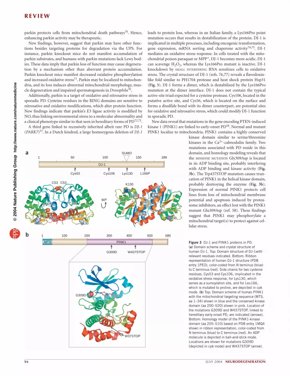
A third gene linked to recessively inherited albeit rare PD is DJ-1(PARK7)57. In a Dutch kindred, a large homozygous deletion of DJ-1
leads to protein loss, whereas in an Italian family, a Lys166Pro pointmutation occurs that results in destabilization of the protein. DJ-1 isimplicated in multiple processes, including oncogenic transformation,gene expression, mRNA sorting and chaperone activity74,75. DJ-1mediates an oxidative stress response. In cells treated with the mito-chondrial poison paraquat or MPP+, DJ-1 becomes more acidic. DJ-1can scavenge H2O2, whereas the Lys166Pro mutant is inactive. DJ-1knockdown by SMALL INTERFERING RNA sensitizes cells to oxidativestress. The crystal structure of DJ-1 (refs. 76,77) reveals a flavodoxin-like fold similar to PH1704 protease and heat shock protein Hsp31(Fig. 3). DJ-1 forms a dimer, which is destabilized by the Lys166Promutation at the dimer interface. DJ-1 does not contain the typicalactive-site triad expected for a cysteine protease. Cys106, located in theputative active site, and Cys56, which is located on the surface andforms a disulfide bond with its dimer counterpart, are potential sitesfor oxidative and nitrosative stress, which could modify DJ-1 functionin sporadic PD.
New data reveal that mutations in the gene encoding PTEN-inducedkinase 1 (PINK1) are linked to early-onset PD58. Normal and mutantPINK1 localize to mitochondria. PINK1 contains a highly conserved
kinase domain similar to serine/threoninekinases in the Ca2+-calmodulin family. Twomutations associated with PD reside in thisdomain, and homology modeling reveals thatthe MISSENSE MUTATION Gly309Asp is locatedin its ADP binding site, probably interferingwith ADP binding and kinase activity (Fig.3b). The Trp437STOP mutation causes trun-cation of PINK1 in the helical kinase domain,probably destroying the enzyme (Fig. 3b).Expression of normal PINK1 protects celllines from loss of mitochondrial membranepotential and apoptosis induced by protea-some inhibitors, an effect lost with the PINK1mutant Glu309Asp (ref. 58). These findingssuggest that PINK1 may phosphorylate amitochondrial target(s) to protect against cel-lular stress.
a
b
Cys53 Cys106 Lys130 L166P
1 50 100 150 189SUMO
G309D W437STOP
1 100 200 300 400 500 580
PINK1MTS
G309D ADPADP
W37STOP
C53C53 C53
C53C53C53C53C106C106 C106C106
C106C106
K130K130 K130K130
L166L166 L166L166L166L166
L166L166
90o
K130
K130K130
DJ-1
Figure 3 DJ-1 and PINK1 proteins in PD. (a) Domain scheme and crystal structure ofhuman DJ-1. Top: Domain structure of DJ-1withrelevant residues indicated. Bottom: Ribbonrepresentation of human DJ-1 structure (PDBentry 1PE0), color-coded from N terminus (blue)to C terminus (red). Side chains for two cysteineresidues, Cys53 and Cys106, implicated in theoxidative stress response, for Lys130, whichserves as a sumoylation site, and for Leu166,which is mutated to proline, are depicted in cpkmode. (b) Top: Domain scheme of human PINK1with the mitochondrial targeting sequence (MTS,aa 1–34) shown in blue and the conserved kinasedomain (aa 200–520) shown in pink. Location ofthe mutations G309D and W437STOP, linked tohereditary early-onset PD, are indicated (arrows).Bottom: Homology model of the PINK1-kinasedomain (aa 205–510) based on PDB entry 1MQ4shown in ribbon representation, color-coded fromN terminus (blue) to C terminus (red). An ADPmolecule is depicted in ball-and-stick mode.Locations are shown for mutations G309D(depicted in cpk mode) and W437STOP (arrow).
©20
04 N
atur
e P
ublis
hing
Gro
up
http
://w
ww
.nat
ure.
com
/nat
urem
edic
ine

R E V I E W
NEURODEGENERATION JULY 2004 S7
Pathways to HDHD is an autosomal dominant inherited neurodegenerative disorderaffecting 1 in 10,000 individuals. It is caused by an insertion of multi-ple CAG repeats in the huntingtin gene. This results in an N-terminalpolyglutamine (polyQ) expansion of the large protein huntingtin(Htt), similar to other polyQ-related neurodegenerative disorders.Disease severity depends on the length of the polyQ stretch, withrepeats ≥40 clearly linked to HD. The polyQ expansion is thought toconfer a toxic gain of function with selective loss of neurons in thestriatum and cerebral cortex.
Htt expression is ubiquitous but is greatest in neurons. Htt isreported in membranes, cytosol, nuclei, and mitochondria, and inassociation with microtubules78–80. The expanded polyQ stretch inmutant Htt induces a conformational change resulting in aggregates indendrites and nuclei (Fig. 4a). It is not clear whether aggregates,smaller accumulations or soluble aberrant protein are important forHD pathogenesis.
The function of normal Htt and the mechanism whereby mutantHtt mediates harmful effects remain unclear81. Htt may act as a molec-ular scaffold, regulating several cellular processes including endocyto-sis, vesicle transport, excitatory synapses, transcriptional events andmitochondrial function82,83.
The Htt domain model reveals a polyQ and proline-rich region atthe N terminus followed by three HEAT domains, named for the fourproteins in which they were first detected: Htt, elongation factor 3, theregulatory A subunit of protein phosphatase 2A, and TOR1 (ref. 84)(Fig. 4b).
Several proteins interact with the Htt proline-rich domain. Anexample is endophilin 3, which regulates dynamin and synaptotagminfunction85. Htt also binds to postsynaptic density protein-95 (PSD-95), a molecule that interacts with excitatory aa receptors, includingNMDA receptors, and synaptic GTPase-activating protein(SynGAP)86. SynGAP in turn inhibits Ras and thereby regulates excita-tory synapses. Thus, Htt may modulate synaptic transmission, learn-ing and memory via its interaction with PSD-95.
HEAT domains mediate protein-protein interactions. Several pro-teins have been isolated that bind to the HEAT repeat. Among themare Htt-interacting protein (HIP)-1, HIP14 and Htt-associated pro-tein (HAP1), which have roles in endocytosis and membrane traffick-ing87–90. Impaired axonal and dendritic trafficking are also implicatedin HD91, and transgenic KNOCK-IN mice for mutant Htt 150Q manifest
axonal pathology suggestive of transportdefects92. Similarly, expression of mutant Httin Drosophila produces an axonal transportphenotype93.
Mutant Htt may be neurotoxic via tran-scriptional dysregulation, binding andsequestering selective transcription factorsand coactivators. These include cAMPresponse element binding protein (CREB)
binding protein (CBP), p53, co-repressor C-terminal binding protein(CtBP), Sp1 and TAFII-130 (refs. 94–97). Histone deacetylaseinhibitors, which increase transcription, ameliorate polyQ-mediatedneurodegeneration in Drosophila94.
Mitochondrial dysfunction is also implicated in HD pathogenesis.The mitochondrial toxin 3-nitropropionate produces neuropathologyin animals similar to that observed in human HD98,99. Mitochondrialmembrane potential in lymphocytes of HD patients is depolarized atlower calcium concentrations than normal80, and brain mitochondriaisolated from transgenic mice expressing full-length mutant Htt showsimilar deficiencies in calcium handling. These findings suggest thatmutant Htt may make neurons susceptible to excitotoxicity, associatedwith increases in cytosolic calcium.
Molecular pathways to ALSALS (or Lou Gehrig’s disease) involves degeneration of motor neu-rons, resulting in progressive muscle wasting and weakness, culminat-ing in paralysis, respiratory failure and death. Perhaps 10% of cases arefamilial, and of those, ∼ 2–3% are caused by mutations in the geneencoding Cu/Zn superoxide dismutase-1 (SOD1), producing a toxicgain of function rather than loss of (catalytic) function100. The precisepathogenic mechanism is not clear, but implicated in motor neurondysfunction and death are protein misfolding and aggregation, defec-tive axonal transport, mitochondrial dysfunction and excitotoxicityvia faulty glutamate reuptake into glial cells. Recent structural evi-dence suggests that some Cu/Zn SOD1 mutations result in destabiliza-tion of normal dimers of the enzyme and foster aggregation, formingamyloid or pores depending on the conditions, not unlike familialamyloid polyneuropathy101,102. Stabilization of dimers has thereforebeen proposed as a therapeutic intervention103.
Recent findings in SOD1 mutant mice indicate that motor neurondeath is not cell autonomous and depends on surrounding cells.Neurons that express mutant SOD1 protein can be rescued by nearbynon-neuronal wild-type cells104.
A recent report on sporadic ALS (representing the vast majority ofcases) revealed abnormal RNA editing in GluR2 subunits of glutamatereceptors, producing increased Ca2+ entry into neurons105,106. Thismechanism may contribute to neuronal demise, suggesting possibletherapeutic targets, such as counteracting overly active calcium-per-meable glutamate receptors or compensating for potentially dysfunc-tional RNA-editing enzymes.
Merge HcRed 104Q-GFP
Huntingtin Htt(P42858)
1 1,000 2,000 3,144
HEATpolyQ
PRD
HEAT HEAT
SH3GL3/EndophilinPSD-95,P53
HAP1HIP1, HIP14,CBP, TAFII-130CtBP
a
b
Figure 4 Aggregation and domain structure of Httin HD. (a) Aggregates of polyQ repeats indendrites and nuclei in a primary neuron co-transfected with vectors encoding 104Q-GFP andHcRed (Clontech). (b) Huntingtin (Htt) domainmodel indicating the location of the polyQ repeat,proline-rich domain (PRD) and HEAT repeats.Selected interaction partners are listed for thepolyQ and PRD region as well as the N-terminalregion, including the first HEAT repeat section.See text and references for abbreviations.
©20
04 N
atur
e P
ublis
hing
Gro
up
http
://w
ww
.nat
ure.
com
/nat
urem
edic
ine

R E V I E W
S8 JULY 2004 NEURODEGENERATION
ConclusionsHere we review common themes occurring in several neurodegenera-tive disorders. Slowly progressive neurodegenerative diseases are prob-ably not the result of a single hit-and-run event, but rather aseveral-step process involving environmental, epigenetic and geneticevents. Thus, the next generation of drug treatment will focus on com-bined therapies selective for several decisive events that characterizethese disorders. Lowering the burden of protein aggregation, oxidativeand nitrosative stress, mitochondrial injury, inflammatory responseand heavy metal accumulation in the brain so as to re-establish neuro-transmission and block excitotoxicity may prove beneficial in thetreatment of several neurodegenerative diseases.
ACKNOWLEDGMENTSWe thank Eliezer Masliah and Mark Barsoum for providing histological images,Huaxi Xu for reading the manuscript and our colleagues for helpful discussions.This work was supported by NIH grants R01 NS44314 and R01 NS047456 (to E.B-W.) and P01 HD29587, R01 EY05477, R01 EY09024, R01 NS43242, R01 NS44326and R01 NS41207 (to S.A.L.).
COMPETING INTERESTS STATEMENTThe authors declare competing financial interests (see the Nature Medicine websitefor details).
HOW TO CITE THIS ARTICLEPlease cite this article as supplement to volume 10 of Nature Medicine, pagesS2–S9.
Received 27 April; accepted 24 May 2004Published online at http://www.nature.com/focus/neurodegen/
1. Selkoe, D.J. Folding proteins in fatal ways. Nature 426, 900–904 (2003).2. Roses, A.D. Alipoprotein E in neurology. Curr. Opin. Neurol. 4, 265–270 (1996).3. Hardy, J. Amyloid, the presenilins and Alzheimer’s disease. Trends Neurosci. 20,
154–159 (1997).4. Selkoe, D.J. Translating cell biology into therapeutic advances in Alzheimer’s dis-
ease. Nature 399, A23–A31 (1999).5. Delacourte, A. & Buee, L. Tau pathology: a marker of neurodegenerative disorders.
Curr. Opin. Neurol. 13, 371–376 (2000).6. Rossjohn, J. et al. Crystal structure of the N-terminal, growth factor–like domain of
Alzheimer amyloid precursor protein. Nat. Struct. Biol. 6, 327–331 (1999).7. Barnham, K.J. et al. Structure of the Alzheimer’s disease amyloid precursor bind-
ing protein. A regulator of neuronal copper homeostasis. J. Biol. Chem. 278,17401–17407 (2003).
8. Watson, A.A., Fairlie, D.P. & Craik, D.J. Solution structure of methionine-oxidizedamyloid β-peptide (1–40). Does oxidation affect conformational switching?Biochemistry 37, 12700–12706 (1998).
9. Curtain, C.C. et al. Alzheimer’s disease amyloid-β binds copper and zinc to gener-ate an allosterically ordered membrane-penetrating structure containing superox-ide dismutase-like subunits. J. Biol. Chem. 276, 20466–20473 (2001).
10. Singleton, A., Myers, A. & Hardy, J. The law of mass action applied to neurode-generative disease: a hypothesis concerning the etiology and pathogenesis of com-plex diseases. Hum. Mol. Genet. 13 (Spec. no. 1), R123–R126 (2004).
11. Haas, C. & Steiner, H. Alzheimer disease-γ-secretase: a complex story of GxGD-type presenilin proteases. Trends Cell Biol. 12, 556–562 (2002).
12. Edbauer, D. et al. Reconstitution of γ-secretase activity. Nat. Cell Biol. 5,486–488 (2003).
13. Haass, C. Take five-BACE and the γ-secretase quartet conduct Alzheimer’s amy-loid β-peptide generation. EMBO J. 23, 483–488 (2004).
14. Hutton, M., Perez-Tur, J. & Hardy, J. Genetics of Alzheimer’s disease. EssaysBiochem. 33, 117–131 (1998).
15. Scheuner, D. et al. Secreted amyloid β-protein similar to that in the senile plaquesof Alzheimer’s disease is increased in vivo by the presenilin 1 and 2 and APPmutations linked to familial Alzheimer’s disease. Nat. Med. 2, 864–870 (1996).
16. Borchelt, D.R. et al. Accelerated amyloid deposition in the brains of transgenicmice coexpressing mutant presenilin 1 and amyloid precursor proteins. Neuron19, 939–945 (1997).
17. De Strooper, B. et al. Deficiency of presenilin-1 inhibits the normal cleavage ofamyloid precursor protein. Nature 391, 387–390 (1998).
18. Naruse, S. et al. Effects of PS1 deficiency on membrane protein trafficking inneurons. Neuron 21, 1213–1221 (1998).
19. Lu, D.C. et al. A second cytotoxic proteolytic pepide derived from amyloid β-pro-tein precursor. Nat. Med. 6, 385–386 (2000).
20. Stewart, W.F., Kawas, C., Corrada, M. & Metter, E.J. Risk of Alzheimer’s diseaseand duration of NSAID use. Neurology 48, 626–632 (1997).
21. Weggen, S. et al. A subset of NSAIDs lower amyloidogenic Aβ42 independently ofcyclooxygenase activity. Nature 414, 212–216 (2001).
22. Lipton, S.A. Concepts: turning down but not off—neuroprotection requires a para-digm shift in drug development. Nature 428, 473 (2004).
23. Kamal, A., Stokin, G.B., Yang, Z., Xia, C.H. & Goldstein, L.S. Axonal transport ofamyloid precursor protein is mediated by direct binding to the kinesin light chainsubunit of kinesin-I. Neuron 28, 449–459 (2000).
24. Kamal, A., Almenar-Queralt, A., LeBlanc, J.F., Roberts, E.A. & Goldstein, L.S.Kinesin-mediated axonal transport of a membrane compartment containing β-sec-retase and presenilin-1 requires APP. Nature 414, 643–648 (2001).
25. Pigino, G. et al. Alzheimer’s presenilin 1 mutations impair kinesin-based axonaltransport. J. Neurosci. 23, 4499–4508 (2003).
26. Selkoe, D.J. Alzheimer’s disease is a synaptic failure. Science 298, 789–791(2002).
27. Chapman, P.F. et al. Impaired synaptic plasticity and learning in aged amyloid pre-cursor protein transgenic mice. Nat. Neurosci. 2, 271–276 (1999).
28. Walsh, D.M. et al. Naturally secreted oligomers of amyloid β protein potentlyinhibit hippocampal long-term potentiation in vivo. Nature 416, 535–539(2002).
29. Stephan, A., Laroche, S. & Davis, S. Generation of aggregated β-amyloid in the rathippocampus impairs synaptic transmission and plasticity and causes memorydeficits. J. Neurosci. 21, 5703–5714 (2001).
30. Bush, A.I. et al. A novel zinc(II) binding site modulates the function of the βA4amyloid protein precursor of Alzheimer’s disease. J. Biol. Chem. 268,16109–16112 (1993).
31. Bush, A.I. et al. Rapid induction of Alzheimer A β amyloid formation by zinc.Science 265, 1464–1467 (1994).
32. Lovell, M.A., Robertson, J.D., Teesdale, W.J., Campbell, J.L. & Markesbery, W.R.Copper, iron and zinc in Alzheimer’s disease senile plaques. J. Neurol. Sci. 158,47–52 (1998).
33. Dong, J. et al. Metal binding and oxidation of amyloid-β within isolated senileplaque cores: Raman microscopic evidence. Biochemistry 42, 2768–2773(2003).
34. Opazo, C. et al. Metalloenzyme-like activity of Alzheimer’s disease β-amyloid. Cu-dependent catalytic conversion of dopamine, cholesterol, and biological reducingagents to neurotoxic H2O2. J. Biol. Chem. 277, 40302–40308 (2002).
35. Bush, A.I. Copper, zinc, and the metallobiology of Alzheimer disease. AlzheimerDis. Assoc. Disord. 17, 147–150 (2003).
36. Huang, X. et al. The A β peptide of Alzheimer’s disease directly produces hydrogenperoxide through metal ion reduction. Biochemistry 38, 7609–7616 (1999).
37. Bossy-Wetzel, E. et al. Crosstalk between nitric oxide and zinc pathways to neu-ronal cell death involving mitochondrial dysfunction and p38-activated K+ chan-nels. Neuron 41, 351–365 (2004).
38. Bush, A.I. Metal complexing agents as therapies for Alzheimer’s disease.Neurobiol. Aging 23, 1031–1038 (2002).
39. Cherny, R.A. et al. Treatment with a copper-zinc chelator markedly and rapidlyinhibits β-amyloid accumulation in Alzheimer’s disease transgenic mice. Neuron30, 665–676 (2001).
40. Ritchie, C.W. et al. Metal-protein attenuation with iodochlorhydroxyquin (clio-quinol) targeting Aβ amyloid deposition and toxicity in Alzheimer disease: a pilotphase 2 clinical trial. Arch. Neurol. 60, 1685–1691 (2003).
41. Hirai, K. et al. Mitochondrial abnormalities in Alzheimer’s disease. J. Neurosci.21, 3017–3023 (2001).
42. Anandatheerthavarada, H.K., Biswas, G., Robin, M.A. & Avadhani, N.G.Mitochondrial targeting and a novel transmembrane arrest of Alzheimer’s amyloidprecursor protein impairs mitochondrial function in neuronal cells. J. Cell Biol.161, 41–54 (2003).
43. Casley, C.S. et al. β-Amyloid fragment 25–35 causes mitochondrial dysfunction inprimary cortical neurons. Neurobiol. Dis. 10, 258–267 (2002).
44. Casley, C.S., Canevari, L., Land, J.M., Clark, J.B. & Sharpe, M.A. β-Amyloidinhibits integrated mitochondrial respiration and key enzyme activities. J. Neurochem. 80, 91–100 (2002).
45. Lustbader, J.W. et al. ABAD directly links Aβ to mitochondrial toxicity inAlzheimer’s disease. Science 304, 448–452 (2004).
46. Xu, J. et al. Dopamine-dependent neurotoxicity of α-synuclein: a mechanism forselective neurodegeneration in Parkinson disease. Nat. Med. 8, 600–606 (2002).
47. Hardy, J., Cookson, M.R. & Singleton, A. Genes and parkinsonism. Lancet Neurol.2, 221–228 (2003).
48. Langston, J.W., Ballard, P., Tetrud, J.W. & Irwin, I. Chronic Parkinsonism inhumans due to a product of meperidine-analog synthesis. Science 219, 979–980(1983).
49. Langston, J.W. et al. Evidence of active nerve cell degeneration in the substantianigra of humans years after 1-methyl-4-phenyl-1,2,3,6-tetrahydropyridine expo-sure. Ann. Neurol. 46, 598–605 (1999).
50. Rose, S. et al. Age-related effects of 1-methyl-4-phenyl-1,2,3,6-tetrahydropyri-dine treatment of common marmosets. Eur. J. Pharmacol. 230, 177–185 (1993).
51. Irwin, I., DeLanney, L.E. & Langston, J.W. MPTP and aging. Studies in theC57BL/6 mouse. Adv. Neurol. 60, 197–206 (1993).
52. Ovadia, A., Zhang, Z. & Gash, D.M. Increased susceptibility to MPTP toxicity inmiddle-aged rhesus monkeys. Neurobiol. Aging 16, 931–937 (1995).
53. Kaur, D. et al. Genetic or pharmacological iron chelation prevents MPTP-inducedneurotoxicity in vivo: a novel therapy for Parkinson’s disease. Neuron 37,899–909 (2003).
54. Betarbet, R. et al. Chronic systemic pesticide exposure reproduces features ofParkinson’s disease. Nat. Neurosci. 3, 1301–1306 (2000).
55. Polymeropoulos, M.H. et al. Mutation in the α-synuclein gene identified in fami-
©20
04 N
atur
e P
ublis
hing
Gro
up
http
://w
ww
.nat
ure.
com
/nat
urem
edic
ine

R E V I E W
NEURODEGENERATION JULY 2004 S9
lies with Parkinson’s disease. Science 276, 2045–2047 (1997).56. Kitada, T. et al. Mutations in the parkin gene cause autosomal recessive juvenile
parkinsonism. Nature 392, 605–608 (1998).57. Bonifati, V. et al. Mutations in the DJ-1 gene associated with autosomal recessive
early-onset parkinsonism. Science 299, 256–259 (2003).58. Valente, E.M. et al. Hereditary early-onset Parkinson’s disease caused by muta-
tions in PINK1. Science 304, 1158–1160 (2004)59. Eriksen, J.L., Dawson, T.M., Dickson, D.W. & Petrucelli, L. Caught in the act.
α-Synuclein is the culprit in Parkinson’s disease. Neuron 40, 453–456 (2003).60. Baptista, M.J., Cookson, M.R. & Miller, D.W. Parkin and α-synuclein: opponent
actions in the pathogenesis of Parkinson’s disease. Neuroscientist 10, 63–72(2004).
61. Sakata, E. et al. Parkin binds the Rpn10 subunit of 26S proteasomes through itsubiquitin-like domain. EMBO Rep. 4, 301–306 (2003).
62. Cookson, M.R. Parkin’s substrates and the pathways leading to neuronal damage.Neuromolecular Med. 3, 1–13 (2003).
63. Cookson, M.R. et al. RING finger 1 mutations in Parkin produce altered localiza-tion of the protein. Hum. Mol. Genet. 12, 2957–2965 (2003).
64. Imai, Y. et al. An unfolded putative transmembrane polypeptide, which can lead toendoplasmic reticulum stress, is a substrate of Parkin. Cell 105, 891–902(2001).
65. Petrucelli, L. et al. Parkin protects against the toxicity associated with mutant α-synuclein: proteasome dysfunction selectively affects catecholaminergic neu-rons. Neuron 36, 1007–1019 (2002).
66. Staropoli, J.F. et al. Parkin is a component of an SCF-like ubiquitin ligase complexand protects postmitotic neurons from kainate excitotoxicity. Neuron 37,735–749 (2003).
67. Darios, F. et al. Parkin prevents mitochondrial swelling and cytochrome c releasein mitochondria-dependent cell death. Hum. Mol. Genet. 12, 517–526 (2003).
68. Yang, Y., Nishimura, I., Imai, Y., Takahashi, R. & Lu, B. Parkin suppressesdopaminergic neuron-selective neurotoxicity induced by Pael-R in Drosophila.Neuron 37, 911–924 (2003).
69. Tsai, Y.C., Fishman, P.S., Thakor, N.V. & Oyler, G.A. Parkin facilitates the elimina-tion of expanded polyglutamine proteins and leads to preservation of proteasomefunction. J. Biol. Chem. 278, 22044–22055 (2003).
70. Greene, J.C. et al. Mitochondrial pathology and apoptotic muscle degeneration inDrosophila parkin mutants. Proc. Natl. Acad. Sci. USA 100, 4078–4083 (2003).
71. Palacino, J.J. et al. Mitochondrial dysfunction and oxidative damage in Parkin-deficient mice. J. Biol. Chem. 279, 18614–18622 (2004).
72. Chung, K.K. et al. S-Nitrosylation of parkin regulates ubiquitination and compro-mises parkin’s protective function. Science 304, 1328–1331 (2004)
73. Yao, D., et al. Nitrosative stress linked to sporadic Parkinson’s disease: S-nitrosy-lation of parkin regulates its E3 ligase activity. Proc. Natl. Acad. Sci. USA (2004),in press.
74. Cookson, M.R. Pathways to parkinsonism. Neuron 37, 7–10 (2003).75. Taira, T. et al. DJ-1 has a role in antioxidative stress to prevent cell death. EMBO
Rep. 5, 213–218 (2004).76. Honbou, K. et al. The crystal structure of DJ-1, a protein related to male fertility
and Parkinson’s disease. J. Biol. Chem. 278, 31380–31384 (2003).77. Wilson, M.A., Collins, J.L., Hod, Y., Ringe, D. & Petsko, G.A. The 1.1-Å resolution
crystal structure of DJ-1, the protein mutated in autosomal recessive early-onsetParkinson’s disease. Proc. Natl. Acad. Sci. USA 100, 9256–9261 (2003).
78. Trottier, Y. et al. Polyglutamine expansion as a pathological epitope inHuntington’s disease and four dominant cerebellar ataxias. Nature 378, 403–406(1995).
79. DiFiglia, M. et al. Huntingtin is a cytoplasmic protein associated with vesicles inhuman and rat brain neurons. Neuron 14, 1075–1081 (1995).
80. Panov, A.V. et al. Early mitochondrial calcium defects in Huntington’s disease area direct effect of polyglutamines. Nat. Neurosci. 5, 731–736 (2002).
81. Young, A.B. Huntingtin in health and disease. J. Clin. Invest. 111, 299–302
(2003).82. Harjes, P. & Wanker, E.E. The hunt for huntingtin function: interaction partners
tell many different stories. Trends Biochem. Sci. 28, 425–433 (2003).83. Li, H., Gu, X., Dawson, V.L. & Dawson, T.M. Identification of calcium- and nitric
oxide–regulated genes by differential analysis of library expression (DAzLE). Proc.Natl. Acad. Sci. USA 101, 647–652 (2004).
84. Andrade, M.A. & Bork, P. HEAT repeats in the Huntington’s disease protein. Nat.Genet. 11, 115–116 (1995).
85. Sittler, A. et al. SH3GL3 associates with the Huntingtin exon 1 protein and pro-motes the formation of polygln-containing protein aggregates. Mol. Cell 2,427–436 (1998).
86. Sun, Y., Savanenin, A., Reddy, P.H. & Liu, Y.F. Polyglutamine-expanded huntingtinpromotes sensitization of N-methyl-D-aspartate receptors via post-synaptic density95. J. Biol. Chem. 276, 24713–24718 (2001).
87. Wanker, E.E. et al. HIP-I: a huntingtin interacting protein isolated by the yeasttwo-hybrid system. Hum. Mol. Genet. 6, 487–495 (1997).
88. Kalchman, M.A. et al. HIP1, a human homologue of S. cerevisiae Sla2p, interactswith membrane-associated huntingtin in the brain. Nat. Genet. 16, 44–53(1997).
89. Li, X.J. et al. A huntingtin-associated protein enriched in brain with implicationsfor pathology. Nature 378, 398–402 (1995).
90. Singaraja, R.R. et al. HIP14, a novel ankyrin domain-containing protein, linkshuntingtin to intracellular trafficking and endocytosis. Hum. Mol. Genet. 11,2815–2828 (2002).
91. Feany, M.B. & La Spada, A.R. Polyglutamines stop traffic: axonal transport as acommon target in neurodegenerative diseases. Neuron 40, 1–2 (2003).
92. Li, H., Li, S.H., Yu, Z.X., Shelbourne, P. & Li, X.J. Huntingtin aggregate-associatedaxonal degeneration is an early pathological event in Huntington’s disease mice. J. Neurosci. 21, 8473–8481 (2001).
93. Gunawardena, S. et al. Disruption of axonal transport by loss of huntingtin orexpression of pathogenic polyQ proteins in Drosophila. Neuron 40, 25–40 (2003).
94. Steffan, J.S. et al. Histone deacetylase inhibitors arrest polyglutamine-dependentneurodegeneration in Drosophila. Nature 413, 739–743 (2001).
95. Dunah, A.W. et al. Sp1 and TAFII130 transcriptional activity disrupted in earlyHuntington’s disease. Science 296, 2238–2243 (2002).
96. Obrietan, K. & Hoyt, K.R. CRE-mediated transcription is increased in Huntington’sdisease transgenic mice. J. Neurosci. 24, 791–796 (2004).
97. Sugars, K.L., Brown, R., Cook, L.J., Swartz, J. & Rubinsztein, D.C. DecreasedcAMP response element–mediated transcription: an early event in exon 1 and full-length cell models of Huntington’s disease that contributes to polyglutaminepathogenesis. J. Biol. Chem. 279, 4988–4999 (2004).
98. Sipione, S. & Cattaneo, E. Modeling Huntington’s disease in cells, flies, and mice.Mol. Neurobiol. 23, 21–51 (2001).
99. Rubinsztein, D.C. Lessons from animal models of Huntington’s disease. TrendsGenet. 18, 202–209 (2002).
100. Rosen, D.R. et al. Mutations in Cu/Zn superoxide dismutase gene are associatedwith familial amyotrophic lateral sclerosis. Nature 362, 59–62 (1993).
101. Hough, M.A. et al. Dimer destabilization in superoxide dismutase may result indisease-causing properties: structures of motor neuron disease mutants. Proc.Natl. Acad. Sci. USA 101, 5976–5981 (2004).
102. Koo, E.H., Lansbury, P.T., Jr. & Kelly, J.W. Amyloid diseases: abnormal proteinaggregation in neurodegeneration. Proc. Natl. Acad. Sci. USA 96, 9989–9990(1999).
103. Ray, S.S. & Lansbury, P.T., Jr. A possible therapeutic target for Lou Gehrig’s dis-ease. Proc. Natl. Acad. Sci. USA 101, 5701–5702 (2004).
104. Clement, A.M. et al. Wild-type nonneuronal cells extend survival of SOD1 mutantmotor neurons in ALS mice. Science 302, 113–117 (2003).
105. Kawahara, Y. et al. Glutamate receptors: RNA editing and death of motor neurons.Nature 427, 801 (2004).
106. Lipton, S.A. Sporadic ALS: blame it on the editor. Nat. Med. 10, 347 (2004).
©20
04 N
atur
e P
ublis
hing
Gro
up
http
://w
ww
.nat
ure.
com
/nat
urem
edic
ine