dopaminergic response to drug words in cocaine addiction
TRANSCRIPT
Dopaminergic response to drug words in cocaine addiction
Rita Z. Goldstein, Ph.D.a,*, Dardo Tomasi, Ph.D.a, Nelly Alia-Klein, Ph.D.a, Jean HonorioCarrillo, M.Sc.a,b, Thomas Maloney, Ph.D.a, Patricia A. Woicik, Ph.D.a, Ruiliang Wang,Ph.D.a, Frank Telang, M.D.a,c, and Nora D. Volkow, M.D.c,d
a Department of Medical Research, Center for Translational Neuroimaging, Brookhaven NationalLaboratory, Upton, NY, 11973-5000
b SUNY at Stony Brook, Stony Brook, NY 11794-4400
c National Institute on Alcohol Abuse and Alcoholism, Rockville, MD 20857
d National Institute on Drug Abuse, Bethesda, MD 20892
AbstractWhen exposed to drug conditioned cues (stimuli associated with the drug) addicted individualsexperience an intense desire for the drug, which is associated with increased dopamine cell firing.We hypothesized that drug related words can trigger activation in the mesencephalon wheredopaminergic cells are located. During functional magnetic resonance imaging (fMRI) 15 individualswith cocaine use disorders and 15 demographically matched healthy control subjects pressed forcolor of drug-related vs. neutral words. Results showed that the drug but not neutral words activatedthe mesencephalon in the cocaine users only. Further, in the cocaine users only, these increased drug-related mesencephalic responses were associated with enhanced verbal fluency specifically for drugwords. Our results for the first time demonstrate fMRI response to drug words in cocaine addictedindividuals in mesencephalic regions as possibly associated with dopaminergic mechanisms and withconditioning to language (in this case drug words). The correlation between the brief verbal fluencytest, which can be easily administered (crucial for clinical studies), and fMRI cue-reactivity couldbe used as a biomarker of neurobiological changes in addiction.
KeywordsfMRI BOLD; addiction Stroop; semantic fluency; neuropsychology; cue reactivity; ventral tegmentalarea; substantia nigra; dopamine
INTRODUCTIONThe mesencephalon (location of the ventral tegmental area and substantia nigra) is a majorsource of dopaminergic release in response to motivationally salient or conditioned stimuli(Robinson and Berridge, 1993; McClure et al., 2003). Possibly due to supraphysiological andchronic reactions to drugs of abuse, a marked decrease in baseline (tonic) dopamine receptoravailability and in dopamine release has been reliably documented in drug addicted individuals(Volkow et al., 2004). When exposed to drug conditioned cues (stimuli associated with thedrug), however, addicted individuals experience an intense desire for the drug as associatedwith an increase in dopamine cell firing (Volkow et al., 2006), supporting a crucial role for themesencephalon in cue-induced relapse to drug seeking (Bossert et al., 2004). The goal of the
*Correspondence and requests for materials should be addressed to: Rita Z. Goldstein, Brookhaven National Laboratory, P. O. Box 5000,Upton, NY, 11973-5000; tel. (631) 344-2657; fax (631) 344-5260; E-mail: [email protected].
NIH Public AccessAuthor ManuscriptJ Neurosci. Author manuscript; available in PMC 2009 November 6.
Published in final edited form as:J Neurosci. 2009 May 6; 29(18): 6001–6006. doi:10.1523/JNEUROSCI.4247-08.2009.
NIH
-PA Author Manuscript
NIH
-PA Author Manuscript
NIH
-PA Author Manuscript
current study was to investigate the role of verbal descriptors of drug stimuli (words), whichhave been conditioned to drug use in humans, in evoking similar dopaminergic responses inaddicted individuals as estimated with novel behavioral and functional magnetic resonanceimaging (fMRI) tasks. We hypothesized that drug but not neutral words would be associatedwith enhanced cue-reactivity as measured at both the behavioral (assessed by correct verbaloutput on a neuropsychological semantic fluency test) and mesencephalon response levels, andthat these brain-behavior responses would be intercorrelated in cocaine addicted individualsbut not in demographically matched healthy control participants.
MATERIALS AND METHODSParticipants
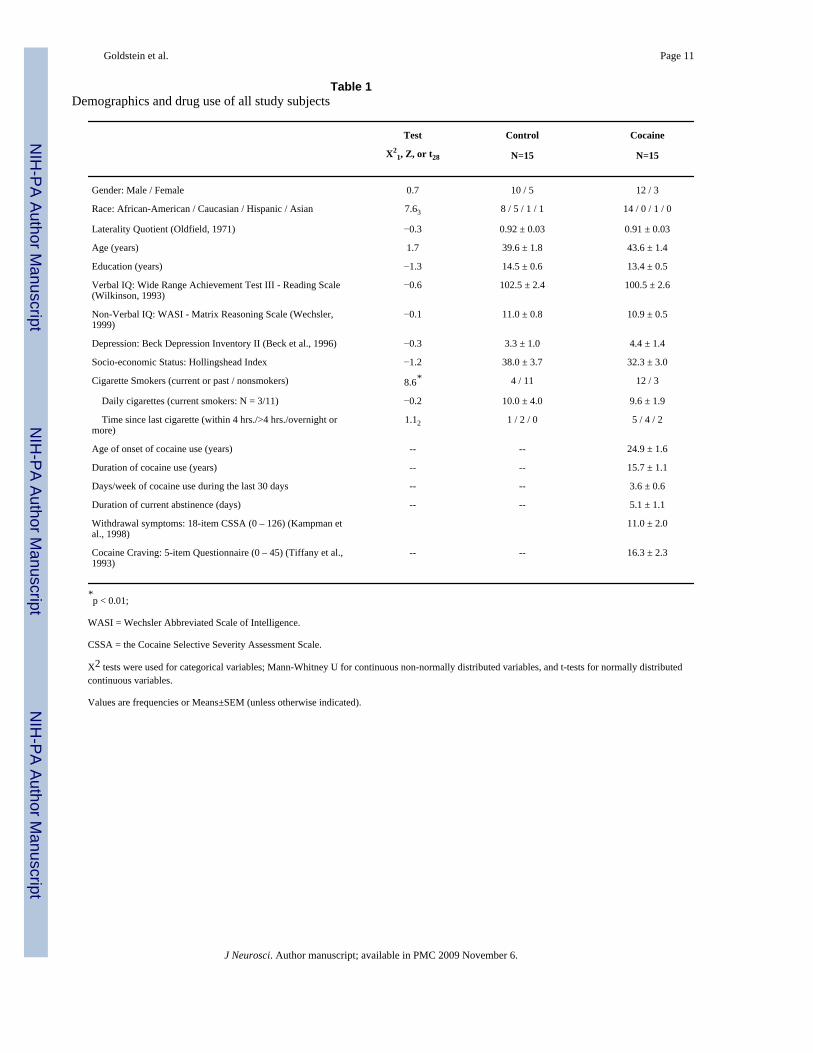
A total of 30 right-handed native English speakers, 15 individuals with current cocaine usedisorders (CUD) and 15 healthy control participants, were recruited using advertisements inlocal newspapers and by word-of mouth. All subjects were able to understand and giveinformed consent (for inclusion/exclusion criteria see Supplementary Material). Healthycontrols and CUD were matched on sex, age, education, and general intellectual functioning(Table 1).
The CUD were not currently treatment-seeking as ascertained during an extensive psychiatricscreen that included the Structured Clinical Interview for DSM-IV Axis I Disorders [researchversion (First et al., 1996; Ventura et al., 1998)] and the Addiction Severity Index (McLellanet al., 1992). Indeed all CUD used crack/cocaine (mostly smoked route) in the past 30 daysand met DSM-IV criteria for current cocaine dependence (N = 12) or abuse (N = 3; these threesubjects met criteria for cocaine dependence in remission). One CUD also reported currentalcohol abuse. Current use or dependence on other drugs was denied and corroborated byprescan urine tests in all subjects (urine was positive for cocaine in 10 CUD; urine was negativefor all drugs in all other subjects). Subjects were fully informed of all study procedures andrisks associated with MRI and provided written consent for their involvement in this study inaccordance with the local Institutional Review Board.
The drug word fMRI taskAll participants were scanned during a drug word fMRI task, while viewing, in a blockedfashion, drug or neutral words (2000 msec per word). Specifically, this fMRI task (developedin E-prime, Psychology Software Tools, Inc., Pittsburgh, PA) uses 40 drug words matchedwith 40 household words on length, part of speech (noun, adjective, adverb, verb), andfrequency in the English language [here we referred to the most widely used dictionarydesigned specifically for this purpose, (Francis and Kučera, 1982): the mean frequency for thedrug words was 43.7 and that of the neutral words was 42.6] (Goldstein et al., 2007b). We didnot include non-English or slang drug words that may have not been recognized by the controlsubjects. Similarly to other emotional fMRI tasks (Whalen et al., 1998), the two word typeswere presented via MRI-compatible goggles in a blocked on-off or off-on order (i.e., drug-neutral or neutral-drug), counterbalanced between subjects. There were eight 3.4-min taskrepetitions (four drug, four neutral), each containing two blocks of 20 drug or neutral words,interleaved with a 20 sec fixation cross. Each word trial was comprised of a 500 ms fixationcross, a 2000 ms word presentation (for word reading), a 500 ms response window, and a 500ms feedback slide. During the 500 ms response window subjects had to press one of four buttons(yellow, blue, red, green) on a commercially available response pad (Cedrus brand Luminamodel LP-400), matching the color of the word they had just read; word color order waspseudorandomized across all task runs. Note that the overt behavioral Stroop effect was notexpected because, to encourage processing of the meaning of the word and minimize theexpected working-memory difficulties in CUD (Woicik et al., 2008), we separated word
Goldstein et al. Page 2
J Neurosci. Author manuscript; available in PMC 2009 November 6.
NIH
-PA Author Manuscript
NIH
-PA Author Manuscript
NIH
-PA Author Manuscript
reading from pressing for its color (by 2000 ms), thereby decreasing the conflict inherent insuch a task [as further described in Supplementary Material]. Lastly, each word sequence wasperformed under one of four counterbalanced monetary reward amounts (50¢, 25¢, 1¢, or 0¢),gained for correct performance for up to $75 received at the completion of this study ($66.6 ±1.1 with no differences between the groups in this amount, t28 = 0.5, p > 0.7).
The word fluency taskAll subjects also performed a standard semantic fluency task, naming as many words from twospecified semantic categories (animals and fruits or vegetables) for one minute per category(Lezak, 1995). Subjects also participated in the emotional variant of this task, naming drug-related words (names of drugs, people, places, or states of mind related to getting, using, orrecovering from drugs) for the same duration (Goldstein et al., 2007a). Correct responses(excluding repetitions and errors, i.e., words clearly not related to the selected semanticcategory) were summed for each category. Following the fMRI measures, we calculated adifferential drug > neutral (averaged across both non-drug semantic categories) fluencymeasure; here we first corrected, with regression analyses, for verbal IQ that correlated withthe fluency measures in the CUD (Supplementary Material). The unstandardized residuals wereused for all subsequent analyses.
MRI data acquisitionMRI scanning was performed on a 4T whole-body Varian/Siemens MRI scanner. The bloodoxygenation level dependent (BOLD) responses were measured as a function of time using aT2*-weighted single-shot gradient-echo EPI sequence (TE/TR = 20/1600 ms, 4 mm slicethickness, 1 mm gap, typically 33 coronal slices, 20 cm FOV, 64 × 64 matrix size, 90°-flipangle, 200 kHz bandwidth with ramp sampling, 128 time points, and 4 dummy scans to bediscarded to avoid non-equilibrium effects in the fMRI signal). Padding was used to minimizemotion, which was also monitored immediately after each fMRI run (Caparelli et al., 2003).Earplugs and headphones were used to minimize the interference effect of scanner noise duringfMRI (Tomasi et al., 2005). Anatomical images were collected using a T1-weighted 3D-MDEFT sequence (Lee et al., 1995) and a modified T2-weighted Hyperecho sequence (Hennigand Scheffler, 2001)and reviewed to rule out gross brain morphological abnormalities.
MRI data processingSubsequent analyses were performed with the statistical parametric mapping package (SPM2;Welcome Department of Cognitive Neurology, London UK). A six-parameter rigid bodytransformation (3 rotations, 3 translations) was used for image realignment and to correct forhead motion; 2 mm displacement and 2° rotation in any of the axes in any of the task repetitionswere used as criteria for acceptable motion. The realigned datasets were spatially normalizedto the standard frame (Talairach) with a 12-parameter affine transformation (Ashburner et al.,1997), using a voxel size of 3 × 3 × 3 mm. An 8-mm full-width-half-maximum Gaussian kernelwas used to smooth the data.
BOLD-fMRI analysesA general linear model (Friston et al., 1995) and a box-car design convolved with a canonicalhemodynamic response function and high-pass filter (cut-off frequency: 1/520 sec) was usedto calculate individual BOLD-fMRI maps for the fMRI task. Two contrasts per subject,reflecting % signal change for the drug or neutral words from the fixation baseline, were usedto calculate the direct drug > neutral differential contrast of interest, which was used in allsubsequent analyses in SPM2: two one-way t-tests (drug > neutral comparison separately forCUD and controls), a 2-way t-test (direct group comparison, CUD vs. controls, for the drug >neutral contrast), and simple regression analyses (using the drug > neutral contrast maps) with
Goldstein et al. Page 3
J Neurosci. Author manuscript; available in PMC 2009 November 6.
NIH
-PA Author Manuscript
NIH
-PA Author Manuscript
NIH
-PA Author Manuscript
selected variables as seed values (drug > neutral word fluency or mesencephalon drug > neutral% BOLD signal change for every participant) [note that subtraction between different taskconditions, and not just between a task condition and a given baseline, allows for a rigorouscontrol of all processes that are extraneous to the effect of interest (e.g., comparing drug wordsto neutral words allows for the control of all the sensory-motor properties that are common tothese two conditions)]. Brain activation clusters were corrected for multiple comparisons usingthe continuous random field calculation (Adler, 1981). This approach, as implemented inSPM2, takes into account the local smoothness of the data and is considered more accurate forlocal smoothness > 3 voxels than the more conservative Bonferroni correction (Worsley et al.,1996; Worsley, 2007). In the present study, the random field calculation was based on theexpected Euler characteristics of the regions above a puncorr < 0.005 threshold (voxel-leveluncorrected). Clusters with at least 4 voxels (108 mm3) and pcorr < 0.05 (cluster-level correctedfor multiple comparisons) were considered significant. In all SPM analyses, anatomicalspecificity was corroborated with a co-planar stereotaxic atlas of the human brain (Talairachand Tournoux, 1988).
For follow-up statistical analyses and to confirm the whole-brain voxel-based analyses,functional regions of interest (ROIs) with an isotropic volume of 27 voxels (729 mm3) weredefined at the mesencephalon level (fixed across subjects and conditions, boldface in Table 2)to extract (with a custom program written in IDL, ITT Visual Information Solutions, Boulder,CO) the average (and variability) BOLD-fMRI signal amplitudes in these coordinates. TheseROI measures were used in the appropriate (e.g., t-test, correlation) analyses in SPSS 11.5(SPSS Inc., Chicago, IL). Statistical significance for these ROI analyses was defined as p <0.05uncorr. These ROI analyses were only performed for the regions that survived the whole-brain correction threshold as described above, which provided protection against Type I error.
Finally, all continuous and normally distributed variables were inspected with parametric tests(within groups: paired t-test; between groups: independent t-tests; correlations: Pearson r).Otherwise, the respective non-parametric tests were used [Wilcoxon, Mann-Whitney U, orSpearman r (rS)]. To study potential contribution to results of history of cigarette smoking (thatsignificantly differed between the study groups, Table 1), differences in all dependent variablesbetween the cigarette smokers and non-smokers were inspected with t-tests or the equivalentnon-parametric test; we also inspected correlations between all of our dependent variables anddaily frequency of cigarettes smoked (Stevens, 1992). Note that these latter analyses were onlyperformed across all study subjects as within-group analyses would be statisticallyunderpowered (there were only four current or past cigarette smokers within the control groupand only three nonsmokers within the CUD group).
RESULTSDrug fluency
A repeated measures analysis of variance (ANOVA) with verbal fluency (corrected for verbalintellectual functioning, Supplementary Material) revealed a group (CUD, control) by word(drug, neutral) interaction (F1,28 = 9.0, p < 0.01; Figure 1A). Post-hoc t-tests showed that thisinteraction was explained by higher drug than neutral responses in the CUD (t14 = 2.2, p <0.05) but not in healthy participants (who showed a trend toward the reverse pattern, t14 = −2.1,p = 0.056) and a significant difference between the study groups for the drug words only (drug:t28 = 2.2, p < 0.05; neutral: t28 = −.9, p > 0.3).
SPM and ROIsVoxel-by-voxel whole-brain analyses (two one-way t-tests for the direct drug > neutralcomparison as a function of study group) revealed a bilateral mesencephalon response in the
Goldstein et al. Page 4
J Neurosci. Author manuscript; available in PMC 2009 November 6.
NIH
-PA Author Manuscript
NIH
-PA Author Manuscript
NIH
-PA Author Manuscript
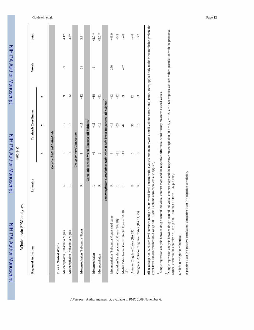
CUD only (Figure 1 top right: x = 6/−6, y = −12/−15, z = −9/−12, 39 voxels, t = 4.1) (Table2). Moreover, a direct whole-brain group comparison (2-way t-test using the direct drug >neutral contrasts) showed a significant group by word interaction in the right mesencephalon(x = 3, y = −15, z = −12, 21 voxels, t = 3.3). The BOLD-fMRI signal (% change from baseline)extracted from this region for each participant and used in a repeated measures ANOVAvalidated this interaction (F1,28 = 10.6, p < 0.01; Figure 1B). Post-hoc t-tests showed that,similarly to the verbal fluency results, this interaction was driven by higher drug than neutralresponses in the CUD (t14 = 3.5, p < 0.01) but not healthy control participants (t14 = −1.3, p >0.2). Note that there were no main effects for repetition/block or money on the neural responsesto this task (specifically for the direct drug > neutral contrast) as further described inSupplementary Material.
Whole-brain SPM correlationsMost interestingly, a whole-brain simple regression analysis showed a significant correlationbetween the differential drug > neutral verbal fluency (used as seed values) and the respectivemesencephalon responses across all study participants (observed bilaterally: x = −6/3, y = −15/−18, z = −18/−21, 9 voxels, t = 2.7). The BOLD-fMRI signal (% change from the direct drug> neutral contrasts extracted from the cluster peak in the left mesencephalon) confirmed thatthis correlation was driven by the CUD (r = 0.58, p < 0.05) and not healthy control participants(r = 0.15, p > 0.5) (Figure 1C). The test of coincidence of these regression lines was significant(F2,26 = 3.7, p < 0.05) confirming that these specific brain-behavior correlations differedsignificantly between the study groups (this result is especially sound given that the whole-brain regression analyses were conducted across all study subjects, enhancing the possibilityfor uncovering similar group results but decreasing the possibility for differential group results,which were nevertheless significant in the current study). Thus, the more the mesencephalonresponded to the drug words as compared to the neutral words, the higher was the drug thanneutral verbal fluency in the CUD but not healthy control participants, together attesting to aconcomitant brain-behavior drug cue-reactivity in cocaine addiction.
Correlations between the mesencephalic drug cue responsivity (seed values were the % BOLDsignal change for every participant at x = 3, y = −15, z = −12, or the mesencephalic region thatshowed a differential drug > neutral BOLD response as a function of study group, Table 2)with other brain regions that showed similar responses (i.e., the direct drug > neutral contrastmaps were used), revealed positive correlations with the parahippocampal gyrus and negativecorrelations with a cluster that included the medial orbitofrontal cortex/rectal gyrus androstroventral/pregenual anterior cingulate cortex (Table 2). Thus, in all study subjects, thehigher the drug > neutral mesencephalic BOLD response during the drug word fMRI task, thehigher the respective response in the parahippocampal gyrus and the lower this relativeresponse in the limbic prefrontal cortex.
Effect of cigarette smoking on all behavioral measures and brain activation resultsThere were no significant associations between any of the study’s dependent variables(calculated as absolute: drug or neutral; or differential scores: drug > neutral, for all behavioralmeasures and ROIs) with history of cigarette smoking (t < |1.3|, p > 0.2). Also, there were nosignificant correlations between any of the study’s dependent variables with current cigarettesmoking frequency (rS < |0.4|, p > 0.07).
DISCUSSIONPerformance on standard (i.e., non drug-related) neuropsychological tasks is frequentlycompromised in drug addicted individuals as compared to healthy control subjects (Woicik etal., 2008). In contrast, compared to neutral stimuli (including words), drug stimuli/words can
Goldstein et al. Page 5
J Neurosci. Author manuscript; available in PMC 2009 November 6.
NIH
-PA Author Manuscript
NIH
-PA Author Manuscript
NIH
-PA Author Manuscript
enhance behavioral responses in drug addicted individuals; although relatively better, theseunique drug-related behavioral responses predict disadvantageous treatment outcome in thispopulation (Cox et al., 2006). Our results show, for the first time, that drug words (uniquelyhuman learned verbal descriptors of stimuli) increased fMRI BOLD responses in themesencephalon, a major source of dopaminergic release to motivationally salient orconditioned stimuli (Robinson and Berridge, 1993; McClure et al., 2003), in cocaine addictedindividuals. These results may reflect the strong conditioned incentive properties of the drugwords in the addicted group. It is therefore possible that this increased mesencephalic responseto drug words reflects activation from prefrontal glutamaterigc projections that regulatedopamine cell firing and drive enhanced dopamine responses to conditioned stimuli (Kalivas,2004; Wise, 2009). Further, possibly through mesencephalon’s extensive connections with thelimbic prefrontal cortex (Devinsky et al., 1995), these cue-reactive neural responses mayculminate in drug-biased behaviors (e.g., uncontrollable drug seeking or craving) (Goldsteinand Volkow, 2002) in drug addicted/susceptible individuals. This latter interpretation isconsistent with the significant correlations in our study between the drug-related responses inthe mesencephalon with similar responses in the prefrontal cortex (Table 2); however, thisinterpretation remains to be validated with additional direct measures of drug-biased responsesin drug addicted individuals.
A confound in our study is the use of monetary gain in this task. Here, evidence suggests thatrewarding stimuli are directly related to enhanced dopaminergic tone and increasedmesencephalic activation (Koch et al., 2008). Indeed, altered fMRI responses to reward in drugusers have been previously reported by our group (Goldstein et al., 2007c) and others (Bjorket al., 2008) as indicative of dopaminergic alterations in addiction (Volkow et al., 2004).Nevertheless, in the current study, the word by group interaction in the mesencephalon wasobserved after averaging the fMRI BOLD signal across all monetary reward conditions,suggesting that the contribution of drug-word cue-reactivity in this region in addiction issignificant and unique. Another limitation is that results could be attributed to severalneuropsychological mechanisms, including enhanced drug word familiarity, salience, attentionbias and memory processes in the CUD as compared to controls. The fMRI task controlled forsome of these processes (e.g., the drug and neutral words were matched for frequency in theEnglish language). However, similar processes remain to be tested in the self-generated drugfluency task (e.g., with simultaneous fMRI or recordings of autonomic responses). Finally, thecurrent results need to be replicated in other subgroups of drug addicted individuals (e.g.,treatment-seekers recruited from treatment centers and larger samples where the potentialimpact on results of individual variables such as sex and race can be studied).
In summary, our results are consistent with the effect of drug words on other (less localized/scalp) psychophysiological responses (Herrmann et al., 2000) and with neuroimaging studiesshowing similar cue-induced increases in dopaminergic responses when addicted individualsview drug images (pictures or movies) as associated with craving, withdrawal symptoms andaddiction severity (Heinz et al., 2004; Volkow et al., 2006). Our results for the first timedemonstrate that, in addicted individuals, drug words alone can elicit an fMRI BOLDmesencephalic response, as possibly associated with dopaminergic (and glutamatergic)mechanisms (Schott et al., 2008) that are crucial to conditioning (D’Ardenne et al., 2008).Moreover, the correlation between a very brief verbal fluency test, which can be easilyadministered (crucial for clinical studies), and fMRI cue-reactivity could be used as a biomarkerof neurobiological changes in drug addiction.
Supplementary MaterialRefer to Web version on PubMed Central for supplementary material.
Goldstein et al. Page 6
J Neurosci. Author manuscript; available in PMC 2009 November 6.
NIH
-PA Author Manuscript
NIH
-PA Author Manuscript
NIH
-PA Author Manuscript
AcknowledgmentsWe thank Muhammad A. Parvaz for help with task administration, Alex Panagopoulos and Dimitris Samaras for helpwith early data analyses, and Gene-Jack Wang for help with medical screens. We also would like to thank Sahib S.Khalsa and Steve Berry for help with word selection, matching and ratings and Suparna Rajaram for help with earlytask design. This study was supported by grants from the National Institute on Drug Abuse (to RZG: 1R01DA023579and R21DA02062) and the General Clinical Research Center (5-MO1-RR-10710).
ReferencesAdler, RJ. The Geometry of Random Fields. Chichester: John Wiley & Sons; 1981.Ashburner J, Neelin P, Collins DL, Evans A, Friston K. Incorporating prior knowledge into image
registration. Neuroimage 1997;6:344–352. [PubMed: 9417976]Beck, AT.; Steer, RA.; Brown, GK. Beck Depression Inventory Manual. Vol. 2. San Antonio: The
Psychological Corporation; 1996.Bjork JM, Smith AR, Hommer DW. Striatal sensitivity to reward deliveries and omissions in substance
dependent patients. Neuroimage 2008;42:1609–1621. [PubMed: 18672069]Bossert JM, Liu SY, Lu L, Shaham Y. A role of ventral tegmental area glutamate in contextual cue-
induced relapse to heroin seeking. J Neurosci 2004;24:10726–10730. [PubMed: 15564590]Caparelli EC, Tomasi D, Arnold S, Chang L, Ernst T. k-Space based summary motion detection for
functional magnetic resonance imaging. NeuroImage 2003;20:1411–1418. [PubMed: 14568510]Cox WM, Fadardi JS, Pothos EM. The addiction-stroop test: Theoretical considerations and procedural
recommendations. Psychol Bull 2006;132:443–476. [PubMed: 16719569]D’Ardenne K, McClure SM, Nystrom LE, Cohen JD. BOLD responses reflecting dopaminergic signals
in the human ventral tegmental area. Science 2008;319:1264–1267. [PubMed: 18309087]Devinsky O, Morrell MJ, Vogt BA. Contributions of anterior cingulate cortex to behaviour. Brain
1995;118 (Pt 1):279–306. [PubMed: 7895011]First, MB.; Spitzer, RL.; Gibbon, M.; Williams, J. Structured Clinical Interview for DSM-IV Axis I
disorders - Patient Edition (SCID-I/P, Version 2.0). New York: Biometrics Research Department,New York State Psychiatric Institute; 1996.
Francis, NW.; Kuera, H. Frequency Analysis of English Usage. Boston: Houghton Mifflin; 1982.Friston KJ. Testing for anatomically specified regional effects. Hum Brain Mapp 1997;5:133–136.
[PubMed: 10096418]Friston KJ, Holmes AP, Worsley KJ, Poline JB, Frith CD, Frackowiak RS. Statistical parametric maps
in functional imaging: a general approach. Human Brain Mapping 1995;2:189–210.Goldstein RZ, Volkow ND. Drug addiction and its underlying neurobiological basis: neuroimaging
evidence for the involvement of the frontal cortex. Am J Psychiatry 2002;159:1642–1652. [PubMed:12359667]
Goldstein RZ, Woicik PA, Lukasik T, Maloney T, Volkow ND. Drug fluency: a potential marker forcocaine use disorders. Drug Alcohol Depend 2007a;89:97–101. [PubMed: 17234364]
Goldstein RZ, Tomasi D, Rajaram S, Cottone LA, Zhang L, Maloney T, Telang F, Alia-Klein N, VolkowND. Role of the anterior cingulate and medial orbitofrontal cortex in processing drug cues in cocaineaddiction. Neuroscience 2007b;144:1153–1159. [PubMed: 17197102]
Goldstein RZ, Alia-Klein N, Tomasi D, Zhang L, Cottone LA, Maloney T, Telang F, Caparelli EC, ChangL, Ernst T, Samaras D, Squires NK, Volkow ND. Is decreased prefrontal cortical sensitivity tomonetary reward associated with impaired motivation and self-control in cocaine addiction? Am JPsychiatry 2007c;164:43–51. [PubMed: 17202543]
Heinz A, Siessmeier T, Wrase J, Hermann D, Klein S, Grusser SM, Flor H, Braus DF, Buchholz HG,Grunder G, Schreckenberger M, Smolka MN, Rosch F, Mann K, Bartenstein P. Correlation betweendopamine D(2) receptors in the ventral striatum and central processing of alcohol cues and craving.Am J Psychiatry 2004;161:1783–1789. [PubMed: 15465974]
Hennig J, Scheffler K. Hyperechoes. Magnetic Resonance in Medicine 2001;46:6–12. [PubMed:11443704]
Goldstein et al. Page 7
J Neurosci. Author manuscript; available in PMC 2009 November 6.
NIH
-PA Author Manuscript
NIH
-PA Author Manuscript
NIH
-PA Author Manuscript
Herrmann MJ, Weijers HG, Wiesbeck GA, Aranda D, Boning J, Fallgatter AJ. Event-related potentialsand cue-reactivity in alcoholism. Alcohol Clin Exp Res 2000;24:1724–1729. [PubMed: 11104120]
Kalivas PW. Glutamate systems in cocaine addiction. Curr Opin Pharmacol 2004;4:23–29. [PubMed:15018835]
Kampman KM, Volpicelli JR, McGinnis DE, Alterman AI, Weinrieb RM, D’Angelo L, Epperson LE.Reliability and validity of the Cocaine Selective Severity Assessment. Addict Behav 1998;23:449–461. [PubMed: 9698974]
Koch K, Schachtzabel C, Wagner G, Reichenbach JR, Sauer H, Schlosser R. The neural correlates ofreward-related trial-and-error learning: an fMRI study with a probabilistic learning task. Learn Mem2008;15:728–732. [PubMed: 18832559]
Lee JH, Garwood M, Menon R, Adriany G, Andersen P, Truwit CL, Ugurbil K. High contrast and fastthree-dimensional magnetic resonance imaging at high fields. Magnetic Resonance in Medicine1995;34:308–312. [PubMed: 7500867]
Lezak, MD. Neuropsychological Assessment. Vol. 3. New York, NY: Oxford University Press; 1995.McClure SM, Daw ND, Montague PR. A computational substrate for incentive salience. Trends Neurosci
2003;26:423–428. [PubMed: 12900173]McLellan AT, Kushner H, Metzger D, Peters R, Smith I, Grissom G, Pettinati H, Argeriou M. The Fifth
Edition of the Addiction Severity Index. J Subst Abuse Treat 1992;9:199–213. [PubMed: 1334156]Oldfield RC. The assessment and analysis of handedness: the Edinburgh inventory. Neuropsychologia
1971;9:97–113. [PubMed: 5146491]Robinson TE, Berridge KC. The neural basis of drug craving: an incentive-sensitization theory of
addiction. Brain Res Brain Res Rev 1993;18:247–291. [PubMed: 8401595]Schott BH, Minuzzi L, Krebs RM, Elmenhorst D, Lang M, Winz OH, Seidenbecher CI, Coenen HH,
Heinze HJ, Zilles K, Duzel E, Bauer A. Mesolimbic functional magnetic resonance imagingactivations during reward anticipation correlate with reward-related ventral striatal dopamine release.J Neurosci 2008;28:14311–14319. [PubMed: 19109512]
Stevens, J. Applied multivariate statistics for the social sciences. Vol. 2. Lawrence Erlbaum Associates;NewJersey: 1992.
Talairach, J.; Tournoux, P. Co-Planar Stereotaxic Atlas of the Human Brain. New York: Thieme MedicalPublishers, Inc; 1988.
Tiffany ST, Singleton E, Haertzen CA, Henningfield JE. The development of a cocaine cravingquestionnaire. Drug and Alcohol Dependence 1993;34:19–28. [PubMed: 8174499]
Tomasi D, Caparelli EC, Chang L, Ernst T. fMRI-acoustic noise alters brain activation during workingmemory tasks. NeuroImage 2005;27:377–386. [PubMed: 15893942]
Ventura J, Liberman RP, Green MF, Shaner A, Mintz J. Training and quality assurance with the StructuredClinical Interview for DSM-IV (SCID-I/P). Psychiatry Res 1998;79:163–173. [PubMed: 9705054]
Volkow ND, Fowler JS, Wang GJ, Swanson JM. Dopamine in drug abuse and addiction: results fromimaging studies and treatment implications. Mol Psychiatry 2004;9:557–569. [PubMed: 15098002]
Volkow ND, Wang GJ, Telang F, Fowler JS, Logan J, Childress AR, Jayne M, Ma Y, Wong C. Cocainecues and dopamine in dorsal striatum: mechanism of craving in cocaine addiction. J Neurosci2006;26:6583–6588. [PubMed: 16775146]
Wechsler, D. Wechsler abbreviated scale of intelligence. San Antonio, TX: Psychological Corporation;1999.
Whalen PJ, Bush G, McNally RJ, Wilhelm S, McInerney SC, Jenike MA, Rauch SL. The emotionalcounting Stroop paradigm: a functional magnetic resonance imaging probe of the anterior cingulateaffective division. Biol Psychiatry 1998;44:1219–1228. [PubMed: 9861465]
Wilkinson, G. The Wide-Range Achievement Test 3- Administration Manual. Wilmington, DE: WideRange Inc; 1993.
Wise RA. Ventral tegmental glutamate: a role in stress-, cue-, and cocaine-induced reinstatement ofcocaine-seeking. Neuropharmacology 2009;56 Suppl 1:174–176. [PubMed: 18598707]
Woicik PA, Moeller SJ, Alia-Klein N, Maloney T, Lukasik TM, Yeliosof O, Wang GJ, Volkow ND,Goldstein RZ. The Neuropsychology of Cocaine Addiction: Recent Cocaine Use Masks Impairment.Neuropsychopharmacology. 2008
Goldstein et al. Page 8
J Neurosci. Author manuscript; available in PMC 2009 November 6.
NIH
-PA Author Manuscript
NIH
-PA Author Manuscript
NIH
-PA Author Manuscript
Worsley, K. Statistical Parametric Mapping: The Analysis of Functional Brain Images. London: Elsevier;2007. Random Field Theory.
Worsley KJ, Marrett S, Neelin P, Vandal AC, Friston KJ, Evans AC. A unified statistical approach fordetermining significant signals in images of cerebral activation. Human Brain Mapping 1996;4:58–73.
Goldstein et al. Page 9
J Neurosci. Author manuscript; available in PMC 2009 November 6.
NIH
-PA Author Manuscript
NIH
-PA Author Manuscript
NIH
-PA Author Manuscript
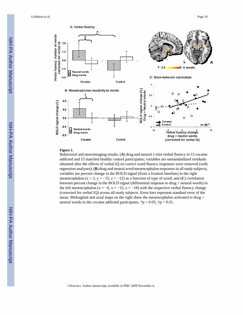
Figure 1.Behavioral and neuroimaging results. (A) drug and neutral 1-min verbal fluency in 15 cocaineaddicted and 15 matched healthy control participants; variables are unstandardized residualsobtained after the effects of verbal IQ on correct word fluency responses were removed (withregression analyses); (B) drug and neutral word mesencephalon responses in all study subjects;variables are percent change in the BOLD signal (from a fixation baseline) in the rightmesencephalon (x = 3, y = −15, z = −12) as a function of type of word; and (C) correlationbetween percent change in the BOLD signal (differential response to drug > neutral words) inthe left mesencephalon (x = −6, x = −15, z = −18) with the respective verbal fluency change(corrected for verbal IQ) across all study subjects. Error bars represent standard error of themean. Midsagittal and axial maps on the right show the mesencephalon activated to drug >neutral words in the cocaine addicted participants. *p < 0.05; †p < 0.01.
Goldstein et al. Page 10
J Neurosci. Author manuscript; available in PMC 2009 November 6.
NIH
-PA Author Manuscript
NIH
-PA Author Manuscript
NIH
-PA Author Manuscript
NIH
-PA Author Manuscript
NIH
-PA Author Manuscript
NIH
-PA Author Manuscript
Goldstein et al. Page 11
Table 1Demographics and drug use of all study subjects
Test Control Cocaine
X21, Z, or t28 N=15 N=15
Gender: Male / Female 0.7 10 / 5 12 / 3
Race: African-American / Caucasian / Hispanic / Asian 7.63 8 / 5 / 1 / 1 14 / 0 / 1 / 0
Laterality Quotient (Oldfield, 1971) −0.3 0.92 ± 0.03 0.91 ± 0.03
Age (years) 1.7 39.6 ± 1.8 43.6 ± 1.4
Education (years) −1.3 14.5 ± 0.6 13.4 ± 0.5
Verbal IQ: Wide Range Achievement Test III - Reading Scale(Wilkinson, 1993)
−0.6 102.5 ± 2.4 100.5 ± 2.6
Non-Verbal IQ: WASI - Matrix Reasoning Scale (Wechsler,1999)
−0.1 11.0 ± 0.8 10.9 ± 0.5
Depression: Beck Depression Inventory II (Beck et al., 1996) −0.3 3.3 ± 1.0 4.4 ± 1.4
Socio-economic Status: Hollingshead Index −1.2 38.0 ± 3.7 32.3 ± 3.0
Cigarette Smokers (current or past / nonsmokers) 8.6* 4 / 11 12 / 3
Daily cigarettes (current smokers: N = 3/11) −0.2 10.0 ± 4.0 9.6 ± 1.9
Time since last cigarette (within 4 hrs./>4 hrs./overnight ormore)
1.12 1 / 2 / 0 5 / 4 / 2
Age of onset of cocaine use (years) -- -- 24.9 ± 1.6
Duration of cocaine use (years) -- -- 15.7 ± 1.1
Days/week of cocaine use during the last 30 days -- -- 3.6 ± 0.6
Duration of current abstinence (days) -- -- 5.1 ± 1.1
Withdrawal symptoms: 18-item CSSA (0 – 126) (Kampman etal., 1998)
11.0 ± 2.0
Cocaine Craving: 5-item Questionnaire (0 – 45) (Tiffany et al.,1993)
-- -- 16.3 ± 2.3
*p < 0.01;
WASI = Wechsler Abbreviated Scale of Intelligence.
CSSA = the Cocaine Selective Severity Assessment Scale.
X2 tests were used for categorical variables; Mann-Whitney U for continuous non-normally distributed variables, and t-tests for normally distributedcontinuous variables.
Values are frequencies or Means±SEM (unless otherwise indicated).
J Neurosci. Author manuscript; available in PMC 2009 November 6.
NIH
-PA Author Manuscript
NIH
-PA Author Manuscript
NIH
-PA Author Manuscript
Goldstein et al. Page 12Ta
ble
2W
hole
-bra
in S
PM a
naly
ses
Reg
ion
of A
ctiv
atio
nL
ater
ality
Tal
aira
ch C
oord
inat
esV
oxel
st-s
tat
xy
z
Coca
ine
Addi
cted
Indi
vidu
als
Dru
g >
Neu
tral
Wor
ds
Mes
ence
phal
on (S
ubst
antia
Nig
ra)
R6
−12
−939
4.1*
Mes
ence
phal
on (S
ubst
antia
Nig
ra)
L−6
−15
−12
3.4*
Gro
up b
y W
ord
Inte
ract
ion
Mes
ence
phal
on (S
ubst
antia
Nig
ra)
R3
−15
−12
213.
3*
Cor
rela
tions
with
Wor
d Fl
uenc
y: A
ll Su
bjec
ts†
Mes
ence
phal
onL
−6
−15
−18
9+2
.7**
Mes
ence
phal
onR
3−1
8−2
1+2
.6**
Mes
ence
phal
on C
orre
latio
ns w
ith O
ther
Who
le-b
rain
Res
pons
es: A
ll Su
bjec
ts‡
Mes
ence
phal
on (S
ubst
antia
Nig
ra):
seed
val
ueR
3−1
5−1
225
0+4
3.9
Cin
gulu
m/P
arah
ippo
cam
pal G
yrus
(BA
28)
L−2
1−2
4−1
2+3
.5
Med
ial O
rbito
fron
tal C
orte
x, R
ecta
l Gyr
us (B
A 1
0,11
)L
−15
42−9
497
−4.8
Ant
erio
r Cin
gula
te C
orte
x (B
A 2
4)B
036
12−4
.0
Subg
enua
l Ant
erio
r Cin
gula
te C
orte
x (B
A 1
1, 2
5)R
315
−3−3
.7
All
resu
lts: p
< 0
.05
clus
ter-
leve
l cor
rect
ed (a
nd p
< 0
.005
vox
el-le
vel u
ncor
rect
ed),
4 vo
xels
min
imum
, *w
ith a
smal
l vol
ume
corr
ectio
n (F
risto
n, 1
997)
app
lied
only
to th
e m
esen
ceph
alon
(**h
ere
the
voxe
l-lev
el u
ncor
rect
ed th
resh
old
was
p <
0.0
1, sm
all v
olum
e co
rrec
tion
was
als
o ap
plie
d).
† Sim
ple
regr
essi
on a
naly
sis b
etw
een
drug
> n
eutra
l ind
ivid
ual c
ontra
st m
aps a
nd th
e re
spec
tive
diff
eren
tial w
ord
fluen
cy m
easu
res a
s see
d va
lues
.
‡ Sim
ple
regr
essi
on a
naly
sis b
etw
een
drug
> n
eutra
l ind
ivid
ual c
ontra
st m
aps a
nd th
e re
spec
tive
mes
ence
phal
on (a
t x =
3, y
= −
15, z
= −
12) r
espo
nses
as s
eed
valu
es (c
orre
latio
n w
ith th
e pr
efro
ntal
corti
cal c
lust
er in
the
cont
rols
: r =
−0.
7, p
< 0
.01;
in th
e C
UD
: r =
−0.
6, p
< 0
.05)
.
L =
left;
R =
righ
t; B
= b
ilate
ral.
A p
ositi
ve t-
stat
(+):
posi
tive
corr
elat
ion;
a n
egat
ive
t-sta
t (−)
: neg
ativ
e co
rrel
atio
n.
J Neurosci. Author manuscript; available in PMC 2009 November 6.