protein kinase c isozymes and addiction
TRANSCRIPT
Introduction
PKC is a family of 10 structurally relatedphospholipid-dependent kinases that transducesignals involving lipid second messengers (1,2).The first to be isolated were the “conventional”cPKCs (α, βI, βII, and γ), which are activated bycalcium and diacylglycerol (DAG). Additional“novel” nPKCs (δ, ε, η, and θ) were subse-quently cloned and characterized as being acti-vated by DAG but not by calcium. Finally, twoadditional “atypical” aPKC isozymes (ζ and
λ/ι) were discovered that are insensitive to cal-cium and DAG. PKC λ and PKCι are the respec-tive human and mouse homologs of the sameenzyme. Additional kinases, related to the PKCfamily are the PKC-related kinases PRK1 andPRK2, which are insensitive to calcium andDAG but show increased activity when boundto activated RhoA GTPase (3). Two otherenzymes, PKCµ and PKCν, like novel PKCs, areactivated by DAG but not by calcium. However,they contain additional functional domains anddisplay a different pattern of substrate speci-ficity, and have now been classified separatelyas members of the PKD family (4).
One of the major signal transduction cas-cades that leads to PKC activation is the
Molecular Neurobiology 139 Volume 29, 2004
Molecular NeurobiologyCopyright © 2004 Humana Press Inc.All rights of any nature whatsoever reserved.ISSN 0893-7648/04/29(2): 139–153/$25.00
Protein Kinase C Isozymes and Addiction
M. Foster Olive* and Robert O. Messing
Ernest Gallo Clinic and Research Center, Department of Neurology, University of California at San Francisco, Emeryville, CA 94608
Abstract
Protein kinase C (PKC) has long been recognized an important family of enzymes that regulatenumerous aspects of neuronal signal transduction, neurotransmitter synthesis, release and reup-take, receptor and ion channel function, neuronal excitability, development, and gene expression.Much evidence has implicated PKCs in the effects of several drugs of abuse, and in behavioralresponses to these drugs. The present review summarizes the effects of both acute and chronicexposure to various drugs of abuse on individual PKC isozymes in the brain. In addition, wesummarize recent studies utilizing mice with targeted deletions of the genes for PKCγ and PKCε.These studies suggest that individual PKC isozymes play a role in the development of drugdependence and addiction.
Index Entries: Protein kinase C; isozymes; ethanol; cocaine; amphetamine; opiates; knock-out mouse.
Received August 25, 2003; Accepted September 25, 2003.* Author to whom all correspondence and reprint
requests should be addressed. E-mail: [email protected]
phosphoinositide (PI) signaling pathway (5).Stimulation of certain G protein-coupled cellsurface receptors activates phospholipase C,which hydrolyzes phosphatidylinositol-4,5-bis-phosphate to form inositol triphosphate (IP3)and DAG. IP3 binds to intracellular receptorscausing release of calcium from stores in theendoplasmic reticulum, whereas DAG binds toand activates most PKC isozymes. DAG mayalso be formed as a downstream consequenceof receptor-mediated activation of phospholi-pase D (6). In addition, cis-unsaturated fattyacids, arachidonic acid, and lysophosphatidy-choline produced by phopholipase A2 can acti-vate or enhance activation of several PKCisozymes (6–8). Likewise, phospatidylinositol-3,4,5-triphhosphate, produced by receptor-mediated activation of PI-3 kinases canactivate several PKC isozymes (9).
Before PKCs can respond to second messen-gers, they must be phosphorylated at threesites in the kinase domain. The first and rate-limiting reaction occurs in the activation loopand is catalyzed by phosphoinositide-depen-dent kinase 1 (PDK-1), which is in turn regu-lated by 3′ phosphoinositide products of PI-3kinase (10) This phosphorylation correctlyaligns residues at the active site, promotingautophosphoylation of two additional residuesat the C-terminus to produce a mature enzymecapable of responding to lipid second messen-gers. For the cPKCs, PDK-1 phosphorylation isconstitutive, whereas for the nPKCs and aPKCs,phosphorylation by PDK1 is under partial regu-lation by 3′ phosphoinositides providing anadditional mode of PKC regulation (10,11).
PKC function is also regulated by subcellu-lar localization, which depends on bothkinase:lipid and kinase:protein interactions.PKC activation is generally associated withtranslocation of PKC from one cellular com-partment to another containing lipid activatorsand proteins that bind the activated form ofthe enzyme in proximity to substrates. Thus,subcellular fractionation of cells or brain tissueand analysis of PKC content in lipid-contain-ing crude membrane fractions is often used asan indirect measure of PKC activation. Once at
the site of a particular substrate, PKCs inducephorphorylation at serine/threonine residues,which in turn alters the function of the sub-strate protein.
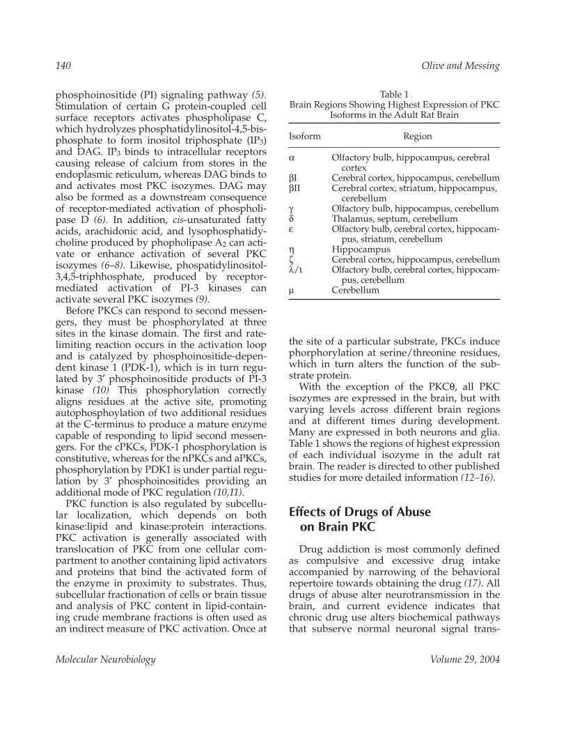
With the exception of the PKCθ, all PKCisozymes are expressed in the brain, but withvarying levels across different brain regionsand at different times during development.Many are expressed in both neurons and glia.Table 1 shows the regions of highest expressionof each individual isozyme in the adult ratbrain. The reader is directed to other publishedstudies for more detailed information (12–16).
Effects of Drugs of Abuse on Brain PKC
Drug addiction is most commonly definedas compulsive and excessive drug intakeaccompanied by narrowing of the behavioralrepertoire towards obtaining the drug (17). Alldrugs of abuse alter neurotransmission in thebrain, and current evidence indicates thatchronic drug use alters biochemical pathwaysthat subserve normal neuronal signal trans-
140 Olive and Messing
Molecular Neurobiology Volume 29, 2004
Table 1Brain Regions Showing Highest Expression of PKC
Isoforms in the Adult Rat Brain
Isoform Region
α Olfactory bulb, hippocampus, cerebralcortex
βI Cerebral cortex, hippocampus, cerebellumβII Cerebral cortex, striatum, hippocampus,
cerebellumγ Olfactory bulb, hippocampus, cerebellumδ Thalamus, septum, cerebellumε Olfactory bulb, cerebral cortex, hippocam-
pus, striatum, cerebellumη Hippocampusζ Cerebral cortex, hippocampus, cerebellumλ/ι Olfactory bulb, cerebral cortex, hippocam-
pus, cerebellumµ Cerebellum

duction (18–20). Such drug-induced alterationsin brain function are thought to alter therewarding and aversive qualities of drugresponses in such a way as to promote compul-sive drug use. Since PKC plays a major role intransducing signals carried by lipid secondmessengers, it is not surprising that drugs ofabuse can alter PKC signaling and that PKCcan modulate responses to drugs of abuse.
Ethanol
At anesthetic concentrations, ethanol andother aliphatic n-alkanols inhibit purified ratbrain cPKCs when assayed in a lipid-indepen-dent assay using protamine sulfate as activatorand substrate (21). Ethanol and short-chainalcohols also inhibit PKCα phosphorylation ofprotamine sulfate when activated in lipid vesi-cles containing PS and DAG, whereas long-chain alcohols enhance this activity throughdifferential interaction with high- and low-affinity binding sites within the regulatory por-tion of the kinase (22). However, studies usingmixed micelles or sonicated lipids and histoneas the substrate have failed to demonstrate aneffect of ethanol on PKC activity in vitro (23,24).
Ethanol has been found to alter PKC local-ization in some cell systems. For example, brief(30 s) exposure of cultured astroglial cells to25–200 mM ethanol induces translocation ofPKC (isoform unknown) from the cytosol tothe crude membrane fraction (25). In addition,incubation of NG108–15 neuroblastoma-glioma cells with 200 mM ethanol for 2 dinduces a translocation of PKCδ from the Golgiapparatus to the perinuclear region and atranslocation of PKCε from perinuclear regionto the cytoplasm (26). It is not known whetherthese changes in subcellular localization areassociated with increased phosphotransferaseactivity and whether they facilitate normalPKC signaling or sequester PKC isozymesaway from physiological substrates.
Early in vitro studies by our group revealedthat certain cellular responses to chronicethanol exposure are mediated by PKCδ andPKCε. Exposure of PC12 cells to 50–200 mM
ethanol for 2–6 d selectively upregulates theexpression of the PKCδ and ε in PC12 cellswithout altering levels of the α, β, or ζisozymes (24). Subsequent studies showed thatPKCε mediates up-regulation of N-type cal-cium channels (27) and enhancement of nervegrowth factor-stimulated neurite outgrowth(28,29) induced by chronic ethanol exposure.In contrast, PKCδ is required for up-regulationof L-type calcium channels by chronic expo-sure to ethanol in these cells (30,31).
Chronic ethanol exposure can also alter PKCabundance in the nervous system. Chronicadministration of ethanol to rats (via liquiddiet) reduces radiolabelled phorbol ester bind-ing to cortical and hippocampal homogenates(32,33), synaptosomes (34) and brain slices(34), as well as general PKC activity (35). How-ever, none of these studies determined whichPKC isozymes are involved in these changes.More recently, it was shown that chronicethanol consumption (via liquid diet) increasesmembrane bound PKCα and PKCγ in limbicforebrain regions while decreasing total levelsof these isozymes in the frontal cortex (36).Interestingly, these investigators found nochange in levels of PKCε in either region afterchronic ethanol, which contrasts with findingsin cell culture (see above). These studies arelimited by heterogeneity of the tissues exam-ined. Also the effects of ethanol on PKCisozyme levels and activity in adult animalsmay be highly dependent on factors such ascell type, duration of ethanol exposure, andethanol concentration.
Prenatal ethanol exposure also been shownto modulate PKC isozyme levels in the brain.Injection of 1 g/kg ethanol into chicken eggscaused time-dependent decreases in overalllevels of PKC α, γ, and ε in the postnatal chickbrain, with no effects on PKCι (37). A recentstudy examining the effects of pre- and postna-tal ethanol exposure on PKC isozymes wasalso conducted (38). In this study, pregnantfemale rats were fed an ethanol-containing dietfor at least 2 mo prior to birth. In addition, theoffspring of intoxicated mothers were given anethanol-containing liquid diet for 8, 30, or 90
PKC Isozymes and Addiction 141
Molecular Neurobiology Volume 29, 2004

postnatal days. Extraction and subcellular frac-tionation of the cerebral cortex showedincreased overall levels of PKC activity, whichcould be attributed to increases in total andmembrane-associated PKCβI, βII and ζ. Nochanges in the levels of the PKCα, γ, ε and ηwere found, whereas decreases in the level ofPKCδ were observed. The results of these twostudies are not easily compared, however, asthey not only utilized different species but alsoused different timing, doses, and durations ofethanol exposure. Nonetheless, they demon-strate the ability of ethanol to alter PKCisozymes in the brain when given duringembryonic development.
The predominant inhibitory ligand-gatedion channel in the nervous system is theGABAA receptor/ionophore complex which,when activated, hyperpolarizes neuronalmembranes via increasing chloride influx.Intoxicating concentrations of ethanol canpotentiate GABAA receptor function in sev-eral neuronal preparations (39–41). SeveralGABAA receptor subunits contain consensussequences for phosphorylation by proteinkinases including Src family tyrosine kinases,PKA, PKC, and Ca2+-calmodulin-dependentkinase II (42), and evidence suggests that PKCplays an important role in regulating GABAAreceptor sensitivity to ethanol. PKC inhibitorssuppress enhancement of GABAA receptorfunction by ethanol in hippocampal neurons,suggesting that PKC-mediated phosphoryla-tion is required for ethanol sensitivity in thatbrain region (43,44). Recent studies in knock-out mice have implicated PKCγ and ε as keymodulators of ethanol’s actions at GABAAreceptors (see below). PKCγ, but not PKCδ orPKCε, can be co-immunoprecipitated with theα1 and α4 subunits of the GABAA receptorfrom rat cerebral cortex and chronic ethanolexposure (in liquid diet) increases PKCγ asso-ciation with α4 while decreasing its associa-tion with α1 (45). In addition, the ability ofethanol to potentiate chloride currents gener-ated by another inhibitory receptor, the strych-nine-sensitive glycine receptor, in rat ventraltegmental neurons can be blocked by a spe-
cific peptide inhibitor of PKCε (46). These dataprovide evidence that PKCγ and PKCε regu-late the response of inhibitory ligand-gatedion channels to ethanol.
Psychostimulants
Psychostimulants such as cocaine andamphetamines act in the brain by eitherinhibiting the reuptake of monoamines viablockade of presynaptic transporters (as is thecase for cocaine) or by reversing vesicular andplasma membrane transporters to induce arelease of monoamine neurotransmitters (as isthe case for amphetamine-related drugs). Psy-chostimulants are highly addictive, and pro-duce robust behavioral changes in rodents andhumans, including arousal, hyperactivity,stereotyped behaviors, and motor sensitization(i.e., increasing motor responses to the drugwith each repeated exposure).
Psychostimulants produce hyperactivity pri-marily by increasing dopamine neurotransmis-sion in the mesolimbic system (i.e., ventraltegmental area—nucleus accumbens pathway)(47–49). This process appears to require theactivation of PKC, as the locomotor stimulanteffects of intra-accumbens amphetamine in ratsis attenuated by local infusion of the nonselec-tive PKC inhibitor Ro31-8220 but not the inac-tive analog bisindoylmaleimide V (50). This ispresumably due to an inhibition of ampheta-mine-induced dopamine release, as has beendemonstrated by PKC inhibitors in vitro(50,51). Similarly, injection of the protein kinaseinhibitor H7 intracerebroventricularly (52) orinto the ventral tegmental area (53) reducesacute cocaine-induced hyperlocomotion, aswell as cocaine-induced increases in nucleusaccumbens dopamine release (53). Intra-tegmental H7 also delays the onset of behav-ioral sensitization associated with repeatedcocaine exposure (54,55). However, H7 inhibitsthe activity of numerous protein kinases asidefrom PKC, and thus the specificity of theseeffects on PKC is not certain. Nonetheless,given the established role of PKC in regulationof dopamine transporter function (56), investi-
142 Olive and Messing
Molecular Neurobiology Volume 29, 2004
gations into the role of specific PKC isozymesin the locomotor and dopamine-releasingeffects of psychostimulants are warranted.
Psychostimulants also produce a phenome-non known as conditioned place preference(CPP), where subjects that are repeatedlyexposed to cocaine in a particular environmentwill demonstrate increased preference for thatenvironment (over a neutral or saline-pairedenvironment) in a drug-free state. This presum-ably results from the subject’s ability to pair orassociate the pleasurable effects of the drugwith the environment in which the drug isreceived. There is some evidence that PKC isinvolved in cocaine CPP, as intracerebroventric-ular injection of H7 or the more selective PKCinhibitor chelerythrine in rats inhibits the devel-opment of a cocaine CPP (52). However, theseinvestigators found that PKC was only involvedin the consolidation of cocaine CPP (i.e., estab-lishing the cocaine-environment associations),as these PKC inhibitors were effective in block-ing cocaine CPP only when administered imme-diately after the cocaine-environment pairings,and not when administered before the drug-environment pairings or prior to the post-train-ing CPP test session. Thus, PKC appears to beinvolved in drug-associated memory formation,which is not surprising given the role of PKC inlong-term potentiation and learning and mem-ory (57–60).
To our knowledge, only one study has beenconducted on the role of individual PKCisozymes in the establishment of psychostimu-lant-environment associations. Thomas andEveritt (61) trained rats to press a lever in orderto receive an intravenous infusion of cocainethat was accompanied by a distinct environ-mental stimuli (i.e., illumination of a light). Acontrol group of rats were exposed to cocaineand the environmental stimuli, but in a non-contingent manner (i.e., presentation of thelight or tone did not depend on whether the ratexecuted the lever press for the cocaine infu-sion). After a 3-d period when no cocaine orenvironmental cues were given, the rats wereplaced back in the training environment todetermine if they would perform the operant
task to obtain cocaine-associated cues. Exami-nation of PKCγ mRNA expression by in situhybridization revealed that animals that hadlearned the light stimulus was predictive ofcocaine infusion demonstrated increased PKCγmRNA abundance in the amygdala anddecreased abundance in the prefrontal cortex.Interestingly, no changes in PKCγ mRNA lev-els were observed in the hippocampus, despiteevidence that PKCγ in this structure is impor-tant for various forms of learning and memory(62–67). However, rats that were presentedassociations between cocaine and the lightstimulus in a noncontingent manner showedchanges in PKCγ mRNA levels in the nucleusaccumbens and anterior cingulate cortex.Results of this study indicate not only thatPKCγ in multiple brain regions may contributeto the formation of drug-environment associa-tions, but also that distinct neural circuits mayunderlie such associations based on whetherdrug-associated environmental cues are pre-dictive of obtaining the drug itself.
Repeated cocaine administration has beenreported to alter the levels of individual PKCisoforms in a heterogeneous manner. Steketeeand colleagues found that repeated adminis-tration of cocaine (15 or 30 mg/kg/d for atleast 4 d) produced an overall increase in PKCactivity in the ventral tegmental area (68), butdid not produce any changes in absolute levelsof PKCα, βI, βII, or γ in this region. However,repeated cocaine exposure did increase levelsof membrane associated PKCβI in the frontalcortex, and decreased levels of PKCα andPKCβII in the striatum when assayed 24 h afterthe last injection (69). In a more recent studyutilizing oligonucleotide microarrays withposthoc immunoblot confirmation, Vrana andco-workers found that a more prolongedadministration of cocaine to rats (45 mg/kg/dfor 14 d) caused increases in PKCα and PKCεmRNA in the hippocampus of rats (70).
The recreational drug 3,4-methylenedioxy-methamphetamine (MDMA, “Ecstasy”) is a sub-stituted amphetamine that acts by inducingmassive release and impaired reuptake of syn-aptic serotonin and dopamine. Recent evidence
PKC Isozymes and Addiction 143
Molecular Neurobiology Volume 29, 2004
suggests that MDMA may also alter PKC sig-naling in the brain. Acute administration ofMDMA to rats increases membrane boundPKC in cortical synaptosomes, as measured byphorbol ester binding (71,72); however, thePKC isozymes involved in this are currentlyunknown. Nonetheless, these effects appear tobe dose- and time-dependent (72) and requireintact nerve terminals that release serotonin(71), which in turn activates receptors that arecoupled to phosphoinositide/PKC signaling,such as 5-HT2A/2C receptors (72,73).
Opiates
Opiate drugs such as morphine and heroinact on one or more of three subtypes of recep-tors, namely µ, δ, and κ opioid receptors. Whilethese opioid receptors are primarily linked tocyclic AMP and protein kinase A signalingpathways, recent evidence suggests that the µreceptor, which is crucial for the actions ofmorphine and heroin, may also be linked tophospholipase D and possibly PKCε (74,75).
The work of Garcia-Sevilla and colleagueshas provided robust evidence that chronicexposure to opiate drugs such as heroin andmorphine alter brain levels of various PKCisozymes. While an acute exposure of rats tomorphine (30 mg/kg) up-regulates corticallevels of PKCα and β (76), chronic exposure tomorphine (10–100 mg/kg/d for 5+ d) resultsin reductions in cortical levels of theseisozymes (76–78). These effects are reversedby spontaneous or naloxone-induced with-drawal (76–78). Similar effects of acute andchronic treatment with other opiate drugs(i.e., heroin, methadone) have also beenobserved (78). In addition, the frontal cortexof brains from humans addicted to heroin orother opiates has shown similar decreases inPKCα (77,79), PKCβ (77), but not in the atypi-cal isoform PKCζ (79). Thus, levels of PKCαand PKCβ appear to be consistently reducedfollowing chronic opiate exposure in bothrodents and humans. The functional conse-quence of this loss of PKCα and β is currentlyunknown and needs to be determined.
The data from studies by Garcia-Sevilla etal. are in contrast to those of Narita et al. (80),who found that intermittent morphine expo-sure (1–5 mg/kg, every other day for 3 d) pro-duced no changes in PKCα, βI, βII and ε in thelimbic forebrain of mice, but did significantlyincrease PKCγ levels. No changes in lowermidbrain levels of any PKC isozymes weredetected. Interesting, this same study alsofound that intracerebroventricular adminis-tration of the PKC inhibitor calphostin Creduced the conditioned place preferenceproduced by repeated morphine exposure(see below).
Prenatal exposure to opiates such as heroin hasalso been shown to alter PKC isozyme expres-sion. When pregnant mice were given heroin 10mg/kg/d subcutaneously on gestational d 9–18,the hippocampi of offspring mice showed nochanges in total levels of PKCα and PKCγ (81).However, this study did reveal that prenatalheroin exposure resulted in blunted translocationof PKCγ, but not PKCα, from the cytosol to themembrane in response to cholinergic stimulationof the hippocampus with carbachol. This studyemphasizes an important point—that prenatalexposure to opiates (or other drugs of abuse)may not change overall levels of individual PKCisozymes, but may alter their function indirectlyvia changes in neurotransmitter receptor-medi-ated stimulation.
Lessons From Mice LackingIndividual PKC Isozymes
Although mice with targeted deletions forseveral of the known PKC genes have beengenerated (82), the only studies to date exam-ining addiction-related phenomena have beenperformed in mice with targeted mutations inPKCγ and PKCε.
PKCγ Null Mutant Mice
PKCγ is expressed exclusively in the nervoussystem and mice lacking this isozyme were gen-erated over a decade ago to examine its role in
144 Olive and Messing
Molecular Neurobiology Volume 29, 2004
long-term potentiation and memory (64,65). Afew years later the behavioral and biochemicaleffects of ethanol and other drugs active atGABAA receptors were examined in these mice(83) with some interesting findings. Corticalmicrosacs from these mice showed no differ-ences in the effects of the direct GABAA agonistmuscimol (2 and 30 µM) on 36Cl– flux, but theaddition of ethanol (15 mM) to this preparationpotentiated GABAA receptor function in tissuefrom wild-type but not PKCγ null mutant mice.However, other positive allosteric modulatorssuch as flunitrazepam (0.1–1 µM) and pentobar-bital (10–30 µM) were still able potentiateGABAA receptor function in the presence of 2–3µM muscimol in both wild-type and PKCγ nullmice, indicating a degree of specificity forethanol. Behaviorally, PKCγ null mice demon-strated reduced hypnotic and hypothermicresponses to 3.5 g/kg ethanol when comparedwith wild-type mice. But in parallel to the invitro findings on GABAA receptor function,hypnotic responses to pentobarbital (62 mg/kg)and hypothermic responses to pentobarbital (50mg/kg) and flunitrazepam (100 mg/kg) werenot different from wild-type controls. Thesedata suggest that PKCγ null mice are specifi-cally less sensitive to ethanol but not to otherpositive allosteric modulators of the GABAAreceptor. These genotypic differences could notbe explained by differences in rates of bloodethanol clearance in PKCγ null mutant mice,which has been reported to be similar to that ofwild-types (83,84). The mechanisms underlyingPKCγ regulation of ethanol-induced changes inGABAA receptor function are unknown, but arethought to involve altered receptor functionbrought on by reduced PKCγ phosphorylationof an unknown substrate that regulates GABAAreceptor sensitivity to ethanol.
A few years later it was demonstrated thatafter repeated exposure to ethanol (either byliquid diet consumption or repeated intraperi-toneal injections), PKCγ null mutant mice donot become tolerant to the ability of ethanol toproduce hypothermia or hypnosis, as mea-sured by loss of the righting reflex (LORR)(85,86). This is consistent with the earlier find-
ings of Harris et al. (83) that PKCγ null mutantmice display reduced sensitivity to ethanol.However, this lack of behavioral tolerance toethanol was originally performed in PKCγ nullmutant mice on a mixed C57BL/6J × 129/SvJgenetic background, and interestingly, thisbehavioral phenotype was no longer evidentwhen mice were backcrossed to C57BL/6Jmice for six generations. One of the behavioralphenotypes (decreased LORR) reappearedwhen the mice were subsequently outcrossedto a C57BL/6J × 129/SvEvTac mixed geneticbackground. These studies emphasize theimportance of alleles in the genetic back-ground that can modify or mask phenotypesinduced by a null mutation (for a further dis-cussion of this issue, see refs. 87,88).
In humans (89,90) and rodents (91,92) thereis often an inverse relationship observedbetween sensitivity to the acute effects ofethanol and alcohol consumption. The reducedethanol sensitivity phenotype of PKCγ nullmice was associated with increased ethanolconsumption in the two bottle choice para-digm (93). No differences in saccharin or nico-tine consumption were noted, indicating thatthe effects were specific to ethanol. Also con-sistent with reduced sensitivity to ethanol,PKCγ null mutant mice showed decreasedsensitivity to the anxiolytic effects of ethanolin the elevated plus maze test (94). Ironically,however, PKCγ null mutant mice were moresensitive to the anxiolytic effects of fluni-trazepam in the light–dark box test, despiteearlier reports that their hypothermic res-ponses to flunitrazepam were similar to that ofwild-types (83). PKCγ null mice also showdecreased basal levels of anxiety-like behavioras measured in three separate testing para-digms (95), which may account for theiraltered sensitivity to flunitrazepam observedin the light–dark box test.
Electrophysiological experiments haverevealed further alterations in response toethanol in PKCγ null mutant mice. In hippo-campal slices, application of 80 mM ethanol tothe CA1 region enhances GABAA receptor-mediated inhibitory postsynaptic current (IPSC)
PKC Isozymes and Addiction 145
Molecular Neurobiology Volume 29, 2004
amplitude in wild-type mice but not PKCγ nullmutant mice (84), providing an electrophysio-logical basis for reduced sensitivity to ethanol inthese mice. Interestingly, opposite results werefound in PKCε null mutant mice (see below).
To our knowledge, only one study on theeffects of drugs of abuse other than ethanol hasbeen conducted using PKCγ null mutant mice.Narita and colleagues (80) demonstrated thatPKCγ null mutant mice fail to display a condi-tioned place preference for morphine follow-ing 3 d of training at 5 mg/kg. These resultsare interesting, because the authors alsoshowed that intracerebroventricular adminis-tration of the PKC inhibitor calphostin C priorto each drug conditioning session attenuatedmorphine-induced place preference. Thesedata suggest a role for PKCγ in the conditionedrewarding effects of opiates.
PKCε Null Mice
Since previous work in cultured cells sug-gested that PKCε mediates certain actions ofethanol (see above) we generated PKCε nullmice to determine the function of this isozymein responses to ethanol in vivo (96,97). Thebrains of these mice displayed no differencesin protein levels of other PKC isozymes (asdetected by Western blot analysis), indicating alack of developmental compensation in theabsence of PKCε. However, changes in expres-sion of other related genes have not yet beenexamined. Behaviorally, we found that PKCεnull mice consumed approx 75% less alcoholand showed reduced ethanol preference com-pared with wild-type mice in a two-bottlechoice paradigm (96). There were no differ-ences in total fluid consumption or intake ofsaccharin or quinine containing fluids. Thus,reduced intake was specific for ethanol.Restoration of PKCε by means of tetracycline-regulated transgenic expression elevated alco-hol intake to levels observed in wild-type mice,indicating that reduced alcohol consumptionin PKCε null mice is due to loss of PKCε inadult neurons (98). Compared with wild-typemice, PKCε null mice also self-administered
about 50% less ethanol in an operant self-administration paradigm (99).
Rodents show increased consumption whenallowed to self-administer ethanol after aperiod of forced abstinence (“alcohol depriva-tion effect”) (100). When mice were allowed toself administer ethanol only 2 d per wk, PKCεnull mice self-administered 60% less ethanolthan did wild-type mice following 104 h ofalcohol deprivation (99). These findings wereassociated with less severe alcohol withdrawalseizures (101) and markedly reduced dopaminerelease in the nucleus accumbens followingacute systemic administration of 1 or 2 g/kgethanol (99). These results suggest that the pos-itive rewarding and reinforcing properties ofalcohol, as well as the negative effects of alco-hol withdrawal, are reduced in PKCε null mice.
Decreased self administration of ethanol inPKCε null mice was associated with enhancedsensitivity to the acute hypnotic effects ofethanol, as measured by the duration of theethanol induced LORR (96,102). Blood ethanolconcentrations following intraperitoneal injec-tion of 4 g/kg ethanol were similar in wild-typeand PKCε null mice, indicating that thisresponse was not due altered ethanol clearance.Since many of the acute effects of ethanol,including sedation and ataxia, are postulated tobe due to actions at GABAA receptors, weexamined responses to other drugs that act atthese receptors. LORR duration was also pro-longed in PKCε null mice in response to pento-barbital, diazepam, and the neurosteroidpregnanolone. Since sedatives can elicit loco-motor activation at low doses, we also exam-ined the effect of GABAA receptor agonists onlocomotor activity. We found that ethanol (2g/kg) elicited much greater locomotor activa-tion in PKCε null mice than in wild-type mice.Diazepam (0.5–4.0 mg/kg), which also alloster-ically enhances the action of GABA at GABAAreceptors, elicited much greater locomotor acti-vation in PKCε null mice. However, muscimol,a direct-acting GABAA receptor agonist,elicited a similar degree of locomotor activationat 0.4 mg/kg in both genotypes. These findingssuggest that GABAA receptors in PKCε null
146 Olive and Messing
Molecular Neurobiology Volume 29, 2004
mice are supersensitive to positive allostericmodulators but not to direct agonists ofGABAA receptors.
To test whether GABAA receptor function isactually altered in these mice, we measured36Cl– uptake in crude synaptosomes (microsacs)isolated from frontal cortex (96,102). Direct acti-vation of receptors by muscimol evoked similarresponses in wild-type and PKCε null mice, butethanol, flunitrazepam, and the neurosteroidalphaxalone caused much greater enhancementof muscimol (1 µM) -stimulated 36Cl– uptake inmicrosacs from PKCε null mice. This enhancedsensitivity could be reproduced in microsacsfrom wild-type mice following preincubationwith the peptide εV1-2, which specificallyinhibits PKCε (103). This peptide had no effecton 36Cl– uptake in microsacs from PKCε nullmice. These results indicate that supersensitiv-ity to allosteric activators results from loss ofPKCε in adult tissue, and not from a develop-mental abnormality. Ethanol (80 mM) alsoenhanced proximal GABAA receptor-mediatedinhibitory postsynaptic currents in CA1 hip-pocampal neurons from PKCε null mice butnot in neurons from wild-type mice (84). Thus,supersensitivity to ethanol is not limited toreceptors found in frontal cortex but can also bedetected in hippocampal neurons from PKCεnull mice using electrophysiological methods.Taken together, these findings demonstrate thatPKCε inhibition enhances GABAA receptorfunction, and suggest that PKCε normally sup-presses regulation of GABAA receptors by posi-tive allosteric modulators.
Consistent with known effects of GABAAreceptors on anxiety, PKCε null mice showedreduced anxiety-like behavior in an open fieldand on the elevated plus maze (102). In addition,these mice showed reduced levels of plasmaACTH and corticosterone, indicating reducedfunction of the hypothalamic–pituitary–adrenal(HPA) axis. This was due to reduced stimulationfrom outside the HPA axis rather than fromimpaired HPA function since responses to corti-cotrophin releasing factor and the corticosteronesynthesis inhibitor, metyrapone, were normal.Treatment with a low dose of the GABAA recep-
tor antagonist, bicuculline, increased corticos-terone levels and induced behavior resemblingwild-type mice on the elevated plus maze. Thesestudies suggest that absence of PKCε leads toincreased GABAA receptor sensitivity to anendogenous positive modulator (e.g., neuros-teroids), thereby reducing anxiety-like behaviorand suppressing the HPA axis. Given that anxi-ety promotes relapse drinking in humans, andthat stress can induce relapse drinking in alco-hol-deprived rodents, reduced anxiety may con-tribute to decreased alcohol consumption inPKCε null mice.
PKCε is activated by bradykinin and con-tributes to bradykinin-mediated sensitizationof nociceptors to heat (104). In addition,PKCε null mice show decreased nociceptorsensitization by epinephrine (97). Moreover,subcutaneous administration of a peptideinhibitor of PKCε (εV1-2 peptide) reducesnociceptor sensitization induced by epineph-rine, NGF, or carrageenan in rats. Theseresults indicate a key role for PKCε in noci-ceptor sensitization. Chronic ethanol expo-sure causes excitatory changes in specificmembrane currents in cultured sensory neu-rons (105). Using a rat model of alcohol-induced hyperalgesia (106) we found thatthis hyperalgesia is acutely attenuated bynonselective protein kinase C (PKC) inhibi-tors and by the selective peptide inhibitor ofPKCε, εV1-2, injected intradermally at thesite of nociceptive testing. Chronic ethanolexposure also induced a higher level of PKCεin dorsal root ganglia (DRG) from ethanol-fed rats, supporting a role for enhancedPKCε signaling in nociceptors in ethanol-induced hyperalgesia. When ethanol waswithdrawn following 12 wk of chronicadministration, a marked enhancement ofhyperalgesia developed. Treatment with εV1-2 peptide reversed alcohol-induced hyperal-gesia with an efficacy similar to that achievedusing bisindoylmaleimide I, a PKC inhibitorthat is not isozyme-selective (107). This sug-gests that PKCε is the sole PKC isozymethat mediates pain associated with alcohol-induced hyperalgesia.
PKC Isozymes and Addiction 147
Molecular Neurobiology Volume 29, 2004
Conclusions
It is clear that several drugs of abuse per-turb the localization and abundance of PKC.However, there is preciously little data on theeffects of abused drugs on specific PKCisozymes. Considering that different isozymesmay have totally opposite effects, as exempli-fied by PKCγ and PKCε regulation of GABAAreceptor sensitivity to ethanol, viewing thePKC family as a unit without regard toisozyme specificity is inadequate. In addition,although several drugs of abuse alter mem-brane localization or PKC abundance, studiesof PKC function are seriously lacking.Whereas PKC translocation to lipid rich sub-cellular compartments is necessary for PKCactivation, relocalization of PKCs does notnecessarily equate with their activation. Oneapproach, not utilized yet in the addictionfield is to use autophosphorylation as amarker for PKC isozyme activitation. It wouldalso be helpful to identify isozyme-specificsubstrates to use as markers of PKC activation.Until the effects of abused drugs on PKCisozyme function are known, the meaning ofstudies that examine changes in PKC localiza-tion and abundance in response to drugs ofabuse is unclear.
Studies utilizing null mutant mice have, incontrast, shed much light on the roles of PKCγand PKCε in host responses to ethanol. In par-ticular, findings in PKCε null mice suggest thatinhibitors of PKCε might be useful in reducingethanol consumption, anxiety, and alcohol-induced hyperalgesia. Caveats associated withgene deletion studies may temper such a con-clusion, though several phenotypes in PKCεnull mice have been confirmed in inbred linesand using pharmacological and gene reconsti-tution approaches, making it less likely thatphenotypes in PKCε null mice are due todevelopmental abnormalities or genetic back-ground effects. The fact that PKCε and PKCγnull mice demonstrate opposite phenotypeswith regards to ethanol sensitivity and con-sumption underscores the need for individual-ized study of PKC isozymes in drug addiction.
Future studies with other drugs and with addi-tional PKC null mutants (82) will undoubtedlyprovide us with a more complete picture of therole of PKC isozymes in behavioral responsesto drugs of abuse.
The genetic component of alcoholism vul-nerability appears to be related to allelic vari-ants of alcohol metabolizing enzymes (108),high levels of impulsivity or disinhibition(109), comorbid psychiatric disorders such asschizophrenia and bipolar disease (110,111)and a low level of response (LR) to alcohol(112). LR can be determined by observing areduced effect of ethanol on self-rated subjec-tive responses and on objective measures suchas balance and blood cortisol or prolactin lev-els at a given blood ethanol concentration(110). LR has also been assessed through self-report of a relatively high number of drinksrequired to generate specific effects of ethanol(113). An inverse association between LR andalcohol preference has also been observed insome inbred strains of mice and rat (114) andin several knockout mouse lines including nullmutants for PKCγ and PKCε. Studies usingC57BL/6 × DBA/2 or long sleep × short sleeprecombinant inbred mouse strains have notidentified quantitative trait loci for alcoholconsumption or ataxic and sedative-hypnoticresponses to alcohol that lie near the mousePKCε gene on chromosome 17E4. However, itis very interesting that the human PKCε geneon chromosome 2p21 lies within a regionrecently identified in linkage studies of alco-holism susceptibility or low LR to ethanol(115,116). Future studies will determinewhether allelic variants of PKCε contribute toalcoholism risk in humans.
Acknowledgments
M. Foster Olive is supported by grantsAA13276 and AA013852 from the NationalInstitute on Alcohol Abuse and Alcoholism. R.O. Messing is supported by AA08117 andAA13588 from the National Institute on Alco-hol Abuse and Alcoholism. Both authors are
148 Olive and Messing
Molecular Neurobiology Volume 29, 2004
also supported by funds provided by the Stateof California for medical research on alcoholand substance abuse through the University ofCalifornia at San Francisco.
References
1. Nishizuka Y. (2001). The protein kinase c fam-ily and lipid mediators for transmembranesignaling and cell regulation. Alcohol. Clin.Exp. Res. 25, 3S–7S.
2. Nishizuka Y. (1988). The molecular hetero-geneity of protein kinase C and its implica-tions for cellular regulation. Nature 334,661–665.
3. Mellor H., and Parker P. J. (1998). Theextended protein kinase C superfamily.Biochem. J. 332 (Pt 2), 281–292.
4. Rykx A., De Kimpe L., Mikhalap S., Vantus T.,Seufferlein T., Vandenheede J. R., and Van LintJ. (2003). Protein kinase D: a family affair.FEBS Lett. 546, 81–86.
5. Fisher S. K., Heacock A. M., and Agranoff B.W. (1992). Inositol lipids and signal transduc-tion in the nervous system—an update. J. Neu-rochem. 58, 18–38.
6. Nishizuka Y. (1995). Protein kinase C and lipidsignaling for sustained cellular responses.FASEB J. 9, 484–496.
7. Murakami K., Chan S. Y., and Routtenberg A.(1986). Protein kinase C activation by cis-fattyacid in the absence of Ca2+ and phospholipids.J. Biol. Chem. 261, 15,424–15,429.
8. Shinomura T., Asaoka Y., Oka M., Yoshida K.,and Nishizuka Y. (1991). Synergistic action ofdiacylglycerol and unsaturated fatty acid forprotein kinase C activation: its possible impli-cations. Proc. Natl. Acad. Sci. USA 88,5149–5153.
9. Toker A., Meyer M., Reddy K. K., et al. (1994).Activation of protein kinase C family mem-bers by the novel polyphosphoinositidesPtdIns-3,4-P2 and PtdIns-3,4,5-P3. J. Biol. Chem.269, 32,358–32,367.
10. Parekh D. B., Ziegler W., and Parker P. J.(2000). Multiple pathways control proteinkinase C phosphorylation. EMBO J. 19,496–503.
11. Toker A. (2002). Phosphoinositides and signaltransduction. Cell. Mol. Life Sci. 59, 761–779.
12. Minami H., Owada Y., Handa Y., and KondoH. (2001). Localization of mRNAs for novel,
atypical as well as conventional protein kinaseC (PKC) isoforms in the brain of developingand mature rats. J. Mol. Neurosci. 15, 121–135.
13. Naik M. U., Benedikz E., Hernandez I., et al.(2000). Distribution of protein kinase Mζ andthe complete protein kinase C isoform familyin rat brain. J. Comp. Neurol. 426, 243–258.
14. Young W. S. III (1988). Expression of three (anda putative four) protein kinase C genes inbrains of rat and rabbit. J. Chem. Neuroanat. 1,177–194.
15. Huang F. L., Yoshida Y., Nakabayashi H.,Young W. S. 3d., and Huang K. P. (1988).Immunocytochemical localization of proteinkinase C isozymes in rat brain. J. Neurosci. 8,4734–4744.
16. Tanaka C. and Saito N. (1992). Localization ofsubspecies of protein kinase C in the mam-malian central nervous system. Neurochem.Intl. 21, 499–512.
17. American Psychiatric Association. (1994).Diagnostic and Statistical Manual of Mental Dis-orders, 4th Edition. American Psychiatric Press,Arlington, VA.
18. Koob G. F. and Nestler E. J. (1997). The neuro-biology of drug addiction. J. NeuropsychiatryClin. Neurosci. 9, 482–497.
19. Nestler E. J. (2001). Molecular basis of long-term plasticity underlying addiction. NatureRev. Neurosci. 2, 119–128.
20. Self D. W. (1998). Neural substrates of drugcraving and relapse in drug addiction. Ann.Med. 30, 379–389.
21. Slater S. J., Cox K. J., Lombardi J. V., Ho C.,Kelly M. B., Rubin E., and Stubbs C. D. (1993).Inhibition of protein kinase C by alcohols andanaesthetics. Nature 364, 82–84.
22. Slater S. J., Kelly M. B., Larkin J. D., et al.(1997). Interaction of alcohols and anestheticswith protein kinase Cα. J. Biol. Chem. 272,6167–6173.
23. Machu T. K., Olsen R. W., and Browning M. D.(1991). Ethanol has no effect on cAMP-depen-dent protein kinase-, protein kinase C-, orCa2+-calmodulin-dependent protein kinase II-stimulated phosphorylation of highly purifiedsubstrates in vitro. Alcohol. Clin. Exp. Res. 15,1040–1044.
24. Messing R. O., Petersen P. J., and Henrich C. J.(1991). Chronic ethanol exposure increases lev-els of protein kinase C δ and ε and proteinkinase C-mediated phosphorylation in culturedneural cells. J. Biol. Chem. 266, 23,428–23,432.
PKC Isozymes and Addiction 149
Molecular Neurobiology Volume 29, 2004

25. Skwish S. and Shain W. (1990). Ethanol anddiolein stimulate PKC translocation inastroglial cells. Life Sci. 47, 1037–1042.
26. Gordon A. S., Yao L., Wu Z. L., Coe I. R., andDiamond I. (1997). Ethanol alters the subcellu-lar localization of δ and ε protein kinase C inNG108-15 cells. Mol. Pharmacol. 52, 554–559.
27. McMahon T., Andersen R., Metten P., Crabbe J.C., and Messing R. O. (2000). Protein kinase Cε mediates up-regulation of N-type calciumchannels by ethanol. Mol. Pharmacol. 57, 53–58.
28. Hundle B., McMahon T., Dadgar J., Chen C. H.,Mochly-Rosen D., and Messing R. O. (1997).An inhibitory fragment derived from proteinkinase C ε prevents enhancement of nervegrowth factor responses by ethanol and phor-bol esters. J. Biol. Chem. 272, 15,028–15,035.
29. Roivainen R., McMahon T., and Messing R. O.(1993). Protein kinase C isozymes that mediateenhancement of neurite outgrowth by ethanoland phorbol esters in PC12 cells. Brain Res.624, 85–93.
30. Walter H. J., McMahon T., Dadgar J., Wang D.,and Messing R. O. (2000). Ethanol regulatescalcium channel subunits by protein kinase Cδ-dependent and -independent mechanisms. J.Biol. Chem. 275, 25,717–25,722.
31. Gerstin E. H., McMahon T., Dadgar J., andMessing R. O. (1998). Protein kinase C δ medi-ates ethanol-induced up-regulation of L-typecalcium channels. J. Biol. Chem. 273,16,409–16,414.
32. Pandey S. C., Dwivedi Y., Piano M. R., SchwertzD. W., Davis J. M., and Pandey G. N. (1993).Chronic ethanol-consumption decreases thephorbol ester binding to membranal but notcytosolic protein kinase C in rat brain. Alcohol10, 259–262.
33. Battaini F., Delvesco R., Govoni S., and Trabuc-chi M. (1989). Chronic alcohol intake modifiesphorbol ester binding in selected rat brainareas. Alcohol 6, 169–172.
34. Kruger H., Wilce P. A., and Shanley B. C.(1993). Ethanol and protein kinase C in ratbrain. Neurochem. Intl. 22, 575–581.
35. Pascale A., Battaini F., Govoni S., PersichellaM., DeSalvia M. A., and Cuomo V. (1997).Chronic low doses of ethanol affect brain pro-tein kinase C and ultrasonic calls in rats. Alco-hol 14, 557–561.
36. Narita M., Tamaki H., Kobayashi M., Soma M.,and Suzuki T. (2001). Changes in Ca2+-depen-dent protein kinase C isoforms induced by
chronic ethanol treatment in mice. Neurosci.Lett. 307, 85–88.
37. McIntyre T. A., Souder M. G., Hartl M. W., andShibley I. A. (1999). Ethanol-induced decreaseof developmental PKC isoform expression inthe embryonic chick brain. Dev. Brain. Res. 117,191–197.
38. Mahadev K. and Vemuri M. C. (1998). Selec-tive changes in protein kinase C isoforms andphosphorylation of endogenous substrate pro-teins in rat cerebral cortex during pre- andpostnatal ethanol exposure. Arch. Biochem. Bio-phys. 356, 249–257.
39. Allan A. and Harris R. (1987). Acute andchronic ethanol treatments alter GABA recep-tor-operated chloride channels. Pharmacol.Biochem. Behav. 27, 665–670.
40. Mehta A. K. and Ticku M. K. (1988). Ethanolpotentiation of GABAergic transmission incultured spinal cord neurons involves γ-aminobutyric acidA-gated chloride channels. J.Pharmacol. Exp. Ther. 246, 558–564.
41. Suzdak P. D., Schwartz R. D., Skolnick P., andPaul S. M. (1986). Ethanol stimulates γ-aminobutyric acid receptor-mediated chloridetransport in rat brain synaptoneurosomes.Proc. Natl. Acad. Sci. USA 83, 4071–4075.
42. Moss S. J. and Smart T. G. (1996). Modulationof amino acid-gated ion channels by proteinphosphorylation. Intl. Rev. Neurobiol. 39, 1–52.
43. Weiner J. L., Valenzuela C. F., Watson P. L., Fra-zier C. J., and Dunwiddie T. V. (1997). Eleva-tion of basal protein kinase C activity increasesethanol sensitivity of GABAA receptors in rathippocampal CA1 pyramidal neurons. J. Neu-rochem. 68, 1949–1959.
44. Weiner J. L., Zhang L., and Carlen P. L. (1994).Potentiation of GABAA-mediated synapticcurrent by ethanol in hippocampal CA1 neu-rons: possible role of protein kinase C. J. Phar-macol. Exp. Ther. 268, 1388–1395.
45. Kumar S., Sieghart W., and Morrow A. L. (2002).Association of protein kinase C with GABAAreceptors containing α1 and α4 subunits in thecerebral cortex: selective effects of chronicethanol consumption. J. Neurochem. 82, 110–117.
46. Jiang Z. L. and Ye J. -H. (2003). Protein kinaseCε is involved in ethanol potentiation ofglycine-gated Cl– current in rat neurons ofventral tegmental area. Neuropharmacology 44,493–502.
47. Swerdlow N. R., Vaccarino F. J., Amalric M.,and Koob G. F. (1986). The neural substrates
150 Olive and Messing
Molecular Neurobiology Volume 29, 2004
for the motor-activating properties of psychos-timulants: a review of recent findings. Pharma-col. Biochem. Behav. 25, 233–248.
48. Johanson C. E. and Fischman M. W. (1989).The pharmacology of cocaine related to itsabuse. Pharmacol. Rev. 41, 3–52.
49. Gold L. H., Geyer M. A., and Koob G. F. (1989).Neurochemical mechanisms involved inbehavioral effects of amphetamines andrelated designer drugs. NIDA Res. Monogr. 94,101–126.
50. Browman K. E., Kantor L., Richardson S.,Badiani A., Robinson T. E., and Gnegy M. E.(1998). Injection of the protein kinase Cinhibitor Ro31–8220 into the nucleus accum-bens attenuates the acute response to amphet-amine: tissue and behavioral studies. BrainRes. 814, 112–119.
51. Kantor L. and Gnegy M. E. (1998). Proteinkinase C inhibitors block amphetamine-medi-ated dopamine release in rat striatal slices. J.Pharmacol. Exp. Ther. 284, 592–598.
52. Cervo L., Mukherjee S., Bertaglia A., andSamanin R. (1997). Protein kinases A and C areinvolved in the mechanisms underlying con-solidation of cocaine place conditioning. BrainRes. 775, 30–36.
53. Steketee J. D. (1993). Injection of the proteinkinase inhibitor H7 into the A10 dopamineregion blocks the acute responses to cocaine:behavioral and in vivo microdialysis studies.Neuropharmacology 32, 1289–1297.
54. Steketee J. D. (1997). Intra-ventral tegmentalarea administration of H7 delays, but does notprevent the development of cocaine-inducedsensitization. Brain Res. Bull. 43, 565–571.
55. Steketee J. D. (1994). Intra-A10 injection of H7blocks the development of sensitization tococaine. Neuroreport 6, 69–72.
56. Reith M. E., Xu C., and Chen N. H. (1997).Pharmacology and regulation of the neuronaldopamine transporter. Eur. J. Pharmacol. 324,1–10.
57. Micheau J. and Riedel G. (1999). Proteinkinases: which one is the memory molecule?Cell. Mol. Life Sci. 55, 534–548.
58. Fagnou D. D. and Tuchek J. M. (1995). The bio-chemistry of learning and memory. Mol. Cell.Biochem. 149–150, 279–286.
59. Nogues X. (1997). Protein kinase C, learningand memory: a circular determinism betweenphysiology and behaviour. Prog. Neuropsy-chopharmacol. Biol. Psychiatry 21, 507–529.
60. Van der Zee E. A. and Douma B. R. (1997). His-torical review of research on protein kinase Cin learning and memory. Prog. Neuropsy-chopharmacol. Biol. Psychiatry 21, 379–406.
61. Thomas K. L. and Everitt B. J. (2001). Limbic-cortical-ventral striatal activation duringretrieval of a discrete cocaine-associated stim-ulus: a cellular imaging study with γ proteinkinase C expression. J. Neurosci. 21, 2526–2535.
62. Van der Zee E. A., Luiten P. G., and DisterhoftJ. F. (1997). Learning-induced alterations inhippocampal PKC-immunoreactivity: areview and hypothesis of its functional signifi-cance. Prog. Neuropsychopharmacol. Biol. Psychi-atry 21, 531–572.
63. Van der Zee E. A., Kronforst-Collins M. A.,Maizels E. T., Hunzicker-Dunn M., and Dister-hoft J. F. (1997). γ Isoform-selective changes inPKC immunoreactivity after trace eyeblinkconditioning in the rabbit hippocampus. Hip-pocampus 7, 271–285.
64. Abeliovich A., Chen C., Goda Y., Silva A. J.,Stevens C. F., and Tonegawa S. (1993). Modi-fied hippocampal long-term potentiation inPKC γ-mutant mice. Cell 75, 1253–1262.
65. Abeliovich A., Paylor R., Chen C., Kim J. J.,Wehner J. M., and Tonegawa S. (1993). PKC γmutant mice exhibit mild deficits in spatialand contextual learning. Cell 75, 1263–1271.
66. Van der Zee E. A., Compaan J. C., de Boer M.,and Luiten P. G. (1992). Changes in PKC γimmunoreactivity in mouse hippocampusinduced by spatial discrimination learning. J.Neurosci. 12, 4808–4815.
67. Douma B. R., Van der Zee E. A., and Luiten P.G. (1998). Translocation of protein kinase Cγoccurs during the early phase of acquisition offood rewarded spatial learning. Behav. Neu-rosci. 112, 496–501.
68. Steketee J. D. (1997). Cocaine-induced behav-ioral sensitization is associated with increasedprotein kinase C activity in the ventraltegmental area. Neurosci. Res. Comm. 20, 59–67.
69. Steketee J. D., Rowe L. A., and Chandler L. J.(1998). The effects of acute and repeatedcocaine injections on protein kinase C activityand isoform levels in dopaminergic brainregions. Neuropharmacology 37, 339–347.
70. Freeman W. M., Brebner K., Lynch W. J., PatelK. M., Robertson D. J., Roberts D. C. S., andVrana K. E. (2002). Changes in rat frontal cor-tex gene expression following chronic cocaine.Mol. Brain Res. 104, 11–20.
PKC Isozymes and Addiction 151
Molecular Neurobiology Volume 29, 2004
71. Kramer H. K., Poblete J. C., and Azmitia E. C.(1995). 3,4-Methylenedioxymethamphetamine(“Ecstasy”) promotes the translocation of pro-tein kinase C (PKC): requirement of viableserotonin nerve terminals. Brain Res. 680, 1–8.
72. Kramer H. K., Poblete J. C., and Azmitia E. C.(1997). Activation of protein kinase C (PKC) by3,4-methylenedioxymethamphetamine(MDMA) occurs through the stimulation ofserotonin receptors and transporter. Neuropsy-chopharmacology 17, 117–129.
73. Kramer H. K., Poblete J. C., and Azmitia E. C.(1998). Characterization of the translocation ofprotein kinase C (PKC) by 3,4-methylene-dioxymethamphetamine (MDMA/ecstasy) insynaptosomes: evidence for a presynapticlocalization involving the serotonin trans-porter (SERT). Neuropsychopharmacology 19,265–277.
74. Mangoura D. and Dawson G. (1993). Opioidpeptides activate phospholipase D and proteinkinase C-ε in chicken embryo neuron cultures.Proc. Natl. Acad. Sci. USA 90, 2915–2919.
75. Chen L. and Huang L. Y. (1991). Sustainedpotentiation of NMDA receptor-mediated glu-tamate responses through activation of proteinkinase C by a mu opioid. Neuron 7, 319–326.
76. Escriba P. V. and Garcia-Sevilla J. A. (1999).Parallel modulation of receptor for activated Ckinase 1 and protein kinase C-α and β iso-forms in brains of morphine-treated rats. Br. J.Pharmacol. 127, 343–348.
77. Busquets X., Escriba P. V., Sastre M., and Gar-cia-Sevilla J. A. (1995). Loss of protein kinaseC-α β in brain of heroin addicts and morphine-dependent rats. J. Neurochem. 64, 247–252.
78. Ventayol P., Busquets X., and Garcia-Sevilla J.A. (1997). Modulation of immunoreactive pro-tein kinase C-α and β isoforms and G proteinsby acute and chronic treatments with mor-phine and other opiate drugs in rat brain.Naunyn-Schmiedeberg’s. Arch. Pharmacol. 355,491–500.
79. Garcia-Sevilla J. A., Ventayol P., Busquets X.,La Harpe R., Walzer C., and Guimon J. (1997).Regulation of immunolabelled µ-opioid recep-tors and protein kinase C-α and ζ isoforms inthe frontal cortex of human opiate addicts.Neurosci. Lett. 226, 29–32.
80. Narita M., Aoki T., Ozaki S., Yajima Y., andSuzuki T. (2001). Involvement of proteinkinase Cγ isoform in morphine-induced rein-forcing effects. Neuroscience 103, 309–314.
81. Shahak H., Slotkin T. A., and Yanai J. (2003).Alterations in PKCγ in the mouse hippocam-pus after prenatal exposure to heroin: a linkfrom cell signaling to behavioral outcome.Dev. Brain. Res. 140, 117–125.
82. Choi D. S. and Messing R. O. (2003). Animalmodels in the study of protein kinase Cisozymes. Methods Mol. Biol. 233, 455–473.
83. Harris R. A., McQuilkin S. J., Paylor R., Abe-liovich A., Tonegawa S., and Wehner J. M.(1995). Mutant mice lacking the γ isoform ofprotein kinase C show decreased behavioralactions of ethanol and altered function of γ-aminobutyrate type A receptors. Proc. Natl.Acad. Sci. USA 92, 3658–3662.
84. Proctor W. R., Poelchen W., Bowers B. J.,Wehner J. M., Messing R. O., and Dunwiddie T.V. (2003). Ethanol differentially enhances hip-pocampal GABAA receptor-mediated responsesin protein kinase Cγ (PKCγ) and PKCε nullmice. J. Pharmacol. Exp. Ther. 305, 264–270.
85. Bowers B. J., Owen E. H., Collins A. C., Abe-liovich A., Tonegawa S., and Wehner J. M.(1999). Decreased ethanol sensitivity and toler-ance development in γ-protein kinase C nullmutant mice is dependent on genetic back-ground. Alcohol. Clin. Exp. Res. 23, 387–397.
86. Bowers B. J., Collins A. C., and Wehner J. M.(2000). Background genotype modulates theeffects of γ-PKC on the development of rapidtolerance to ethanol-induced hypothermia.Addict. Biol. 5, 47–58.
87. Gerlai R. (1996). Gene-targeting studies ofmammalian behavior: is it the mutation or thebackground genotype? Trends Neurosci. 19,177–181.
88. Crawley J. N. (2000). What’s Wrong With MyMouse? Behavioral Phenotyping of Transgenic andKnockout Mice. Wiley-Liss, New York.
89. Schuckit M. A. (1994). Low level of response toalcohol as a predictor of future alcoholism.Am. J. Psychiatry 151, 184–189.
90. Schuckit M. A. (1994). Alcohol sensitivity anddependence. Exs 71, 341–348.
91. Kurtz D. L., Stewart R. B., Zweifel M., Li T.-K.,and Froehlich J. C. (1996). Genetic differencesin tolerance and sensitization to the seda-tive/hypnotic effects of alcohol. Pharmacol.Biochem. Behav. 53, 585–591.
92. Thiele T. E., Marsh D. J., Ste. Marie L., BernsteinI. L., and Palmiter R. D. (1998). Ethanol con-sumption and resistance are inversely related toneuropeptide Y levels. Nature 396, 366–369.
152 Olive and Messing
Molecular Neurobiology Volume 29, 2004

93. Bowers B. J. and Wehner J. M. (2001). Ethanolconsumption and behavioral impulsivity areincreased in protein kinase Cγ null mutantmice. J. Neurosci. 21, RC180:1–5.
94. Bowers B. J., Elliott K. J., and Wehner J. M.(2001). Differential sensitivity to the anxiolyticeffects of ethanol and flunitrazepam in PKCγnull mutant mice. Pharmacol. Biochem. Behav.69, 99–110.
95. Bowers B. J., Collins A. C., Tritto T., andWehner J. M. (2000). Mice lacking PKCγ exhibitdecreased anxiety. Behav. Genet. 30, 111–121.
96. Hodge C. W., Mehmert K. K., Kelley S. P., et al.(1999). Supersensitivity to allosteric GABAAreceptor modulators and alcohol in mice lack-ing PKCε. Nature Neurosci. 2, 997–1002.
97. Khasar S. G., Lin Y.-H., Martin A., et al. (1999).A novel nociceptor signaling pathway demon-strated in protein kinase Cε mutant mice. Neu-ron 24, 253–260.
98. Choi D. S., Wang D., Dadgar J., Chang W. S.,and Messing R. O. (2002). Conditional rescueof protein kinase C ε regulates ethanol prefer-ence and hypnotic sensitivity in adult mice. J.Neurosci. 22, 9905–9911.
99. Olive M. F., Mehmert K. K., Messing R. O., andHodge C. W. (2000). Reduced operant ethanolself-administration and in vivo mesolimbicdopamine responses to ethanol in PKCε defi-cient mice. Eur. J. Neurosci. 12, 4131–4140.
100. Spanagel R. and Hölter S. M. (1999). Long-term alcohol self-administration with repeatedalcohol deprivation phases: an animal modelof alcoholism? Alcohol Alcohol. 34, 231–243.
101. Olive M. F., Mehmert K. K., Nannini M. A.,Camarini R., Messing R. O., and Hodge C. W.(2001). Reduced ethanol withdrawal severityand altered withdrawal-induced c-fos expres-sion in various brain regions of mice lackingprotein kinase Cε. Neuroscience 103, 171–179.
102. Hodge C. W., Raber J., McMahon T., et al.(2002). Decreased anxiety-like behavior,reduced stress hormones, and neurosteroidsupersensitivity in mice lacking protein kinaseCε. J. Clin. Invest. 110, 1003–1010.
103. Johnson J. A., Gray M. O., Chen C.-H., andMochly-Rosen D. (1996). A protein kinase Ctranslocation inhibitor as an isozyme-selectiveantagonist of cardiac function. J. Biol. Chem.271, 24,962–24,966.
104. Cesare P., Dekker L. V., Sardini A., Parker P. J.,and McNaughton P. A. (1999). Specificinvolvement of PKC-ε in sensitization of the
neuronal response to painful heat. Neuron 23,617–624.
105. Bunting T. A. and Scott BS (1989). Aging andethanol alter neuronal electric membraneproperties. Brain Res. 501, 105–115.
106. Dina O. A., Barletta J., Chen X., Mutero A.,Martin A., Messing R. O., and Levine J. D.(2000). Key role for the epsilon isoform of pro-tein kinase C in painful alcoholic neuropathyin the rat. J. Neurosci. 20, 8614–8619.
107. Toullec D., Pianetti P., Coste H., et al. (1991).The bisindolylmaleimide GF 109203X is apotent and selective inhibitor of protein kinaseC. J. Biol. Chem. 266, 15,771–15,781.
108. Li T.-K. (2000). Pharmacogenetics of responsesto alcohol and genes that influence alcoholdrinking. J. Stud. Alcohol. 61, 5–12.
109. Begleiter H. and Porjesz B. (1999). What isinherited in the predisposition toward alco-holism? A proposed model. Alcohol. Clin. Exp.Res. 23, 1125–1135.
110. Schuckit M. A. (1998). Biological, psychologi-cal and environmental predictors of the alco-holism risk: a longitudinal study. J. Stud.Alcohol. 59, 485–494.
111. Raimo E. B. and Schuckit M. A. (1998). Alcoholdependence and mood disorders. Addict.Behav. 23, 933–946.
112. Schuckit M. A. and Smith T. L. (2000). The rela-tionships of a family history of alcohol depen-dence, a low level of response to alcohol andsix domains of life functioning to the develop-ment of alcohol use disorders. J. Stud. Alcohol.61, 827–835.
113. Schuckit M. A., Kraft H. S., Hurtado S. L.,Tschinkel S. A., Minagawa R., and Shaffer R. A.(2001). A measure of the intensity of responseto alcohol in a military population. Am. J.Drug. Alcohol Abuse 27, 749–757.
114. Li T.-K., Lumeng L., and Doolittle D. P. (1993).Selective breeding for alcohol preference andassociated responses. Behav. Genet. 23, 163–170.
115. Foroud T., Edenberg H. J., Goate A., et al.(2000). Alcoholism susceptibility loci: confir-mation studies in a replicate sample and fur-ther mapping. Alcohol. Clin. Exp. Res. 24,933–945.
116. Wilhelmsen K. C., Schuckit M., Smith T. L., LeeJ. V., Segall S. K., Feiler H. S., and Kalmijn J.(2003). The search for genes related to a low-level response to alcohol determined by alco-hol challenges. Alcohol. Clin. Exp. Res. 27,1041–1047.
PKC Isozymes and Addiction 153
Molecular Neurobiology Volume 29, 2004