serotonin decreases generation of dopaminergic neurons from mesencephalic precursors via serotonin...
TRANSCRIPT
Serotonin Decreases Generation of DopaminergicNeurons from Mesencephalic Precursors viaSerotonin Type 7 and Type 4 Receptors
J. Parga, J. Rodriguez-Pallares, A. Munoz, M. J. Guerra, and J. L. Labandeira-Garcia
Laboratory of Neuroanatomy and Experimental Neurology, Department of Morphological Sciences,Faculty of Medicine, University of Santiago de Compostela, 15782 Santiago de Compostela, Spain
Received 28 December 2005; accepted 3 May 2006
ABSTRACT: Inductive signals mediating the dif-
ferentiation of neural precursors into serotonergic
(5-HT) or dopaminergic neurons have not been clarified.
We have recently shown that in cell aggregates obtained
from rat mesencephalic precursors, reduction of sero-
tonin levels induces a marked increase in generation of
dopaminergic neurons. In the present study we treated
rat neurospheres with antagonists of the main subtypes
of 5-HT receptors, 5-HT transport inhibitors, or 5-HT
receptor agonists, and studied the effects on generation
of dopaminergic neurons. Cultures treated with Methio-
thepin (5-HT1,2,5,6,7 receptor antagonist), the 5-HT4 recep-
tor antagonist GR113808, or the 5-HT7 receptor antago-
nist SB 269970 showed a significant increase in generation
of dopaminergic cells. Treatment with the 5-HT1B/1D an-
tagonist GR 127935, the 5-HT2 antagonist Ritanserin, the
5-HT transporter inhibitor Fluoxetine, the dopamine and
norepinephrine transport inhibitor GBR 12935, or with
both inhibitors together, or 5-HT4 or 5-HT7 receptor ago-
nists induced significant decreases in generation of dopa-
minergic cells. Cultures treated with WAY100635 (5-HT1A
receptor antagonist), the 5-HT3 receptor antagonist On-
dasetron, or the 5-HT6 receptor antagonist SB 258585 did
not show any significant changes. Therefore, 5-HT4 and
5-HT7 receptors are involved in the observed serotonin-
induced decrease in generation of dopaminergic neurons
from proliferating neurospheres of mesencephalic pre-
cursors. 5-HT4 and 5-HT7 receptors were found in astro-
cytes and serotonergic cells using double immunolabeling
and laser confocal microscopy, and the glial receptors
appeared to play a major role. ' 2006 Wiley Periodicals, Inc.
J Neurobiol 67: 10–22, 2007
Keywords: serotonin; dopamine; progenitor cells; serotonin
receptors; Parkinson’s disease
INTRODUCTION
There is increasing evidence that neurotransmitter
signaling may be involved in early developmental
events, some of which occur prior to synapse forma-
tion (Nguyen et al., 2001; Owens and Kriegstein,
2002). Serotonin (5-HT) is involved in early embryo-
genesis, even before neurons appear (Loric et al.,
1995; Walther and Bader, 1999). 5-HT is one of the
first transmitters that appears during development,
and is involved in proliferation and differentiation of
5-HT neurons, as well as many growth events in their
target areas (Zhou et al., 2000; Buznikov et al., 2001;
Branchereau et al., 2002; Suppress Pflieger et al.,
2002). Data on the role of 5-HT in the differentiation
and growth of dopamine (DA) neurons are contradic-
tory (e.g., both stimulatory and inhibitory effects
have been reported; Lauder et al., 1985; Liu and
Lauder, 1992a,b; Azmitia, 2001; Witaker-Azmitia,
2001). Furthermore, it has been suggested that seroto-
nergic and dopaminergic cells share a common pre-
cursor (Hynes et al., 1995; Mouillet-Richard et al.,
2000). However, inductive signals that mediate the
differentiation of neural precursors into 5-HT or DA
neurons have not been clarified. Clarification of these
Correspondence to: J.L. Labandeira-Garcia ([email protected]).Contract grant sponsor: Galician Government (XUGA).Contract grant sponsor: Spanish Ministry of Education (MEC).
' 2006 Wiley Periodicals, Inc.Published online 20 October 2006 in Wiley InterScience (www.interscience.wiley.com).DOI 10.1002/neu.20306
10
signals is highly relevant not only in understanding
brain development but also in obtaining DA neurons
for possible cell therapy in Parkinson’s disease (Lind-
vall and Bjorklund, 2004; Winkler et al., 2005). In
addition, clarification of the relationships between
5-HT and DA populations differentiated within the
grafts may be relevant for understanding of L-dopa-
induced dyskinesias observed in some grafted patients
(Lopez et al., 2001; Freed et al., 2001; Munoz et al.,
2003; Olanow et al., 2003).
The search for alternative sources of DA neurons
for grafting other than primary fetal tissue has be-
come a major research topic. Precursor cells can be
isolated from the embryonic mesencephalic flexure,
where DA neurons are formed during normal devel-
opment, and can proliferate in culture under serum-
free conditions as floating neurospheres in response
to mitogens such as epidermal growth factor (EGF).
They spontaneously convert to neurons and glia fol-
lowing mitogen withdrawal, but rarely express the DA
neuron phenotype. However, differentiation of precur-
sor cells to DA neurons can be increased by different
methodological approaches (Wang et al., 1995; Rodri-
guez-Pallares et al., 2001, 2004; Riaz et al., 2002). We
have recently shown that in cell aggregates obtained
from rat mesencephalic precursors, reduction of the
number of 5-HT cells or serotonin levels induces a
marked increase in generation of DA neurons in neu-
rospheres (Rodriguez-Pallares et al., 2003, 2005).
However, the mechanisms involved in the inhibitory
effects of serotonin on generation of DA neurons from
proliferating neurospheres of mesencephalic precur-
sors have not been clarified. Serotonin interacts with
seven families of receptors with different and some-
times opposing functions in a variety of cellular and
developmental processes (Mouillet-Richard et al., 2000;
Branchereau et al., 2002; Rumajogee et al., 2004). It is
known that 5-HT receptors are functional and capable
of playing a role in neuronal development from very
early stages of ontogenesis (Walther and Bader, 1999;
Nguyen et al., 2001), although little is known about
the receptors and mechanisms involved. Furthermore,
the developmental actions of serotonin may not only
be initiated through binding to a cell surface receptor
but also through the regulation of intracellular 5-HT
via serotonin transporter activity (Fanburg and Lee,
1997; Rumajogee et al., 2004).
The aim of the present study was to investigate
which 5-HT receptors and/or 5-HT transporters are
associated with the inhibitory effects of serotonin on
generation of DA neurons from proliferating neuro-
spheres of mesencephalic precursors. We treated pro-
liferating neurospheres with antagonists of the main
subtypes of 5-HT receptors, 5-HT receptor agonists,
or 5-HT transport inhibitors, and studied the effects on
generation of DA neurons. In addition, we investi-
gated the location of receptors involved in the nega-
tive effects of 5-HT on generation of DA cells by dou-
ble immunolabeling and laser confocal microscopy.
METHODS
Isolation and Culture of MesencephalicPrecursor Cells
Mesencephalic precursor cells were obtained from Sprague-
Dawley rat embryos of 12 days of gestation (E12). All
experiments were carried out in accordance with the Euro-
pean Communities Council Directive of 24 November 1986
(86/609/EEC), and approved by the corresponding commit-
tee at the University of Santiago de Compostela. Rats were
deeply anesthetized with chloral hydrate (400 mg/kg). Ventral
mesencephalic tissue was dissected from the embryos, and
incubated in 0.1% trypsin (Sigma, St Louis, MO), 0.05%
DNase (Sigma), and Dulbecco’s modified Eagle’s medium
(DMEM; Gibco, Paisley, UK) for 20 min at 378C. The tis-
sue was rinsed in DNase/DMEM and mechanically dissoci-
ated. The resulting cell suspension was centrifuged at 50 g
for 5 min, the supernatant was carefully removed, and the
pellet resuspended in 0.05% DNase/DMEM to the final vol-
ume required. The number of viable cells in the suspension
was estimated using acridine orange/ethidium bromide, and
cells were plated onto 35 mm culture dishes (Nunc, Roskilde,
Denmark) at a density of 5 � 105 cells/mL. Cells were main-
tained in a humidified CO2 incubator (5% CO2; 378C). Theproliferation medium was composed of HAMS F12/DMEM
(1:3), B27 supplement (1:50; Gibco), and EGF (20 ng/mL;
Sigma). After 7 days in vitro, cells formed floating clusters
(i.e., cell aggregates) that were mechanically dissociated and
seeded onto fresh EGF-containing medium, and maintained
for another 7 days to purify the long-term propagating pre-
cursor cells and obtain a second generation of cell aggre-
gates. After 2 weeks, neurospheres were gently triturated
and plated onto culture dishes previously coated with poly-
L-lysine (1 mg/mL; Sigma) in the absence of EGF. Cells
from each batch of neurospheres were divided into control
cultures (i.e., untreated; group A), and cultures subjected to
different treatments at plating (groups B–E), and processed
in the same culture batch as follows. In control cultures
(group A), the neurospheres were seeded in a differentiation
culture medium composed of HAMS F12/DMEM (1:1) and
10% fetal bovine serum (FBS; Biochrom KG, Berlin, Ger-
many). Group B cells were plated onto the differentiation
medium supplemented with antagonists of different 5-HT
receptors (see below) in order to investigate the effects of
the corresponding receptors on the differentiation of progen-
itors. Group C cultures were seeded in the presence of the
selective 5-HT reuptake inhibitor Fluoxetine ((6)-N-methyl-g-
[4-(trifluoromethyl)phenoxy]benzenepropanamine hydrochlor-
ide, 10�5–10�7 M; Sigma) to study if the effects of 5-HT on
generation of DA neurons occur through internalization of
Serotonin Type 7 and Type 4 Receptors 11
Journal of Neurobiology. DOI 10.1002/neu

5-HT by the 5-HT transporter. It has been reported that
the dopamine transporter (DAT) may mediate 5-HT trans-
port into neurons, therefore we also tested the effect of
the dopamine and norepinephrine transport blocker GBR
12935 (1-[2-(diphenylmethoxy)ethyl]-4-(3-phenylpropyl)-
piperazine dihydrochloride, 10�5–10�7 M; Sigma), as well
as the effect of Fluoxetine + GBR 12935. Because group B
experiments indicated that 5-HT4 and 5-HT7 receptors were
involved in the 5-HT-induced decrease in generation of DA
neurons, group D cultures were treated with the serotoner-
gic neurotoxin 5,7-DHT (5,7-dihydroxytryptamine creati-
nine sulfate, 10�5 M in sterile saline containing 0.2% ascor-
bic acid; Fluka, Buchs) to reduce endogenous 5-HT, and
seeded in the presence of Zacopride hydrochloride (Zacopride,
5-HT4 agonist/5-HT3 antagonist; 10�5–10�7 M; Tocris) or
[(6)-8-hydroxy-2-(dipropylamino)tetralin hydrobromide (8-
OH-DPAT; 5-HT7/1A agonist; 10�7–5 � 10�7 M); Sigma]
and WAY 100635 (5-HT1A antagonist, see below). Given that
5-HT receptors that decreased generation of DA neurons
were located in glial cells (i.e., 5-HT7 and 5-HT4 receptors,
see below), group E cultures were seeded in the presence
of 5-HT7 or 5-HT4 antagonists and a cell cycle inhibitor,
the antimitotic agent cytosine-�-D-arabinofuranoside, dur-ing the differentiation period (Ara-C, 1 �M; 7 days; Sigma),
or the same doses of Ara-C alone, to inhibit replication of
non-neuronal cells, and to study if the effects of these anta-
gonists are mediated by glial cells, given that the number of
astrocytes (GFAP-ir cells, see below) is 10–15% of that
observed in control cultures (see also Michel et al., 1997).
Treatments were added to the medium for an incubation
period of 7 days (i.e., 7 days of treatment) and cells were
maintained in cultures during 7 days in vitro. The range of
concentrations of a specific antagonist, agonist, or transport
blocker was chosen on the basis of the results of previous
studies. When used in combination the doses of the drug
were determined on the basis of the maximal response ob-
tained from among the concentrations tested. The following
5-HT receptor antagonists were used: Methiothepin mesylate
(Methiothepin, a 5-HT1,2,5,6,7 receptor antagonist, 10�5–
10�7 M; Sigma); N-[2-[4-(2-methoxyphenyl)-1-piperazinyl]
ethyl]-N-2-pyridinylcyclohexanecarboxamide maleate
(WAY100635, potent and selective 5-HT1A receptor antagonist,
10�6–10�8 M; Sigma); N-[4-methoxy-3-(4-methyl-1-pipera-
zinyl)phenyl]-20-methyl-40-(5-methyl-1,2,4-oxadiazol-3-yl)-
1,10-biphenyl-4-carboxamide hydrochloride (GR 127935,
5-HT1B/1D antagonist, 10�5–10�7 M; Sigma); Ritanserin
(5-HT2A/2B/2C/7 antagonist, 10�5–10�7 M; Sigma); Ondase-
tron hydrochloride (Ondasetron; 5-HT3 antagonist, 10�5–
10�6 M; Sigma); 1-methyl-1H-indole-3-carboxylic acid, [1-[2-[(methylsulfonyl)amino]ethyl]-4-piperidinyl]methyl ester
(GR113808, selective 5-HT4 receptor antagonist, 10�5–
10�7 M; Tocris); 4-iodo-N-[4-methoxy-3-(4-methylpipera-
zin-1-yl)phenyl]benzene-sulfonamide (SB 258585, selective
5-HT6 receptor antagonist, 10�5–10�7 M; Sigma); (2R)-1-[(3-hydroxyphenyl)sulfonyl]-2-[2-(4-methyl-1-piperidinyl)
ethyl]pyrrolidine hydrochloride (SB 269970, selective 5-HT7
receptor antagonist, 10�5–10�7 M; Tocris). Stock solutions
of the antagonists (2� 10�4 M, except WAY 100635 used at
2 � 10�5 M) were prepared in DMEM, filter sterilized,
divided into aliquots, and frozen. Aliquots were thawed im-
mediately before use. As Ritanserin and Ondasetron required
dilution in 10% methanol, additional control cultures con-
taining methanol alone were used. Similarly, additional con-
trol cultures containing 0.2% ascorbate saline were used in
groups treated with 5,7-DHT (group D cultures).
Immunolabeling
Cultures were fixed with 4% paraformaldehyde in Dulbec-
co’s phosphate buffered saline (DPBS; pH 7.4) for 20 min,
and washed three times in DPBS. Cultures were preincu-
bated with a blocking solution containing 10% normal
serum in DPBS with 1% bovine serum albumin (DPBS-
BSA) and 0.3% Triton X-100 (Sigma) for 1 h. The cultures
were then incubated overnight at 48C with a mouse mono-
clonal antityrosine hydroxylase antibody (TH; 1:30,000;
Sigma) as a DA marker. Cultures were then washed and
incubated for 1 h with the corresponding biotinylated sec-
ondary antibody (horse antimouse; Vector) diluted 1:500.
After washing, cultures were incubated for 90 min with avi-
din-biotin-peroxidase complex (ABC; 1:150; Vector).
Finally, the labeling was revealed with 0.04% hydrogen
peroxide and 0.05% 3,30-diaminobenzidine (DAB; Sigma).
Given that 5-HT7 and 5-HT4 receptors were identified as
those related to the 5-HT-induced decrease in generation of
DA cells (see below), their effects on the serotonergic cell
population were also tested. Cultures treated with 5-HT4 or
5-HT7 receptor antagonists were immunostained as above,
using a rabbit polyclonal antiserum against 5-HT (1:7500;
Incstar) or a mouse monoclonal antitryptophan hydroxylase
antibody (TPH; 1:1000; Sigma) as markers of 5-HT cells,
and the corresponding biotinylated secondary antibodies.
In addition, control cultures were processed for double
immunofluorescence against 5-HT7- or 5HT4-receptors and
TH, TPH, or GFAP (glial fibrillary acidic protein, as a
marker of astrocytes) to identify the 5-HT4 or 5-HT7 recep-
tor-expressing cells. Representative cultures were also proc-
essed for double immunofluorescence against TH or 5-HT
and DAT or TPH, as second DA or 5-HT markers to confirm
the DA or 5-HT phenotype. Cultures were incubated over-
night at 48C using primary antibodies against TH (1:15,000),
TPH (1:500), or GFAP (1:1000; Chemicon), containing 1%
normal serum and 0.3% Triton X-100 diluted in DPBS-
BSA. After rinsing with DPBS, the cultures were incubated
for 180 min with the corresponding secondary antibodies
conjugated with cyanine 3.18 (Cy3; 1:250; Chemicon). For
the second labeling, cultures were incubated overnight at
48C with primary antibodies against 5-HT4 (1:200; Chemi-
con), 5-HT7 (1:160; Diasorin), 5-HT (1:4000; Incstar), or
DAT (1:25; Chemicon). Cultures were washed and incubated
with the corresponding secondary antibodies conjugated with
fluorescein isothiocyanate (FITC; 1:100; Chemicon). To visu-
alize the fluorescent labeling, we used a laser confocal micro-
scope (TCS-SP2; Leica, Heildelberg, Germany), and a se-
quential scanning method to avoid any possible overlap. In all
experiments, the control cultures, in which primary antibody
was omitted, were immunonegative for these markers.
12 Parga et al.
Journal of Neurobiology. DOI 10.1002/neu
Quantification and Data Analysis
Cells were always counted by the same investigator who
was unaware of the treatment history and using phase con-
trast or epi-fluorescence microscopy (Nikon’s Eclipse
inverted microscope, Tokyo, Japan) at 100X magnification.
In each experiment, we counted 15 culture dishes and 15
randomly chosen cell aggregates per dish. A few culture
dishes that did not contain at least 15 aggregates were
excluded from counting. In each cell aggregate, we esti-
mated the number of DA or 5-HT cells by focusing down
through the aggregate and counting all immunoreactive
cells that came into focus. Thus, 1264 6 146 TH-ir cells
were counted by experiment in a control group. All experi-
ments were replicated at least three times using new
groups of EGF-responsive mesencephalic precursor cells.
The results were normalized to the counts of the control-
group of the same batch (i.e., expressed as a percentage of
the control-group counts) to counteract any possible varia-
tion from one batch to another. Statistical differences
between groups (mean6 SEM) were tested using one-way
ANOVA followed by posthoc Tukey test (p < 0.05). Nor-
mality of populations and homogeneity of variances were
tested before each ANOVA. Statistical analysis was per-
formed using SigmaStat 2.0 from Jandel Scientific (San
Rafael, CA).
RESULTS
Effect of 5-HT Receptor Antagonistsand 5-HTand/or DATransporterInhibition on Generation of TH-ir Cellsfrom Neurospheres of MesencephalicPrecursor Cells
Differentiation of the mesencephalic precursors was ini-
tiated by removal of the mitogen and addition of FBS.
After 7 days, cells differentiated from the proliferating
neurospheres showed different morphologies. Most
cells appeared as differentiated neurons or glia. How-
ever, numerous cells of undifferentiated morphology
(i.e., rounded cell bodies without processes) were still
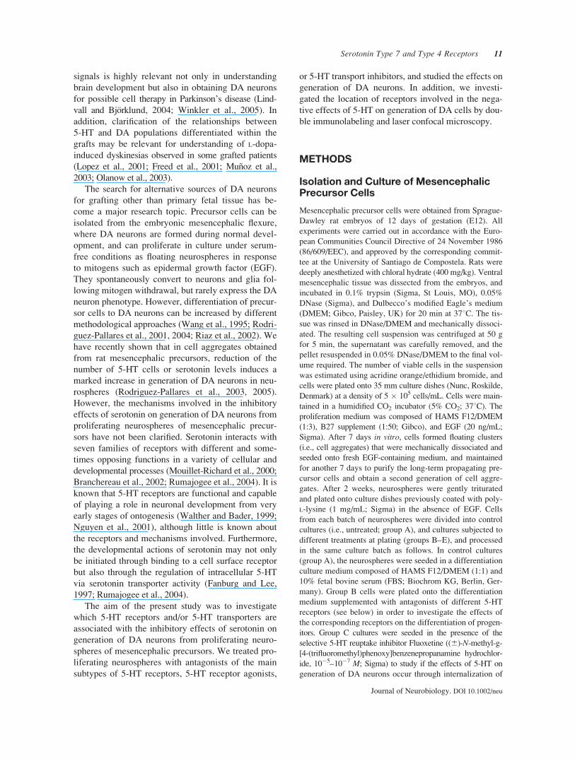
present in the aggregates. Most TH-ir cells appeared as
mature neurons with long and branching processes, and
only a few had immature appearance (Fig. 1). Immuno-
reactivity for DAT was also observed in TH-ir neurons
using double immunolabeling and laser confocal mi-
croscopy, and immunoreactivity for TPH was also ob-
served in 5-HT-ir cells, which confirmed the dopami-
nergic and serotonergic phenotypes, respectively (not
shown, see Rodriguez-Pallares et al., 2003, 2004).
Figure 1 Microphotographs of dopaminergic (i.e., TH-ir) neurons differentiated from aggregates
of mesencephalic precursor cells. Cultures of precursor cells treated with the 5-HT7 receptor antago-
nist SB269970 (B) or the 5-HT4 receptor antagonist GR113808 (C) contained more TH-ir cells than
the nontreated control cell cultures (A). However, cultures treated with the 5-HT1B/1D antagonist
GR127935 showed a significant decrease in the number of TH-ir cells (D). Scale bar¼ 100 �m.
Serotonin Type 7 and Type 4 Receptors 13
Journal of Neurobiology. DOI 10.1002/neu

Figure 2 Effects of treatment with different doses of 5-HT antagonists on the number of TH-ir
cells differentiated from neurospheres of mesencephalic precursors. The generation of TH-ir cells
was reduced by treatment with 5-HT1B/1D [GR 127935; (A)] or 5-HT2 [Ritanserin; (B)] receptor
antagonists, but was not significantly affected by treatment with 5-HT1A (WAY 100635), 5-HT3
(Ondasetron), or 5-HT6 (SB 258585) receptor antagonists (C–E). Additional control cultures con-
taining 10% methanol (i.e., vehicle) were included in (B) and (D). The data are expressed as per-
centages of the number of TH-ir cells obtained in the respective control cultures (100%). Data rep-
resent means 6 SEM. Means that differ significantly are indicated by a different letter (p < 0.05,
one-way ANOVA and posthoc Tukey tests).
14 Parga et al.
Journal of Neurobiology. DOI 10.1002/neu
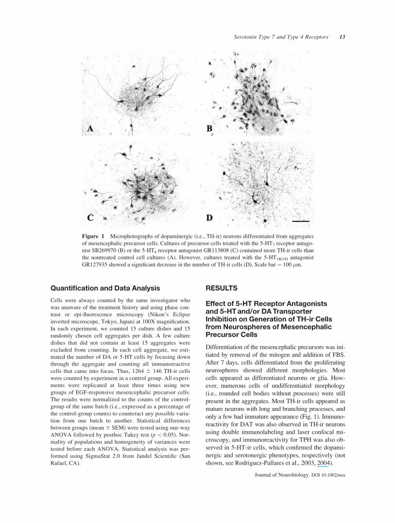
Cultures treated for 7 days with Methiothepin
(5-HT1,2,5,6,7 receptor antagonist) showed significantly
more TH-ir cells than the controls (group A). This
effect was maximal at doses of 10�7 M (about 240%
of controls). However, the results obtained after treat-
ing the neurospheres with more selective antagonists
were different (Figs. 1–3). Cultures treated with
WAY100635 (potent and selective 5-HT1A receptor
antagonist) did not show any significant change in the
number of TH-ir neurons generated in the neuro-
spheres [Fig. 2(C)]. Treatment with the 5-HT1B/1D an-
tagonist GR 127935 induced a significant decrease in
generation of TH-ir cells in the neurospheres (approxi-
mately 45% of controls), and a similar decrease (ap-
proximately 35% of controls) was observed in cultures
treated with the 5-HT2 antagonist Ritanserin [Figs. 1(D)
and 2(A,B)]. Treatment with the selective 5-HT3 re-
ceptor antagonist Ondasetron or with the selective 5-
HT6 receptor antagonist SB 258585 did not induce
any significant change in the number of TH-ir cells
observed in the neurospheres [Fig. 2(D,E)]. However,
cultures treated with the selective 5-HT7 receptor an-
tagonist SB 269970 or the selective 5-HT4 receptor
antagonist GR113808 showed a significant increase in
generation of TH-ir neurons [about 260 and 225% of
controls, respectively; Figs. 1(B,C) and 3].
Cultures treated with the 5-HT transporter inhibitor
Fluoxetine showed a significant reduction in the num-
ber of TH-ir neurons [Fig. 4(A)], and treatment with
the dopamine and norepinephrine transport blocker
GBR 12935 induced a similar reduction in generation
of TH-ir neurons from the proliferating neurospheres
[Fig. 4(B)]. Finally, simultaneous treatment with Fluox-
etine and GBR 12935 induced a marked decrease in
generation of TH-ir neurons [Fig. 4(C)].
Effect of 5-HT7 and 5-HT4 ReceptorAgonists on Generation of TH-ir Cells
The results obtained with 5-HT7 and 5HT4 receptor
antagonists were confirmed by using 5-HT7 and 5HT4
receptor agonists in cultures treated with the serotoner-
gic neurotoxin 5,7-DHT to reduce the levels of endog-
enous 5-HT. In addition, the 5-HT1A/7 agonist 8-OH-
DPAT was used in the presence of the 5-HT1A antagonist
WAY100635. Cultures treated with 5,7-DHT showed a
Figure 3 Effects of treatment with different doses of 5-
HT antagonists on the number of TH-ir cells differentiated
from neurospheres of mesencephalic precursors. The genera-
tion of TH-ir cells was increased by treatment with Methio-
thepin [5-HT1,2,5,6,7 antagonist; (A)], SB 269970 [5-HT7
receptor antagonist; (B)], or GR 113808 [5-HT4 receptor
antagonist; (C)]. The data are expressed as percentages of
the number of TH-ir cells obtained in the respective control
cultures (100%). Data represent means 6 SEM. Means that
differ significantly are indicated by a different letter (p <0.05, one-way ANOVA and posthoc Tukey tests).
Serotonin Type 7 and Type 4 Receptors 15
Journal of Neurobiology. DOI 10.1002/neu

marked increase in generation of TH-ir cells. However,
the 5,7-DHT-induced increase in TH-ir cells was signifi-
cantly reduced by simultaneous treatment with the 5-HT4or 5-HT7 agonist (Fig. 5).
Effect of 5-HT7 and 5-HT4 ReceptorAntagonists on the SerotonergicPopulation
Because 5-HT4 and 5-HT7 receptors appeared to be
involved in serotonin-induced decrease in generation
of DA neurons, we also studied the effects of treat-
ment with the selective 5-HT4 receptor antagonist
GR113808 and with the selective 5-HT7 receptor an-
tagonist SB 269970 on the generation of 5-HT neu-
rons. Neurospheres treated with the selective 5-HT7
receptor antagonist SB 269970 showed a significant
decrease in the number of 5-HT cells with respect to
controls (68 6 6%). Treatment with the 5-HT4 recep-
tor antagonist GR113808 resulted in a nonsignificant
decrease in the number of 5-HT cells with respect to
controls (89 6 5%).
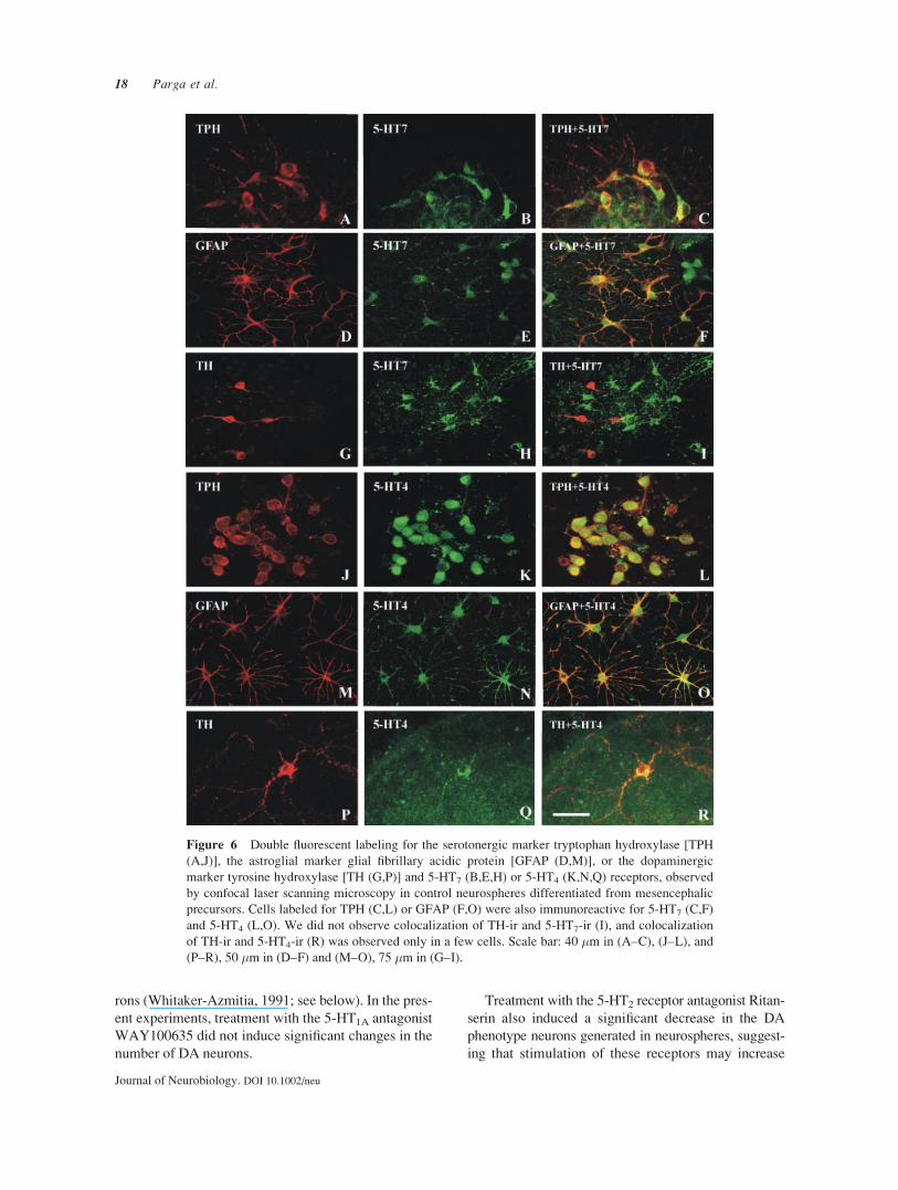
Location of 5-HT7 and 5-HT4 Receptors
The location of 5-HT7 and 5HT4 receptors was studied
using double immunolabeling and laser confocal mi-
croscopy (Fig. 6). Double immunolabeling for 5-HT7
and GFAP or TPH revealed that 5-HT7 receptors were
located in astrocytes and 5-HT cells. However, we did
not observe double labeling for 5-HT7 and TH, sug-
gesting that these receptors are not located in DA neu-
rons. Similarly, double immunolabeling for 5-HT4 and
GFAP or TPH revealed that 5-HT4 receptors were
located in astrocytes and 5-HT cells. Most TH-ir cells
did not express 5-HT4 immunoreactivity. However, a
few TH-ir cells were also immunoreactive for 5-HT4.
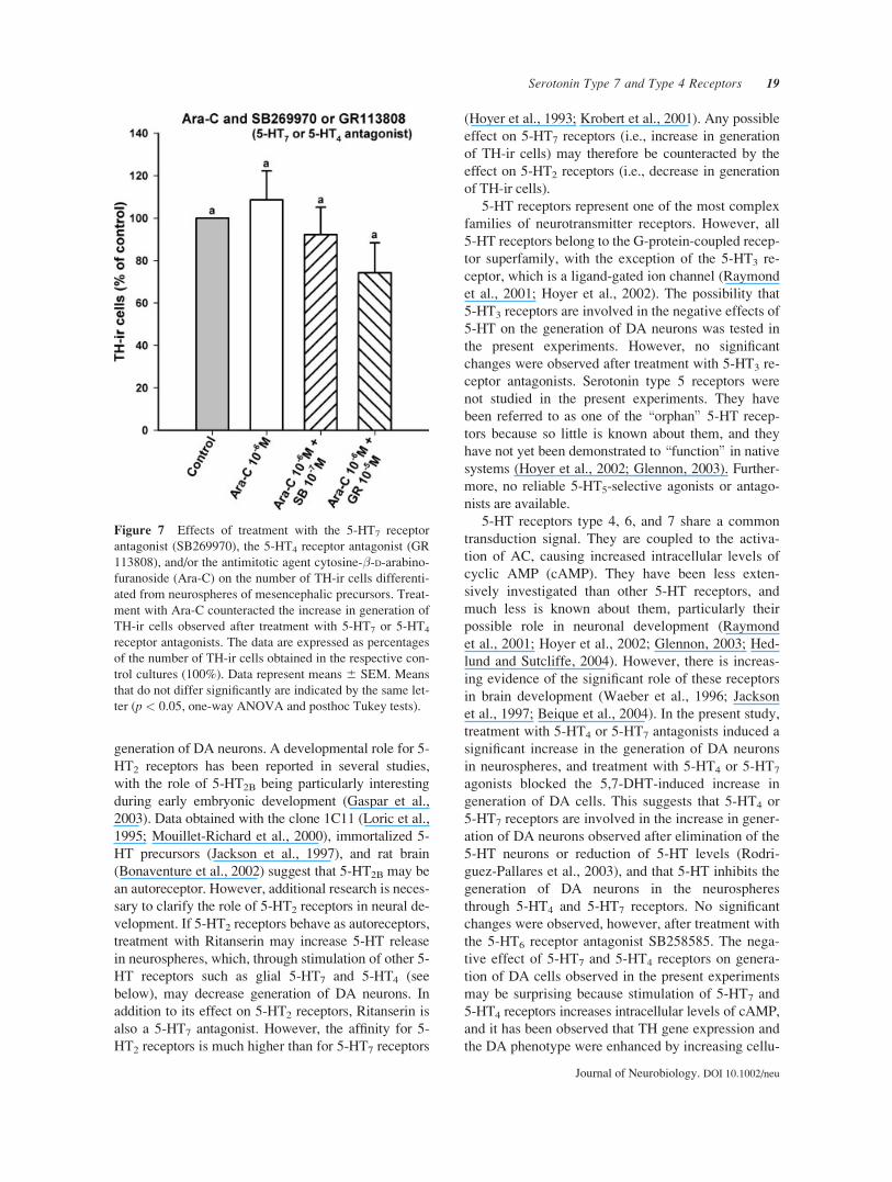
Because 5-HT7 and 5-HT4 receptors were located in
glial cells, group E cultures were seeded in the pres-
ence of 5-HT7 or 5-HT4 antagonists and the antimitotic
agent Ara-C to inhibit replication of non-neuronal cells.
Treatment with Ara-C during the differentiation period
prevented the 5-HT7 or 5-HT4 antagonist-induced
increase in generation of TH-ir neurons, indicating that
glial 5-HT7 or 5-HT4 receptors played an important
role in the negative effect of serotonin on generation of
DA cells in the proliferating neurospheres (Fig. 7).
DISCUSSION
In the present study, we investigated the 5-HT recep-
tors and/or transporters involved in the inhibitory
effects of serotonin on generation of dopaminergic
neurons from proliferating neurospheres of mesence-
Figure 4 Treatment with different doses of the 5-HT
transporter inhibitor Fluoxetine (A), the dopamine and nor-
epinephrine transporter inhibitor GBR 12935 (B), or Fluox-
etine + GBR 12935 significantly decreased the number of
TH-ir cells differentiated from neurospheres of mesence-
phalic precursors. The data are expressed as percentages of
the number of TH-ir cells obtained in the respective control
cultures (100%). Data represent means 6 SEM. Means that
differ significantly are indicated by a different letter (p <0.05, one-way ANOVA and posthoc Tukey tests).
16 Parga et al.
Journal of Neurobiology. DOI 10.1002/neu

phalic precursors. We previously observed an impor-
tant increase in the number DA cells after inducing
partial elimination of the 5-HT neurons with the sero-
tonergic toxin 5,7-DHT, or after reducing 5-HT levels
with the 5-HT synthesis inhibitor DL-p-chloropheny-
lalanine (pCPA; Rodriguez-Pallares et al., 2003). In
addition to the 5-HT released by serotonergic cells, it
is known that there is also a low 5-HT content in the
FBS used in the differentiation medium, which may
contribute to stimulation of 5-HT receptors. How-
ever, the results of several experiments suggest that
the 5-HT content in FBS is below the usual effective
levels (Zhou and Iacovitti, 2000; Riaz et al., 2004).
According to effects observed after reducing the
levels of 5-HT (Rodriguez-Pallares et al., 2003), anta-
gonists affecting most subtypes of 5-HT receptors,
such as Methiothepin (5-HT1A,1B,1D,2A,2C,5A,5B,6,7 an-
tagonist), induced an increase in the number of TH-
positive cells generated in the neurospheres. However,
the results obtained after treating the neurospheres
with more selective antagonists were different. Treat-
ment with the 1B/1D receptor antagonist GR127935
induced a significant decrease in the number of DA
phenotype neurons generated in the neurospheres,
suggesting that stimulation of these receptors may
potentiate the DA phenotype. The results of several
recent studies suggest that in very early stages of de-
velopment 5-HT1B/1D and 5-HT1A receptors may act
as 5-HT autoreceptors, so that 5-HT synthesis, stor-
age, and uptake may be down-regulated by 5-HT itself
(Hery et al., 1999; Mouillet-Richardet al., 2000; Rob-
erts et al., 2001; Rumajogee et al., 2004). It has been
suggested that 5-HT1B autoreceptors diminish 5-HT
release from axon terminals and varicosities (Galzin
et al., 1992), while 5-HT1A autoreceptors inhibit firing
of serotonergic neurons (Sprouse and Aghanian, 1987;
Hery et al., 1999; Azmitia, 2001). Furthermore, it has
recently been observed that the lack of 5-HT1B or
5-HT1A receptors is associated with a significant in-
crease in the number of serotonergic neurons in primary
cultures from embryonic raphe (Rumajogee et al.,
2004). Therefore, treatment with 5-HT1B/D antagonists
may increase 5-HT release in neurospheres, which
through other 5-HT receptors (i.e., glial 5-HT4, 5-HT7
receptors, see below) may decrease generation of DA
neurons. However, it has also been reported that glial
5-HT1A receptors promote the growth of 5-HT neu-
Figure 5 Effects of treatment with different doses of 5-HT4or 5-HT7 agonists on the number of TH-ir cells differenti-
ated from neurospheres of mesencephalic precursors. Cul-
tures were treated with the serotonergic neurotoxin 5,7-DHT
(in 0.2% ascorbic acid, AA) to reduce endogenous 5-HT,
which induced a marked increase in generation of TH-ir
cells. The 5,7-DHT-induced increase in TH-ir cells was sig-
nificantly reduced by simultaneous treatment with the 5-HT4
agonist/5-HT3 antagonist Zacopride or with the 5-HT7/1A
agonist 8-OH-DPAT in the presence of WAY100635 (5-HT1A
antagonist). Additional control cultures containing vehicle
(0.2% ascorbate saline, AA) were also included. The data
are expressed as percentages of the number of TH-ir cells
obtained in the respective control cultures (100%). Data rep-
resent means 6 SEM. Means that differ significantly are
indicated by a different letter (p < 0.05, one-way ANOVA
and posthoc Tukey tests).
Serotonin Type 7 and Type 4 Receptors 17
Journal of Neurobiology. DOI 10.1002/neu
rons (Whitaker-Azmitia, 1991; see below). In the pres-
ent experiments, treatment with the 5-HT1A antagonist
WAY100635 did not induce significant changes in the
number of DA neurons.
Treatment with the 5-HT2 receptor antagonist Ritan-
serin also induced a significant decrease in the DA
phenotype neurons generated in neurospheres, suggest-
ing that stimulation of these receptors may increase
Figure 6 Double fluorescent labeling for the serotonergic marker tryptophan hydroxylase [TPH
(A,J)], the astroglial marker glial fibrillary acidic protein [GFAP (D,M)], or the dopaminergic
marker tyrosine hydroxylase [TH (G,P)] and 5-HT7 (B,E,H) or 5-HT4 (K,N,Q) receptors, observed
by confocal laser scanning microscopy in control neurospheres differentiated from mesencephalic
precursors. Cells labeled for TPH (C,L) or GFAP (F,O) were also immunoreactive for 5-HT7 (C,F)
and 5-HT4 (L,O). We did not observe colocalization of TH-ir and 5-HT7-ir (I), and colocalization
of TH-ir and 5-HT4-ir (R) was observed only in a few cells. Scale bar: 40 �m in (A–C), (J–L), and
(P–R), 50 �m in (D–F) and (M–O), 75 �m in (G–I).
18 Parga et al.
Journal of Neurobiology. DOI 10.1002/neu
generation of DA neurons. A developmental role for 5-
HT2 receptors has been reported in several studies,
with the role of 5-HT2B being particularly interesting
during early embryonic development (Gaspar et al.,
2003). Data obtained with the clone 1C11 (Loric et al.,
1995; Mouillet-Richard et al., 2000), immortalized 5-
HT precursors (Jackson et al., 1997), and rat brain
(Bonaventure et al., 2002) suggest that 5-HT2B may be
an autoreceptor. However, additional research is neces-
sary to clarify the role of 5-HT2 receptors in neural de-
velopment. If 5-HT2 receptors behave as autoreceptors,
treatment with Ritanserin may increase 5-HT release
in neurospheres, which, through stimulation of other 5-
HT receptors such as glial 5-HT7 and 5-HT4 (see
below), may decrease generation of DA neurons. In
addition to its effect on 5-HT2 receptors, Ritanserin is
also a 5-HT7 antagonist. However, the affinity for 5-
HT2 receptors is much higher than for 5-HT7 receptors
(Hoyer et al., 1993; Krobert et al., 2001). Any possible
effect on 5-HT7 receptors (i.e., increase in generation
of TH-ir cells) may therefore be counteracted by the
effect on 5-HT2 receptors (i.e., decrease in generation
of TH-ir cells).
5-HT receptors represent one of the most complex
families of neurotransmitter receptors. However, all
5-HT receptors belong to the G-protein-coupled recep-
tor superfamily, with the exception of the 5-HT3 re-
ceptor, which is a ligand-gated ion channel (Raymond
et al., 2001; Hoyer et al., 2002). The possibility that
5-HT3 receptors are involved in the negative effects of
5-HT on the generation of DA neurons was tested in
the present experiments. However, no significant
changes were observed after treatment with 5-HT3 re-
ceptor antagonists. Serotonin type 5 receptors were
not studied in the present experiments. They have
been referred to as one of the \orphan" 5-HT recep-
tors because so little is known about them, and they
have not yet been demonstrated to \function" in nativesystems (Hoyer et al., 2002; Glennon, 2003). Further-
more, no reliable 5-HT5-selective agonists or antago-
nists are available.
5-HT receptors type 4, 6, and 7 share a common
transduction signal. They are coupled to the activa-
tion of AC, causing increased intracellular levels of
cyclic AMP (cAMP). They have been less exten-
sively investigated than other 5-HT receptors, and
much less is known about them, particularly their
possible role in neuronal development (Raymond
et al., 2001; Hoyer et al., 2002; Glennon, 2003; Hed-
lund and Sutcliffe, 2004). However, there is increas-
ing evidence of the significant role of these receptors
in brain development (Waeber et al., 1996; Jackson
et al., 1997; Beique et al., 2004). In the present study,
treatment with 5-HT4 or 5-HT7 antagonists induced a
significant increase in the generation of DA neurons
in neurospheres, and treatment with 5-HT4 or 5-HT7
agonists blocked the 5,7-DHT-induced increase in
generation of DA cells. This suggests that 5-HT4 or
5-HT7 receptors are involved in the increase in gener-
ation of DA neurons observed after elimination of the
5-HT neurons or reduction of 5-HT levels (Rodri-
guez-Pallares et al., 2003), and that 5-HT inhibits the
generation of DA neurons in the neurospheres
through 5-HT4 and 5-HT7 receptors. No significant
changes were observed, however, after treatment with
the 5-HT6 receptor antagonist SB258585. The nega-
tive effect of 5-HT7 and 5-HT4 receptors on genera-
tion of DA cells observed in the present experiments
may be surprising because stimulation of 5-HT7 and
5-HT4 receptors increases intracellular levels of cAMP,
and it has been observed that TH gene expression and
the DA phenotype were enhanced by increasing cellu-
Figure 7 Effects of treatment with the 5-HT7 receptor
antagonist (SB269970), the 5-HT4 receptor antagonist (GR
113808), and/or the antimitotic agent cytosine-�-D-arabino-furanoside (Ara-C) on the number of TH-ir cells differenti-
ated from neurospheres of mesencephalic precursors. Treat-
ment with Ara-C counteracted the increase in generation of
TH-ir cells observed after treatment with 5-HT7 or 5-HT4
receptor antagonists. The data are expressed as percentages
of the number of TH-ir cells obtained in the respective con-
trol cultures (100%). Data represent means 6 SEM. Means
that do not differ significantly are indicated by the same let-
ter (p < 0.05, one-way ANOVA and posthoc Tukey tests).
Serotonin Type 7 and Type 4 Receptors 19
Journal of Neurobiology. DOI 10.1002/neu
lar levels of cAMP (Riaz et al., 2002; Pliego-Rivero
et al., 1999). However, the results of our double im-
munolabeling studies suggest that there is no signifi-
cant expression of these receptors in TH-ir cells, and
that they are located in glial cells and serotonergic
cells (i.e., cells immunoreactive for GFAP and TPH,
respectively). A few TH-ir cells also immunoreactive
for 5-HT4 may be immature DA cells transitorily
expressing 5-HT4 receptors or non-DA cells transito-
rily expressing TH. Type 6 and 7 5-HT receptors had
previously been observed in astrocytes (Hirst et al.,
1997, 1998). In addition, Ara-C prevented the 5-HT7
or 5-HT4 antagonist-induced increase in generation of
TH-ir neurons. This suggests a relevant role for glial
5-HT7 and 5-HT4 receptors in the negative effect of 5-
HT on generation of DA cells in proliferating neuro-
spheres, although additional glial-derived factors may
also be involved in the effects of Ara-C. It has previ-
ously been suggested that some of the effects of 5-HT
on neurogenesis may be mediated by glia (Liu and
Lauder, 1992a,b; Rumajogee et al., 2005), and that
glial 5-HT1A receptors elicit the release of S100� via
activation of AC, which promotes the growth of 5-HT
neurons (Whitaker-Azmitia, 1991). We observed a si-
multaneous positive effect on the number of 5-HT
cells (i.e., there was a significant decrease in the num-
ber of 5-HT cells after treatment with the 5-HT7 an-
tagonist), which suggests that glial-derived factors
induced by 5-HT7 and 5-HT4 receptor stimulation
may potentiate the induction and/or survival of 5-HT
neurons from precursors at the expense of DA neu-
rons. In addition, we observed immunolabeling for 5-
HT7 and 5-HT4 receptors in cells also labeled for
TPH, which suggests that the serotonergic phenotype
may also be enhanced via activation of cAMP in 5-HT
cells (Galter and Unsicker, 2000; Rumajogee et al.,
2004). Several previous studies have detected 5-HT7
receptors in the raphe nuclei (To et al., 1995; Ruat
et al., 1993; Duncan et al., 1999) and it has been sug-
gested that 5-HT receptors may act as autoreceptors
on 5-HT neurons (Roberts et al., 2001). Interestingly,
5-HT7 gene expression has been observed in immor-
talized serotonergic precursors, and this expression
was lost after cell differentiation (Jackson et al.,
1997). However, the present experiments using the
toxin 5,7-DHT + 5-HT4 or 5-HT7 agonists, and those
using Ara-C + 5-HT4 or 5-HT7 antagonists show that
the glial receptors play a major role, and additional
studies are necessary to clarify the role of the 5-HT7
and 5-HT4 receptors observed in serotonergic cells in
the present study.
Most cellular transduction systems that have been
described for 5-HT appear to depend on initiation of
the signal by a cell surface receptor. However, the
results of several studies have shown that some devel-
opmental actions are undertaken through internaliza-
tion of 5-HT by 5-HT transporters (Fanburg and Lee,
1997; Rumajogee et al., 2004), which are expressed
early in neuronal precursors (Yamamoto et al., 1981;
Stringer et al., 1994). If the 5-HT-induced decrease in
generation of DA neurons is brought about via inter-
nalization of 5-HT by transporters, the blockage of
5-HT transport should induce the same effect as a
decrease in 5-HT levels (i.e. an increase in generation
of DA neurons). In the present study, however, we
observed that the selective 5-HT reuptake inhibitor
Fluoxetine decreased the number of DA neurons gen-
erated in the neurospheres, suggesting that the nega-
tive effects induced by 5-HT on generation of DA
neurons were not brought about via 5-HT transporters
but via cell surface receptors. In fact, the Fluoxetine-
induced decrease in generation of DA neurons may
be explained by an increased stimulation of 5-HT4
and 5-HT7 receptors, because inhibition of the 5-HT
reuptake mechanism should result in an increased
concentration of 5-HT.
It has been observed that when the 5-HT trans-
porter is compromised, DA neurons can take up 5-HT
using the DAT (Zhou et al., 2002). Therefore, we pre-
vented any internalization of 5-HT by carrying out si-
multaneous treatment with the 5-HT uptake blocker
Fluoxetine and the DA uptake blocker GBR-12935.
Again, we observed a decrease in the number of DA
neurons. Interestingly, treatment with the DA uptake
blocker alone also decreased generation of DA cells
(see Du and Iacovitti, 1995; Riaz et al., 2004).
In conclusion, the present results show that inhibi-
tion of glial 5-HT7 and 5-HT4 receptors increased
generation of DA neurons in proliferating neuro-
spheres of mesencephalic precursors. This appears to
be responsible for the increase in generation of DA
neurons observed after elimination of the 5-HT neu-
rons or reduction of 5-HT levels.
The authors thank Pilar Aldrey for her excellent techni-
cal assistance.
REFERENCES
Azmitia EC. 2001. Modern views on an ancient chemical:
Serotonin effects on cell proliferation, maturation, and
apoptosis. Brain Res Bull 56:413–424.
Beique JC, Chapin-Penick EM, Mladenovic L, Andrade R.
2004. Serotonergic facilitation of synaptic activity in the
developing rat prefrontal cortex. J Physiol 556:739–754.
Bonaventure P, Guo H, Tian B, Liu X, Bittner A, Roland S,
Salunga R, et al. 2002. Nuclei and subnuclei gene expres-
sion profiling in mammalian brain. Brain Res 943:38–47.
20 Parga et al.
Journal of Neurobiology. DOI 10.1002/neu
Branchereau P, Chapron J, Meyrand P. 2002. Descending
5-hydroxytryptamine raphe inputs repress the expression
of serotonergic neurons and slow the maturation of inhib-
itory systems in mouse embryonic spinal cord. J Neurosci
22:2598–2606.
Buznikov GA, Lambert HW, Lauder JM. 2001. Serotonin
and serotonin-like substances as regulators of early em-
bryogenesis and morphogenesis. Cell Tissue Res 305:
177–186.
Du X, Iacovitti L. 1995. Synergy between growth factors
and transmitters required for catecholamine differentia-
tion in brain neurons. J Neurosci 15:5420–5427.
Duncan MJ, Short J, Wheeler DL. 1999. Comparison of the
effects of aging on 5-HT7 and 5-HT1A receptors in dis-
crete regions of the circadian timing systems in hamsters.
Brain Res 829:39–45.
Fanburg BL, Lee SL. 1997. A new role for an old molecule:
serotonin as a mitogen. Am J Physiol 272:L795–806.
Freed CR, Greene PE, Breeze RE, Tsai WY, DuMouchel W,
Kao R, Dillon S, et al. 2001. Transplantation of embryonic
dopamine neurons for severe Parkinson’s disease. N Engl
J Med 344:710–719.
Galter D, Unsicker K. 2000. Brain-derived neurotrophic factor
and trkB are essential for cAMP-mediated induction of sero-
tonergic neuronal phenotype. J Neurosci Res 61:295–301.
Galzin AM, Poirier MF, Lista A, Chodkiewicz JP, Blier P,
Ramdine R, Loo H, et al. 1992. Characterization of the
5-hydroxytryptamine receptor modulating the release of
5-[3H]hydroxytryptamine in slices of the human neocor-
tex. J Neurochem 59:1293–1301.
Gaspar P, Cases O, Maroteaux L. 2003. The developmental
role of the serotonin: news from mouse molecular genet-
ics. Nature Rev Neurosci 4:1–11.
Glennon RA. 2003. Higher-end serotonin receptors: 5-HT5,
5-HT6, 5-HT7. J Med Chem 14:2795–2812.
Hedlund PB, Sutcliffe JG. 2004. Functional, molecular and
pharmacological advances in 5-HT7 receptor research.
Trends Pharmacol Sci 25:481–486.
Hery F, Boulenguez P, Semont A, Hery M, Pesce G, Becquet D,
Faudon M, et al. 1999. Identification and role of serotonin
5-HT1A and 5-HT1B receptors in primary cultures of rat
embryonic rostral raphe nucleus neurons. J Neurochem
72:1791–1801.
Hirst WD, Cheung NY, Rattray M, Price GW, Wilkin GP.
1998. Cultured astrocytes express messenger RNA for
multiple serotonin receptor subtypes, without functional
coupling of 5-HT1 receptor subtypes to adenylyl cyclase.
Brain Res Mol Brain Res 6:90–99.
Hirst WD, Price GW, Rattray M, Wilkin GP. 1997. Identifica-
tion of 5-hydroxytryptamine receptors positively coupled
to adenylyl cyclase in rat cultured astrocytes. Br J Pharma-
col 120:509–515.
Hoyer D, Clarke DE, Fozard JR, Hartig PR, Martin GR,
Mylecharane EJ, Saxena PR, et al. 1993. International
union of pharmacology classification of receptors for 5-hy-
droxytryptamine (Serotonin). Pharmacol Rev 46:157–203.
Hoyer D, Hannon JP, Martin GR. 2002. Molecular, pharma-
cological and functional diversity of 5-HT receptors.
Pharmacol Biochem Behav 71:533–554.
Hynes M, Porter JA, Chiang C, Chang D, Tessier-Lavigne M,
Beachy PA, Rosenthal A. 1995. Induction of midbrain
dopaminergic neurons by Sonic Hedgehog. Neuron 80:
95–101.
Jackson ZE, Stringer BM, Foster GA. 1997. Identification
of 5-HT receptor sub-types in a homogeneous population
of presumptive serotoninergic neurones. Neuropharma-
cology 36:543–548.
Krobert KA, Bach T, Syversveen T, Kvingedal AM, Levy FO.
2001. The cloned human 5-HT7 receptor splice variants:
a comparative characterization of their pharmacology,
function and distribution. Naunyn Schmiedebergs Arch
Pharmacol 363:620–632.
Lauder JM, Towle AC, Patrick K, Henderson P, Krebs H.
1985. Decreased serotonin content of embryonic raphe neu-
rons following maternal administration of p-chlorophenyl-
alanine: A quantitative immunocytochemical study. Dev
Brain Res 20:107–114.
Lindvall O, Bjorklund A. 2004. Cell therapy in Parkinson’s
disease. NeuroRx 1:382–393.
Liu J, Lauder JM. 1992a. Serotonin promotes region-spe-
cific glial influences on cultured serotonin and dopamine
neurons. Glia 5:306–317.
Liu JP, Lauder JM. 1992b. S-100 beta and insulin-like
growth factor-II differentially regulate growth of devel-
oping serotonin and dopamine neurons in vitro. J Neuro-
sci Res 33:248–256.
Lopez A, Munoz A, Guerra MJ, Labandeira-Garcia JL.
2001. Mechanisms of the effects of exogenous levodopa
on the dopamine-denervated striatum. Neuroscience 103:
639–651.
Loric S, Maroteaux L, Kellermann O, Launay JM. 1995.
Functional serotonin-2B receptors are expressed by a ter-
atocarcinoma-derived cell line during serotonergic differ-
entiation. Mol Pharmacol 47:458–466.
Michel PP, Ruberg M, Agid Y. 1997. Rescue of mesence-
phalic dopamine neurons by anticancer drug cytosine
arabinoside. J Neurochem 69:1499–1507.
Mouillet-Richard S, Mutel V, Loric S, Tournois C, Launay JM,
Kekkermann O. 2000. Regulation by neurotransmitter re-
ceptors of serotonergic or catecholaminergic neuronal cell
differentiation. J Biol Chem 275:9186–9192.
Munoz A, Rodriguez-Pallares J, Guerra MJ, Labandeira-
Garcia JL. 2003. Host brain regulation of dopaminergic
grafts function: the role of the serotonergic and noradren-
ergic systems in amphetamine-induced responses. Synapse
47:66–76.
Nguyen L, Rigo JM, Rocher V, Belachew S, Malgrange B,
Rogister B, Leprince P, et al. 2001. Neurotransmitters as
early signals for central nervous system development.
Cell Tissue Res 305:187–202.
Olanow CW, Goetz CG, Kordower JH, Stoessl AJ, Sossi V,
Brin MF, Shannon KM, et al. 2003. A double-blind con-
trolled trial of bilateral fetal nigral transplantation in
Parkinson’s disease. Ann Neurol 54:403–414.
Owens DF, Kriegstein AR. 2002. Developmental neuro-
transmitters? Neuron 36:989–995.
Pliego Rivero FB, McCormack WJ, Jauniaux E, Stern GM,
Bradford HF. 1999. Forskolin-induced expression of
Serotonin Type 7 and Type 4 Receptors 21
Journal of Neurobiology. DOI 10.1002/neu

tyrosine hydroxylase in human foetal brain cortex. Brain
Res Dev Brain Res 114:201–206.
Raymond JR, Mukhin YV, Gelasco A, Turner J, Collins-
worth G, Gettys TW, Grewal JS, et al. 2001. Multiplicity
of mechanisms of serotonin receptor signal transduction.
Pharmacol Ther 92:179–212.
Riaz SS, Jauniaux E, Stern GM, Bradford HF. 2002. The
controlled conversion of human neural progenitor cells
derived from foetal ventral mesencephalon into dopami-
nergic neurons in vitro. Brain Res Dev Brain Res 136:
27–34.
Riaz SS, Spyridon T, Jauniaux E, Stern GM, Bradford HF.
2004. The differentiation potential of human fetal neuronal
progenitor cells in vitro. Brain Res Dev Brain Res 153:
39–51.
Roberts C, Price GW, Middlemiss DN. 2001. Ligands for
the investigation of 5-HT autoreceptor function. Brain Res
Bull 56:463–469.
Rodriguez-Pallares J, Guerra MJ, Labandeira-Garcia JL.
2003. Elimination of serotonergic cells induces a marked
increase in generation of dopaminergic neurons from
mesencephalic precursors. Eur J Neurosci 18:2166–2174.
Rodriguez-Pallares J, Parga JA, Rey P, Guerra MJ, Labandeira-
Garcia JL. 2005. Expanded mesencephalic precursors
treated with antibodies against FGF4 to increase dopami-
nergic differentiation survive and induce recovery after
transplantation in a parkinsonian rat model. Synapse 58:
13–22.
Rodriguez-Pallares J, Quiroz CR, Parga JA, Guerra MJ,
Labandeira-Garcia JL. 2004. Angiotensin II increases
differentiation of dopaminergic neurons from mesence-
phalic precursors via angiotensin type 2 receptors. Eur
J Neurosci 20:1489–1498.
Rodriguez-Pallares J, Rey P, Soto-Otero R, Labandeira-
Garcia JL. 2001. N-Acetyl-cysteine enhances production
of dopaminergic neurons from mesencephalic-derived
precursor cells. NeuroReport 12:3935–3938.
Ruat M, Traiffort E, Leurs R, Tardivel-Lacombe J, Diaz J,
Arrang JM, Schwartz JC. 1993. Molecular cloning, char-
acterization, and localization of high-affinity serotonin
receptor (5-HT7) activating cAMP formation. Proc Natl
Acad Sci USA 90:8547–8551.
Rumajogee P, Verge D, Darmon M, Brisorgueil MJ, Hamon M,
Miquel MC. 2005. Rapid up-regulation of the neuronal
serotoninergic phenotype by brain-derived neurotrophic
factor and cyclic adenosine monophosphate: relations with
raphe astrocytes. J Neurosci Res 81:481–487.
Rumajogee P, Verge D, Hanoun N, Brisorgueil M-J, Hen R,
Lesch K-P, Hamon M, et al. 2004. Adaption of serotoner-
gic neuronal phenotype in the absence of 5-HT autorecep-
tors or the 5-HT transporter: involvement of BDNF and
cAMP. Eur J Neurosci 19:937–944.
Sprouse JS, Aghajanian GK. 1987. Electrophysiological
responses of serotoninergic dorsal raphe neurons to 5-HT1Aand 5-HT1B agonists. Synapse 1:3–9.
Stringer BM, Verhofstad AA, Foster GA. 1994. Raphe neural
cells immortalized with a temperature-sensitive oncogene:
differentiation under basal conditions down an APUD cell
lineage. Brain Res Dev Brain Res 79:267–274.
To ZP, Bonnaus DW, Eglen RM, Jakeman RB. 1995. Char-
acterization and distribution of putative 5-HT receptors
in guinea-pig brain. Br J Pharmacol 115:107–116.
Waeber C, Sebben M, Bockaert J, Dumuis A. 1996. Re-
gional distribution and ontogeny of 5-HT4 binding sites
in rat brain. Behav Brain Res 73:259–262.
Walther DJ, Bader M. 1999. Serotonin synthesis in murine
embryonic stem cells. Mol Brain Res 68:55–63.
Wang MZ, Jin P, Bumcrot DA, Marigo V, McMahon AP,
Wang EA, Woolf T, et al. 1995. Induction of dopaminer-
gic neuron phenotype in the midbrain by Sonic hedgehog
protein. Nat Med 1:1184–1188.
Whitaker-Azmitia PM. 1991. Role of serotonin and other
neurotransmitter receptors in brain development: basis
for developmental pharmacology. Pharmacol Rev 43:
553–561.
Whitaker-Azmitia PM. 2001. Serotonin and brain develop-
ment: Role in the human developmental diseases. Brain
Res Bull 56:479–485.
Winkler C, Kirik D, Bjorklund A. 2005. Cell transplanta-
tion in Parkinson’s disease: how can we make it work?
Trends Neurosci 28:86–92.
Yamamoto M, Steinbusch HW, Jessell TM. 1981. Differen-
tiated properties of identified serotonin neurons in disso-
ciated cultures of embryonic rat brain stem. J Cell Biol
91:142–152.
Zhou FC, Lesch KP, Murphy DL. 2002. Serotonin uptake
into dopamine neurons via dopamine transporters: a com-
pensatory alternative. Brain Res 942:109–119.
Zhou FC, Saru Y, Zhang JK. 2000. Expression of serotonin
transporter protein in developing rat brain. Dev Brain
Res 119:33–45.
Zhou J, Iacovitti L. 2000. Mechanisms governing the differ-
entiation of a serotonergic phenotype in culture. Brain Res
877:37–46.
22 Parga et al.
Journal of Neurobiology. DOI 10.1002/neu