a new structure of a serotonin salt: comparison and conformational analysis of all known serotonin...
TRANSCRIPT

A new structure of a serotonin salt:comparison and conformationalanalysis of all known serotonincomplexes
Denis Rychkov,a,b* Elena V. Boldyrevaa,b* and Nikolay A.
Tumanova,b,c
aREC-008, Novosibirsk State University, Pirogova 2, Novosibirsk 630090, Russian
Federation, bInstitute of Solid State Chemistry, Siberian Branch of the Russian
Academy of Sciences, Kutateladze 18, Novosibirsk 630128, Russian Federation, andcInstitute of Condensed Matter and Nanosciences, Universite Catholique de Louvain,
Place Louis Pasteur 1, Louvain-La-Neuve, Belgium
Correspondence e-mail: [email protected], [email protected]
Received 25 June 2013
Accepted 17 July 2013
Four serotonin salt structures (serotonin adipate, C10H13-
N2O+�C6H9O4
�, is a previously unknown structure) were
analysed to understand the influence of the anion on serotonin
conformation. Hydrogen bonding alone favours a flat
conformation, whereas additional stacking interactions
between ions may possibly account for the nonplanar
conformation. Since molecular conformation, stability and
biological activity are interrelated, one can consider influen-
cing the chemical and biological properties of serotonin by
selecting an appropriate counter-ion for salt formation.
Keywords: crystal structure; serotonin adipate; conformation;hydrogen bonding; stacking interactions; Hirshfeld surfaces.
1. Introduction
Biologically active substances are at the focus of biological,
pharmaceutical and chemical research. Serotonin, one of the
most common neurotransmitters, is widely studied in relation
to its effect on humans at different levels, from cellular up to
mental (Mohammad-Zadeh et al., 2008; Seo et al., 2008; Berger
et al., 2009). Although serotonin plays a key role in some
biological processes, its chemistry and crystallography have
not been well studied.
Many biologically active compounds are crystallized with
additional components as salts or cocrystals to improve
stability or solubility or to decrease hygroscopicity
(Almarsson & Zaworotko, 2004; Libbrecht, 2005; Schultheiss
& Newman, 2009; Babu & Nangia, 2011; Brittain, 2012).
Serotonin is not an exception. Its low thermal stability in
solution can be overcome if a second component, like adipic
acid, is added on crystallization. Serotonin adipate has been
used in medical formulations in Russia and Eastern Europe as
an analogue of serotonin creatinine sulfate monohydrate, but
the crystal structure of the adipate salt remained unknown.
Another interesting issue is that the conformation of serotonin
creatinine sulfate monohydrate is different from that
predicted theoretically for a ‘free’ serotonin molecule in the
gas phase (Chothia, 1969; Pratuangdejkul, Jaudon et al., 2006).
It was worth investigating whether the conformation of sero-
tonin in the adipate salt is the same as in creatinine sulfate,
remembering that they are pharmacological analogues.
The aim of the present study was to crystallize serotonin
adipate, (I), determine its crystal structure and analyse it in a
comparison with all the other previously known crystal
structures of serotonin (Karle et al., 1965; Thewalt & Bugg,
1972; Aniy et al., 1978), paying special attention to the inter-
relation between the crystalline environment of a molecule
and the molecular conformation.
2. Experimental
2.1. Synthesis and crystallization
Serotonin adipate (purity not less than 98%, according to
high-performance liquid chromatography data) was crystal-
lized from an aqueous solution at 313 K. The crystals used in
the diffraction experiments were almost transparent, with a
light-brown tinge.
2.2. Refinement
Crystal data, data collection and structure refinement
details are summarized in Table 1. All H atoms were located in
a difference Fourier map, and their positions and isotropic
displacement parameters were refined freely.
3. Results and discussion
The crystal structure of serotonin adipate, (I) (Fig. 1), consists
of sheets of molecules, in which chains of serotonin cations
with protonated hydroxy and amino groups, linked via N—
H� � �O hydrogen bonds (Table 2), alternate with chains
formed by adipate anions, linked via O—H� � �O hydrogen
bonds (Table 2). Additional O—H� � �O and N—H� � �O
hydrogen bonds link serotonin cations to adipate anions, so
that a two-dimensional network containing interconnected
rings is formed (Fig. 2 and Table 2).
The serotonin conformations in four different known
crystal structures (including the present structure of serotonin
adipate) are compared in Fig. 3. The values of the bond
organic compounds
Acta Cryst. (2013). C69, 1055–1061 doi:10.1107/S0108270113019823 # 2013 International Union of Crystallography 1055
Acta Crystallographica Section C
Crystal StructureCommunications
ISSN 0108-2701

lengths and angles are quite similar in all four structures. The
torsion angles are rather close in three of the four compounds,
in which the molecules are almost flat, but differ significantly
in serotonin picrate, in which the side chain of serotonin is
almost orthogonal to the planar bicyclic system (Fig. 3 and
Table 3).
To understand the relationship between the molecular
conformations and the crystalline environments, the molecular
packing in the four structures was compared in relation to
organic compounds
1056 Rychkov et al. � C10H13N2O+�C6H9O4
� Acta Cryst. (2013). C69, 1055–1061
Table 1Experimental details.
Crystal dataChemical formula C10H13N2O+
�C6H9O4�
Mr 322.36Crystal system, space group Triclinic, P1Temperature (K) 298a, b, c (A) 7.1582 (3), 10.5984 (7), 12.1715 (7)�, �, � (�) 113.202 (6), 101.233 (4), 98.045 (4)V (A3) 808.04 (8)Z 2Radiation type Mo K�� (mm�1) 0.10Crystal size (mm) 0.15 � 0.12 � 0.05
Data collectionDiffractometer Oxford Gemini Ultra R
diffractometerAbsorption correction Multi-scan (CrysAlis PRO; Oxford
Diffraction, 2008)Tmin, Tmax 0.994, 1.000No. of measured, independent and
observed [I > 2�(I)] reflections10092, 3286, 1849
Rint 0.061(sin �/�)max (A�1) 0.625
RefinementR[F 2 > 2�(F 2)], wR(F 2), S 0.051, 0.121, 1.00No. of reflections 3286No. of parameters 296No. of restraints 0H-atom treatment All H-atom parameters refined�max, �min (e A�3) 0.16, �0.20
Computer programs: CrysAlis PRO (Oxford Diffraction, 2008), SHELXS97 (Sheldrick,2008), SHELXL97 (Sheldrick, 2008), OLEX2 (Dolomanov et al., 2009), Mercury (Macraeet al., 2008), PLATON (Spek, 2009), CrystalExplorer (McKinnon et al., 1998; Spackman& McKinnon, 2002), enCIFer (Allen et al., 2004) and publCIF (Westrip, 2010).
Figure 1The molecular structure of serotonin adipate, (I), showing the atom-labelling scheme. Displacement ellipsoids are drawn at the 50%probability level for non-H atoms. H atoms are shown as spheres of anarbitrary radius.
Figure 2Fragments of the crystal structure of serotonin adipate, viewed in the bcand ac planes. Hydrogen bonds are shown as dashed lines. [Symmetrycodes: (i) x, y + 1, z; (ii) x, y� 1, z; (iii)�x + 1,�y,�z + 1; (iv) x + 1, y, z.]
Figure 3Serotonin conformations in different salts, showing overlay pictures ofadipate (medium dark colour; green in the electronic version of thejournal) with (a) oxalate, (b) creatinine sulfate and (c) picrate. Themolecules are almost flat and identical in (a) and (b), but differ and arenot flat in the case of (c).
Table 2Hydrogen-bond geometry (A, �).
D—H� � �A D—H H� � �A D� � �A D—H� � �A
O1—H1� � �O4i 1.04 (3) 1.53 (3) 2.569 (2) 172 (2)N1—H1A� � �O3ii 0.99 (3) 1.80 (3) 2.762 (3) 163 (2)N1—H1B� � �O5iii 1.00 (3) 1.80 (3) 2.782 (3) 166 (2)O5—H5� � �O2iv 0.84 (3) 1.82 (3) 2.647 (3) 164 (3)
Symmetry codes: (i) x; yþ 1; z; (ii) �x;�y;�zþ 1; (iii) x; y� 1; z; (iv) x; y; zþ 1.

intermolecular interactions. We start with discussing the
hydrogen bonds, since they most often control crystal struc-
tures and the ability of molecules to change conformation. The
geometric parameters of the hydrogen bonds are summarized
in Table 2. In all four structures, the strengths of the hydrogen
bonds are classified as moderate; donor–acceptor distances lie
in the range 2.7–3.1 A for N—H� � �O and O—H� � �N hydrogen
bonds, and in the range 2.5–3.1 A for O—H� � �O hydrogen
bonds (Fig. 5).
Serotonin oxalate and serotonin adipate not only have
similar geometric parameters of their hydrogen bonds but also
organic compounds
Acta Cryst. (2013). C69, 1055–1061 Rychkov et al. � C10H13N2O+�C6H9O4
� 1057
Table 3Geometric parameters of the serotonin molecule in different serotonin salts.
’1, ’2, ’3 and ’4 are as defined in Fig. 4. Gp denotes the +gauche, Gm the�gauche and ‘At’ the anti conformation. The H atoms with similar values of the ’3 torsionangle were chosen as HN1A. Serotonin picrate angles and distances can hardly be compared due to significant changes in dihedral angles. S.u. values are not givendue to their absence in the original articles.
Angle (�) N1—C7—C8 C7—C8—C9 C8—C9—C16 C8—C9—C10 O5—C12—C11 O5—C12—C13
Adipate 110.1 (2) 113.9 (2) 128.9 (2) 124.5 (2) 122.2 (2) 116.0 (2)Creatine sulfate 108.0 111.1 131.3 124.6 121.9 115.9Oxalate 115.4 113.9 129.7 123.4 121.4 118.1Picrate 111.0 114.7 127.8 126.4 117.5 120.7
Distance (A) N1—C7 C7—C8 C8—C9 C9—C16 C9—C10 O5—C12
Adipate 1.483 (3) 1.510 (3) 1.490 (3) 1.368 (3) 1.427 (3) 1.380 (3)Creatine sulfate 1.51 1.53 1.48 1.37 1.47 1.38Oxalate 1.51 1.48 1.50 1.37 1.43 1.37Picrate 1.49 1.51 1.50 1.37 1.44 1.39
Dihedral angle (�) Conformation of ’1’2 ’1 ’2 ’3 ’4
Adipate AtAt 178.7 177.2 60.0 4.0Creatinine GpGp 166.7 172.6 60.0† 180.0†Oxalate GpAt 171.7 179.7 63.9 174.2Picrate GmGm‡ �67.5 �66.6 57.01 161.1
Figure 5Hydrogen-bond-length distribution in serotonin complexes, showing(top) the N� � �O (and O� � �N) donor–acceptor distances (A) and (bottom)the O� � �O donor–acceptor distances (A).
Figure 4Definition of serotonin atom numbering and dihedral angles. The fourtorsion angles are defined as ’1 (C10—C9—C8—C7), ’2 (C9—C8—C7—N1), ’3 (C8—C7—N1—HN1A) and ’4 (C11—C12—O5—H5).
Figure 6Chains of molecules in (a) serotonin adipate [symmetry codes: (i) x, y + 1,z; (ii) x, y � 1, z] and (b) serotonin oxalate [symmetry codes: (i) �x + 1,�y + 2, �z + 1; (ii) x, y + 1, z; (iii) �x + 1, �y + 3, �z + 1; (iv) x, y � 1, z;(v) �x + 1, �y + 1, �z + 1].
similar structural motifs, with chains of oxalic and adipic acid
molecules, respectively (Fig. 6).
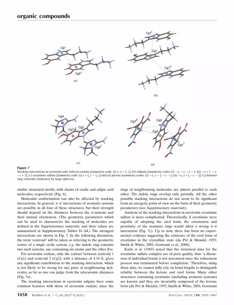
Molecular conformation can also be affected by stacking
interactions. In general, – interactions of aromatic systems
are possible in all four of these structures, but their strength
should depend on the distances between the -systems and
their mutual orientation. (The geometric parameters which
can be used to characterize the stacking of molecules are
defined in the Supplementary materials, and their values are
summarized in Supplementary Tables S1–S4.) The strongest
interactions are shown in Fig. 7. In the following discussion,
the term ‘centroid’ will be taken as referring to the geometric
centre of a single cyclic system, e.g. the indole ring contains
two such systems, one containing six atoms and the other five.
For serotonin oxalate, only the contact between centroid 1
(Cg1) and centroid 2 (Cg2), with a distance of 4.19 A, gives
any significant contribution to the stacking interaction, which
is not likely to be strong for any pairs of neighbouring mol-
ecules, as far as one can judge from the interatomic distances
(Fig. 7a).
The stacking interactions in serotonin adipate have some
common features with those of serotonin oxalate, since the
rings of neighbouring molecules are almost parallel to each
other. The indole rings overlap only partially. All the other
possible stacking interactions do not seem to be significant
from an energetic point of view on the basis of their geometric
parameters (see Supplementary materials).
Analysis of the stacking interactions in serotonin creatinine
sulfate is more complicated. Theoretically, if creatinine were
capable of adopting the enol form, the orientation and
proximity of the aromatic rings would allow a strong –interaction (Fig. 7c). Up to now, there has been no experi-
mental evidence suggesting the existence of the enol form of
creatinine in the crystalline state (du Pre & Mendel, 1955;
Smith & White, 2001; Goswami et al., 2006).
Karle et al. (1965) noted that the structural data for the
creatinine sulfate complex are of poor quality, thus ‘a discus-
sion of individual bonds is not warranted since the refinement
process was terminated before completion.’ Therefore, using
these data, we cannot fully rely on bond lengths to distinguish
reliably between the ketone and enol forms. Many other
structures containing creatinine (including aromatic systems)
are known and they are invariably composed of the ketone
form (du Pre & Mendel, 1955; Smith & White, 2001; Goswami
organic compounds
1058 Rychkov et al. � C10H13N2O+�C6H9O4
� Acta Cryst. (2013). C69, 1055–1061
Figure 7Stacking interactions in serotonin salts with (a) oxalate [symmetry code: (i) x, y + 1, z], (b) adipate [symmetry codes: (i) �x, �y, �z + 1; (ii) �x + 1, �y,�z + 1], (c) creatinine sulfate [symmetry code: (i) x + 1
2, y� 12, z] and (d) picrate [symmetry codes: (i)�x, y� 1
2,�z� 12; (ii)�x, y + 1
2,�z� 12]. Cg denotes
ring centroids (indicated by large spheres).

et al., 2006). Therefore, we conclude that – interactions
between the serotonin fragments in creatinine sulfate are
likely to be absent. The only possible interaction is that of the
indole -system of serotonin with the conjugated -system of
the creatinine molecule. This interaction yields a lower stabi-
lization energy than could be expected for related systems in
which – stacking interactions between aromatic rings exist.
From the geometric criteria (see Supplementary materials), it
can be assumed that the only serotonin structure that contains
a strong – stacking interaction is serotonin picrate, in which
the whole indole system is involved in an interaction with
another -ring. Another point about intermolecular inter-
actions is an additional donor–acceptor interaction with the
indole -system that can also influence the energy distribution
in the crystal structure and complement the stacking inter-
action (see Supplementary materials).
Thus, only the structure of serotonin picrate has obvious
– interactions involving the indole -system, and addi-
tionally Y—X(—N O)� � � interactions, while the other
three structures have only minor – contributions to the
overall intermolecular interaction.
Additional insight into the interactions between serotonin
and its immediate crystalline environment can be obtained by
analysing the Hirshfeld surfaces (McKinnon et al., 1998;
Spackman & McKinnon, 2002) (Fig. 8). It is quite obvious that
three out of the four surfaces are quite similar to each other,
while the last one, corresponding to another conformation, is
significantly different. Some minor differences in the surfaces
of serotonin adipate, oxalate and creatinine sulfate are related
to the three-dimensional orientation of potentially reactive
sites and the curviness of the surface. This can be explained by
the difference in quality of the diffraction data.
Qualitative and quantitative differences in the Hirshfeld
fingerprint plots are even more remarkable. The plot for
serotonin adipate (bottom row of Fig. 8, leftmost plot) displays
characteristics consistent with hydrogen bonding, similar to
that in carboxylic acids: symmetric sharp features point to the
lower left quadrant of the plot, the upper one corresponding
to the hydrogen-bond donors and the lower one to the
acceptors. –NH3+, NH in the indole ring and OH groups act as
donors, while the only acceptor is the OH group (the O atom is
an acceptor for another NH3+). The green colour shows that
there are many more donor groups than acceptors (blue
colour of another band). Another feature are the wings at the
lower right (-acceptor) and upper left (C—H donor) corners
of the plot, corresponding to an ‘aromatic’ C—H� � � inter-
organic compounds
Acta Cryst. (2013). C69, 1055–1061 Rychkov et al. � C10H13N2O+�C6H9O4
� 1059
Figure 8The molecular conformation (top row), Hirshfeld surfaces (middle) and fingerprint plots (bottom) for the serotonin molecule in various complexes. Fromleft to right: adipate, oxalate, creatinine sulfate and picrate. Each molecule is shown with the Hirshfeld surface mapped with dnorm [coloured between�0.500 (red in the electronic version of the journal) and 1.500 (blue)], where dnorm is the normalized contact distance, which takes the van der Waals radiiof the atoms into account.

action. The indole ring in serotonin acts as an acceptor for the
CH2 groups of adipic acid which lie above. Moreover, the CH2
group of ethylamine donates an H atom in a C—H� � �interaction with another molecule of serotonin. There are no
indications of – interactions in the region of de = di = 1.8 A.
A very similar plot is calculated for serotonin oxalate
(bottom row of Fig. 8, second plot from left). The same
features characterizing the hydrogen bonding can be
observed, namely the two symmetric bands at the lower left
corner of the plot. More differences can be seen for the
‘aromatic’ interactions: there are no ‘wings’ typical of a C—
H� � � interaction; neither is there any evidence of –stacking interactions. Another difference is the appearance of
a significant number of widely spread dots in the upper right
corner, which may indicate the presence of ‘cavities’ in the
structure, and hence of a non-optimum packing.
The fingerprint plot calculated for serotonin creatinine
sulfate (bottom row of Fig. 8, third plot from left) should be
considered with care, given that the published data did not
contain H-atom coordinates. In this work we calculated the
H-atom positions using the Mercury feature ‘Add missing
H-atoms’ (Macrae et al., 2008). The unusual shape of the plot
seems to be a consequence of the poor structural data. The
features related to hydrogen bonding (such as the huge wing
in the bottom left) cannot therefore be discussed. Never-
theless, a – stacking interaction is independent of H-atom
positions, and the fingerprint shows some evidence of such an
interaction, which cannot be classified as ‘strong’ (Fig. 9).
The fingerprint plot of serotonin picrate (bottom row of
Fig. 8, rightmost plot) gives evidence of a large number of
hydrogen-bond donors, so that the colour of the lower left
band is red at the position de = 1.5 A and di = 1.15 A. This can
be explained by the existence of additional hydrogen bonds
between the NO2 group of the picrate and the C—H atoms of
the indole ring. C—H� � � interactions are absent. The
presence of – stacking interactions in this structure is also
apparent. This can be better visualized by comparing the
fingerprint plots of serotonin creatinine sulfate and serotonin
picrate, in which C� � �C contacts are considered as isolated
(Fig. 9).
To sum up this analysis of different interactions from
hydrogen bonds to – stacking using different techniques, we
should discuss some key features that may cause conforma-
tional changes in serotonin. The hydrogen-bond motifs are
quite different in almost all of these structures, so no clear
correlation between molecular structure and hydrogen
bonding is observed. C—H� � � interactions apparently also
do not play a key role. However, only one structure (the
picrate salt) out of the four has a strong – stacking inter-
action and this structure shows conformational changes. Thus,
on the basis of the current evidence, it looks as though the flat
conformation of serotonin may be obtained only in the
absence of stacking interactions, even when the molecules
form a hydrogen-bonded network.
We have not found any clear influence of water molecules
on the conformational changes of serotonin in hydrates
(serotonin creatinine sulfate monohydrate and serotonin
picrate monohydrate), although it is very complicated to
divide strictly the contribution of each molecule to the overall
hydrogen-bond network and as a result establish its exact role
in the whole structure. It should also be mentioned that,
according to Karle et al. (1965) and Thewalt & Bugg (1972),
these hydrates are stable under ambient conditions.
Another remarkable feature is that the conformation of
serotonin in the present serotonin adipate structure differs
from the most stable conformation predicted for a single
molecule by adiabatic conformational analysis using quantum
chemistry calculations: a ‘free’ serotonin molecule in cationic
form should be nonplanar (Chothia, 1969; Pratuangdejkul,
Jaudon et al., 2006). It also differs from the serotonin
conformations in all other known complexes, but the changes
are not very significant among the ‘flat’ conformations. We
should point out that some differences between serotonin
adipate and serotonin creatinine sulfate that are described
below can be noteworthy, but do not affect the overall
conformation to any great extent.
The most significant difference between the adipate salt and
the creatinine sulfate complex of serotonin is the orientation
of the OH group (Table 3, ’4), which is explained by forming
the most energetically favourable hydrogen bond but, as
mentioned above, it does not change the overall similarity of
these structures. Depending on the values of the two dihedral
angles, ’1 and ’2, serotonin can be in either a +gauche (Gp),
�gauche (Gm) or anti (At) conformation (Figs. 3 and 4), and
the serotonin conformers will be named hereinafter as, for
example, GpAt when ’1 corresponds to the +gauche confor-
mation and ’2 to the anti conformation. We will name as ‘At’
only those structures with dihedral angles very close (�2�) to
the ideal value of 180�, understanding that these angles can
still be quite close to the other angles despite their different
names. In this terminology, the serotonin conformation in the
adipate salt is AtAt. The corresponding energy should be
about 9 kcal mol�1 (1 kcal mol�1 = 4.184 kJ mol�1) greater
than that calculated for the most stable GmGp and GpGp
conformations predicted for the ‘free’ molecule in the gas
phase (Pratuangdejkul, Jaudon et al., 2006). Three other
conformations (GpGp, GmGm and GpAt) were reported for
organic compounds
1060 Rychkov et al. � C10H13N2O+�C6H9O4
� Acta Cryst. (2013). C69, 1055–1061
Figure 9Hirshfeld fingerprint plots for the serotonin molecule in complexes with(left) creatinine sulfate and (right) picrate. Only C� � �C interactions arehighlighted. The highlighted surface area is 2.8% for the creatininesulfate salt and 5.8% for the picrate salt.

serotonin in different crystalline environments, viz. for crea-
tinine sulfate, picrate and oxalate, respectively. The GmGm
and GmGp conformations were presumed to be stabilized by
the interaction of the cationic group of serotonin (NH3+) with
the -system of the indole ring (Pratuangdejkul, Jaudon et al.,
2006). Interestingly, there are no such interactions in the
known crystalline forms of serotonin. In the solid state, the
conformation seems to be changed solely because of the
stacking interactions.
We can suppose that the crystalline environment defines the
molecular conformation. The calculated conformational
energy difference between creatinine sulfate and picrate is
only about 2 kcal mol�1, while the difference in lattice energy
is 274 kcal mol�1 (Caillet et al., 1977). Exact values aside, it is
important to note the magnitude of the difference in confor-
mational and lattice energies.
4. Conclusions
This work has shown the influence of different interactions on
the molecular conformation of serotonin in the crystalline
state. Three main conclusions may be drawn on the basis of the
experimental data available so far: (i) the crystalline envir-
onment may define the conformation of serotonin molecules;
(ii) ‘flat’ (thermodynamically unfavourable) conformations
can be stabilized in the crystalline state if hydrogen bonds are
the only intermolecular interactions; and (iii) additional
stacking and donor–acceptor interactions change the mol-
ecular conformation dramatically, such that the molecules are
no longer flat.
From these results we can suggest an intriguing theory that
the intramolecular geometry of serotonin can be changed by
varying the counter-ions in the crystal structures of its salts or
molecular complexes. It is worth remembering that forming
salts with different anions is a common technique for modi-
fying the biological activity, pharmacological and therapeutic
effect, and some physicochemical properties, such as pKa, of
the substance (Pratuangdejkul, Nosoongnoen et al., 2006). We
can also highlight that, according to one hypothesis (Akhrem
et al., 1978), the physiological effects of serotonin can be
altered by varying its conformation. It is well documented that
conformational polymorphs of one-component pharmaceu-
tical crystals show pronounced differences in solubility and
biological activity, potentially resulting from the retention of
molecular conformation following dissolution, thus affecting
subsequent substrate–receptor interactions in biochemical
pathways (Leonidov, 1997). With an increased understanding
of the effects of the crystalline environment on the confor-
mation of biologically active molecules, one can imagine the
possibility of explicitly inducing a desired conformation of the
target molecule, thus leading to a specific augmentation in
bioactivity. This prospect opens an avenue to advanced drug
design, keeping these synergetic effects in mind.
It seems very promising to make an attempt at crystallizing
serotonin salts with different anions and co-formers and to test
the existence of a correlation between crystalline environ-
ment, molecular conformation and biological activity for a
larger series of salts and complexes.
The authors thank Professor O. I. Lomovsky for providing
the sample of serotonin adipate and for general support. The
work was supported by RFFI grants 12-03-31663 and 12-03-
33117.
Supplementary data for this paper are available from the IUCr electronicarchives (Reference: YF3040). Services for accessing these data aredescribed at the back of the journal.
References
Akhrem, A., Golubovich, V. P., Galaktionov, S. & Shibut, V. P. (1978). Theor.Exp. Chem. 13, 682–686.
Allen, F. H., Johnson, O., Shields, G. P., Smith, B. R. & Towler, M. (2004).J. Appl. Cryst. 37, 335–338.
Almarsson, O. & Zaworotko, M. J. (2004). Chem. Commun. pp. 1889–1896.Aniy, A., Mester, L., Klewe, B. & Furberg, S. (1978). Acta Chem. Scand. Ser. A,
32, 267–270.Babu, N. J. & Nangia, A. (2011). Cryst. Growth Des. 11, 2662–2679.Berger, M., Gray, J. A. & Roth, B. L. (2009). Annu. Rev. Med. 60, 355–366.Brittain, H. G. (2012). Cryst. Growth Des. 12, 5823–5832.Caillet, J., Claverie, P. & Pullman, B. (1977). Acta Cryst. A33, 885–889.Chothia, C. (1969). Proc. Natl Acad. Sci. 63, 1063–1070.Dolomanov, O. V., Bourhis, L. J., Gildea, R. J., Howard, J. A. K. & Puschmann,
H. (2009). J. Appl. Cryst. 42, 339–341.Goswami, S., Jana, S., Hazra, A., Fun, H.-K. & Anjum, S. (2006).
CrystEngComm, 8, 712–718.Karle, I. L., Dragonette, K. S. & Brenner, S. A. (1965). Acta Cryst. 19, 713–716.Leonidov, N. B. (1997). Russ. Chem. J. 41(5), 5–23.Libbrecht, K. G. (2005). Rep. Prog. Phys. 68, 855–895.Macrae, C. F., Bruno, I. J., Chisholm, J. A., Edgington, P. R., McCabe, P.,
Pidcock, E., Rodriguez-Monge, L., Taylor, R., van de Streek, J. & Wood,P. A. (2008). J. Appl. Cryst. 41, 466–470.
McKinnon, J. J., Mitchell, A. S. & Spackman, M. A. (1998). Chem. Eur. J. 4,2136–2141.
Mohammad-Zadeh, L. F., Moses, L. & Gwaltney-Brant, S. M. (2008). J. Vet.Pharmacol. Ther. 31, 187–199.
Oxford Diffraction (2008). CrysAlis PRO. Oxford Diffraction Ltd, Abingdon,Oxfordshire, England.
Pratuangdejkul, J., Jaudon, P., Ducrocq, C., Nosoongnoen, W., Guerin, G.-A.,Conti, M., Loric, S., Launay, J.-M. & Manivet, P. (2006). J. Chem. TheoryComput. 2, 746–760.
Pratuangdejkul, J., Nosoongnoen, W., Guerin, G.-A., Loric, S., Conti, M.,Launay, J.-M. & Manivet, P. (2006). Chem. Phys. Lett. 420, 538–544.
Pre, S. du & Mendel, H. (1955). Acta Cryst. 8, 311–313.Schultheiss, N. & Newman, A. (2009). Cryst. Growth Des. 9, 2950–2967.Seo, D., Patrick, C. J. & Kennealy, P. J. (2008). Aggress. Violent Behav. 13, 383–
395.Sheldrick, G. M. (2008). Acta Cryst. A64, 112–122.Smith, G. & White, J. (2001). Aust. J. Chem. 54, 3–7.Spackman, M. A. & McKinnon, J. J. (2002). CrystEngComm, 4, 378–392.Spek, A. L. (2009). Acta Cryst. D65, 148–155.Thewalt, U. & Bugg, C. E. (1972). Acta Cryst. B28, 82–92.Westrip, S. P. (2010). J. Appl. Cryst. 43, 920–925.
organic compounds
Acta Cryst. (2013). C69, 1055–1061 Rychkov et al. � C10H13N2O+�C6H9O4
� 1061

supplementary materials
sup-1Acta Cryst. (2013). C69, 1055-1061
supplementary materials
Acta Cryst. (2013). C69, 1055-1061 [doi:10.1107/S0108270113019823]
A new structure of a serotonin salt: comparison and conformational analysis of
all known serotonin complexes
Denis Rychkov, Elena V. Boldyreva and Nikolay A. Tumanov
Computing details
Data collection: CrysAlis PRO (Oxford Diffraction, 2008); cell refinement: CrysAlis PRO (Oxford Diffraction, 2008);
data reduction: CrysAlis PRO (Oxford Diffraction, 2008); program(s) used to solve structure: SHELXS97 (Sheldrick,
2008); program(s) used to refine structure: SHELXL97 (Sheldrick, 2008); molecular graphics: OLEX2 (Dolomanov et al.,
2009), Mercury (Macrae et al., 2008), PLATON (Spek, 2009) and CrystalExplorer (McKinnon et al., 1998; Spackman &
McKinnon, 2002); software used to prepare material for publication: OLEX2 (Dolomanov et al., 2009), enCIFer (Allen
et al., 2004) and publCIF (Westrip, 2010).
2-(5-Hydroxy-1H-indol-3-yl)ethanaminium 3-carboxypropanoate
Crystal data
C10H13N2O+·C6H9O4−
Mr = 322.36Triclinic, P1a = 7.1582 (3) Åb = 10.5984 (7) Åc = 12.1715 (7) Åα = 113.202 (6)°β = 101.233 (4)°γ = 98.045 (4)°V = 808.04 (8) Å3
Z = 2
F(000) = 344322.37Dx = 1.325 Mg m−3
Mo Kα radiation, λ = 0.7107 ÅCell parameters from 2039 reflectionsθ = 1.9–26.3°µ = 0.10 mm−1
T = 298 KPlate, colourless0.15 × 0.12 × 0.05 mm
Data collection
Oxford Gemini Ultra R diffractometer
Radiation source: Enhance (Mo) X-ray SourceGraphite monochromatorDetector resolution: 10.3457 pixels mm-1
ω scansAbsorption correction: multi-scan
(CrysAlis PRO; Oxford Diffraction, 2008)Tmin = 0.994, Tmax = 1.000
10092 measured reflections3286 independent reflections1849 reflections with I > 2σ(I)Rint = 0.061θmax = 26.4°, θmin = 1.9°h = −8→8k = −13→13l = −12→15
Refinement
Refinement on F2
Least-squares matrix: fullR[F2 > 2σ(F2)] = 0.051wR(F2) = 0.121
S = 1.003286 reflections296 parameters0 restraints
supplementary materials
sup-2Acta Cryst. (2013). C69, 1055-1061
Primary atom site location: structure-invariant direct methods
Secondary atom site location: difference Fourier map
Hydrogen site location: difference Fourier mapAll H-atom parameters refined
w = 1/[σ2(Fo2) + (0.0429P)2]
where P = (Fo2 + 2Fc
2)/3(Δ/σ)max < 0.001Δρmax = 0.16 e Å−3
Δρmin = −0.20 e Å−3
Special details
Experimental. CrysAlisPro, Agilent Technologies, Version 1.171.35.15 (release 03-08-2011 CrysAlis171 .NET) (compiled Aug 3 2011,13:03:54) Empirical absorption correction using spherical harmonics, implemented in SCALE3 ABSPACK scaling algorithm.Geometry. All e.s.d.'s (except the e.s.d. in the dihedral angle between two l.s. planes) are estimated using the full covariance matrix. The cell e.s.d.'s are taken into account individually in the estimation of e.s.d.'s in distances, angles and torsion angles; correlations between e.s.d.'s in cell parameters are only used when they are defined by crystal symmetry. An approximate (isotropic) treatment of cell e.s.d.'s is used for estimating e.s.d.'s involving l.s. planes.Refinement. Refinement of F2 against ALL reflections. The weighted R-factor wR and goodness of fit S are based on F2, conventional R-factors R are based on F, with F set to zero for negative F2. The threshold expression of F2 > σ(F2) is used only for calculating R-factors(gt) etc. and is not relevant to the choice of reflections for refinement. R-factors based on F2 are statistically about twice as large as those based on F, and R-factors based on ALL data will be even larger.
Fractional atomic coordinates and isotropic or equivalent isotropic displacement parameters (Å2)
x y z Uiso*/Ueq
N1 0.2468 (4) −0.1305 (3) 0.7987 (2) 0.0523 (6)N2 0.2581 (3) 0.0764 (2) 0.4250 (2) 0.0570 (6)O1 0.2346 (2) 0.81214 (18) 0.16669 (15) 0.0549 (5)O2 0.3050 (3) 0.65419 (18) 0.00795 (17) 0.0699 (5)O3 0.1196 (2) 0.08911 (16) 0.18057 (15) 0.0518 (4)O4 0.3235 (2) −0.01023 (16) 0.07900 (15) 0.0524 (4)O5 0.2448 (3) 0.5954 (2) 0.7695 (2) 0.0704 (6)C1 0.2592 (3) 0.6863 (2) 0.1048 (2) 0.0414 (5)C2 0.2307 (4) 0.5868 (2) 0.1627 (2) 0.0445 (6)C3 0.2515 (4) 0.4404 (2) 0.0852 (2) 0.0444 (6)C4 0.2290 (4) 0.3395 (2) 0.1442 (2) 0.0464 (6)C5 0.2498 (4) 0.1940 (2) 0.0616 (2) 0.0450 (6)C6 0.2288 (3) 0.0849 (2) 0.1118 (2) 0.0392 (5)C7 0.2618 (4) −0.1017 (3) 0.6905 (2) 0.0489 (6)C8 0.2533 (4) 0.0492 (3) 0.7188 (2) 0.0481 (6)C9 0.2578 (3) 0.0857 (2) 0.6129 (2) 0.0431 (6)C10 0.2552 (3) 0.2220 (2) 0.6173 (2) 0.0429 (6)C11 0.2508 (3) 0.3511 (3) 0.7107 (2) 0.0454 (6)C12 0.2476 (3) 0.4643 (3) 0.6829 (2) 0.0523 (6)C13 0.2482 (4) 0.4537 (3) 0.5646 (3) 0.0621 (7)C14 0.2534 (4) 0.3290 (3) 0.4724 (3) 0.0614 (7)C15 0.2547 (3) 0.2131 (3) 0.4985 (2) 0.0474 (6)C16 0.2572 (3) 0.0000 (3) 0.4943 (2) 0.0526 (6)H1 0.261 (3) 0.879 (3) 0.124 (2) 0.080 (8)*H1A 0.123 (4) −0.117 (3) 0.821 (3) 0.094 (10)*H1B 0.249 (3) −0.232 (3) 0.775 (2) 0.070 (8)*H1C 0.337 (4) −0.072 (3) 0.871 (3) 0.099 (11)*H2 0.236 (4) 0.040 (3) 0.342 (3) 0.101 (11)*
supplementary materials
sup-3Acta Cryst. (2013). C69, 1055-1061
H2A 0.325 (3) 0.631 (2) 0.245 (2) 0.050 (6)*H2B 0.101 (3) 0.584 (2) 0.181 (2) 0.071 (8)*H3A 0.156 (3) 0.400 (2) 0.003 (2) 0.051 (6)*H3B 0.375 (3) 0.448 (2) 0.069 (2) 0.060 (7)*H4A 0.101 (3) 0.332 (2) 0.162 (2) 0.060 (7)*H4B 0.328 (3) 0.380 (2) 0.225 (2) 0.070 (8)*H5 0.254 (4) 0.598 (3) 0.841 (3) 0.088 (11)*H5A 0.143 (3) 0.154 (2) −0.021 (2) 0.059 (7)*H5B 0.370 (3) 0.204 (2) 0.044 (2) 0.067 (8)*H7A 0.145 (3) −0.172 (2) 0.618 (2) 0.054 (6)*H7B 0.382 (3) −0.123 (2) 0.675 (2) 0.056 (7)*H8A 0.129 (3) 0.064 (2) 0.748 (2) 0.069 (7)*H8B 0.359 (3) 0.112 (2) 0.791 (2) 0.056 (7)*H11 0.250 (3) 0.360 (2) 0.794 (2) 0.051 (6)*H14 0.249 (3) 0.321 (2) 0.395 (2) 0.062 (7)*H13 0.238 (3) 0.540 (3) 0.548 (2) 0.067 (7)*H16 0.252 (3) −0.104 (3) 0.453 (2) 0.069 (8)*
Atomic displacement parameters (Å2)
U11 U22 U33 U12 U13 U23
N1 0.0699 (16) 0.0382 (14) 0.0561 (15) 0.0199 (11) 0.0242 (13) 0.0224 (12)N2 0.0695 (13) 0.0595 (16) 0.0422 (13) 0.0134 (10) 0.0188 (11) 0.0213 (12)O1 0.0824 (11) 0.0379 (11) 0.0620 (11) 0.0245 (8) 0.0337 (9) 0.0296 (9)O2 0.1189 (14) 0.0511 (12) 0.0653 (12) 0.0333 (10) 0.0526 (11) 0.0340 (10)O3 0.0702 (10) 0.0449 (10) 0.0571 (10) 0.0217 (8) 0.0327 (9) 0.0294 (8)O4 0.0643 (9) 0.0426 (10) 0.0717 (11) 0.0251 (8) 0.0336 (8) 0.0352 (9)O5 0.1139 (15) 0.0494 (13) 0.0589 (13) 0.0287 (10) 0.0290 (11) 0.0293 (11)C1 0.0473 (12) 0.0350 (14) 0.0469 (14) 0.0126 (10) 0.0168 (11) 0.0198 (12)C2 0.0528 (15) 0.0377 (14) 0.0511 (15) 0.0154 (11) 0.0183 (13) 0.0242 (12)C3 0.0544 (15) 0.0345 (14) 0.0503 (15) 0.0126 (11) 0.0160 (13) 0.0229 (12)C4 0.0590 (16) 0.0359 (14) 0.0552 (16) 0.0157 (11) 0.0218 (14) 0.0264 (12)C5 0.0599 (16) 0.0354 (14) 0.0475 (15) 0.0137 (11) 0.0199 (14) 0.0225 (12)C6 0.0451 (12) 0.0313 (13) 0.0423 (13) 0.0067 (10) 0.0103 (11) 0.0186 (11)C7 0.0565 (16) 0.0475 (16) 0.0502 (15) 0.0154 (12) 0.0207 (13) 0.0250 (13)C8 0.0571 (16) 0.0412 (15) 0.0470 (15) 0.0109 (12) 0.0152 (13) 0.0199 (12)C9 0.0448 (12) 0.0440 (15) 0.0450 (14) 0.0125 (10) 0.0147 (10) 0.0220 (12)C10 0.0435 (12) 0.0472 (15) 0.0423 (13) 0.0099 (10) 0.0131 (10) 0.0232 (12)C11 0.0542 (14) 0.0466 (16) 0.0425 (14) 0.0148 (10) 0.0180 (11) 0.0235 (13)C12 0.0635 (15) 0.0442 (17) 0.0502 (15) 0.0137 (11) 0.0151 (12) 0.0213 (13)C13 0.0840 (18) 0.0558 (19) 0.0565 (17) 0.0152 (14) 0.0174 (14) 0.0358 (16)C14 0.0804 (18) 0.064 (2) 0.0460 (16) 0.0101 (13) 0.0168 (14) 0.0328 (16)C15 0.0506 (13) 0.0493 (16) 0.0423 (14) 0.0103 (10) 0.0130 (11) 0.0203 (13)C16 0.0583 (15) 0.0474 (18) 0.0494 (16) 0.0117 (11) 0.0161 (12) 0.0180 (14)
Geometric parameters (Å, º)
N1—C7 1.483 (3) C5—H5A 1.02 (2)N1—H1A 0.99 (3) C5—H5B 0.93 (2)N1—H1B 1.00 (3) C6—C5 1.508 (3)

supplementary materials
sup-4Acta Cryst. (2013). C69, 1055-1061
N1—H1C 0.90 (3) C7—H7A 1.02 (2)N2—C16 1.381 (3) C7—H7B 0.95 (2)N2—H2 0.89 (3) C8—C7 1.510 (3)O1—H1 1.04 (3) C8—C9 1.490 (3)O3—C6 1.245 (2) C8—H8A 1.03 (2)O4—C6 1.271 (2) C8—H8B 0.96 (2)O5—H5 0.84 (3) C9—C16 1.368 (3)O5—C12 1.380 (3) C10—C9 1.427 (3)C1—O1 1.305 (3) C10—C11 1.402 (3)C1—O2 1.217 (3) C11—C12 1.371 (3)C1—C2 1.493 (3) C11—H11 0.99 (2)C2—C3 1.511 (3) C12—C13 1.400 (3)C2—H2A 0.98 (2) C13—C14 1.367 (4)C2—H2B 1.00 (2) C13—H13 1.02 (2)C3—H3A 0.98 (2) C14—H14 0.91 (2)C3—H3B 0.94 (2) C15—N2 1.377 (3)C4—C3 1.512 (3) C15—C10 1.411 (3)C4—C5 1.520 (3) C15—C14 1.385 (3)C4—H4A 0.98 (2) C16—H16 1.00 (3)C4—H4B 0.99 (2)
N1—C7—C8 110.1 (2) C8—C7—H7A 111.4 (11)N1—C7—H7A 105.7 (12) C8—C7—H7B 113.9 (13)N1—C7—H7B 105.1 (13) C9—C8—C7 113.9 (2)N2—C15—C10 107.3 (2) C9—C8—H8A 111.8 (13)N2—C15—C14 130.9 (2) C9—C8—H8B 110.9 (14)N2—C16—H16 119.2 (15) C9—C16—N2 109.9 (2)O1—C1—C2 115.3 (2) C9—C16—H16 130.8 (15)O2—C1—O1 121.8 (2) C10—C9—C8 124.5 (2)O2—C1—C2 122.9 (2) C10—C11—H11 120.5 (12)O3—C6—O4 122.7 (2) C11—C10—C15 119.0 (2)O3—C6—C5 120.0 (2) C11—C10—C9 133.4 (2)O4—C6—C5 117.3 (2) C11—C12—O5 122.2 (2)O5—C12—C13 116.0 (2) C11—C12—C13 121.8 (2)C1—O1—H1 112.4 (14) C12—O5—H5 112 (2)C1—C2—C3 113.5 (2) C12—C11—C10 118.4 (2)C1—C2—H2A 106.7 (12) C12—C11—H11 121.1 (12)C1—C2—H2B 108.4 (13) C12—C13—H13 118.1 (14)C2—C3—C4 114.2 (2) C13—C14—C15 118.3 (3)C2—C3—H3A 109.8 (12) C13—C14—H14 120.9 (15)C2—C3—H3B 108.9 (14) C14—C15—C10 121.8 (2)C3—C2—H2A 112.2 (12) C14—C13—C12 120.7 (3)C3—C2—H2B 111.8 (14) C14—C13—H13 121.1 (14)C3—C4—C5 111.8 (2) C15—N2—C16 108.7 (2)C3—C4—H4A 109.7 (13) C15—N2—H2 124 (2)C3—C4—H4B 108.9 (14) C15—C10—C9 107.5 (2)C4—C3—H3A 109.5 (12) C15—C14—H14 120.7 (16)C4—C3—H3B 109.4 (14) C16—N2—H2 126 (2)C4—C5—H5A 108.4 (12) C16—C9—C10 106.5 (2)

supplementary materials
sup-5Acta Cryst. (2013). C69, 1055-1061
C4—C5—H5B 108.8 (15) C16—C9—C8 128.9 (2)C5—C4—H4A 110.0 (13) H1A—N1—H1B 108 (2)C5—C4—H4B 110.4 (13) H1A—N1—H1C 102 (3)C6—C5—C4 116.0 (2) H1B—N1—H1C 112 (2)C6—C5—H5A 106.4 (12) H2A—C2—H2B 103.7 (19)C6—C5—H5B 109.5 (15) H3A—C3—H3B 104.6 (19)C7—N1—H1A 112.9 (16) H4A—C4—H4B 106 (2)C7—N1—H1B 107.4 (14) H5A—C5—H5B 107 (2)C7—N1—H1C 115 (2) H7A—C7—H7B 110.1 (18)C7—C8—H8A 106.9 (13) H8A—C8—H8B 103.9 (19)C7—C8—H8B 108.9 (13)
Hydrogen-bond geometry (Å, º)
D—H···A D—H H···A D···A D—H···A
O1—H1···O4i 1.04 (3) 1.53 (3) 2.569 (2) 172 (2)N1—H1A···O3ii 0.99 (3) 1.80 (3) 2.762 (3) 163 (2)N1—H1B···O5iii 1.00 (3) 1.80 (3) 2.782 (3) 166 (2)O5—H5···O2iv 0.84 (3) 1.82 (3) 2.647 (3) 164 (3)
Symmetry codes: (i) x, y+1, z; (ii) −x, −y, −z+1; (iii) x, y−1, z; (iv) x, y, z+1.
Geometric parameters of the serotonin molecule in different serotonin salts
Angle (°) N1-C1-C2 C1-C2-C3 C2-C3-C4 C2-C3-C5 O5-C7-C6 O5-C7-C8Adipate 110.2 (2) 113.9 (2) 128.9 (2) 124.5 (2) 122.2 (2) 116.0 (2)Creatine sulfate 108.0 111.1 131.3 124.6 121.9 115.9Oxalate 115.4 113.9 129.7 123.4 121.4 118.1Picrate 111.0 114.7 127.8 126.4 117.5 120.7
Distance (Å) N1-C1 C1-C2 C2-C3 C3-C4 C3-C5 O5-C7Adipate 1.484 (4) 1.510 (5) 1.489 (4) 1.37 1.427 (3) 1.380 (3)Creatine sulfate 1.51 1.53 1.48 1.37 1.47 1.38Oxalate 1.51 1.48 1.50 1.37 1.43 1.37Picrate 1.49 1.51 1.50 1.37 1.44 1.39
Dihedral angle (°)
Conf φ1φ2 φ1 φ2 φ3 φ4
Adipate AtAt 178.7 177.2 60.0 4.0Creatinine GpGp 166.7 172.6 60.0* 180.0*Oxalate GpAt 171.7 179.7 63.9 174.2Picrate GmGm** -67.5 -66.6 57.01 161.1
Notes: +gauche (Gp), -gauche (Gm) and anti (At) conformation. The H atoms with similar values of the φ3 torsion angle were choosen as HN11. (*) H atoms were calculated from Mercury `Edit structure′ (Macrae et al., 2008). (**) In our view, it is more appropriate to name the creatinine sulfate conformation GmGm rather than GpGm (Pratuangdejkul, Jaudon et al., 2006). Serotonin picrate angles and distances can hardly be compared due to significant changes in dihedral angles. S.u. values are not given due to their absence in the original articles.