age, sex, and reproductive hormone effects on brain serotonin-1a and serotonin-2a receptor binding...
TRANSCRIPT
Age, Sex, and Reproductive Hormone Effects on BrainSerotonin-1A and Serotonin-2A Receptor Binding in a HealthyPopulation
Eydie L Moses-Kolko*,1, Julie C Price2, Nilesh Shah3, Sarah Berga4, Susan M Sereika3,5, Patrick M Fisher6,Rhaven Coleman3, Carl Becker2, N Scott Mason2, Tammy Loucks4 and Carolyn C Meltzer7
1Department of Psychiatry, Western Psychiatric Institute and Clinic, University of Pittsburgh Medical Center, Pittsburgh, PA, USA; 2Department of
Radiology, University of Pittsburgh Medical Center, Pittsburgh, PA, USA; 3Department of Biostatistics and Graduate School of Public Health,
University of Pittsburgh and University of Pittsburgh Medical Center, Pittsburgh, PA, USA; 4Departments of Gynecology and Obstetrics and
Psychiatry, Emory University School of Medicine, Atlanta, GA, USA; 5Department of Health and Community Systems, School of Nursing,
Pittsburgh, PA, USA; 6Center for Neuroscience and Center for the Neural Basis of Cognition, University of Pittsburgh, Pittsburgh, PA, USA;7Department of Radiology and Imaging Sciences, Emory University School of Medicine, Atlanta, GA, USA
There is a need for rigorous positron emission tomography (PET) and endocrine methods to address inconsistencies in the literature
regarding age, sex, and reproductive hormone effects on central serotonin (5HT) 1A and 2A receptor binding potential (BP). Healthy
subjects (n¼ 71), aged 20–80 years, underwent 5HT1A and 2A receptor imaging using consecutive 90-min PET acquisitions with
[11C]WAY100635 and [18F]altanserin. Logan graphical analysis was used to derive BP using atrophy-corrected distribution volume (VT) in
prefrontal, mesiotemporal, occipital cortices, and raphe nucleus (5HT1A only). We used multivariate linear regression modeling to
examine BP relationships with age, age2, sex, and hormone concentrations, with post hoc regional significance set at po0.008. There
were small postsynaptic 5HT1A receptor BP increases with age and estradiol concentration in women (p¼ 0.004–0.005) and a tendency
for small 5HT1A receptor BP declines with age and free androgen index in men (p¼ 0.05–0.06). Raphe 5HT1A receptor BP decreased
4.5% per decade of age (p¼ 0.05), primarily in men. There was a trend for 15% receptor reductions in prefrontal cortical regions in
women relative to men (post hoc p¼ 0.03–0.10). The significant decline in 5HT2A receptor BP relative to age (8% per decade;
po0.001) was not related to sex or hormone concentrations. In conclusion, endocrine standardization minimized confounding
introduced by endogenous hormonal fluctuations and reproductive stage and permitted us to detect small effects of sex, age, and
endogenous sex steroid exposures upon 5HT1A binding. Reduced prefrontal cortical 5HT1A receptor BP in women vs men, but
increased 5HT1A receptor BP with aging in women, may partially explain the increased susceptibility to affective disorders in women
during their reproductive years that is mitigated in later life. 5HT1A receptor decreases with age in men might contribute to the known
increased risk for suicide in men over age 75 years. Low hormone concentrations in adults o50 years of age may be associated with
more extreme 5HT1A receptor BP values, but remains to be studied further. The 5HT2A receptor declines with age were not related to
sex or hormone concentrations in this sample. Additional study in clinical populations is needed to further examine the affective role of
sex–hormone–serotonin receptor relationships.
Neuropsychopharmacology (2011) 36, 2729–2740; doi:10.1038/npp.2011.163; published online 17 August 2011
Keywords: serotonin receptors; age; sex; [11C]WAY100635; [18F ]altanserin; PET imaging
���������������������������������������������������������������������
INTRODUCTION
Epidemiological studies highlight differential sex and sex-by-age interactional patterns in the incidence of depressionand suicide. The life onset risk for major depression is
1.6-fold increased in women relative to men aged 10–50years (Kessler et al, 1993), but equalizes between womenand men after age 55 years (Bebbington et al, 1998). Suicideattempts are fourfold more lethal in men than women, withthe highest suicide rate occurring in men over age 75 years(Szanto et al, 2002). To what extent these patterns relate tobrain effects of age-related decline in sex hormones(Feldman et al, 2002; Soules et al, 2001) or sex differencesremain unknown.
The serotonin (5HT) 1A and 2A receptors systems inprefrontal, mesiotemporal, and occipital cortices and rapheReceived 9 March 2011; revised 24 June 2011; accepted 14 July 2011
*Correspondence: EL Moses-Kolko, Western Psychiatric Institute andClinic, University of Pittsburgh Medical Center, 3811 O’Hara Street,Pittsburgh, PA 15213, USA, Tel: + 1 412 624 9152, Fax: + 1 412 3838336, E-mail: [email protected]
Neuropsychopharmacology (2011) 36, 2729–2740
& 2011 American College of Neuropsychopharmacology. All rights reserved 0893-133X/11
www.neuropsychopharmacology.org
nucleus (5HT1A only) have been explored as potentialmediators of sex, age, and hormone effects on mood, givenabnormalities noted in affective disorders (Drevets et al,2007; Meyer, 2008) and modulation by sex hormones. 5HTacting at 5HT1A somatodendritic autoreceptors on raphenucleus serotonin neurons negatively regulates downstream5HT release and 5HT activity at postsynaptic 5HT1A, and5HT2A receptors have inhibitory and excitatory actions oncortical targets, respectively (Barnes and Sharp, 1999; Sharpet al, 2007), making these receptors critical modulators ofglobal 5HT tone.
Age and sex relationships to 5HT1A receptor binding inhealthy adults have been inconsistent, potentially because oflimitations in postmortem and positron emission tomog-raphy (PET) methods (Arango et al, 1995; Cheetham et al,1990; Matsubara et al, 1991; Palego et al, 1997; Costes et al,2005; Jovanovic et al, 2008; Meltzer et al, 2001; Parseyet al, 2002; Rabiner et al, 2002; Stein et al, 2008; Tauscheret al, 2001). Potential sex differences in nonspecific binding(Parsey et al, 2002) and menstrual cycle-related receptorfluctuations (Jovanovic et al, 2006) in [11C]WAY100635PET studies might have presented confounds. Furthermore,potential inverse relationships between hormones (estra-diol; androgens, as mediated by aromatization of testoster-one to estradiol) and 5HT1A receptor expression (Osterlundet al, 2000; Osterlund and Hurd, 1998; Pecins-Thompsonand Bethea, 1999; Ricci et al, 2006; Simon et al, 1998; Zhanget al, 1999) deserve study in humans to explicate age–sex–5HT relationships.
Although the weight of evidence reveals an absence of sexdifferences in 5HT2A BPP (Adams et al, 2004; Biver et al,1996; Frokjaer et al, 2009; Meyer et al, 1999; Rosier et al,1996), 5HT2A receptor binding was positively associatedwith increases in estradiol in men and women (Frokjaeret al, 2010; Kugaya et al, 2003; Moses et al, 2000), similar toexperimental animals (Cyr et al, 1998; Sumner and Fink,1995, 1998). Whether the well-described inverse associationbetween age and 5HT2A receptor binding (Meltzer et al,1998) differs by sex, as suggested by nonlinear 5HT2Areceptor decreases that were greater in midlife (ages 20–45years) relative to older subjects (ages 46–70 years) in apredominantly female sample (Sheline et al, 2002), remainsto be studied.
In the current study, we addressed prior limitations andunanswered questions through scanning of a large cohort(n¼ 71) of healthy subjects aged 20–80 years with arterial-based quantification of PET data with assessment of sex,age, and hormone effects on nonspecific tracer factors. We‘standardized’ women endocrinologically by scanningovulatory, premenopausal women in their follicular men-strual cycle phase and by confirming hypogondal status inpostmenopausal women. Regions of interest includedprefrontal, mesiotemporal, and occipital cortices as well asraphe nucleus (5HT1A only). We hypothesized that theabrupt menopausal decline in estradiol would be associatedwith increases of 5HT1A receptor binding potential (BP)with aging that would be less pronounced in men, givenmore gradual androgen decreases with aging. We also testedthe hypothesis that the age-related decline in 5HT2A wasassociated with decreased gonadal steroids in late life(Moses et al, 2000), and sought to replicate the curvilinearinverse relationship between age and 5HT2A receptor
binding in women (Sheline et al, 2002) and the linearinverse relationship between age and 5HT2A receptorbinding in men (Meltzer et al, 1998). We hypothesized that5HT receptor binding in all brain regions examined wouldhave consistent relationships with age, sex, and steroids assuggested by consistent 5HT receptor genomic expressionacross brain regions (David et al, 2005).
SUBJECTS AND METHODS
Subjects
5HT receptor data in relation to amygdala activity tonegative emotional faces within a subset of this cohort havebeen previously published (Fisher et al, 2006, 2009).Subjects provided written informed consent as approvedby the University of Pittsburgh Biomedical InstitutionalReview Board. Healthy subjects screened by the StructuredClinical Interview for DSM-IV (First et al, 1998) and theHamilton Scale for Depression (Hamilton, 1960) wererecruited in a sex-balanced fashion from age 18 through80 years. To optimize scan acquisition in women duringtimes of low circulating steroids, we excluded perimeno-pausal women, whose hormone concentrations are lesspredictable. Premenopausal women were younger than age50 years and were scanned after confirmation of anovulatory cycle (21-day progesterone X25 nmol/l) andbetween days 2–9 of a subsequent menstrual cycle.Postmenopausal women were X50 years of age withestradiol concentrations o40 pg/ml, FSH 430 IU/l, and41 year of amenorrhea. Premenopausal women were freefrom oral contraceptives for 3 months or depo-medroxy-progesterone acetate for 1 year, were more than 6 monthspost-breastfeeding, and more than 2 years postpartum.Postmenopausal women were free from hormone treatmentin the preceding 2 years and had o5 years of lifetimehormone therapy. Although prior hysterectomy or oophe-rectomy were excluded initially, we ultimately included 3women with remote hysterectomy/oopherectomy because itwas so common in women over 60 years.
Subjects had no personal history of a major Axis Idisorder and no family history of a mood or psychoticdisorder. Subjects were excluded if they had medical orneurological illnesses likely to affect cerebral physiology oranatomy, gross abnormalities of brain structure evident bymagnetic resonance images (MRI), suicidal intent, sub-stance abuse within 1 year, lifetime history of substancedependence (other than nicotine), eating disorders, orexposure to medications likely to alter cerebral physiology.After consent and screening of 176 subjects, 115 wereeligible, of whom 79 were scanned and 70 and 62 producedusable WAY100635 and altanserin data, respectively (Sup-plementary Figure A).
Endogenous hormones were characterized through men-strual cycle charting and scan day measurement (0945–1215 hours) of thyroid-stimulating hormone (TSH), freethyroxine (fT4), dehydroepiandrosterone sulfate (DHEAS),and sex hormone-binding globulin (SHBG) in all subjectsand estradiol and progesterone in women and totaltestosterone (TT) in men. Free androgen index (FAI) wascalculated as (total testosterone (nmol/l)Csex hormone-binding globulin (nmol/l))� 100. Estradiol, progesterone,
Age/sex/hormone effects on 5HT1A and 2A receptorsEL Moses-Kolko et al
2730
Neuropsychopharmacology
TT, fT4 (Coat-A-Count, Siemens, Los Angeles, CA), andDHEAS (Diagnostic Systems Laboratories, Webster, TX)were measured by radioimmunoassay. TSH and SHBG weremeasured by time-resolved fluroimmunometric assay(DELFIA, Perkin Elmer, Boston, MA). Intra- and inter-assay coefficients of variation (CVs) for each assay wereo10%. All specimens were analyzed in duplicate and in thesame assay run to reduce variability. Log-base 10 transfor-mation was used to induce a normal distribution for use inmodels of receptor binding.
Image Acquisition
To provide an anatomical framework for PET data analysis,MRIs were obtained using a 1.5 T Signa Scanner (GEHealthcare, Milwaukee, WI) and a three-dimensional (3D)spoiled gradient recalled sequence. PET scans were acquiredon an ECAT HR + PET scanner (Siemens, Erlangen,Germany) in 3D mode (Meltzer et al, 2004). A 10-mintransmission scan was obtained for attenuation correctionusing rotating 68Ge/68Ga rods. Subjects underwent con-secutive 90-min PET acquisitions of both [11C]WAY100635and [18F]altanserin, separated by 10 min, which allowed for100 min (five half-lives) between radioligand injections.Radiosyntheses of [carbonyl-11C]WAY100635 (McCarronet al, 1996) and [18F]altanserin (Lemaire et al, 1991; Soloffet al, 2010) were performed as previously described. The[11C]WAY100635 dynamic emission scan (34 frames ofincreasing length over 90 min) occurred after IV bolusadministration of 8.0 to 16.6 mCi (mean±SD¼ 13.9±2.0)of high specific activity [11C]WAY100635 (2.1±1.1 mCi/nmol at the time of injection). The [18F]altanserin dynamicemission scan (22 frames of increasing length over 90 min)occurred after IV bolus administration of 6.3 to 7.8 mCi(mean±SD¼ 7.2±0.3) of high specific activity [18F]altan-serin (11.3±17.2 mCi/nmol at time of injection). Arterialblood sampled during scanning was corrected for radiola-beled metabolites to compute the plasma input function for[11C]WAY100635 (Bailer et al, 2005) and [18 F]altanserin(Henry et al, 2004) scans. Plasma samples of[11C]WAY100635 and [18F]altanserin were obtained at 2,10, 30, 60, and 90 min after injection to correct the inputfunction for radiolabeled metabolites. Plasma proteinbinding of [11C]WAY100635 and [18F]altanserin wasmeasured by ultracentrifugation using the Centrifreemembranes to determine the free tracer fraction (fp).
Image Analysis
Brain regions of interest (ROIs) (average of right and lefthemispheres) selected a priori based upon regional 5HT1Aand 5HT2A receptor abnormalities in depression andsuicide and associations with sex, age, and hormoneswere: amygdala, hippocampus, lateral orbitofrontal cortex,pregenual cortex, subgenual cortex, anterior cingulategyrus, occipital cortex, and raphe nucleus (encompassingmedian and dorsal rapheF5HT1A receptor studies only).Because the raphe nuclei cannot be resolved on MR, ROIplacement was guided along the z axis by PET scan andwas drawn on seven MR planes superior to the inter-peduncular cistern (Meltzer et al, 2001). A reference regionfor assessing nonspecifically bound and free radioligand,
[11C]WAY100635 VND, was defined in cerebellar gray matterusing guidelines that excluded the vermis (Parsey et al,2005) and minimized the spill-in effects from neighboringcortex (Bailer et al, 2005; Drevets et al, 1999; Meltzer et al,2001, 2004; Price et al, 2002b). The cerebellar white matterwas not used because it did not exhibit in vivo kineticsconsistent with nondisplaceable uptake in 5HT1A receptor-rich areas. The cerebellum was assumed to have minimallevels of specific [18F]altanserin binding and to provide areasonable estimate of [18F]altanserin VND, despite over-estimation of the nonspecific component as a result of theblood– brain barrier-permeable radiometabolites (Lammertsmaand Hume, 1996).
PET images were aligned with MR images usingautomated image registration (Woods et al, 1993). ROIswere manually traced on the MR image using a modifiedversion of the IDL-based (Interactive Data Language,Boulder, CO) computer program, ROITOOL, of CTI PETSystems (Knoxville, TN) according to published guidelines(Drevets et al, 1999; Meltzer et al, 2001). Regional tissuetime-activity concentrations were obtained from the dy-namic PET image for each ROI. Logan graphical analysiswith generalized linear least squares smoothing (Bailer et al,2005; Logan et al, 2001; Price et al, 2002a) was applied to thearterial input function and regional tissue time-activityconcentrations to derive [11C]WAY100635 and [18F]altan-serin distribution volume (VT). To control for the dilutionaleffect of expanded CSF spaces on brain radioactivityconcentrations, we corrected VT for atrophy in all subjectsusing MR-based correction factors that varied from 0 to 1(no dilution) (Meltzer et al, 1990, 1999). The[11C]WAY100635 and [18F]altanserin binding potentialmeasures (BPP and BPND) were derived according totraditional relationships: BPP¼VT–VND and BPND¼VT/VND–1 (Innis et al, 2007), where VT is regional distributionvolume of tracer and VND is cerebellar reference tissuevolume of distribution. We derived both BPP and BPND asboth measures have been used in prior 5HT1A and 5HT2Areceptor studies. BPP relies upon fp in plasma whereas BPND
relies upon free and nonspecific binding in the cerebellarreference tissue (VND). Because our study results are largelyconsistent for the two BP outcome measures, tabulated andgraphical data are presented herein for BPP and inSupplementary Tables for BPND.
Statistical Analysis
We used multivariate general linear regression modeling inwhich we tested the main effects of age, age2, sex, and anage-by-sex interaction on atrophy correction factors, fp,VND, BPND, and BPP, for all regions of interest, separatelyfor 5HT1A and 2A receptor binding. 5HT1A receptor BPwas evaluated in two separate regression models givendifferent physiology of postsynaptic from presynaptic(raphe) regions, whereas 5HT2A receptor BP was evaluatedin a single regression model. In all models, age was centeredto limit nonessential multicollinearity that is induced withcreation of the interaction term and higher-order effectsthat are a function of age. We present tabulated regres-sion coefficients (b) (Tables 2 and 3) as centered age ‘perdecade’ (b divided by 10) to aid interpretation becauseregression coefficient values were small. We followed the
Age/sex/hormone effects on 5HT1A and 2A receptorsEL Moses-Kolko et al
2731
Neuropsychopharmacology

same procedure in sex-stratified groups, in which log10
(estradiol) and log10 (free androgen index) were added ascovariates to the models. Our threshold for significance waspo0.05 for each multivariate six-region regression as wellas regressions for cerebellar VT and raphe BP, which werecontrol and secondary ROIs, respectively. We set a¼ 0.008(0.05/6) to adjust for multiple post hoc exploratoryunivariate tests by region. Linear mixed modeling was usedin the analysis of age and sex effects on the metabolism ofparent radioligand over the course of scanning withdenominator degrees of freedom approximated based onthe Satterthwaite method.
We evaluated the effects of potential confoundingvariables by examining the relationship of sex, age, andBP with subject characteristics (Table 1) using linearregression and contingency table analysis with w2 tests ofindependence. Variables that were at least marginallyassociated with the dependent measure (po0.10; bodymass index (BMI), hormone concentrations (TSH, free T4,and DHEAS), and injected radioligand mass) were thenadded together to the regression models. All of these
covariates were ultimately dropped from the finalmodel because of absence of significant association withreceptor BP.
RESULTS
Demographics
Of the 71 subjects with usable [11C]WAY100635 or[18F]altanserin scan data, there were 37 men and 34 womenparticipants with age that ranged from 20.1 to 80.6 years(Table 1). The sample was largely Caucasian (83%) and welleducated with 62% having received a college or postgrad-uate degree. Depressive and anxiety scale scores were notdifferent on the basis of sex or age.
Reproductive Hormone Data
Three postmenopausal women (ages 67–80 years) hadpast hysterectomy and/or oopherectomy between ages40–42 years. Estradiol concentration was higher in the
Table 1 Sample Characteristics (Mean±SD)
Men Women
n 37 34
Mean±SD or proportion % Mean±SD or proportion %
Age 47.88±18.00 48.57±16.98
Education X college degree 22/37 59.46% 22/34 64.71%
Caucasian 32/37 86.49% 27/34 79.41%
Hamilton depression rating scale score (17-item) 1.74±1.87 2.27±2.20
STAI raw score trait anxiety 27.35±5.41 29.03±6.60
Body mass index 25.63±3.65 25.59±4.76
TSH (mIU/ml) 1.53±1.14 1.88±2.05
Free thyroxine (ng/dl) 1.02±0.27 1.37±1.57
Estradiola (pg/ml) ± 20.97±29.67
Progesterone F 0.5±0.2
Free androgen index (ng/ml)a 47.64±20.75 F
DHEAS*a (mg/dl) 188.68±258.36 96.38±72.32
Radioligand characteristics
WAY100635 cerebellar VT (ml/cm3) 0.99±0.24 0.98±0.29
WAY100635 injected dose (mCi) 14.83±1.90 13.68±2.01
WAY100635 specific activity (mCi/nmol) 2.14±1.05 1.95±1.02
WAY100635 injected mass (mg) 3.62±2.17 3.69±1.79
WAY100635 fp 0.11±0.04 0.11±0.07
Altanserin cerebellar VT** (ml/cm3) 1.36±0.39 1.53±0.35
Altanserin injected dose (mCi) 7.12±0.37 7.19±0.29
Altanserin specific activity (mCi/nmol) 11.06±11.73 12.23±22.43
Altanserin injected mass*** (mg) 0.70±0.70 0.93±0.92
Altanserin fp 0.01±0.01 0.01±0.01
aPearson’s correlations relative to age: Estradiol �0.50, po0.01. Free androgen index �0.69, po0.001. DHEAS (male) �0.30, 0.5opo0.10. DHEAS (female) �0.44,po0.05.For age and sex effect, *po0.05.For age� sex interaction, age, and sex effect, **po0.05.For age� sex interaction and age effect, ***0.5opo0.10.
Age/sex/hormone effects on 5HT1A and 2A receptorsEL Moses-Kolko et al
2732
Neuropsychopharmacology
postmenopausal intact women (7.40 vs 4.00 ng/ml). Meanscan day estradiol concentration among all women partici-pants was low (20.4±29.5 pg/ml), as was progesterone inpremenopausal women (0.5±0.2 ng/ml), indications ofaccurate scan timing during the early follicular phase ormenopause (Table 1). FAI in men was 4.7±2.1. As expected,estradiol and FAI concentrations were inversely correlatedwith age, but were not thought to cause multicollinearity inour regressions because correlations were o0.8. Pearsonbivariate correlations and traditional multicollinearitystatistics were examined in univariate models withoutobserved multicollinearity.
Potentially Confounding PET Variables: AtrophyCorrection, Radioligand CER VT, Free Fraction, andMetabolism
Atrophy-correction factors across all ROIs decreased withaging as expected (increasing atrophy) (mean bo�0.05 perdecade, po0.001). Atrophy-correction factors were in-creased in women relative to men (mean b¼ 0.014 perdecade, p¼ 0.004). There were no significant associationsfor age or sex, or age-by-sex interactions on[11C]WAY100635 and [18F]Altanserin injected mass or onAltanserin metabolism over the scan duration. There was asignificant sex-by-age interaction (p¼ 0.013) on the freefraction (fp) of [11C]WAY100635 in plasma, with an age-associated increase in men and analogous decrease inwomen, that did not impact relationships of age, sex, andhormone concentration with WAY100635 BPP (Supplemen-tary Table B). Altanserin fp, low as described previously(Haugbol et al, 2007), was not related to age or sexvariables.
In the full sample of men and women, 5HT1A cerebellarVT trended toward a decline with age (b¼�0.03 per decadeof life, p¼ 0.07), that plateaued at later ages, as described bya significant age2 association with VT (b o0.01 per decade,p¼ 0.01). This later-age plateauing was also noted forwomen (b¼ 0.01 per decade, p¼ 0.002; Table 2).
5HT2A receptor cerebellar VT increased with age in thefull sample (b¼ 0.08 per decade, p¼ 0.002), as driven bythis relationship in women (b¼ 0.16 per decade, p¼ 0.001).In women, estradiol concentration was also associated withincreased cerebellar VT (b¼ 0.30; p¼ 0.03; Table 3).
5HT1A Receptor PET Data
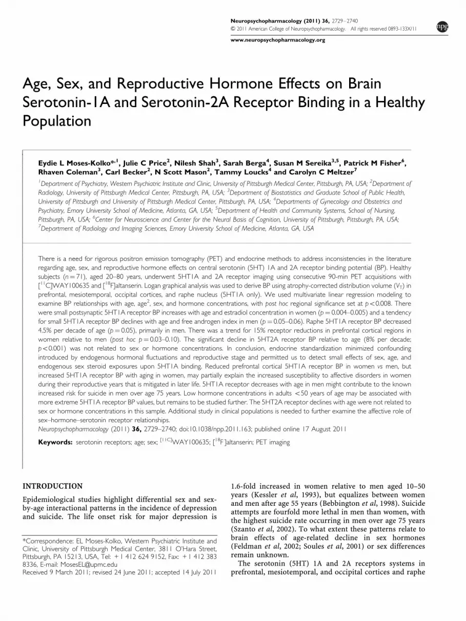
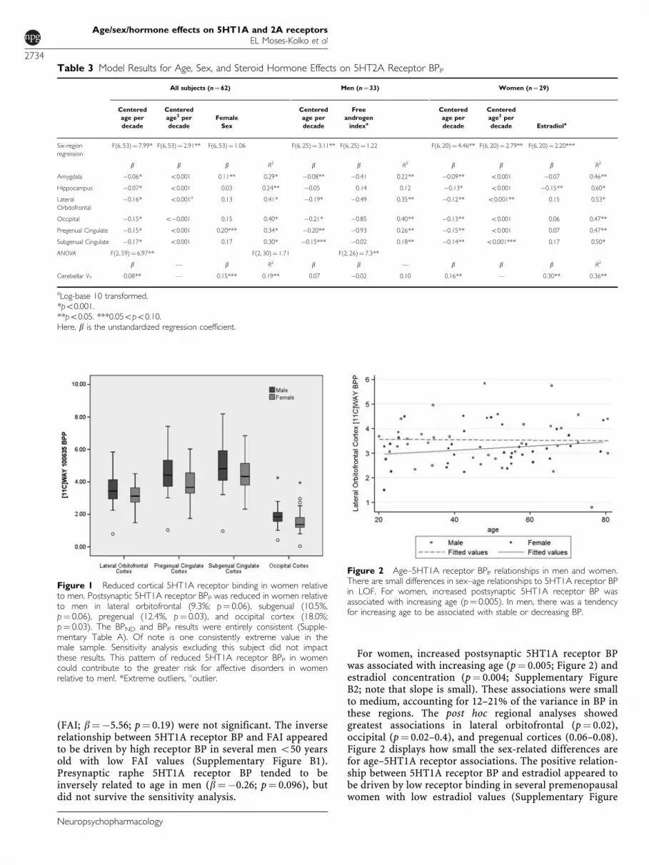
There was no significant age-by-sex interaction for 5HT1Areceptor BP (see Table 2). 5HT1A receptor BP was reducedin women relative to men in postsynaptic prefrontal (LOF,SGC, PRE) and occipital (OCC) regions in the range of 16–19% (BPND) and 9–18% (BPP) (post hoc p¼ 0.03–0.10;Figure 1), but did not survive the post hoc regionalcorrection for multiple testing (po0.008). 5HT1A receptorBP was nonsignificantly lower in women relative to men inamygdala and raphe regions as well. Raphe 5HT1A receptorBP was inversely related to age or age2 (4.5% decrease perdecade; p¼ 0.05).
In the sex-stratified model for postsynaptic 5HT1Areceptor BP in men, there was a small inverse relationshipbetween BP and age (p¼ 0.05) and FAI (p¼ 0.06), account-ing for only 1–9% of the variance in BP. These resultsremained at the borderline of significance in a sensitivityanalysis that excluded an older male outlier with low BPvalues. Large magnitude inverse relationships betweenhippocampal BP and age (b¼�0.74; p¼ 0.09) and log10
Table 2 Model Results for Age, Sex, and Steroid Hormone Effects on 5HT1A Receptor BPP
All subjects (n¼70) Men (n¼ 36) Women (n¼34)
Centered ageper decade
Femalesex
Centered ageper decade
Free androgenindexa
Centered ageper decade Estradiola
Six-region regression F(6, 62)¼ 2.28* F(6, 62)¼ 1.10 F(6, 28)¼ 2.47* F(6, 28)¼ 2.46* F(6, 26)¼ 4.04* F(6, 26)¼ 4.37*
b b R2 b b R2 b b R2
Amygdala 0.01 �0.21 0.003 0.07 0.50 0.002 �0.09 �0.29 0.01
Hippocampus �0.25 0.26 0.03 �0.74** �5.56 0.09 �0.19 �0.21 0.01
Lateral Orbitofrontal 0.03 �0.35** 0.04 �0.13 �1.68 0.05 0.23* 0.63* 0.19*
Occipital �0.05 �0.31** 0.07** �0.09 �0.79 0.03 0.07 0.65* 0.21*
Pregenual Cingulate 0.09 �0.54** 0.07** �0.09 �2.21 0.06 0.26** 0.70** 0.12
Subgenual Cingulate 0.04 �0.50** 0.04 �0.09 �1.21 0.01 0.22 0.62 0.07
ANOVA F(2, 67)¼ 2.31 F(2, 33)¼ 1.56 F(3, 30)¼ 1.92
Raphe �0.14* �0.16 0.06 �0.26** �1.38 0.09 �0.11 �0.07 0.16
ANOVA F(3, 66)¼ 3.30* F(2, 33)¼ 1.13 F(3, 30)¼ 5.13*
Cerebellar VTb �0.03** 0.02 0.13* �0.04 �0.44 0.06 �0.04 3� 10�3 0.34*
aLog-base 10 transformed.bNot reported in table is the significant, small relationship between age2 and Cerebellar VT in the full sample and women subsample.*po0.05.**0.05opo0.10.Here, b is the unstandardized regression coefficient.
Age/sex/hormone effects on 5HT1A and 2A receptorsEL Moses-Kolko et al
2733
Neuropsychopharmacology
(FAI; b¼�5.56; p¼ 0.19) were not significant. The inverserelationship between 5HT1A receptor BP and FAI appearedto be driven by high receptor BP in several men o50 yearsold with low FAI values (Supplementary Figure B1).Presynaptic raphe 5HT1A receptor BP tended to beinversely related to age in men (b¼�0.26; p¼ 0.096), butdid not survive the sensitivity analysis.
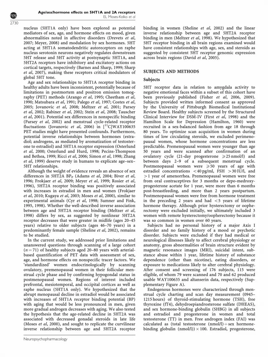
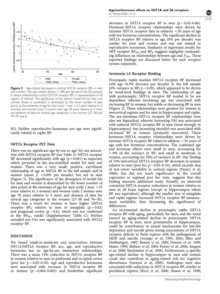
For women, increased postsynaptic 5HT1A receptor BPwas associated with increasing age (p¼ 0.005; Figure 2) andestradiol concentration (p¼ 0.004; Supplementary FigureB2; note that slope is small). These associations were smallto medium, accounting for 12–21% of the variance in BP inthese regions. The post hoc regional analyses showedgreatest associations in lateral orbitofrontal (p¼ 0.02),occipital (p¼ 0.02–0.4), and pregenual cortices (0.06–0.08).Figure 2 displays how small the sex-related differences arefor age–5HT1A receptor associations. The positive relation-ship between 5HT1A receptor BP and estradiol appeared tobe driven by low receptor binding in several premenopausalwomen with low estradiol values (Supplementary Figure
Table 3 Model Results for Age, Sex, and Steroid Hormone Effects on 5HT2A Receptor BPP
All subjects (n¼62) Men (n¼33) Women (n¼ 29)
Centered
age per
decade
Centered
age2 per
decade
Female
Sex
Centered
age per
decade
Free
androgen
indexa
Centered
age per
decade
Centered
age2 per
decade Estradiola
Six-region
regression
F(6, 53)¼ 7.99* F(6, 53)¼ 2.91** F(6, 53)¼ 1.06 F(6, 25)¼ 3.11** F(6, 25)¼ 1.22 F(6, 20)¼ 4.46** F(6, 20)¼ 2.79** F(6, 20)¼ 2.20***
b b b R2 b b R2 b b b R2
Amygdala �0.06* o0.001 0.11** 0.29* �0.08** �0.41 0.22** �0.09** o0.001 �0.07 0.46**
Hippocampus �0.07* o0.001 0.03 0.24** �0.05 0.14 0.12 �0.13* o0.001 �0.15** 0.60*
Lateral
Orbitofrontal
�0.16* o0.001a 0.13 0.41* �0.19* �0.49 0.35** �0.12** o0.001** 0.15 0.53*
Occipital �0.15* o�0.001 0.15 0.40* �0.21* �0.85 0.40** �0.13** o0.001 0.06 0.47**
Pregenual Cingulate �0.15* o0.001 0.20*** 0.34* �0.20** �0.93 0.26** �0.15** o0.001 0.07 0.47**
Subgenual Cingulate �0.17* o0.001 0.17 0.30* �0.15*** �0.02 0.18** �0.14** o0.001*** 0.17 0.50*
ANOVA F(2, 59)¼ 6.97** F(2, 30)¼ 1.71 F(2, 26)¼ 7.3**
b F b R2 b b F b b b R2
Cerebellar VT 0.08** F 0.15*** 0.19** 0.07 �0.02 0.10 0.16** F 0.30** 0.36**
aLog-base 10 transformed.*po0.001.**po0.05. ***0.05opo0.10.Here, b is the unstandardized regression coefficient.
Figure 1 Reduced cortical 5HT1A receptor binding in women relativeto men. Postsynaptic 5HT1A receptor BPP was reduced in women relativeto men in lateral orbitofrontal (9.3%; p¼ 0.06), subgenual (10.5%,p¼ 0.06), pregenual (12.4%, p¼ 0.03), and occipital cortex (18.0%;p¼ 0.03). The BPND and BPP results were entirely consistent (Supple-mentary Table A). Of note is one consistently extreme value in themale sample. Sensitivity analysis excluding this subject did not impactthese results. This pattern of reduced 5HT1A receptor BPP in womencould contribute to the greater risk for affective disorders in womenrelative to men!. *Extreme outliers, 1outlier.
Figure 2 Age–5HT1A receptor BPP relationships in men and women.There are small differences in sex–age relationships to 5HT1A receptor BPin LOF. For women, increased postsynaptic 5HT1A receptor BP wasassociated with increasing age (p¼ 0.005). In men, there was a tendencyfor increasing age to be associated with stable or decreasing BP.
Age/sex/hormone effects on 5HT1A and 2A receptorsEL Moses-Kolko et al
2734
Neuropsychopharmacology
B2). Neither reproductive hormones nor age were signifi-cantly related to raphe BP.
5HT2A Receptor PET Data
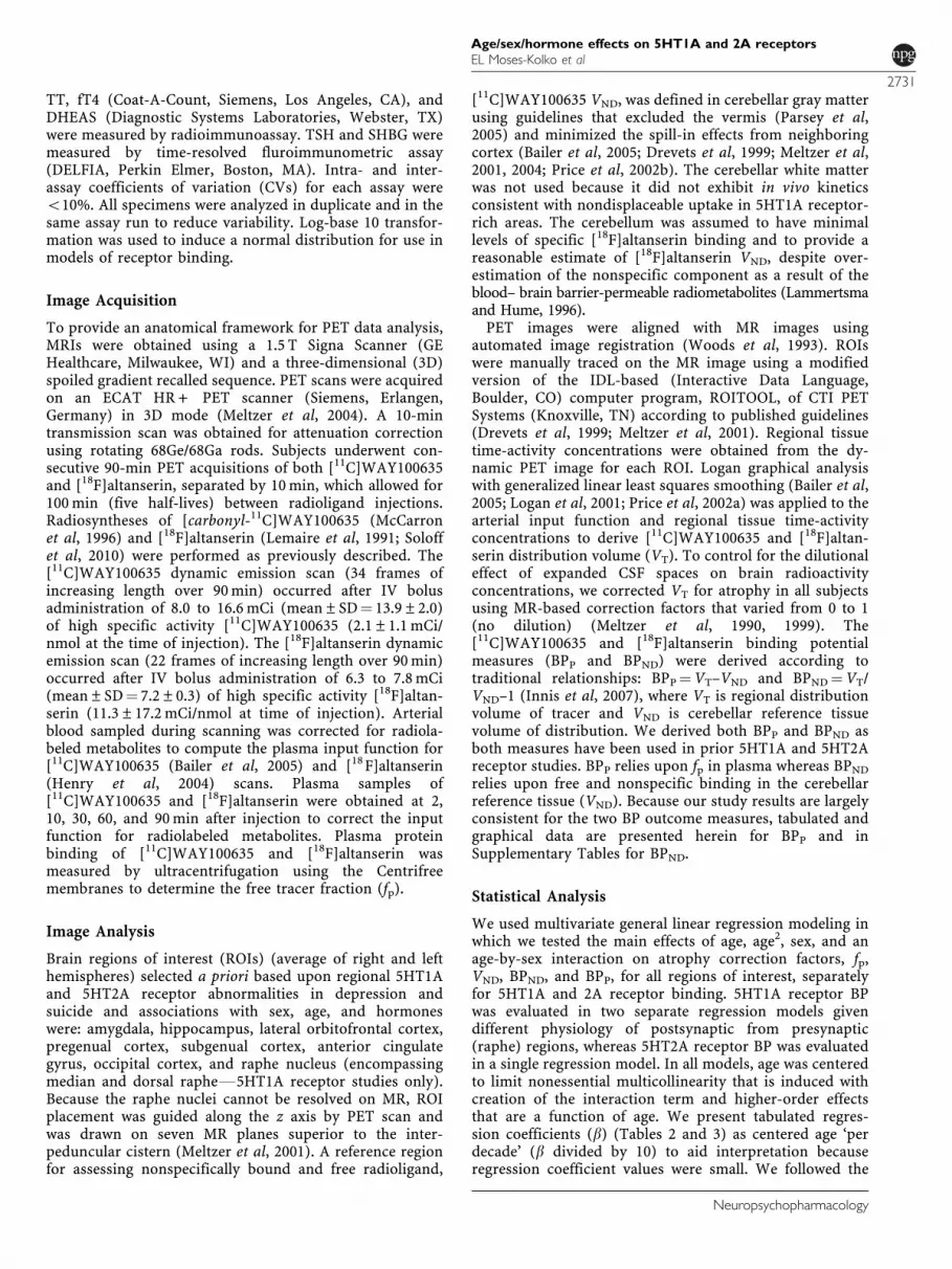
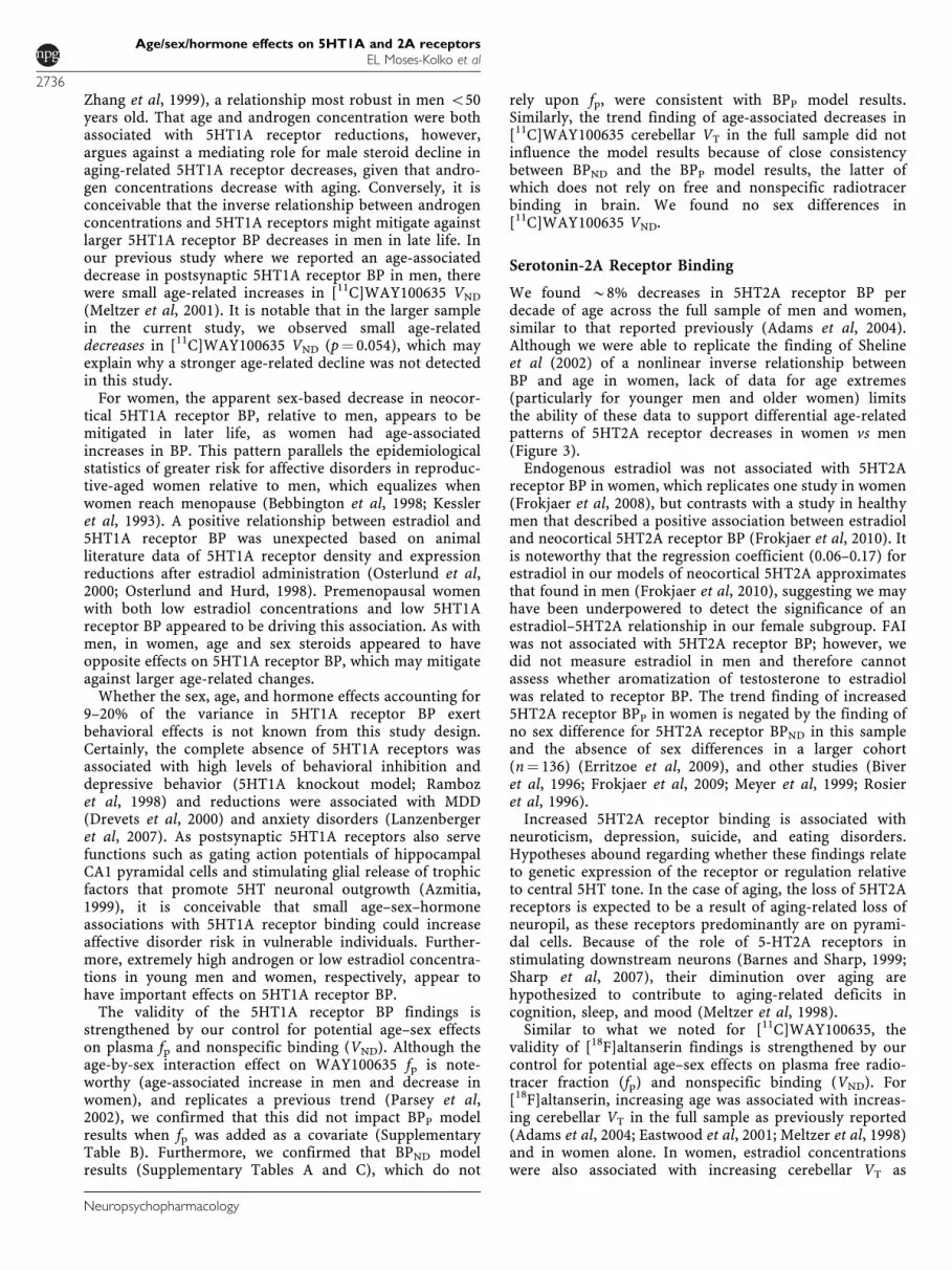
There was no significant age-by-sex or age2-by-sex associa-tion with 5HT2A receptor BP (see Table 3). 5HT2A receptorBP decreased significantly with age (po0.001) as expected,which persisted in the sex-stratified model for men andwomen. There was a very small, positive, curvilinearrelationship of age to 5HT2A BP in the full sample and inwomen (mean b o0.001 per decade), but not in men(Figure 3). The significance of the divergence of model fitsfor men and women is diminished by the lower number ofdata points at the extremes of age for men (only 1 man o23years relative to 3 women) and women (only 2 women overage 70 years relative to 4 men) and absence of data forseveral age categories in the women (27–38 and 70–78).There was a trend for women to have higher 5HT2Areceptor BPP relative to men in amygdala (p¼ 0.02)and pregenual cortex (p¼ 0.6), which was not confirmedin the BPND model (Supplementary Table C). Neitherestradiol nor FAI was significantly associated with 5HT2Areceptor BP.
DISCUSSION
We found small-to-moderate size associations between5HT1A/5HT2A receptor BP, sex, age, and reproductivehormones but no age-by-sex interaction relationships.There was a mean 15% reduction in 5HT1A receptor BPin women relative to men in prefrontal and occipital cortex(post hoc p¼ 0.03–0.10). Age and reproductive hormoneswere associated with increases in 5HT1A receptor BPin women (p¼ 0.004–0.005) and borderline significant
decreases in 5HT1A receptor BP in men (p¼ 0.05–0.06).Hormone-5HT1A receptor relationships were driven byextreme 5HT1A receptor data in subjects o50 years of agewith low hormone concentrations. The significant decline in5HT2A receptor BP relative to age (8% per decade) wassimilar for men and women and was not related toreproductive hormones. Similarity of regression results for5HT receptor BPND and BPP suggests negligible confound-ing influences on relationships between age and VND. Thesereported findings are discussed below for each receptorsystem separately.
Serotonin-1A Receptor Binding
Presynpatic raphe nucleus 5HT1A receptor BP decreasedwith age (4.5% decrease per decade) in the full sample(6% variance in BP; p¼ 0.05), which appeared to be drivenby trend-level findings in men. The relationship of agewith postsynaptic 5HT1A receptor BP tended to be sexdependent, wherein increasing age was associated withincreasing BP in women, but stable or decreasing BP in men(Figure 2). These relationships were greatest for women inneocortical regions and for men in hippocampus and raphe.The sex–hormone–5HT1A receptor BP relationships werealso sex dependent, wherein increasing FAI was associatedwith reduced 5HT1A receptor BP in men (most strongly inhippocampus), but increasing estradiol was associated withincreased BP in women (primarily neocortex). Thesehormone–5HT1A receptor relationships were driven byextreme 5HT1A receptor BP values in subjects o50 years ofage with low hormone concentrations. The combined ageand hormone effects were small in men, accounting for1–9% of the variance in BP, and small to moderate inwomen, accounting for 20% of variance in BP. Our findingof 15% neocortical 5HT1A receptor BP decreases in womenrelative to men (post hoc p¼ 0.03–0.10), which exceeds thatof test–retest variability in cortical regions (Parsey et al,2000), but did not reach significance in the overallregression or regional post hoc tests, suggests that thisfinding warrants additional study. Notably, we observedconsistent 5HT1A receptor reductions in women relative tomen in all brain regions (except in hippocampus whereBP was equivalent), although the smaller size of amygdalaand raphe regions increased 5HT1A receptor BP measure-ment variability, thus decreasing the significance ofdifferences.
An incremental decline in presynaptic raphe 5HT1Areceptor BP with aging, particularly for men, and the trendtoward an aging-related decline in postsynaptic 5HT1Areceptor BP in men, most prominently in hippocampus,could be contributory to neural mechanisms for late-lifedepression and suicide given strong associations of 5HT1Areceptor deficits in these regions with the pathogenesis ofMDD and suicide (Arango et al, 1995, 2001; Blier andDeMontigny, 1987; Bowen et al, 1989; Drevets et al, 1999;Mann, 1999; Meltzer et al, 2004; Parsey et al, 2006; Sargentet al, 2000; Stockmeier et al, 1998). Furthermore, a specificage-related decline in hippocampus in men and womencould also contribute to aging-related risk for cognitivedisturbance (Sarnyai et al, 2000). Androgens were alsoassociated with reductions in 5HT1A receptor BP, similar topreclinical reports (Ricci et al, 2006; Simon et al, 1998;
Figure 3 Age-related decreases in cortical 5HT2A receptor BPP in menand women. The age-related decline (B8% per decade in the full sample)in lateral orbitofrontal cortical 5HT2A receptor BPP is representative of allregions of interest. The significance of the distinct model fits for men andwomen (linear vs curvilinear) is diminished by the lower number of datapoints at the extremes of age for men (only 1 man o23 years relative to 3women) and women (only 2 women over age 70 years relative to 4 men)and absence of data for several age categories in the women (27–38 and70–78 years).
Age/sex/hormone effects on 5HT1A and 2A receptorsEL Moses-Kolko et al
2735
Neuropsychopharmacology
Zhang et al, 1999), a relationship most robust in men o50years old. That age and androgen concentration were bothassociated with 5HT1A receptor reductions, however,argues against a mediating role for male steroid decline inaging-related 5HT1A receptor decreases, given that andro-gen concentrations decrease with aging. Conversely, it isconceivable that the inverse relationship between androgenconcentrations and 5HT1A receptors might mitigate againstlarger 5HT1A receptor BP decreases in men in late life. Inour previous study where we reported an age-associateddecrease in postsynaptic 5HT1A receptor BP in men, therewere small age-related increases in [11C]WAY100635 VND
(Meltzer et al, 2001). It is notable that in the larger samplein the current study, we observed small age-relateddecreases in [11C]WAY100635 VND (p¼ 0.054), which mayexplain why a stronger age-related decline was not detectedin this study.
For women, the apparent sex-based decrease in neocor-tical 5HT1A receptor BP, relative to men, appears to bemitigated in later life, as women had age-associatedincreases in BP. This pattern parallels the epidemiologicalstatistics of greater risk for affective disorders in reproduc-tive-aged women relative to men, which equalizes whenwomen reach menopause (Bebbington et al, 1998; Kessleret al, 1993). A positive relationship between estradiol and5HT1A receptor BP was unexpected based on animalliterature data of 5HT1A receptor density and expressionreductions after estradiol administration (Osterlund et al,2000; Osterlund and Hurd, 1998). Premenopausal womenwith both low estradiol concentrations and low 5HT1Areceptor BP appeared to be driving this association. As withmen, in women, age and sex steroids appeared to haveopposite effects on 5HT1A receptor BP, which may mitigateagainst larger age-related changes.
Whether the sex, age, and hormone effects accounting for9–20% of the variance in 5HT1A receptor BP exertbehavioral effects is not known from this study design.Certainly, the complete absence of 5HT1A receptors wasassociated with high levels of behavioral inhibition anddepressive behavior (5HT1A knockout model; Rambozet al, 1998) and reductions were associated with MDD(Drevets et al, 2000) and anxiety disorders (Lanzenbergeret al, 2007). As postsynaptic 5HT1A receptors also servefunctions such as gating action potentials of hippocampalCA1 pyramidal cells and stimulating glial release of trophicfactors that promote 5HT neuronal outgrowth (Azmitia,1999), it is conceivable that small age–sex–hormoneassociations with 5HT1A receptor binding could increaseaffective disorder risk in vulnerable individuals. Further-more, extremely high androgen or low estradiol concentra-tions in young men and women, respectively, appear tohave important effects on 5HT1A receptor BP.
The validity of the 5HT1A receptor BP findings isstrengthened by our control for potential age–sex effectson plasma fp and nonspecific binding (VND). Although theage-by-sex interaction effect on WAY100635 fp is note-worthy (age-associated increase in men and decrease inwomen), and replicates a previous trend (Parsey et al,2002), we confirmed that this did not impact BPP modelresults when fp was added as a covariate (SupplementaryTable B). Furthermore, we confirmed that BPND modelresults (Supplementary Tables A and C), which do not
rely upon fp, were consistent with BPP model results.Similarly, the trend finding of age-associated decreases in[11C]WAY100635 cerebellar VT in the full sample did notinfluence the model results because of close consistencybetween BPND and the BPP model results, the latter ofwhich does not rely on free and nonspecific radiotracerbinding in brain. We found no sex differences in[11C]WAY100635 VND.
Serotonin-2A Receptor Binding
We found B8% decreases in 5HT2A receptor BP perdecade of age across the full sample of men and women,similar to that reported previously (Adams et al, 2004).Although we were able to replicate the finding of Shelineet al (2002) of a nonlinear inverse relationship betweenBP and age in women, lack of data for age extremes(particularly for younger men and older women) limitsthe ability of these data to support differential age-relatedpatterns of 5HT2A receptor decreases in women vs men(Figure 3).
Endogenous estradiol was not associated with 5HT2Areceptor BP in women, which replicates one study in women(Frokjaer et al, 2008), but contrasts with a study in healthymen that described a positive association between estradioland neocortical 5HT2A receptor BP (Frokjaer et al, 2010). Itis noteworthy that the regression coefficient (0.06–0.17) forestradiol in our models of neocortical 5HT2A approximatesthat found in men (Frokjaer et al, 2010), suggesting we mayhave been underpowered to detect the significance of anestradiol–5HT2A relationship in our female subgroup. FAIwas not associated with 5HT2A receptor BP; however, wedid not measure estradiol in men and therefore cannotassess whether aromatization of testosterone to estradiolwas related to receptor BP. The trend finding of increased5HT2A receptor BPP in women is negated by the finding ofno sex difference for 5HT2A receptor BPND in this sampleand the absence of sex differences in a larger cohort(n¼ 136) (Erritzoe et al, 2009), and other studies (Biveret al, 1996; Frokjaer et al, 2009; Meyer et al, 1999; Rosieret al, 1996).
Increased 5HT2A receptor binding is associated withneuroticism, depression, suicide, and eating disorders.Hypotheses abound regarding whether these findings relateto genetic expression of the receptor or regulation relativeto central 5HT tone. In the case of aging, the loss of 5HT2Areceptors is expected to be a result of aging-related loss ofneuropil, as these receptors predominantly are on pyrami-dal cells. Because of the role of 5-HT2A receptors instimulating downstream neurons (Barnes and Sharp, 1999;Sharp et al, 2007), their diminution over aging arehypothesized to contribute to aging-related deficits incognition, sleep, and mood (Meltzer et al, 1998).
Similar to what we noted for [11C]WAY100635, thevalidity of [18F]altanserin findings is strengthened by ourcontrol for potential age–sex effects on plasma free radio-tracer fraction (fp) and nonspecific binding (VND). For[18F]altanserin, increasing age was associated with increas-ing cerebellar VT in the full sample as previously reported(Adams et al, 2004; Eastwood et al, 2001; Meltzer et al, 1998)and in women alone. In women, estradiol concentrationswere also associated with increasing cerebellar VT as
Age/sex/hormone effects on 5HT1A and 2A receptorsEL Moses-Kolko et al
2736
Neuropsychopharmacology
previously described in men (Frokjaer et al, 2010). Age andestradiol relationships with VND may be contributory, butare not thought to influence the BP outcomes, as the sameage and estradiol-associated decreases observed for altan-serin BPND were also present for altanserin BPP. Thetendency for increased cerebellar VT in women relative tomen concurs with one (Adams et al, 2004) but not anotherstudy (Erritzoe et al, 2009), and does not appear to impactBP in our study. Some have considered using an alternativereference region, although the pons is fraught with its ownproblems (see Adams et al, 2004).
In contrast to a prior study (Adams et al, 2004), we foundno age–sex relationships to metabolism of [18F]altanserinover the scan duration, which suggests that there were noage–sex discrepancies in blood–brain barrier-permeableradiolabeled metabolites (Smith et al, 1998). Additionally,there were no sex or age effects on altanserin fp, whichconcurs with absence of sex differences in fp reportedpreviously (Adams et al, 2004). Nonetheless, it is importantto note that the plasma-derived Logan BP is expected tounderestimate the dual-input compartmental modeling BPvalue (this model accounts for blood–brain barrier passageof radiometabolites) by about 30%, whereas correlationbetween the BP values is very high (rB1.0) (Price et al,2002a, b). This underestimation reflects overestimation ofnonspecific binding in the cerebellar reference region.
Although BMI has been related to 5HT2A receptor BP inother studies (Erritzoe et al, 2009), the effect size in thisdata set was too small (b¼ 1� 10�5) to be consideredclinically meaningful and therefore was excluded from themodel.
CONCLUSIONS
The strengths of this study included: (1) robust and well-validated methods for quantification of atrophy-corrected5HT1A and 5HT2A receptor BP with careful control fornonspecific effects of age and sex on radioligand freefraction and cerebellar VT, (2) characterization andstandardization of reproductive status and associatedendogenous hormonal exposures, (3) thorough screeningfor and exclusion of individuals with personal psychiatrichistory, and (4) relatively large subject sample by PETstandards with a wide age span of men and women,although a higher number of altanserin scans in subjectsover age 70 years would have been desirable.
In conclusion, aside from the well-known age effects todecrease 5HT2A receptor BP, at most 9–20% of 5HT1A and2A receptor BP variance was explained by sex, age, andreproductive hormone effects and there were no age-by-sexinteractions with receptor binding. Endocrine standardiza-tion minimized confounding introduced by endogenoushormonal fluctuations and reproductive stage and per-mitted us to detect small effects of sex, age, and endogenoussex steroid exposures upon 5HT1A binding. Although theseeffect sizes are small, they could potentially lower thethreshold for illness in individuals with genetic and/orenvironmental vulnerabilities for affective disorders. Inaddition, low hormone concentrations in adults o50 yearsof age may be associated with more extreme 5HT1Areceptor BP values, but remains to be studied further.
ACKNOWLEDGEMENTS
This research study was supported by R01s MH67602 andMH71944. Support to CC Meltzer, EL Moses-Kolko, S Berga,SM Sereika, JC Price, R Coleman, and C Becker wasprovided by R01 MH067602. Dr Price received additionalsupport through K02 AG027998, and EL Moses-Kolkoreceived additional support through RO1 MH079164.Support for Nilesh Shah was through 5T32 GM074927.Support for Patrick M Fisher was through the Multi-modalNeuroimaging Training Program at the University ofPittsburgh (DA023420).
DISCLOSURE
Dr Moses-Kolko received a small honorarium as a guestspeaker for La Leche League regional conference, Summer2009. Over the past 3 years, N Scott Mason has receivedcompensation from Janssen Pharmaceuticals, Elan Pharma-ceuticals, Banner Alzheimer’s Institute, and the GollmanGroup (Dallas, TX). Dr Sarah Berga has served on the Boardof Directors and as a consultant for numerous organizations(itemized below). Member of University of Virginia MedicalAlumni Association Board of Directors, 2007 to present(Gratis); consultant for Agile Therapeutics Medical AdvisoryBoardFMarch 2011; AHC Media, LLCFconsultant,Annual business meetingFApril 2008, April 2009, June2010, Noven Pharmaceutical Medical Advisory BoardFFeb2010, Promedica CommunicationsFBayer PharmaceuticalMedical Advisory Board, MeetingFJune 2009, August 2009,Watson Pharmaceutical Women’s Health Strategic AdvisoryBoardFApril 2010; Legal consulting for Kirkland and Ellis,LLCLeydig, Voit & Mayer, LLC, and Reed Smith, LLC;Editorial board for ACOG, Editorial Committee, Guidelinesfor Women’s Health Care, 2009–2011 (Gratis), AmericanJournal of Obstetrics and Gynecology, 2003 to present(Gratis), Advisory Board for Subspecialty Neuroendocrino-logy and Reproductive Neurobiology, The EndocrineSociety Member, Endocrine Self-Assessment ProgramCommittee (June 2007–June 2011), Clinical PracticeGuideline Task Force on Hypothalamic Amenorrhea,January 2011 to present (Gratis), Editorial Board forEndocrinology, January 2010 to present (Gratis),Menopause, Editorial Board, 1999 to present (Gratis). NIHStudy Section reviewer; Society for Women’s HealthResearch, ISIS CVD Network Member, 2009 to present;UpToDate Peer Review Board, 2005 to present. Julie C Price,Susan M Sereika, Patrick M Fisher, Rhaven Coleman, CarlBecker, Tammy Loucks, Carolyn C Meltzer, and Nilesh Shahdeclare no conflict of interest.
REFERENCES
Adams KH, Pinborg LH, Svarer C, Hasselbalch SG, Holm S,Haugbol S et al (2004). A database of [(18)F]-altanserin bindingto 5-HT(2A) receptors in normal volunteers: normative data andrelationship to physiological and demographic variables. Neuro-image 21: 1105–1113.
Arango V, Underwood MD, Boldrini M, Tamir H, Kassir SA,Hsiung S et al (2001). Serotonin 1A receptors, serotonintransporter binding and serotonin transporter mRNA expression
Age/sex/hormone effects on 5HT1A and 2A receptorsEL Moses-Kolko et al
2737
Neuropsychopharmacology
in the brainstem of depressed suicide victims. Neuropsycho-pharmacology 25: 892–903.
Arango V, Underwood MD, Gubbi AV, Mann JJ (1995). Localizedalterations in pre- and postsynaptic serotonin binding sites inthe ventrolateral prefrontial cortex of suicide victims. Brain Res688: 121–133.
Azmitia EC (1999). Serotonin neurons, neuroplasticity, andhomeostasis of neural tissue. Neuropsychopharmacology 21: 2S.
Bailer UF, Frank GK, Henry SE, Price JC, Meltzer CC, Weissfeld Let al (2005). Altered brain serotonin 5-HT1A receptor bindingafter recovery from anorexia nervosa measures by positronemission tomography and [11C]WAY 100635. Arch GenPsychiatry 62: 1032–1041.
Barnes NM, Sharp T (1999). A review of central 5-HT receptorsand their function. Neuropharmacology 38: 1083–1152.
Bebbington P, Dunn G, Jenkins R, Lewis G, Brugha T, Farrell Met al (1998). The influence of age and sex on the prevalence ofdepressive conditions: report from the National Survey ofPsychiatric Morbidity. Psychol Med 28: 9–19.
Biver F, Lotstra F, Monclus M, Wikler D, Damhaut P, Mendlewicz Jet al (1996). Sex difference in 5HT2 receptor in the living humanbrain. Neurosci Lett 204: 25–28.
Blier P, DeMontigny C (1987). Modification of 5-HT neuronproperties by sustained administration of the 5HT1A agonistgepirone: electrophysiological studies in the rat brain. Synapse 1:470–480.
Bowen DM, Najlerahim A, Procter AW, Francis PT, Murphy E(1989). Circumscribed changes of the cerebral cortex inneuropsychiatric disorders of late life. Proc Natl Acad Sci USA86: 9504–9508.
Cheetham SC, Crompton MR, Katona CL, Horton RW (1990).Brain 5-HT1 binding sites in depressed suicides. Psychopharma-cology (Berl) 102: 544–548.
Costes N, Merlet I, Ostrowsky K, Faillenot I, Lavenne F, Zimmer Let al (2005). A 18F-MPPF PET normative database of 5-HT1Areceptor binding in men and women over aging. J Nucl Med 46:1980–1989.
Cyr M, Bosse R, Di Paolo T (1998). Gonadal hormones modulate 5-hydroxytryptamine2A receptors: emphasis on the rat frontalcortex. Neuroscience 83: 829–836.
David SP, Murthy NV, Rabiner EA, Munafo MR, Johnstone EC,Jacob R et al (2005). A functional genetic variation of theserotonin (5-HT) transporter affects 5-HT1A receptor binding inhumans. J Neurosci 25: 2586–2590.
Drevets WC, Frank E, Price JC, Kupfer DJ, Greer PJ, Mathis C(2000). Serotonin type-1A receptor imaging in depression. NuclMed Biol 27: 499–507.
Drevets WC, Frank E, Price JC, Kupfer DJ, Holt D, Greer PJ et al(1999). PET imaging of serotonin 1A receptor binding indepression. Biol Psychiatry 46: 1375–1387.
Drevets WC, Thase ME, Moses-Kolko EL, Price J, Frank E, KupferDJ et al (2007). Serotonin-1A receptor imaging in recurrentdepression: replication and literature review. Nucl Med Biol 34:865–877.
Eastwood SL, Burnet PW, Gittins R, Baker K, Harrison PJ (2001).Expression of serotonin 5-HT(2A) receptors in the human cere-bellum and alterations in schizophrenia. Synapse 42: 104–114.
Erritzoe D, Frokjaer VG, Haugbol S, Marner L, Svarer C, Holst Ket al (2009). Brain serotonin 2A receptor binding: relationsto body mass index, tobacco and alcohol use. Neuroimage 46:23–30.
Feldman HA, Longcope C, Derby CA, Johannes CB, Araujo AB,Coviello AD et al (2002). Age trends in the level of serumtestosterone and other hormones in middle-aged men: long-itudinal results from the Massachusetts male aging study. J ClinEndocrinol Metab 87: 589–598.
First MB, Spitzer RL, Gibbon M, Williams JBW (1998). StructuredClinical Interview for DSM-IV Axis I Disorders - Patient Edition.
New York State Psychiatric Institute, Biometrics ResearchDepartment: New York.
Fisher P, Meltzer C, Ziolko S, Price J, Moses-Kolko E, Serga S et al(2006). Capacity for 5-HT1A-mediated autoregulation predictsamygdala reactivity. Nat Neurosci 9: 1362–1363.
Fisher PM, Meltzer CC, Price JC, Coleman RL, Ziolko SK, Becker Cet al (2009). Medial prefrontal cortex 5-HT2A density iscorrelated with amygdala reactivity, response habituation, andfunctional coupling. Cereb Cortex 19: 2499–2507.
Frokjaer VG, Erritzoe D, Juul A, Nielsen FA, Holst K, Svarer C et al(2010). Endogenous plasma estradiol in healthy men is positivelycorrelated with cerebral cortical serotonin 2A receptor binding.Psychoneuroendocrinology 35: 1311–1320.
Frokjaer VG, Erritzoe D, Madsen J, Paulson OB, Knudsen GM(2009). Gender and the use of hormonal contraception in womenare not associated with cerebral cortical 5-HT 2A receptorbinding. Neuroscience 163: 640–645.
Frokjaer VG, Mortensen EL, Nielsen FA, Haugbol S, Pinborg LH,Adams KH et al (2008). Frontolimbic serotonin 2A receptorbinding in healthy subjects is associated with personality riskfactors for affective disorder. Biol Psychiatry 63: 569–576.
Hamilton M (1960). A rating scale for depression. J NeurolNeurosurg Psychiatry 23: 56–62.
Haugbol S, Pinborg LH, Arfan HM, Frokjaer VM, Madsen J,Dyrby TB et al (2007). Reproducibility of 5-HT2A receptormeasurements and sample size estimations with [18F]altanserinPET using a bolus/infusion approach. Eur J Nucl Med MolImaging 34: 910–915.
Henry SE, Bailer UF, Frank GK, Meltzer CC, Price JC, Mathis CAet al (2004). Positive relationships between 5HT2A receptoractivity in frontal cortical regions after recovery from bulimianervosa. Biol Psychiatry 55: 105S.
Innis RB, Cunningham VJ, Delforge J, Fujita M, Gjedde A, GunnRN et al (2007). Consensus nomenclature for in vivo imaging ofreversibly binding radioligands. J Cereb Blood Flow Metab 27:1533–1539.
Jovanovic H, Cerin A, Karlsson P, Lundberg J, Halldin C,Nordstrom AL (2006). A PET study of 5-HT1A receptors atdifferent phases of the menstrual cycle in women with pre-menstrual dysphoria. Psychiatry Res Neuro 148: 185–193.
Jovanovic H, Lundberg J, Karlsson P, Cerin A, Saijo T, Varrone Aet al (2008). Sex differences in the serotonin 1A receptor andserotonin transporter binding in the human brain measured byPET. Neuroimage 39: 1408–1419.
Kessler RC, McGonagle KA, Swartz M, Blazer DG, Nelson CB(1993). Sex and depression in the National Comorbidity SurveyI: lifetime prevalence, chronicity and recurrence. J Affect Disord29: 85–96.
Kugaya A, Epperson CN, Zoghbi S, van Dyck CH, Hou Y, Fujita Met al (2003). Increase in prefrontal cortex serotonin 2A receptorsfollowing estrogen treatment in postmenopausal women. Am JPsychiatry 160: 1522–1524.
Lammertsma AA, Hume SP (1996). Simplified reference tissuemodel for PET receptor studies. Neuroimage 4(3 Part 1): 153–158.
Lanzenberger RR, Mitterhauser M, Spindelegger C, Wadsak W,Klein N, Mien L-K et al (2007). Reduced serotonin-1Areceptor binding in social anxiety disorder. Biol Psychiatry 61:1081–1089.
Lemaire C, Cantineau R, Guillaume M, Plenevaux A, Christiaens L(1991). Fluorine-18-altanserin: a radioligand for the study ofserotonin receptors with PET: radiolabeling and in vivo biologicbehavior in rats. J Nucl Med 32: 2266–2272.
Logan J, Fowler JS, Volkow ND, Ding YS, Wang G-J, Alexoff DL(2001). A strategy for removing the bias in the graphical analysismethod. J Cereb Blood Flow Metab 21: 307–320.
Mann JJ (1999). Role of the serotonergic system in the pathogen-esis of major depression and suicidal behavior. Neuropsycho-pharmacology 21(2 Suppl): 99S–105S.
Age/sex/hormone effects on 5HT1A and 2A receptorsEL Moses-Kolko et al
2738
Neuropsychopharmacology
Matsubara S, Arora RC, Meltzer HY (1991). Serotonergic measuresin suicide brain: 5-HT1A binding sites in frontal cortex ofsuicide victims. J Neural Transm Gen Sect 85: 181–194.
McCarron JA, Turton D, Pike VW, Poole K (1996). Remotelycontrolled production of the 5HT1A receptor radioligand,[carbonyl-11C]WAY-100635, via 11C-carboxylation of an im-mobilized Grignard reagent. J Labelled Comp Radiopharm 38:941–953.
Meltzer C, Kinahan P, Nichols T, Greer P, Comtat C,Cantwell M et al (1999). Comparative evaluation of MR-basedpartial volume correction schemes for PET. J Nucl Med 40:2053–2065.
Meltzer CC, Drevets WC, Price JC, Mathis CA, Lopresti B, Greer PJet al (2001). Gender-specific aging effects on the serotonin 1Areceptor. Brain Res 895: 9–17.
Meltzer CC, Leal JP, Mayberg HS, Wagner HJ, Frost JJ (1990).Correction of PET data for partial volume effects inhuman cerebral cortex by MR imaging. J Comput Assist Tomogr14: 561–570.
Meltzer CC, Price JC, Mathis CA, Butters MA, Ziolko SK, Moses-Kolko E et al (2004). Serotonin 1A receptor binding andtreatment response in late-life depression. Neuropsychopharma-cology 29: 2258–2265.
Meltzer CC, Smith G, DeKosky ST, Pollock BG, Mathis CA, MooreRY et al (1998). Serotonin in aging, late-life depression, andAlzheimer’s disease: the emerging role of functional imaging (seecomment). Neuropsychopharmacology 18: 407–430.
Meyer JH (2008). Applying neuroimaging ligands to study majordepressive disorder. Semin Nucl Med 38: 287–304.
Meyer JH, Kapur S, Houle S, DaSilva J, Owczarek B, Brown GMet al (1999). Prefrontal cortex 5-HT2 receptors in depression:an [18F]setoperone PET imaging study. Am J Psychiatry 156:1029–1034.
Moses EL, Drevets WC, Smith G, Mathis CA, Kalro BN, Butters MAet al (2000). Effects of estradiol and progesterone administrationon human serotonin 2A receptor binding: a PET study. BiolPsychiatry 48: 854–860.
Osterlund MK, Halldin C, Hurd YL (2000). Effects of chronic17beta-estradiol treatment on the serotonin 5-HT(1A)receptor mRNA and binding levels in the rat brain. Synapse35: 39–44.
Osterlund MK, Hurd YL (1998). Acute 17 beta-estradiol treatmentdown-regulates serotonin 5HT1A receptor mRNA expression inthe limbic system of female rats. Brain Res Mol Brain Res 55:169–172.
Palego L, Marazziti D, Rossi A, Giannaccini G, Naccarato AG,Lucacchini A et al (1997). Apparent absence of aging and gendereffects on serotonin 1A receptors in human neocortex andhippocampus. Brain Res 758: 26–32.
Parsey RV, Arango V, Olvet DM, Oquendo MA, Van Heertum RL,Mann JJ (2005). Regional heterogeneity of 5-HT1A receptors inhuman cerebellum as assessed by positron emission tomogra-phy. J Cereb Blood Flow Metab 25: 785–793.
Parsey RV, Oquendo MA, Ogden RT, Olvet DM, Simpson N,Huang Y et al (2006). Altered serotonin 1A binding in majordepression: a [carbonyl-C-11]WAY 100635 positron emissiontomography study. Biol Psychiatry 59: 106–113.
Parsey RV, Oquendo MA, Simpson NR, Ogden RT, Van HeertumR, Arango V et al (2002). Effects of sex, age, and aggressivetraits in man on brain serotonin 5-HT1A receptor bindingpotential measured by PET using [C-11]WAY-100635. Brain Res954: 173–182.
Parsey RV, Slifstein M, Hwang DR, Abi-Dargham A, Simpson N,Mawlawi O et al (2000). Validation and reproducibility ofmeasurement of 5HT1A receptor parameters with [carbonyl-11C]WAY-100635 in human: comparison of arterial andreference tissue input functions. J Cereb Blood Flow Metab 20:1111–1133.
Pecins-Thompson M, Bethea CL (1999). Ovarian steroidregulation of serotonin-1A autoreceptor messenger RNA ex-pression in the dorsal raphe of rhesus macaques. Neuroscience89: 267–277.
Price JC, Kelley DE, Ryan CM, Meltzer CC, Drevets WC, Mathis CAet al (2002b). Evidence of increased serotonin-1A receptorbinding in type 2 diabetes: a positron emission tomographystudy. Brain Res 927: 97–103.
Price JC, Xu L, Mazumdar S, Meltzer CC, Drevets WC, Mathis CAet al (2002a). Impact of graphical analysis bias on groupcomparisons of regional [carbonyl-11C]WAY binding potentialmeasures. Neuroimage 16: S72.
Rabiner EA, Messa C, Sargent PA, Husted-Kjaer K, Montgomery A,Lawrence AD et al (2002). A database of [(11)C]WAY-100635binding to 5-HT(1A) receptors in normal male volunteers:normative data and relationship to methodological, demo-graphic, physiological, and behavioral variables. Neuroimage15: 620–632.
Ramboz S, Oosting R, Amara DA, Kung HF, Blier P,Mendelsohn M et al (1998). Serotonin receptor 1A knockout:an animal model of anxiety-related disorder. Neurobiology 95:14476–14481.
Ricci LA, Rasakham K, Grimes JM, Melloni JRH (2006). Serotonin-1A receptor activity and expression modulate adolescentanabolic/androgenic steroid-induced aggression in hamsters.Pharmacol Biochem Behav 85: 1–11.
Rosier A, Dupont P, Peuskens J, Bormans G, Vandenberghe R,Maes M et al (1996). Visualisation of loss of 5-HT2A receptorswith age in healthy volunteers using [18F]altanserinand positron emission tomographic imaging. Psychiatry Res68: 11–22.
Sargent PA, Kjaer KH, Bench CJ, Rabiner EA, Messa C, Meyer Jet al (2000). Brain serotonin 1A receptor binding measured bypositron emission tomography with [11C]WAY-100635: effectsof depression and antidepressant treatment. Arch Gen Psychiatry57: 174–180.
Sarnyai Z, Sibille EL, Pavlides C, Fenster RJ, McEwen BS, Toth M(2000). Impaired hippocampal-dependent learning andfunctional abnormalities in the hippocampus in micelacking serotonin(1A) receptors. Proc Natl Acad Sci USA 97:14731–14736.
Sharp T, Boothman L, Raley J, Queree P (2007). Importantmessages in the ‘post’: recent discoveries in 5-HT neuronefeedback control. Trends Pharmacol Sci 28: 629–636.
Sheline YI, Mintun MA, Moerlein SM, Snyder AZ (2002). Greaterloss of 5-HT(2A) receptors in midlife than in late life. Am JPsychiatry 159: 430–435.
Simon NG, Cologer-Clifford A, Lu S-f, McKenna SE, Hu S (1998).Testosterone and its metabolites modulate 5HT1A and 5HT1Bagonist effects on intermale aggression. Neurosci Biobehav Rev23: 325–336.
Smith GS, Price JC, Lopresti BJ, Huang Y, Simpson N, Holt D et al(1998). Test-retest variability of serotonin 5-HT2A receptorbinding measured with positron emission tomography and[18F]altanserin in the human brain. Synapse 30: 380–392.
Soloff PH, Price JC, Mason NS, Becker C, Meltzer CC (2010).Gender, personality, and serotonin-2A receptor binding inhealthy subjects. Psychiatry Res 181: 77–84.
Soules MR, Sherman S, Parrott E, Rebar R, Santoro N, Utian Wet al (2001). Executive summary: Stages of Reproductive AgingWorkshop (STRAW). Fertil Steril 76: 874–878.
Stein P, Savli M, Wadsak W, Mitterhauser M, Fink M, SpindeleggerC et al (2008). The serotonin-1A receptor distribution in healthymen and women measured by PET and [carbonyl-11C]WAY-100635 (see comment). Eur J Nucl Med Mol Imaging 35:2159–2168.
Stockmeier CA, Shapiro LA, Dilley GE, Kolli TN, Friedman L,Rajkowska G (1998). Increase in serotonin-1A autoreceptors in
Age/sex/hormone effects on 5HT1A and 2A receptorsEL Moses-Kolko et al
2739
Neuropsychopharmacology
the midbrain of suicide victims with major depression-postmortem evidence for decreased serotonin activity. J Neurosci18: 7394–7401.
Sumner BE, Fink G (1995). Estrogen increases the density of 5-HT2A receptors in cerebral cortex and nucleus accumbens in thefemale rat. J Steroid Biochem Mol Biol 54(1/2): 15–20.
Sumner BE, Fink G (1998). Testosterone as well as estrogenincreases serotonin2A receptor mRNA and binding site densitiesin the male rat brain. Brain Res Mol Brain Res 59: 205–214.
Szanto K, Gildengers A, Mulsant BH, Brown G, Alexopoulos GS,Reynolds III CF (2002). Identification of suicidal ideation and
prevention of suicidal behaviour in the elderly. Drugs Aging 19:11–24.
Tauscher J, Verhoeff NP, Christensen BK, Hussey D, Meyer JH,Kecojevic A et al (2001). Serotonin 5-HT1A receptor bindingpotential declines with age as measured by [11C]WAY-100635and PET. Neuropsychopharmacology 24: 522–530.
Woods RP, Mazziotta JC, Cherry SR (1993). MRI-PET registrationwith automated algorithm. J Comput Assist Tomogr 17: 536–546.
Zhang L, Ma W, Barker JL, Rubinow DR (1999). Sex differences inexpression of serotonin receptors (subtypes 1A and 2A) in ratbrain: a possible role of testosterone. Neuroscience 94: 251–259.
Supplementary Information accompanies the paper on the Neuropsychopharmacology website (http://www.nature.com/npp)
Age/sex/hormone effects on 5HT1A and 2A receptorsEL Moses-Kolko et al
2740
Neuropsychopharmacology