effects of gaba and gaba receptor inhibition on differentiation of mesencephalic precursors into...
TRANSCRIPT
Effects of GABA and GABA Receptor Inhibition onDifferentiation of Mesencephalic Precursors intoDopaminergic Neurons In Vitro
J.A. Parga, J. Rodriguez-Pallares, M.J. Guerra, J.L. Labandeira-Garcia
Laboratory of Neuroanatomy and Experimental Neurology, Department of Morphological Sciences,Faculty of Medicine, University of Santiago de Compostela, 15782 Santiago de Compostela, Spain
Received 18 January 2007; revised 13 April 2007; accepted 17 April 2007
ABSTRACT: Neurotransmitters have been shown
to control CNS neurogenesis, and GABA-mediated sig-
naling is thought to be involved in the regulation of
nearly all key developmental stages. Generation of do-
paminergic (DA) neurons from stem/precursor cells
for cell therapy in Parkinson’s disease has become a
major focus of research. However, the possible effects
of GABA on generation of DA neurons from prolifer-
ating neurospheres of mesencephalic precursors have
not been studied. In the present study, GABAA, and
GABAB receptors were found to be located in DA cells.
Treatment of cultures with GABA did not cause signif-
icant changes in generation of DA cells from precur-
sors. However, treatment with the GABAA receptor
antagonist bicuculline (10�5 M) led to a significant
increase in the number DA cells, and treatment with
the GABAB receptor antagonist CGP 55845 (10�5 M)
to a significant decrease. Simultaneous treatment with
bicuculline and CGP 55845 did not induce significant
changes. Apoptotic cell death studies and bromodeox-
yuridine immunohistochemistry indicated that the
aforementioned differences in generation of DA neu-
rons are not due to changes in survival or proliferation
of DA cells, but rather to increased or decreased dif-
ferentiation of mesencephalic precursors towards the
DA phenotype. The results suggest that these effects
are exerted via GABA receptors located on DA pre-
cursors, and are not an indirect consequence of effects
on the serotonergic or glial cell population. Adminis-
tration of GABAA receptor antagonists in the differen-
tiation medium may help to obtain higher rates of DA
neurons for potential use in cell therapy for Parkin-
son’s disease. ' 2007 Wiley Periodicals, Inc. Develop Neurobiol
67: 1549–1559, 2007
Keywords: GABA; dopamine; progenitor cells; cell
therapy; Parkinson’s disease
INTRODUCTION
The search for alternative sources of dopaminergic
(DA) neurons for grafting other than primary fetal
tissue has become a major topic in Parkinson’s dis-
ease (PD) research. Previous studies have shown
that precursor cells can be proliferated in culture
under serum free conditions as floating neurospheres
in response to mitogens such as epidermal growth
factor (EGF). After differentiation in vitro, the pre-
cursors contained in these neurosphere cultures are
capable of generating neurons with phenotypes simi-
lar to those normally derived from the corresponding
region in vivo (Parmar et al., 2002; Riaz and Brad-
ford, 2005). Precursor cells can be isolated from the
embryonic mesencephalic flexure, where DA neu-
rons are formed during normal development. They
spontaneously convert to neurons and glia following
mitogen withdrawal, but the number of neurons
expressing the DA phenotype is low. However, dif-
Correspondence to: J.L. Labandeira-Garcia ([email protected]).Contract grant sponsors: Galician Government (XUGA) and
Spanish Ministry of Education (MEC).
' 2007 Wiley Periodicals, Inc.Published online 24 May 2007 in Wiley InterScience (www.interscience.wiley.com).DOI 10.1002/dneu.20531
1549
ferentiation of precursor cells to DA neurons can be
increased by different methodological approaches
(Wang et al., 1995; Rodriguez-Pallares et al., 2001;
Riaz et al., 2002; Rodriguez-Pallares et al., 2004).
Clarification of the signals involved in DA neuron
induction is highly relevant in understanding brain
development and also in obtaining DA neurons for
possible cell therapy in PD (Lindvall and Bjorklund,
2004; Winkler et al., 2005).
There is increasing evidence that neurotransmitter
signaling is involved in early developmental events,
some of which occur prior to synapse formation
(Lauder, 1993; Nguyen et al., 2001; Owens and
Kriegstein, 2002a,b). Functional neurotransmitter
receptors are expressed by a wide variety of neuro-
nal progenitors during embryonic development, and
several studies have shown that neurotransmitters
participate in the extrinsic control of CNS neurogen-
esis involving progenitor cell proliferation, migra-
tion, differentiation, and cell death (Haydar et al.,
2000; Nguyen et al., 2001). Several recent studies
have shown that neurotransmitters regulate genera-
tion of DA neurons from proliferating cell aggre-
gates (Du and Iacovitti, 1995). Administration of
DA or DA agonists induces an increase in generation
of DA neurons in vitro (Guo et al., 2002; Riaz et al.,
2002, 2004; Li et al., 2006). On the contrary, we
have recently shown that serotonin (5-HT) induces a
decrease in generation of DA neurons from prolifer-
ating neurospheres of mesencephalic precursors via
5-HT4 and 5-HT7 receptors (Rodriguez-Pallares
et al., 2003; Parga et al., 2006). During embryonic
development, GABA appears long before the onset
of inhibitory synaptogenesis, and GABA-mediated
signaling is thought to be involved in the regulation
of nearly all key developmental stages ranging from
cell proliferation, differentiation, and migration to
circuit refinement (Varju et al., 2001; Owens and
Kriegstein, 2002a,b). Furthermore, GABA receptors
are present in neural progenitors from the very be-
ginning of neuronal determination, and both GABAB
and particularly GABAA receptors have been
involved in development regulation (Wang et al.,
2003; Jelitai et al., 2004; Schlesinger et al., 2004).
However, the possible effects of GABA on genera-
tion of DA neurons from proliferating neurospheres
of mesencephalic precursors have not been studied.
In the present study, we investigated the possible
effects that GABA and GABA receptors may have
on the generation of DA phenotype neurons from
mesencephalic precursors, and whether the observed
effects may be the result of indirect effects on other
cell populations such as glial cells or serotonergic
neurons.
METHODS
Isolation and Culture of MesencephalicPrecursor Cells
Mesencephalic precursor cells were obtained from
Sprague-Dawley rat embryos of 12 days gestation (E12).
All experiments were carried out in accordance with the
European Communities Council Directive of 24 Novem-
ber 1986 (86/609/EEC), and approved by the correspond-
ing committee at the University of Santiago de Compos-
tela. Rats were deeply anesthetized with chloral hydrate
(400 mg/kg). Ventral mesencephalic tissue was dissected
from the embryos and incubated in 0.1% trypsin (Sigma,
St Louis, MO), 0.05% DNase (Sigma) and Dulbecco’s
Modified Eagle’s Medium (DMEM; Gibco, Paisley, UK)
for 20 min at 378C. The tissue was rinsed in DNase/
DMEM and mechanically dissociated. The resulting cell
suspension was centrifuged at 50g for 5 min, the superna-
tant was carefully removed and the pellet resuspended in
0.05% DNase/DMEM to the final volume required. The
number of viable cells in the suspension was estimated by
acridine orange/ethidium bromide staining, and cells were
plated onto 35-mm culture dishes (BD Falcon, Le Pont de
Claix, France) at a density of 5 3 105 cells/mL. Cells were
maintained in a humidified CO2 incubator (5% CO2;
378C). The proliferation medium was composed of HAMS
F12/DMEM (1:3), B27 supplement (1:50; Gibco), and
EGF (20 ng/mL; Sigma). After 7 days in vitro, cells
formed floating clusters (i.e. cell aggregates) that were
mechanically dissociated and seeded onto fresh EGF-con-
taining medium, and maintained for another 7 days to
purify the long-term propagating precursor cells and
obtain a second generation of cell aggregates. After
2 weeks, neurospheres were gently triturated and plated
onto culture dishes previously coated with poly-L-lysine
(1 mg/mL; Sigma) in the absence of EGF.
Cells from each batch of neurospheres were divided into
control cultures (i.e. untreated), and cultures subjected to
different treatments at plating, and processed in the same
culture batch, as follows. In control cultures, the neuro-
spheres were seeded into a differentiation culture medium
composed of HAMS F12/DMEM (1:1) and 10% fetal bo-
vine serum (FBS; Biochrom KG, Berlin, Germany). A sec-
ond group of cultures were seeded in the presence of
GABA (10�4 M; Sigma). A third group of cultures were
plated on to the differentiation medium supplemented with
GABAA (Bicuculline; (�)-Bicuculline methiodide, 10�6 to
10�5 M; Sigma) or GABAB (CGP 55845; (2S)-3-[[(15)-1-(3,4-dichlorophenyl)ethyl]amino-2-hydroxypropyl](phenyl-
methyl)phosphinic acid, 10�7 to 10�5 M; Tocris Cookson,
Bristol, UK) receptor antagonists to investigate the effects
of the corresponding receptors on the differentiation of pro-
genitors. Stock solutions of the antagonists (2 3 10�4 M)
were prepared in DMEM, filter sterilized, divided into ali-
quots and frozen. Aliquots were thawed immediately before
use. The range of concentrations of specific antagonists or
GABA was chosen on the basis of the results of previous
studies. When used in combination the doses of the drug
1550 Parga et al.
Developmental Neurobiology. DOI 10.1002/dneu
were determined based on the maximal response obtained
from among the concentrations tested. A fourth group of
cultures were seeded in the presence of GABA receptor
antagonists and Bromodeoxyuridine (BrdU; 1 lM, 7 days,
Sigma) to obtain an index of DA cell replication (see later).
Other cultures were seeded in the presence of GABA recep-
tor antagonists and a cell cycle inhibitor, the antimito-
tic agent Cytosine-b-D-arabinofuranoside (Ara-C, 1 lM;
7 days; Sigma) during the differentiation period or the same
doses of Ara-C alone, to inhibit replication of non-neuronal
cells and to study if the effects of these antagonists are
mediated by glial cells. We have previously observed that
after treatment with Ara-C the number of astrocytes
(GFAP-ir cells, see later) is 10–15% of that observed in
control cultures (Parga et al., 2006; see also Michel et al.,
1997). Treatments were added to the culture medium for an
incubation period of 7 days and cells were maintained in
cultures during 7 days in vitro. However, some cultures
treated with the GABAB receptor antagonist CGP 55845
and the corresponding controls were incubated for only
2 days to study apoptosis at early stages of development of
the cell aggregates (see later). Finally, the specificity of the
effect of CGP 55845 on GABAB receptor inhibition was
verified with a second GABAB receptor antagonist (CGP
54626; [S-(R*,R*)]-[3-[[1-(3,4-dichlorophenyl)ethyl]amino]-
2-hydroxypropyl](cyclohexylmethyl) phosphinic acid, 10�6
to 10�5 M; Tocris Cookson), and the possible effect of inhib-
iting glycine mediated responses by bicuculline treatment
was studied in cultures treated with the glycine receptor an-
tagonist Strychnine (Strychnine hydrochloride, 5 3 10�7 M;
Sigma). It has previously been shown that 0.5 lM Strychnine
abolishes the glycinergic responses without affecting
GABAA receptor-mediated responses (O’Brien and Berger,
1999).
Immunolabeling
Cultures were fixed with 4% paraformaldehyde in Dulbec-
co’s phosphate buffered saline (DPBS; pH 7.4) for 20 min,
and washed three times in DPBS. Cultures were preincu-
bated with a blocking solution containing 10% normal se-
rum in DPBS with 1% bovine serum albumin (DPBS-BSA)
and 0.3% Triton X-100 (Sigma) for 1 h. The cultures were
then incubated overnight at 48C with a mouse monoclonal
anti-tyrosine hydroxylase antibody (TH, Sigma; 1:30,000)
as a marker of DA neuron phenotype, or a rabbit polyclonal
antiserum against serotonin (5-HT; Incstar; 1:7500) or a
mouse monoclonal anti-tryptophan hydroxylase antibody
(TPH, Sigma; 1:1000) as markers of 5-HT cells, or a rabbit
polyclonal antibody against GABA (Sigma; 1:5000) as a
marker of GABAergic cells, or a rabbit polyclonal antibody
against a3 subunit (GABAA a3, Sigma; 1:2000) as marker
of GABAA receptors, or a guinea pig polyclonal antibody
against GABAB receptor 1 subunit (GABABR1, Chemicon;
1:20,000) as a marker of GABAB receptors. We have previ-
ously observed that rat cholinergic, DA, noradrenergic and
serotonergic neurons express GABAA receptors derived
from the alpha3 subunit (Rodriguez-Pallares et al., 2001).
Cultures were washed and incubated for 1 h with the corre-
sponding biotinylated secondary antibody diluted 1:500.
The cultures were washed then incubated for 90 min with
avidin–biotin–peroxidase complex (ABC, Vector, 1:150).
Finally, the labeling was revealed with 0.04% hydrogen
peroxide and 0.05% 3,30diaminobenzidine (DAB; Sigma).
Representative cultures were also processed for double
immunofluorescence against TH and GABAA or GABAB
receptor markers to identify the expression of these recep-
tors in DA neurons. Cultures were incubated overnight at
48C with the corresponding primary antibodies (see earlier),
at double antibody concentration containing 1% normal se-
rum and 0.3% Triton X-100 diluted in DPBS-BSA. The cul-
tures were rinsed with DPBS, then incubated for 180 min
with the corresponding secondary antibodies conjugated
with cyanine 3.18 (Cy3; Chemicon; 1:250). For the second
labeling, cultures were incubated overnight at 48C with the
corresponding primary antibodies. Cultures were then
washed and incubated with secondary antibodies conju-
gated with fluorescein isothiocyanate (FITC; Chemicon;
1:100). To visualize the fluorescent labeling, we used a
laser confocal microscope (TCS-SP2, Leica, Heildelberg,
Germany) and a sequential scanning method to avoid any
possible overlap. In all experiments the control cultures, in
which primary antibody was omitted, were immunonega-
tive for these markers.
Apoptotic Cell Death Measurement
Apoptotic cell death of TH-ir cells was determined by DNA
staining and the TUNEL assay in control cultures and cul-
tures treated with GABA receptor antagonists for 7 days or
2 days. Cultures were processed for immunofluorescence of
TH (see earlier), and then all cell nuclei were stained with
bis-benzimide (Hoechst 33342; Sigma) added to a final con-
centration of 3 3 10�6 M (Hilwig and Gropp, 1975; Pardo
et al., 1997). Apoptotic cells were identified by chromatin
condensation.
The TUNEL assay involves measurement of the frag-
mented DNA of the cells by use of the enzyme terminal de-
oxynucleotidyl transferase to incorporate fluorescein-12-
dUTP at the 30-OH ends of the DNA (Gavrieli et al., 1992).
Cultures were processed for immunofluorescence of TH
(see earlier) and the TUNEL assay was then performed
with a DeadEndTM Colorimetric TUNEL System (Promega
Corporation, Madison, WI, USA) and following the manu-
facturer’s instructions. In cultures treated for 2 days with
the GABAB receptor antagonist CGP 55845, the total num-
ber of apoptotic cells was also counted to estimate any pos-
sible decrease in the number of TH-ir neurons due to
enhanced death of precursors. The labeling was visualized
by epi-fluorescence microscopy.
Bromodeoxyuridine Labeling
BrdU staining was used to obtain an index of DA cell
replication. For this, cells from control groups and
groups treated with GABAA or GABAB antagonists
GABA and Dopaminergic Differentiation 1551
Developmental Neurobiology. DOI 10.1002/dneu
were incubated in the corresponding differentiation me-
dium with 1 lM BrdU for 7 days (i.e. during the differ-
entiation period; Arsenijevic and Weiss, 1998). The
cells were then fixed, incubated with 2 M HCl for
30 min at 378C, and washed in borate buffer (pH 8.9) to
neutralize the acid. Cells were processed for double im-
munofluorescence against TH (see earlier) and BrdU
(Sigma; 1:20). The immunolabeling was visualized by
epi-fluorescence microscopy
Quantification and Data Analysis
The same investigator, who was unaware of the treatment
history, always counted the cells using phase contrast or
epi-fluorescence microscopy (Nikon’s Eclipse inverted
microscope, Tokyo, Japan) at X100 magnification. In each
experiment, 15 culture dishes, and 15 randomly chosen cell
aggregates per dish were counted. A few culture dishes that
did not contain at least 15 aggregates were excluded from
counting. In each cell aggregate, the number of DA or 5-
HT cells was estimated by focusing down through the ag-
gregate and counting all immunoreactive cells that came
into focus, or all cells double labeled for TH and BdrU or
TH and apoptosis markers. The thickness of the aggregates
was 44.3 6 3.4 lm (n ¼ 50), as estimated with an Olympus
CAST-Grid System. Thus, 1389 6 161 TH-ir cells were
counted by experiment in a control group. All experiments
were replicated at least three times using new groups of
EGF-responsive mesencephalic precursor cells. The results
were normalized to the counts of the control group of the
same batch (i.e. expressed as a percentage of the control
group counts) to counteract any possible variation from one
batch to another. Statistical differences between groups
(mean 6 SEM) were tested by one-way ANOVA followed
by post hoc Tukey test (p < 0.05). Normality of populations
and homogeneity of variances were tested before each
ANOVA. Statistical analysis was performed by SigmaStat
3.0 from Jandel Scientific (San Rafael, CA).
RESULTS
Location of GABA-ir Cells and GABAReceptors Within the Cell Aggregates
After 7 days, cells differentiated from the prolifer-
ating neurospheres showed different morphologies.
Most cells appeared as differentiated neurons or
glia. However, numerous cells with undifferenti-
ated morphology (i.e. rounded cell bodies without
processes) were still present in the aggregates.
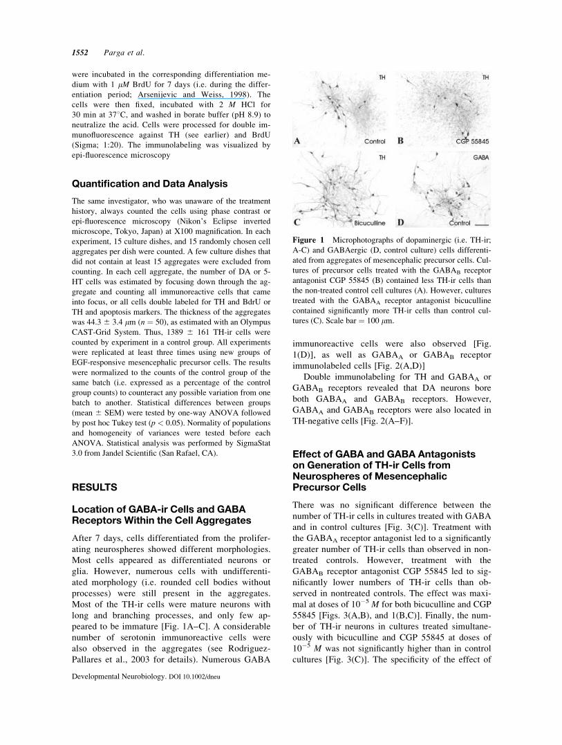
Most of the TH-ir cells were mature neurons with
long and branching processes, and only few ap-
peared to be immature [Fig. 1A–C]. A considerable
number of serotonin immunoreactive cells were
also observed in the aggregates (see Rodriguez-
Pallares et al., 2003 for details). Numerous GABA
immunoreactive cells were also observed [Fig.
1(D)], as well as GABAA or GABAB receptor
immunolabeled cells [Fig. 2(A,D)]
Double immunolabeling for TH and GABAA or
GABAB receptors revealed that DA neurons bore
both GABAA and GABAB receptors. However,
GABAA and GABAB receptors were also located in
TH-negative cells [Fig. 2(A–F)].
Effect of GABA and GABA Antagonistson Generation of TH-ir Cells fromNeurospheres of MesencephalicPrecursor Cells
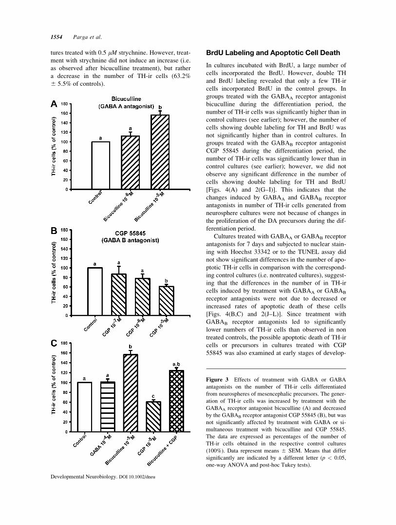
There was no significant difference between the
number of TH-ir cells in cultures treated with GABA
and in control cultures [Fig. 3(C)]. Treatment with
the GABAA receptor antagonist led to a significantly
greater number of TH-ir cells than observed in non-
treated controls. However, treatment with the
GABAB receptor antagonist CGP 55845 led to sig-
nificantly lower numbers of TH-ir cells than ob-
served in nontreated controls. The effect was maxi-
mal at doses of 10�5 M for both bicuculline and CGP
55845 [Figs. 3(A,B), and 1(B,C)]. Finally, the num-
ber of TH-ir neurons in cultures treated simultane-
ously with bicuculline and CGP 55845 at doses of
10�5 M was not significantly higher than in control
cultures [Fig. 3(C)]. The specificity of the effect of
Figure 1 Microphotographs of dopaminergic (i.e. TH-ir;
A-C) and GABAergic (D, control culture) cells differenti-
ated from aggregates of mesencephalic precursor cells. Cul-
tures of precursor cells treated with the GABAB receptor
antagonist CGP 55845 (B) contained less TH-ir cells than
the non-treated control cell cultures (A). However, cultures
treated with the GABAA receptor antagonist bicuculline
contained significantly more TH-ir cells than control cul-
tures (C). Scale bar ¼ 100 lm.
1552 Parga et al.
Developmental Neurobiology. DOI 10.1002/dneu
CGP 55845 on GABAB receptor inhibition was con-
firmed with the second GABAB receptor antagonist
(CGP 54626), which also induced a significant
decrease in the number of TH-ir neurons at doses of
10�5 M (45.1% 6 4.4% of the control values) or
10�6 M (48.7% 6 8.2% of the control values). The
possible effect of inhibiting glycine mediated re-
sponses by bicuculline treatment was studied in cul-
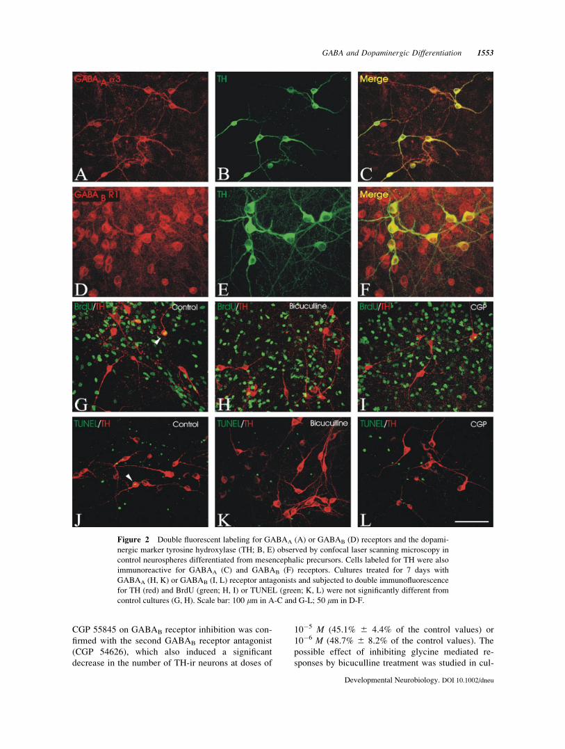
Figure 2 Double fluorescent labeling for GABAA (A) or GABAB (D) receptors and the dopami-
nergic marker tyrosine hydroxylase (TH; B, E) observed by confocal laser scanning microscopy in
control neurospheres differentiated from mesencephalic precursors. Cells labeled for TH were also
immunoreactive for GABAA (C) and GABAB (F) receptors. Cultures treated for 7 days with
GABAA (H, K) or GABAB (I, L) receptor antagonists and subjected to double immunofluorescence
for TH (red) and BrdU (green; H, I) or TUNEL (green; K, L) were not significantly different from
control cultures (G, H). Scale bar: 100 lm in A-C and G-L; 50 lm in D-F.
GABA and Dopaminergic Differentiation 1553
Developmental Neurobiology. DOI 10.1002/dneu
tures treated with 0.5 lM strychnine. However, treat-
ment with strychnine did not induce an increase (i.e.
as observed after bicuculline treatment), but rather
a decrease in the number of TH-ir cells (63.2%
6 5.5% of controls).
BrdU Labeling and Apoptotic Cell Death
In cultures incubated with BrdU, a large number of
cells incorporated the BrdU. However, double TH
and BrdU labeling revealed that only a few TH-ir
cells incorporated BrdU in the control groups. In
groups treated with the GABAA receptor antagonist
bicuculline during the differentiation period, the
number of TH-ir cells was significantly higher than in
control cultures (see earlier); however, the number of
cells showing double labeling for TH and BrdU was
not significantly higher than in control cultures. In
groups treated with the GABAB receptor antagonist
CGP 55845 during the differentiation period, the
number of TH-ir cells was significantly lower than in
control cultures (see earlier); however, we did not
observe any significant difference in the number of
cells showing double labeling for TH and BrdU
[Figs. 4(A) and 2(G–I)]. This indicates that the
changes induced by GABAA and GABAB receptor
antagonists in number of TH-ir cells generated from
neurosphere cultures were not because of changes in
the proliferation of the DA precursors during the dif-
ferentiation period.
Cultures treated with GABAA or GABAB receptor
antagonists for 7 days and subjected to nuclear stain-
ing with Hoechst 33342 or to the TUNEL assay did
not show significant differences in the number of apo-
ptotic TH-ir cells in comparison with the correspond-
ing control cultures (i.e. nontreated cultures), suggest-
ing that the differences in the number of in TH-ir
cells induced by treatment with GABAA or GABAB
receptor antagonists were not due to decreased or
increased rates of apoptotic death of these cells
[Figs. 4(B,C) and 2(J–L)]. Since treatment with
GABAB receptor antagonists led to significantly
lower numbers of TH-ir cells than observed in non
treated controls, the possible apoptotic death of TH-ir
cells or precursors in cultures treated with CGP
55845 was also examined at early stages of develop-
Figure 3 Effects of treatment with GABA or GABA
antagonists on the number of TH-ir cells differentiated
from neurospheres of mesencephalic precursors. The gener-
ation of TH-ir cells was increased by treatment with the
GABAA receptor antagonist bicuculline (A) and decreased
by the GABAB receptor antagonist CGP 55845 (B), but was
not significantly affected by treatment with GABA or si-
multaneous treatment with bicuculline and CGP 55845.
The data are expressed as percentages of the number of
TH-ir cells obtained in the respective control cultures
(100%). Data represent means 6 SEM. Means that differ
significantly are indicated by a different letter (p < 0.05,
one-way ANOVA and post-hoc Tukey tests).
1554 Parga et al.
Developmental Neurobiology. DOI 10.1002/dneu
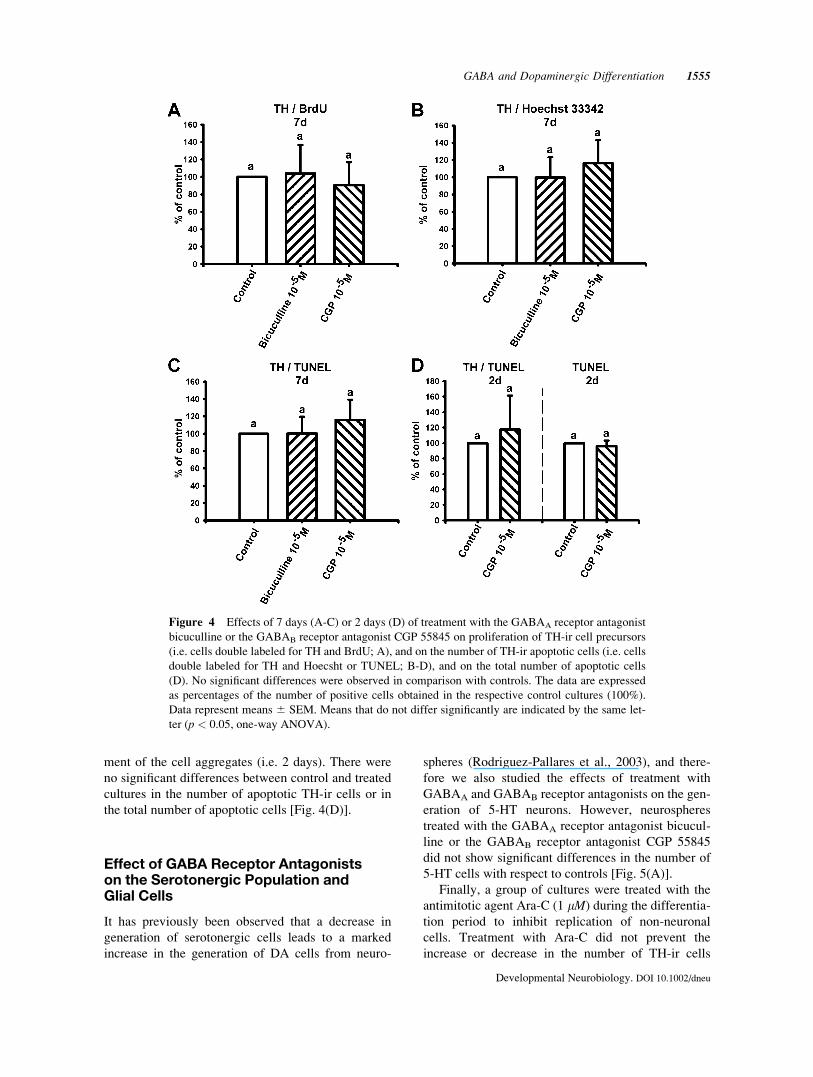
ment of the cell aggregates (i.e. 2 days). There were
no significant differences between control and treated
cultures in the number of apoptotic TH-ir cells or in
the total number of apoptotic cells [Fig. 4(D)].
Effect of GABA Receptor Antagonistson the Serotonergic Population andGlial Cells
It has previously been observed that a decrease in
generation of serotonergic cells leads to a marked
increase in the generation of DA cells from neuro-
spheres (Rodriguez-Pallares et al., 2003), and there-
fore we also studied the effects of treatment with
GABAA and GABAB receptor antagonists on the gen-
eration of 5-HT neurons. However, neurospheres
treated with the GABAA receptor antagonist bicucul-
line or the GABAB receptor antagonist CGP 55845
did not show significant differences in the number of
5-HT cells with respect to controls [Fig. 5(A)].
Finally, a group of cultures were treated with the
antimitotic agent Ara-C (1 lM) during the differentia-
tion period to inhibit replication of non-neuronal
cells. Treatment with Ara-C did not prevent the
increase or decrease in the number of TH-ir cells
Figure 4 Effects of 7 days (A-C) or 2 days (D) of treatment with the GABAA receptor antagonist
bicuculline or the GABAB receptor antagonist CGP 55845 on proliferation of TH-ir cell precursors
(i.e. cells double labeled for TH and BrdU; A), and on the number of TH-ir apoptotic cells (i.e. cells
double labeled for TH and Hoecsht or TUNEL; B-D), and on the total number of apoptotic cells
(D). No significant differences were observed in comparison with controls. The data are expressed
as percentages of the number of positive cells obtained in the respective control cultures (100%).
Data represent means 6 SEM. Means that do not differ significantly are indicated by the same let-
ter (p < 0.05, one-way ANOVA).
GABA and Dopaminergic Differentiation 1555
Developmental Neurobiology. DOI 10.1002/dneu
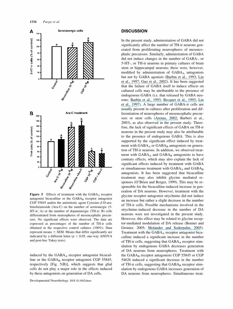
induced by the GABAA receptor antagonist bicucul-
line or the GABAB receptor antagonist CGP 55845,
respectively [Fig. 5(B)], which suggests that glial
cells do not play a major role in the effects induced
by these antagonists on generation of DA cells.
DISCUSSION
In the present study, administration of GABA did not
significantly affect the number of TH-ir neurons gen-
erated from proliferating neurospheres of mesence-
phalic precursors. Similarly, administration of GABA
did not induce changes in the number of GABA-, or
5-HT-, or TH-ir neurons in primary cultures of brain
stem or hippocampal neurons; these were, however,
modified by administration of GABAA antagonists
but not by GABA agonists (Barbin et al., 1993; Liu
et al., 1997; Guo et al., 2002). It has been suggested
that the failure of GABA itself to induce effects on
cultured cells may be attributable to the presence of
endogenous GABA (i.e. that released by GABA neu-
rons; Barbin et al., 1993; Becquet et al., 1993; Liu
et al., 1997). A large number of GABA-ir cells are
usually present in cultures after proliferation and dif-
ferentiation of neurospheres of mesencephalic precur-
sors or stem cells (Arenas, 2002; Barberi et al.,
2003), as also observed in the present study. There-
fore, the lack of significant effects of GABA on TH-ir
neurons in the present study may also be attributable
to the presence of endogenous GABA. This is also
supported by the significant effect induced by treat-
ment with GABAA or GABAB antagonists on genera-
tion of TH-ir neurons. In addition, we observed treat-
ment with GABAA and GABAB antagonists to have
contrary effects, which may also explain the lack of
significant effects induced by treatment with GABA
or simultaneous treatment with GABAA and GABAB
antagonists. It has been suggested that bicuculline
treatment may also inhibit glycine mediated re-
sponses (O’Brien and Berger, 1999). This may be re-
sponsible for the bicuculline-induced increase in gen-
eration of DA neurons. However, treatment with the
glycine receptor antagonist strychnine did not induce
an increase but rather a slight decrease in the number
of TH-ir cells. Possible mechanisms involved in the
strychnine-induced decrease in the number of DA
neurons were not investigated in the present study.
However, this effect may be related to glycine recep-
tor-mediated modulation of DA release (Bennet and
Gronier, 2005; Molander and Soderpalm, 2005).
Treatment with the GABAA receptor antagonist bicu-
culline induced a significant increase in the number
of TH-ir cells, suggesting that GABAA receptor stim-
ulation by endogenous GABA decreases generation
of DA neurons from neurospheres. Treatment with
the GABAB receptor antagonists CGP 55845 or CGP
54626 induced a significant decrease in the number
of TH-ir cells, suggesting that GABAB receptor stim-
ulation by endogenous GABA increases generation of
DA neurons from neurospheres. Simultaneous treat-
Figure 5 Effects of treatment with the GABAA receptor
antagonist bicuculline or the GABAB receptor antagonist
CGP 55845 and/or the antimitotic agent Cytosine-b-D-ara-binofuranoside (Ara-C) on the number of serotonergic (5-
HT-ir; A) or the number of dopaminergic (TH-ir; B) cells
differentiated from neurospheres of mesencephalic precur-
sors. No significant effects were observed. The data are
expressed as percentages of the number of TH-ir cells
obtained in the respective control cultures (100%). Data
represent means 6 SEM. Means that differ significantly are
indicated by a different letter (p < 0.05, one-way ANOVA
and post-hoc Tukey tests).
1556 Parga et al.
Developmental Neurobiology. DOI 10.1002/dneu
ment with GABAA and GABAB antagonists did not
induce any significant difference in the number of
TH-ir cells, suggesting that GABA does not have any
significant effect on the generation of DA neurons via
mechanisms different to GABAA or GABAB receptor
stimulation.
The changes induced by treatment with GABAA
or GABAB antagonists in the number of TH-ir neu-
rons obtained from precursors may be related to an
increase in the generation of the DA phenotype from
precursor cells, and also to an increase in the survival
of cells with the DA phenotype. Several studies have
reported that GABA promotes survival (Liu et al.,
1997; Luk and Sadikot, 2001) or has no effects
(Fiszman et al., 1999; Nguyen et al., 2003) on sur-
vival of different types of neuronal progenitors or
developing neurons. It appears that GABA may
induce different effects depending on the type of cell
or experimental conditions. The data obtained in the
present study with Hoechst staining and the TUNEL
assay suggest that the increase or decrease in the
number of TH-ir neurons after treatment with
GABAA or GABAB receptor antagonists is not due to
an increase in the survival of TH-ir neurons.
The increase in the number of TH-ir neurons
appears to be related to an increase in the generation
of TH-ir neurons. This may be due to either increased
proliferation of precursors of DA neurons or to an
increase in differentiation of DA neurons from mes-
encephalic precursor cells. Negative, positive and no
significant effects of GABA on different types of neu-
ronal progenitors have been reported (Fiszman et al.,
1999; Luk and Sadikot, 2001; Owens and Kriegstein,
2002a,b; Nguyen et al., 2003). It has also been
observed that GABAA receptor activation is mito-
genic in the proliferative neocortical ventricular zone
and antiproliferative in the subventricular zone
(Haydar et al., 2000). The present data from BrdU
immunohistochemistry suggest that there is no signif-
icant increase in the proliferation of DA precursors
during the differentiation period in cultures treated
with GABAA or GABAB receptor antagonists, and
also that the observed differences in the generation of
TH-ir neurons are due to an increase in differentiation
of DA neurons from mesencephalic precursor cells.
Previous studies have shown that GABA can affect
different aspects of neuronal differentiation in vitro
(Owens and Kriegstein, 2002a,b; Jelitai et al., 2004).
Although the mechanisms of the differentiation
effects exerted by GABA have not been totally clari-
fied, they are usually mediated by activation of
GABA receptors, as GABA receptor antagonists have
shown to block the effects in the present and other
experiments.
In the present and other studies both GABAA and
GABAB receptors were located in DA cells (Kim
et al., 1997; Wirtsshafter and Sheppard, 2001; Schle-
singer et al., 2004) as well as in glial cells and other
cell populations such as serotonergic cells (Wirtssh-
after and Sheppard, 2001), which have been shown to
regulate generation of DA cells from proliferating
neurospheres of mesencephalic precursors. We have
previously shown that elimination of serotonergic
cells or inhibition of serotonin synthesis induces a
marked increase in generation of DA cells from pro-
liferating neurospheres of mesencephalic precursors
(Rodriguez-Pallares et al., 2003), and it has been
reported that GABAA receptor antagonists signifi-
cantly reduced the survival of serotonergic neurons in
primary cultures from rat brainstem (Liu et al., 1997).
Therefore, the increase in generation of DA neurons
observed in the present study after treatment with the
GABAA receptor antagonist bicuculline may be the
consequence of a bicuculline-induced reduction in
the number of serotonergic cells. However, we did
not observe significant differences in the number of
serotonergic cells generated from the neurospheres
after treatment with GABAA or GABAB antagonist.
It is also possible that GABA receptor antagonists or
GABA receptor activation induce significant changes
in 5-HT release without significant changes in the
number of serotonergic neurons. It has been observed
that inhibition of GABAA receptors or activation of
GABAB receptors decreases 5-HT release in primary
cultures of raphe cells (Becquet et al., 1993), and this
may explain the results observed in the present
experiments after treatment of cultures with GABAA
or GABAB receptor antagonists. However, we have
recently observed that 5-HT4 and 5-HT7 receptors
located in glial cells play a major role in the decreas-
ing effect of serotonin on generation of DA neurons
from neurospheres, which is inhibited by elimination
of glial cells with Ara-C (Parga et al., 2006). In the
present study, the effects of the GABA antagonists on
generation of DA neurons were not significantly
modified by treatment with Ara-C, which indicates
that they are not caused by changes in levels of sero-
tonin and that glial cells do not play a major role, and
also suggests that the effects are exerted by GABA
receptors located on the DA precursors.
A number of compounds have been reported to
increase generation of TH-ir neurons from precursors;
most of these compounds induce increases in the
number of TH-ir neurons of between 150 and 400%
with respect to control values (Riaz and Bradford,
2005 for review). We have obtained increases of
300–400% with antioxidant treatments (Rodriguez-
Pallares et al., 2001, 2002). Treatment with different
GABA and Dopaminergic Differentiation 1557
Developmental Neurobiology. DOI 10.1002/dneu
cytokines has led to increases of between 150 and
350% (Ling et al., 1998; Rodriguez-Pallares et al.,
2004, 2005). Neurotransmitters have also been shown
to regulate generation of DA neurons from precur-
sors. Treatment with DA alone induced small or non
significant increases in DA neuron generation (Du
and Iacovitti, 1995; Riaz et al., 2002, 2004), and sero-
tonin receptor antagonists induced an increase of
250% in the generation of TH-ir neurons (Parga
et al., 2006). Interestingly, however, it has been
shown that treatment with combinations of com-
pounds that induce moderate increases in generation
of DA neurons led to greater increases in the number
DA cells (Potter et al., 1999; Riaz and Bradford,
2005; Riaz et al., 2002, 2004). Co-treatment with
neurotransmitters or neurotransmitter antagonists and
growth factors has been particularly effective (Du
and Iacovitti, 1995; Riaz et al., 2002, 2004). In the
present study, treatment with the GABAA receptor
antagonist bicuculline induced a 160% increase in
generation of TH-ir neurons, which is lower than the
increase induced by some of the aforementioned anti-
oxidants or cytokines and higher than that observed
after treatment with DA alone or growth factors such
as BDNF or GDNF alone (Du and Iacovitti, 1995;
Riaz et al., 2002, 2004; Riaz and Bradford, 2005). As
previously observed for DA, cotreatment with
GABAA receptor antagonists and growth factors and/
or other compounds may induce further increases in
generation of DA neurons from precursors or stem
cells.
In conclusion, the present results show that
GABAA receptor activation decreases and GABAB
receptor activation increases generation of DA neu-
rons from proliferating neurospheres of mesence-
phalic precursors. A large number of GABAergic
cells and a considerable level of endogenous GABA
are usually found together with DA neurons differen-
tiated from precursor or stem cells, and the present
results suggest that administration of GABAA recep-
tor antagonists in the differentiation medium may
help in obtaining greater numbers of DA neurons for
potential use in cell therapy for PD.
The authors thank Pilar Aldrey for her excellent techni-
cal assistance.
REFERENCES
Arenas A. 2002. Stem cells in the treatment of Parkinson’s
disease. Brain Res Bull 57:785–808.
Arsenijevic Y, Weiss S. 1998. Insulin-like growth factor-I is
a differentiation factor for postmitotic CNS stem cell-
derived neuronal precursors: Distinct actions from those
of brain-derived neurotrophic factor. J Neurosci 18:2118–
2128.
Barberi T, Klivenyi P, Calingasan N, Hyojin L, Kawamata
H, Loonam K, Perrier AL, et al. 2003. Neural subtype
specification of fertilization and nuclear transfer embry-
onic stem cells and application in parkinsonian mice. Nat
Biotechnol 21:1200–1207.
Barbin G, Pollard H, Gaiarsa JL, Ben-Ari Y. 1993. Involve-
ment of GABAA receptors in the outgrowth of cultured
hippocampal neurons. Neurosci Lett 152:150–154.
Becquet D, Hery M, Francois-Bellan AM, Giraud P,
Deprez P, Faudon M, Fache MP, Hery F. 1993. Gluta-
mate, GABA, glycine and taurine modulate serotonin
synthesis and release in rostral and caudal rhombence-
phalic raphe cells in primary cultures. Neurochem Int
23:269–283.
Bennett S, Gronier B. 2005. Modulation of striatal dopa-
mine release in vitro by agonists of glycineB site of
NMDA receptors; interaction with antipsychotics. Eur J
Pharmacol 527:52–59.
Du X, Iacovitti L. 1995. Synergy between growth factors
and transmitters required for catecholamine differentia-
tion in brain neurons. J Neurosci 15:5420–5427.
Fiszman ML, Borodinsky LN, Neale JH. 1999. GABA
induces proliferation of immature cerebellar granule cells
grown in vitro. Brain Res Dev Brain Res 115:1–8.
Gavrieli Y, Sherman Y, Ben-Sasson SA. 1992. Identifica-
tion of programmed cell death in situ via specific
labeling of nuclear DNA fragmentation. J Cell Biol
119:493–501.
Guo H, Tang Z, Yu Y, Xu L, Jin G, Zhou J. 2002. Apomor-
phine induces trophic factors that support fetal rat mesen-
cephalic dopaminergic neurons in cultures. Eur J Neuro-
sci 16:1861–1870.
Haydar TF, Wang F, Schwartz ML, Rakic P. 2000. Differen-
tial modulation of proliferation in the neocortical ventric-
ular and subventricular zones. J Neurosci 20:5764–5774.
Hilwig I, Gropp A. 1975. pH-dependent fluorescence of
DNA and RNA in cytologic staining with \33258"Hoechst. Exp Cell Res 91:457–460.
Jelitai M, Anderova M, Marko K, Kekesi K, Koncz P,
Sykova E, Madarasz E. 2004. Role of gamma-aminobu-
tyric acid in early neuronal development: Studies with an
embryonic neuroectodermal stem cell clone. J Neurosci
Res 76:801–811.
Kim KM, Nakajima S, Nakajima Y. 1997. Dopamine and
GABA receptors in cultured substantia nigra neurons:
Correlation of electrophysiology and immunocytochem-
istry. Neuroscience 78:759–769.
Lauder JM. 1993. Neurotransmitters as growth regulatory
signals: Role of receptors and second messengers. Trends
Neurosci 16:233–240.
Li A, Guo H, Luo X, Sheng J, Yang S, Yin Y, Zhou J, Zhou
J. 2006. Apomorphine-induced activation of dopamine
receptors modulates FGF-2 expression in astrocytic cul-
tures and promotes survival of dopaminergic neurons.
FASEB J 20:1263–1265.
Lindvall O, Bjorklund A. 2004. Cell therapy in Parkinson’s
disease. NeuroRx 1:382–393.
1558 Parga et al.
Developmental Neurobiology. DOI 10.1002/dneu
Ling ZD, Potter ED, Lipton JW, Carvey PM. 1998. Differ-
entiation of mesencephalic progenitor cells into dopami-
nergic neurons by cytokines. Exp Neurol 149:411–423.
Liu J, Morrow AL, Devaud L, Grayson DR, Lauder JM.
1997. GABAA receptors mediate trophic effects of
GABA on embryonic brainstem monoamine neurons in
vitro. J Neurosci 17:2420–2428.
Luk KC, Sadikot AF. 2001. GABA promotes survival but
not proliferation of parvalbumin-immunoreactive inter-
neurons in rodent neostriatum: An in vivo study with
stereology. Neuroscience 104:93–103.
Michel PP, Ruberg M, Agid Y. 1997. Rescue of mesence-
phalic dopamine neurons by anticancer drug cytosine
arabinoside. J Neurochem 69:1499–1507.
Molander A, Soderpalm B. 2005. Glycine receptors regu-
late dopamine release in the rat nucleus accumbens.
Alcohol Clin Exp Res 29:17–26.
Nguyen L, Malgrange B, Breuskin I, Bettendorff L,
Moonen G, Belachew S, Rigo JM. 2003. Autocrine/para-
crine activation of the GABA(A) receptor inhibits the
proliferation of neurogenic polysialylated neural cell ad-
hesion molecule-positive (PSA-NCAMþ) precursor cells
from postnatal striatum. J Neurosci 23:3278–3294.
Nguyen L, Rigo JM, Rocher V, Belachew S, Malgrange B,
Rogister B, Leprince P, Moonen G. 2001. Neurotrans-
mitters as early signals for central nervous system devel-
opment. Cell Tissue Res 305:187–202.
O’Brien JA, Berger AJ. 1999. Cotransmission of GABA
and glycine to brain stem motoneurons. J Neurophysiol
82:1638–1641.
Owens DF, Kriegstein AR. 2002a. Developmental neuro-
transmitters? Neuron 36:989–995.
Owens DF, Kriegstein AR. 2002b. Is there more to GABA
than synaptic inhibition? Nat Rev Neurosci 3:715–727.
Pardo B, Paino CL, Casarejos MJ, Mena MA. 1997. Neuro-
nal-enriched cultures from embryonic rat ventral mesen-
cephalon for pharmacological studies of dopamine neu-
rons. Brain Res Protocols 1:127–132.
Parga J, Rodriguez-Pallares J, Munoz A, Guerra MJ, Laban-
deira-Garcia JL. 2006. Serotonin decreases generation of do-
paminergic neurons from mesencephalic precursors via se-
rotonin type 7 and type 4 receptors. J Neurobiol 67:10–22.
Parmar M, Skogh C, Bjorklund A, Campbell K. 2002. Re-
gional specification of neurosphere cultures derived from
subregions of the embryonic telencephalon. Mol Cell
Neurosci 21:645–656.
Potter ED, Ling ZD, Carvey PM. 1999. Cytokine-induced
conversion of mesencephalic-derived progenitor cells
into dopamine neurons. Cell Tissue Res 296:235–246.
Riaz SS, Bradford HF. 2005. Factors involved in the deter-
mination of the neurotransmitter phenotype of develop-
ing neurons of the CNS: Applications in cell replacement
treatment for Parkinson’s disease. Prog Neurobiol
76:257–278.
Riaz SS, Jauniaux E, Stern GM, Bradford HF. 2002. The
controlled conversion of human neural progenitor cells
derived from foetal ventral mesencephalon into dopami-
nergic neurons in vitro. Brain Res Dev Brain Res 136:
27–34.
Riaz SS, Spyridon T, Jauniaux, Stern GM, Bradford HF.
2004. The differentiation potential of human fetal neuro-
nal progenitor cells in vitro. Brain Res Dev Brain Res
153:39–51.
Rodriguez-Pallares J, Caruncho HJ, Guerra MJ, Laban-
deira-Garcia JL. 2002. Dipyridamole-induced increase in
production of rat dopaminergic neurons from mesence-
phalic precursors. Neurosci Lett 320:65–68.
Rodriguez-Pallares J, Caruncho HJ, Lopez-Real A, Wojcik
S, Guerra MJ, Labandeira-Garcia JL. 2001. Rat brain
cholinergic, dopaminergic, noradrenergic and serotoner-
gic neurons express GABAA receptors derived from the
alpha3 subunit. Receptors Channels 7:471–478.
Rodriguez-Pallares J, Guerra MJ, Labandeira-Garcia JL.
2003. Elimination of serotonergic cells induces a marked
increase in generation of dopaminergic neurons from
mesencephalic precursors. Eur J Neurosci 18:2166–2174.
Rodriguez-Pallares J, Guerra MJ, Labandeira-Garcia JL.
2005. Angiotensin II and interleukin-1 interact to
increase generation of dopaminergic neurons from neuro-
spheres of mesencephalic precursors. Brain Res Dev
Brain Res 158:120–122.
Rodriguez-Pallares J, Quiroz CR, Parga JA, Guerra MJ,
Labandeira-Garcia JL. 2004. Angiotensin II increases
differentiation of dopaminergic neurons from mesence-
phalic precursors via angiotensin type 2 receptors. Eur J
Neurosci 20:1489–1498.
Rodriguez-Pallares J, Rey P, Soto-Otero R, Labandeira-
Garcia JL. 2001. N-Acetyl-cysteine enhances production
of dopaminergic neurons from mesencephalic-derived
precursor cells. Neuro Report 12:3935–3938.
Schlesinger F, Meywirth J, Krampfl K, Grosskreutz J, Petri
S, Mauth C, Just L, Bader A. 2004. Bufler J. Ligand-
gated channels in early mesencephalic neuronal precur-
sors: Immunocytochemical and electrophysiological
analysis. Eur J Neurosci 19:2371–2376.
Varju P, Katarova Z, Madarasz E, Szabo G. 2001. GABA
signalling during development: New data and old ques-
tions. Cell Tissue Res 305:239–246.
Wang MZ, Jin P, Bumcrot DA, Marigo V, McMahon AP,
Wang EA, Woolf T, Pang K. 1995. Induction of dopami-
nergic neuron phenotype in the midbrain by Sonic hedge-
hog protein. Nat Med 1:1184–1188.
Wang DD, Krueger DD, Bordey A. 2003. GABA depolar-
izes neuronal progenitors of the postnatal subventricular
zone via GABAA receptor activation. J Physiol 550:
785–800.
Winkler C, Kirik D, Bjorklund A. 2005. Cell transplanta-
tion in Parkinson’s disease: How can we make it work?
Trends Neurosci 28:86–92.
Wirtshafter D, Sheppard AC. 2001. Localization of
GABA(B) receptors in midbrain monoamine containing
neurons in the rat. Brain Res Bull 56:1–5.
GABA and Dopaminergic Differentiation 1559
Developmental Neurobiology. DOI 10.1002/dneu