release of gaba and activation of gaba a in the spinal cord mediates the effects of tens in rats
TRANSCRIPT
Release of GABA and activation of GABAA in the spinal cordmediates the effects of TENS in rats
Y. Maeda, T.L. Lisi, C.G.T. Vance, and K.A. Sluka*Physical Therapy and Rehabilitation Science Graduate Program, Neuroscience Graduate Program,Pain Research Program, 1-242 MEB, University of Iowa, Iowa City, IA 52242, USA
AbstractTranscutaneous electrical nerve stimulation (TENS) is a commonly utilized non-pharmacological,non-invasive treatment for pain. GABA is a neurotransmitter in the dorsal horn of the spinal cordthat mediates analgesia locally, and also through activation of supraspinal sites. TENS reduceshyperalgesia through activation of receptor-mediated pathways at the level of the spinal cord, andsupraspinally. The current study tested the hypothesis that either high or low frequency TENS appliedto the inflamed knee joint increases GABA in the spinal cord dorsal horn and activates GABAreceptors spinally. We utilized microdialysis to sample the extracellular fluid before, during and afterTENS and analyzed GABA in dialysates with high performance liquid chromatography. We analyzedthe extracellular GABA concentrations in animals with and without knee joint inflammation inducedby intra-articular injection of kaolin and carrageenan. We further tested if spinal blockade of GABAreceptors prevents the antihyperalgesia produced by TENS in rats with joint inflammation. We showthat high frequency TENS increases extracellular GABA concentrations in the spinal cord in animalswith and without joint inflammation. The increases in GABA do not occur in response to lowfrequency TENS, and there are no increases in glycine in response to low or high frequency TENS.However, the reduction in primary hyperalgesia by both high and low frequency TENS is preventedby spinal blockade of GABAA receptors with bicuculline. Thus, high frequency TENS increasesrelease of GABA in the deep dorsal horn of the spinal cord, and both high and low frequency TENSreduce primary hyperalgesia by activation of GABAA receptors spinally.
KeywordsPain; Electrical stimulation; Microdialysis; Bicuculline; Hyperalgesia
1. IntroductionTranscutaneous electrical nerve stimulation (TENS) is the application of surface electrodes tothe skin for pain relief. Clinically, TENS is applied at varying frequencies of stimulation, high(>50 Hz) or low (<10 Hz). Previously, we show different mechanisms of action for the abilityof low and high frequency TENS to reduce hyperalgesia. Specifically, low frequency TENSactivates mu-opioid, serotonin, and cholinergic receptors in the spinal cord and mu-opioidreceptors supraspinally (Sluka et al., 1999; Kalra et al., 2001; Radhakrishnan and Sluka,2003; Radhakrishnan et al., 2003). On the other hand, antihyperalgesia produced by highfrequency TENS activates delta-opioid receptors and cholinergic receptors spinally, and delta-
© 2006 Elsevier B.V. All rights reserved.*Corresponding author. Fax: +1 319 335 9707. E-mail address: [email protected] (K.A. Sluka)..
NIH Public AccessAuthor ManuscriptBrain Res. Author manuscript; available in PMC 2009 September 18.
Published in final edited form as:Brain Res. 2007 March 9; 1136(1): 43–50. doi:10.1016/j.brainres.2006.11.061.
NIH
-PA Author Manuscript
NIH
-PA Author Manuscript
NIH
-PA Author Manuscript
opioid receptors suprapspinally (Sluka et al., 1999; Kalra et al., 2001; Radhakrishnan andSluka, 2003; Radhakrishnan et al., 2003).
Activation of descending inhibitory pathways from the periaqueductal gray relays through theRVM and utilizes opioids to produce analgesia (see Fields and Basbaum, 1999). Stimulationof the PAG inhibits spinothalamic tract cells through activation of GABAA receptors in thespinal cord (Lin et al., 1994). Supraspinal activation of mu-opioid receptors increases releaseof GABA in the spinal cord that is prevented by 5-HT3 receptor antagonists (Kawamata et al.,2002). Indeed, dorsal column stimulation at high frequency (100 Hz), which would activatelarge diameter sensory afferents like TENS, also increases spinal release of GABA in normalanimals and those with nerve injury (Linderoth et al., 1994; Stiller et al., 1996), and reducesglutamate release through activation of GABAB receptors (Cui et al., 1997). These data suggestthat analgesia mediated by supraspinal μ-opioid receptors releases serotonin in the spinal cordthat in turn activates 5-HT3 receptors on GABAergic neurons resulting in release of GABA.We suggest a similar pattern for effects of low frequency TENS: supraspinal μ-opioid receptorsreleases serotonin in the spinal cord that in turn activates 5-HT3 receptors on GABAergicneurons resulting in release of GABA that subsequently decreases activity of dorsal hornneurons (Fig. 1A). We suggest that high frequency TENS activates supraspinal delta-opioidreceptors that releases GABA and enkephalins in the spinal cord resulting in decreased releaseof glutamate and decreased activity of dorsal horn neurons (Fig. 1B). We are therefore testingthe hypothesis that either high or low frequency TENS applied to the inflamed knee jointincreases GABA in the spinal cord dorsal horn and activates GABA receptors spinally. Thehypothesis will be tested by 1) using in vivo microdialysis of the dorsal horn to examine thetime course of neurotransmitter release in response to TENS and 2) using behavioral tests toexamine if blockade of GABAA receptors spinally prevents the antihyperalgesia produced byTENS.
2. Results2.1. GABA
Baseline concentrations for GABA in the spinal cord were 2.9 pg/ml ± 0.3 (mean ± S.E.M.).GABA concentrations increased in the dorsal horn of the spinal cord in the sample beginningafter the termination of high frequency TENS but not during application of high frequencyTENS. For the changes in GABA concentrations there was a significant effect for time(F5,115 =4.6, p=0.001, repeated ANOVA) and for frequency (F2,23=4.1, p=0.03, repeatedANOVA). The high frequency TENS group was significantly higher than the no TENS group(p=0.02; Tukey’s post hoc) (Fig. 2). However, there was no significant difference between theinflamed and the non-inflamed groups with similar increases in GABA occurring in responseto high frequency TENS for both the inflamed and the non-inflamed groups. For analysis ofthe area under the curve, there was an overall effect for time for the period after TENS(F5,41=4.3, p=0.03), but not during TENS with increases in concentrations of GABA in thegroup treated with high frequency TENS (p=0.01) when compared to the animals that did notreceive TENS (Fig. 3).
2.2. GlycineBaseline concentrations for glycine in the spinal cord were 46.8±2.4 pg/ml. No significantdifferences in glycine either during or after treatment with either high or low frequency TENS,in inflamed or non-inflamed animals (Figs. 3 and 4).
2.3. Primary hyperalgesiaInduction of knee joint inflammation reduced the compression withdrawal threshold of theknee on the side of inflammation 24 h later. Application of high (p=0.001) or low (p=0.004)
Maeda et al. Page 2
Brain Res. Author manuscript; available in PMC 2009 September 18.
NIH
-PA Author Manuscript
NIH
-PA Author Manuscript
NIH
-PA Author Manuscript
frequency TENS, with ACSF infusion, increased the compression withdrawal threshold of theknee joint when compared to ACSF infusion with sham TENS (Fig. 5). Pretreatment withbicuculline prior to either high or low frequency TENS significantly prevented the reductionin hyperalgesia by both high and low frequency TENS (F10,42=7.8, p=0.0001, time*groupeffect). Post hoc testing showed that the high frequency TENS+bicuculline group wassignificantly less than the high frequency TENS+ACSF group (p=0.02; Tukey’s test) and thelow frequency+bicuculline group less than the low frequency+ACSF group (p=0.03, Tukey’stest). The sham TENS+ACSF group was not different with the sham TENS+bicuculline group.The percent inhibition by TENS was also significantly different between groups (F5,26=7.1,p=0.001, one-way ANOVA) with the high frequency+bicuculline group less than the highfrequency+ACSF group (p=0.01; Tukey’s test) and the low frequency+bicuculline group lessthan the low frequency+ACSF group (p=0.03, Tukey’s test). There was no difference betweenthe rats treated with ACSF and sham TENS when compared to ACSF and bicuculline. Therewere no changes for the compression withdrawal threshold of the contralateral hindlimb afterinflammation, or after TENS with ACSF or TENS with bicuculline.
3. Discussion3.1. GABA and high frequency TENS
The current study shows that high frequency TENS increases extracellular concentrations ofGABA in the L3/L4 spinal segments, and these increases in extracellular concentration ofGABA begin after removal from the stimulus. The current study also shows that blockade ofGABAA receptors in the spinal cord prevents the antihyperalgesia produced by high frequencyTENS. Similarly, prior studies show increased extracellular concentrations of GABA in thespinal cord during dorsal column stimulation, at high frequency, also begins after removal ofthe stimulation and utilizes GABA receptors in the spinal cord to produce analgesia (Linderothet al., 1994; Stiller et al., 1996). Dorsal column stimulation, delivered at sensory intensity andhigh frequency, would activate large diameter afferent fibers similar to TENS delivered atsensory intensity and high frequency (Radhakrishnan and Sluka, 2005). Thus, high frequencystimulation at intensities that activate large diameter afferents results in increases in GABA inthe spinal cord dorsal horn.
The time course of changes in extracellular GABA differs from that observed in prior studiesexamining the release of neurotransmitters and TENS (Sluka et al., 2005,2006). For highfrequency TENS, decreases in glutamate occur during TENS and return to baselineconcentrations after removal of TENS (Sluka et al., 2005). Further the inhibition ofextracellular glutamate by TENS only occurs in animals with joint inflammation, and not thosewithout (Sluka et al., 2005). Since GABA increases are delayed in relation to the time ofstimulation in the current study, GABA is likely not increased in direct response to thestimulation, but rather indirectly as a result of other long-lasting neurotransmitter and/orcellular mechanisms.
The current study also shows equivalent increases in extracellular GABA during highfrequency TENS in animals with and without joint inflammation that begins after removal ofTENS. It is thus, unlikely that the increases in GABA are responsible for the decreases inglutamate by high frequency TENS observed in a prior study (Sluka et al., 2005). Further, weshow that the decreases in glutamate are prevented by prior treatment with naltrindole, a delta-opioid receptor antagonist, suggesting that endogenous opioid release mediates the decreasesin glutamate (Sluka et al., 2005). We hypothesize that the increases in extracellular GABA area parallel, spinal inhibitory system that enhances the antihyperalgesia produced by TENS. Insupport, early work shows that spinal transection reduces, but does not eliminate, the analgesiceffects of TENS in animals without tissue injury (Woolf et al., 1980). Approximately 50% ofthe analgesic effect of high frequency TENS remains after spinal transection. The delayed
Maeda et al. Page 3
Brain Res. Author manuscript; available in PMC 2009 September 18.
NIH
-PA Author Manuscript
NIH
-PA Author Manuscript
NIH
-PA Author Manuscript
increases in extracellular GABA in the spinal cord in response to TENS parallel the timing ofTENS antihyperalgesia testing in prior studies (Sluka et al., 1998). In addition toantihyperalgesia occurring after removal of TENS, inhibition of dorsal horn neuronsensitization also continues after removal of TENS (Ma and Sluka, 2001). Thus, delayedincreases in GABA follow the time course of effectiveness of high frequency TENS forreduction in hyperalgesia and thus likely represent one mechanism by which TENS reducespain.
3.2. GABA and low frequency TENSIn the current study we show that low frequency TENS, unlike the results with high frequencyTENS, had no effect on extracellular GABA concentrations in the L3/L4 dorsal horn of thespinal cord, which does not support our initial hypothesis. However, in the current study,blockade of GABAA receptors in the spinal cord reduced the antihyperalgesia produced bylow and high frequency TENS, supporting our initial hypothesis. One explanation for thisdiscrepancy between release and behavior data for low frequency TENS could be that therewas an increase in GABA extracellularly in animals with joint inflammation in other spinalsegments such as the L5 or L6 segments. Alternatively, increases in GABA could occur in thesuperficial dorsal horn that we were unable to measure in the deep dorsal horn by microdialysis.Bicuculline, delivered by microdialysis, is expected to diffuse at least 2–4 mm in therostrocaudal direction and dorsoventral directions. Thus, while sampling the extracellularenvironment is restricted to the area surrounding the microdialysis fiber, drug delivery wouldencompass multiple spinal sections and the superficial dorsal horn. Further the release studieswere performed while the rat was anesthetized with sodium pentobarbital. The behavioralstudies, on the other hand, were performed with the animal awake except for the short durationwhen TENS was applied under light halothane anesthesia (1–2%). Thus, the effects of blockadeof GABA receptors spinally during low frequency TENS could be to reduce the binding toGABAA receptors in the L5/L6 spinal cord and/or superficial dorsal horn, or related to use ofanesthetic during collection.
Prior studies support a role for GABA in analgesia that involves suprspinal stimulation andserotonin. Specifically, these prior data show 1) electrical stimulation of supraspinal sites(PAG) decreases dorsal horn neuron activity through activation of GABA receptors (Lin et al.,1994; Peng et al., 1996; Lin et al., 1996a), 2) the analgesia and reduction in dorsal horn neuronactivity as a result of stimulation of supraspinal sites (PAG and RVM) utilizes serotonin (Linet al., 1996b; Cui et al., 1999; Hammond et al., 1985, 1998; Sorkin et al., 1993; Bowker andAbhold, 1990), 3) activation of 5-HT3 receptors in the spinal cord increases the release ofGABA in the spinal cord (Kawamata et al., 2002), and 4) low frequency TENS increases releaseof serotonin and activates 5-HT2 and 5-HT3 receptors in the spinal cord (Sluka et al., 2006;Radhakrishnan et al., 2003).
3.3. Neurotransmitter mechanisms of TENSThe differential changes in extracellular GABA concentrations in response to low and highfrequency TENS has been shown previously for other neurotransmitters and receptors (Slukaet al., 1999; Kalra et al., 2001; Radhakrishnan et al., 2003; Sluka et al., 2005, 2006).Specifically, in the spinal cord serotonin is increased in response to low frequency TENS, butnot high frequency TENS. The reduction in hyperalgesia by low frequency TENS is preventedby spinal blockade of mu-opioid, or 5-HT2 and 5-HT3 receptors, and supraspinal blockade ofmu-opioid receptors. Spinal blockade of delta-opioid or serotonin receptors has no effect onthe reduction in hyperalgeisa by high frequency TENS. In contrast, for high frequency TENSwe previously show that there is a reduction in glutamate release in the spinal cord, and theantihyperalgesia is prevented by spinal or supraspinal blockade of delta-opioid receptors, but
Maeda et al. Page 4
Brain Res. Author manuscript; available in PMC 2009 September 18.
NIH
-PA Author Manuscript
NIH
-PA Author Manuscript
NIH
-PA Author Manuscript
not mu-opioid receptors. Thus, different frequencies of stimulation produce antihyperalgesiathrough distinct mechanisms in the spinal dorsal horn and supraspinal pathways.
3.4. Extracellular GABA release mechanismsThe increased extracellular concentrations of GABA that occur in response to TENS could bea result of increased neuronal release, decreased reuptake, and/or increased glial release (seedel Arco et al., 2003; Timmerman and Westerink, 1997). Basal concentrations of GABA likelyreflect metabolic or glial release since these basal concentrations are independent of calciummechanisms and tetrototoxin-insensitive (Timmerman and Westerink, 1997). However,increased extracellular concentrations evoked by peripherally applied electrical stimulation areless clear. Traditionally, it was thought that extracellular GABA was supplied by spillover fromthe synaptic cleft. However, GABAergic synapses are surrounded a large number of high andlow affinity transporters in both neurons and glia. These GABA transporters tightly regulateGABA and blockade of these transporters increases extracellular GABA as measured bymicrodialysis (Schousboe, 2003). Further, glia can release GABA into the extracellular fluid,and increases in GABA concentrations extracellularly are likely to effect both neurons and gliathough receptors located on astrocytes and extrasynaptically on neurons (Sykova, 2004). Thisgeneral increase in GABA in the extracellular fluid has been hypothesized to modulate neuronalexcitability and gene expression by modifying membrane potential and intracellular cascadesthrough extrasynaptic receptors (del Arco et al., 2003).
4. SummaryIn summary, increases in extracellular GABA in the L3/L4 spinal cord occur in response tohigh frequency TENS, but not low frequency TENS. The increases in extracellular GABAoccur in both the inflamed and the non-inflamed animals. These increases in GABA are delayedin onset with respect to the time of application of TENS, and parallel the time in whichantihyperalgesia occurs in response to TENS. In parallel, blockade of GABAA in the spinalcord reduced the antihyperalgesia produced by both low and high frequency TENS. Thus, thesedata suggest that increased spinal release of GABA activates GABAA receptors in the spinalcord to reduce hyperalgesia.
5. Experimental proceduresAll experiments were approved by the Animal Care and Use Committee at the University ofIowa. A total of 69 male Sprague—Dawley rats (Harlan, St. Louis, MO, 250–350 g) were usedfor this study.
5.1. Injection of kaolin and carrageenan into the knee jointMale Sprague—Dawley rats were injected with a mixture of 3% kaolin and 3% carrageenan(0.1 ml in sterile saline, pH 7.2–7.4) into the knee joint while the rat was anesthetized withhalothane, 2–5% (Sluka and Westlund, 1993). For release of neurotransmitters knee joints wereinjected bilaterally since the microdialysis fiber encompasses both dorsal horns. For thebehavioral studies, one knee joint was injected.
5.2. Measurement of compression withdrawal threshold of the kneeThe compression withdrawal threshold of the knee was measured as previously described(Skyba et al., 2005). Rats were acclimated to the restraining device for 5 min 3 times per dayfor 2 consecutive days. On the day of testing, the experimenter extended one hind limb and theknee joint was compressed using measuring device. The measuring device consists of twostrain gauges attached to the inner arm of a forceps. Compression was stopped when the animalwithdrew the limb forcefully or when the animal vocalized. The maximum force applied at
Maeda et al. Page 5
Brain Res. Author manuscript; available in PMC 2009 September 18.
NIH
-PA Author Manuscript
NIH
-PA Author Manuscript
NIH
-PA Author Manuscript
withdrawal was recorded as the threshold in grams as an average of 3 trials for each time period.A reduction in withdrawal threshold of the knee is interpreted as primary hyperalgesia.
5.3. Application of TENSTENS was applied to the inflamed knee(s) for 20 min while the rat was anesthetized with 1–2% halothane (Sluka et al., 1999). Electrodes were placed on the medial and lateral aspects ofthe joint and high frequency (100 Hz), low frequency (4 Hz), or placebo TENS was applied.All other parameters were kept constant as follows: pulse width, 100 μs; amplitude/intensity,sensory level (just below motor contraction). Previously we demonstrated that reduction ofsecondary mechanical or heat hyperalgesia was equivalent with high or low frequency TENSat sensory intensity (Sluka et al., 1999; King and Sluka, 2001).
The TENS units used in these studies are utilized clinically (EMPI Eclipse+). The waveformis a balanced asymmetrical biphasic square wave. Amplitude is adjustable from 0–60 mA;pulse width is adjustable from 30 to 250 μs; pulse rate (frequency) is adjustable from 2–125Hz. Electrodes are 0.5″ diameter round pregelled and used clinically for TENS treatment (ofsmall areas such as the hand/fingers). The size of electrodes used in these experiments comparesto the area of tissue that would be covered by electrodes in human subjects receiving TENS tothe knee joint.
5.4. Placement of microdialysis fibersMicrodialysis fibers (200 μm o.d., Hospal Filtral AN69) were covered with epoxy except fora 2 mm gap (Sluka and Westlund, 1992) and placed the day before the experiment while therat was anesthetized with halothane (2–4%). Specifically, the T13 vertebra was cleared ofmuscle and small holes drilled into the lateral aspect on each side to expose a small portion ofthe spinal cord. Microdialysis fibers were inserted transversely across the dorsal horn of thespinal cord through the two holes and then fixed to the bone with dental cement. The free endsof the microdialysis fiber were inserted into PE20 tubing and the connection secured withepoxy. The incision was sutured closed and animals recovered for 24 h.
5.5. Analysis of glycine and GABASamples from microdialysis experiments were analyzed for glycine and GABA usingfluorescent detection after derivitization with o-phthaldialdehyde (OPT; Sigma)(Zahn et al.,2002)4. All samples were stored at −70 °C until analysis. Aliquots of 20 μl of the sample werediluted with 160 μl ACSF, and 20 μl 10 ng/ml of the internal standard homoserine. Samplesunderwent pre-column derivitization with OPT and injection with an auto-injector. ASupelcosil LC-18 HPLC column (5 μmparticle diameter, 4.6 mm i.d., 15 cm long) and a mobilephase composed of 17% methanol and 0.05 M sodium acetate was used with a pumping rateof 1.0 ml/min. The fluorescence detector was set at 330 nm for excitation and 420 nm foremission. Standards were dispersed through the run so that there were 3 standards at thebeginning of the run, one standard every 5 samples, and two standards at the end of the run.The limit of detection for glycine is 0.25 ng/ml, the limit of quantification is 0.77 pg/ml, andprecision is 4%. The limit of detection for GABA is 0.01 ng/ml, the limit of quantification is0.02 ng/ml and precision is 4%. Peak areas were first normalized to the internal standardhomoserine and concentrations calculated based on normalized peak areas with externalstandards run simultaneously. With the current techniques glycine and GABA are well abovethe levels of detection for samples collected from the spinal cord.
5.6. Experimental protocol5.6.1. Microdialysis—The day after placement of the microdialysis fiber, rats wereanesthetized with sodium pentobarbital (50 mg/kg, i.p.). An intravenous line was inserted to
Maeda et al. Page 6
Brain Res. Author manuscript; available in PMC 2009 September 18.
NIH
-PA Author Manuscript
NIH
-PA Author Manuscript
NIH
-PA Author Manuscript
maintain anesthesia throughout the sampling period with sodium pentobarbital (2–4 mg/kg/h,i.v.). TENS electrodes were placed on the knee joint prior to collection of samples. Artificialcerebrospinal fluid (ACSF) was infused through the microdialysis fiber at 5 μl/min. All sampleswere collected on ice, immediately frozen on dry ice, and stored at −70 °C until analysis. After1 h of washout, 4, 10 min baseline samples were collected. TENS was then applied for 20 minto the knee joint and 2, 10 min samples collected during TENS treatment. TENS was thenstopped and 4, 10 min samples collected.
Animals were divided into the following groups as follows: 1) Inflammation, no TENS (n=6);2) Inflammation, high frequency TENS (n=4); 3) Inflammation, low frequency TENS (n=10);4) Normal, no TENS (n=7); 5) Normal, high frequency TENS (n=9); 6) Normal, low frequencyTENS (n=6). At the end of the experiment, rats were euthanized with an overdose of sodiumpentobarbital, the spinal cord removed, fixed in 10% formalin and analyzed for microdialysisfiber placement. Fifteen animals were removed from the study due to placement in L5 or L6spinal segments. The animals included in this manuscript had microdialysis fibers were placedin the L3/L4 spinal segments and the deep dorsal horn (laminae III—VI).
5.6.2. Behavior—Prior to placement of microdialysis fibers the compression withdrawalthreshold of the knee was measured (baseline, pre-inflammation). After baseline testing oneknee joint was injected with a mixture of 3% kaolin and 3% carrageenan under halothaneanesthesia (4%) and the rats were returned to their cage. After 2–3 h, the rats were re-anesthetized with halothane (2–4%) and a microdialysis fiber was placed in lumbarenlargement of the spinal cord at the level of the T13 vertebra.
The next day, the compression withdrawal threshold of the knee was assessed afterinflammation to ensure development of hyperalgesia (post-inflammation). Rats were thenrandomly divided into the following groups as follows: 1) ACSF (n=5)+high frequency TENS,2) bicuculline+high frequency TENS (n=5), 3) ACSF+low frequency TENS (n=4), 4)bicuculline+low frequency TENS (n=4), 5) ACSF+sham TENS (n=4) and 6) bicuculline+shamTENS (n=5). Preliminary experiments in an additional four animals determined that 1 hinfusion of 0.1 mM bicuculline blocked the analgesic effects of the GABA agonist muscimol,and thus we utilized 0.1 mM bicuculline to block GABAA receptors in the spinal cord.
ACSF or 0.1 mM bicuculline was infused with the animal awake for 30 min. The rats werethen lightly anesthetized with 1–2% halothane and TENS was applied to the inflamed kneejoint for 20 min. ACSF or bicuculline was infused throughout the application of TENS for atotal infusion time of approximately 1 h. Approximately 10–15 min after application of TENSand recovery from anesthesia the compression withdrawal threshold of the knee joint was re-assessed. Total testing time was 10–15 min. Rats were euthanized and the spinal cord wasremoved to confirm placement of microdialysis fibers in the lumbar enlargement.
5.7. Statistical analysisFor GABA and glycine data was converted to a percent of baseline. Data were analyzed witha repeated measures ANOVA for time and frequency of stimulation. The area under the curvefor the GABA and glycine release data was analyzed for the period during the application ofTENS, and for the time period after application of TENS. A one-way ANOVA compareddifferences between groups. Post hoc testing was done with Tukey’s test. For behavioralexperiments, data was analyzed with a repeated measures ANOVA for time and for group.Post hoc testing with a Tukey’s test compared differences between groups. Behavioral datawere also converted to a percent of baseline so that 100% inhibition resulted in a full reversalof hyperalgesia, 0% inhibition was no change in hyperalgesia, and >100% was analgesic.Analysis with a one-way ANOVA compared differences between groups.
Maeda et al. Page 7
Brain Res. Author manuscript; available in PMC 2009 September 18.
NIH
-PA Author Manuscript
NIH
-PA Author Manuscript
NIH
-PA Author Manuscript
AcknowledgmentsFunded by the Arthritis Foundation, National Institutes of Health K0202201.
REFERENCESBowker RM, Abhold RH. Evoked changes in 5-hydroxytryptamine and norepinephrine release: in vivo
dialysis of the rat dorsal horn. Eur. J. Pharmacol 1990;175:101–106. [PubMed: 1691099]Cui J-G, O’Connor WT, Ungerstedt U, Linderoth B, Meyerson BA. Spinal cord stimulation attenuates
augmented dorsal horn release of excitatory amino acids in mononeuropathy via a GABAergicmechanism. Pain 1997;73:87–95. [PubMed: 9414060]
Cui M, Feng Y, McAdo DJ, Willis WD. Periaqueductal gray stimulation-induced inhibition of nociceptivedorsal horn neurons in rats is associated with the release of norepinephrine, serotonin, and amino acids.J. Pharmacol. Exp. Ther 1999;289:868–876. [PubMed: 10215665]
del Arco A, Segovia G, Fuxe K, Mora F. Changes in dialysate concentrations of glutamate and GABAin the brain: an index of volume transmission mediated actions? J. Neurochem 2003;85:23–33.[PubMed: 12641724]
Fields, HL.; Basbaum, AI. Central nervous system mechanisms of pain modulation. In: Wall, PD.;Melzack, R., editors. Textbook of Pain. Churchill Livingstone; New York: 1999. p. 243-257.
Hammond DL, Tyce GM, Yaksh TL. Efflux of 5-hydroxytryptamine and noradrenaline into spinal cordsuperfusates during stimulation of the rat medulla. J. Physiol. (London) 1985;359:151–162. [PubMed:2582112]
Hammond DL, Nelson V, Thomas DA. Intrathecal methysergide antagonizes the antinociception, butnot the hyperalgesia produced by microinjection of baclofen in the ventromedial medulla of the rat.Neurosci. Lett 1998;244:93–96. [PubMed: 9572593]
Kalra A, Urban MO, Sluka KA. Blockade of opioid receptors in rostral ventral medulla preventsantihyperalgesia produced by transcutaneous electrical nerve stimulation (TENS). J. Pharmacol. Exp.Ther 2001;298:257–263. [PubMed: 11408550]
Kawamata T, Omote K, Toriyabe M, Kawamata M, Namiki A. Intracerebroventricular morphineproduces antinociception by evoking gamma-aminobutyric acid release through activation of 5-hydroxytryptamine 3 receptors in the spinal cord. Anesthesiology 2002;96:1175–1182. [PubMed:11981159]
King EW, Sluka KA. The effect of varying frequency and intensity of transcutaneous electrical nervestimulation on secondary mechanical hyperalgesia in an animal model of inflammation. J. Pain2001;2:128–133. [PubMed: 14622834]
Lin Q, Peng YB, Willis WD. Glycine and GABA(A) antagonists reduce the inhibition of primatespinothalamic tract neurons produced by stimulation in periaqueductal gray. Brain Res1994;654:286–302. [PubMed: 7987678]
Lin Q, Peng YB, Willis WD. Role of GABA receptor subtypes in inhibition of primate spinothalamictract neurons: difference between spinal and periaqueductal gray inhibition. J. Neurophysiol 1996a;75:109–123. [PubMed: 8822545]
Lin Q, Peng YB, Willis WD. Antinociception and inhibition from the periaqueductal gray are mediatedin part by spinal 5HT1A receptors. J. Pharmacol. Exp. Ther 1996b;276:958–967. [PubMed: 8786576]
Linderoth B, Stiller CO, Gunasekera L, O’Connor WT, Ungerstedt U, Brodin E. Gamma-aminobutyricacid is released in the dorsal horn by electrical spinal cord stimulation: an in vivo microdialysis studyin the rat. Neurosurgery 1994;34:484–488. [PubMed: 8190224]
Ma YT, Sluka KA. Reduction in inflammation-induced sensitization of dorsal horn neurons bytranscutaneous electrical nerve stimulation in anesthetized rats. Exp. Brain Res 2001;137:94–102.[PubMed: 11310176]
Peng YB, Lin Q, Willis WD. Effects of GABA and glycine receptor antagonists on the activity and PAG-induced inhibition of rat dorsal horn neurons. Brain Res 1996;736:189–201. [PubMed: 8930324]
Radhakrishnan R, Sluka KA. Spinal muscarinic receptors are activated during low or high frequencyTENS-induced antihyperalgesia in rats. Neuropharmacology 2003;45:1111–1119. [PubMed:14614954]
Maeda et al. Page 8
Brain Res. Author manuscript; available in PMC 2009 September 18.
NIH
-PA Author Manuscript
NIH
-PA Author Manuscript
NIH
-PA Author Manuscript
Radhakrishnan R, Sluka KA. Deep tissue afferents, but not cutaneous afferents, mediate TENS-inducedantihyperalgesia. J. Pain 2005;6:673–680. [PubMed: 16202960]
Radhakrishnan R, King EW, Dickman J, Richtsmeier C, Schardt N, Spurgin M, Sluka KA. Blockade ofspinal 5-HT receptor subtypes prevents low, but not high, frequency TENS-induced antihyperalgesiain rats. Pain 2003;105:205–213. [PubMed: 14499437]
Schousboe A. Role of astrocytes in the maintenance and modulation of glutamatergic and GABAergicneurotransmission. Neurochem. Res 2003;28:347–352. [PubMed: 12608708]
Skyba DA, Radhakrishnan R, Sluka KA. Characterization of a method for measuring primaryhyperalgesia of deep somatic tissue. J. Pain 2005;6:41–47. [PubMed: 15629417]
Sluka KA, Westlund KN. An experimental arthritis in rat: dorsal horn aspartate and glutamate increases.Neurosci. Lett 1992;145:141–144. [PubMed: 1361220]
Sluka KA, Westlund KN. Behavioral and immunohistochemical changes in an experimental arthritismodel in rats. Pain 1993;55:367–377. [PubMed: 7510059]
Sluka KA, Bailey K, Bogush J, Olson R, Ricketts A. Treatment with either high or low frequency TENSreduces the secondary hyperalgesia observed after injection of kaolin and carrageenan into the kneejoint. Pain 1998;77:97–102. [PubMed: 9755024]
Sluka KA, Deacon M, Stibal A, Strissel S, Terpstra A. Spinal blockade of opioid receptors prevents theanalgesia produced by TENS in arthritic rats. J. Pharmacol. Exp. Ther 1999;289:840–846. [PubMed:10215661]
Sluka KA, Vance CGT, Lisi TL. High, but not low, frequency transcutaneous electrical nerve stimulation(TENS) reduces aspartate and glutamate release in the spinal cord dorsal horn. J. Neurochem2005;95:1794–1801. [PubMed: 16236028]
Sluka KA, Lisi TL, Westlund KN. Increased release of serotonin in the spinal cord during low, but nothigh, frequency TENS. Arch. Phys. Med. Rehabil. 2006
Sorkin LS, Mcadoo DJ, Willis WD. Raphe magnus stimulation induced antinociception in the cat isassociated with release of amino acids as well as serotonin in the lumbar dorsal horn. Brain Res1993;618:95–108. [PubMed: 8402183]
Stiller C-O, Cui J-G, O’Conner WT, Brodin E, Meyerson BA, Linderoth B. Release of y-aminobutyricacid in the dorsal horn and suppression of tactile allodynia by spinal cord stimulation inmononeuropathic rats. Neurosurgery 1996;39:367–374. [PubMed: 8832675]
Sykova E. Extrasynaptic volume transmission and diffusion parameters of the extracellular space.Neuroscience 2004;129:861–876. [PubMed: 15561404]
Timmerman W, Westerink BHC. Brain microdialysis of GABA and glutamate: what does it signify?Synapse 1997;27:242–261. [PubMed: 9329159]
Woolf CJ, Mitchell D, Barrett GD. Antinociceptive effect of peripheral segmental electrical stimulationin the rat. Pain 1980;8:237–252. [PubMed: 7402687]
Zahn PK, Sluka KA, Brennan TJ. Excitatory amino acid release in the spinal cord caused by plantarincision in the rat. Pain 2002;100:65–76. [PubMed: 12435460]
Maeda et al. Page 9
Brain Res. Author manuscript; available in PMC 2009 September 18.
NIH
-PA Author Manuscript
NIH
-PA Author Manuscript
NIH
-PA Author Manuscript
Fig. 1.Schematic diagram representing potential pathways involved in low (A) or high (B) frequencyTENS antihyperalgesia. RVM=rostra ventral medulla; Endo=endomorphin-2; Glu=glutamate;STT=spinothalamic tract; SRT=spinoreticular path; 5-HT=serotonin.
Maeda et al. Page 10
Brain Res. Author manuscript; available in PMC 2009 September 18.
NIH
-PA Author Manuscript
NIH
-PA Author Manuscript
NIH
-PA Author Manuscript
Fig. 2.Time course of changes in extracellular concentrations of GABA during (10–20 min) and after(30–60 min) treatment with low frequency (middle panel), high frequency (bottom panel), orno TENS (top panel) for animals with (open circles) and without (closed circles) knee jointinflammation. Increases in GABA in the extracellular fluid were delayed beginningimmediately after removal of TENS and continuing through 60 min. The increases were similarbetween the group with joint inflammation and the group without joint inflammation. Nochanges in GABA were observed in the group that received low frequency TENS, or the groupthat did not receive TENS. Data are expressed as a percent of baseline with baseline set at100% as the mean±S.E.M. *, P <0.05, significantly different than no TENS.
Maeda et al. Page 11
Brain Res. Author manuscript; available in PMC 2009 September 18.
NIH
-PA Author Manuscript
NIH
-PA Author Manuscript
NIH
-PA Author Manuscript
Fig. 3.Summary of the responses during and after TENS for changes in GABA and glycine. Theaverage increase after treatment with high frequency TENS was signicantly greater than thegroup that did not receive TENS or the group that received low frequency TENS. There wasno change in GABA during TENS, and no change in glycine either during or after TENS. Dataare the mean±S.E.M. *, P <0.05.
Maeda et al. Page 12
Brain Res. Author manuscript; available in PMC 2009 September 18.
NIH
-PA Author Manuscript
NIH
-PA Author Manuscript
NIH
-PA Author Manuscript
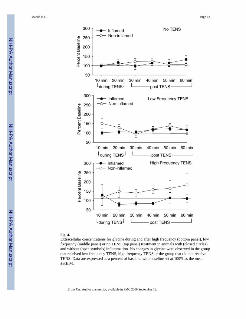
Fig. 4.Extracellular concentrations for glycine during and after high frequency (bottom panel), lowfrequency (middle panel) or no TENS (top panel) treatment in animals with (closed circles)and without (open symbols) inflammation. No changes in glycine were observed in the groupthat received low frequency TENS, high frequency TENS or the group that did not receiveTENS. Data are expressed as a percent of baseline with baseline set at 100% as the mean±S.E.M.
Maeda et al. Page 13
Brain Res. Author manuscript; available in PMC 2009 September 18.
NIH
-PA Author Manuscript
NIH
-PA Author Manuscript
NIH
-PA Author Manuscript
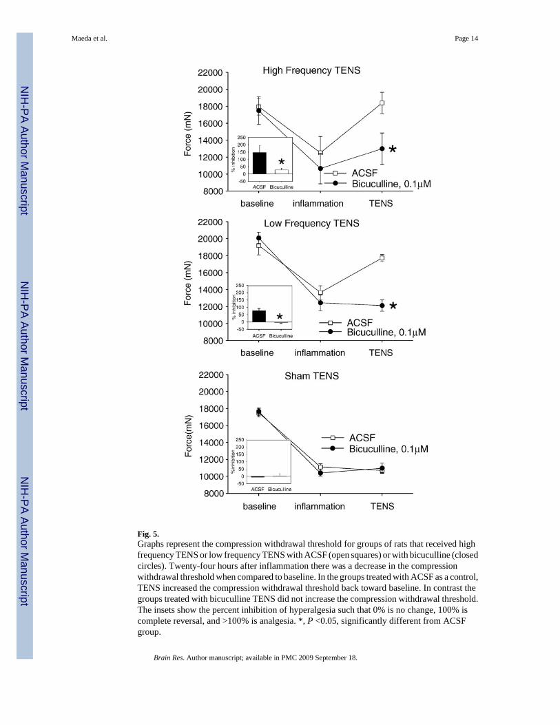
Fig. 5.Graphs represent the compression withdrawal threshold for groups of rats that received highfrequency TENS or low frequency TENS with ACSF (open squares) or with bicuculline (closedcircles). Twenty-four hours after inflammation there was a decrease in the compressionwithdrawal threshold when compared to baseline. In the groups treated with ACSF as a control,TENS increased the compression withdrawal threshold back toward baseline. In contrast thegroups treated with bicuculline TENS did not increase the compression withdrawal threshold.The insets show the percent inhibition of hyperalgesia such that 0% is no change, 100% iscomplete reversal, and >100% is analgesia. *, P <0.05, significantly different from ACSFgroup.
Maeda et al. Page 14
Brain Res. Author manuscript; available in PMC 2009 September 18.
NIH
-PA Author Manuscript
NIH
-PA Author Manuscript
NIH
-PA Author Manuscript