gaba effects during neuronal differentiation of stem cells
TRANSCRIPT

REVIEW ARTICLE
GABA Effects During Neuronal Differentiation of Stem Cells
Patricia Salazar Æ Marco A. Velasco-Velazquez ÆIvan Velasco
Accepted: 21 February 2008 / Published online: 21 March 2008
� Springer Science+Business Media, LLC 2008
Abstract Gamma-amino butyrate (GABA) is the most
prevalent inhibitory neurotransmitter in the adult brain. In this
review, we summarize the pharmacology and regulation of
GABAergic transmission components (biosynthetic enzymes,
receptors and transporters) in adult non-neurogenic brain
regions. The effects of targeted mutations in genes relevant for
GABAergic functions and how they influence specific neu-
ronal circuits and pathological states are presented. We then
review GABA actions on neuronal differentiation. During
brain development, GABA has depolarizing activity in cere-
brocortical neural precursors, controlling cell division and
contributing to neuronal migration and maturation. In the
adult forebrain there are two neurogenic regions exposed to
synaptic and non-synaptic GABA release. Neural stem cells
and neuronal progenitors express GABA receptors in sub-
ventricular and subgranular zones. GABA effects in these
cells are very similar to those found in embryonic cortical
precursor cells, and therefore it is possible that this amino acid
has important roles during adult brain plasticity.
Keywords GAD � GABA receptors � GAT � GABAergic
depolarization � Neuronal migration � Adult neurogenesis
GABA Synthesis
GABA is synthesized preferentially in central nervous
system (CNS) from glutamate by glutamate decarboxylase
(GAD) enzymes. Early kinetic and inhibition studies
showed two GAD isoforms with different affinity for its
coenzyme, pyridoxal-50-phosphate, GAD65 and GAD67 [1–
4]. These proteins are encoded by different, independently
regulated genes. GAD67, located on cell bodies and den-
drites, synthesizes a cytoplasmic pool of GABA, and
GAD65 synthesizes synaptic GABA in nerve endings [5].
Once produced, GABA is stored into secretory vesicles by
an active transporter called vesicular GABA transporter
(VGAT), that has been cloned and characterized [6].
Vesicular GABA will be released by exocytosis.
Glutamate decarboxylase knockout mice were generated
a decade ago. Since then, it is known that GAD67-deficient
mice present cleft palate and die after birth [7], and that
GAD65 knockout mice show susceptibility to seizures,
indicating a less severe weakness of GABAergic transmis-
sion [8, 9]. Currently, GAD-mediated GABA synthesis has
been implicated in the regulation of activity in specific
GABAergic neurons, and seems to cooperate in the estab-
lishment of proper neuronal networks in definite regions of
the Nervous System. GABA synthesized by GADs is
required for development of the neural network that controls
the respiratory system in rodents. Mouse fetuses lacking
GAD67 die from respiratory failure, even when their CNS is
not affected macroscopically [10]. Given that in these mice a
small amount of GABA is still synthesized by GAD65, Fujii
et al. [11] studied the bursting activity of respiratory motor
nerves and the activity of single respiratory neurons in
GAD67/ GAD65 double knockout mice. These mice die after
birth similarly to GAD67 knockouts, but their forebrain
is totally depleted of GABA [11, 12]. In these
Special issue article in honor of Dr. Ricardo Tapia.
P. Salazar � I. Velasco (&)
Departamento de Neurociencias, Instituto de Fisiologıa Celular,
Universidad Nacional Autonoma de Mexico, AP 70-253,
Mexico, DF 04510, Mexico
e-mail: [email protected]
M. A. Velasco-Velazquez
Departamento de Farmacologıa, Facultad de Medicina,
Universidad Nacional Autonoma de Mexico, Mexico,
DF 04510, Mexico
123
Neurochem Res (2008) 33:1546–1557
DOI 10.1007/s11064-008-9642-8
GAD67-/-:GAD65
-/- mice, respiratory discharges recorded
from ventral roots are decreased in embryonic day (E)15
fetuses, and totally absent in E18 fetuses [11]. Accordingly,
neurons from the rostroventrolateral medulla show no
respiratory discharges. Application of substance P, which
produces hyperpolarization, induces respiratory discharges
in those neurons. In contrast, substance P has no effect in
neurons from VGAT-deficient mice, suggesting that also
glycine (affected because it is also introduced to synaptic
vesicles by VGAT) is essential for the function of respiratory
neurons [11]. Another cerebral region in which GABAergic
transmission is important is the visual cortex. Maturation of
perisomatic synapses in the adolescent visuocortex of
GAD67 knockdown mice is significantly impaired. Basket
interneurons from GAD67+/- and GAD67
-/- but not GAD65-/-
mice show reduced axonal branching and synapse formation
in organotypic cultures [13]. These abnormalities can be
partially rescued by inhibiting the GABA transport system
with 1,2,5,6-tetrahydro-1-[2-[[(diphenylmethylene)amino]
oxy]ethyl]-3-pyridinecarboxylic acid (NO-711), or by the
administration of modulators of the GABAA (diazepam), or
GABAB (baclofen) receptors [13]. In contrast, perisomatic
innervation in visual cortex is normal in GAD65-knockdown
mice [13]. These results show that GAD67-mediated GABA
synthesis is specifically involved in GABAergic axonal and
synaptic morphogenesis.
Some of the genetic modifications to alter GAD expres-
sion make organisms unviable, and even before development
of knockout technology, other approaches have been tested
to chemically inhibit GAD activity in the postnatal period.
The consistent result of these experiments is that low GAD
activity results in epileptic convulsions [14–17].
GABA Receptors
Once GABA is formed, it is released, synaptically or non-
synaptically, to act on specific receptors. There are many
different compounds that modify responses of the GABAergic
system by acting on three types of widely described GABA
receptors: the ionotropic GABAA and GABAC receptors [18,
19], and the metabotropic GABAB receptor [20].
GABAA Receptors
GABAA receptors are ligand-gated ion channels permeable
to chloride, distributed throughout the mammalian CNS.
These receptors have shown high diversity by the different
combination of subunits: a1-6, b1-4, c1-4, d, e, p, and h,
that confer distinct characteristics, including differences in
channel kinetics, affinity for GABA, and rate of desensi-
tization, that will ultimately affect GABA function as
anxiolitic, anesthetic or anticonvulsant [19, 21].
The first GABA receptor described, GABAA, was iden-
tified by its sensitivity to inhibition by the specific
antagonists bicuculline and picrotoxin [22]. Even when
different agonists (i.e. muscimol) of GABAA receptor have
been discovered, modulators are the most important GABAA
receptor-interacting drugs. GABAA receptors have different
high-affinity drug-binding sites that confer depressant and
antiepileptic properties [23–25]. Benzodiazepines, neuros-
teroids and barbiturates bind to different sites of GABAA
receptors with the same consequence: an increase in the
period of pore opening and an enhancement in Cl- current
through the receptors [21]. The pharmacology of these
receptors depends on subunit composition [19].
Benzodiazepines are positive allosteric modulators of
GABAA receptors. Diazepam potentiates GABA inhibitory
actions, and therefore has CNS depressant properties [23,
24]. Benzodiazepines have been used for treatment of
epilepsy and chronic anxiety. Binding studies in transfected
cells revealed that benzodiazepine pharmacology was
dependent on subunit composition, particularly of the asubunit [26]. Compounds that show preferential affinity for
a1 subunit, such as zolpidem, have mainly hypnotic
activity. In contrast, a2 containing receptors, but not those
that contain a3, mediate the anxiolytic activity and myo-
relaxant effects of diazepam [18, 27, 28]. Another subunit
involved in the potentiating effect of benzodiazepines is the
c subunit, while b2 subunit is responsible for the receptor
desensitization [19].
Neurosteroids, such as allopregnanolone, enhance Cl-
passing through GABAA receptors, and protect against
different models of epilepsy like those induced by picro-
toxin or 4-aminopyridine in rat hippocampal slices [29].
Chronic administration of allopregnanolone increases
expression of the a4 and d subunits of the GABAA
receptors in several areas of the CNS [30]. Barbiturates
actions depend largely on the b subunit, although their
GABA-potentiating activity is influenced by a subunit
type. Pentobarbital is more effective than GABA only
when receptors contain an a6 subunit [31].
Subunit-specific GABAA receptor knockouts have been
generated. These studies have provided evidence that some
subunits are more important for a given nervous system
disorder or pharmacological response. Genetic deletion of
the a1-subunit produces a variable phenotype that can
include strong body tremor, spontaneous seizures, lower
body weight [32, 33], and increased susceptibility to the
locomotor stimulants effects of ethanol [34, 35]. A pro-
nounced up-regulation of other GABAA receptor subunits,
without functional compensation, ocurrs in regions where
a1-GABA receptors are absent [36]. In vitro studies have
shown that inhibitory postsynaptic currents (IPSC) are
especially altered in visual cortex [37] and hippocampus
[36]. Moreover, cerebral Purkinje cells show a complete
Neurochem Res (2008) 33:1546–1557 1547
123
loss of GABAA mediated transmission [38]. The fact that
neurons lacking a1 subunit have different functional
properties suggest that the plasticity of GABAergic circuits
probably includes formation of novel synaptic connections
in order to ensure stable function of neuronal networks [36,
39]. b3 subunit of the GABAA receptor is important for
normal CNS function, including the response to anesthetics
[40]. This subunit also participates in the pathogenesis of
neurodevelopmental disorders such as Angelman Syn-
drome [41–43] and autism spectrum disorder [44–46].
GABAA receptor b3-subunit knockout mice show cleft
palate abnormalities [47] and high neonatal mortality [48].
In addition, these mice present compensatory adaptations
[49, 50] and numerous behavioral abnormalities. Recently,
a system that allows conditional inactivation of the b3 gene
in a tissue and/or developmentally specific manner was
engineered [51]. When this subunit is deleted only from
CNS, palate development occurs normally, but survival
ratio is low. In contrast, mice with a selective forebrain
knockout of GABAA b3 survived the neonatal period, but
show reduced reproductive fitness, decreased sensitivity to
the anesthetic etomidate, were hyperactive, and some
became obese [51]. These results confirm that the b3
subunit of the GABAA receptor is involved in develop-
mental processes, normal physiology of behavior and the
onset of pharmacological responses, and suggest that it
may be involved in body weight control. Knockout of the dsubunit produces altered behavioral responses to alcohol
[52] or neurosteroids [53].
GABAA receptors have also shown to be located
extrasynaptically. These receptors could modulate GABA
release to control neuronal excitability in cerebellum and
hippocampus, where GABAA receptors away from the
GABA release sites could interfere with the propagation of
the action potentials by inducing tonic inhibition [54, 55].
Such extrasynaptic GABA receptors have also been found
in retina, where drugs and neurohormones that act on
GABAA receptors could affect the firing patterns critical
for the establishment of adult neural circuits [56].
As we will discuss later, activation of GABAA
receptors in embryonic or adult neural precursors causes
depolarization. This phenomenon, possibly related to a
locally reversed Cl- gradient, can also occur in some
non-neurogenic adult regions. However, in the majority
of adult CNS, the principal action for GABA is to
produce inhibition.
GABAB Receptors
The metabotropic GABAB receptor is coupled to G pro-
teins and its activation causes a presynaptic inhibition by
(i) suppressing the activation of N- and P/Q-type voltage-
gated Ca2+ channels that allow Ca2+ entrance important to
trigger neurotransmitter release, and (ii) by increasing the
opening of voltage-gated K+ channels [57]. GABAB
receptor is not blocked by bicuculline and is activated by
the specific agonist baclofen [58]. Even when other specific
agonists have been discovered, baclofen is still the most
studied GABAB receptor activator. Baclofen induces a
centrally mediated muscle relaxant effect and thus is
effective treating spasticity. This GABAB agonist presents
antinociceptive effects, reduces the craving for cocaine,
show anti-bronchoconstriction and antitussive activities
[59], as well as anxiolytic activity in some clinical assays
[60, 61]. Two allosteric positive modulators of GABAB
receptors have been reported: CGP7930 and GS39783 [62,
63]. Both compounds accentuate the effects of GABA and
baclofen, even when they do not have a direct agonist
activity.
GABAB receptors consist of two subunits: GABAB1
and GABAB2. Deletion of either of the subunits results
in a complete loss of GABAB functions and induces a
highly anxious phenotype [64, 65]. For GABAB1 subunit,
one gene generates two isoforms: GABAB1a is expressed
as a presynaptic heteroreceptor in the hippocampus and
lateral amigdala, and GABAB1b is predominantly located
postsynaptically in these structures [66]. Mice with
genetic mutation of GABAB1a show impaired object
recognition [66, 67] and fail to acquire a conditioned
taste aversion [68], indicating that this isoform is
essential for object recognition and discrimination. In
contrast, GABAB1b-/- mice acquired conditional taste
aversion normally, but failed to extinguish the learned
avoidance up to 30 days later [68], suggesting that this
isoform participates in the retrieval or long-term storage
of memory. Both isoform-deficient mice show similar
innate anxiety and impairments in a test indicative of
spatial working memory [67]. Together, these studies
indicate that both isoforms of the GABAB1 subunit
contribute to cognitive capability, but each conveys
specific components of cognitive processes.
GABAC Receptors
GABAC ionotropic receptors are formed by q1-3 subunits
[69]. This receptor does not respond to bicuculline or
baclofen [70] and can be specifically inhibited by TPMPA
[(1,2,5,6-tetrahydropyridine-4-yl) methylphosphinic acid]
[71] and activated by CACA (cis-4-amino-crotonic acid) or
CAMP (cis-2-aminomethyl-cyclopropane carboxilic acid)
[72]. Being both ionotropic receptors, GABAC receptor
shares with GABAA receptor: (i) the susceptibility to pic-
rotoxin, and (ii) the capability of being activated by TACA
(trans-4-aminocrotonic acid) or muscimol [73].
The physiological roles of GABAC q1 subunit are
poorly understood compared to GABAA, given that this
1548 Neurochem Res (2008) 33:1546–1557
123
subunit is relatively new discovered. GABAC receptor q1
subunit is highly expressed in the retina [74]. Mice lacking
q1 subunit present retinal alterations with deficient visual
information processing [75]. Recently, q1 subunit was
found to be highly expressed in mitral cells of the olfactory
bulb. When this GABAC subunit is eliminated by genetic
deletion, olfactory sensitivity is enhanced, which may be
caused by an increased sensitivity and over-excitation of
the primary olfactory neurons, secondary to an attenuated
inhibition in the mitral cell layer of the olfactory bulb [76].
Thus, GABAC q1 subunit has also a specific role in the
physiological function of the olfactory sensory system.
GABA Uptake
After GABA has acted on its neuronal receptors, it is
removed from extracellular space by specific transporters
expressed on neuronal and glial cells. Inside glial cells,
GABA is catabolized by GABA transaminase to succinic
semialdehyde, with concomitant transamination of a-keto-
glutarate to form glutamate. Then, glutamate is transformed
to glutamine, via glutamine synthetase, to leave the cell and
reach a GABAergic neuron, where a glutaminase enzyme
transforms glutamine to glutamate, the GABA precursor
[5].
GABA uptake is carried out by high-affinity sodium/
chloride-dependent transporters. Four GABA transporters
(GATs) have been identified. The classical GABA uptake
inhibitors nipecotic acid, guvacine, and THPO (4,5,6,7-
tetrahydroisoxazolo [4,5-c]pyridin-3-ol) were essential for
the structural elucidation and classification of GATs [77].
Guvacine and nipecotic acid show high affinity for rat
(r)GAT-1 and rGAT-2 [mouse (m)GAT3 homologue], and
low affinity for rGAT-3 (mGAT4 homologue) or human
BGT-1 (mGAT2 homologue) [78]. Tiagabine and NO-711
are lipophilic derivatives of nipecotic acid. Interestingly,
these compounds are typically at least four orders of
magnitude more potent at GAT-1 than at any of the other
GATs [79]. Classical GABA uptake inhibitors show a low
permeability at the blood–brain barrier; therefore, these
compounds are inefficient in systemic therapy [77]. On the
other hand, their lipophilic derivatives have been subjected
to extensive animal behavioral studies primarily as anti-
convulsant agents. Tiagabine as well as other derivatives of
nipecotic acid are used clinically as a therapeutic agents for
the treatment of epilepsy [80] as well as for anxiety dis-
orders [81].
The major plasma membrane transporter responsible for
GABA uptake in adult CNS is GAT-1, which is abundant
in axonal terminals and present in some astroglia processes
[82–84]. In GAT-1-/- mice, extracellular levels of GABA
are increased, resulting in a largely enhanced GABAA
receptor-mediated tonic conductance in several brain
regions [85, 86]. These animals displayed altered behav-
ioral response to ethanol [87], tremor, ataxia and
nervousness [86], reduced aggression and lower level of
depression and anxiety-like behaviors, suggesting that
GAT1 is involved in the pathophysiology of depression
[88].
GAT-2 is located in ependymal cells, meninges and
choroid plexus, with rare expression in neurons or astro-
cytes, and therefore is thought that it participates in the
regulation of GABA concentration in cerebrospinal fluid.
GAT-3, on the other hand, is present in neurons and glia in
adult CNS, but at lower levels than GAT-1 [89]. However,
during development, GAT-3 is the most prevalent GAT in
the cerebral cortex: it appears in neuropil, around blood
vessels, and in numerous cells in late embryonic life
[90, 91].
The uptake of GABA is a very efficient process. For
example, in cortical cultures containing neurons and glial
cells, it has been shown that addition of 100 lM GABA is
decreased to 20 lM after 6 h, and only the addition of
100 lM GABA together with uptake inhibitors caused an
increase in GABA concentration around 50 lM, which was
sufficient to inhibit glutamate neurotoxicity [92].
Although a physiologic role for GABA outside the
nervous system is unclear, this amino acid has been found
in peripheral tissues, including liver, kidney, pancreas,
testis, oviduct, adrenal, sympathetic ganglia, gastrointesti-
nal tract and circulating erythrocytes [93]. It is reported
that insulinoma pancreatic cells possess GAD activity,
synthesize GABA and present low GABA uptake [94].
However, different from that reported for brain, pancreatic
GAD activity was not enhanced by addition of pyridoxal-
50-phosphate. Immunofluorescence and electron micros-
copy studies in pancreatic islets of Langerhans, show that
a- and b-cells contain GABA in their microvesicles and
secretory granules, and these cells also contain VGAT and
a plasma membrane transporter (GAT-3) for GABA uptake
[95]. These findings suggest that pancreatic cells possess
the required components to synthesize and release GABA,
which could participate in paracrine signaling.
After reviewing some aspects of GABAergic transmis-
sion in adult CNS, the next sections are devoted to
descriptions of GABA actions during cerebral cortex
development, and the effects of GABA on adult neural stem
cells from the subventricular zone (SVZ) and subgranular
zone (SGZ) of the hippocampus. Neural stem cells are
undifferentiated cells that self-renew and can differentiate
to neurons and glial (astrocytes and oligodendrocytes) cells
[96, 97]. Only neuronal differentiation will be considered
here. There is compelling evidence that GABA regulates
different aspects of neurogenesis in these three cerebral
regions.
Neurochem Res (2008) 33:1546–1557 1549
123
Neurogenesis in the Developing Cerebral Cortex
During early CNS development, the region close to the
ventricle is a neurogenic epithelium constituted by asyn-
chronously dividing cells presenting interkinetic nuclear
movement. Cerebrum cortices are thus formed from multi-
potent neural stem cells that proliferate initially in the
ventricular zone (VZ), and later on the SVZ. These NSC
produce neuroblasts that migrate away from VZ to pass the
intermediate zone (IZ) and stopping just before the marginal
zone (MZ). Neuroblasts reaching the cortical plate (CP)
produce neurons that accommodate themselves in one of the
six layers present in the mature cerebral cortex. One of the
defining features of cortical development is the presence of
glial fibrillary acidic protein (GFAP)-positive cells called
radial glia (RG), that span the thickness of this structure from
the VZ to the CP, and serve as scaffolds for radial migration
of neurons to the CP [98]. Although there is also tangential
migration, especially relevant for GABAergic interneurons
generation, radial migration has been studied extensively
[99]. Early-born neurons can be found in deeper layers and
late-born cells are closer to the pial surface. During this well-
described developmental process, dividing progenitors can
be found in VZ and SVZ. This latter region prevails into
adulthood, producing neural precursors that generate neu-
rons that migrate to the olfactory bulb, which will be
described later. No cell divisions can be observed in the CP
until the gliogenic phase of cortical development, which is
delayed compared with neurogenesis. Glial production will
not be discussed here. Recently, a portion of RG cells have
been identified as the neural stem cells in cerebrocortical
region [100, 101]. Once asymmetrically generated, neurons
migrate to the CP (Fig. 1a). This migration through RG
shafts has been associated with a leading process directed to
the pial end. Migration is dependent on a high number of
membrane proteins, quimioactive molecules and transcrip-
tion factors. One converging mechanism for regulating
radial neuronal migration is the synaptic-independent regu-
lation of intracellular calcium. Cells stop migrating when
they pass previously generated neurons and reach the CP/MZ
interphase, as they adopt their final position in the CP. Cor-
tical neurons are not generated exclusively in the cortex, but
are also born in the ganglionic eminence of the ventral tel-
encephalon and follow migratory routes through the IZ to
reach the cortex and contribute with GABAergic interneu-
rons [102, 103].
The appearance of GABA in cerebral cortex is very
early in development, much before that synaptic contacts
are apparent. At E10 in mice, GABA immunoreactivity
appears with strong staining in the neuroepithelial pial
surface, although diffuse labeling can be found in the entire
neural tissue. At E12 and E14, GABA labeling is still
stronger in the outer layers above the germinal area. By
E16, essentially there is no GABA mark in the proliferative
zone but the labeling remains in the CP and IZ [104].
Knock-in mice that express Green Fluorescent Protein
(GFP) under GAD67 promoter have shown that this enzyme
is present from E15 in IZ and MZ of cerebral cortices
[105]. It is very unlikely that synaptic release is occurring
at these stages, since no structural evidence of synapses are
in place at VZ nor IZ. GATs are also present in early stages
of forebrain development. At E18, GAT-1 and GAT-3 are
expressed in brain cortex, and after birth extended to the
entire cortex [106]. Ambient GABA in developing cerebral
cortex could be released by growth cones [107] or by
GATs from GABAergic neurons [108]. In addition to the
biosynthetic machinery for GABA, during brain develop-
ment, GABAA receptor subunits are expressed and show
different regional and temporal expression. The subunits
a2, a3, and a5 are expressed during the embryonic stages,
while the others are expressed after birth [109, 110].
The inhibitory hyperpolarizing action of GABA in mature
CNS is not present in developing cerebral cortex. In fact,
activation of GABAA receptors in proliferating neuroblast of
the VZ causes a depolarization that lasts longer and required
lower GABA concentrations than GABA responses in
mature neurons. The first evidence that GABA regulates
cortical neurogenesis came from explants where it was
demonstrated that GABA depolarizes neural cells at the VZ,
causing decreased DNA synthesis. This cell cycle arrest was
correlated with depolarization and calcium entry, since high
potassium and glutamate had the same effects. GABA
depolarization was not present with furosemide, indicating
that chloride currents were responsible of this excitatory
action. Endogenous tonic GABA release is present in the
cortical slices, since adding bicuculline caused a shift in
current [111]. A more detailed analysis of GABA effects on
proliferation evidenced that in fact 30 lM GABA caused a
shortening in cell cycle duration in VZ cells, but a decrease in
proliferation of SVZ cells. When analyzed as a general
proliferating population, the overall effect of GABA or
muscimol was to prevent proliferation, which are essentially
the results reported earlier. Regarding neuronal differentia-
tion, GABA prevents neurogenesis by maintaining
precursors of the VZ proliferating, because GABA-treated
slices had less neuroblasts migrating above the SVZ and
therefore decreased number of postmitotic neurons in the CP
[104]. Cortical precursor cells divide in culture when
exposed to basic fibroblast growth factor (bFGF). This
cytokine caused an increase in the proportion of cells
expressing a1 GABAA receptor subunit. GABA blocked
bFGF-induced proliferation (Fig. 1a) and caused neuronal
differentiation of cortical precursors [112].
Later on, it was shown that neuroblasts contained a
higher content (around 30 mM) of Cl- than mature neu-
rons (close to 10 mM), and since GABAA receptors are
1550 Neurochem Res (2008) 33:1546–1557
123
chloride channels, the net effect in the first case is excita-
tion, and hyperpolarization in the second [113]. This high
Cl- concentration is due to the expression of NKCC1, a
Na+/K+/2Cl- inward transporter that causes intracellular
accumulation of chloride in precursor cells. Mature neu-
rons, in addition to NKCC1, also express KCC2, a K+/Cl-
transporter that extrudes chloride from the cells and shift
GABA action from excitation to inhibition in cortical [114]
and hippocampal cells [115]. Recently, in utero electro-
poration of KCC2 switched GABA action from excitation
to inhibition in VZ cells. Postnatal analysis of KCC2
overexpressor cells showed correct migration and cortical
layering. However, a maturation defect was observed
in vivo, consisting of decreased length and number of
branches in neuronal processes. To correlate GABA-
induced depolarization with these alterations, prenatal
transfection was performed with Kir2.1 potassium channel,
which resulted in cells with lower membrane potential, and
caused similar structural changes in postnatal cerebral
cortex [116]. These findings can be related to experiments
showing that GABA regulates neurite outgrowth in cul-
tured neurons in a chloride-dependent fashion [117].
Interestingly, knocking out Dlx1 caused a reduced number
in a subset of GABAergic interneurons in the cortex, and
the surviving cells had smaller dendrite length and reduced
branching; these mutant animals developed epilepsy [118].
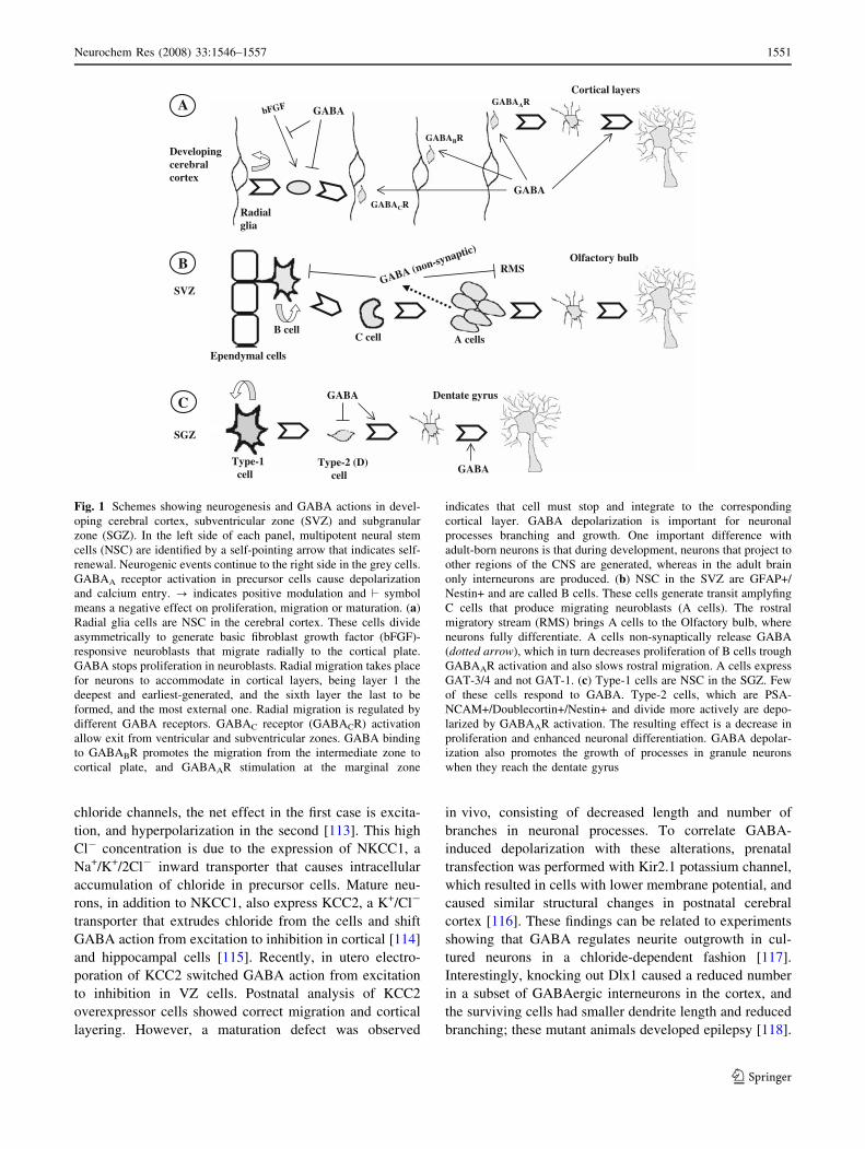
Cortical layers
A GABAARGABA
A cellsB cell
C cell
GABAnaptic)
GABABR
GABACRRadialglia
bFGF
Developingcerebralcortex
GABA
B Olfactory bulb(non-sy RMS
SVZ
Ependymal cells
Type-1cell
CGABA Dentate gyrus
SGZ
Type-2 (D)cell GABA
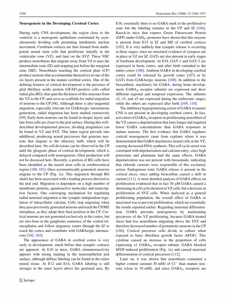
Fig. 1 Schemes showing neurogenesis and GABA actions in devel-
oping cerebral cortex, subventricular zone (SVZ) and subgranular
zone (SGZ). In the left side of each panel, multipotent neural stem
cells (NSC) are identified by a self-pointing arrow that indicates self-
renewal. Neurogenic events continue to the right side in the grey cells.
GABAA receptor activation in precursor cells cause depolarization
and calcium entry. ? indicates positive modulation and ‘ symbol
means a negative effect on proliferation, migration or maturation. (a)
Radial glia cells are NSC in the cerebral cortex. These cells divide
asymmetrically to generate basic fibroblast growth factor (bFGF)-
responsive neuroblasts that migrate radially to the cortical plate.
GABA stops proliferation in neuroblasts. Radial migration takes place
for neurons to accommodate in cortical layers, being layer 1 the
deepest and earliest-generated, and the sixth layer the last to be
formed, and the most external one. Radial migration is regulated by
different GABA receptors. GABAC receptor (GABACR) activation
allow exit from ventricular and subventricular zones. GABA binding
to GABABR promotes the migration from the intermediate zone to
cortical plate, and GABAAR stimulation at the marginal zone
indicates that cell must stop and integrate to the corresponding
cortical layer. GABA depolarization is important for neuronal
processes branching and growth. One important difference with
adult-born neurons is that during development, neurons that project to
other regions of the CNS are generated, whereas in the adult brain
only interneurons are produced. (b) NSC in the SVZ are GFAP+/
Nestin+ and are called B cells. These cells generate transit amplyfing
C cells that produce migrating neuroblasts (A cells). The rostral
migratory stream (RMS) brings A cells to the Olfactory bulb, where
neurons fully differentiate. A cells non-synaptically release GABA
(dotted arrow), which in turn decreases proliferation of B cells trough
GABAAR activation and also slows rostral migration. A cells express
GAT-3/4 and not GAT-1. (c) Type-1 cells are NSC in the SGZ. Few
of these cells respond to GABA. Type-2 cells, which are PSA-
NCAM+/Doublecortin+/Nestin+ and divide more actively are depo-
larized by GABAAR activation. The resulting effect is a decrease in
proliferation and enhanced neuronal differentiation. GABA depolar-
ization also promotes the growth of processes in granule neurons
when they reach the dentate gyrus
Neurochem Res (2008) 33:1546–1557 1551
123
GABA produces chemotaxis (directed migration to
GABA source) at submicromolar concentrations, and che-
mokinesis (random motility) when applied in micromolar
ranges in dissociated E15 cortical cells [119]. GABA has
also been shown to affect neuroblast radial migration: E18
cerebrocortical slices were incubated with the mitotic
marker BrdU and analyzed 48 h later. Controls show
BrdU-positive cells that migrated to CP. Addition of
saclofen (GABAB antagonist) caused accumulation in IZ,
whereas picrotoxin (GABAA+C receptor blocker) caused
labeled cells to remain in VZ/SVZ. On the other hand,
bicuculline did not block, but enhanced migration to the CP
and caused increased thickness. A model was proposed in
which transit from VZ/SVZ was dependent on GABAC
receptors, the pass from IZ to CP was promoted by GABAB
activation and the stop signal at the MZ required GABAA
receptors [120]. In vivo application of GABAergic drugs
also alters migration. Implanting slabs that acutely release
muscimol or bicuculline in the surface of cerebral cortex of
neonatal rats caused heterotopia (an increased number of
neurons in the surface of the cortex), just underneath the
application points, after 7 or 14 days. In brain slices,
bicuculline, and to a lesser extent muscimol, caused an
increase in migration of cells to the cortical surface. No
mechanism is provided to explain the fact that GABAA
agonist and antagonist had the same effect [121].
Thus, GABA has depolarizing effects on cerebrocortical
cells, causes decreased proliferation of neuroblasts, par-
ticipate in neuronal migration and promotes neurite
extension during development. Collectively, the evidence
points to very important roles of GABA during neuronal
circuit formation in rodents.
Neurogenesis in Adult Brain
Subventricular Zone
The region in the adult brain that contains the largest
germinal zone for neurons is the SVZ. This area contains
multipotent neural stem cells [122–125] that are thought to
originate from the embryonic VZ [126], and produce
neuroblasts that migrate to their final differentiation site,
the olfactory bulb, through the rostral migratory stream
(RMS, Fig. 1b). This migration is independent of RG or
other glial elements [127]. Structural and functional anal-
yses have provided a detailed architecture of the SVZ (see
Fig. 1b). Neural stem cells (called B cells) express GFAP
and are in close apposition with ependymal cells of the
ventricle. These cells divide and generate transit amplify-
ing (C) cells that differentiate into migratory neuroblast
(A cells) that generates GABAergic and dopaminergic
interneurons in the olfactory bulb. Migrating neuroblast,
similar to what it is observed during CNS development,
express doublecourtin [128, 129].
Similar to the effects reported in cortical precursors dur-
ing development, GABA elicited a depolarizing (+14 to
+19 mV) chloride current resulting from GABAA receptor
activation in precursor cells from postnatal and adult SVZ
[130, 131]. Precursors in culture express GAD67 and GAD65,
and are immunopositive for GABA. Their subunit combi-
nation of GABAA receptor include a2-5, b1-3 and c1-3, since
these subunits were detected by RT-PCR [131, 132]. A
proportion of PSA-NCAM+ precursor cells, cultured as
neurospheres, responded with an intracellular calcium rise
upon GABA application, which caused decreased prolifer-
ation via mitogen-activated protein kinases MEK 1 and 2.
Release of endogenous GABA to the culture medium is
likely, since GABAA antagonists prevented these responses,
and the positive GABAergic modulators clonazepam and
pentobarbital decreased proliferation. Interestingly, EGF
application lowered GABA concentration in the cultures,
suggesting a feedback loop where EGF directly stimulates
proliferation and indirectly decreased GABA extrusion to
prevent GABA anti-proliferative effects [132].
In saggital cerebral slices that encompass SVZ and RMS,
application of 10 lM GABA or pentobarbital decreased A
cells’ rostral migratory rate, which was in fact activated by
bicuculline, pointing to endogenous GABA modulation of
migration in the RMS. This was further supported by the fact
that elevating GABA levels, by causing depolarization-
induced release, or by inhibition of GATs, decreased
migration speed [133]. These ex-vivo studies were extended
by the same group using coronal slices of transgenic animals
that express GFP in GFAP-positive B cells. When 100 lM
GABA was applied, all GFP+ cells presented a GABAA
receptor-mediated inward current close to chloride equilib-
rium potential that resulted in cell depolarization. Addition
of exogenous GABA was potentiated by an uptake blocker
selective for GAT 3/4, and unaffected by NO-711 (GAT1-
specific). Slices were electrically stimulated and a GABAA
response was observed in GFP-expressing cells. GABA
release by neuroblasts (A cells) was taking effect, although
amino acid extrusion was not synaptic in nature, since it was
tetrodotoxin-, vesicular release- and extracellular calcium-
independent. This group nicely showed that GABA release
had a non-proliferating effect on SVZ B cells (NSC) and
conversely, bicuculline augmented NSC division [134]. In
this scheme, GABA secreted by A cells would act as a par-
acrine factor for B cells regulating cell number, and also as an
autocrine molecule to influence migration in the RMS.
Subgranular Zone in the Hippocampus
Hippocampal multipotent neural stem cells reside in the
SGZ of the dentate gyrus, between the hilus and the
1552 Neurochem Res (2008) 33:1546–1557
123
granule cell layer, and express GFAP [135] and Nestin.
These type-1 cells divide slowly and generate highly
dividing progenitors (type-2 cells, D cells) that migrate a
short distance to integrate as neurons into the granule cell
layer of the dentate gyrus (Fig. 1c). Migrating neuroblasts
express doublecourtin and PSA-NCAM, and terminally
differentiated neurons are positive for NeuN and Calbindin
[136]. There is also evidence for hippocampal neurogenesis
in humans [137]. Since hippocampal formation has been
related to several memory and learning functions, the
possibility that adult hippocampal neurogenesis could play
a physiological role in such tasks has been suggested [138,
139]. Significant numbers of adult-born neurons are pro-
duced in the hippocampus [140], although a proportion
undergoes cell death. There are several factors that modify
the amount of neurons produced in this structure such as
hormones, enriched environment, exercise, stroke and
small modulatory RNAs [141–145].
Using mice that express GFP under Nestin regulatory
elements, two groups described fluorescent type-1 neural
stem cells that have one thick process and enter cell cycle
sporadically, whereas GFP+ type-2 cells are PSA-
NCAM+, have a more round morphology and actively
divide. Type-2 cells showed spontaneous and evoked
depolarizing currents sensitive to bicuculline [146, 147].
Intracellular chloride concentration in these cells was 30
vs. 5 mM in mature granule cells. This was correlated with
NKCC1 expression in type-2 cells, and not in granule
neurons. GABA application caused increased intracellular
calcium, and this was followed by expression of the neu-
rogenic transcription factor Neuro D [146]. Other works
have established that early-born neurons do not respond to
zolpidem, a GABAA receptor a1 subunit potentiator, while
mature neurons show increased GABA hyperpolarizing
responses in its presence [148, 149]; a1 is expressed by
full-grown neurons but absent in newborn neurons [149].
Recently, a double retroviral labeling technique, with
two different fluorescent proteins injected at E15 and in
adulthood, allowed to study the functional integration of
embryonic-born hippocampal neurons and adult-differen-
tiated granule cells in the same slice. After analyzing
evoked responses in both neuronal populations, they con-
clude that recorded synaptic responses are very similar, and
just subtle differences are present [150]. In these studies, it
was established that newborn neurons receive GABA
afferents first and then glutamatergic synaptic contacts,
similar to what is observed in developing cerebral cortex.
Using GFP retrovirus, it has been shown that there is
tonic GABAA receptor-mediated depolarization in new-
born neurons. The depolarizing action of GABA is due to a
high chloride content in immature cells that express
NKCC1, and as cells mature they start expressing KCC2,
reducing chloride concentration and responding to GABA
by hyperpolarization. Injecting retrovirus that express short
hairpin RNA targeting Nkcc1, the authors knock down
expression of NKCC1 and reversed the depolarizing action
of GABA. This was accompanied by a delayed and reduced
integration of the newborn neurons to GABA and gluta-
mate synaptic contacts. Remarkably similar to what was
observed in developing cerebral cortex, manipulation of
cells to lower intracellular chloride concentration, and
therefore inhibition of GABA-induced depolarization,
caused a marked decrease in the length and arborization of
dendrites [151].
In vivo application of GABAA receptor-interacting
drugs had no effect on type-1 cells, but increased prolif-
eration (antagonists) or arrested (agonists) type-2 cells.
Positive modulators of GABAA receptors caused a long-
term increase in the number BrdU+ Calbindin+ neurons 28
days after treatment [146]. These results are hard to
interpret because both mature hippocampal circuitry and
progenitor cells would be exposed to these pharmacologi-
cal agents.
In conclusion, the actions of GABA in developing CNS
and germinal niches in adult brains are different from
GABAergic inhibitory hyperpolarization in non-neuro-
genic regions of adult CNS. In neural precursors, GABA
causes depolarization and affects proliferation, migration,
and neuronal maturation. Therefore, GABAergic players
are important during developmental and adult neurogene-
sis. The results summarized here show that genetic or
pharmacological modifications of the GABAergic system
cause alterations in neuronal circuit formation, emphasiz-
ing the role of GABA in CNS plasticity.
Acknowledgements Our laboratories are supported by grants from
PAPIIT of Universidad Nacional Autonoma de Mexico, Conacyt
(M.A.V-V. and I.V.), National Institute for Neurological Disorders
and Stroke, Fundacion Aleman and TWAS (I.V.).
References
1. Bayon A, Possani LD, Tapia M et al (1977) Kinetics of brain
glutamate decarboxylase. Interactions with glutamate, pyridoxal
50-phosphate and glutamate-pyridoxal 50-phosphate Schiff base.
J Neurochem 29:519–525
2. Covarrubias M, Tapia R (1980) Brain glutamate decarboxylase:
properties of its calcium-dependent binding to liposomes and
kinetics of the bound and the free enzyme. J Neurochem
34:1682–1688
3. Erlander MG, Tobin AJ (1991) The structural and functional
heterogeneity of glutamic acid decarboxylase: a review. Neu-
rochem Res 16:215–226
4. Kaufman DL, Houser CR, Tobin AJ (1991) Two forms of the
gamma-aminobutyric acid synthetic enzyme glutamate decar-
boxylase have distinct intraneuronal distributions and cofactor
interactions. J Neurochem 56:720–723
5. Soghomonian JJ, Martin DL (1998) Two isoforms of glutamate
decarboxylase: why? Trends Pharmacol Sci 19:500–505
Neurochem Res (2008) 33:1546–1557 1553
123
6. McIntire SL, Reimer RJ, Schuske K et al (1997) Identification
and characterization of the vesicular GABA transporter. Nature
389:870–876
7. Asada H, Kawamura Y, Maruyama K et al (1997) Cleft palate
and decreased brain gamma-aminobutyric acid in mice lacking
the 67-kDa isoform of glutamic acid decarboxylase. Proc Natl
Acad Sci USA 94:6496–6499
8. Kash SF, Johnson RS, Tecott LH et al (1997) Epilepsy in mice
deficient in the 65-kDa isoform of glutamic acid decarboxylase.
Proc Natl Acad Sci USA 94:14060–14065
9. Asada H, Kawamura Y, Maruyama K et al (1996) Mice lacking
the 65 kDa isoform of glutamic acid decarboxylase (GAD65)
maintain normal levels of GAD67 and GABA in their brains but
are susceptible to seizures. Biochem Biophys Res Commun
229:891–895
10. Kuwana S, Okada Y, Sugawara Y et al (2003) Disturbance of
neural respiratory control in neonatal mice lacking GABA
synthesizing enzyme 67-kDa isoform of glutamic acid decar-
boxylase. Neuroscience 120:861–870
11. Fujii M, Arata A, Kanbara-Kume N et al (2007) Respiratory
activity in brainstem of fetal mice lacking glutamate decar-
boxylase 65/67 and vesicular GABA transporter. Neuroscience
146:1044–1052
12. Ji F, Kanbara N, Obata K (1999) GABA and histogenesis in fetal
and neonatal mouse brain lacking both the isoforms of glutamic
acid decarboxylase. Neurosci Res 33:187–194
13. Chattopadhyaya B, Di Cristo G, Wu CZ et al (2007) GAD67-
mediated GABA synthesis and signaling regulate inhibitory
synaptic innervation in the visual cortex. Neuron 54:889–903
14. Tapia R, Pasantes-Morales H, Taborda E et al (1975) Seizure
susceptibility in the developing mouse and its relationship to
glutamate decarboxylase and pyridoxal phosphate in brain.
J Neurobiol 6:159–170
15. Salazar P, Montiel T, Brailowsky S et al (1994) Decrease of
glutamate decarboxylase activity after in vivo cortical infusion
of gamma-aminobutyric acid. Neurochem Int 24:363–368
16. Massieu L, Rivera A, Tapia R (1994) Convulsions and inhibition
of glutamate decarboxylase by pyridoxal phosphate-gamma-
glutamyl hydrazone in the developing rat. Neurochem Res
19:183–187
17. Arias C, Valero H, Tapia R (1992) Inhibition of brain glutamate
decarboxylase activity is related to febrile seizures in rat pups.
J Neurochem 58:369–373
18. Mohler H (2006) GABA(A) receptor diversity and pharmacol-
ogy. Cell Tissue Res 326:505–516
19. Benarroch EE (2007) GABAA receptor heterogeneity, function,
and implications for epilepsy. Neurology 68:612–614
20. Lujan R (2007) Subcellular regulation of metabotropic GABA
receptors in the developing cerebellum. Cerebellum 6:123–129
21. Macdonald RL, Olsen RW (1994) GABAA receptor channels.
Annu Rev Neurosci 17:569–602
22. Bormann J (1988) Electrophysiology of GABAA and GABAB
receptor subtypes. Trends Neurosci 11:112–116
23. Atack JR (2005) The benzodiazepine binding site of GABA(A)
receptors as a target for the development of novel anxiolytics.
Expert Opin Investig Drugs 14:601–618
24. Banfi S, Cornelli U, Fonio W et al (1982) A screening method
for substances potentially active on learning and memory.
J Pharmacol Method 8:255–263
25. Macdonald RL, Kelly KM (1994) Mechanisms of action of
currently prescribed and newly developed antiepileptic drugs.
Epilepsia 35(Suppl 4):S41–50
26. Pritchett DB, Luddens H, Seeburg PH (1989) Type I and type II
GABAA-benzodiazepine receptors produced in transfected
cells. Science 245:1389–1392
27. Crestani F, Low K, Keist R et al (2001) Molecular targets for
the myorelaxant action of diazepam. Mol Pharmacol 59:
442–445
28. Crestani F, Martin JR, Mohler H et al (2000) Mechanism of
action of the hypnotic zolpidem in vivo. Br J Pharmacol
131:1251–1254
29. Salazar P, Tapia R, Rogawski MA (2003) Effects of neurosteroids
on epileptiform activity induced by picrotoxin and 4-aminopyri-
dine in the rat hippocampal slice. Epilepsy Res 55:71–82
30. Smith SS, Shen H, Gong QH et al (2007) Neurosteroid regula-
tion of GABA(A) receptors: Focus on the alpha4 and delta
subunits. Pharmacol Ther 116:58–76
31. Drafts BC, Fisher JL (2006) Identification of structures within
GABAA receptor alpha subunits that regulate the agonist action
of pentobarbital. J Pharmacol Exp Ther 318:1094–1101
32. Sur C, Wafford KA, Reynolds DS et al (2001) Loss of the major
GABA(A) receptor subtype in the brain is not lethal in mice.
J Neurosci 21:3409–3418
33. Kralic JE, Korpi ER, O’Buckley TK et al (2002) Molecular and
pharmacological characterization of GABA(A) receptor alpha1
subunit knockout mice. J Pharmacol Exp Ther 302:1037–1045
34. Kralic JE, Wheeler M, Renzi K et al (2003) Deletion of
GABAA receptor alpha 1 subunit-containing receptors alters
responses to ethanol and other anesthetics. J Pharmacol Exp
Ther 305:600–607
35. Blednov YA, Walker D, Alva H et al (2003) GABAA receptor
alpha 1 and beta 2 subunit null mutant mice: behavioral
responses to ethanol. J Pharmacol Exp Ther 305:854–863
36. Schneider Gasser EM, Duveau V, Prenosil GA et al (2007)
Reorganization of GABAergic circuits maintains GABAA
receptor-mediated transmission onto CA1 interneurons in
alpha1-subunit-null mice. Eur J Neurosci 25:3287–3304
37. Bosman LW, Rosahl TW, Brussaard AB (2002) Neonatal
development of the rat visual cortex: synaptic function of
GABAA receptor alpha subunits. J Physiol 545:169–181
38. Fritschy JM, Panzanelli P (2006) Molecular and synaptic orga-
nization of GABAA receptors in the cerebellum: effects of
targeted subunit gene deletions. Cerebellum 5:275–285
39. Marder E, Goaillard JM (2006) Variability, compensation and
homeostasis in neuron and network function. Nat Rev Neurosci
7:563–574
40. Boehm SL II, Ponomarev I, Jennings AW et al (2004) gamma-
Aminobutyric acid A receptor subunit mutant mice: new per-
spectives on alcohol actions. Biochem Pharmacol 68:1581–1602
41. DeLorey TM, Handforth A, Anagnostaras SG et al (1998) Mice
lacking the beta3 subunit of the GABAA receptor have the
epilepsy phenotype and many of the behavioral characteristics
of Angelman syndrome. J Neurosci 18:8505–8514
42. Saitoh S, Kubota T, Ohta T et al (1992) Familial Angelman
syndrome caused by imprinted submicroscopic deletion
encompassing GABAA receptor beta 3-subunit gene. Lancet
339:366–367
43. Wagstaff J, Knoll JH, Fleming J et al (1991) Localization of the
gene encoding the GABAA receptor beta 3 subunit to the
Angelman/Prader-Willi region of human chromosome 15. Am J
Hum Genet 49:330–337
44. Shao Y, Cuccaro ML, Hauser ER et al (2003) Fine mapping of
autistic disorder to chromosome 15q11-q13 by use of pheno-
typic subtypes. Am J Hum Genet 72:539–548
45. DeLorey TM (2005) GABRB3 gene deficient mice: a potential
model of autism spectrum disorder. Int Rev Neurobiol 71:
359–382
46. Buxbaum JD, Silverman JM, Smith CJ et al (2002) Association
between a GABRB3 polymorphism and autism. Mol Psychiatry
7:311–316
1554 Neurochem Res (2008) 33:1546–1557
123
47. Culiat CT, Stubbs LJ, Montgomery CS et al (1994) Phenotypic
consequences of deletion of the gamma 3, alpha 5, or beta 3
subunit of the type A gamma-aminobutyric acid receptor in
mice. Proc Natl Acad Sci USA 91:2815–2818
48. Homanics GE, DeLorey TM, Firestone LL et al (1997) Mice
devoid of gamma-aminobutyrate type A receptor beta3 subunit
have epilepsy, cleft palate, and hypersensitive behavior. Proc
Natl Acad Sci USA 94:4143–4148
49. Ramadan E, Fu Z, Losi G et al (2003) GABA(A) receptor beta3
subunit deletion decreases alpha2/3 subunits and IPSC duration.
J Neurophysiol 89:128–134
50. Wong SM, Cheng G, Homanics GE et al (2001) Enflurane
actions on spinal cords from mice that lack the beta3 subunit of
the GABA(A) receptor. Anesthesiology 95:154–164
51. Ferguson C, Hardy SL, Werner DF et al (2007) New insight into
the role of the beta3 subunit of the GABAA-R in development,
behavior, body weight regulation, and anesthesia revealed by
conditional gene knockout. BMC Neurosci 8:85
52. Mihalek RM, Bowers BJ, Wehner JM et al (2001) GABA(A)-
receptor delta subunit knockout mice have multiple defects in
behavioral responses to ethanol. Alcohol Clin Exp Res 25:
1708–1718
53. Zhu WJ, Wang JF, Krueger KE et al (1996) Delta subunit
inhibits neurosteroid modulation of GABAA receptors. J Neu-
rosci 16:6648–6656
54. Glykys J, Mody I (2007) The main source of ambient GABA
responsible for tonic inhibition in the mouse hippocampus.
J Physiol 582:1163–1178
55. Kullmann DM, Ruiz A, Rusakov DM et al (2005) Presynaptic,
extrasynaptic and axonal GABAA receptors in the CNS: where
and why? Prog Biophys Mol Biol 87:33–46
56. Wang CT, Blankenship AG, Anishchenko A et al (2007)
GABA(A) receptor-mediated signaling alters the structure of
spontaneous activity in the developing retina. J Neurosci
27:9130–9140
57. MacDermott AB, Role LW, Siegelbaum SA (1999) Presynaptic
ionotropic receptors and the control of transmitter release. Annu
Rev Neurosci 22:443–485
58. Bormann J (2000) The ‘ABC’ of GABA receptors. Trends
Pharmacol Sci 21:16–19
59. Bowery NG, Bettler B, Froestl W et al (2002) International
Union of Pharmacology. XXXIII. Mammalian gamma-amino-
butyric acid(B) receptors: structure and function. Pharmacol Rev
54:247–264
60. Drake RG, Davis LL, Cates ME et al (2003) Baclofen treatment
for chronic posttraumatic stress disorder. Ann Pharmacother
37:1177–1181
61. Breslow MF, Fankhauser MP, Potter RL et al (1989) Role of
gamma-aminobutyric acid in antipanic drug efficacy. Am J
Psychiatry 146:353–356
62. Urwyler S, Pozza MF, Lingenhoehl K et al (2003) N,N0-di-
cyclopentyl-2-methylsulfanyl-5-nitro-pyrimidine-4,6-diamine
(GS39783) and structurally related compounds: novel allosteric
enhancers of gamma-aminobutyric acidB receptor function.
J Pharmacol Exp Ther 307:322–330
63. Urwyler S, Mosbacher J, Lingenhoehl K et al (2001) Positive
allosteric modulation of native and recombinant gamma-ami-
nobutyric acid(B) receptors by 2,6-Di-tert-butyl-4-(3-hydroxy-
2,2-dimethyl-propyl)-phenol (CGP7930) and its aldehyde analog
CGP13501. Mol Pharmacol 60:963–971
64. Bowery NG (2006) GABAB receptor: a site of therapeutic
benefit. Curr Opin Pharmacol 6:37–43
65. Mombereau C, Kaupmann K, Froestl W et al (2004) Genetic
and pharmacological evidence of a role for GABA(B) receptors
in the modulation of anxiety- and antidepressant-like behavior.
Neuropsychopharmacology 29:1050–1062
66. Vigot R, Barbieri S, Brauner-Osborne H et al (2006) Differen-
tial compartmentalization and distinct functions of GABAB
receptor variants. Neuron 50:589–601
67. Jacobson LH, Kelly PH, Bettler B et al (2007) Specific roles of
GABA(B(1)) receptor isoforms in cognition. Behav Brain Res
181:158–162
68. Jacobson LH, Kelly PH, Bettler B et al (2006) GABA(B(1))
receptor isoforms differentially mediate the acquisition and
extinction of aversive taste memories. J Neurosci 26:8800–8803
69. Zhang D, Pan ZH, Zhang X et al (1995) Cloning of a gamma-
aminobutyric acid type C receptor subunit in rat retina with a
methionine residue critical for picrotoxinin channel block. Proc
Natl Acad Sci USA 92:11756–11760
70. Feigenspan A, Bormann J (1998) GABA-gated Cl- channels in
the rat retina. Prog Retin Eye Res 17:99–126
71. Chebib M, Mewett KN, Johnston GA (1998) GABA(C) receptor
antagonists differentiate between human rho1 and rho2 receptors
expressed in Xenopus oocytes. Eur J Pharmacol 357:227–234
72. Kusama T, Spivak CE, Whiting P et al (1993) Pharmacology of
GABA rho 1 and GABA alpha/beta receptors expressed in
Xenopus oocytes and COS cells. Br J Pharmacol 109:200–206
73. Bormann J, Feigenspan A (1995) GABAC receptors. Trends
Neurosci 18:515–519
74. Cutting GR, Lu L, O’Hara BF et al (1991) Cloning of the
gamma-aminobutyric acid (GABA) rho 1 cDNA: a GABA
receptor subunit highly expressed in the retina. Proc Natl Acad
Sci USA 88:2673–2677
75. McCall MA, Lukasiewicz PD, Gregg RG et al (2002) Elimi-
nation of the rho1 subunit abolishes GABA(C) receptor
expression and alters visual processing in the mouse retina.
J Neurosci 22:4163–4174
76. Chen Y, Zhou D, Zhou K et al (2007) Study on olfactory
function in GABA(C) receptor/channel rho(1) subunit knockout
mice. Neurosci Lett 427:10–15
77. Krogsgaard-Larsen P, Frolund B, Frydenvang K (2000) GABA
uptake inhibitors. Design, molecular pharmacology and thera-
peutic aspects. Curr Pharm Des 6:1193–1209
78. Borden LA (1996) GABA transporter heterogeneity: pharma-
cology and cellular localization. Neurochem Int 29:335–356
79. Dhar TG, Borden LA, Tyagarajan S et al (1994) Design, syn-
thesis and evaluation of substituted triarylnipecotic acid
derivatives as GABA uptake inhibitors: identification of a ligand
with moderate affinity and selectivity for the cloned human
GABA transporter GAT-3. J Med Chem 37:2334–2342
80. LaRoche SM (2007) A new look at the second-generation
antiepileptic drugs: a decade of experience. Neurologist 13:
133–139
81. Crane D (2003) Tiagabine for the treatment of anxiety. Depress
Anxiety 18:51–52
82. Gadea A, Lopez-Colome AM (2001) Glial transporters for
glutamate, glycine, and GABA: II. GABA transporters. J Neu-
rosci Res 63:461–468
83. Guastella J, Nelson N, Nelson H et al (1990) Cloning and
expression of a rat brain GABA transporter. Science 249:
1303–1306
84. Schousboe A, Sarup A, Larsson OM et al (2004) GABA trans-
porters as drug targets for modulation of GABAergic activity.
Biochem Pharmacol 68:1557–1563
85. Jensen K, Chiu CS, Sokolova I et al (2003) GABA transporter-1
(GAT1)-deficient mice: differential tonic activation of GABAA
versus GABAB receptors in the hippocampus. J Neurophysiol
90:2690–2701
86. Chiu CS, Brickley S, Jensen K et al (2005) GABA transporter
deficiency causes tremor, ataxia, nervousness, and increased
GABA-induced tonic conductance in cerebellum. J Neurosci
25:3234–3245
Neurochem Res (2008) 33:1546–1557 1555
123
87. Cai YQ, Cai GQ, Liu GX et al (2006) Mice with genetically altered
GABA transporter subtype I (GAT1) expression show altered
behavioral responses to ethanol. J Neurosci Res 84:255–267
88. Liu GX, Cai GQ, Cai YQ et al (2007) Reduced anxiety and
depression-like behaviors in mice lacking GABA transporter
subtype 1. Neuropsychopharmacology 32:1531–1539
89. Conti F, Minelli A, Melone M (2004) GABA transporters in the
mammalian cerebral cortex: localization, development and
pathological implications. Brain Res Brain Res Rev 45:196–212
90. Evans JE, Frostholm A, Rotter A (1996) Embryonic and post-
natal expression of four gamma-aminobutyric acid transporter
mRNAs in the mouse brain and leptomeninges. J Comp Neurol
376:431–446
91. Minelli A, Barbaresi P, Conti F (2003) Postnatal development of
high-affinity plasma membrane GABA transporters GAT-2 and
GAT-3 in the rat cerebral cortex. Brain Res Dev Brain Res
142:7–18
92. Velasco I, Tapia R (2002) High extracellular gamma-aminobu-
tyric acid protects cultured neurons against damage induced
by the accumulation of endogenous extracellular glutamate.
J Neurosci Res 67:406–410
93. Tillakaratne NJ, Medina-Kauwe L, Gibson KM (1995) gamma-
Aminobutyric acid (GABA) metabolism in mammalian neural
and nonneural tissues. Comp Biochem Physiol A Physiol
112:247–263
94. Salazar P, del Carmen Sanchez-Soto M, Hiriart M et al (2001)
Biochemical characteristics of the gamma-aminobutyric acid
system in the insulinoma cell lines HIT-T15, RIN-m5F, betaTC3,
and comparison with rat brain. Arch Med Res 32:419–428
95. Gammelsaeter R, Froyland M, Aragon C et al (2004) Glycine,
GABA and their transporters in pancreatic islets of Langerhans:
evidence for a paracrine transmitter interplay. J Cell Sci
117:3749–3758
96. Gage FH (2000) Mammalian neural stem cells. Science
287:1433–1438
97. McKay R (1997) Stem cells in the central nervous system.
Science 276:66–71
98. Rakic P (2006) A century of progress in corticoneurogenesis:
from silver impregnation to genetic engineering. Cereb Cortex
16(Suppl 1):13–17
99. Metin C, Baudoin JP, Rakic S et al (2006) Cell and molecular
mechanisms involved in the migration of cortical interneurons.
Eur J Neurosci 23:894–900
100. Noctor SC, Flint AC, Weissman TA et al (2001) Neurons
derived from radial glial cells establish radial units in neocortex.
Nature 409:714–720
101. Noctor SC, Flint AC, Weissman TA et al (2002) Dividing pre-
cursor cells of the embryonic cortical ventricular zone have
morphological and molecular characteristics of radial glia.
J Neurosci 22:3161–3173
102. Ang ES Jr, Haydar TF, Gluncic V et al (2003) Four-dimensional
migratory coordinates of GABAergic interneurons in the
developing mouse cortex. J Neurosci 23:5805–5815
103. Lopez-Bendito G, Sturgess K, Erdelyi F et al (2004) Preferential
origin and layer destination of GAD65-GFP cortical interneu-
rons. Cereb Cortex 14:1122–1133
104. Haydar TF, Wang F, Schwartz ML et al (2000) Differential
modulation of proliferation in the neocortical ventricular and
subventricular zones. J Neurosci 20:5764–5774
105. Tamamaki N, Yanagawa Y, Tomioka R et al (2003) Green
fluorescent protein expression and colocalization with calretinin,
parvalbumin, and somatostatin in the GAD67-GFP knock-in
mouse. J Comp Neurol 467:60–79
106. Jursky F, Nelson N (1996) Developmental expression of GABA
transporters GAT1 and GAT4 suggests involvement in brain
maturation. J Neurochem 67:857–867
107. Taylor J, Docherty M, Gordon-Weeks PR (1990) GABAergic
growth cones: release of endogenous gamma-aminobutyric acid
precedes the expression of synaptic vesicle antigens. J Neuro-
chem 54:1689–1699
108. Taylor J, Gordon-Weeks PR (1991) Calcium-independent
gamma-aminobutyric acid release from growth cones: role of
gamma-aminobutyric acid transport. J Neurochem 56:273–
280
109. Fritschy JM, Paysan J, Enna A et al (1994) Switch in the
expression of rat GABAA-receptor subtypes during postnatal
development: an immunohistochemical study. J Neurosci
14:5302–5324
110. Laurie DJ, Wisden W, Seeburg PH (1992) The distribution of
thirteen GABAA receptor subunit mRNAs in the rat brain. III.
Embryonic and postnatal development. J Neurosci 12:4151–
4172
111. LoTurco JJ, Owens DF, Heath MJ et al (1995) GABA and
glutamate depolarize cortical progenitor cells and inhibit DNA
synthesis. Neuron 15:1287–1298
112. Antonopoulos J, Pappas IS, Parnavelas JG (1997) Activation of
the GABAA receptor inhibits the proliferative effects of bFGF
in cortical progenitor cells. Eur J Neurosci 9:291–298
113. Owens DF, Boyce LH, Davis MB et al (1996) Excitatory GABA
responses in embryonic and neonatal cortical slices demon-
strated by gramicidin perforated-patch recordings and calcium
imaging. J Neurosci 16:6414–6423
114. Owens DF, Kriegstein AR (2002) Is there more to GABA than
synaptic inhibition? Nat Rev Neurosci 3:715–727
115. Rivera C, Voipio J, Payne JA et al (1999) The K+/Cl-
co-transporter KCC2 renders GABA hyperpolarizing during
neuronal maturation. Nature 397:251–255
116. Cancedda L, Fiumelli H, Chen K et al (2007) Excitatory GABA
action is essential for morphological maturation of cortical
neurons in vivo. J Neurosci 27:5224–5235
117. Maric D, Liu QY, Maric I et al (2001) GABA expression
dominates neuronal lineage progression in the embryonic rat
neocortex and facilitates neurite outgrowth via GABA(A)
autoreceptor/Cl- channels. J Neurosci 21:2343–2360
118. Cobos I, Calcagnotto ME, Vilaythong AJ et al (2005) Mice
lacking Dlx1 show subtype-specific loss of interneurons,
reduced inhibition and epilepsy. Nat Neurosci 8:1059–1068
119. Behar TN, Li YX, Tran HT et al (1996) GABA stimulates
chemotaxis and chemokinesis of embryonic cortical neurons via
calcium-dependent mechanisms. J Neurosci 16:1808–1818
120. Behar TN, Schaffner AE, Scott CA et al (2000) GABA receptor
antagonists modulate postmitotic cell migration in slice cultures
of embryonic rat cortex. Cereb Cortex 10:899–909
121. Heck N, Kilb W, Reiprich P et al (2007) GABA-A receptors
regulate neocortical neuronal migration in vitro and in vivo.
Cereb Cortex 17:138–148
122. Reynolds BA, Weiss S (1992) Generation of neurons and
astrocytes from isolated cells of the adult mammalian central
nervous system. Science 255:1707–1710
123. Lois C, Alvarez-Buylla A (1994) Long-distance neuronal
migration in the adult mammalian brain. Science 264:1145–
1148
124. Luskin MB (1993) Restricted proliferation and migration of
postnatally generated neurons derived from the forebrain sub-
ventricular zone. Neuron 11:173–189
125. Doetsch F, Caille I, Lim DA et al (1999) Subventricular zone
astrocytes are neural stem cells in the adult mammalian brain.
Cell 97:703–716
126. Tramontin AD, Garcia-Verdugo JM, Lim DA et al (2003)
Postnatal development of radial glia and the ventricular zone
(VZ): a continuum of the neural stem cell compartment. Cereb
Cortex 13:580–587
1556 Neurochem Res (2008) 33:1546–1557
123
127. Wichterle H, Garcia-Verdugo JM, Alvarez-Buylla A (1997)
Direct evidence for homotypic, glia-independent neuronal
migration. Neuron 18:779–791
128. Gleeson JG, Lin PT, Flanagan LA et al (1999) Doublecortin is a
microtubule-associated protein and is expressed widely by
migrating neurons. Neuron 23:257–271
129. Leker RR, Soldner F, Velasco I et al (2007) Long-lasting
regeneration after ischemia in the cerebral cortex. Stroke
38:153–161
130. Wang DD, Krueger DD, Bordey A (2003) GABA depolarizes
neuronal progenitors of the postnatal subventricular zone via
GABAA receptor activation. J Physiol 550:785–800
131. Stewart RR, Hoge GJ, Zigova T et al (2002) Neural progenitor
cells of the neonatal rat anterior subventricular zone express
functional GABA(A) receptors. J Neurobiol 50:305–322
132. Nguyen L, Malgrange B, Breuskin I et al (2003) Autocrine/
paracrine activation of the GABA(A) receptor inhibits the pro-
liferation of neurogenic polysialylated neural cell adhesion
molecule-positive (PSA-NCAM+) precursor cells from postnatal
striatum. J Neurosci 23:3278–3294
133. Bolteus AJ, Bordey A (2004) GABA release and uptake regulate
neuronal precursor migration in the postnatal subventricular
zone. J Neurosci 24:7623–7631
134. Liu X, Wang Q, Haydar TF et al (2005) Nonsynaptic GABA
signaling in postnatal subventricular zone controls proliferation
of GFAP-expressing progenitors. Nat Neurosci 8:1179–1187
135. Seri B, Garcia-Verdugo JM, McEwen BS et al (2001) Astro-
cytes give rise to new neurons in the adult mammalian
hippocampus. J Neurosci 21:7153–7160
136. Christie BR, Cameron HA (2006) Neurogenesis in the adult
hippocampus. Hippocampus 16:199–207
137. Eriksson PS, Perfilieva E, Bjork-Eriksson T et al (1998) Neu-
rogenesis in the adult human hippocampus. Nat Med 4:1313–
1317
138. Schinder AF, Gage FH (2004) A hypothesis about the role of
adult neurogenesis in hippocampal function. Physiology
(Bethesda) 19:253–261
139. Aimone JB, Wiles J, Gage FH (2006) Potential role for adult
neurogenesis in the encoding of time in new memories. Nat
Neurosci 9:723–727
140. Cameron HA, McKay RD (2001) Adult neurogenesis produces a
large pool of new granule cells in the dentate gyrus. J Comp
Neurol 435:406–417
141. Nakatomi H, Kuriu T, Okabe S et al (2002) Regeneration of
hippocampal pyramidal neurons after ischemic brain injury by
recruitment of endogenous neural progenitors. Cell 110:429–441
142. Cameron HA, McKay RD (1999) Restoring production of hip-
pocampal neurons in old age. Nat Neurosci 2:894–897
143. Kempermann G, Kuhn HG, Gage FH (1997) More hippocampal
neurons in adult mice living in an enriched environment. Nature
386:493–495
144. Kuwabara T, Hsieh J, Nakashima K et al (2004) A small
modulatory dsRNA specifies the fate of adult neural stem cells.
Cell 116:779–793
145. van Praag H, Kempermann G, Gage FH (1999) Running
increases cell proliferation and neurogenesis in the adult mouse
dentate gyrus. Nat Neurosci 2:266–270
146. Tozuka Y, Fukuda S, Namba T et al (2005) GABAergic exci-
tation promotes neuronal differentiation in adult hippocampal
progenitor cells. Neuron 47:803–815
147. Wang LP, Kempermann G, Kettenmann H (2005) A subpopu-
lation of precursor cells in the mouse dentate gyrus receives
synaptic GABAergic input. Mol Cell Neurosci 29:181–189
148. Karten YJ, Jones MA, Jeurling SI et al (2006) GABAergic
signaling in young granule cells in the adult rat and mouse
dentate gyrus. Hippocampus 16:312–320
149. Overstreet Wadiche L, Bromberg DA, Bensen AL et al (2005)
GABAergic signaling to newborn neurons in dentate gyrus.
J Neurophysiol 94:4528–4532
150. Laplagne DA, Kamienkowski JE, Esposito MS et al (2007)
Similar GABAergic inputs in dentate granule cells born during
embryonic and adult neurogenesis. Eur J Neurosci 25:2973–
2981
151. Ge S, Goh EL, Sailor KA et al (2006) GABA regulates synaptic
integration of newly generated neurons in the adult brain. Nature
439:589–593
Neurochem Res (2008) 33:1546–1557 1557
123