gamma-amino butyric acid (gaba) release in the ciliated protozoon paramecium occurs by neuronal-like...
TRANSCRIPT
1251
INTRODUCTIONEukaryotic cells release molecules by fusing secretory vesicles withthe plasma membrane, and, in fact, all cell types appear to have aconstitutive pathway for secretion. Some specialized cells, such asneurons, also maintain a separate reservoir of vesicles that secretetheir content only in response to specific stimuli, a phenomenoncalled regulated exocytosis (Kelly, 1985).
Vesicular exocytosis is the physiological mechanism ofneurotransmitter release from axon terminals. Classical exocytosisoccurs when the terminal plasma membrane depolarizes and voltage-sensitive calcium channels (VSCCs) open. This influx of Ca2+ causespreviously docked vesicles to undergo Ca2+-dependent fusion andto release their messenger content directly into the extracellular space(Schweizer et al., 1995; Südhof, 1995). There is convincing evidencethat exocytosis involves the assembly of core complexes from theso-called SNARE proteins. Core complexes are composed of theplasma membrane target proteins syntaxin and SNAP-25(synaptosome-associated protein of 25kDa) and different isoformsof the synaptic vesicle protein VAMP (vesicle-associated membraneprotein) (Schweizer et al., 1995; Südhof, 1995). The core complexformation allows vesicles to dock at the nerve terminal plasmamembrane, the first step of regulated exocytosis. Disassembly ofthe SNARE complex also involves other proteins that prime vesiclesand make them competent for exocytosis (Fasshauer et al., 1997;
Fukuda et al., 2000; Katz and Brennwald, 2000; Weber et al., 1998).These proteins include homohexameric ATPase, NSF and itsadaptor protein -soluble NSF attachment protein (-SNAP).Additional proteins, such as the Ca2+ sensor synaptotagmin, facilitatefusion with the plasma membrane.
In the motile, ciliated, protozoon Paramecium, constitutive andstimulated exocytosis of dense-core vesicles (trichocysts) is athoroughly studied process (Plattner et al., 1991; Vayssié et al.,2000). Trichocysts are defensive organelles that are secreted througha pathway of regulated exocytosis in response to extracellularstimulation (Plattner, 2002; Plattner and Kissmehl, 2003; Vayssiéet al., 2000). Evidence originating from biochemical,electrophysiological and calcium-imaging experiments shows thata transient subcortical increase of intracellular free Ca2+ is necessaryfor exocytotic membrane fusion in Paramecium. This processinvolves a stimulus-dependent influx of Ca2+ from the externalmedium and a stimulus-dependent release of Ca2+ from internalstores (Erxleben et al., 1997; Kerboeuf and Cohen, 1990; Klaukeand Plattner, 1997; Stelly et al., 1995).
We have previously reported that GABAA and GABAB receptorsare expressed in Paramecium, where they regulate motor behaviorand membrane potential (Bucci et al., 2005; Ramoino et al., 2003).In the present study, we investigated the presence and release of -amino butyric acid (GABA), the natural agonist of the above
The Journal of Experimental Biology 213, 1251-1258© 2010. Published by The Company of Biologists Ltddoi:10.1242/jeb.039594
-Amino butyric acid (GABA) release in the ciliated protozoon Paramecium occurs byneuronal-like exocytosis
P. Ramoino1,*, M. Milanese2, S. Candiani3, A. Diaspro4, M. Fato5, C. Usai6 and G. Bonanno2,7,8
1Department for the Study of Territory and its Resources (DIP.TE.RIS.), University of Genoa, Corso Europa 26, 16132 Genova, Italy,2Department of Experimental Medicine, Section of Pharmacology and Toxicology, University of Genoa, Viale Cembrano 4, 16148
Genova, Italy, 3Department of Biology, University of Genoa, Viale Benedetto XV, 16132 Genova, Italy, 4The Italian Institute ofTechnology (IIT), Nanophysics Unit, Via Morego 30, 16163 Genova, Italy, 5Department of Communication, Computer and SystemSciences (DIST), University of Genoa, Viale Causa 13, 16145 Genova, Italy, 6Institute of Biophysics, CNR Genoa, Via De Marini 6,
16149 Genova, Italy, 7Center of Excellence for Biomedical Research, University of Genoa, Viale Benedetto XV, 16132 Genova, Italyand 8National Institute of Neuroscience, Corso Raffaello 30, 10125 Torino, Italy
*Author for correspondence ([email protected])
Accepted 6 January 2010
SUMMARYParamecium primaurelia expresses a significant amount of -amino butyric acid (GABA). Paramecia possess both glutamatedecarboxylase (GAD)-like and vesicular GABA transporter (vGAT)-like proteins, indicating the ability to synthesize GABA fromglutamate and to transport GABA into vesicles. Using antibodies raised against mammalian GAD and vGAT, bands with anapparent molecular weight of about 67kDa and 57kDa were detected. The presence of these bands indicated a similarity betweenthe proteins in Paramecium and in mammals. VAMP, syntaxin and SNAP, putative proteins of the release machinery that form theso-called SNARE complex, are present in Paramecium. Most VAMP, syntaxin and SNAP fluorescence is localized in spots thatvary in size and density and are primarily distributed near the plasma membrane. Antibodies raised against mammal VAMP-3,sintaxin-1 or SNAP-25 revealed protein immunoblot bands having molecular weights consistent with those observed in mammals.Moreover, P. primaurelia spontaneously releases GABA into the environment, and this neurotransmitter release significantlyincreases after membrane depolarization. The depolarization-induced GABA release was strongly reduced not only in the absenceof extracellular Ca2+ but also by pre-incubation with bafilomycin A1 or with botulinum toxin C1 serotype. It can be concluded thatGABA occurs in Paramecium, where it is probably stored in vesicles capable of fusion with the cell membrane; accordingly,GABA can be released from Paramecium by stimulus-induced, neuronal-like exocytotic mechanisms.
Key words: GABA exocytosis, membrane depolarization, SNARE proteins, ciliated protozoa.
THE JOURNAL OF EXPERIMENTAL BIOLOGY
1252
receptors. We demonstrated that this amino acid and the machineryfor its synthesis, storage and release are present in Paramecium. Inaddition, we showed that GABA is released into the environmentin both a spontaneous and stimulus-dependent manner. This releasemay exert GABA’s autocrine regulatory functions. Finally, we foundthat GABA release depends on the presence of extracellular calciumand is blocked by bafilomycin and botulinum toxin C1 serotype(BoNT/C1).
MATERIALS AND METHODSCell and culture conditions
Experiments were carried out on Paramecium primaureliaSonneborn, stock 90, cultured at 25°C in lettuce medium (pH 6.9)inoculated with Enterobacter aerogenes (Sonneborn, 1970). Cellswere harvested in the late log phase of growth.
AntibodiesMonoclonal antibodies anti-sintaxin-1 and anti-SNAP25 as well aspolyclonal rabbit anti-VAMP-3 (cellulobrevin) were purchased fromSynapticSystem (Goettingen, Germany); the polyclonal guinea piganti-vGAT (vesicular GABA transporter), rabbit anti-GAD65/67
(glutamate decarboxylase) and rabbit anti-GABA were obtainedfrom Chemicon International (Temecula, CA, USA); the monoclonalantibody anti-GABA was obtained from Sigma Chemical Co. (StLouis, MO, USA). The secondary antibodies anti-mouse Alexa Fluor488, anti-rabbit Alexa Fluor 488, anti-guinea pig Alexa Fluor 488,anti-mouse Alexa Fluor 594 and anti-rabbit Alexa Fluor 594 wereobtained from Molecular Probes, Invitrogen (Carlsbad, CA, USA).
ChemicalsPre-stained molecular mass markers were obtained from Amersham(Buckinghamshire, England); BoNT/C1 was purchased from Wako(Osaka, Japan); remaining chemicals were purchased from Sigmaunless otherwise specified in the text.
ImmunolabelingCells were fixed in 4% paraformaldehyde in phosphate bufferedsaline (PBS) buffer (0.01moll–1, pH 7.4) for 30min, washed threetimes with PBS and incubated for 60min with 3% bovine serumalbumin (BSA) in PBS plus 1% Triton X-100. The blockingpermeabilizing buffer was removed, and the cells were incubatedovernight at 4°C with one or two of the following antibodies: mouseanti-GABA (1:1000), mouse anti-syntaxin-1 (1:100), mouse anti-SNAP25 (1:500), rabbit anti-VAMP3 (1:100), rabbit anti-GABA(1:400), rabbit anti-GAD65/67 (1:400) or guinea pig anti-vGAT(1:2000). After three washes in 1% BSA in PBS plus 0.1% TritonX-100 for 10min each, the appropriate secondary antibodyconjugated to Alexa Fluor 488 (dilution 1:300) was applied for 1hat 37°C. After extensive washing in PBS, cells were mounted inglycerol/buffer. In control experiments, the absence of cross-reactivity between the secondary antibodies was verified by omittingone of the primary antibodies during incubation. In addition, forevery combination involving double labeling, singly labeled vesicleswere always observed in the cells. The specificity of primaryantibodies was tested by preabsorbion with the appropriateimmunogen for GABA, GAD and vGAT (Chemicon) for 2h at roomtemperature before processing Paramecium cells. No staining wasdetected in the negative control experiments.
Image acquisition and analysisImages (1024�1024�8bit) were acquired with a Leica TCS SP2Confocal Scanner (Mannheim, Germany) equipped with an Ar
(457–476–488–514nm) 100mW laser and a HeNe (543nm) 1.5mWlaser using a one Airy disk unit pinhole diameter and an oilimmersion HCX PL APO �100/1.4 objective (Diaspro et al., 2006).Alexa Fluor 488 was excited with the 488nm line of the Ar laser,and its fluorescence was collected in a spectral window of500–530nm. Alexa 594 fluorescence was collected using the543nm excitation wavelength and a 590–620nm spectral windowemission. Spectral windows were selected by the acousto-optic beamsplitter of the Leica SP2 scanning head. Serial optical sections weretaken through the cell at a z-step of 75nm. The image acquisitionthrough green and red channels was performed according to asequential acquisition protocol to reject possible cross-talk artefacts.
The Leica Confocal Software programme was used for imageacquisition, storage and analysis. Labeling experiments wererepeated 4–5 times, and images are representative of observationsof an average of 30 cells in each sample.
Western blotCells were centrifuged at 600g to a density of 200�103cellsml–1
and re-suspended in water containing a protease inhibitor cocktail.Samples were homogenized, sonicated and subjected to SDS-polyacrylamide gel electrophoresis (SDS-PAGE) followed bywestern blotting. 100g of Paramecium protein and 10g of ratcerebral cortex protein per lane were used. The appropriate pre-stained molecular mass markers were run concomitantly. Transferto nitrocellulose (NC) membranes was performed electrophoreticallyat 100V for 75min. After saturation with 5% non-fat dry milk, theNC membranes were incubated for 1h with the following primaryantibodies: mouse anti-syntaxin-1 (1:5000), mouse anti-SNAP25(1:1000), rabbit anti-VAMP3 (1:10,000), rabbit anti-GAD65/67
(1:1000) and guinea pig anti-vGAT (1:1000). The membrane wasincubated with the appropriate horseradish peroxidase-conjugated(sheep) secondary antibody and coated using the ECL westernblotting detection system for 1min. The membrane was immediatelyexposed to Amersham autoradiography film at room temperaturefor various periods (from 5s to 60min) in a film cassette.
BoNT/C1 treatment of cell lysatesBoNT/C1 was activated by reduction in 200mmoll–1 Tris-HCl(pH8.0), 500mmoll–1 NaCl and 50moll–1 ZnCl2 with 5mmoll–1
dithiothreitol for 30min at 37°C to release the light chain (Schildeet al., 2008). Crude cell lysates from P. primaurelia were incubatedwith BoNT/C1 (final concentration 500nmoll–1) for 1h at 37°C.The protein was methanol precipitated and analyzed by SDS-PAGE.Syntaxin was detected on western blots with an anti-rat syntaxin-1monoclonal antibody.
GABA releaseCells (100�103ml–1) were adapted in the adaptation solution(CaCl2 1mmoll–1, Hepes 1mmoll–1, pH7.4) for 10min and werethen treated with 20, 30 or 40mmoll–1 KCl (final concentration)for 10min. After treatment, the free-cell medium was analyzed byhigh performance liquid chromatography (HPLC). GABA releasewas studied in standard or Ca2+-free medium. In some experiments,Paramecium cells were pre-treated with bafilomycin (500moll–1;60min) or with BoNT/C1 (40nmoll–1; 24h). The control releasesamples were cells that had been adapted in medium for 10minwithout depolarization.
HPLCThe amount of intracellular and released GABA was determined byHPLC. The analytical method involved pre-column derivatization with
P. Ramoino and others
THE JOURNAL OF EXPERIMENTAL BIOLOGY
1253GABA release in Paramecium
o-phthalaldehyde followed by separation on a C18 reverse-phasechromatography column (Chrompack, Middleburg, The Netherlands;10mm � 4.6mm, 3m; 30°C) coupled with fluorometric detection(excitation wavelength 350nm; emission wavelength 450nm) (Raiteriet al., 2000). The amount of GABA in the biological samples wasdetermined by interpolating the peak area of the Paramecium sampleusing a curve obtained with different standard GABA solutions. Areashave been normalized using homoserine as an internal standard. Thesensitivity of the assay amounted to about 0.1pmol GABA.
Statistical analysisDifferences between means (±s.e.m.) were determined using theStudent’s t-test or analysis of variance (ANOVA) with Dunnett’sor Newman–Keuls post-hoc tests (GraphPad Prism, GraphPad, SanDiego, CA, USA). Tests were repeated on four different occasionsover several weeks.
RESULTSGABA localization in Paramecium
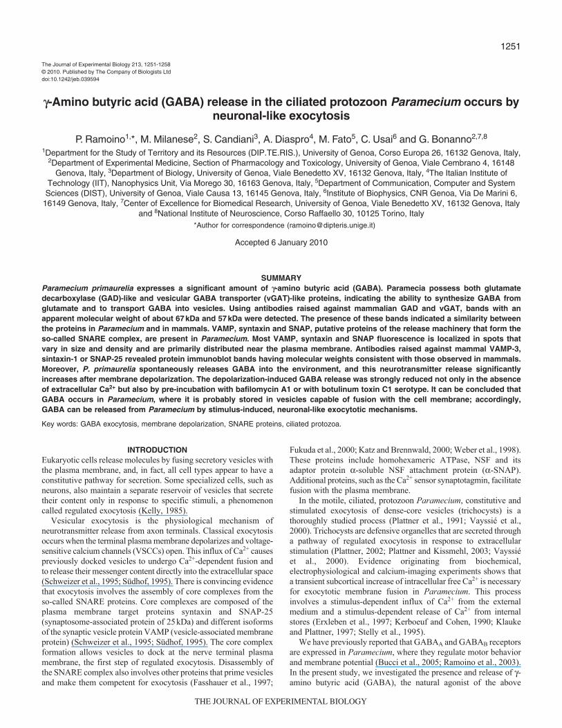
The localization and distribution of GABA in P. primaurelia wereexamined using a polyclonal antibody and laser confocalmicroscopy. Labeling was detected primarily at the cell surface andin the food vacuoles of cells in the late log phase of growth (Fig.1A).Fig.1B documents staining of dot-like structures located on theoutlines of the regularly arranged surface fields (kinetics) that arecharacteristic of Paramecium cells.
The presence of the biosynthetic enzyme GAD and vGAT wasdetermined by both immunofluorescence and immunoblotting.Using confocal microscopy, GAD- and vGAT-likeimmunoreactivity was detected at the cell surface and inside thecytoplasm (Fig.1C,D). No immunostaining was observed in thenegative controls when the primary antibody was omitted or whenthe primary antibody was pre-absorbed by GABA or the specificGAD or vGAT immunogen peptides.
Immunoblots showed two bands with an apparent molecularweight of about 67kDa and 57kDa, using antibodies raised againstmammalian GAD and vGAT, respectively (Fig.1E, lane 2). These
molecular mass values were consistent with those obtained insynaptosomes prepared from rat cerebral cortex under the sameexperimental conditions (Fig.1E, lane 1).
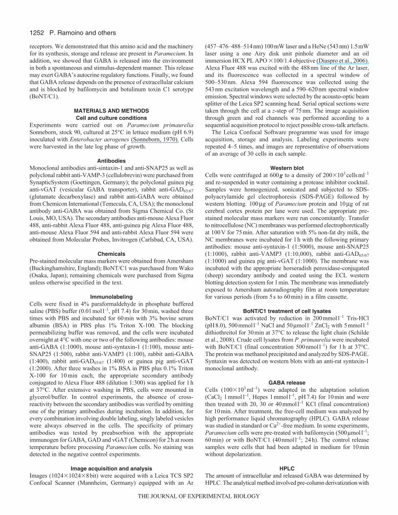
Proteins of the release machinery in ParameciumTransmitter release at the nerve terminal involves assembly of theSNARE complex proteins. VAMPs, in conjunction with syntaxinsand SNAP-25, are thought to play a role in docking synaptic vesiclesto the presynaptic plasma membrane (Rothman and Wieland, 1996;Sudhof, 1995). Evidence from both confocal microscopy andimmunoblot indicated the presence in Paramecium of putativeVAMP, syntaxin and SNAP proteins, which were identified usingantibodies raised against mammalian proteins (monoclonal anti-syntaxin-1 and anti-SNAP-25 antibodies and the polyclonal antibodyanti-cellubrevin/VAMP3 that cross-reacts with both synaptobrevinsVAMP1 and VAMP2). By confocal microscopy, immunoreactivityappeared localized as a punctate pattern on the cell surface(Fig.2A–C). Negative controls, obtained by omitting the primaryantibody, showed no immunostaining.
Western blots of proteins derived from Paramecium cells,performed using anti-syntaxin-1 antibody, revealed two bands withestimated molecular masses of about 32kDa and 36kDa (Fig.2D,line 2). Bands with an apparent molecular mass of about 20kDaand 25kDa were detected using a broad-spectrum anti-VAMPantibody and an anti-SNAP-25 antibody, respectively (Fig.2D, line2). These values are consistent with those obtained for rat cerebralcortex synaptosomes (Fig.2D, line 1).
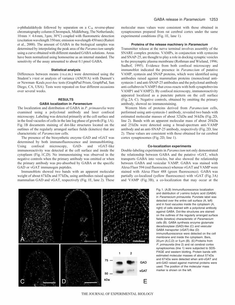
Co-localization experimentsDouble-labeling experiments in Paramecium not only demonstratedthe relationship between GABA and the putative vGAT, whichtransports GABA into vesicles, but also showed the relationshipbetween GABA and vesicular VAMP. GABA was stained withAlexa Fluor 594 (red fluorescence) whereas vGAT and VAMP werestained with Alexa Fluor 488 (green fluorescence). GABA waspartially co-localized (yellow fluorescence) with vGAT (Fig.3A)and VAMP (Fig.3B), a co-localization that may occur at the
Fig.1. (A,B) Immunofluorescence localizationand distribution of -amino butyric acid (GABA)in Paramecium primaurelia. Punctate label wasdetected over the entire cell surface (A, left)and in food vacuoles inside the cytoplasm (A,right) of cells stained with a polyclonal antibodyagainst GABA. Dot-like structures are stainedon the outlines of the regularly arranged surfacefields (kinetics) characteristic of Parameciumcells (B). GABA synthesis enzyme glutamatedecarboxylase (GAD)-like (C) and vesicularGABA transporter (vGAT)-like (D)immunofluorescence were detected on the cellmembrane and inside the cytoplasm. Bars,20m (A,C,D) or 5m (B). (E)Proteins fromP. primaurelia (line 2) and rat cerebral cortexsynaptosomes (line 1) were subjected to SDS-PAGE and western blotting. Protein bands withestimated molecular masses of about 57kDaand 67kDa were detected when anti-vGAT andanti-GAD raised against mammal proteins wereused. The position of the molecular massmarker is shown on the left.
THE JOURNAL OF EXPERIMENTAL BIOLOGY
1254
vesicular level. Co-localization along the z-axes was demonstratedby the similarity of the green and red z-profiles of the fluorescenceintensity of three double-stained vesicles collected at the top(Fig.3Ai,Bi), middle (Fig.3Aii,Bii) and bottom (Fig.3Aiii,Biii)planes of a 2-m thick z-stack.
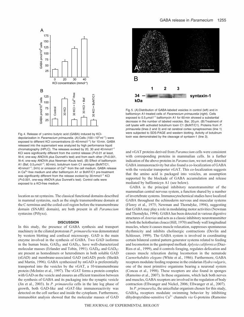
KCl-induced GABA releaseWe used KCl depolarization (from 0mmoll–1 up to 40mmoll–1 KCl;10min exposure) in the presence of Ca2+ to induce GABA releasein Paramecium. The amino acid was spontaneously released intothe environment (control condition, 0mmoll–1 KCl) but the amountof released amino acid increased after K+ depolarization in aconcentration-dependent manner (Fig.4A). The GABA released inthe 10-min fraction collected before the onset of K+ stimulationamounted to 32±3.6pmol104 cells–1. The K+-evoked overflow was65±7.0, 120±7.8 and 160±10.3pmol104 cells–1 at 20, 30 and40mmoll–1 KCl, respectively. In these sets of experiments, thetotal GABA content in the paramecia amounted to about2450±6.0pmol104 cells–1.
A lack of extracellular Ca2+ abolished the depolarization-inducedGABA release (Fig.4B), suggesting the involvement of regulatedexocytosis. The involvement of vesicular GABA in amino acidrelease is supported by experiments with bafilomycin-A1, whichwas expected to prevent the accumulation of GABA into vesicles(Moriyama and Futai, 1990; Roseth et al., 1995). Cells were firstincubated with bafilomycin-A1 (0.5moll–1, 60min) and thenexposed to 30mmoll–1 KCl. Pre-incubation with bafilomycin A1
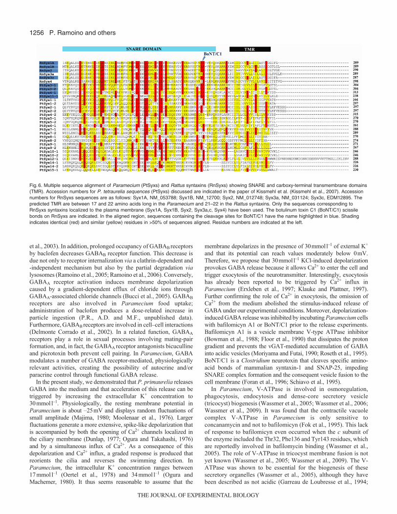
greatly reduced the depolarization-evoked GABA release (Fig.4B).As revealed by examination of GABA-labeled vesicle abundanceusing confocal microscopy, there was a substantial decrease in thenumber of labeled vesicles in Paramecium cells exposed tobafilomycin A1 (Fig.5A).
We also tested the ability of BoNT/C1, which hampersneurotransmitter exocytosis by cleaving syntaxin-1 and SNAP-25in mammals (Foran et al., 1996; Schiavo et al., 1995), to blockGABA release. Incubation of cells for 24h in the presence of theactivated toxin (40nmoll–1) significantly inhibited GABA release(Fig.4B). Activity of botulinum toxin was demonstrated in westernblot experiments by directly measuring the amount of Parameciumsyntaxin present after exposure of cell extracts to the botulinumtoxin. Syntaxin, detected with the anti-rat syntaxin-1 monoclonalantibody used above, was totally cleaved by BoNT/C1 (Fig.5B).
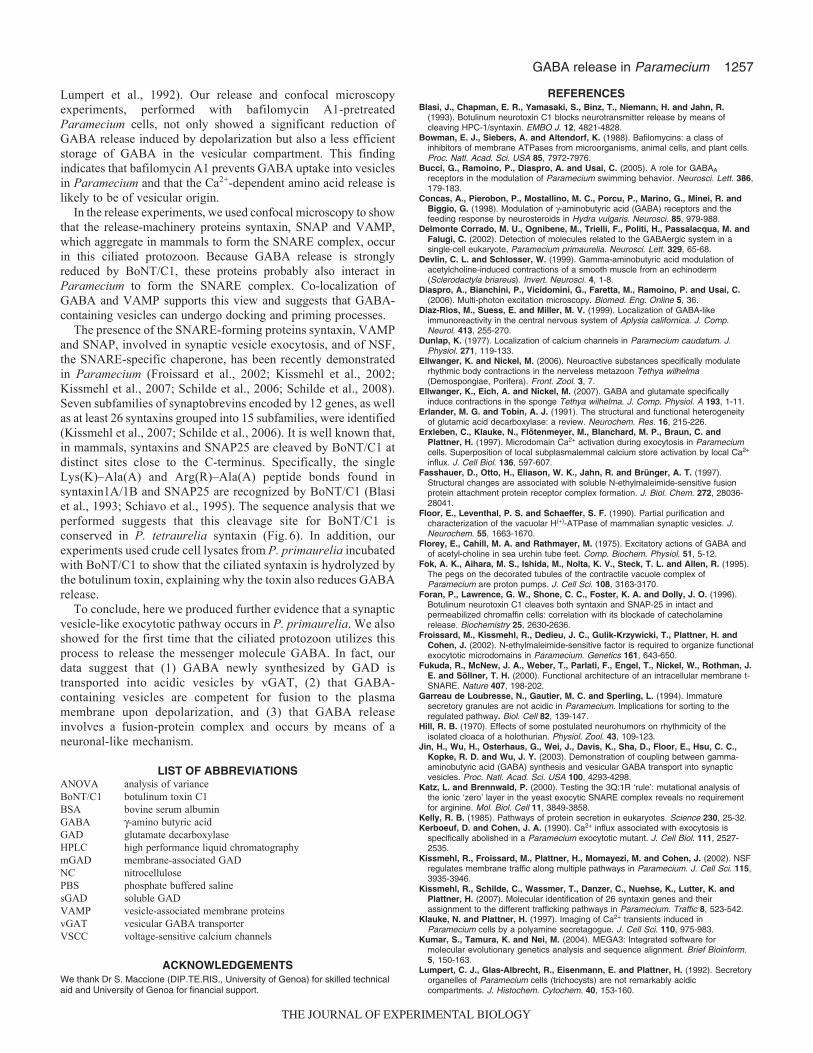
To identify putative cleavage sites for BoNT/C1 in theParamecium syntaxin (PtSyx) sequences, a multiple sequencealignment was performed using the Rattus norvegicus syntaxin asconsensus sequences (Fig.6). Sequences were aligned using theClustalW program in the Molecular Evolutionary GeneticsAnalysis (MEGA) software package (Kumar et al., 2004)(http://www.megasoftware.net/). Domain predictions (SNARE andtransmembrane domains) were determined by PROSITE andTMHMM version 2.0 (http://www.cbs.dtu.dk/services/TMHMM/).Our analysis showed that some Paramecium syntaxin homologs(PtSyx 3-1/3-2), as well as some ciliate-specific syntaxins (PtSyx4-1and PtSyx 11-1), possess a single Lys–Ala peptide bond in the same
P. Ramoino and others
Fig.2. Localization and distribution of vesicle-associated membrane protein(VAMP)- (A), syntaxin- (B) and SNAP-like (C) immunofluorescence inParamecium primaurelia detected using antibodies raised against mammalproteins. The label was detected over the entire cell membrane using cellsin the late log phase of growth. Bars, 20m. (D)Proteins from P.primaurelia (line 2) and rat cerebral cortex synaptosomes (line 1) weresubjected to SDS-PAGE and western blotting. Bands with apparentmolecular mass of 25kDa and 20kDa were detected using the SNAP-25and VAMP-3 antibodies, respectively, and two bands with estimatedmolecular masses of 32kDa and 36kDa were detected using thesyntaxin-1 antiserum. The position of the molecular mass marker is shownon the left.
Fig.3. (Left) Co-localization of -amino butyric acid (GABA) and thevesicular GABA transporter (vGAT)-like (A), as well as co-localization ofGABA and vesicle-associated membrane protein (VAMP)-like (B), inParamecium primaurelia cells labeled with an antibody against GABA,conjugated with Alexa 594 (red), and antibodies against mammal vGAT orVAMP-3, conjugated with Alexa Fluor 488 (green). Co-localization is shownas yellow fluorescence. (Right) z-stack profiles of fluorescence intensity indouble-labeled vesicles; data represent the fluorescence-intensitydistribution along the z-axis of three yellow-labeled vesicles selected fromdifferent optical planes (Ai, Bi, top; Aii, Bii, middle; Aiii, Biii, bottom)(green, �; red, �). Bars, 10m.
THE JOURNAL OF EXPERIMENTAL BIOLOGY
1255GABA release in Paramecium
location as rat syntaxins. The classical functional domains describedin mammal syntaxins, such as the single transmembrane domain atthe C-terminus and the coiled-coil region before the transmembranedomain (SNARE domain), are both present in all Parameciumsyntaxins (PtSyxs).
DISCUSSIONIn this study, the presence of GABA synthesis and transportmachinery in the ciliated protozoan P. primaurelia was demonstratedby western blotting and confocal microscopy. GAD is the mainenzyme involved in the synthesis of GABA. Two GAD isoformsin the human brain, GAD65 and GAD67, have well-characterizedmolecular masses (Erlander and Tobin, 1991). GAD65 and GAD67
are present as homodimers or heterodimers in both soluble GAD(sGAD) and membrane-associated GAD (mGAD) pools (Sheikhand Martin, 1996). GABA synthesized by mGAD is preferentiallytransported into the vesicles by the vGAT, a 10-transmembraneprotein (McIntire et al., 1997). The vGAT forms a protein complexwith GAD on the vesicle and ensures an efficient transition betweenthe synthesis of GABA and its packaging into the synaptic vesicle(Jin et al., 2003). In P. primaurelia cells in the late log phase ofgrowth, both GAD-like and vGAT-like immunoreactivity wasdetected on the cell surface and inside the cytoplasm. Furthermore,immunoblot analysis showed that the molecular masses of GAD
and vGAT proteins derived from Paramecium cells were consistentwith corresponding proteins in mammalian cells. In a furtherindication of the above proteins in Paramecium, we not only detectedGABA immunoreactivity but also found a co-localization of GABAwith the vesicular transporter vGAT. This co-localization suggeststhat the amino acid is packaged into vesicles, an assumptionsupported by the blockade of GABA accumulation and releasemediated by bafilomicyn A1 (see below).
GABA is the principal inhibitory neurotransmitter of themammalian central nervous system, a function shared by a numberof invertebrate systems. Immunocytochemical studies have localizedGABA throughout the echinoderm nervous and muscular systems(Florey et al., 1975; Newman and Thorndyke, 1994), suggestingthat GABA may play a role in modulating motor activities (Newmanand Thorndyke, 1994). GABA has been detected in various digestivestructures of Asterias and acts as a classic inhibitory neurotransmitterin both the holothurian cloaca (Hill, 1970) and body wall longitudinalmuscles, where it causes muscle relaxation, suppresses spontaneousrhythmicity and inhibits cholinergic contractions (Devlin andSchlosser, 1999). The GABA system is involved in coordinatingcertain bilateral central pattern generator systems related to feedingand locomotion in the gastropod mollusk Aplysia californica (Diaz-Rios et al., 1999), and it controls foraging, regulates defecation andcauses muscle relaxation during locomotion in the nematodeCaenorhabditis elegans (White et al., 1986). Furthermore, GABAreceptors modulate feeding response in the cnidarian Hydra vulgaris,one of the most primitive organisms bearing a neuronal system(Concas et al., 1998). These receptors are also found in sponges(Ramoino et al., 2007). In these organisms, which lack both nervesand muscles, GABA receptors are involved in the regulation of bodycontraction (Ellwanger and Nickel, 2006; Ellwanger et al., 2007).
In P. primaurelia, the unicellular organism chosen for this study,GABAB receptors modulate swimming behavior by inhibitingdihydropyridine-sensitive Ca2+ channels via G-proteins (Ramoino
Fig.4. Release of -amino butyric acid (GABA) induced by KCl-depolarization in Paramecium primaurelia. (A)Cells (100�103ml–1) wereexposed to different KCl concentrations (0–40mmoll–1) for 10min. GABAreleased into the supernatant was analyzed by high performance liquidchromatography (HPLC). The releases evoked by 20, 30 and 40mmoll–1
KCl were significantly different from the control release (P<0.01 at least,N4; one-way ANOVA plus Dunnett’s test) and from each other (P<0.001,N4; one-way ANOVA plus Newman–Keuls test). (B) Effect of bafilomycinA1 (Baf, 0.5moll–1; 60min), botulinum toxin C1 serotype (BoNT/C1,40nmoll–1; 24h) or omission of Ca2+ from the cell medium. GABA releasein Ca2+-free medium and after bafilomycin A1 or BoNT/C1 pre-treatmentwas significantly different from the release evoked by 30mmoll–1 KCl(P<0.001, one-way ANOVA plus Dunnett’s test). Control cells wereexposed to a KCl-free medium.
Fig.5. (A)Distribution of GABA-labeled vesicles in control (left) and inbafilomicyn A1-treated cells of Paramecium primaurelia (right). Cellsexposed to 0.5moll–1 bafilomycin A1 for 60min showed a substantialdecrease in the number of labeled vesicles. Bar, 20m. (B)Treatment ofcell lysate with activated botulinum toxin C1 (BoNT/C1). Proteins from P.primaurelia (lines 2 and 3) and rat cerebral cortex synaptosomes (line 1)were subjected to SDS-PAGE and western blotting. Activity of botulinumtoxin was demonstrated by the cleavage of syntaxin-1 (line 3).
THE JOURNAL OF EXPERIMENTAL BIOLOGY
1256
et al., 2003). In addition, prolonged occupancy of GABAB receptorsby baclofen decreases GABAB receptor function. This decrease isdue not only to receptor internalization via a clathrin-dependent and-independent mechanism but also by the partial degradation vialysosomes (Ramoino et al., 2005; Ramoino et al., 2006). Conversely,GABAA receptor activation induces membrane depolarizationcaused by a gradient-dependent efflux of chloride ions throughGABAA-associated chloride channels (Bucci et al., 2005). GABAB
receptors are also involved in Paramecium food uptake;administration of baclofen produces a dose-related increase inparticle ingestion (P.R., A.D. and M.F., unpublished data).Furthermore, GABAB receptors are involved in cell–cell interactions(Delmonte Corrado et al., 2002). In a related function, GABAA
receptors play a role in sexual processes involving mating-pairformation, and, in fact, the GABAA receptor antagonists bicucullineand picrotoxin both prevent cell pairing. In Paramecium, GABAmodulates a number of GABA receptor-mediated, physiologicallyrelevant activities, creating the possibility of autocrine and/orparacrine control through functional GABA release.
In the present study, we demonstrated that P. primaurelia releasesGABA into the medium and that acceleration of this release can betriggered by increasing the extracellular K+ concentration to30mmoll–1. Physiologically, the resting membrane potential inParamecium is about –25mV and displays random fluctuations ofsmall amplitude (Majima, 1980; Moolenaar et al., 1976). Largerfluctuations generate a more extensive, spike-like depolarization thatis accompanied by both the opening of Ca2+ channels localized inthe ciliary membrane (Dunlap, 1977; Ogura and Takahashi, 1976)and by a simultaneous influx of Ca2+. As a consequence of thisdepolarization and Ca2+ influx, a graded response is produced thatreorients the cilia and reverses the swimming direction. InParamecium, the intracellular K+ concentration ranges between17mmoll–1 (Oertel et al., 1978) and 34mmoll–1 (Ogura andMachemer, 1980). It thus seems reasonable to assume that the
membrane depolarizes in the presence of 30mmoll–1 of external K+
and that its potential can reach values moderately below 0mV.Therefore, we propose that 30mmoll–1 KCl-induced depolarizationprovokes GABA release because it allows Ca2+ to enter the cell andtrigger exocytosis of the neurotransmitter. Interestingly, exocytosishas already been reported to be triggered by Ca2+ influx inParamecium (Erxleben et al., 1997; Klauke and Plattner, 1997).Further confirming the role of Ca2+ in exocytosis, the omission ofCa2+ from the medium abolished the stimulus-induced release ofGABA under our experimental conditions. Moreover, depolarization-induced GABA release was inhibited by incubating Paramecium cellswith bafilomicyn A1 or BoNT/C1 prior to the release experiments.Bafilomicyn A1 is a vesicle membrane V-type ATPase inhibitor(Bowman et al., 1988; Floor et al., 1990) that dissipates the protongradient and prevents the vGAT-mediated accumulation of GABAinto acidic vesicles (Moriyama and Futai, 1990; Roseth et al., 1995).BoNT/C1 is a Clostridium neurotoxin that cleaves specific amino-acid bonds of mammalian syntaxin-1 and SNAP-25, impedingSNARE complex formation and the consequent vesicle fusion to thecell membrane (Foran et al., 1996; Schiavo et al., 1995).
In Paramecium, V-ATPase is involved in osmoregulation,phagocytosis, endocytosis and dense-core secretory vesicle(tricocyst) biogenesis (Wassmer et al., 2005; Wassmer et al., 2006;Wassmer et al., 2009). It was found that the contractile vacuolecomplex V-ATPase in Paramecium is only sensitive toconcanamycin and not to bafilomicyn (Fok et al., 1995). This lackof response to bafilomicyn even occurred when the c subunit ofthe enzyme included the Thr32, Phe136 and Tyr143 residues, whichare reportedly involved in bafilomycin binding (Wassmer et al.,2005). The role of V-ATPase in tricocyst membrane fusion is notyet known (Wassmer et al., 2005; Wassmer et al., 2009). The V-ATPase was shown to be essential for the biogenesis of thesesecretory organelles (Wassmer et al., 2005), although they havebeen described as not acidic (Garreau de Loubresse et al., 1994;
P. Ramoino and others
Fig.6. Multiple sequence alignment of Paramecium (PtSyxs) and Rattus syntaxins (RnSyxs) showing SNARE and carboxy-terminal transmembrane domains(TMR). Accession numbers for P. tetraurelia sequences (PtSyxs) discussed are indicated in the paper of Kissmehl et al. (Kissmehl et al., 2007). Accessionnumbers for RnSyxs sequences are as follows: Syx1A, NM_053788; Syx1B, NM_12700; Syx2, NM_012748; Syx3a, NM_031124; Syx3c, EDM12895. Thepredicted TMR are between 17 and 22 amino acids long in the Paramecium and 21–22 in the Rattus syntaxins. Only the sequences corresponding toRnSyxs syntaxins localized to the plasma membrane (Syx1A, Syx1B, Syx2, Syx3a,c, Syx4) have been used. The botulinum toxin C1 (BoNT/C1) scissilebonds on RnSyxs are indicated. In the aligned region, sequences containing the cleavage sites for BoNT/C1 have the name highlighted in blue. Shadingindicates identical (red) and similar (yellow) residues in >50% of sequences aligned. Residue numbers are indicated at the left.
THE JOURNAL OF EXPERIMENTAL BIOLOGY
1257GABA release in Paramecium
Lumpert et al., 1992). Our release and confocal microscopyexperiments, performed with bafilomycin A1-pretreatedParamecium cells, not only showed a significant reduction ofGABA release induced by depolarization but also a less efficientstorage of GABA in the vesicular compartment. This findingindicates that bafilomycin A1 prevents GABA uptake into vesiclesin Paramecium and that the Ca2+-dependent amino acid release islikely to be of vesicular origin.
In the release experiments, we used confocal microscopy to showthat the release-machinery proteins syntaxin, SNAP and VAMP,which aggregate in mammals to form the SNARE complex, occurin this ciliated protozoon. Because GABA release is stronglyreduced by BoNT/C1, these proteins probably also interact inParamecium to form the SNARE complex. Co-localization ofGABA and VAMP supports this view and suggests that GABA-containing vesicles can undergo docking and priming processes.
The presence of the SNARE-forming proteins syntaxin, VAMPand SNAP, involved in synaptic vesicle exocytosis, and of NSF,the SNARE-specific chaperone, has been recently demonstratedin Paramecium (Froissard et al., 2002; Kissmehl et al., 2002;Kissmehl et al., 2007; Schilde et al., 2006; Schilde et al., 2008).Seven subfamilies of synaptobrevins encoded by 12 genes, as wellas at least 26 syntaxins grouped into 15 subfamilies, were identified(Kissmehl et al., 2007; Schilde et al., 2006). It is well known that,in mammals, syntaxins and SNAP25 are cleaved by BoNT/C1 atdistinct sites close to the C-terminus. Specifically, the singleLys(K)–Ala(A) and Arg(R)–Ala(A) peptide bonds found insyntaxin1A/1B and SNAP25 are recognized by BoNT/C1 (Blasiet al., 1993; Schiavo et al., 1995). The sequence analysis that weperformed suggests that this cleavage site for BoNT/C1 isconserved in P. tetraurelia syntaxin (Fig.6). In addition, ourexperiments used crude cell lysates from P. primaurelia incubatedwith BoNT/C1 to show that the ciliated syntaxin is hydrolyzed bythe botulinum toxin, explaining why the toxin also reduces GABArelease.
To conclude, here we produced further evidence that a synapticvesicle-like exocytotic pathway occurs in P. primaurelia. We alsoshowed for the first time that the ciliated protozoon utilizes thisprocess to release the messenger molecule GABA. In fact, ourdata suggest that (1) GABA newly synthesized by GAD istransported into acidic vesicles by vGAT, (2) that GABA-containing vesicles are competent for fusion to the plasmamembrane upon depolarization, and (3) that GABA releaseinvolves a fusion-protein complex and occurs by means of aneuronal-like mechanism.
LIST OF ABBREVIATIONSANOVA analysis of varianceBoNT/C1 botulinum toxin C1BSA bovine serum albuminGABA -amino butyric acidGAD glutamate decarboxylaseHPLC high performance liquid chromatographymGAD membrane-associated GADNC nitrocellulosePBS phosphate buffered salinesGAD soluble GADVAMP vesicle-associated membrane proteinsvGAT vesicular GABA transporterVSCC voltage-sensitive calcium channels
ACKNOWLEDGEMENTSWe thank Dr S. Maccione (DIP.TE.RIS., University of Genoa) for skilled technicalaid and University of Genoa for financial support.
REFERENCESBlasi, J., Chapman, E. R., Yamasaki, S., Binz, T., Niemann, H. and Jahn, R.
(1993). Botulinum neurotoxin C1 blocks neurotransmitter release by means ofcleaving HPC-1/syntaxin. EMBO J. 12, 4821-4828.
Bowman, E. J., Siebers, A. and Altendorf, K. (1988). Bafilomycins: a class ofinhibitors of membrane ATPases from microorganisms, animal cells, and plant cells.Proc. Natl. Acad. Sci. USA 85, 7972-7976.
Bucci, G., Ramoino, P., Diaspro, A. and Usai, C. (2005). A role for GABAA
receptors in the modulation of Paramecium swimming behavior. Neurosci. Lett. 386,179-183.
Concas, A., Pierobon, P., Mostallino, M. C., Porcu, P., Marino, G., Minei, R. andBiggio, G. (1998). Modulation of -aminobutyric acid (GABA) receptors and thefeeding response by neurosteroids in Hydra vulgaris. Neurosci. 85, 979-988.
Delmonte Corrado, M. U., Ognibene, M., Trielli, F., Politi, H., Passalacqua, M. andFalugi, C. (2002). Detection of molecules related to the GABAergic system in asingle-cell eukaryote, Paramecium primaurelia. Neurosci. Lett. 329, 65-68.
Devlin, C. L. and Schlosser, W. (1999). Gamma-aminobutyric acid modulation ofacetylcholine-induced contractions of a smooth muscle from an echinoderm(Sclerodactyla briareus). Invert. Neurosci. 4, 1-8.
Diaspro, A., Bianchini, P., Vicidomini, G., Faretta, M., Ramoino, P. and Usai, C.(2006). Multi-photon excitation microscopy. Biomed. Eng. Online 5, 36.
Diaz-Rios, M., Suess, E. and Miller, M. V. (1999). Localization of GABA-likeimmunoreactivity in the central nervous system of Aplysia californica. J. Comp.Neurol. 413, 255-270.
Dunlap, K. (1977). Localization of calcium channels in Paramecium caudatum. J.Physiol. 271, 119-133.
Ellwanger, K. and Nickel, M. (2006). Neuroactive substances specifically modulaterhythmic body contractions in the nerveless metazoon Tethya wilhelma(Demospongiae, Porifera). Front. Zool. 3, 7.
Ellwanger, K., Eich, A. and Nickel, M. (2007). GABA and glutamate specificallyinduce contractions in the sponge Tethya wilhelma. J. Comp. Physiol. A 193, 1-11.
Erlander, M. G. and Tobin, A. J. (1991). The structural and functional heterogeneityof glutamic acid decarboxylase: a review. Neurochem. Res. 16, 215-226.
Erxleben, C., Klauke, N., Flötenmeyer, M., Blanchard, M. P., Braun, C. andPlattner, H. (1997). Microdomain Ca2+ activation during exocytosis in Parameciumcells. Superposition of local subplasmalemmal calcium store activation by local Ca2+
influx. J. Cell Biol. 136, 597-607.Fasshauer, D., Otto, H., Eliason, W. K., Jahn, R. and Brünger, A. T. (1997).
Structural changes are associated with soluble N-ethylmaleimide-sensitive fusionprotein attachment protein receptor complex formation. J. Biol. Chem. 272, 28036-28041.
Floor, E., Leventhal, P. S. and Schaeffer, S. F. (1990). Partial purification andcharacterization of the vacuolar H(+)-ATPase of mammalian synaptic vesicles. J.Neurochem. 55, 1663-1670.
Florey, E., Cahill, M. A. and Rathmayer, M. (1975). Excitatory actions of GABA andof acetyl-choline in sea urchin tube feet. Comp. Biochem. Physiol. 51, 5-12.
Fok, A. K., Aihara, M. S., Ishida, M., Nolta, K. V., Steck, T. L. and Allen, R. (1995).The pegs on the decorated tubules of the contractile vacuole complex ofParamecium are proton pumps. J. Cell Sci. 108, 3163-3170.
Foran, P., Lawrence, G. W., Shone, C. C., Foster, K. A. and Dolly, J. O. (1996).Botulinum neurotoxin C1 cleaves both syntaxin and SNAP-25 in intact andpermeabilized chromaffin cells: correlation with its blockade of catecholaminerelease. Biochemistry 25, 2630-2636.
Froissard, M., Kissmehl, R., Dedieu, J. C., Gulik-Krzywicki, T., Plattner, H. andCohen, J. (2002). N-ethylmaleimide-sensitive factor is required to organize functionalexocytotic microdomains in Paramecium. Genetics 161, 643-650.
Fukuda, R., McNew, J. A., Weber, T., Parlati, F., Engel, T., Nickel, W., Rothman, J.E. and Söllner, T. H. (2000). Functional architecture of an intracellular membrane t-SNARE. Nature 407, 198-202.
Garreau de Loubresse, N., Gautier, M. C. and Sperling, L. (1994). Immaturesecretory granules are not acidic in Paramecium. Implications for sorting to theregulated pathway. Biol. Cell 82, 139-147.
Hill, R. B. (1970). Effects of some postulated neurohumors on rhythmicity of theisolated cloaca of a holothurian. Physiol. Zool. 43, 109-123.
Jin, H., Wu, H., Osterhaus, G., Wei, J., Davis, K., Sha, D., Floor, E., Hsu, C. C.,Kopke, R. D. and Wu, J. Y. (2003). Demonstration of coupling between gamma-aminobutyric acid (GABA) synthesis and vesicular GABA transport into synapticvesicles. Proc. Natl. Acad. Sci. USA 100, 4293-4298.
Katz, L. and Brennwald, P. (2000). Testing the 3Q:1R ‘rule’: mutational analysis ofthe ionic ‘zero’ layer in the yeast exocytic SNARE complex reveals no requirementfor arginine. Mol. Biol. Cell 11, 3849-3858.
Kelly, R. B. (1985). Pathways of protein secretion in eukaryotes. Science 230, 25-32.Kerboeuf, D. and Cohen, J. A. (1990). Ca2+ influx associated with exocytosis is
specifically abolished in a Paramecium exocytotic mutant. J. Cell Biol. 111, 2527-2535.
Kissmehl, R., Froissard, M., Plattner, H., Momayezi, M. and Cohen, J. (2002). NSFregulates membrane traffic along multiple pathways in Paramecium. J. Cell Sci. 115,3935-3946.
Kissmehl, R., Schilde, C., Wassmer, T., Danzer, C., Nuehse, K., Lutter, K. andPlattner, H. (2007). Molecular identification of 26 syntaxin genes and theirassignment to the different trafficking pathways in Paramecium. Traffic 8, 523-542.
Klauke, N. and Plattner, H. (1997). Imaging of Ca2+ transients induced inParamecium cells by a polyamine secretagogue. J. Cell Sci. 110, 975-983.
Kumar, S., Tamura, K. and Nei, M. (2004). MEGA3: Integrated software formolecular evolutionary genetics analysis and sequence alignment. Brief Bioinform.5, 150-163.
Lumpert, C. J., Glas-Albrecht, R., Eisenmann, E. and Plattner, H. (1992). Secretoryorganelles of Paramecium cells (trichocysts) are not remarkably acidiccompartments. J. Histochem. Cytochem. 40, 153-160.
THE JOURNAL OF EXPERIMENTAL BIOLOGY
1258
Majima, J. (1980). Membrane potential fluctuation in paramecium. Biophys. Chem. 11,101-108.
McIntire, S. L., Reimer, R. J., Schuske, K., Edwards, R. H. and Jorgensen, E. M.(1997). Identification and characterization of the vesicular GABA transporter. Nature389, 870-876.
Moolenaar, W., de Goede, J. and Verveen, A. (1976). Membrane noise inParamecium. Nature 260, 344-345.
Moriyama, Y. and Futai, M. (1990). H(+)-ATPase, a primary pump for accumulation ofneurotransmitters, is a major constituent of brain synaptic vesicles. Biochem.Biophys. Res. Commun. 173, 443-448.
Newman, S. J. and Thorndyke, M. C. (1994). Localisation of -aminobutyric acid(GABA)-like immunoreactivity in the echinoderm Asterias rubens. Cell Tissue Res.278, 177-185.
Oertel, D., Schein, S. J. and Kung, C. (1978). A potassium channel activated byhyperpolarization in Paramecium. J. Membr. Biol. 43, 169-185.
Ogura, A. and Machemer, H. (1980). Distribution of mechanoreceptor channels in theParamecium surface membrane. J. Comp. Physiol. A 135, 233-242.
Ogura, A. and Takahashi, K. (1976). Artificial deciliation causes loss of calcium-dependent resposes in Paramecium. Nature 264, 170-172.
Plattner, H. (2002). My favorite cell: Paramecium. BioEssays 24, 649-658.Plattner, H. and Kissmehl, R. (2003). Dense-core secretory vesicle docking and
exocytotic membrane fusion in Paramecium cells. Biochim. Biophys. Acta 1641, 183-193.
Plattner, H., Lumpert, C. J., Knoll, G., Kissmehl, R., Höhne, B., Momayezi, M. andGlas-Albrecht, R. (1991). Stimulus-secretion coupling in Paramecium cells. Eur. J.Cell Biol. 55, 3-16.
Raiteri, M., Sala, R., Fassio, A., Rossetto, O. and Bonanno, G. (2000). Entrappingof impermeant probes of different size into non permeabilized synaptosomes as amethod to study presynaptic mechanisms. J. Neurochem. 74, 423-431.
Ramoino, P., Fronte, P., Beltrame, F., Diaspro, A., Fato, M., Raiteri, L., Stigliani, S.and Usai, C. (2003). Swimming behavior regulation by GABAB receptors inParamecium. Exp. Cell Res. 291, 398-405.
Ramoino, P., Usai, C., Beltrame, F., Fato, M., Gallus, L., Tagliaferro, G., Magrassi,R. and Diaspro, A. (2005). GABAB receptor intracellular trafficking afterinternalization in Paramecium. Microsc. Res. Tech. 68, 290-295.
Ramoino, P., Gallus, L., Beltrame, F., Diaspro, A., Fato, M., Rubini, P., Stigliani,S., Bonanno, G. and Usai, C. (2006). Endocytosis of GABAB receptors modulatesmembrane excitability in the single-celled organism Paramecium. J. Cell Sci. 119,2056-2064.
Ramoino, P., Gallus, L., Paluzzi, S., Raiteri, L., Diaspro, A., Fato, M., Bonanno, G.,Tagliafierro, G., Ferretti, C. and Manconi, R. (2007). The GABAergic-like system inthe marine demosponge Chondrilla nucula. Microsc. Res. Tech. 70, 944-951.
Roseth, S., Fykse, E. M. and Fonnum, F. (1995). Uptake of L-glutamate into rat brainsynaptic vesicles: effect of inhibitors that bind specifically to the glutamatetransporter. J. Neorochem. 65, 96-103.
Rothman, J. E. and Wieland, F. T. (1996). Protein sorting by transport vesicles.Science 272, 227.
Schiavo, G., Shone, C. C., Bennett, M. K., Scheller, R. H. and Montecucco, C.(1995). Botulinum neurotoxin type C cleaves a single Lys-Ala bond within thecarboxyl-terminal region of syntaxins. J. Biol. Chem. 270, 10566-10570.
Schilde, C., Wassmer, T., Mansfeld, J., Plattner, H. and Kissmehl, R. (2006). Amultigene family encoding R-SNAREs in the ciliate Paramecium tetraurelia. Traffic 7,440-455.
Schilde, C., Lutter, K., Kissmehl, R. and Plattner, H. (2008). Molecular identificationof a SNAP-25-like SNARE protein in Paramecium. Eukaryot. Cell 7, 1387-1402.
Schweizer, F. E., Betz, H. and Augustine, G. J. (1995). From vesicle docking toendocytosis: intermediate reactions of exocytosis. Neuron 14, 689-696.
Sheikh, S. N. and Martin, D. L. (1996). Heteromers of glutamate decarboxylaseisoforms occur in rat cerebellum. J. Neurochem. 66, 2082-2090.
Sonneborn, T. M. (1970). Methods in Paramecium research. In Methods in CellPhysiology, vol. 4 (ed. D. M. Prescott), pp. 241-339. New York: Academic Press.
Stelly, N., Halpern, S., Nicolas, G., Fragu, P. and Adoutte, A. (1995). Directvisualization of a vast cortical calcium compartment in Paramecium by secondary ionmass spectrometry (SIMS) microscopy: possible involvement in exocytosis. J. CellSci. 108, 1895-1909.
Südhof, T. C. (1995). The synaptic vesicle cycle: a cascade of protein-proteininteractions. Nature 375, 645-653.
Vayssié, L., Skouri, F., Sperling, L. and Cohen, J. (2000). Molecular genetics ofregulated secretion in Paramecium. Biochimie 82, 269-288.
Wassmer, T., Froissard, M., Plattner, H., Kissmehl, R. and Cohen, J. (2005). Thevacuolar proton-ATPase plays a major role in several membrane boundedorganelles in Paramecium. J. Cell Sci. 118, 2813-2825.
Wassmer, T., Kissmehl, R., Cohen, J. and Plattner, H. (2006). Seventeen a-subunitisoforms of Paramecium V-ATPase provide high specialization in localization andfunction. Mol. Biol. Cell 17, 917-930.
Wassmer, T., Sehring, I. M., Kissmehl, R. and Plattner, H. (2009). The V-ATPase inParamecium: functional specialization by multiple gene isoforms. Pflugers Arch. Eur.J. Physiol. 457, 599-607.
Weber, T., Zemelman, B. V., McNew, J. A., Westermann, B., Gmachl, M., Parlati,F., Söllner, T. H. and Rothman, J. E. (1998). SNAREpins: minimal machinery formembrane fusion. Cell 92, 759-772.
White, J. G., Southgate, E., Thomson, J. N. and Brenner, S. (1986). The structureof the nervous system of the nematode Caenorhabditis elegans. Philos. Trans. R.Soc. Lond. B., Biol. Sci. 314, 1-340.
P. Ramoino and others
THE JOURNAL OF EXPERIMENTAL BIOLOGYAll in-text references underlined in blue are linked to publications on ResearchGate, letting you access and read them immediately.