gaba is excitatory in adult vasopressinergic neuroendocrine cells
TRANSCRIPT
Cellular/Molecular
GABA Is Excitatory in Adult VasopressinergicNeuroendocrine Cells
Juhee Haam,1 Ion R. Popescu,1 Linda A. Morton,1 Katalin C. Halmos,1 Ryoichi Teruyama,3 Yoichi Ueta,4
and Jeffrey G. Tasker1,2
1Department of Cell and Molecular Biology and 2Neuroscience Program, Tulane University, New Orleans, Louisiana 70118, 3Department of BiologicalSciences, Louisiana State University, Baton Rouge, Louisiana 70803, and 4Department of Physiology, University of Occupational and Environmental Health,Kitakyushu 807-8555, Japan
Neuronal excitability in the adult brain is controlled by a balance between synaptic excitation and inhibition mediated by glutamate andGABA, respectively. While generally inhibitory in the adult brain, GABAA receptor activation is excitatory under certain conditions inwhich the GABA reversal potential is shifted positive due to intracellular Cl � accumulation, such as during early postnatal developmentand brain injury. However, the conditions under which GABA is excitatory are generally either transitory or pathological. Here, we revealGABAergic synaptic inputs to be uniformly excitatory in vasopressin (VP)-secreting magnocellular neurons in the adult hypothalamusunder normal conditions. The GABA reversal potential (EGABA ) was positive to resting potential and spike threshold in VP neurons, butnot in oxytocin (OT)-secreting neurons. The VP neurons lacked expression of the K �-Cl � cotransporter 2 (KCC2), the predominant Cl �
exporter in the adult brain. The EGABA was unaffected by inhibition of KCC2 in VP neurons, but was shifted positive in OT neurons, whichexpress KCC2. Alternatively, inhibition of the Na �-K �-Cl � cotransporter 1 (NKCC1), a Cl � importer expressed in most cell types mainlyduring postnatal development, caused a negative shift in EGABA in VP neurons, but had no effect on GABA currents in OT neurons. GABAA
receptor blockade caused a decrease in the firing rate of VP neurons, but an increase in firing in OT neurons. Our findings demonstratethat GABA is excitatory in adult VP neurons, suggesting that the classical excitation/inhibition paradigm of synaptic glutamate and GABAcontrol of neuronal excitability does not apply to VP neurons.
IntroductionMagnocellular neuroendocrine cells in the paraventricular nu-cleus (PVN) and supraoptic nucleus (SON) of the hypothalamusplay an important role in regulating fluid balance, reproductivefunctions, and energy homeostasis. Magnocellular neurons se-crete either oxytocin (OT) or vasopressin (VP) (Mohr et al., 1988;Kiyama and Emson, 1990), and neuropeptide secretion fromthese neurons is closely related to their firing frequency and pat-tern (Dreifuss et al., 1971; Dutton and Dyball, 1979). Synapticactivity is a key regulator of the firing activity in magnocellularneurons (MacVicar et al., 1982). Approximately 60% of the totalnumber of synapses in the SON and PVN are GABAergic, indi-cating a significant role for GABA in the synaptic regulation of themagnocellular neurons (Decavel and Van den Pol, 1990; El Ma-jdoubi et al., 1997). GABA is generally inhibitory in the adult
brain, but it also can mediate excitatory synaptic responses underconditions of high intracellular Cl� concentration. A low intra-cellular Cl� concentration, as it is in most neurons of the adultbrain, causes EGABA to be negative to resting membrane potential,which leads to outward membrane currents and inhibitory syn-aptic signals upon opening of GABAA receptor channels. A highintracellular Cl� concentration, however, can cause EGABA to bepositive to resting potential and GABAA receptor activation togenerate inward membrane currents and depolarizing synapticsignals (Misgeld et al., 1986; Prescott et al., 2006; Choi et al.,2008).
The concentration of intracellular Cl� ions in neurons ismainly controlled by two Cl� transporters, NKCC1 and KCC2.NKCC1 accumulates Cl� ions inside cells by the cotransport ofCl� into cells using the Na� concentration gradient; KCC2, onthe other hand, exports Cl� from cells by the cotransport of Cl�
out of cells using the K� concentration gradient (Payne et al.,1996; Plotkin et al., 1997). The expression and activity of the Cl�
transporters are regulated by various factors, including develop-ment, activity, and stress (Rivera et al., 1999; Wardle and Poo,2003; Woodin et al., 2003; Cordero-Erausquin et al., 2005;Fiumelli et al., 2005; Hewitt et al., 2009). Recent studies haveshown that the Cl � transporters are expressed in a cell type-specific manner. For example, VP neurons in the hypothala-mus have been shown not to express detectable levels of KCC2in immunohistochemical studies (Kanaka et al., 2001; Belenkyet al., 2008). Interestingly, GABA was shown to reduce the
Received July 26, 2011; revised Oct. 18, 2011; accepted Nov. 9, 2011.Author contributions: J.H., I.R.P., and J.G.T. designed research; J.H., I.R.P., L.A.M., K.C.H., and R.T. performed
research; Y.U. contributed unpublished reagents/analytic tools; J.H. and I.R.P. analyzed data; J.H., I.R.P., and J.G.T.wrote the paper.
This work was supported by NIH Grant NS042081, the Tulane Research Enhancement Fund, and the Catherineand Hunter Pierson Chair in Neuroscience. We thank Drs. Harold Gainer and Alan Robinson for generously providingantibodies.
The authors declare no competing financial interests.Correspondence should be addressed to Jeffrey Tasker, Department of Cell and Molecular Biology, 2000 Percival
Stern Hall, Tulane University, New Orleans, LA 70118. E-mail: [email protected]:10.1523/JNEUROSCI.3826-11.2012
Copyright © 2012 the authors 0270-6474/12/320572-11$15.00/0
572 • The Journal of Neuroscience, January 11, 2012 • 32(2):572–582
firing activity of OT neurons but not VP neurons in vivo,suggesting a lack of inhibitory action of GABA in VP neurons(Engelmann et al., 2004).
Previous in vitro studies showing GABA as an inhibitoryneurotransmitter in the PVN and SON were conducted usingintracellular or patch-clamp recordings (Wuarin and Dudek,1993; Boudaba et al., 1996), which disrupt the normal Cl �
concentration gradient. In the present study, we used grami-cidin-perforated patch-clamp recordings and loose-seal patchextracellular recordings, both of which do not disturb the Cl �
concentration gradient, as well as immunohistochemical anal-yses, to study GABA-mediated synaptic currents and actionpotential generation in OT and VP magnocellular neurons ofthe SON and PVN.
Materials and MethodsAnimals. We used 5–12 week old male wild-type and transgenic Wistarrats that express VP-eGFP fusion protein in VP neurons according to aprotocol approved by the Tulane University Institutional Animal Careand Use Committee and in accordance with US Public Health Serviceguidelines. The VP-eGFP transgenic rat colony was established fromfounders provided by Dr. Yoichi Ueta of the University of Occupationaland Environmental Health in Japan (Ueta et al., 2005). Wild-type ratswere purchased from Harlan and were allowed to acclimate to their livingquarters for at least a week before being used for experiments. All rats hadad libitum access to water and food.
Slice preparation. Rats were deeply anesthetized with isoflurane inha-lation (VetOne, Meridian, ID) and decapitated using a rodent guillotine.The brain was quickly removed from the cranial cavity after cutting theoptic nerves and immersed in a cooled (1–2°C) artificial CSF (aCSF)bubbled with 100% O2. The composition of the aCSF for dissection andelectrophysiological recordings was (in mM): 140 NaCl, 3 KCl, 1.3MgSO4, 1.4 NaH2PO4, 2.4 CaCl2, 11 glucose, and 5 HEPES; pH wasadjusted to 7.2–7.4 with NaOH. The hypothalamus was blocked andglued to the chuck of a vibrating microtome with the rostral side up. Twoor three 300-�m-thick coronal hypothalamic slices containing the PVNand/or SON were sectioned and bisected along the midline, and thehemi-slices were maintained submerged in a holding chamber in oxy-genated aCSF at room temperature, where they were allowed to equili-brate for at least 1 h before being transferred to the recording chamber.
Electrophysiological recording materials and methods. All electrophysi-ological recordings were performed in visualized individual PVN andSON magnocellular neurons in acute hypothalamic slices maintained ata temperature of 30°C. Patch-clamp electrodes were pulled from boro-silicate glass capillary tubes (1.65 mm outer diameter, 1.2 mm innerdiameter; KG33; King Precision Glass) with a Flaming/Brown P-97 mi-cropipette puller (Sutter Instruments) to a resistance of 3– 6 M�. Pipettesolutions contained (in mM) 120 K-gluconate, 10 KCl, 1 NaCl, 1 MgCl2,1 CaCl2, 10 EGTA, 2 Mg-ATP, 0.3 Na-GTP, and 10 HEPES; the pH of thepipette solution was adjusted to 7.3 with KOH and the osmolarity wasadjusted to 300 mOsmol with 20 mM D-sorbitol. Magnocellular neuroen-docrine cells were identified in the PVN based on their large soma size,their location within the lateral magnocellular division of the nucleus,and the presence of a distinct transient outward rectification during re-cordings (Tasker and Dudek, 1991; Luther et al., 2000). Vasopressinneurons were distinguished by the presence of eGFP expression (Ueta etal., 2005); eGFP-negative neurons were considered to be putative OTneurons (Fig. 1 A). We used 488 nm for excitation of GFP fluores-cence. Glutamate receptor antagonists 6,7-dinitroquinoxaline-2,3-dione(DNQX, 15 �M) and DL-2-amino-5-phosphonopentanoic acid (DL-AP5,50 �M) were bath-applied to block glutamatergic synaptic inputs andisolate GABA synaptic currents. For some experiments, the GABAA re-ceptor antagonist bicuculline methiodide (10 – 60 �M) was bath-appliedto block synaptic GABA currents. For perforated patch-clamp record-ings, gramicidin was added to the intracellular solution. Gramicidin wasfirst dissolved in DMSO (0.05 mg/�l) and then diluted in the patchpipette solution to a final concentration of 100 �g/ml (Kyrozis and
Reichling, 1995). All recordings were performed using a Multiclamp700A patch-clamp amplifier (Molecular Devices) and data were low-passfiltered at 2 kHz and sampled at 10 kHz with pClamp software (Molec-ular Devices). To determine the EGABA, synaptic currents were evoked byelectrical stimuli (30 –50 V for 0.3 ms) applied every 10 s with a bipolarelectrode placed dorsomedial to the PVN or SON. A Grass S48 stimulatorand SIU5 stimulus isolation unit (Grass Technologies) were used to de-liver the extracellular stimulation. For loose-seal cell-attached patch re-cordings, pipettes with a larger tip diameter (pipette resistance � 1–3M�) were used to prevent the spontaneous formation of a high-resistance seal. Cells were recorded at the resting potential (i.e., injectioncurrent � 0 pA), and a high-K � (10 mM) extracellular solution was usedto depolarize cells, consisting of (in mM) 133 NaCl, 10 KCl, 1.3 MgSO4,1.4 NaH2PO4, 2.4 CaCl2, 11 glucose, and 5 HEPES; pH was adjusted to7.2–7.4 with NaOH. The 10 mM K � did not have a significant effect onEGABA in VP cells (�33.1 � 3.4 mV in 3 mM aCSF, n � 15 vs �29.8 � 4.1mV in 10 mM K � solution, n � 4; p � 0.639). Only recordings with a sealresistance of �40 M� were considered to have a stable loose-seal config-uration and were used for data analysis.
Drug application. The following drugs were stored as stock solutions infrozen aliquots (�20°C) and were thawed and dissolved in aCSF to theirfinal concentrations immediately before experiments (final concentration):DNQX (15 �M, Tocris Bioscience), AP5 (50 �M, Tocris Bioscience), bicuc-ulline methiodide (10–60 �M, Ascent Scientific), bumetanide (20 or 40 �M,Tocris Bioscience), and VU0240551 (75 �M, Tocris Bioscience). Bumetanideand VU0240551 were dissolved in DMSO or a mixture of DMSO and etha-nol. All other drugs were dissolved in sterilized deionized water.
Immunohistochemical identification of recorded cells. Some SON andPVN magnocellular neurons in slices from wild-type Wistar rats wereinfused with biocytin (Tocris Bioscience) during recordings for subse-quent immunohistochemical identification. In these recordings, 0.3%biocytin was included in the patch solution. Following perforated patchrecordings and characterization of EGABA, the whole-cell configurationwas achieved for 2–5 min to allow dialysis of the cell with biocytin-containing patch solution. After recording from and filling 1–2 cells in aslice with biocytin, the slices were collected and immersion fixed in 4%paraformaldehyde/0.1% picric acid in 0.15 M sodium phosphate buffer,pH 7.2, at 4°C for at least 24 h.
Fixed slices were incubated for 24 h at 4°C in a mixture of primaryantibodies that included a rabbit polyclonal antibody to VP-neurophysin(1:20,000; generously provided by Alan Robinson, UCLA, Los Ange-les, CA) (Roberts et al., 1993) and a mouse monoclonal antibody toOT-neurophysin (1:500; PS38, generously provided by Harold Gainer,National Institutes of Health, Bethesda, MD) (Ben-Barak et al., 1985).Slices were then incubated overnight at 4°C in a mixture of secondaryantibodies, including DyLight 488-conjugated goat anti-rabbit and Dy-Light 594-conjugated goat anti-mouse IgG (Jackson ImmunoResearch),and avidin-AMCA (7-amino-4methylcoumarin-3-acetic acid; VectorLabs) to visualize the biocytin-filled neurons. All antibodies and labelingreagents were dissolved in PBS containing 0.5% Triton X-100, and sliceswere washed several times in PBS/Triton X-100 between steps. Neuronswere identified as either vasopressinergic or oxytocinergic only if positivestaining for one of the two antibodies was complemented by a negativereaction for the other.
In vivo immunohistochemistry. Rats were deeply anesthetized with iso-flurane inhalation (VetOne, Meridian, ID) and intracardially perfusedwith heparinized saline followed by 4% paraformaldehyde in PBS untilthe blood was sufficiently cleared from the circulatory system. Brainswere dissected and transferred to 30% sucrose in PBS overnight. Thehypothalamus was blocked and 30 �m sections containing the SON andPVN were obtained using a cryostat (Model CM3050, Leica Microsys-tems). Sections were treated with 3% normal goat serum (NGS) with0.01% Triton-X and then incubated in a mouse monoclonal antibody toOT (PS 38, 1:500, generously provided by Dr. H. Gainer, National Insti-tutes of Health,) and a guinea pig polyclonal antibody to VP (T-5048,1:500, Peninsula Laboratories, LLC) or anti-KCC2 polyclonal antibody(1:500, Millipore) in 1% NGS in PBS overnight at 4°C. After rinsing fivetimes for 5 min with PBS, slices were treated with goat anti-mouse sec-ondary antibody conjugated to Alexa Fluor 488 (A-11001, 1:400) at room
Haam et al. • GABA Is Excitatory in Vasopressin Neurons J. Neurosci., January 11, 2012 • 32(2):572–582 • 573
temperature for 1 h to stain OT neurons, with goat anti-guinea pig IgGconjugated to Alexa Fluor 546 (A-11074, 1:400) at room temperature for1 h to stain VP neurons, or goat anti-rabbit IgG conjugated to Alexa Fluor546 (A-11010, 1:400) at room temperature for 1 h to stain KCC2-expressing neurons. All secondary antibodies were purchased fromInvitrogen. After washing five times for 5 min with PBS, slices weremounted on coverslips with Vectashield (Vector Laboratories) andimaged on a confocal microscope (Zeiss LSM 510, Carl Zeiss AG).
Statistical analysis. Synaptic current detection analysis was performedusing Mini Analysis 6.0.9 software (Synaptosoft) and pClampfit 10.2(Molecular Devices). All data were expressed as � SEM. The Student’spaired or unpaired t test was used for within-cell comparisons andbetween-cell comparisons, respectively. For non-normally distributeddata, determined by the Shapiro–Wilk test, the Mann–Whitney U test orthe Wilcoxon signed-rank test was used for within-cell and between-cellcomparisons, respectively. For comparisons of more than two groups, weused the ANOVA followed by the Newman–Keuls test. Differences wereconsidered to be statistically significant at p � 0.05.
ResultsIdentification of vasopressin and oxytocin neuronsTo compare GABA actions in OT and VP magnocellular neurons,we used transgenic rats that express a VP-eGFP fusion protein
under the control of the VP promoter. Previous studies haveshown the specific expression of eGFP in VP neurons in the SONand the PVN of these VP-eGFP transgenic rats (Ueta et al., 2005).To confirm these previous findings, we performed a qualitativeimmunohistochemical analysis of VP and OT labeling in SONand PVN sections from VP-eGFP rats (n � 3). These experimentsconfirmed that GFP is expressed selectively in vasopressinergicneurons and not in oxytocinergic neurons in the SON and PVN(Fig. 1A).
GABA synaptic currents in GFP-positive andGFP-negative neuronsTo study GABA-mediated synaptic currents without disturbingthe intracellular Cl� concentration, we used the gramicidin-perforated patch-clamp recording technique. After a high-resistance seal was formed, cells were held in cell-attached modeat �60 mV and the access resistance was periodically monitored.In most of the cells, the access resistance gradually dropped to�80 M� and the amplitude of capacitative currents reached�200 pA within 40 min after seal formation. Perforated-patchrecordings were performed after the access resistance stabilized at
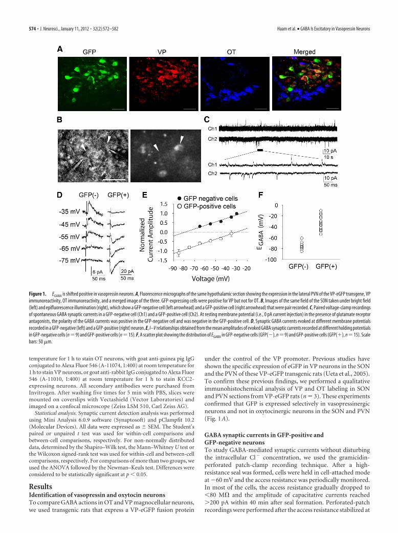
Figure 1. EGABA is shifted positive in vasopressin neurons. A, Fluorescence micrographs of the same hypothalamic section showing the expression in the lateral PVN of the VP-eGFP transgene, VPimmunoreactivity, OT immunoreactivity, and a merged image of the three. GFP-expressing cells were positive for VP but not for OT. B, Images of the same field of the SON taken under bright field(left) and epifluorescence illumination (right), which show a GFP-negative cell (left arrowhead) and a GFP-positive cell (right arrowhead) that were pair recorded. C, Paired voltage-clamp recordingsof spontaneous GABA synaptic currents in a GFP-negative cell (Ch1) and a GFP-positive cell (Ch2). At resting membrane potential (i.e., 0 pA current injection) in the presence of glutamate receptorantagonists, the polarity of the GABA currents was positive in the GFP-negative cell and was negative in the GFP-positive cell. D, Synaptic GABA currents evoked at different membrane potentialsrecorded in a GFP-negative (left) and a GFP-positive (right) neuron. E, I–V relationships obtained from the mean amplitudes of evoked GABA synaptic currents recorded at different holding potentialsin GFP-negative cells (n �9) and GFP-positive cells (n �15). F, A scatter plot showing the distribution of EGABA in GFP-negative cells (GFP(�), n �9) and GFP-positive cells (GFP(�), n �15). Scalebars: 50 �m.
574 • J. Neurosci., January 11, 2012 • 32(2):572–582 Haam et al. • GABA Is Excitatory in Vasopressin Neurons
between 30 and 80 M�. All perforated-patch recordings of syn-aptic currents were performed in the presence of the ionotropicglutamate receptor antagonists DNQX (15 �M) and DL-AP5 (50�M) to isolate GABAergic synaptic currents (Wuarin and Dudek,1993). In GFP-negative, putative OT neurons recorded with littleor no holding current (i.e., near resting potential, n � 12), spon-taneous synaptic currents were outward. GFP-positive, VP neu-rons recorded at resting potential (n � 18) generated inwardspontaneous synaptic currents. In simultaneous recordings ofmixed pairs of GFP-positive cells and GFP-negative cells re-corded at resting potential, spontaneous synaptic currents wereinward in the GFP-positive cells and outward in the GFP-negative cells (Fig. 1B,C). Both the spontaneous outward synap-tic currents recorded in putative OT neurons (n � 5) and thespontaneous inward currents recorded in VP neurons (n � 6)were completely blocked with the GABAA receptor antagonistbicuculline methiodide (60 �M), indicating that they wereGABAergic synaptic currents and suggesting that GABA is inhib-itory at resting potential in OT neurons (Vm � �53.3 � 2.3 mV,n � 15), and that, surprisingly, GABA is excitatory at restingpotential in VP neurons (Vm � �53.3 � 1.5 mV, n � 22).
GABA reversal potential in GFP-positive andGFP-negative neuronsThe striking difference in the polarity of synaptic GABA currentsat resting membrane potential between VP neurons and putativeOT neurons prompted us to examine whether there is a differ-ence in the EGABA between the two cell groups. A current–voltage( I–V) relationship of GABA currents was generated by evokingsynaptic currents with extracellular electrical stimulation of af-ferent inputs (see Materials and Methods) at different holdingpotentials with ionotropic glutamate receptors blocked (inDNQX and DL-AP5) to isolate GABA currents (Fig. 1D). TheEGABA in putative OT neurons was �75.1 � 2.6 mV (n � 9),whereas the EGABA in VP neurons was significantly more positive,at �33.1 � 3.4 mV (n � 15, Student’s t test, p � 0.001) (Fig.1E,F). The EGABA was negative with respect to resting potential in
OT neurons, resulting in outward GABA currents in OT neuronsat rest, whereas it was positive to the resting membrane potentialin VP neurons, resulting in inward GABA currents at rest.
Since male rats undergo sexual maturation at �6 weeks of age,one possibility is that many of the magnocellular neurons that werecorded from were from rats that had not yet reached pubertyand in which an excitatory-to-inhibitory developmental switchof the GABA response had not yet occurred. To control for thispossibility, we divided recordings into those from young rats,aged 34 –39 d, and from older rats, aged 53– 80 d, and comparedthe EGABA in the two groups. The EGABA in VP neurons from theolder rats (�28.6 � 3.5 mV, n � 12) was not significantly differ-ent from that in neurons from the young rats (�39.4 � 3.9 mV,n � 6; p � 0.075, Student’s t test) (data not shown), suggestingthat the positive EGABA is not due to immaturity of the VPneurons.
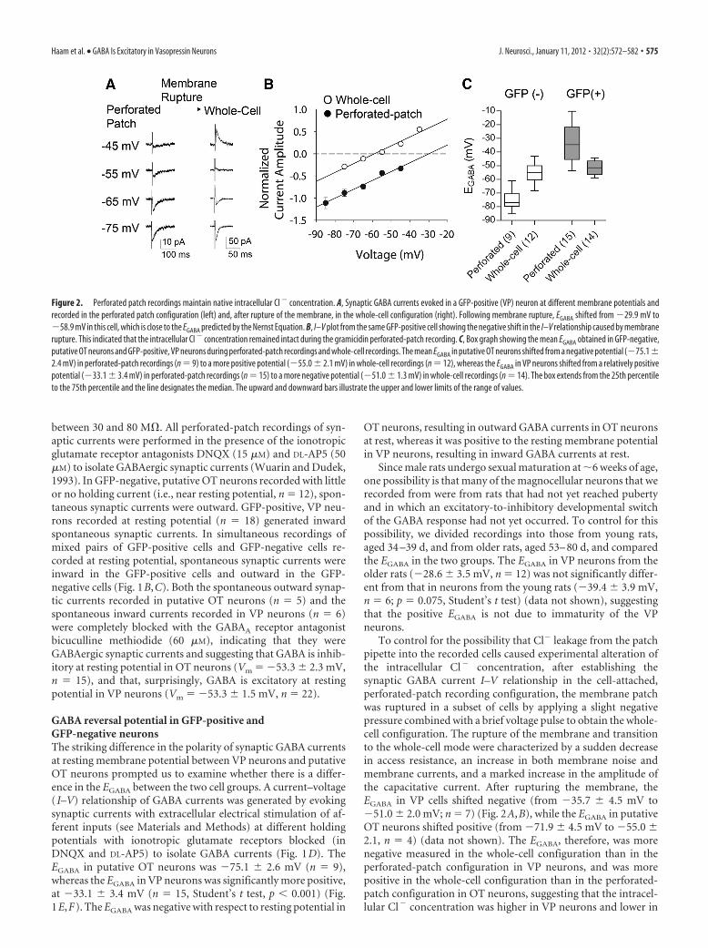
To control for the possibility that Cl� leakage from the patchpipette into the recorded cells caused experimental alteration ofthe intracellular Cl� concentration, after establishing thesynaptic GABA current I–V relationship in the cell-attached,perforated-patch recording configuration, the membrane patchwas ruptured in a subset of cells by applying a slight negativepressure combined with a brief voltage pulse to obtain the whole-cell configuration. The rupture of the membrane and transitionto the whole-cell mode were characterized by a sudden decreasein access resistance, an increase in both membrane noise andmembrane currents, and a marked increase in the amplitude ofthe capacitative current. After rupturing the membrane, theEGABA in VP cells shifted negative (from �35.7 � 4.5 mV to�51.0 � 2.0 mV; n � 7) (Fig. 2A,B), while the EGABA in putativeOT neurons shifted positive (from �71.9 � 4.5 mV to �55.0 �2.1, n � 4) (data not shown). The EGABA, therefore, was morenegative measured in the whole-cell configuration than in theperforated-patch configuration in VP neurons, and was morepositive in the whole-cell configuration than in the perforated-patch configuration in OT neurons, suggesting that the intracel-lular Cl� concentration was higher in VP neurons and lower in
Figure 2. Perforated patch recordings maintain native intracellular Cl � concentration. A, Synaptic GABA currents evoked in a GFP-positive (VP) neuron at different membrane potentials andrecorded in the perforated patch configuration (left) and, after rupture of the membrane, in the whole-cell configuration (right). Following membrane rupture, EGABA shifted from �29.9 mV to�58.9 mV in this cell, which is close to the EGABA predicted by the Nernst Equation. B, I–V plot from the same GFP-positive cell showing the negative shift in the I–V relationship caused by membranerupture. This indicated that the intracellular Cl � concentration remained intact during the gramicidin perforated-patch recording. C, Box graph showing the mean EGABA obtained in GFP-negative,putative OT neurons and GFP-positive, VP neurons during perforated-patch recordings and whole-cell recordings. The mean EGABA in putative OT neurons shifted from a negative potential (�75.1�2.4 mV) in perforated-patch recordings (n � 9) to a more positive potential (�55.0 � 2.1 mV) in whole-cell recordings (n � 12), whereas the EGABA in VP neurons shifted from a relatively positivepotential (�33.1 � 3.4 mV) in perforated-patch recordings (n � 15) to a more negative potential (�51.0 � 1.3 mV) in whole-cell recordings (n � 14). The box extends from the 25th percentileto the 75th percentile and the line designates the median. The upward and downward bars illustrate the upper and lower limits of the range of values.
Haam et al. • GABA Is Excitatory in Vasopressin Neurons J. Neurosci., January 11, 2012 • 32(2):572–582 • 575
OT neurons than that in the patch solution. This suggests that thenative intracellular Cl� concentration in VP neurons (� 41 mM)is higher than that in OT neurons (� 8 mM, calculated accordingto the Nernst Equation), and that the positive EGABA measured inVP neurons seen in perforated patch recordings is not contami-nated by leakage of Cl� from the patch pipette into the recordedcell. In whole-cell recordings, there was no significant differencein the EGABA between putative OT and VP neurons (�55.0 � 2.1mV, n � 12 putative OT cells; � 51.6 � 1.3 mV, n � 14 VP cells;p � 0.166, Mann–Whitney U test) (Fig. 2C).
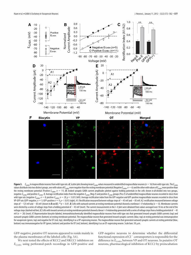
GABA reversal potential in magnocellular neurons fromwild-type ratsPrevious studies have shown that GFP expression does not affectthe intracellular Cl� concentration or EGABA (Kuner and Augus-tine, 2000; DeFazio et al., 2002; Fu and van den Pol, 2007), andthat the expression of VP in the VP-eGFP transgenic rats is notsignificantly different from that in wild-type rats (Fujio et al.,2006). To confirm that the positive EGABA in VP-eGFP-expressing cells was not a result of the GFP expression or thegenetic changes in the VP neurons, we determined the EGABA inmagnocellular neurons from the SON and PVN of age-matchedwild-type male Wistar rats. Consistent with the data from theVP-eGFP transgenic rats, the measured values of EGABA in mag-nocellular neurons from wild-type rats distributed into twogroups, one group with an EGABA negative to the resting mem-brane potential (�75.4 � 3.8 mV mV, n � 6) and the other groupwith an EGABA positive to the resting potential (�37.2 � 2.6 mV,n � 11) (Fig. 3A,B). The cells assigned to each of the two groupson the basis of their EGABA were tested for membrane propertiescharacteristic of OT or VP neurons. There was a significant dif-ference in the rectification index, defined as (I�45 mV �I�85 mV)/(I�85 mV � I�125 mV), between the two cell groups(0.40 � 0.04 in cells with outward GABA currents at restingmembrane potential, n � 8; 0.83 � 0.04 in cells with inwardGABA currents at resting membrane potential, n � 20, p � 0.001,Student’s t test) (Fig. 3C). Thus, the cells that showed a negativeEGABA generated a hyperpolarization-induced inward rectifica-tion, which is manifested as a non-linearity in the membranepotential range between �85 mV and �125 mV and is charac-teristic of OT, but not VP, neurons (Hirasawa et al., 2003) (Fig.3D). The cells that showed a positive EGABA had a linear or near-linear I–V relationship, which is characteristic of VP neurons(Fig. 3E). Additionally, the cells with a hyperpolarized EGABA
were found in the ventromedial part of the PVN and the dorso-lateral part of the SON, where OT neurons are concentrated,while the cells with a depolarized EGABA were found in the dor-solateral part of the PVN and in the ventromedial part of theSON, where VP neurons are concentrated (data not shown) (Ka-naka et al., 2001; Kawasaki et al., 2006; Simmons and Swanson,2009).
Finally, to confirm the identity of the two populations ofmagnocellular neurons in wild-type rats as oxytocinergic andvasopressinergic, following characterization of synaptic GABAcurrents with perforated-patch recordings, we labeled neuronsby including biocytin in the patch solution and infusing biocytinintracellularly by rupturing the membrane at the end of record-ings to achieve the whole-cell configuration. After recording andinjecting one or two neurons in a slice, the slice was processedfor immunohistochemistry using antibodies against OT and VP(see Materials and Methods). Of 16 neurons that generated in-ward spontaneous synaptic currents at resting membrane poten-tial (with glutamate receptors blocked), 15 stained positively for
VP and negatively for OT (Fig. 3F, top); of three neurons thatgenerated outward spontaneous synaptic currents at resting po-tential, two stained positively for OT and negatively for VP (Fig.3F, bottom). Together, these results in slices from wild-type ratsconfirmed our findings in VP-eGFP rats of a positive EGABA in VPneurons and a negative EGABA in OT neurons.
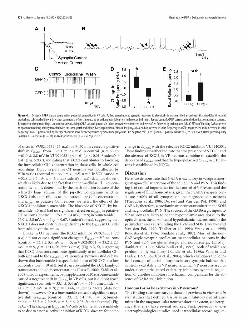
GABA excites vasopressinergic magnocellular neuronsTo determine whether depolarizing GABA synaptic currentsin VP neurons are excitatory and can trigger action potentials,cells were recorded in the perforated patch mode and wereheld at a membrane potential close to spike threshold, wherespontaneous action potential currents were often observed. Atthis potential, spontaneous inward GABA currents were ob-served, indicating that GABA is excitatory near spike thresh-old. In addition, extracellular electrical stimulation elicitedinward synaptic GABA currents, which often triggered actionpotential currents (Fig. 4 A). In current-clamp mode, VP neu-rons held near spike threshold in the presence of ionotropicglutamate receptor antagonists showed spontaneous GABA-mediated depolarizing synaptic potentials that were often fol-lowed by action potential firing (Fig. 4 B).
We next examined whether basal GABA levels have an excit-atory effect on spontaneous spiking activity in VP cells. To recordspiking activity without influencing the EGABA, we performedextracellular loose-seal cell-attached patch recordings, which al-low the plasma membrane at the patch to remain intact and thusthe recorded cell to maintain its intracellular Cl� concentration.This provides an accurate measurement of spontaneous firingactivity with minimal influence of the recording conditions(Nunemaker et al., 2003; Perkins, 2006). The frequency of spon-taneous spiking activity in VP neurons recorded in the loose-sealmode was significantly reduced by bath application of the GABAA
receptor antagonist bicuculline methiodide (10 �M) (from1.34 � 0.50 Hz to 0.64 � 0.32 Hz, n � 7, p � 0.05, Wilcoxonsigned-rank test) (Fig. 4C,D), suggesting that tonic GABA releasehas an excitatory influence on spontaneous spike generation inVP neurons. Blocking GABAA receptor activity had the oppositeeffect on spiking activity in GFP-negative, putative OT neurons.Thus, bicuculline (10 �M) caused a significant increase in thefiring frequency of GFP-negative cells (from 0.69 � 0.71 Hz to1.60 � 0.63 Hz, n � 6, p � 0.05, Wilcoxon signed-rank test) (Fig.4C,D), indicating that GABA acts basally to inhibit spiking in OTneurons. Interestingly, the average basal spike frequency in VPneurons was significantly greater than that of OT neurons(1.55 � 0.36 in VP neurons, n � 25, vs 0.52 � 0.30 Hz in OTneurons, n � 11, Mann–Whitney U test, p � 0.01) (Fig. 4E), anddecreased to below the spike frequency of OT neurons whenGABAA receptors in both cell types were blocked (0.64 � 0.32 Hzin VP neurons vs 1.60 � 0.63 Hz in OT neurons).
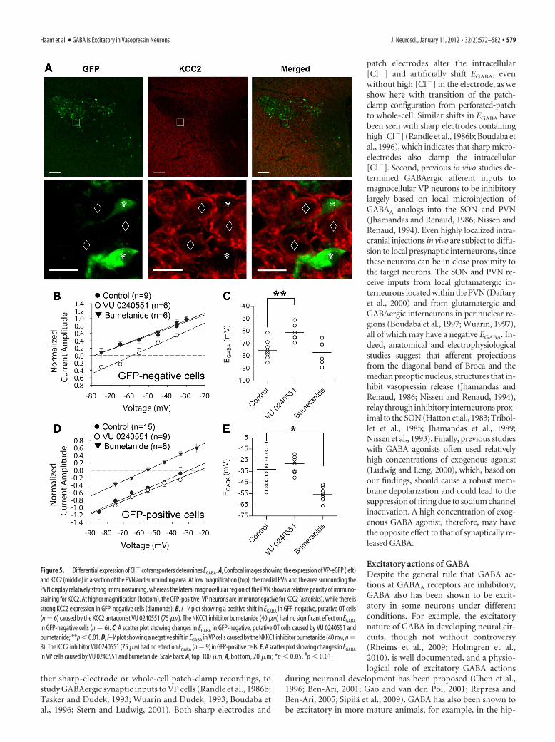
Differential chloride transport in vasopressin andoxytocin neuronsPrevious immunohistochemical studies have shown a relativepaucity of expression of the KCC2 cotransporter in hypothalamicVP neurons (Kanaka et al., 2001; Belenky et al., 2010). Our im-munohistochemistry findings showed strong KCC2 immunola-beling in the hypothalamic areas surrounding the SON and PVN,but relatively weaker labeling in the SON and PVN (Fig. 5A).Some labeling was seen in GFP-negative, putative OT neurons,but no KCC2 immunolabeling was seen in GFP-positive VP neu-rons (Fig. 5A), suggesting a relative lack of KCC2 expression inGFP-positive VP neurons. The KCC2 labeling that was seen in
576 • J. Neurosci., January 11, 2012 • 32(2):572–582 Haam et al. • GABA Is Excitatory in Vasopressin Neurons
GFP-negative, putative OT neurons appeared to reside mainly inthe plasma membranes of the labeled cells (Fig. 5A).
We next tested the effects of KCC2 and NKCC1 inhibitors onEGABA using perforated-patch recordings in GFP-positive and
GFP-negative neurons to determine whether the differentialfunctional expression of Cl� cotransporters is responsible for thedifference in EGABA between VP and OT neurons. In putative OTneurons, pharmacological inhibition of KCC2 by preincubation
Figure 3. EGABA in magnocellular neurons from wild-type rats. A, Scatter plot showing mean EGABA values measured in unidentified magnocellular neurons (n�16) from wild-type rats. The EGABA
values distribute into two distinct groups, one with values of EGABA more negative than the resting membrane potential (Negative EGABA, n � 6) and the other with values of EGABA more positive thanthe resting membrane potential (Positive EGABA, n � 11). B, Evoked synaptic GABA current amplitudes plotted against holding potentials in the cells shown in A divided into two groups,negative-EGABA and positive-EGABA. C, Average rectification index from the negative-EGABA (Neg-E) and positive-EGABA groups (Pos-E) of unidentified magnocellular neurons recorded in slices fromwild-type rats (negative-EGABA, n � 8; positive-EGABA, n � 20, p � 0.01) (left). Average rectification index from the GFP-negative and GFP-positive magnocellular neurons recorded in slices fromVP-GFP rats (GFP-negative, n � 5; GFP-positive n � 9, p � 0.01) (right). A1: Rectification measured between voltage steps of �45 mV and �85 mV; A2: rectification measured between voltagesteps of �125 mV and �85 mV (shown in D and E); **p � 0.01. D, Cells with outward currents at resting membrane potential showed a nonlinear I–V relationship (n � 8). Membrane currentswere elicited by a series of voltage steps from a holding potential of �45 mV (inset). The current measurements in the I–V plot were obtained from values averaged over 10 ms at the end of thevoltage steps (dashed red line). E, Cells with inward currents at resting membrane potential showed a linear I–V relationship generated with a series of voltage steps from a holding potential of �45mV (n � 20) (inset). F, Representative biocytin-labeled, immunohistochemically identified magnocellular neurons from wild-type rats that generated inward synaptic GABA currents (top) andoutward synaptic GABA currents (bottom) at resting membrane potential. The magnocellular neuron that generated inward synaptic currents (blue, top) at resting potential was immunopositivefor vasopressin (green, top) and negative for OT (red, top), identifying it as a VP-expressing neuron. The magnocellular neuron that generated outward synaptic currents at resting potential (blue,bottom) was immunonegative for VP (green, bottom) and positive for OT (red, bottom), identifying it as an OT-expressing neuron. Scale bars: 50 �m.
Haam et al. • GABA Is Excitatory in Vasopressin Neurons J. Neurosci., January 11, 2012 • 32(2):572–582 • 577
of slices in VU0240551 (75 �M) for � 30 min caused a positiveshift in EGABA, from �75.1 � 2.4 mV in control (n � 9) to�61.0 � 2.8 mV in VU0240551 (n � 6) (p � 0.01, Student’s ttest) (Fig. 5B,C), indicating that KCC2 contributes to loweringthe intracellular Cl� concentration in these cells. In whole-cellrecordings, EGABA in putative OT neurons was not affected byVU0240551 (control � �55.0 � 3.1 mV, n � 8; in VU0240551 ��52.8 � 3.5 mV, n � 4; n.s., Student’s t test) (data not shown),which is likely due to the fact that the intracellular Cl� concen-tration is mainly determined by the patch solution because of therelatively large volume of the pipette. To examine whetherNKCC1 also contributes to the intracellular Cl� concentrationand EGABA in putative OT neurons, we tested the effect of theNKCC1 inhibitor bumetanide. The blockade of NKCC1 by bu-metanide (40 �M) had no significant effect on EGABA in putativeOT neurons (control: � 75.1 � 2.4 mV, n � 9; in bumetanide: �77.0 � 3.8 mV, n � 6, p � 0.67, Student’s t test), suggesting thatNKCC1 does not contribute significantly to the EGABA in OT cellsfrom adult hypothalamus.
Unlike in OT neurons, the KCC2 inhibitor VU0240551 (75�M) did not cause a significant change in EGABA in VP neurons(control: � 33.1 � 3.4 mV, n � 15; in VU0240551: � 28.2 � 2.5mV, n � 9, p � 0.311, Student’s t test) (Fig. 5D,E), suggestingthat KCC2 does not contribute significantly to intracellular Cl�
buffering and to the EGABA in VP neurons. Previous studies haveshown that bumetanide is a specific inhibitor of NKCC1 at a lowconcentration (�10 �M), but it can also inhibit the KCC family oftransporters at higher concentrations (Russell, 2000; Kahle et al.,2008). In our experiments, bath application of 20 �M bumetanidecaused a negative shift in EGABA in VP cells, but it did not reachsignificance (control: � 33.1 � 3.4 mV, n � 15; bumetanide: �44.7 � 5.3 mV, n � 9; p � 0.066, Student’s t test) (data notshown); however, 40 �M bumetanide caused a significant nega-tive shift in EGABA (control: � 33.1 � 3.4 mV, n � 15; bumet-anide: � 55.7 � 2.2 mV, n � 8, p � 0.05, Student’s t test) (Fig.5D,E). The change in EGABA in VP cells by bumetanide is unlikelyto be due to a nonselective inhibition of KCC2 since we found no
change in EGABA with the selective KCC2 inhibitor VU0240551.These findings together indicate that the presence of NKCC1 andthe absence of KCC2 in VP neurons combine to establish thedepolarized EGABA, and that the hyperpolarized EGABA in OT neu-rons is established by KCC2.
DiscussionHere, we demonstrate that GABA is excitatory in vasopressiner-gic magnocellular neurons of the adult SON and PVN. This find-ing is of critical importance for the control of VP release and theregulation of fluid homeostasis, given that GABA synapses con-stitute �60% of all synapses on the magnocellular neurons(Theodosis et al., 1986; Decavel and Van den Pol, 1990), andGABA is, therefore, a predominant neurotransmitter in the SONand magnocellular PVN. The sources of the GABAergic inputs toVP neurons are likely to be the hypothalamic area dorsal to theoptic chiasm, the dorsomedial hypothalamic nucleus, and/or theperinuclear areas surrounding the PVN and SON (Decavel andVan den Pol, 1990; Thellier et al., 1994; Vrang et al., 1995;Boudaba et al., 1996; Boudaba et al., 1997). Most of the non-GABAergic synaptic profiles on magnocellular neurons in thePVN and SON are glutamatergic and noradrenergic (El Maj-doubi et al., 1997; Michaloudi et al., 1997), both of which arepredominantly excitatory (Randle et al., 1986a; Wuarin andDudek, 1993; Boudaba et al., 2003), which challenges the long-held concept of an inhibitory-excitatory synaptic balance thatcontrols excitability in VP neurons. Either VP neurons are notunder a counterbalanced excitatory-inhibitory synaptic regula-tion, or another inhibitory mechanism compensates for the ab-sence of GABAergic inhibition.
How can GABA be excitatory in VP neurons?This finding runs contrary to those of previous in vitro and invivo studies that defined GABA as an inhibitory neurotrans-mitter in the magnocellular neuroendocrine system, a discrep-ancy that may have several sources. First, previous in vitroelectrophysiological studies used intracellular recordings, ei-
Figure 4. Synaptic GABA signals cause action potential generation in VP cells. A, Two superimposed synaptic responses to electrical stimulation (filled arrowhead) that straddled threshold,producing a subthreshold inward synaptic current to the first stimulus and an action potential current to the second stimulus. Evoked synaptic GABA currents often induced action potential currents.B, In current-clamp recordings, spontaneous depolarizing GABA synaptic potentials (black arrows) were observed and were often followed by action potentials. C, Effect of blocking GABA currentson spontaneous firing activity recorded with the loose-patch technique. Bath application of bicuculline (10 �M) caused an increase in spike frequency in a GFP-negative cell and a decrease in spikefrequency in a GFP-positive cell. D, Average change in spike frequency caused by bicuculline (10 �M) in GFP-negative cells (n � 6) and GFP-positive cells (n � 7; *p � 0.05). E, Basal spike frequency(in Hz) in GFP-negative (n � 11) and GFP-positive cells (n � 25); **p � 0.01.
578 • J. Neurosci., January 11, 2012 • 32(2):572–582 Haam et al. • GABA Is Excitatory in Vasopressin Neurons
ther sharp-electrode or whole-cell patch-clamp recordings, tostudy GABAergic synaptic inputs to VP cells (Randle et al., 1986b;Tasker and Dudek, 1993; Wuarin and Dudek, 1993; Boudaba etal., 1996; Stern and Ludwig, 2001). Both sharp electrodes and
patch electrodes alter the intracellular[Cl�] and artificially shift EGABA, evenwithout high [Cl�] in the electrode, as weshow here with transition of the patch-clamp configuration from perforated-patchto whole-cell. Similar shifts in EGABA havebeen seen with sharp electrodes containinghigh [Cl�] (Randle et al., 1986b; Boudaba etal., 1996), which indicates that sharp micro-electrodes also clamp the intracellular[Cl�]. Second, previous in vivo studies de-termined GABAergic afferent inputs tomagnocellular VP neurons to be inhibitorylargely based on local microinjection ofGABAA analogs into the SON and PVN(Jhamandas and Renaud, 1986; Nissen andRenaud, 1994). Even highly localized intra-cranial injections in vivo are subject to diffu-sion to local presynaptic interneurons, sincethese neurons can be in close proximity tothe target neurons. The SON and PVN re-ceive inputs from local glutamatergic in-terneurons located within the PVN (Daftaryet al., 2000) and from glutamatergic andGABAergic interneurons in perinuclear re-gions (Boudaba et al., 1997; Wuarin, 1997),all of which may have a negative EGABA. In-deed, anatomical and electrophysiologicalstudies suggest that afferent projectionsfrom the diagonal band of Broca and themedian preoptic nucleus, structures that in-hibit vasopressin release (Jhamandas andRenaud, 1986; Nissen and Renaud, 1994),relay through inhibitory interneurons prox-imal to the SON (Hatton et al., 1983; Tribol-let et al., 1985; Jhamandas et al., 1989;Nissen et al., 1993). Finally, previous studieswith GABA agonists often used relativelyhigh concentrations of exogenous agonist(Ludwig and Leng, 2000), which, based onour findings, should cause a robust mem-brane depolarization and could lead to thesuppression of firing due to sodium channelinactivation. A high concentration of exog-enous GABA agonist, therefore, may havethe opposite effect to that of synaptically re-leased GABA.
Excitatory actions of GABADespite the general rule that GABA ac-tions at GABAA receptors are inhibitory,GABA also has been shown to be excit-atory in some neurons under differentconditions. For example, the excitatorynature of GABA in developing neural cir-cuits, though not without controversy(Rheims et al., 2009; Holmgren et al.,2010), is well documented, and a physio-logical role of excitatory GABA actions
during neuronal development has been proposed (Chen et al.,1996; Ben-Ari, 2001; Gao and van den Pol, 2001; Represa andBen-Ari, 2005; Sipila et al., 2009). GABA has also been shown tobe excitatory in more mature animals, for example, in the hip-
Figure 5. Differential expression of Cl � cotransporters determines EGABA. A, Confocal images showing the expression of VP-eGFP (left)and KCC2 (middle) in a section of the PVN and surrounding area. At low magnification (top), the medial PVN and the area surrounding thePVN display relatively strong immunostaining, whereas the lateral magnocellular region of the PVN shows a relative paucity of immuno-staining for KCC2. At higher magnification (bottom), the GFP-positive, VP neurons are immunonegative for KCC2 (asterisks), while there isstrong KCC2 expression in GFP-negative cells (diamonds). B, I–V plot showing a positive shift in EGABA in GFP-negative, putative OT cells(n � 6) caused by the KCC2 antagonist VU 0240551 (75 �M). The NKCC1 inhibitor bumetanide (40 �M) had no significant effect on EGABA
in GFP-negative cells (n � 6). C, A scatter plot showing changes in EGABA in GFP-negative, putative OT cells caused by VU 0240551 andbumetanide; **p�0.01. D, I–V plot showing a negative shift in EGABA in VP cells caused by the NKKC1 inhibitor bumetanide (40 mM, n�8). The KCC2 inhibitor VU 0240551 (75 �M) had no effect on EGABA (n�9) in GFP-positive cells. E, A scatter plot showing changes in EGABA
in VP cells caused by VU 0240551 and bumetanide. Scale bars: A, top, 100 �m; A, bottom, 20 �m; *p � 0.05, #p � 0.01.
Haam et al. • GABA Is Excitatory in Vasopressin Neurons J. Neurosci., January 11, 2012 • 32(2):572–582 • 579
pocampus, cortex, amygdala, and basal ganglia (Golding andOertel, 1996; Martina et al., 2001; Chavas and Marty, 2003;Gulledge and Stuart, 2003; Woodruff et al., 2006; Viitanen et al.,2010), although these findings were from immature neuronssince the studies were conducted in rodents aged 5 weeks oryounger, which leaves open the question of whether theexcitatory-to-inhibitory switch in these neurons may occur withpuberty. GABA has also been shown to be excitatory in matureanimals under specific conditions, such as during phases of thecircadian cycle (Wagner et al., 1997; De Jeu and Pennartz, 2002;Choi et al., 2008), in areas where neurogenesis occurs (Tozuka etal., 2005), and during stress (Hewitt et al., 2009; Kim et al., 2011).GABA’s excitatory actions are important in the adult brain notonly for the regulation of excitability, but also for synapse forma-tion and synaptic integration (Albus et al., 2005; Tozuka et al.,2005; Ge et al., 2006), and depend on a number of factors, includ-ing GABA channel conductance, cellular compartment, cell type,and basal neuronal activity (DeFazio et al., 2002; Han et al., 2002;Moenter and DeFazio, 2005; Fu and van den Pol, 2007; Herbisonand Moenter, 2011; Song et al., 2011).
The excitatory influence of GABA and the capacity of GABAto drive spike firing in VP cells is quite robust compared with thatreported in most other brain areas (Golding and Oertel, 1996;Bracci and Panzeri, 2006; Woodruff et al., 2009), where often thedepolarizing GABA signals maintain their inhibitory influencedue to shunting (Randle and Renaud, 1987; Zhang and Jackson,1993; Hewitt et al., 2009). The physiological significance of theexcitatory GABA input to VP neurons in the PVN and SON is notyet clear, although the robust suppression of spiking activity inVP neurons by blocking GABAA receptors suggests that the ex-citatory GABAergic input contributes to the higher firing fre-quency of VP neurons relative to OT neurons (Armstrong et al.,1994). Oxytocin neurons increased their firing frequency signif-icantly under GABAA receptor blockade, which indicates a robusttonic inhibitory GABAergic input to OT cells (Li et al., 2007;Popescu et al., 2010) and suggests that the tonic synaptic GABAinput to the two cell types largely determines their spontaneousspiking activity under these recording conditions.
A recent study showed that chronic osmotic stress induces apositive shift in EGABA in unidentified magnocellular neurons,making synaptic GABA inputs excitatory (Kim et al., 2011). Inthat study, GABA was inhibitory in 75% and excitatory in 25% ofunidentified magnocellular neurons from control animals. Ourfindings suggest that the excitatory GABA inputs in that studywere in VP neurons, and that the shift in EGABA with salt-loadingwas specific, therefore, to the OT neurons. Another study alsoreported depolarizing GABA responses in magnocellular neu-rons, also in a subset of recorded cells that may have been VPneurons (Randle and Renaud, 1987).
Differential expression of chloride cotransportersConsistent with our immunohistochemical confirmation of alack of KCC2 expression in VP neurons (Kanaka et al., 2001;Belenky et al., 2008), inhibition of KCC2 activity had no effect onEGABA in VP neurons, but shifted EGABA positive in OT neurons.Blockade of NKCC1, on the other hand, caused a significant neg-ative shift of EGABA in VP neurons, but had no effect in OT neu-rons, suggesting that NKCC1 accumulates intracellular chloridein VP cells, but not in OT cells. The lack of KCC2 expressionfound in VP neurons in perfused brain tissue suggests that theshift in chloride gradient that we observed in vitro is present invivo, and that it was not, therefore, the result of disrupted chlo-
ride transport induced selectively in VP neurons by the slicingprocedure.
Mechanisms that control the excitability ofvasopressin neuronsOur demonstration that GABA is excitatory in VP neurons raisesthe question of how the excitability of VP neurons is controlled, ifneither of the two main neurotransmitters, glutamate and GABA,is inhibitory. A complete loss of synaptic inhibition would beexpected to render the cells hyperexcitable, and implies that otherinhibitory inputs may take the place of GABA synapses on VPneurons to maintain appropriate levels of excitability. Furtherstudies are required to determine whether other neurotransmit-ters or gliotransmitters are responsible for providing an inhibi-tory input to VP neurons to counter the glutamate and GABAexcitatory synaptic inputs.
Thus, GABAergic synaptic inputs to VP neurons, and to OTneurons under some conditions, contribute to the excitatory syn-aptic control of the hypothalamic-neurohypophysial system. Itremains to be determined how this control integrates into abroader regulation of the excitability and activation of the hypo-thalamic magnocellular neurons, how plastic this control is un-der different physiological conditions, and what the molecularmechanisms are that are responsible for causing the positive shiftin EGABA in vasopressinergic neuroendocrine cells.
ReferencesAlbus H, Vansteensel MJ, Michel S, Block GD, Meijer JH (2005) A GABAe-
rgic mechanism is necessary for coupling dissociable ventral and dorsalregional oscillators within the circadian clock. Curr Biol 15:886 – 893.
Armstrong WE, Smith BN, Tian M (1994) Electrophysiological characteris-tics of immunochemically identified rat oxytocin and vasopressin neu-rones in vitro. J Physiol 475:115–128.
Belenky MA, Yarom Y, Pickard GE (2008) Heterogeneous expression ofgamma-aminobutyric acid and gamma-aminobutyric acid-associated re-ceptors and transporters in the rat suprachiasmatic nucleus. J Comp Neu-rol 506:708 –732.
Belenky MA, Sollars PJ, Mount DB, Alper SL, Yarom Y, Pickard GE (2010)Cell-type specific distribution of chloride transporters in the rat suprachi-asmatic nucleus. Neuroscience 165:1519 –1537.
Ben-Ari Y (2001) Developing networks play a similar melody. Trends Neu-rosci 24:353–360.
Ben-Barak Y, Russell JT, Whitnall MH, Ozato K, Gainer H (1985) Neuro-physin in the hypothalamo-neurohypophysial system. I. Production andcharacterization of monoclonal antibodies. J Neurosci 5:81–97.
Boudaba C, Szab o K, Tasker JG (1996) Physiological mapping of local in-hibitory inputs to the hypothalamic paraventricular nucleus. J Neurosci16:7151–7160.
Boudaba C, Schrader LA, Tasker JG (1997) Physiological evidence for localexcitatory synaptic circuits in the rat hypothalamus. J Neurophysiol77:3396 –3400.
Boudaba C, Di S, Tasker JG (2003) Presynaptic noradrenergic regulation ofglutamate inputs to hypothalamic magnocellular neurones. J Neuroen-docrinol 15:803– 810.
Bracci E, Panzeri S (2006) Excitatory GABAergic effects in striatal projec-tion neurons. J Neurophysiol 95:1285–1290.
Chavas J, Marty A (2003) Coexistence of excitatory and inhibitory GABAsynapses in the cerebellar interneuron network. J Neurosci 23:2019 –2031.
Chen G, Trombley PQ, van den Pol AN (1996) Excitatory actions of GABAin developing rat hypothalamic neurones. J Physiol 494:451– 464.
Choi HJ, Lee CJ, Schroeder A, Kim YS, Jung SH, Kim JS, Kim do Y, Son EJ,Han HC, Hong SK, Colwell CS, Kim YI (2008) Excitatory actions ofGABA in the suprachiasmatic nucleus. J Neurosci 28:5450 –5459.
Cordero-Erausquin M, Coull JA, Boudreau D, Rolland M, De Koninck Y(2005) Differential maturation of GABA action and anion reversal po-tential in spinal lamina I neurons: impact of chloride extrusion capacity.J Neurosci 25:9613–9623.
Daftary SS, Boudaba C, Tasker JG (2000) Noradrenergic regulation of par-
580 • J. Neurosci., January 11, 2012 • 32(2):572–582 Haam et al. • GABA Is Excitatory in Vasopressin Neurons
vocellular neurons in the rat hypothalamic paraventricular nucleus. Neu-roscience 96:743–751.
Decavel C, Van den Pol AN (1990) GABA: a dominant neurotransmitter inthe hypothalamus. J Comp Neurol 302:1019 –1037.
DeFazio RA, Heger S, Ojeda SR, Moenter SM (2002) Activation of A-typegamma-aminobutyric acid receptors excites gonadotropin-releasing hor-mone neurons. Mol Endocrinol 16:2872–2891.
De Jeu M, Pennartz C (2002) Circadian modulation of GABA function inthe rat suprachiasmatic nucleus: excitatory effects during the night phase.J Neurophysiol 87:834 – 844.
Dreifuss JJ, Kalnins I, Kelly JS, Ruf KB (1971) Action potentials and releaseof neurohypophysial hormones in vitro. J Physiol 215:805– 817.
Dutton A, Dyball RE (1979) Phasic firing enhances vasopressin release fromthe rat neurohypophysis. J Physiol 290:433– 440.
El Majdoubi M, Poulain DA, Theodosis DT (1997) Lactation-induced plas-ticity in the supraoptic nucleus augments axodendritic and axosomaticGABAergic and glutamatergic synapses: an ultrastructural analysis usingthe disector method. Neuroscience 80:1137–1147.
Engelmann M, Bull PM, Brown CH, Landgraf R, Horn TF, Singewald N,Ludwig M, Wotjak CT (2004) GABA selectively controls the secretoryactivity of oxytocin neurons in the rat supraoptic nucleus. Eur J Neurosci19:601– 608.
Fiumelli H, Cancedda L, Poo MM (2005) Modulation of GABAergic trans-mission by activity via postsynaptic Ca 2�-dependent regulation of KCC2function. Neuron 48:773–786.
Fu LY, van den Pol AN (2007) GABA excitation in mouse hilar neuropep-tide Y neurons. J Physiol 579:445– 464.
Fujio T, Fujihara H, Shibata M, Yamada S, Onaka T, Tanaka K, Morita H,Dayanithi G, Kawata M, Murphy D, Ueta Y (2006) Exaggerated re-sponse of arginine vasopressin-enhanced green fluorescent protein fusiongene to salt loading without disturbance of body fluid homeostasis in rats.J Neuroendocrinol 18:776 –785.
Gao XB, van den Pol AN (2001) GABA, not glutamate, a primary transmit-ter driving action potentials in developing hypothalamic neurons. J Neu-rophysiol 85:425– 434.
Ge S, Goh EL, Sailor KA, Kitabatake Y, Ming GL, Song H (2006) GABAregulates synaptic integration of newly generated neurons in the adultbrain. Nature 439:589 –593.
Golding NL, Oertel D (1996) Context-dependent synaptic action of glycin-ergic and GABAergic inputs in the dorsal cochlear nucleus. J Neurosci16:2208 –2219.
Gulledge AT, Stuart GJ (2003) Excitatory actions of GABA in the cortex.Neuron 37:299 –309.
Han SK, Abraham IM, Herbison AE (2002) Effect of GABA on GnRH neu-rons switches from depolarization to hyperpolarization at puberty in thefemale mouse. Endocrinology 143:1459 –1466.
Hatton GI, Ho YW, Mason WT (1983) Synaptic activation of phasic burst-ing in rat supraoptic nucleus neurones recorded in hypothalamic slices.J Physiol 345:297–317.
Herbison AE, Moenter SM (2011) Depolarising and hyperpolarising actionsof GABA(A) receptor activation on gonadotrophin-releasing hormoneneurones: towards an emerging consensus. J Neuroendocrinol23:557–569.
Hewitt SA, Wamsteeker JI, Kurz EU, Bains JS (2009) Altered chloride ho-meostasis removes synaptic inhibitory constraint of the stress axis. NatNeurosci 12:438 – 443.
Hirasawa M, Mouginot D, Kozoriz MG, Kombian SB, Pittman QJ (2003)Vasopressin differentially modulates non-NMDA receptors in vasopres-sin and oxytocin neurons in the supraoptic nucleus. J Neurosci23:4270 – 4277.
Holmgren CD, Mukhtarov M, Malkov AE, Popova IY, Bregestovski P, Zil-berter Y (2010) Energy substrate availability as a determinant of neuro-nal resting potential, GABA signaling and spontaneous network activityin the neonatal cortex in vitro. J Neurochem 112:900 –912.
Jhamandas JH, Renaud LP (1986) A gamma-aminobutyric-acid-mediatedbaroreceptor input to supraoptic vasopressin neurones in the rat.J Physiol 381:595– 606.
Jhamandas JH, Raby W, Rogers J, Buijs RM, Renaud LP (1989) Diagonalband projection towards the hypothalamic supraoptic nucleus: light andelectron microscopic observations in the rat. J Comp Neurol 282:15–23.
Kahle KT, Staley KJ, Nahed BV, Gamba G, Hebert SC, Lifton RP, Mount DB
(2008) Roles of the cation-chloride cotransporters in neurological dis-ease. Nat Clin Pract Neurol 4:490 –503.
Kanaka C, Ohno K, Okabe A, Kuriyama K, Itoh T, Fukuda A, Sato K (2001)The differential expression patterns of messenger RNAs encoding K-Clcotransporters (KCC1,2) and Na-K-2Cl cotransporter (NKCC1) in therat nervous system. Neuroscience 104:933–946.
Kawasaki M, Yamaga C, Onaka T, Saito J, Mera T, Hashimoto H, Fujihara H,Okimoto N, Ohnishi H, Nakamura T, Ueta Y (2006) The short chainsugar acid, 2-buten-4-olide, activates oxytocin-secreting neurons but notarginine vasopressin-secreting neurons in the hypothalamus of rats. BrainRes 1086:133–141.
Kim JS, Kim WB, Kim YB, Lee Y, Kim YS, Shen FY, Lee SW, Park D, Choi HJ,Hur J, Park JJ, Han HC, Colwell CS, Cho YW, Kim YI (2011) Chronichyperosmotic stress converts GABAergic inhibition into excitation in va-sopressin and oxytocin neurons in the rat. J Neurosci 31:13312–13322.
Kiyama H, Emson PC (1990) Evidence for the co-expression of oxytocin and vaso-pressinmessengerribonucleicacids inmagnocellularneurosecretorycells: simul-taneous demonstration of two neurohypophysin messenger ribonucleic acids byhybridization histochemistry. J Neuroendocrinol 2:257–259.
Kuner T, Augustine GJ (2000) A genetically encoded ratiometric indicatorfor chloride: capturing chloride transients in cultured hippocampal neu-rons. Neuron 27:447– 459.
Kyrozis A, Reichling DB (1995) Perforated-patch recording with gramici-din avoids artifactual changes in intracellular chloride concentration.J Neurosci Methods 57:27–35.
Li C, Tripathi PK, Armstrong WE (2007) Differences in spike train variabil-ity in rat vasopressin and oxytocin neurons and their relationship tosynaptic activity. J Physiol 581:221–240.
Ludwig M, Leng G (2000) GABAergic projection from the arcuate nucleusto the supraoptic nucleus in the rat. Neurosci Lett 281:195–197.
Luther JA, Halmos KC, Tasker JG (2000) A slow transient potassium cur-rent expressed in a subset of neurosecretory neurons of the hypothalamicparaventricular nucleus. J Neurophysiol 84:1814 –1825.
MacVicar BA, Andrew RD, Dudek FE, Hatton GI (1982) Synaptic inputsand action potentials of magnocellular neuropeptidergic cells: intracellu-lar recording and staining in slices of rat hypothalamus. Brain Res Bull8:87–93.
Martina M, Royer S, Par e D (2001) Cell-type-specific GABA responses andchloride homeostasis in the cortex and amygdala. J Neurophysiol86:2887–2895.
Michaloudi HC, el Majdoubi M, Poulain DA, Papadopoulos GC, TheodosisDT (1997) The noradrenergic innervation of identified hypothalamicmagnocellular somata and its contribution to lactation-induced synapticplasticity. J Neuroendocrinol 9:17–23.
Misgeld U, Deisz RA, Dodt HU, Lux HD (1986) The role of chloride trans-port in postsynaptic inhibition of hippocampal neurons. Science232:1413–1415.
Moenter SM, DeFazio RA (2005) Endogenous gamma-aminobutyric acidcan excite gonadotropin-releasing hormone neurons. Endocrinology146:5374 –5379.
Mohr E, Bahnsen U, Kiessling C, Richter D (1988) Expression of the vaso-pressin and oxytocin genes in rats occurs in mutually exclusive sets ofhypothalamic neurons. FEBS Lett 242:144 –148.
Nissen R, Renaud LP (1994) GABA receptor mediation of median preopticnucleus-evoked inhibition of supraoptic neurosecretory neurones in rat.J Physiol 479:207–216.
Nissen R, Cunningham JT, Renaud LP (1993) Lateral hypothalamic lesionsalter baroreceptor-evoked inhibition of rat supraoptic vasopressin neu-rones. J Physiol 470:751–766.
Nunemaker CS, DeFazio RA, Moenter SM (2003) A targeted extracellularapproach for recording long-term firing patterns of excitable cells: a prac-tical guide. Biol Proced Online 5:53– 62.
Payne JA, Stevenson TJ, Donaldson LF (1996) Molecular characterizationof a putative K-Cl cotransporter in rat brain. A neuronal-specific isoform.J Biol Chem 271:16245–16252.
Perkins KL (2006) Cell-attached voltage-clamp and current-clamp record-ing and stimulation techniques in brain slices. J Neurosci Methods154:1–18.
Plotkin MD, Kaplan MR, Peterson LN, Gullans SR, Hebert SC, Delpire E(1997) Expression of the Na(�)-K(�)-2Cl- cotransporter BSC2 in thenervous system. Am J Physiol 272:C173–183.
Popescu IR, Morton LA, Franco A, Di S, Ueta Y, Tasker JG (2010) Synchro-
Haam et al. • GABA Is Excitatory in Vasopressin Neurons J. Neurosci., January 11, 2012 • 32(2):572–582 • 581
nized bursts of miniature inhibitory postsynaptic currents. J Physiol588:939 –951.
Prescott SA, Sejnowski TJ, De Koninck Y (2006) Reduction of anion reversalpotential subverts the inhibitory control of firing rate in spinal lamina Ineurons: towards a biophysical basis for neuropathic pain. Mol Pain 2:32.
Randle JC, Renaud LP (1987) Actions of gamma-aminobutyric acid on ratsupraoptic nucleus neurosecretory neurones in vitro. J Physiol 387:629 – 647.
Randle JC, Bourque CW, Renaud LP (1986a) Alpha 1-adrenergic receptoractivation depolarizes rat supraoptic neurosecretory neurons in vitro.Am J Physiol 251:R569 –R574.
Randle JC, Bourque CW, Renaud LP (1986b) Characterization of sponta-neous and evoked inhibitory postsynaptic potentials in rat supraopticneurosecretory neurons in vitro. J Neurophysiol 56:1703–1717.
Represa A, Ben-Ari Y (2005) Trophic actions of GABA on neuronal devel-opment. Trends Neurosci 28:278 –283.
Rheims S, Holmgren CD, Chazal G, Mulder J, Harkany T, Zilberter T, Zil-berter Y (2009) GABA action in immature neocortical neurons directlydepends on the availability of ketone bodies. J Neurochem 110:1330 –1338.
Rivera C, Voipio J, Payne JA, Ruusuvuori E, Lahtinen H, Lamsa K, Pirvola U,Saarma M, Kaila K (1999) The K�/Cl- co-transporter KCC2 rendersGABA hyperpolarizing during neuronal maturation. Nature 397:251–255.
Roberts MM, Robinson AG, Fitzsimmons MD, Grant F, Lee WS, HoffmanGE (1993) c-fos expression in vasopressin and oxytocin neurons revealsfunctional heterogeneity within magnocellular neurons. Neuroendocri-nology 57:388 – 400.
Russell JM (2000) Sodium-potassium-chloride cotransport. Physiol Rev80:211–276.
Simmons DM, Swanson LW (2009) Comparison of the spatial distributionof seven types of neuroendocrine neurons in the rat paraventricular nu-cleus: toward a global 3D model. J Comp Neurol 516:423– 441.
Sipila ST, Huttu K, Yamada J, Afzalov R, Voipio J, Blaesse P, Kaila K (2009)Compensatory enhancement of intrinsic spiking upon NKCC1 disrup-tion in neonatal hippocampus. J Neurosci 29:6982– 6988.
Song I, Savtchenko L, Semyanov A (2011) Tonic excitation or inhibition isset by GABA(A) conductance in hippocampal interneurons. Nat Com-mun 2:376.
Stern JE, Ludwig M (2001) NO inhibits supraoptic oxytocin and vasopres-sin neurons via activation of GABAergic synaptic inputs. Am J PhysiolRegul Integr Comp Physiol 280:R1815–R1822.
Tasker JG, Dudek FE (1991) Electrophysiological properties of neurones inthe region of the paraventricular nucleus in slices of rat hypothalamus.J Physiol 434:271–293.
Tasker JG, Dudek FE (1993) Local inhibitory synaptic inputs to neurones of
the paraventricular nucleus in slices of rat hypothalamus. J Physiol469:179 –192.
Thellier D, Moos F, Richard P, Stoeckel ME (1994) Evidence for connec-tions between a discrete hypothalamic dorsochiasmatic area and the su-praoptic and paraventricular nuclei. Brain Res Bull 34:261–274.
Theodosis DT, Paut L, Tappaz ML (1986) Immunocytochemical analysis ofthe GABAergic innervation of oxytocin- and vasopressin-secreting neu-rons in the rat supraoptic nucleus. Neuroscience 19:207–222.
Tozuka Y, Fukuda S, Namba T, Seki T, Hisatsune T (2005) GABAergic ex-citation promotes neuronal differentiation in adult hippocampal progen-itor cells. Neuron 47:803– 815.
Tribollet E, Armstrong WE, Dubois-Dauphin M, Dreifuss JJ (1985) Extra-hypothalamic afferent inputs to the supraoptic nucleus area of the rat asdetermined by retrograde and anterograde tracing techniques. Neurosci-ence 15:135–148.
Ueta Y, Fujihara H, Serino R, Dayanithi G, Ozawa H, Matsuda K, Kawata M,Yamada J, Ueno S, Fukuda A, Murphy D (2005) Transgenic expressionof enhanced green fluorescent protein enables direct visualization forphysiological studies of vasopressin neurons and isolated nerve terminalsof the rat. Endocrinology 146:406 – 413.
Viitanen T, Ruusuvuori E, Kaila K, Voipio J (2010) The K�-Cl cotrans-porter KCC2 promotes GABAergic excitation in the mature rat hip-pocampus. J Physiol 588:1527–1540.
Vrang N, Larsen PJ, Møller M, Mikkelsen JD (1995) Topographical organi-zation of the rat suprachiasmatic-paraventricular projection. J CompNeurol 353:585– 603.
Wagner S, Castel M, Gainer H, Yarom Y (1997) GABA in the mammaliansuprachiasmatic nucleus and its role in diurnal rhythmicity. Nature387:598 – 603.
Wardle RA, Poo MM (2003) Brain-derived neurotrophic factor modulationof GABAergic synapses by postsynaptic regulation of chloride transport.J Neurosci 23:8722– 8732.
Woodin MA, Ganguly K, Poo MM (2003) Coincident pre- and postsynapticactivity modifies GABAergic synapses by postsynaptic changes in Cl-transporter activity. Neuron 39:807– 820.
Woodruff AR, Monyer H, Sah P (2006) GABAergic excitation in the baso-lateral amygdala. J Neurosci 26:11881–11887.
Woodruff A, Xu Q, Anderson SA, Yuste R (2009) Depolarizing effect ofneocortical chandelier neurons. Frontiers in neural circuits 3:15.
Wuarin JP (1997) Glutamate microstimulation of local inhibitory circuitsin the supraoptic nucleus from rat hypothalamus slices. J Neurophysiol78:3180 –3186.
Wuarin JP, Dudek FE (1993) Patch-clamp analysis of spontaneous synapticcurrents in supraoptic neuroendocrine cells of the rat hypothalamus.J Neurosci 13:2323–2331.
Zhang SJ, Jackson MB (1993) GABA-activated chloride channels in secre-tory nerve endings. Science 259:531–534.
582 • J. Neurosci., January 11, 2012 • 32(2):572–582 Haam et al. • GABA Is Excitatory in Vasopressin Neurons