apoe2 exaggerates ptsd-related behavioral, cognitive and neuroendocrine alterations
TRANSCRIPT

ApoE2 Exaggerates PTSD-Related Behavioral, Cognitive, and
Neuroendocrine Alterations
Lance A Johnson1, Damian G Zuloaga1, Erin Bidiman1, Tessa Marzulla1, Sydney Weber1, Helane Wahbeh2 andJacob Raber*,1,2,3
1Department of Behavioral Neuroscience, Oregon Health and Science University, Portland, OR, USA; 2Department of Neurology,Oregon Health and Science University, Portland, OR, USA; 3Department of Radiation Medicine and Division of Neuroscience, ONPRC,Oregon Health and Science University, Beaverton, OR, USA
Apolipoprotein E (apoE) is an essential component of lipoprotein particles in both the brain and periphery, and exists in three isoforms inthe human population: E2, E3, and E4. ApoE has numerous, well-established roles in neurobiology. Most notably, E4 is associated withearlier onset and increased risk of Alzheimer’s disease (AD). Although possession of E2 is protective in the context of AD, E2 appears toconfer an increased incidence and severity of posttraumatic stress disorder (PTSD). However, the biological processes underlying this linkremain unclear. In this study, we began to elucidate these associations by examining the effects of apoE on PTSD severity in combatveterans, and on PTSD-like behavior in mice with human apoE. In a group of 92 veterans with PTSD, we observed significantly higherClinician-Administered PTSD Scale and PTSD Checklist scores in E2+ individuals, as well as alterations in salivary cortisol levels.Furthermore, we measured behavioral and biological outcomes in mice expressing human apoE after a single stressful event as well asfollowing a period of chronic variable stress, a model of combat-related trauma. Mice with E2 showed impairments in fear extinction, andbehavioral, cognitive, and neuroendocrine alterations following trauma. To the best of our knowledge, these data constitute the firsttranslational demonstration of PTSD severity in men and PTSD-like symptoms in mice with E2, and point to apoE as a novel biomarker ofsusceptibility, and potential therapeutic target, for PTSD.Neuropsychopharmacology (2015) 40, 2443–2453; doi:10.1038/npp.2015.95; published online 6 May 2015
�����������������������������������������������
INTRODUCTION
Posttraumatic stress disorder (PTSD) is a debilitating mentaldisorder that is characterized by re-experiencing, avoidance,and hyperarousal (American Psychiatric Association, 2013).The detrimental effects of PTSD are often magnified byaccelerated development of stress-related medical conditions,and PTSD displays a high comorbidity rate with anxiety anddepressive disorders (Gadermann et al, 2012). Following atraumatic event, many people experience traumatic stresssymptoms, but rates appear to diminish quickly over thecourse of a few months (Rubin et al, 2005; Galea et al, 2003).Thus, PTSD has been characterized as a condition in which apositive course of recovery from trauma is impeded (Yehudaand LeDoux, 2007). It has been estimated that close to 90% ofpeople are exposed to at least one traumatic event, such asrape, assault, disaster, rescue work, or combat, over the courseof their lifetime (Kilpatrick et al, 2013). However, PTSDdevelops only in a subgroup of people exposed to a traumaticevent, with the lifetime prevalence of PTSD estimatedbetween 7.8 and 8.7% (Kessler et al, 1995, 2005), thus
supporting a role for genetic risk factors (Pittman et al, 2012;Yehuda et al, 2011).Why some individuals exposed to trauma develop PTSD
and others do not is a longstanding question. Identifyingspecific genes that are associated with PTSD risk couldprovide new understanding of the cause of this disorder, andeventually lead to the design of new therapeutics. Several geneswithin the hypothalamic-pituitary-adrenal (HPA) axis(FKBP5, GCCR, CNR1), dopaminergic (DRD), and serotoner-gic (SLC6A4) systems have been linked to PTSD (Corneliset al, 2010). Genes encoding neurotrophins (BDNF), G-pro-tein signaling (RGS2), and lipoproteins have also have beeninvestigated, including the recent association of the ε2 allele ofthe gene encoding apolipoprotein E (apoE) with PTSD riskand symptomatology (Freeman et al, 2005; Kim et al, 2013).ApoE is an essential component of lipoprotein particles bothin the brain and in the periphery, and exists in three isoformsin the human population: apoE2 (E2), apoE3 (E3), and apoE4(E4) (Mahley and Rall, 2000). ApoE has numerous roles inbehavioral and cognitive functions, including regulation ofanxiety and cognitive performance during normal aging andin the context of neurodegenerative disease (Raber et al, 2003;Raber, 2007; Verghese et al, 2011). Several neurobiologicalfunctions associated with PTSD have been shown to bemodulated by apoE isoform, including hippocampal volume(Hostage et al, 2013), cognitive impairment (Caselli et al,
*Correspondence: Dr J Raber, Department of Behavioral Neuroscience,L470, Oregon Health and Science University, Portland, OR 97239, USA,Tel: +1 503 494 1524, Fax: +1 503 494 6877, E-mail: [email protected] 21 November 2014; revised 23 January 2015; accepted 31March 2015; accepted article preview online 10 April 2015
Neuropsychopharmacology (2015) 40, 2443–2453© 2015 American College of Neuropsychopharmacology. All rights reserved 0893-133X/15
www.neuropsychopharmacology.org
2007), and neuroendocrine alterations related to the HPA axis(Gil-Bea et al, 2010; Peskind et al, 2001; Raber et al, 2000).Compared with E3, E4 has been associated with decreased
brain metabolism (Reiman et al, 2004), deficient repairfollowing injury (Laskowitz et al, 1998), and earlier onset andincreased risk of Alzheimer’s disease (AD), whereas E2 isgenerally protective (Farrer et al, 1997). However, E2 wasrecently associated with an increase in the risk and severityof PTSD in two studies (Freeman et al, 2005; Kim et al,2013). In the first study, E2 was associated with an increasedrisk of PTSD and moderated the interaction between PTSDand alcohol use (Kim et al, 2013). In the second, E2 wasassociated with decreased memory function and more severere-experiencing symptoms in chronic, combat-related PTSDsubjects (Freeman et al, 2005). Consistent with these twohuman studies, we recently demonstrated that homozygousmice expressing only human E2 exhibit impaired extinctionof contextual fear memory (Olsen et al, 2012). Impairedextinction of fear memory has been proposed to reflect theprocesses underlying the recurring and re-experiencingsymptoms of PTSD (Jovanovic and Ressler, 2010).Although this recently identified link between apoE and
PTSD is of great interest, the biological processes underlyingthis association remain unclear. In the current study, wesought to elucidate these associations by investigating theeffects of apoE on symptomatology in humans with PTSD, aswell as examining PTSD-like behavioral and physiologicalchanges in a mouse model of human apoE.
MATERIALS AND METHODS
Participants, Cortisol Collection, ApoE Measurement,and APOE Genotyping
Human data were collected as part of a larger clinical trial(n= 114). Participants were mostly male combat veteransfrom the Vietnam Era. Detailed descriptions of participantsand data collection are available in the SupplementaryMaterials and Methods section. The study was approved bythe Portland Veterans Administration Medical Center andOregon Health and Science University institutional reviewboards. Written informed consent was obtained from allsubjects. Data used in this analysis included genetic data for110 individuals, 6 of whom were female (all E3/E3) and theirdata were not included in this study; complete clinicalmeasures for 92 men; and cortisol measurements for 82 ofthese individuals. Salivary cortisol was collected at waking,30 min after waking, and bedtime using Sarstedt Salivettes(Sarstedt, Germany) on two consecutive weekdays, asdescribed previously (Wahbeh et al, 2008; Wahbeh andOken, 2013). Participants fasted for the waking and 30-minsample, and the importance of collecting the sampleimmediately upon waking and exactly 30 min after wasstressed to the participants. Participants recorded their salivacollection times on a data sheet. Plasma was collected fromunfasted individuals at the screening visit, and apoEconcentrations were determined in diluted plasma (1:300)using a commercial assay (Abcam, Cambridge, MA). APOEgenotype was determined from whole blood DNA samples,and cortisol values were quantified in duplicate with enzyme-linked immunoassay (Salimetrics, State College, PA), by theOregon Clinical and Translational Research Institute.
Experimental Animals
Mice expressing human apoE isoforms under control of themouse apoE promoter (targeted replacement mice) werebackcrossed for at least 10 generations to the C57BL/6background (Knouff et al, 1999; Sullivan et al, 1997, 1998).Mice were fed normal chow diet ad libitum (Prolab IsoProRMH 3000; Agway). All analyses were carried out in mice4–5 months of age. Only male mice were used for the currentstudy. All procedures were in compliance with the NationalInstitutes of Health Guide for the Care and Use ofLaboratory Animals and with IACUC approval at OregonHealth and Science University.
Chronic Variable Stress
To simulate the variable, extreme, and chronic nature ofconditions experienced by military personnel in combat, weused a model of chronic variable stress (CVS) (Goswamiet al, 2013). Mice were exposed daily to multiple distinctstressors over the course of five consecutive days using amodified version of a previously described protocol(McGuire et al, 2010). Mice were exposed daily to two ofthe following stressors over the course of the 5-day period:(1) Cold water swim. Mice were placed in a beaker (diameter12 cm, height 24 cm) filled with cold water (16–18 °C) to aheight of 12 cm for a period of 3 min. (2) Restraint for15 min. (3) Food deprivation, overnight (12 h). (4) Inter-mittent white noise (300–10 000 Hz) was generated usingwhite noise generators (Kinder Scientific) for 1 h. (5) Wetcage. Three hundred milliliters of water was added to thebedding of each cage and mice remained in the wet cage for aperiod of 3 h. (6) 45° Cage tilt. Cages were removed from theventilation rack and placed on a 45° angled cage holder for3 h. (7) Predator threat (odor) for 5 min. Odor exposure wasperformed by adding 10 μl of liquid odorant (5% 2,5-dihydro-2,4,5-trimethylthiazoline; TMT) to two pieces offilter paper (2 × 2 cm2) affixed to the left and right testchamber walls before placing the subject into the chamber.TMT is an active compound derived from fox scent glandsand elicits a prey fear response in mice (Ayers et al, 2013;Fendt and Endres, 2008).
Anxiety-Like Behavior, Nest Building, and ActivityMonitoring
Anxiety-like behavior was measured using the elevated zeromaze as described (Johnson et al, 2014). Nest building wasmeasured using an established protocol (Deacon, 2006), andactivity was monitored using home-cage activity sensors(Biobserve, St Augustin, Germany).
Fear Memory and Extinction: Hippocampus-DependentDeclarative Memory
Fear acquisition and extinction were analyzed using a MedAssociates mouse fear conditioning system, andhippocampus-dependent spatial memory was tested using amodified version of the Morris water maze, similar to apreviously described protocol (Johnson et al, 2014).
ApoE2 and PTSD in humans and miceLA Johnson et al
2444
Neuropsychopharmacology

Biochemical Analyses
Mice were administered a ketamine–xylazine–acepromazinecocktail and intracardially perfused with 20ml PBS. Theadrenal and pituitary glands were carefully dissected andbrains were removed. The right hemisphere was regionallydissected as described (Spijker, 2011), and dissected tissueswere immediately frozen in liquid nitrogen and stored at − 80 °C until analysis. Human apoE protein levels were measured inplasma and RIPA-homogenized tissues using a commerciallyavailable ELISA (Abcam). Corticosterone was measured inplasma using the ImmuChem Double Antibody Corticoster-one 125I RIA Kit (MP Biomedicals, Orangeburg, NY).
Statistical Analyses
Data are expressed as mean± SEM. Multiple groups and/ormultiple time points were analyzed using repeated-measuresor multivariate ANOVA, followed by paired or unpairedt-tests using GraphPad Prism (San Diego, CA) or SPSSsoftware (Chicago, IL).For additional information, see Supplementary Materials
and Methods.
RESULTS
Mice with Human E2 Exhibit Impaired Extinction ofFear Memory
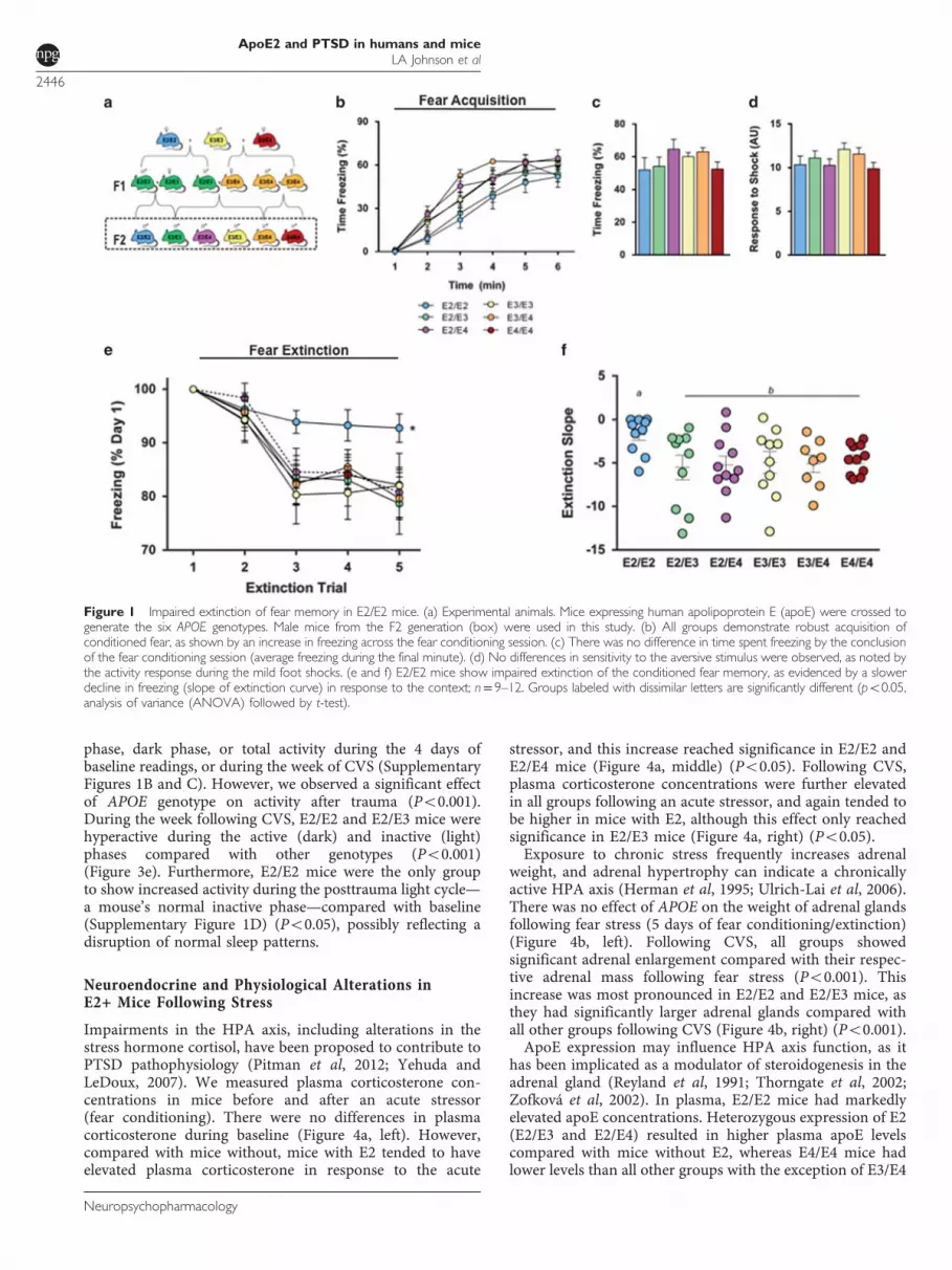
As deficits in the extinction of contextual fear memory likelycontribute to the pathophysiology of PTSD (Jovanovic et al,2009; Maren, 2001), we examined contextual fear extinctionin a mouse model of human apoE (Figure 1a) (Knouff et al,1999; Sullivan et al, 1997, 1998). Acquisition of contextualconditioned fear was determined by comparing the percen-tage of time mice spent freezing during a 1-minute baselineperiod and the time following a mild foot shock. E2/E2 miceshowed a slower increase in freezing rates during trainingcompared with all groups except E2/E3 (Figure 1b)(Po0.05). However, by the conclusion of the training session(final minute), the levels of freezing did not differ bygenotype (Figure 1c), and all groups showed significantlyincreased freezing following shock (Po0.001), suggestinguniversal acquisition of the conditioned fear (Figures 1b andc). Additionally, there was no difference in activity responseduring the foot shocks, suggesting a similar sensitivity andreactivity to the shock (Figure 1d). E2/E2 mice showed asignificantly slower decline in freezing over 4 days ofcontextual fear extinction compared with all other groups(Figure 1e) (Po0.05). Additionally, the rate of extinction,measured as the slope of the percent time freezing across alltrials, was significantly slower in E2/E2 mice compared withall other groups (Figure 1e) (Po0.05).
Cognitive and Behavioral Impairments in Mice with E2Following Conditioned Fear Exposure
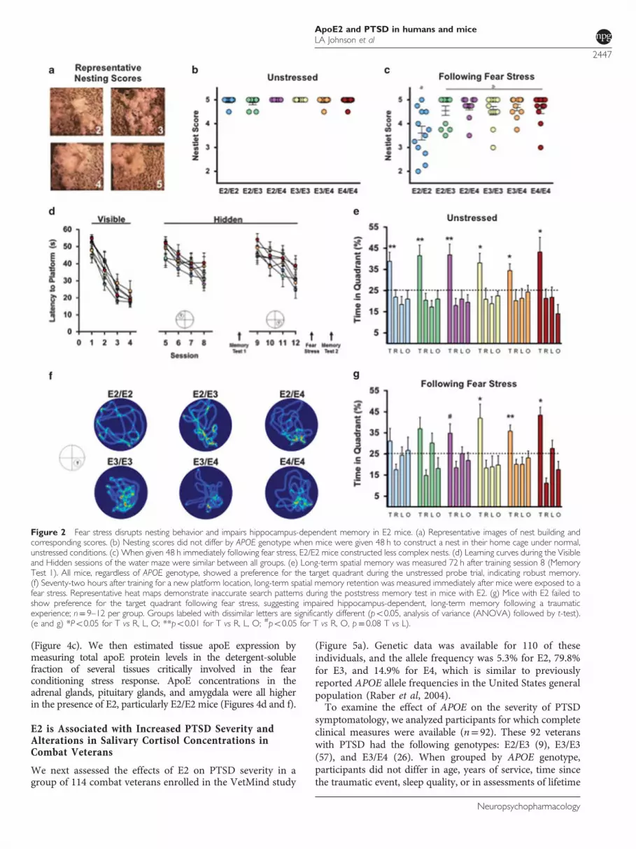
To assess the effects of fear conditioning stress on nestbuilding behavior, we rated nest construction in the homecage at baseline and following fear conditioning (Figure 2a).At baseline, there were no differences in the complexity ofnests constructed (Figure 2b). However, following the stressof fear conditioning, E2/E2 mice constructed less complex
nests compared with other groups (Figure 2c) (Po0.05),demonstrating a disturbance in typical home-cage behavior.To determine whether the apoE isoforms modulate
hippocampus-dependent cognitive function following a trau-matic event, we measured long-term spatial memory beforeand after fear conditioning stress using the water maze. Beforethe stress, mice completed training in the visible and firsthidden sessions. There were no differences in time to reach theplatform or swim speed during the visible platform training,indicating intact task learning, motivation, and sensorimotorskills among all genotypes (Figure 2d, Visible). Likewise, therewas no significant effect of APOE genotype on learning curvesto locate the hidden platform (Figure 2d, Hidden). Seventy-two hours after training to locate the first hidden platformlocation (session 8), mice were given a test of long-termmemory retention (Memory Test 1). All mice, regardless ofAPOE genotype, showed a preference for the target quadrant,suggesting robust long-term memory (Figure 2e).Next, we examined the effects of stress on long-term
memory in these same animals. To avoid the potentialinfluence of varying degrees of memory retention, that is, a‘test–retest’ effect, mice were trained to locate a new platformlocation. Seventy-two hours following the last training trial(session 12), mice were exposed to the fear conditioningstress, and immediately administered a memory test(Memory Test 2). Mice with E2 (E2/E2, E2/E3, and E2/E4)failed to show a preference for the target quadrant duringthis stress-paired memory test (Figures 2f and g).
CVS - A Model of Combat Related PTSD - IncreasesAnxiety and Causes Behavioral and CircadianDisturbances in E2+ Mice
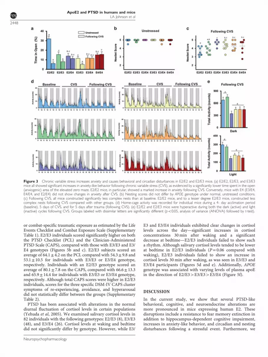
CVS is a model for many stress-related disorders, includingPTSD. In this study, we exposed mice to a different stressordaily over subsequent days to simulate the prolongedand unpredictable stress conditions experienced by militarypersonnel in front-line positions (Supplementary Figure 1A)(Goswami et al, 2013). At baseline, mice homozygous for E2were less anxious, as measured in the elevated zero maze(Po0.001). Following CVS, however, E2/E2 mice showed amarked increase in a measure of anxiety, as evidenced bysignificantly less time spent in the open (anxiogenic) area(Figure 3a). E2/E2, E2/E3, and E3/E3 mice all showedsignificant increases in anxiety-like behavior following CVS(Po0.05). Conversely, mice with E4 (E3/E4, E4/E4, and E2/E4) did not show changes in anxiety-like behavior after theCVS exposure (Figure 3a).Activities of daily living (ADL) are often disrupted during
PTSD (Zatzick et al, 2008), and typical mouse behaviors suchas nest building reflect general well-being and may sharesome psychiatric features with measures of ADL in humans(Deacon, 2012; Jirkof, 2014). CVS reduced nesting scores inall groups compared with baseline (Figures 3b and c)(Po0.05). E2/E2 mice were particularly sensitive, as theyshowed lower nesting scores after CVS than any other groupexcept E2/E3 (Figure 3c) (Po0.05).PTSD is often associated with sleep disturbances (Germain
et al, 2008). To determine the effects of apoE on trauma-induced circadian rhythms, we measured circadian home-cage activity of singly housed mice before, during, and afterCVS (Figure 3d). There were no differences in the light
ApoE2 and PTSD in humans and miceLA Johnson et al
2445
Neuropsychopharmacology
phase, dark phase, or total activity during the 4 days ofbaseline readings, or during the week of CVS (SupplementaryFigures 1B and C). However, we observed a significant effectof APOE genotype on activity after trauma (Po0.001).During the week following CVS, E2/E2 and E2/E3 mice werehyperactive during the active (dark) and inactive (light)phases compared with other genotypes (Po0.001)(Figure 3e). Furthermore, E2/E2 mice were the only groupto show increased activity during the posttrauma light cycle—a mouse’s normal inactive phase—compared with baseline(Supplementary Figure 1D) (Po0.05), possibly reflecting adisruption of normal sleep patterns.
Neuroendocrine and Physiological Alterations inE2+ Mice Following Stress
Impairments in the HPA axis, including alterations in thestress hormone cortisol, have been proposed to contribute toPTSD pathophysiology (Pitman et al, 2012; Yehuda andLeDoux, 2007). We measured plasma corticosterone con-centrations in mice before and after an acute stressor(fear conditioning). There were no differences in plasmacorticosterone during baseline (Figure 4a, left). However,compared with mice without, mice with E2 tended to haveelevated plasma corticosterone in response to the acute
stressor, and this increase reached significance in E2/E2 andE2/E4 mice (Figure 4a, middle) (Po0.05). Following CVS,plasma corticosterone concentrations were further elevatedin all groups following an acute stressor, and again tended tobe higher in mice with E2, although this effect only reachedsignificance in E2/E3 mice (Figure 4a, right) (Po0.05).Exposure to chronic stress frequently increases adrenal
weight, and adrenal hypertrophy can indicate a chronicallyactive HPA axis (Herman et al, 1995; Ulrich-Lai et al, 2006).There was no effect of APOE on the weight of adrenal glandsfollowing fear stress (5 days of fear conditioning/extinction)(Figure 4b, left). Following CVS, all groups showedsignificant adrenal enlargement compared with their respec-tive adrenal mass following fear stress (Po0.001). Thisincrease was most pronounced in E2/E2 and E2/E3 mice, asthey had significantly larger adrenal glands compared withall other groups following CVS (Figure 4b, right) (Po0.001).ApoE expression may influence HPA axis function, as it
has been implicated as a modulator of steroidogenesis in theadrenal gland (Reyland et al, 1991; Thorngate et al, 2002;Zofková et al, 2002). In plasma, E2/E2 mice had markedlyelevated apoE concentrations. Heterozygous expression of E2(E2/E3 and E2/E4) resulted in higher plasma apoE levelscompared with mice without E2, whereas E4/E4 mice hadlower levels than all other groups with the exception of E3/E4
Figure 1 Impaired extinction of fear memory in E2/E2 mice. (a) Experimental animals. Mice expressing human apolipoprotein E (apoE) were crossed togenerate the six APOE genotypes. Male mice from the F2 generation (box) were used in this study. (b) All groups demonstrate robust acquisition ofconditioned fear, as shown by an increase in freezing across the fear conditioning session. (c) There was no difference in time spent freezing by the conclusionof the fear conditioning session (average freezing during the final minute). (d) No differences in sensitivity to the aversive stimulus were observed, as noted bythe activity response during the mild foot shocks. (e and f) E2/E2 mice show impaired extinction of the conditioned fear memory, as evidenced by a slowerdecline in freezing (slope of extinction curve) in response to the context; n= 9–12. Groups labeled with dissimilar letters are significantly different (po0.05,analysis of variance (ANOVA) followed by t-test).
ApoE2 and PTSD in humans and miceLA Johnson et al
2446
Neuropsychopharmacology
(Figure 4c). We then estimated tissue apoE expression bymeasuring total apoE protein levels in the detergent-solublefraction of several tissues critically involved in the fearconditioning stress response. ApoE concentrations in theadrenal glands, pituitary glands, and amygdala were all higherin the presence of E2, particularly E2/E2 mice (Figures 4d and f).
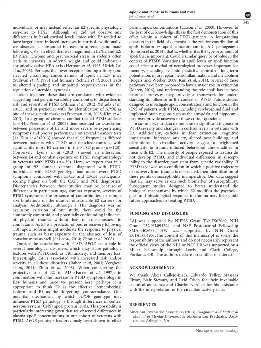
E2 is Associated with Increased PTSD Severity andAlterations in Salivary Cortisol Concentrations inCombat Veterans
We next assessed the effects of E2 on PTSD severity in agroup of 114 combat veterans enrolled in the VetMind study
(Figure 5a). Genetic data was available for 110 of theseindividuals, and the allele frequency was 5.3% for E2, 79.8%for E3, and 14.9% for E4, which is similar to previouslyreported APOE allele frequencies in the United States generalpopulation (Raber et al, 2004).To examine the effect of APOE on the severity of PTSD
symptomatology, we analyzed participants for which completeclinical measures were available (n= 92). These 92 veteranswith PTSD had the following genotypes: E2/E3 (9), E3/E3(57), and E3/E4 (26). When grouped by APOE genotype,participants did not differ in age, years of service, time sincethe traumatic event, sleep quality, or in assessments of lifetime
Figure 2 Fear stress disrupts nesting behavior and impairs hippocampus-dependent memory in E2 mice. (a) Representative images of nest building andcorresponding scores. (b) Nesting scores did not differ by APOE genotype when mice were given 48 h to construct a nest in their home cage under normal,unstressed conditions. (c) When given 48 h immediately following fear stress, E2/E2 mice constructed less complex nests. (d) Learning curves during the Visibleand Hidden sessions of the water maze were similar between all groups. (e) Long-term spatial memory was measured 72 h after training session 8 (MemoryTest 1). All mice, regardless of APOE genotype, showed a preference for the target quadrant during the unstressed probe trial, indicating robust memory.(f) Seventy-two hours after training for a new platform location, long-term spatial memory retention was measured immediately after mice were exposed to afear stress. Representative heat maps demonstrate inaccurate search patterns during the poststress memory test in mice with E2. (g) Mice with E2 failed toshow preference for the target quadrant following fear stress, suggesting impaired hippocampus-dependent, long-term memory following a traumaticexperience; n= 9–12 per group. Groups labeled with dissimilar letters are significantly different (po0.05, analysis of variance (ANOVA) followed by t-test).(e and g) *Po0.05 for T vs R, L, O; **po0.01 for T vs R, L, O; #po0.05 for T vs R, O, p= 0.08 T vs L).
ApoE2 and PTSD in humans and miceLA Johnson et al
2447
Neuropsychopharmacology
or combat-specific traumatic exposure as estimated by the LifeEvents Checklist and Combat Exposure Scale (SupplementaryTable 1). E2/E3 individuals scored significantly higher on boththe PTSD Checklist (PCL) and the Clinician-AdministeredPTSD Scale (CAPS), compared with those with E3/E3 and E3/E4 genotypes (Figures 5b and c). E2/E3 subjects scored anaverage of 64.1± 4.2 on the PCL compared with 54.3± 9.8 and53.1± 10.5 for individuals with E3/E3 or E3/E4 genotype,respectively. Individuals with an E2/E3 genotype scored anaverage of 80.1± 7.8 on the CAPS, compared with 66.6± 13.3and 65.9± 14.4 for individuals with E3/E3 or E3/E4 genotype,respectively. Although total CAPS scores were higher in E2/E3individuals, scores for the three specific DSM-IV CAPS clustersymptoms of re-experiencing, avoidance, and hyperarousaldid not statistically differ between the groups (SupplementaryTable 2).PTSD has been associated with alterations in the normal
diurnal fluctuation of cortisol levels in certain populations(Yehuda et al, 2005). We examined salivary cortisol levels in82 individuals with the following genotypes: E2/E3 (8), E3/E3(48), and E3/E4 (26). Cortisol levels at waking and bedtimedid not significantly differ by genotype. However, while E3/
E3 and E3/E4 individuals exhibited clear changes in cortisollevels across the day—significant increases in cortisolconcentrations 30 min after waking and a significantdecrease at bedtime—E2/E3 individuals failed to show sucha rhythm. Although salivary cortisol levels tended to be lowerat bedtime in E2/E3 individuals (P= 0.06 compared withwaking), E2/E3 individuals failed to show an increase incortisol levels 30 min after waking, as was seen in E3/E3 andE3/E4 participants (Figures 5d and e). Additionally, APOEgenotype was associated with varying levels of plasma apoEin the direction of E2/E34E3/E34E3/E4 (Figure 5f).
DISCUSSION
In the current study, we show that several PTSD-likebehavioral, cognitive, and neuroendocrine alterations aremore pronounced in mice expressing human E2. Thesedisruptions include a resistance to fear memory extinction inaddition to hippocampus-dependent cognitive impairment,increases in anxiety-like behavior, and circadian and nestingdisturbances following a stressful event. Furthermore, we
Figure 3 Chronic variable stress increases anxiety and causes behavioral and circadian disturbances in E2/E2 and E2/E3 mice. (a) E2/E2, E2/E3, and E3/E3mice all showed significant increases in anxiety-like behavior following chronic variable stress (CVS), as evidenced by a significantly lower time spent in the open(anxiogenic) area of the elevated zero maze. E2/E2 mice, in particular, showed a marked increase in anxiety following CVS. Conversely, mice with E4 (E3/E4,E4/E4, and E2/E4) did not show changes in anxiety after CVS. (b) Nesting scores did not differ by APOE genotype under normal, unstressed conditions.(c) Following CVS, all mice constructed significantly less complex nests than at baseline. E2/E2 mice, and to a lesser degree E2/E3 mice, constructed lesscomplex nests following CVS compared with other groups. (d) Home-cage activity was recorded for individual mice during a 4- day acclimation period(baseline), 5 days of CVS, and for 5 days after trauma (following CVS). (e) E2/E2 and E2/E3 mice were hyperactive during both the dark (active) and light(inactive) cycles following CVS. Groups labeled with dissimilar letters are significantly different (po0.05, analysis of variance (ANOVA) followed by t-test).
ApoE2 and PTSD in humans and miceLA Johnson et al
2448
Neuropsychopharmacology
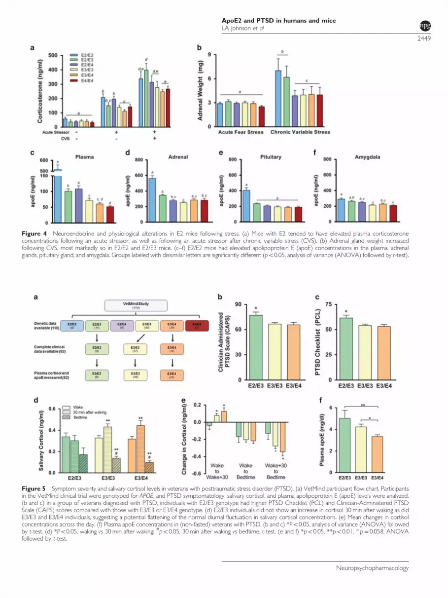
Figure 4 Neuroendocrine and physiological alterations in E2 mice following stress. (a) Mice with E2 tended to have elevated plasma corticosteroneconcentrations following an acute stressor, as well as following an acute stressor after chronic variable stress (CVS). (b) Adrenal gland weight increasedfollowing CVS, most markedly so in E2/E2 and E2/E3 mice. (c–f) E2/E2 mice had elevated apolipoprotein E (apoE) concentrations in the plasma, adrenalglands, pituitary gland, and amygdala. Groups labeled with dissimilar letters are significantly different (po0.05, analysis of variance (ANOVA) followed by t-test).
Figure 5 Symptom severity and salivary cortisol levels in veterans with posttraumatic stress disorder (PTSD). (a) VetMind participant flow chart. Participantsin the VetMind clinical trial were genotyped for APOE, and PTSD symptomatology, salivary cortisol, and plasma apolipoprotein E (apoE) levels were analyzed.(b and c) In a group of veterans diagnosed with PTSD, individuals with E2/E3 genotype had higher PTSD Checklist (PCL) and Clinician-Administered PTSDScale (CAPS) scores compared with those with E3/E3 or E3/E4 genotype. (d) E2/E3 individuals did not show an increase in cortisol 30 min after waking as didE3/E3 and E3/E4 individuals, suggesting a potential flattening of the normal diurnal fluctuation in salivary cortisol concentrations. (e) Mean changes in cortisolconcentrations across the day. (f) Plasma apoE concentrations in (non-fasted) veterans with PTSD. (b and c) *Po0.05, analysis of variance (ANOVA) followedby t-test. (d) *Po0.05, waking vs 30 min after waking; #po0.05, 30 min after waking vs bedtime, t-test. (e and f) *po0.05, **po0.01, ^p= 0.058, ANOVAfollowed by t-test.
ApoE2 and PTSD in humans and miceLA Johnson et al
2449
Neuropsychopharmacology

demonstrate more severe PTSD symptoms and alterations insalivary cortisol in combat veterans with E2. Taken together,these results establish an interspecies demonstration ofexaggerated PTSD symptomatology in the presence of E2.Given the striking effects on PTSD symptomatology in our
cohort of veterans, we turned to a mouse model of humanapoE to further examine the effects of APOE on variousneuropsychological phenomena associated with PTSD. Toour knowledge, this is the first mouse model of human apoEto examine PTSD-like behavioral, cognitive, and physiologi-cal outcomes. To mimic the characteristics of our cohort ofveterans, who were all men and whose median age was 21years at the time of the traumatic incident, we used youngadult male mice (4–5 months of age). Although the vastmajority of human apoE mouse studies use only homo-zygous mice, it is very difficult to recruit a sufficient numberof E2/E2 participants (genotype frequency o1%) (Raberet al, 2004). For this reason, and to dissect out specificisoform contributions and potential opposing effects (forinstance, in E2/E4 individuals), we included all six APOEgenotypes in our animal model.Abnormal regulation of fear is a core feature of PTSD, and
patients suffering from the disorder exhibit deficits inlearning safety in the absence of threat. Much like exposuretherapy is applied to PTSD patients in the clinic, thisphenomenon is commonly modeled in rodents using fearextinction (Kaouane et al, 2012; McGuire et al, 2010; Rauet al, 2005). Consistent with our data showing more severePTSD symptoms in E2+ veterans, we confirmed deficits infear extinction in E2/E2 mice (Olsen et al, 2012), suggestingthat an abnormal processing of fear memory may underliethe apparent sensitivity of E2+ individuals to trauma.Anxiety and avoidance in patients with PTSD can also bemodeled in mice using the elevated zero maze. We observedincreases in anxiety-like behavior in E2/E2, E2/E3, and E3/E3mice following CVS, and this increase in anxiety wasparticularly marked in E2/E2 mice. Interestingly, all themice expressing E4 (E3/E4, E4/E4, and E2/E4) were resistantto this trauma-induced increase in anxiety-like behavior.Previous research in our lab and others suggest that mice andhumans with E4 are generally more anxious (Raber, 2007).In this study, possession of E4 appeared to protect againsttrauma-induced increases in anxiety, even in mice coexpres-sing E2. This may suggest a possible exaggeration by E2 (andconversely a buffering by E4) of the anxiogenic effects ofCVS. Taken together with evidence in the context oftraumatic brain injury (TBI), these results raise an interestingquestion: how is it that E4 appears to be poorer at negotiatingphysical trauma (TBI, stroke) (Shi et al, 2014; Zhou et al,2008), but perhaps more efficient at rectifying the psycho-logical trauma of PTSD?Another prominent feature of PTSD is memory distur-
bances, although these disturbances can vary widely. Forinstance, intrusive memories of the traumatic event fall underthe re-experiencing symtpoms, whereas avoidance criteriainclude an inability to recall critical aspects of the trauma.Additionally, those suffering from PTSD often describeroutine memory problems with emotionally neutral material(Samuelson, 2011). PTSD has also been associated withdiminished hippocampal volume (Smith, 2005) and function(Shin et al, 2006). Stress can significantly affect memory, andevidence suggests that stress and APOE interact to affect
memory (Peavy et al, 2007). We show that when challengedwith a memory test immediately after a fear stress, mice withE2 demonstrate relative impairments in long-term hippo-campus-dependent memory. These E2-associated deficits inpoststress memory recall occurred despite similar learningcurves and similar memory retention in unstressed condi-tions. These data suggest that possession of E2 imparts asusceptibility to stress-induced impairments in memory, afinding in agreement with the memory disturbances reportedin E2+ PTSD patients (Freeman et al, 2005).ADL, a measurement of a patient’s functional status, are
often hindered during PTSD (Zatzick et al, 2008). In mice,nest building has been used to assess distress and suffering,as well as hippocampal damage (Deacon et al, 2002) andneurodegeneration (Cramer et al, 2012; Wesson and Wilson,2011), and may share neuropsychiatric features withmeasures of ADL (Deacon, 2012; Jirkof, 2014). We observeddiminished nest building behavior in E2/E2 mice 2 days afterfear conditioning. Following CVS, deficits were present in allgroups, but the effect was most pronounced in E2/E2 and E2/E3 mice. PTSD is also associated with sleep disturbances(Germain et al, 2008). APOE gene expression and proteinlevels fluctuate daily (Shen et al, 2009) and a patient’scircadian rhythm and sleep quality also appear to bemodulated by APOE, at least in AD (Lim et al, 2013;Robertson et al, 2005; Yeasavage et al, 2004). CVS disruptedthe normal activity patterns of all mice, but this effect wasmost pronounced in E2/E2 and E2/E3 mice. Especiallyimportant may be the increase in activity during the normalinactive (light) cycle, which may reflect sleep disturbances inthe E2/E2 and E2/E3 mice. Although translation of nestbuilding and circadian activity to the human condition iscomplex, these data suggest that E2/E3 and E2/E2 mice areparticularly reactive to trauma. Although we did not observeany disruption in circadian rhythm in E2/E3 veterans withPTSD, at least as measured by sleep quality and disturbances(Supplementary Table 1), future studies using more sophis-ticated methods to analyze circadian rhythm in E2+ patientswith PTSD may be warranted.A possible link between apoE biology and the psychiatric
features of PTSD involves the HPA axis (Freeman et al, 2005;Raber et al, 2000). Low cortisol levels have been frequentlyreported in PTSD patients (Young and Breslau, 2004;Wahbeh and Oken, 2013). However, the effects of APOEon cortisol levels in humans (Fiocco et al, 2008; Peskind et al,2001) and mice (Raber et al, 2000; Reverte et al, 2014;Robertson et al, 2005; Thorngate et al, 2002) are less clear.Additionally, several studies have shown varying resultswhen examining the relationship between APOE and cortisolon stress and cognitive function (Beluche et al, 2010;Gallagher-Thompson et al, 2001; Gerritsen et al, 2011; Leeet al, 2008). Looking beyond basal levels of cortisol, Yehudaet al (2005) showed that the normal circadian rhythm ofcortisol levels is flattened in Holocaust survivors with PTSDcompared with non-exposed subjects. Interestingly, weobserved a similar phenomenon in E2/E3 veterans withPTSD. In contrast to E3/E3 and E3/E4, cortisol levels in E2/E3 individuals did not increase within 30 min of waking.Although our study is limited in power owing to the smallnumber of cortisol samples from E2/E3 patients (n= 8), thisapparent flattening of diurnal cortisol levels may point topre-existing alterations in HPA axis function in E2/E3
ApoE2 and PTSD in humans and miceLA Johnson et al
2450
Neuropsychopharmacology
individuals, or may instead reflect an E2-specific physiologicresponse to PTSD. Although we did not observe anydifferences in basal cortisol levels, mice with E2 tended tohave larger stress-induced increases in cortisol. Additionally,we observed a substantial increase in adrenal gland massfollowing CVS, an effect that was magnified in E2/E2 and E2/E3 mice. Chronic and psychosocial stress in rodents oftenleads to increases in adrenal weight and could indicate achronically active HPA axis (Herman et al, 1995; Ulrich-Laiet al, 2006). Perhaps, the lower receptor binding affinity (andelevated circulating concentrations of apoE in E2+ mice(Sullivan et al, 1998) and humans (Schiele et al, 2000) leadsto altered signaling and impaired responsiveness in theregulation of steroidal synthesis.Taken together, these data are consistent with evidence
suggesting that genetic variability contributes to disparities inrisk and severity of PTSD (Pitman et al, 2012; Yehuda et al,2011), and in particular with the identification of APOE asone of these genetic markers (Freeman et al, 2005; Kim et al,2013). In a group of chronic, combat-related PTSD subjects(n= 44), Freeman et al (2005) demonstrated an associationbetween possession of E2 and more severe re-experiencingsymptoms and poorer performance on several memory tests(1). Kim et al (2013) showed that allele frequencies differedbetween patients with PTSD and matched controls, withsignificantly more E2 carriers in the PTSD group (n= 128).Conversely, Lyons et al (2013) showed an interactionbetween E4 and combat exposure on PTSD symptomatologyin veterans with PTSD (n= 39). Here, we report that in agroup of 92 combat veterans diagnosed with PTSD,individuals with E2/E3 genotype had more severe PTSDsymptoms compared with E3/E3 and E3/E4 participants,scoring higher on both the CAPS and PCL diagnostics.Discrepancies between these studies may be because ofdifferences in participant age, combat exposure, severity ofPTSD symptoms, the presence of comorbidities, or samplesize limitations on the number of available E2 carriers foranalysis. Additionally, although a TBI diagnosis was anexclusion criterion of our study, there could be thecommonly comorbid, and potentially confounding influence,of physical trauma without loss of consciousness incombatants. As E4 is a predictor of poorer recovery followingTBI, apoE isoform might modulate the response to physicaltrauma such as blast exposure in the absence of loss ofconsciousness as well (Shi et al, 2014; Zhou et al, 2008).Outside the association with PTSD, APOE has a role in
several neurological disorders, which may share pathologicfeatures with PTSD, such as TBI, anxiety, and memory loss.Interestingly, E4 is associated with increased risk and/orseverity in all these disorders (Raber et al, 2003; Vergheseet al, 2011; Zhou et al, 2008). When considering theprotective role of E2 in AD (Farrer et al, 1997), incombination with the increase in PTSD symptomatology inE2+ humans and mice we present here, perhaps it isappropriate to think E2 as the effective ‘remembering’isoform and E4 as the ‘forgetting’ counterbalance. Onepotential mechanism by which APOE genotype mayinfluence PTSD pathology is through differences in centralnervous system (CNS) apoE protein levels. This possibility isparticularly interesting given that we observed differences inplasma apoE concentrations in our cohort of veterans withPTSD. APOE genotype has previously been shown to effect
plasma apoE concentrations (Larson et al, 2000). However, tothe best of our knowledge, this is the first demonstration of thiseffect within a cohort of PTSD patients. A longstandingquestion in the field of dementia is the relative contribution ofapoE isoform vs apoE concentration in AD pathogenesis(Johnson et al, 2014), that is, whether it is the type or amount ofapoE that is important. Could a similar query be of value in thecontext of PTSD? Variations in apoE levels or apoE functioncould affect a myriad of neurological processes important formemory, including synaptic plasticity, control of long-termpotentiation, injury repair, neuroinflammation, and metabolism(Rogers and Weeber, 2008; Kim et al, 2014). Several of thesepathways have been proposed to have a major role in extinction(Maren, 2014), and understanding the role apoE has in theseneuronal processes may provide a framework for under-standing its influence in the context of PTSD. Future studiesdesigned to investigate apoE concentrations and function in theCNS of patients with PTSD, including cerebrospinal fluid andimplicated brain regions such as the amygdala and hippocam-pus, may provide answers to these critical questions.In summary, our data demonstrate a significant increase in
PTSD severity and changes in cortisol levels in veterans withE2. Additionally, deficits in fear extinction, cognitiveimpairment, increased anxiety, altered nest building, anddisruptions in circadian activity suggest a heightenedsensitivity to trauma-induced behavioral abnormalities inmice with E2. The majority of people exposed to trauma donot develop PTSD, and individual differences in suscept-ibility to the disorder may stem from genetic variability. IfPTSD is viewed as a condition in which a positive trajectoryof recovery from trauma is obstructed, then identification ofthese points of susceptibility is imperative. Our data suggestthat E2 may serve as one such biomarker of susceptibility.Subsequent studies designed to better understand thebiological mechanisms by which E2 modifies the psycholo-gical and physiological response to trauma may help guidefuture approaches to treating PTSD.
FUNDING AND DISCLOSURE
LAJ was supported by NIEHS Grant T32-ES07060, NIHGrant T32-HL094294, and NSF Postdoctoral FellowshipSMA-1408653. HW was supported by NIH GrantK01AT004951.The content of this manuscript is solely theresponsibility of the authors and do not necessarily representthe official views of the NIH or NSF. EB was supported by aMiller Fellowship through Lewis and Clark College,Portland, OR. The authors declare no conflict of interest.
ACKNOWLEDGMENTS
We thank Alicia Callejo-Black, Eduardo Tellez, MassaraEiwaz, Blair Stewart, and Reid Olsen for their invaluabletechnical assistance and Charles N Allen for his assistancewith the interpretation of the circadian activity data.
REFERENCES
American Psychiatric Association (2013). Diagnostic and StatisticalManual of Mental Disorders5th ednAmerican Psychiatric Asso-ciation: Arlington, VA.
ApoE2 and PTSD in humans and miceLA Johnson et al
2451
Neuropsychopharmacology

Beluche I, Carriere I, Ritchie K, Ancelin ML (2010). A prospectivestudy of diurnal cortisol and cognitive function in community-dwelling elderly people. Psychol Med 40: 1039–1049.
Caselli RJ, Reiman EM, Locke DE, Hutton ML, Hentz JG, Hoffman-Snyder C et al (2007). Cognitive domain decline in healthyapolipopro-tein E 4 homozygotes before the diagnosis of mildcognitiveimpairment. Arch Neurol 64: 1306–1311.
Cornelis MC, Nugent NR, Amstadter AB, Koenen KC (2010).Genetics of post-traumatic stress disorder: review and recom-mendations for genome-wide association studies. Curr PsychiatryRep 12: 313–326.
Cramer PE, Cirrito JR, Wesson DW, Lee CY, Karlo JC,Zinn AE et al (2012). ApoE-directed therapeutics rapidly clearβ-amyloid and reverse deficits in AD mouse models. Science 335:1503–1506.
Deacon RM, Croucher A, Rawlins JN (2002). Hippocampalcytotoxic lesion effects on species-typical behaviours in mice.Behav Brain Res 132: 203–213.
Deacon RM (2006). Assessing nest building in mice. Nat Protoc 1:1117–1119.
Deacon R (2012). Assessing burrowing, nest construction, andhoarding in mice. J Vis Exp e2607.
Farrer LA, Cupples LA, Haines JL, Hyman B, Kukull WA,Mayeux R et al (1997). Effects of age, sex, and ethnicity on theassociation between apolipoprotein E genotype and Alzheimerdisease. A meta-analysis. APOE and Alzheimer Disease MetaAnalysis Consortium. JAMA 278: 1349–1356.
Fendt M, Endres T (2008). 2,3,5-Trimethyl-3-thiazoline (TMT), acomponent of fox odor - just repugnant or really fear-inducing?Neurosci Biobehav Rev 32: 1259–1266.
Fiocco AJ, Poirier J., Joober R., Nair NP, Lupien SJ (2008). Acuteand long-term associations between ApoE genetic polymorphism,cortisol levels, and declarative memory performance inolder adults. Psychoneuroendocrinology 33: 625–633.
Freeman T, Roca V, Guggenheim F, Kimbrell T, Griffin WS (2005).Neuropsychiatric associations of apolipoprotein E alleles insubjects with combat-related posttraumatic stress disorder. JNeuropsychiatry Clin Neurosci 17: 541–543.
Gadermann AM, Alonso J, Vilagut G, Zaslavsky AM, Kessler RC(2012). Comorbidity and disease burden in the NationalComorbidity Survey Replication (NCS-R). Depress Anxiety 29:797–806.
Galea S, Vlahov D, Resnick H, Ahern J, Susser E, Gold J et al (2003).Trends of probable post-traumatic stress disorder in New YorkCity after the September 11 terrorist attacks. Am J Epidemiol 158:514–524.
Gallagher-Thompson D, O'Hara R, Simmons A, Kraemer HC,Murphy GM Jr (2001). Apolipoprotein E epsilon4 allele affects therelationship between stress and depression in caregivers ofpatients with Alzheimer’s disease. J Geriatr Psychiatry Neurol 14:115–119.
Germain A, Buysse DJ, Nofzinger E (2008). Sleep-specific mechan-isms underlying posttraumatic stress disorder: integrative reviewand neurobiological hypotheses. Sleep Med Rev 12: 185–195.
Gerritsen L, Comijs HC, Deeg DJ, Penninx BW, Geerlings MI(2011). Salivary cortisol, APOE-ε4 allele and cognitive declinein a prospective study of older persons. Neurobiol Aging 32:1615–1625.
Gil-Bea FJ, Aisa B, Solomon A, Solas M, del Carmen Mugueta M,Winblad B et al (2010). HPA axis dysregulation associated toapolipoprotein E4 genotype in Alzheimer’s disease. J AlzheimersDis 22: 829–838.
Goswami S, Rodríguez-Sierra O, Cascardi M, Paré D (2013).Animal models of post-traumatic stress disorder: face validity.Front Neurosci 7: 89.
Herman JP, Adams D, Prewitt C (1995). Regulatory changes inneuroendocrine stress-integrative circuitry produced by a variablestress paradigm. Neuroendocrinology 61: 180–190.
Hostage CA, Roy Choudhury K, Doraiswamy PM, Petrella JR(2013). Alzheimer’s disease neuroimaging initiative. Dissectingthe gene dose-effects of the APOE ε4 and ε2 alleles onhippocampal volumes in aging and Alzheimer’s disease. PLoSOne 8: e54483.
Jirkof P (2014). Burrowing and nest building behavior as indicatorsof well-being in mice. J Neurosci Methods 234: 139–146.
Johnson LA, Olsen RH, Merkens LS, DeBarber A, Steiner RD,Sullivan PM et al (2014). Apolipoprotein E-low densitylipoprotein receptor interaction affects spatial memory retentionand brain ApoE levels in an isoform-dependent manner.Neurobiol Dis 64: 150–162.
Jovanovic T, Norrholm SD, Fennell JE, Keyes M, Fiallos AM, Myers KMet al (2009). Posttraumatic stress disorder may be associated withimpaired fear inhibition: relation to symptom severity. Psychiatry Res15: 151–160.
Jovanovic T, Ressler KJ (2010). How the neurocircuitry and geneticsof fear inhibition may inform our understanding of PTSD.Am J Psychiatry 167: 648–662.
Kaouane N, Porte Y, Vallée M, Brayda-Bruno L, Mons N,Calandreau L et al (2012). Glucocorticoids can induce PTSD-like memory impairments in mice. Science 335: 1510–1513.
Kessler RC, Berglund P, Demler O, Jin R, Merikangas KR,Walters EE (2005). Lifetime prevalence and age-of-onsetdistributions of DSM-IV disorders in the national comorbiditysurvey replication. Am Med Assoc 62: 593–602.
Kessler RC, Sonnega A, Bromet E, Hughes M, Nelson CB (1995).Posttraumatic stress disorder in the national comorbidity survey.Arch Gen Psychiatry 52: 1048–1060.
Kilpatrick DG, Resnick HS, Milanak ME, Miller MW, Keyes KM,Friedman MJ (2013). National estimates of exposure to traumaticevents and PTSD prevalence using DSM-IV and DSM-5 criteria. JTrauma Stress 26: 537–547.
Kim J, Yoon H, Basak J, Kim J (2014). Apolipoprotein E in synapticplasticity and Alzheimer's disease: potential cellular and mole-cular mechanisms. Mol Cells 37: 767–776.
Kim TY, Chung HG, Shin HS, Kim SJ, Choi JH, Chung MY et al(2013). Apolipoprotein E gene polymorphism, alcohol use, andtheir interactions in combat-related posttraumatic stress disorder.Depress Anxiety 30: 1194–1201.
Knouff C, Hinsdale ME, Mezdour H, Altenburg MK, Watanabe M,Quarfordt SH et al (1999). ApoE structure determines VLDLclearance and atherosclerosis risk in mice. J Clin Invest 103:1579–1586.
Larson IA, Ordovas JM, DeLuca C, Barnard JR, Feussner G,Schaefer EJ (2000). Association of apolipoprotein (Apo)Egenotype with plasma apo E levels. Atherosclerosis 148: 327–335.
Laskowitz DT, Horsburgh K, Roses AD (1998). Apolipoprotein Eand the CNS response to injury. J Cereb Blood Flow Metab 18:465–471.
Lee BK, Glass TA, Wand GS, McAtee MJ, Bandeen-Roche K,Bolla KI et al (2008). Apolipoprotein E genotype, cortisol, andcognitive function in community-dwelling older adults. Am JPsychiatry 165: 1456–1464.
Lim AS, Yu L, Kowgier M, Schneider JA, Buchman AS, Bennett DA(2013). Modification of the relationship of the apolipoprotein E ε4allele to the risk of Alzheimer disease and neurofibrillary tangledensity by sleep. JAMA Neurol 70: 1544–1551.
Lyons MJ, Genderson M, Grant MD, Logue M, Zink T, McKenzie Ret al (2013). Gene–environment interaction of ApoE genotype andcombat exposure on PTSD. Am J Med Genet B 162B: 762–769.
Mahley RW, Rall SC Jr (2000). Apolipoprotein E: far more than alipid transport protein. Annu Rev Genom Hum Genet 1: 507–537.
Maren S (2001). Neurobiology of Pavlovian fear conditioning. AnnuRev Neurosci 24: 897–931 review.
Maren S (2014). Out with the old and in with the new:synaptic mechanisms of extinction in the amygdala. Brain Respii: S0006-8993.
ApoE2 and PTSD in humans and miceLA Johnson et al
2452
Neuropsychopharmacology
McGuire J, Herma JP, Horn PS, Sallee FR, Sah R (2010). Enhancedfear recall and emotional arousal in rats recovering from chronicvariable stress. Physiol Behav 101: 474–482.
Olsen RH, Agam M, Davis MJ, Raber J (2012). ApoE isoform-dependent deficits in extinction of contextual fear conditioning.Genes Brain Behav 11: 806–812.
Peavy GM, Lange KL, Salmon DP, Patterson TL, Goldman S, GamstAC et al (2007). The effects of prolonged stress and APOE genotypeon memory and cortisol in older adults. Biol Psychiatry 62: 472–478.
Peskind ER, Wilkinson CW, Petrie EC, Schellenberg GD,Raskind MA (2001). Increased CSF cortisol in AD is a functionof APOE genotype. Neurology 56: 1094–1098.
Pitman RK, Rasmusson AM, Koenen KC, Shin LM, Orr SP,Gilbertson MW et al (2012). Biological studies of post-traumaticstress disorder. Nat Rev Neurosci 13: 769–787.
Raber J, Akana SF, Bhatnagar S, Dallman MF, Wong D, Mucke L(2000). Hypothalamic-pituitary-adrenal dysfunction in Apoe(− /− ) mice: possible role in behavioral and metabolic altera-tions. J Neurosci 20: 2064–2071.
Raber J, Huang Y, Ashford JW (2004). ApoE genotype accounts forthe vast majority of AD risk and AD pathology. Neurobiol Aging25: 641–650.
Raber J (2007). Role of apolipoprotein E in anxiety. Neural Plast2007: 91236 review.
Rau V, DeCola JP, Fanselow MS (2005). Stress-induced enhance-ment of fear learning: an animal model of posttraumatic stressdisorder. Neurosci Biobehav Rev 29: 1207–1223.
Reiman EM, Chen K, Alexander GE, Caselli RJ, Bandy D,Osborne D et al (2004). Functional brain abnormalities in youngadults at genetic risk for late-onset Alzheimer's dementia. ProcNatl Acad Sci USA 101: 284–289.
Reverte I, Pujol A, Domingo JL, Colomina MT (2014). Thyroidhormones and fear learning but not anxiety are affected in adultapoE transgenic mice exposed postnatally to decabromodiphenylether (BDE-209). Physiol Behav 133: 81–91.
Reyland ME, Gwynne JT, Forgez P, Prack MM, Williams DL (1991).Expression of the human apolipoprotein E gene suppressessteroidogenesis in mouse Y1 adrenal cells. Proc Natl Acad Sci USA88: 2375–2379.
Robertson J, Curley J, Kaye J, Quinn J, Pfankuch T, Raber J (2005).ApoE isoforms and measures of anxiety in probable AD patientsand Apoe− /− mice. Neurobiol Aging 26: 637–643.
Rogers JT, Weeber EJ (2008). Reelin and apoE actions on signaltransduction, synaptic function and memory formation. NeuronGlia Biol 4: 259–270.
Rubin GJ, Brewin C, Greenberg N, Simpson J, Wessely S (2005).Psychological and behavioural reactions to the bombings inLondon on 7 July 2005: cross sectional survey of a representativesample of Londoners. BMJ 331: 606–611.
Samuelson KW (2011). Post-traumatic stress disorder anddeclarative memory functioning: a review. Dialogues. ClinNeurosci 13: 346–351.
Schiele F, De Bacquer D, Vincent-Viry M, Beisiegel U, Ehnholm C,Evans A et al (2000). Apolipoprotein E serum concentration andpolymorphism in six European countries: the ApoEurope Project.Atherosclerosis 152: 475–488.
Shen L, Carey K, Wang DQ, Woods SC, Liu M (2009). Food-entrained rhythmic expression of apolipoprotein E expression inthe hypothalamus of rats. Brain Res 1273: 66–71.
Shi J, Han P, Kuniyoshi SM (2014). Cognitive impairment inneurological diseases: lessons from apolipoprotein E. J AlzheimersDis 38: 1–9.
Shin LM, Rauch SL, Pitman RK (2006). Amygdala, medialprefrontal cortex, and hippocampal function in PTSD. Ann NYAcad Sci 1071: 67–79.
Smith ME (2005). Bilateral hippocampal volume reduction in adultswith post-traumatic stress disorder: a meta-analysis of structuralMRI studies. Hippocampus 15: 798–807.
Spijker S (2011). Neuroproteomics. Chapter 2: Dissection of RodentBrain Regions Vol 57, pp 13–26.
Sullivan PM, Mezdour H, Aratani Y, Knouff C, Najib J, Reddick RLet al (1997). Targeted replacement of the mouse apolipoprotein Egene with the common human APOE3 allele enhances diet-induced hypercholesterolemia and atherosclerosis. J Biol Chem272: 17972–17980.
Sullivan PM, Mezdour H, Quarfordt SH, Maeda N (1998). Type IIIhyperlipoproteinemia and spontaneous atherosclerosis inmice resulting from gene replacement of mouse Apoe withhuman Apoe*2. J Clin Invest 102: 130–135.
Thorngate FE, Strockbine PA, Erickson SK, Williams DL (2002).Altered adrenal gland cholesterol metabolism in the apoE-deficient mouse. J Lipid Res 43: 1920–1926.
Ulrich-Lai YM, Figueiredo HF, Ostrander MM, Choi DC,Engeland WC, Herman JP (2006). Chronic stress induces adrenalhyperplasia and hypertrophy in a subregion-specific manner. AmJ Physiol Endocrinol Metab 291: E965–E973.
Verghese PB, Castellano JM, Holtzman DM (2011). ApolipoproteinE in Alzheimer's disease and other neurological disorders. LancetNeurol 10: 241–252.
Wahbeh H, Oken BS (2013). Salivary cortisol lower in posttrau-matic stress disorder. J Trauma Stress 26: 241–248.
Wahbeh H, Kishiyama SS, Zajdel D, Oken BS (2008). Salivarycortisol awakening response in mild Alzheimer disease, care-givers, and noncaregivers. Alzheimer Dis Assoc Disord 22:181–183.
Wesson DW, Wilson DA (2011). Age and gene overexpressioninteract to abolish nesting behavior in Tg2576 amyloid precursorprotein (APP) mice. Behav Brain Res 216: 408.
Yehuda R, Golier JA, Kaufman S (2005). Circadian rhythm ofsalivary cortisol in Holocaust survivors with and without PTSD.Am J Psychiatry 162: 998–1000.
Yehuda R, Koenen KC, Galea S, Flory JD (2011). The role of genesin defining a molecular biology of PTSD. Dis Markers 30: 67–76.
Yehuda R, LeDoux J (2007). Response variation following trauma: atranslational neuroscience approach to understanding PTSD.Neuron 56: 19–32.
Yesavage JA, Friedman L, Kraemer H, Tinklenberg JR, Salehi A,Noda A et al (2004). Sleep/wake disruption in Alzheimer’sdisease: APOE status and longitudinal course. J Geriatr PsychiatryNeurol 17: 20–24.
Young EA, Breslau N (2004). Cortisol and catecholamines inposttraumatic stress disorder: an epidemiologic communitystudy. Arch Gen Psychiatry 61: 394–401.
Zatzick D, Jurkovich GJ, Rivara FP, Wang J, Fan MY, Joesch J et al(2008). A national US study of posttraumatic stress disorder,depression, and work and functional outcomes after hospitaliza-tion for traumatic injury. Ann Surg 248: 429–437.
Zhou W, Xu D, Peng X, Zhang Q, Jia J, Crutcher KA (2008). Meta-analysis of APOE4 allele and outcome after traumaticbrain injury. J Neurotrauma 25: 279–290.
Zofková I, Zajícková K, Hill M, Horínek A (2002). Apolipoprotein Egene determines serum testosterone and dehydroepiandro-sterone levels in postmenopausal women. Eur J Endocrinol 147:503–506.
Supplementary Information accompanies the paper on the Neuropsychopharmacology website (http://www.nature.com/npp)
ApoE2 and PTSD in humans and miceLA Johnson et al
2453
Neuropsychopharmacology