corticotropin-releasing factor and urocortin i modulate excitatory glutamatergic synaptic...
TRANSCRIPT

Behavioral/Systems/Cognitive
Corticotropin-Releasing Factor and Urocortin I ModulateExcitatory Glutamatergic Synaptic Transmission
Jie Liu,1 Baojian Yu,1 Volker Neugebauer,1 Dimitri E. Grigoriadis,2 Jean Rivier,3 Wylie W. Vale3
Patricia Shinnick-Gallagher,1 and Joel P. Gallagher1
1Department of Pharmacology and Toxicology, University of Texas Medical Branch, Galveston, Texas 77555-1031, 2Neurocrine Biosciences Incorporated,San Diego, California 92121, and 3Clayton Foundation Laboratories for Peptide Biology, The Salk Institute for Biological Studies, Peptide BiologyLaboratory, La Jolla, California 92037
Corticotropin-releasing factor (CRF)-related peptides serve as hormones and neuromodulators of the stress response and play a role inaffective disorders. These peptides are known to alter complex behaviors and neuronal properties, but their receptor-mediated effects atCNS synapses are not well described. Here we show that excitatory glutamatergic transmission is modulated by two endogenous CRF-related peptide ligands, corticotropin-releasing factor [CRF rat/human (r/h)] and Urocortin I (Ucn I), within the central nucleus of theamygdala (CeA) and the lateral septum mediolateral nucleus (LSMLN). These limbic nuclei are reciprocally innervated, are involved instress and affective disorders, and have high densities of the CRF receptors CRF1 and CRF2. Activation of these receptors exerts diamet-rically opposed actions on glutamatergic transmission in these nuclei. In the CeA, CRF(r/h) depressed excitatory glutamatergic trans-mission through a CRF1-mediated postsynaptic action, whereas Ucn I facilitated synaptic responses through presynaptic and postsyn-aptic CRF2-mediated mechanisms. Conversely, in the LSMLN, CRF caused a CRF1-mediated facilitation of glutamatergic transmission viapostsynaptic mechanisms, whereas Ucn I depressed EPSCs by postsynaptic and presynaptic CRF2-mediated actions. Furthermore,antagonists of these receptors also affected glutamatergic neurotransmission, indicating that endogenous ligands tonically modulatedsynoptic activity at these synapses.
These data show that CRF receptors in CeA and LSMLN synapses exert and maintain a significant synaptic tone and thereby regulateexcitatory glutamatergic transmission. The results also suggest that CRF receptors may provide novel targets in affective disorders andstress.
Key words: CRF; amygdala; lateral septum; glutamatergic synaptic transmission; synaptic homeostasis; urocortin
IntroductionCorticotropin-releasing factor (CRF), a 41 amino acid peptidepurified and characterized initially by Vale et al. (1981), plays amajor role in coordinating endocrine, autonomic, and behav-ioral responses to stress (Vale et al., 1981; Dunn and Berridge,1990; Bale and Vale, 2004). CRF and its peptide family (“CRF-related peptides”) are also implicated in the pathophysiology ofaffective disorders (Steckler and Holsboer, 1999) such as anxietyand depression. Several known mammalian [CRF, Urocortin I(Ucn I), Ucn II, and Ucn III (these latter two having humanhomologs identified as stresscopin-related peptide and stress-copin, respectively; (Hsu and Hsueh, 2001)], and non-mammalian peptides are members of this family (Dautzenbergand Hauger, 2002). CRF-related peptide actions are mediated
through two different G-protein-coupled receptors (GPCRs),CRF1 and CRF2 (Hauger et al., 2003), and are associated withmultiple signaling pathways (Grammatopoulos et al., 2001;Blank et al., 2003).
CRF-related peptides serve as hormones and regulators of pi-tuitary function within the hypothalamic pituitary axis (HPA)but also are synthesized and released in specific brain areas out-side the HPA (Swanson et al., 1983). Muller et al. (2003) demon-strated a non-HPA role for these peptides and their receptors inanxiety and stress using conditional CRF1-receptor knock-outanimals. CRF-related peptides also regulate various behaviors asneurotransmitters or neuromodulators (Valentino, 1989; Owensand Nemeroff, 1993) outside the HPA; however, the roles of thesepeptides and their receptors in CNS synaptic transmission havenot been thoroughly investigated.
The central nucleus of the amygdala (CeA) and lateral septummediolateral nucleus (LSMLN) are two limbic nuclei exhibitinghigh densities for CRF1 and CRF2, respectively (Chalmers et al.,1995; Li et al., 2002). The CeA, a major output nucleus of theamygdala complex, plays an essential role in anxiety, memory,stress, schizophrenia, and emotional reward circuitry (Aggleton,1992; Shinnick-Gallagher et al., 2003). Similar to the CeA, thelateral septum is implicated in various normal and abnormal
Received July 18, 2003; revised March 10, 2004; accepted March 10, 2004.This work was supported by National Institute on Drug Abuse Grants DA 11991 (J.P.G., P.S.-G.) and T32-A07287
(K.A.C.), National Institute of Mental Health Grant MH058327 (P.S.-G.), and a grant from the National Institute ofDiabetes and Digestive and Kidney Diseases (W.W.V., J.R.). We thank Geoffrey Swanson, Kenneth Johnson, KathrynCunningham, and Paul Sawchenko for discussions and input.
Correspondence should be addressed to Dr. Joel P. Gallagher, University of Texas Medical Branch, Department ofPharmacology and Toxicology, 300 University Avenue, Galveston, TX 77555-1031. E-mail: [email protected].
DOI:10.1523/JNEUROSCI.5531-03.2004Copyright © 2004 Society for Neuroscience 0270-6474/04/244020-10$15.00/0
4020 • The Journal of Neuroscience, April 21, 2004 • 24(16):4020 – 4029

behaviors, including fear, memory, food and water intake, anxi-ety, emotions, reward and addiction, schizophrenia, and stress(Numan, 2000). These two limbic nuclei comprise a direct andreciprocal neuronal circuit (Jakab and Leranth, 1995) involved infear, stress, reward, and emotionality.
CRF influences neuronal properties. The slow afterhyperpo-larizing potential (Aldenhoff et al., 1983; Rainnie et al., 1992; Foxand Gruol, 1993; Smith and Dudek, 1994) is inhibited by CRF, aneffect that would augment excitability. CRF also enhances R-typevoltage-gated calcium channels in rat CeA neurons (Yu andShinnick-Gallagher, 1998), suggesting a role in neurotransmis-sion. Furthermore, excitatory glutamatergic transmission is po-tentiated via a CRF2 modulation of NMDA transmission; thiseffect is dependent on CRF-binding protein in mouse ventraltegmental neurons (Ungless et al., 2003). Multiple studies alsoimplicate a role for CRF in long-term potentiation (Wang et al.,2000; Bishop, 2002; Blank et al., 2002) and long-term depression(Miyata et al., 1999) and suggest that different intracellular sig-naling pathways may be responsible for the long-term effects.
The purpose of this study was to use selective pharmacologicalprobes to determine the functional roles of CRF-related peptidesat glutamatergic synapses of limbic nuclei known to express CRF1
and CRF2 receptors.
Materials and MethodsAnimals and tissue collection. Brain slices containing the CeA or LSMLNwere prepared from the same animals. Male rats (Sprague Dawley, 100 –250 gm; Harlan, Indianapolis, IN) were used to examine the actions ofCRF-related peptides on individual neurons while monitoring evokedEPSCs and miniature EPSCs (mEPSCs) in the CeA and lateral LSMLN(Neugebauer et al., 2000; Yu et al., 2002). Rats were decapitated and thebrain rapidly removed and immersed in a cold (�5°C) artificial CSF(ACSF) solution that was bubbled continuously with 95% O2/5% CO2 tomaintain proper pH (7.3–7.4). The composition of this control ACSF is(in mM): 117 NaCl, 3.5 KCl, 1.2 NaH2PO4, 2.5 CaCl2, 25 NaHCO3, and11.5 glucose. To generate septal slices, an isolated brain was quicklyblocked initially to transverse sections �5 mm thick, with the caudal edgeat the level of the optic chiasm. A second 5-mm-thick block of tissue was
cut from the more caudal aspect of the samebrain to generate brain slices containing theamygdaloid complex. Each frontal and caudal 5mm block of tissue was then glued to the spec-imen holder in a Vibroslice chamber, and indi-vidual 500-�m-thick slices containing the ap-propriate nuclei from the same rat weresectioned (Fig. 1) and transferred to individualrecording chambers maintained at 32 � 2°C.
Whole-cell patch-clamp recording. Whole-cellrecordings were obtained from either the amyg-dala (Neugebauer et al., 2000) or septal (Yu etal., 2002) slice preparations. Patch electrodeshad tip resistances of 3–5 M� when filled withan internal solution containing (in mM): 122K-gluconate, 5 NaCl, 0.3 CaCl2, 2 MgCl2, 1EGTA, 10 HEPES, 5 Na2-ATP, 0.4 Na3-GTP ad-justed to pH 7.2–7.3 with KOH and to osmolar-ity of 280 mmol/kg with sucrose.
Evoked EPSCs and mEPSCs. Afferent inputsto the respective nuclei were stimulated via abipolar stimulating electrode (SNE-100, KopfInstruments) placed in ventral amygdala (VAP)(Fig. 1, left top, Stim 1) and the basolateralamygdala (BLA) (Fig. 1, left, Stim 2) pathwayswhile we recorded from the lateral capsular re-gion of the CeA, and in the ipsilateral ventralaspect of the LSMLN while we recorded fromthe LSMLN (Fig. 1, right, Stim). Stimulus pa-
rameters were adjusted to yield consistent responses, e.g., 150 �sec du-ration and 1–25 V intensity at a frequency of 0.1– 0.25 Hz. For evaluationof drug effects on EPSCs, stimulus intensities were adjusted to one-half ofthreshold for orthodromic spike generation. In all experiments in whichEPSC data were reported, brain slices were superfused with a mixture ofpicrotoxin, bicuculline methiodide, and CGP55845 added to the normalACSF. In the paired-pulse paradigm, the first response (EPSC1) and thesecond, or test, response (EPSC2) were elicited in septal afferents atintervals of 35–200 msec. The amplitude of the tail of the first EPSC at theinitiation of the second EPSC was subtracted, and the ratio of facilitationwas calculated according to the following formula: (EPSC2/EPSC1). Theratio of facilitation was plotted as a function of the interstimulus intervalfor control brain slices and after treatment with CRF-related peptides.Evoked currents were acquired and analyzed using pClamp, version 9.1software. Measurements were collected with an Axoclamp-2A amplifierat a switching frequency of 5– 6 kHz (30% duty cycle), gain of 3– 8 nA/mV, and time constant of 20 msec. Phase shift and anti-alias filter wereoptimized. Headstage voltage was monitored on a separate oscilloscopeto ensure optimum performance of the amplifier.
mEPSC activity was analyzed off-line using Synaptosoft software; theminiature events were defined as amplitude above a preset baseline–noise level (5 pA) and reviewed visually by the investigator beforeanalyses.
Drug application. Pharmacological sensitivity and drug testing wereconducted by superfusion with known concentrations of substances un-til equilibrium concentrations were established (minimum 10 min). Af-ter the drug superfusion was discontinued, the return of the electrophys-iological parameters under study to control levels was taken as evidenceof recovery. To control for possible cumulative effects of a series of drugapplications, a random “Latin square design” was used to generate all ofthe concentration–response curves. In addition, an interval of 20 minwas established to minimize any effects of a previous drug exposure.Whenever technically possible, a complete concentration–responsecurve was collected from a single neuron in a single slice. In those slices inwhich a neuron could not be maintained for the time required to collectfive different concentrations points (5 � 10 min for control plus 5 � 10min for drug treatment plus 5 � 20 min for washout period, total � 200min), each and all points were pooled to provide the data presented. In mostcases only a single concentration could be obtained from a single slice. CRF[rat/human CRF(r/h)], [Ucn I(rat)], �-helical CRF(9–41), picrotoxin, bicu-
Figure 1. Illustration of brain slices containing the amygdala (left) and septum (right) showing recording and stimulation sites.Slices (30 �m) were stained with cresyl violet and with drawing applied to denote positioning of stimulating and recordingelectrodes. Single recording electrode is shown within the CeA, with stimulating electrode 1 positioned to activate the VAP–CeApathway and electrode 2 positioned to activate the BLA–CeA pathway. CeA, Central amygdala nucleus; LA, lateral amygdalanucleus; BLA, basolateral anterior amygdala nucleus; VAP, ventral amygdala pathway; BLP, basolateral posterior amygdalanucleus; ec, external capsule; ic, internal capsule; opt, optic tract. Single stimulating electrode is shown within the ventral LSMLNwith the recording electrode positioned in the LSMLN. LSMLN, Lateral septum mediolateral nucleus; MS, medial septum; DLSN,dorsolateral septal nucleus.
Liu et al. • CRF-Related Peptides and Glutamate Transmission J. Neurosci., April 21, 2004 • 24(16):4020 – 4029 • 4021
culline methiodide, CGP55845, and tetrodotoxin were obtained from Sigma(St. Louis, MO); D(�)-2-amino-5-phosphonopentanoic acid (D-APV), andDNQX were from RBI (Natick, MA); stressin1, astressin, astressin2-B (Ast2-B), and mouse urocortin II (Ucn II) were from J. Rivier (Clayton FoundationLaboratories for Peptide Biology, The Salk Institute for Biological Studies,San Diego, CA); NBI 27914 was from D. Grigoriadis (Neurocrine, San Di-ego, CA); and antalarmin was from E. Webster (National Institutes ofHealth, Bethesda MD). Peptides and CRF antagonists were water-solubleand dissolved in the ACSF.
Analysis of data. Experiments were conducted using a paired protocol,i.e., each neuron served as its own control before and subsequent to drugexposure. In analyzing EPSCs, n refers to the number of neurons fromwhich a minimum of 10 EPSCs were sampled, averaged, and compared incontrol solutions. Drug effects were determined statistically using apaired t test with a level of p � 0.05 required for significance.
Statistical significance at the level of p � 0.05 was determined with aKolmogorov–Smirnov (K–S) test when analyzing mEPSC events. TheK–S statistic tested the hypothesis that two data sets were drawn from thesame distribution. The test relied on the fact that the value of the sampledcumulative density function was asymptotically normally distributed.mEPSCs were collected over a 20 min period (control) before and afterapplication of CRF(r/h) or Ucn I. Each collection period provided asample size (number of individual mEPSC events) of �750 events. Thetotal number of events in a sample determined the maximal cumulativefraction (1.0), whereas events of comparable size or frequency were des-ignated as fractions of their cumulative maximum. Averaged values weregiven as the mean � SEM.
ResultsCRF-related peptides lack direct membrane effectsWe initially compared the actions of CRF(r/h) and Ucn I(rat)within the CeA and LSMLN. Under our recording conditions,neither CRF(r/h) nor Ucn I (5–250 nM) affected resting mem-brane potential [�59 � 0.5 mV in CeA (n � 59); �58 � 0.5 mVin LSMLN (n � 49)] or input resistance [105 � 5.1 M� in CeA(n � 40); 198 � 18.5 M� in LSMLN (n � 45)] significantly orconsistently in neurons recorded from either CeA (n � 78; 42with CRF and 36 with Ucn I) or LSMLN (n � 59; 28 with CRF and31 with Ucn I) nuclei. These data suggested that these ligands didnot have direct membrane actions within these nuclei at the con-centrations and recording conditions used in this in vitro study.
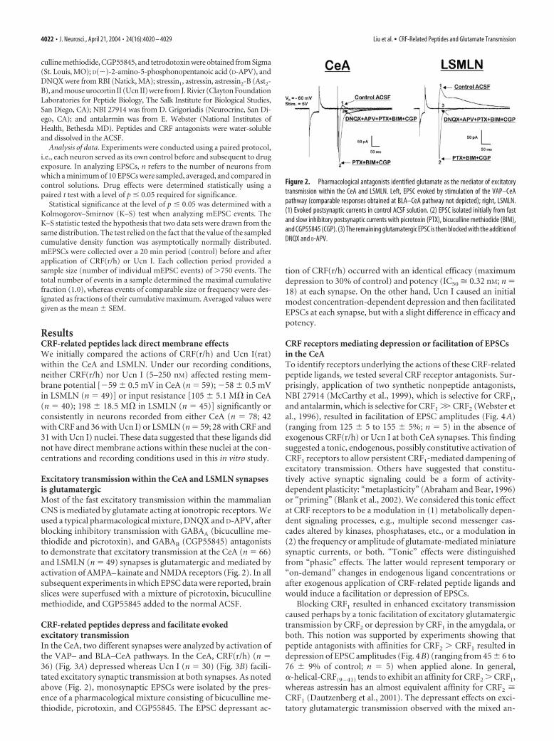
Excitatory transmission within the CeA and LSMLN synapsesis glutamatergicMost of the fast excitatory transmission within the mammalianCNS is mediated by glutamate acting at ionotropic receptors. Weused a typical pharmacological mixture, DNQX and D-APV, afterblocking inhibitory transmission with GABAA (bicuculline me-thiodide and picrotoxin), and GABAB (CGP55845) antagoniststo demonstrate that excitatory transmission at the CeA (n � 66)and LSMLN (n � 49) synapses is glutamatergic and mediated byactivation of AMPA– kainate and NMDA receptors (Fig. 2). In allsubsequent experiments in which EPSC data were reported, brainslices were superfused with a mixture of picrotoxin, bicucullinemethiodide, and CGP55845 added to the normal ACSF.
CRF-related peptides depress and facilitate evokedexcitatory transmissionIn the CeA, two different synapses were analyzed by activation ofthe VAP– and BLA–CeA pathways. In the CeA, CRF(r/h) (n �36) (Fig. 3A) depressed whereas Ucn I (n � 30) (Fig. 3B) facili-tated excitatory synaptic transmission at both synapses. As notedabove (Fig. 2), monosynaptic EPSCs were isolated by the pres-ence of a pharmacological mixture consisting of bicuculline me-thiodide, picrotoxin, and CGP55845. The EPSC depressant ac-
tion of CRF(r/h) occurred with an identical efficacy (maximumdepression to 30% of control) and potency (IC50 � 0.32 nM; n �18) at each synapse. On the other hand, Ucn I caused an initialmodest concentration-dependent depression and then facilitatedEPSCs at each synapse, but with a slight difference in efficacy andpotency.
CRF receptors mediating depression or facilitation of EPSCsin the CeATo identify receptors underlying the actions of these CRF-relatedpeptide ligands, we tested several CRF receptor antagonists. Sur-prisingly, application of two synthetic nonpeptide antagonists,NBI 27914 (McCarthy et al., 1999), which is selective for CRF1,and antalarmin, which is selective for CRF1 �� CRF2 (Webster etal., 1996), resulted in facilitation of EPSC amplitudes (Fig. 4A)(ranging from 125 � 5 to 155 � 5%; n � 5) in the absence ofexogenous CRF(r/h) or Ucn I at both CeA synapses. This findingsuggested a tonic, endogenous, possibly constitutive activation ofCRF1 receptors to allow persistent CRF1-mediated dampening ofexcitatory transmission. Others have suggested that constitu-tively active synaptic signaling could be a form of activity-dependent plasticity: “metaplasticity” (Abraham and Bear, 1996)or “priming” (Blank et al., 2002). We considered this tonic effectat CRF receptors to be a modulation in (1) metabolically depen-dent signaling processes, e.g., multiple second messenger cas-cades altered by kinases, phosphatases, etc., or a modulation in(2) the frequency or amplitude of glutamate-mediated miniaturesynaptic currents, or both. “Tonic” effects were distinguishedfrom “phasic” effects. The latter would represent temporary or“on-demand” changes in endogenous ligand concentrations orafter exogenous application of CRF-related peptide ligands andwould induce a facilitation or depression of EPSCs.
Blocking CRF1 resulted in enhanced excitatory transmissioncaused perhaps by a tonic facilitation of excitatory glutamatergictransmission by CRF2 or depression by CRF1 in the amygdala, orboth. This notion was supported by experiments showing thatpeptide antagonists with affinities for CRF2 � CRF1 resulted indepression of EPSC amplitudes (Fig. 4B) (ranging from 45 � 6 to76 � 9% of control; n � 5) when applied alone. In general,�-helical-CRF(9 – 41) tends to exhibit an affinity for CRF2 � CRF1,whereas astressin has an almost equivalent affinity for CRF2 �CRF1 (Dautzenberg et al., 2001). The depressant effects on exci-tatory glutamatergic transmission observed with the mixed an-
Figure 2. Pharmacological antagonists identified glutamate as the mediator of excitatorytransmission within the CeA and LSMLN. Left, EPSC evoked by stimulation of the VAP–CeApathway (comparable responses obtained at BLA–CeA pathway not depicted); right, LSMLN.(1) Evoked postsynaptic currents in control ACSF solution. (2) EPSC isolated initially from fastand slow inhibitory postsynaptic currents with picrotoxin (PTX), bicuculline methiodide (BIM),and CGP55845 (CGP). (3) The remaining glutamatergic EPSC is then blocked with the addition ofDNQX and D-APV.
4022 • J. Neurosci., April 21, 2004 • 24(16):4020 – 4029 Liu et al. • CRF-Related Peptides and Glutamate Transmission
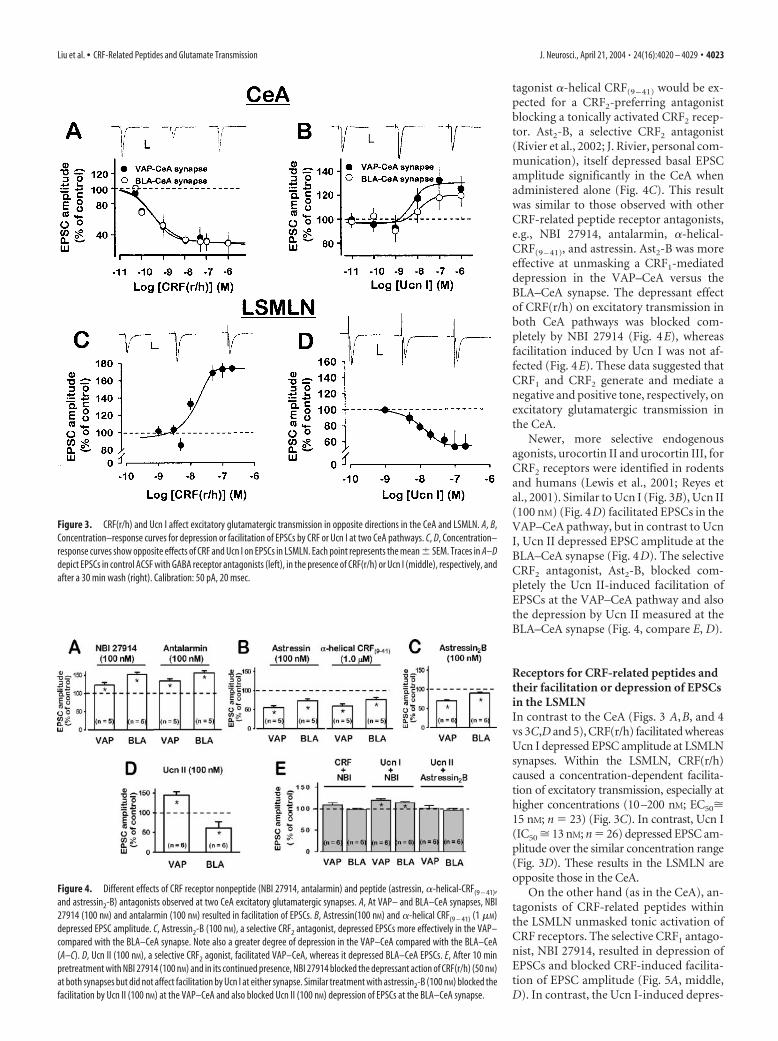
tagonist �-helical CRF(9 – 41) would be ex-pected for a CRF2-preferring antagonistblocking a tonically activated CRF2 recep-tor. Ast2-B, a selective CRF2 antagonist(Rivier et al., 2002; J. Rivier, personal com-munication), itself depressed basal EPSCamplitude significantly in the CeA whenadministered alone (Fig. 4C). This resultwas similar to those observed with otherCRF-related peptide receptor antagonists,e.g., NBI 27914, antalarmin, �-helical-CRF(9 – 41), and astressin. Ast2-B was moreeffective at unmasking a CRF1-mediateddepression in the VAP–CeA versus theBLA–CeA synapse. The depressant effectof CRF(r/h) on excitatory transmission inboth CeA pathways was blocked com-pletely by NBI 27914 (Fig. 4E), whereasfacilitation induced by Ucn I was not af-fected (Fig. 4E). These data suggested thatCRF1 and CRF2 generate and mediate anegative and positive tone, respectively, onexcitatory glutamatergic transmission inthe CeA.
Newer, more selective endogenousagonists, urocortin II and urocortin III, forCRF2 receptors were identified in rodentsand humans (Lewis et al., 2001; Reyes etal., 2001). Similar to Ucn I (Fig. 3B), Ucn II(100 nM) (Fig. 4D) facilitated EPSCs in theVAP–CeA pathway, but in contrast to UcnI, Ucn II depressed EPSC amplitude at theBLA–CeA synapse (Fig. 4D). The selectiveCRF2 antagonist, Ast2-B, blocked com-pletely the Ucn II-induced facilitation ofEPSCs at the VAP–CeA pathway and alsothe depression by Ucn II measured at theBLA–CeA synapse (Fig. 4, compare E, D).
Receptors for CRF-related peptides andtheir facilitation or depression of EPSCsin the LSMLNIn contrast to the CeA (Figs. 3 A,B, and 4vs 3C,D and 5), CRF(r/h) facilitated whereasUcn I depressed EPSC amplitude at LSMLNsynapses. Within the LSMLN, CRF(r/h)caused a concentration-dependent facilita-tion of excitatory transmission, especially athigher concentrations (10–200 nM; EC50�15 nM; n � 23) (Fig. 3C). In contrast, Ucn I(IC50 � 13 nM; n � 26) depressed EPSC am-plitude over the similar concentration range(Fig. 3D). These results in the LSMLN areopposite those in the CeA.
On the other hand (as in the CeA), an-tagonists of CRF-related peptides withinthe LSMLN unmasked tonic activation ofCRF receptors. The selective CRF1 antago-nist, NBI 27914, resulted in depression ofEPSCs and blocked CRF-induced facilita-tion of EPSC amplitude (Fig. 5A, middle,D). In contrast, the Ucn I-induced depres-
Figure 3. CRF(r/h) and Ucn I affect excitatory glutamatergic transmission in opposite directions in the CeA and LSMLN. A, B,Concentration–response curves for depression or facilitation of EPSCs by CRF or Ucn I at two CeA pathways. C, D, Concentration–response curves show opposite effects of CRF and Ucn I on EPSCs in LSMLN. Each point represents the mean � SEM. Traces in A–Ddepict EPSCs in control ACSF with GABA receptor antagonists (left), in the presence of CRF(r/h) or Ucn I (middle), respectively, andafter a 30 min wash (right). Calibration: 50 pA, 20 msec.
Figure 4. Different effects of CRF receptor nonpeptide (NBI 27914, antalarmin) and peptide (astressin, �-helical-CRF(9 – 41),and astressin2-B) antagonists observed at two CeA excitatory glutamatergic synapses. A, At VAP– and BLA–CeA synapses, NBI27914 (100 nM) and antalarmin (100 nM) resulted in facilitation of EPSCs. B, Astressin(100 nM) and �-helical CRF(9 – 41) (1 �M)depressed EPSC amplitude. C, Astressin2-B (100 nM), a selective CRF2 antagonist, depressed EPSCs more effectively in the VAP–compared with the BLA–CeA synapse. Note also a greater degree of depression in the VAP–CeA compared with the BLA–CeA(A–C). D, Ucn II (100 nM), a selective CRF2 agonist, facilitated VAP–CeA, whereas it depressed BLA–CeA EPSCs. E, After 10 minpretreatment with NBI 27914 (100 nM) and in its continued presence, NBI 27914 blocked the depressant action of CRF(r/h) (50 nM)at both synapses but did not affect facilitation by Ucn I at either synapse. Similar treatment with astressin2-B (100 nM) blocked thefacilitation by Ucn II (100 nM) at the VAP–CeA and also blocked Ucn II (100 nM) depression of EPSCs at the BLA–CeA synapse.
Liu et al. • CRF-Related Peptides and Glutamate Transmission J. Neurosci., April 21, 2004 • 24(16):4020 – 4029 • 4023

sion of EPSC amplitude (Fig. 5A, bottom,D) was not altered by NBI 27914, suggest-ing that NBI 27914 released a tonic CRF2-mediated depression of EPSCs within theLSMLN.
A new and highly selective CRF1 pep-tide agonist, stressin1 (100 nM) (Rivier etal., 2001; J. Rivier, personal communica-tion), facilitated LSMLN EPSCs (Fig. 5B)to the same extent (167 � 15%) (Fig. 5E)as a comparable concentration of 100 nM
CRF(r/h) (Fig. 3C). The selective peptideCRF2 antagonist (Rivier et al., 2002),Ast2-B (100 nM) (Fig. 5C, top and bottom,E), enhanced EPSC amplitude, whereas itdid not block the facilitatory action ofCRF(r/h) on excitatory glutamatergictransmission (Fig. 5C, top, E). In contrast,the depressant effect of Ucn I on excitatoryglutamatergic transmission was blockedcompletely by Ast2-B (Fig. 5C, bottom, E),and a facilitatory action of Ucn I on EPSCsin LSMLN neurons was unmasked (Fig.5C, bottom, E). Furthermore, astressin,which has an almost equivalent affinity atCRF2 � CRF1 (Dautzenberg et al., 2001),had little effect on the EPSC amplitude(94.5 � 4.5%; n � 6; p � 0.05) in theLSMLN compared with control. Thesedata support the hypothesis that the facil-itating action of CRF(r/h) at excitatoryglutamatergic synapses within the LSMLNmay be mediated by a CRF1 receptor,whereas the Ucn I depressant action maybe mediated through a CRF2 receptor.
The availability and use of different antagonists provided re-sults that emphasize the complex distribution of these receptorswithin the LSMLN and CeA. Because Ucn II has no affinity for theCRF-binding protein (Reyes et al., 2001), it is unlikely that CRF2
requires the CRF-binding protein for activation in either CeA orLSMLN neurons.
CRFs affect mEPSCsTo examine the presynaptic versus postsynaptic site of action ofthe endogenous ligands CRF(r/h) and Ucn I, we recorded theireffects on the frequency of spontaneous mEPSCs, an indicator ofpresynaptic action, and amplitude, a gauge of postsynaptic ac-tion. mEPSCs were measured in the presence of picrotoxin (50�M), bicuculline (10 �M), CGP55845 (2 �M), and tetrodotoxin (1�M) in standard extracellular solution to block GABAA–GABAB
receptor activity and voltage-dependent sodium channels,respectively.
mEPSCs in the CeABecause the CeA contains CRF neurons, CRF autoreceptors maybe present and innervated by CRF terminals from intrinsic CRFcell bodies (Harrigan et al., 1994). Figure 6, B and D, representsnormalized cumulative amplitude and interevent interval distri-bution curves for a control and either a CRF- or an Ucn I-treatedneuron recorded from the CeA. In the presence of CRF(r/h) (50nM), both curves were shifted to the left, i.e., CRF depressed theamplitude while increasing the frequency (decreasing the inter-event interval) of mEPSCs compared with control. CRF(r/h) de-
creased significantly the mean mEPSC amplitude from 17.4 � 0.4to 14.5 � 0.2 pA ( p � 0.01; n � 6), whereas the frequency wasincreased significantly from 3.9 to 4.7 mEPSCs per second (adecreased interevent interval from 256 � 11 msec in control to215 � 8 msec with CRF). Ucn I (200 nM), on the other hand,significantly increased the amplitude of mEPSCs from 18.6 � 0.3to 21.6 � 0.2 pA ( p � 0.01; n � 6), shown as a shift to the right inFigure 6D. Similar to CRF, but exerting a more robust effect, UcnI shifted the normalized cumulative interevent interval distribu-tion curve to the left (Fig. 6D), with an average increased mEPSCfrequency from 4.5 to 12.3 mEPSCs per second (a decreased in-terevent interval from 220 � 11 msec in control to 81 � 2 msecwith Ucn I). These effects of CRF and Ucn on mEPSCs weresummarized in Figure 8 (bottom left). We have assigned the CRFincrease in mEPSC frequency to a CRF2- rather than a CRF1-mediated action on the basis of our data with evoked EPSCs andCRF receptor selective antagonists (Fig. 4). CRF(r/h) effects onmEPSC amplitude and frequency in the CeA suggested that itspostsynaptic depressant action prevailed over a presynaptic fa-cilitatory action when the effects of CRF on evoked EPSCs werecompared (Figs. 3A, 8, bottom left). Because both CRF1 and CRF2
receptor activation may occur postsynaptically, preferential acti-vation of one or the other postsynaptic receptor in conjunctionwith activation of the presynaptic CRF2 receptor would result indepression (CRF1) or facilitation (CRF2), respectively (Fig. 8).
mEPSCs in the LSMLNIn the LSMLN, CRF(r/h) (50 nM) produced a statistically signif-icant increase in the amplitude (Fig. 7A) (mean amplitude in-
Figure 5. CRF1 and CRF2 receptor activation tonically regulate EPSC amplitude in the LSMLN. A, Top, CRF(r/h, 50 nM) facilitatedwhereas Ucn I (100 nM) depressed LSMLN EPSCs. Middle, NBI 27914 (100 nM) resulted in depression of baseline EPSCs andprevented a 50 nM CRF-induced facilitation. Bottom, NBI 27914 (100 nM) resulted in depression of EPSC and further added to theUcn I (100 nM)-induced depression. B, Selective CRF1 agonist, stressin1 (100 nM), similar to CRF(r/h) (50 nM) (A, top left), facilitatedEPSC. C, Top, Ast2-B(100 nM) resulted in facilitated EPSC, whereas in its presence CRF(r/h) (50 nM) further enhanced the EPSC.Bottom, Ast2-B (100 nM) resulted in EPSC facilitation, whereas in its presence Ucn I (100 nM) now facilitated, rather than depressed,the EPSC. D, Graphic summary of effects of NBI (100 nM), CRF(r/h) (50 nM), and Ucn I (100 nM) on LSMLN EPSCs, and NBI (100 nM)block of CRF(r/h) (50 nM) facilitation, but not Ucn I (100 nM) depression. E, Graphic summary of EPSC facilitation by stressin1 (100nM; CRF1 agonist) and Ast2-B (100 nM; CRF2 antagonist). Ast2-B (100 nM) did not block facilitation by CRF(r/h) (50 nM), whereasAst2-B reversed Ucn I (50 nM) depression to facilitation.
4024 • J. Neurosci., April 21, 2004 • 24(16):4020 – 4029 Liu et al. • CRF-Related Peptides and Glutamate Transmission

creased from 17.0 � 0.5 to 30.1 � 0.9 pA; p � 0.01; n � 5)without affecting the frequency of mEPSCs (the mean intereventinterval was 297 � 14.7 msec in control and 310 � 15.2 msec inCRF-treated neurons; p � 0.05; n � 5). On the other hand, UcnI (200 nM) caused a significant depression of both amplitude andfrequency of mEPSCs (Fig. 7C). The mean amplitude was largerin control (19.9 � 0.5 pA) compared with Ucn I (14.6 � 0.6 pA;n � 6; p � 0.01). Furthermore, the mean interevent interval withUcn I was longer (from 352 � 25.3 to 991 � 62.3 msec; n � 6; p �0.01; mean frequency decreased from 2.8 to 1.0 mEPSCs persecond). Figure 7, B and D, represents normalized cumulativeamplitude and interevent distribution curves for a control andeither CRF- or Ucn I-treated LSMLN neurons. These mEPSCsresults were summarized and interpreted in Figure 8, bottomright.
To confirm a solely postsynaptic action for CRF in theLSMLN, we conducted paired-pulse facilitation (PPF) experi-ments, used as an indicator of a presynaptic locus of action, andmonitored the ratio of the amplitudes of the second to the first ofa pair of evoked EPSCs. CRF did not affect PPF in the LSMLN.These PPF data suggested only a postsynaptic action for CRF inthe LSMLN and supported our findings of a lack of effect of CRFon mEPSC frequency (Figure 7A,B): after Ucn I at 35 msec, 1.6 �0.16 versus 1.9 � 0.10; at 50 msec, 1.3 � 0.12 versus 1.5 � 0.08;and at 150 msec, 1.3 � 0.12 versus 1.5 � 0.08, respectively ( p �0.05 at each of these stimulus intervals). Ucn I enhanced PPF (Fig.7F), indicating a presynaptic inhibitory action because treat-ments decreasing transmitter release increase PPF (Katz andMiledi, 1968; Manabe et al., 1993); these data supported our find-
ings (Fig. 7C,D) of a reduced mEPSC frequency in the LSMLN.Our mEPSC and PPF data indicated the presence of CRF2 recep-tors on the glutamatergic terminals, similar to our previous datafor the CeA. In the LSMLN, however, the presynaptic CRF2 wasinhibitory, whereas in the CeA, the presynaptic CRF2 was facili-tatory. These presynaptic CRF2 inhibitory receptors could be lo-calized on CeA afferents to the LSMLN.
DiscussionThe primary findings of this study are as follows: (1) in the CeA,CRF(r/h) depressed excitatory glutamatergic transmissionthrough a CRF1-mediated postsynaptic action, whereas Ucn Ifacilitated EPSCs via postsynaptic and presynaptic facilitatoryCRF2-mediated mechanisms; (2) conversely, in the LSMLN, CRFcaused a CRF1-mediated facilitation of excitatory glutamatergictransmission via postsynaptic mechanisms, whereas Ucn I de-pressed EPSCs via postsynaptic and presynaptic CRF2-mediatedmechanisms; and (3) endogenous ligands for these receptors ton-ically and phasically affect excitatory glutamatergic transmissionin the CeA and LSMLN. These data provided evidence that CRF-related peptides function endogenously in neuronal circuits out-side the HPA where they activate local synaptic receptors to mod-ulate excitatory glutamatergic transmission.
Two endogenous ligands, CRF(r/h) and Ucn I, each bind toboth CRF1 and CRF2 receptors. In general, when Ucn I is com-pared with CRF(r/h) at either CRF1 or CRF2 receptors, Ucn I ismore potent (Hauger et al., 2003). Furthermore, Ucn I is alsomore potent (Vaughan et al., 1995) in releasing ACTH from an-terior pituitary corticotropes, a CRF1-mediated response. On the
Figure 6. CRF(r/h) and Ucn I affected mEPSCs differently in the CeA. A, Typical traces of mEPSCs before and after CRF(r/h) (50 nM). B, Graph of cumulative fraction of mEPSCs from single celldepicted in A and plotted as a function of amplitude ( p � 0.005; maximum amplitude difference was 0.11 by K–S test). Interevent interval ( p � 0.005; maximum difference was 0.11 by K–S test).C, Typical traces of mEPSCs before and after Ucn I (200 nM). D, Graph of cumulative fraction of mEPSCs from single cell depicted in C as a function of amplitude ( p�0.0001; maximum amplitude difference was0.11 by K–S test). Interevent interval ( p � 0.0001; maximum interevent interval difference was 0.36 by K–S test). Insets depict average changes increase from control at 1.0 (n � 6).
Liu et al. • CRF-Related Peptides and Glutamate Transmission J. Neurosci., April 21, 2004 • 24(16):4020 – 4029 • 4025

other hand, in a different functional assay, CRF(r/h) is morepotent at CRF1 and Ucn I is more potent at CRF2 in stimulatingcAMP (Hauger et al., 2003). Thus, in addition to the specificligand, the coupling of the respective receptors to their effectorsand the site at which the endogenous receptors are expressed canall affect and define the potency of the ligand when function isused to measure outcome. We used CRF(r/h) and Ucn I, theendogenous ligands for CRF1 and CRF2, along with selective ago-nists and antagonists for CRF1 and CRF2 to define CRF receptorsand their synaptic actions within two limbic nuclei.
We investigated two different CeA pathways: the VAP–CeAprovides afferent inputs from brainstem areas, whereas the BLA–CeA pathway carries intra-amygdala information (Alheid andHeimer, 1988). Both CeA synapses exhibited an equivalent levelof depression and an identical IC50. A CRF1-mediated depressantaction of EPSCs within the CeA was based on its antagonism bythe selective CRF1 antagonists NBI 27914 or antalarmin (Fig. 4).On the other hand, Ucn I (Fig. 3), at concentrations 20- to 50-foldgreater than CRF(r/h), facilitated glutamatergic EPSCs. Interest-ingly, Ucn I, unlike CRF(r/h), was more effective and potent inthe VAP– versus BLA–CeA pathway (Fig. 3B). Postsynaptic andpresynaptic CRF2 contributed to Ucn I facilitation because theselective CRF2 antagonist, Ast2-B, blocked the facilitation of EP-SCs induced by the selective CRF2 agonist, Ucn II (Fig. 4D,E).Furthermore, Ucn I and CRF each increased the frequency ofmEPSCs in the CeA (Figure 6). The differences in effective con-centrations between ligands at the specific CeA synapses may berelated to the relative abundance of a receptor type, the differen-tial linking of second messenger systems, and different locations
of receptors within a synapse (presynaptic versus postsynaptic).Figure 8 depicts a summary of our results.
We propose that during “normal” physiological conditions,termed “homeostasis” (Dallman, 2003; McEwen and Wingfield,2003), when lower endogenous CRF(r/h) concentrations mayoccur within the CeA, CRF1 activation results preferably in de-pression of EPSCs. On the other hand, with higher concentra-tions of CRF-related peptides, both CRF1 and CRF2 will be acti-vated. If the endogenous ligand or a drug preferred CRF1
receptors, then depression of the excitatory drive from the VAPor BLA to the CeA would be expected because lower CRF1 agonistconcentrations result in maximal depression of EPSCs. If a ligandpreferred CRF2 receptors (Ucn II), however, then a mechanismto facilitate excitatory drive onto the CeA, especially its inputfrom the VAP–CeA pathway, would be enhanced while simulta-neously depressing EPSCs in the LSMLN (Fig. 3).
Within the LSMLN, the effective concentration range for bothCRF(r/h) and Ucn I was greater than CRF(r/h) but was similar tothat of Ucn I in the CeA. This might be expected because theLSMLN exhibits the highest density for CRF2 receptors in thebrain (Chalmers et al., 1995; Li et al., 2002). Furthermore, unlikethe CeA, CRF(r/h) had no effect on mEPSC frequency or PPF,whereas Ucn I exhibited a lower effective concentration, reducedmEPSC frequency, and enhanced PPF; these latter data provideevidence for a presynaptic inhibitory CRF2. Activation of bothpostsynaptic CRF1 (facilitation) and presynaptic and postsynap-tic CRF2 (depression) could cancel the effects of each. The highereffective concentrations required by either ligand in the LSMLNcompared with the CeA could suggest that unless endogenous
Figure 7. CRF(r/h) and Ucn I have different effects on mEPSCs within the LSMLN. A, Typical traces of mEPSCs before and after CRF(r/h) (50 nM). B, Graph of cumulative fraction of mEPSCs fromsingle cell depicted in A and plotted as a function of amplitude ( p � 0.001; maximum amplitude difference was 0.29 by K–S test). Interevent interval ( p � 0.401 by K–S test). C, Ucn I (200 nM)depressed the amplitude ( p � 0.0001; maximum amplitude difference was 0.36 by K–S test). Maximum difference of the interevent interval was 0.42 by K–S test). Insets depict decrease fromcontrol at 1.0 (n�5) of mEPSCs. D, Graphic plot of the effect of Ucn I on the cumulative fraction of mEPSCs as a function of mEPSC amplitude and interevent interval. E, Plot of EPSC2 /EPSC1 ratio versusinterstimulus intervals of EPSCs before (control) and in the presence of CRF(r/h); CRF did not affect this ratio. F, Ucn I increased the EPSC2 /EPSC1 ratio significantly (*p � 0.05; n � 5), especially atthe shorter intervals.
4026 • J. Neurosci., April 21, 2004 • 24(16):4020 – 4029 Liu et al. • CRF-Related Peptides and Glutamate Transmission

ligand levels are elevated, e.g., during stress, the role for CRF-related peptides during normal physiological conditions withinthese two nuclei would be to depress excitatory transmission inthe CeA.
Our data indicate that CRF1 and CRF2 receptors are colocal-ized postsynaptically in the LSMLN and that their selective orpossibly mixed activation mediates conspicuously opposite ac-tions (Figs. 3, 8). Dautzenberg and Hauger (2002) also depictedsuch a cellular colocalization. Cellular colocalization of differentreceptors for the same or similar ligand (e.g., another member ofthe CRF-related peptides) suggests that different second messen-ger systems and downstream proteins could be responsible formediation of CRF-related peptide transduction mechanisms. Be-cause our data indicate that excitatory glutamatergic synaptictransmission can be affected by the prevailing tone of the endog-enous ligands at CRF1 and CRF2 receptors, an imbalance of CRF-related peptide receptor tone within the LSMLN opens this mod-ulation to possible regulation by various factors andenvironmental conditions. Because LSMLN neurons are linkeddirectly to the CeA (Jakab and Leranth, 1995), an imbalancewithin the LSMLN would also cause an imbalance in the CeA.Furthermore, CRF axons from the CeA project to the LSMLN(Jakab and Leranth, 1995) and would also modulate transmissionwithin the LSMLN.
What might be the functional relevance of the concentration-dependent effects of CRF-related peptides in normal excitatory
glutamatergic synaptic transmission and what is the source ofendogenously released CRF-related peptides acting within theLSMLN or CeA? Importantly, anxiety-like behavioral and auto-nomic effects after centrally administered CRF are not dependenton CRF-induced HPA-released ACTH or corticosteroids, be-cause these anxiety-related outcomes persist in hypophysecto-mized rats (Eaves et al., 1985) and in mice conditionally knockedout for CRF1 (Muller et al., 2003). Bremner et al. (1997) reportedhigher levels of CRF (137%) in CSF of patients diagnosed withchronic combat-related posttraumatic stress disorder versus CS-F–CRF in comparison subjects (100%). Subsequently, Vythil-ingam et al. (2000) concluded that in healthy humans, CSF–CRFrepresented CRF derived primarily from non-HPA CRF neuronsrather than HPA-axis CRF neurons projecting from the paraven-tricular nucleus of the hypothalamus. These non-HPA-mediatedmodulatory actions may be caused by circulating CRF-relatedpeptides (Rothman et al., 2002), or may be the result of CRF-related peptides released locally from neurons where it may becolocalized with a variety of transmitters, including glutamate(Sauvage and Steckler, 2001; King and Bishop, 2002). In thisextra-HPA role, CRF has been linked to stress (Dunn and Ber-ridge, 1990), fear (Koob et al., 1993), and reward (Rivier andVale, 1987). Antagonists of these peptides infused into the amyg-dala block consolidation of fear memory (Liang et al., 1992;Roozendaal et al., 2002). In general, activation of brain CRF1 isassociated with anxiogenic behaviors (Liebsch et al., 1995),whereas activation of brain CRF2 may be involved in coping be-havior in stressful situations (Liebsch et al., 1999). Radulovic et al.(1999) suggested that modulation by CRF1 versus CRF2 in learn-ing and anxiety could be mediated by differential signaling.
Antagonism of CRF2 in the lateral septum or CRF1 in theamygdala reduces stress-induced behavior in the rat (Bakshi etal., 2002). Furthermore, Yadin et al. (1993) suggested that theLSMLN and the amygdala are critical nuclei in the expression andcontrol of emotions, especially those involving fear, depression,anxiety, reward, and stress. These data support the hypothesisthat the opposing actions of endogenous CRF-related peptideson their receptors in these two limbic nuclei may act in concert toregulate these plastic adaptations of the CNS. Altogether our dataimplicate a role for CRF-related peptides to modulate or perhapsregulate synaptic excitatory glutamatergic transmission locallyunder both normal and stressful conditions and emphasize thatthe effects of CRF-related peptides are dependent on multiplevariables, including the brain area, the synapse, and sites within agiven synapse.
In summary, we show a novel and highly crucial role for CRF-related peptides as “modulators” of in vitro excitatory glutama-tergic transmission in two limbic nuclei expressing their recep-tors. In this modulatory role, the receptors would function toestablish a “tone” or homeostasis for normal excitatory glutama-tergic transmission that is finely controlled by the endogenouslevels of the peptides. CRF-receptor activation by CRF-relatedpeptides could also serve as a priming (Blank et al., 2002) ormetaplastic (Abraham and Bear, 1996) mechanism to provideadditional modulation of excitatory transmission, when needed.The synaptic homeostasis of these endogenous CRF-related pep-tides and CRF receptors may also play an important role in initi-ating, expressing, or maintaining cellular signaling processes thatcontribute to learning, memory, anxiety, and depression. Finally,therapeutic agents interacting with CRF-related peptides couldcontribute to reestablishing synaptic homeostasis when per-turbed during stress, e.g., at “stressed synapses,” or in the induc-tion of various mental illnesses associated with stress.
Figure 8. Diagrams depicting presynaptic and postsynaptic distribution and function of CRF1
and CRF2 within two limbic pathways. Tonic and phasic activation of CRF1 or CRF2 receptorsregulates excitatory glutamatergic transmission positively by facilitation (green) or negativelyby depression (red). Endogenous CRF peptides may be co-released from nerve terminals (intra-cellularly; CRF Peptides enclosed inside yellow retangle on nerve terminals) or be circulated andmaintained in the extracellular space (CRF Peptides as unbound yellow rectangle and free insynaptic spaces), or both. Left (at CeA), CRF1 located postsynaptically; activation leads to de-pression of EPSC amplitude. CRF2 located presynaptically and postsynaptically; activation maylead to enhanced glutamate release and facilitation of EPSC amplitude, but end result alsodepends on level of CRF1 activation. Right (at LSMLN), CRF1 located postsynaptically; activationleads to facilitation of EPSC amplitude. CRF2 located postsynaptically and presynaptically; acti-vation leads to depression of EPSC amplitude, but result depends on net CRF1 and CRF2 effects.Tables below diagram (Left, CeA; right, LSMLN) summarize mEPSC and evoked EPSC data andsuggest CRF receptor types mediating the respective results.
Liu et al. • CRF-Related Peptides and Glutamate Transmission J. Neurosci., April 21, 2004 • 24(16):4020 – 4029 • 4027

ReferencesAbraham WC, Bear MF (1996) Metaplasticity: the plasticity of synaptic
plasticity. Trends Neurosci 19:126 –130.Aggleton JP (1992) The amygdala: neurobiological aspects of emotion,
memory, and mental dysfunction. New York: Wiley.Aldenhoff J, Gruol DL, Rivier J, Vale W, Siggins GR (1983) Corticotropin
releasing factor decreases postburst hyperpolarizations and excites hip-pocampal neurons. Science 221:875– 877.
Alheid GF, Heimer L (1988) New perspectives in basal forebrain organiza-tion of special relevance for neuropsychiatric disorders: the striatopalli-dal, amygdaloid, and corticopetal components of substantia innominata.Neuroscience 27:1–39.
Bakshi VP, Smith-Roe S, Newman SM, Grigoriadis DE, Kalin NH (2002)Reduction of stress-induced behavior by antagonism of corticotropin-releasing hormone 2 (CRH2) receptors in lateral septum or CRH1 recep-tors in amygdala. J Neurosci 22:2926 –2935.
Bale TL, Vale WW (2004) CRF and CRF receptors: role in stress responsivityand other behaviors. Annu Rev Pharmacol Toxicol 44:525–557.
Bishop GA (2002) Development of a corticotropin-releasing factor-mediated effect on the firing rate of Purkinje cells in the postnatal mousecerebellum. Exp Neurol 178:165–174.
Blank T, Nijholt I, Eckart K, Spiess J (2002) Priming of long-term potenti-ation in mouse hippocampus by corticotropin-releasing factor and acutestress: implications for hippocampus-dependent learning. J Neurosci22:3788 –3794.
Blank T, Nijholt I, Grammatopoulos DK, Randeva HS, Hillhouse EW, SpiessJ (2003) Corticotropin-releasing factor receptors couple to multipleG-proteins to activate diverse intracellular signaling pathways in mousehippocampus: role in neuronal excitability and associative learning.J Neurosci 23:700 –707.
Bremner JD, Licinio J, Darnell A, Krystal JH, Owens MJ, Southwick SM,Nemeroff CB, Charney DS (1997) Elevated CSF corticotropin-releasingfactor concentrations in posttraumatic stress disorder. Am J Psychiatry154:624 – 629.
Chalmers DT, Lovenberg TW, De Souza EB (1995) Localization of novelcorticotropin-releasing factor receptor (CRF2) mRNA expression to spe-cific subcortical nuclei in rat brain: comparison with CRF1 receptormRNA expression. J Neurosci 15:6340 – 6350.
Dallman MF (2003) Stress by any other name. . . ? Horm Behav 43:18 –20.Dautzenberg FM, Hauger RL (2002) The CRF peptide family and their re-
ceptors: yet more partners discovered. Trends Pharmacol Sci 23:71–77.Dautzenberg FM, Killpatrick GJ, Hauger RL, Moreau J-L (2001) Molecular
biology of the CRH receptors-in the mood. Peptides 22:753–760.Dunn AJ, Berridge CW (1990) Physiological and behavioral responses to
corticotropin-releasing factor administration: is CRF a mediator of anx-iety in the stress response? Brain Res Rev 15:71–100.
Eaves M, Thatcher-Britton K, Rivier J, Vale W, Koob G (1985) Effects ofcorticotropin-releasing factor on locomotor activity in hypophysecto-mized rats. Peptides 6:923–926.
Fox EA, Gruol DL (1993) Corticotropin-releasing factor suppresses the af-terhyperpolarization in cerebellar Purkinje neurons. Neurosci Lett149:103–107.
Grammatopoulos DK, Randeva H, Levine M, Kanellopoulou K, Hillhouse E(2001) Rat cerebral cortex corticotropin-releasing hormone receptors:evidence for receptor coupling to multiple G-proteins. J Neurochem76:509 –519.
Harrigan EA, Magnuson DJ, Thunstedt GM, Gray TS (1994) Corticotropinreleasing factor neurons are innervated by calcitonin gene-related peptideterminals in the rat central amygdaloid nucleus. Brain Res Bull33:529 –534.
Hauger RL, Grigoriadis DE, Dallman M, Plotsky PM, Vale WW, DautzenbergFM (2003) International Union of Pharmacology. XXXVI. Current sta-tus of the nomenclature for receptors for corticotropin-releasing factorand their ligands. Pharmacol Rev 55:21–26.
Hsu SY, Hsueh AJW (2001) Human stresscopin and stresscopin-relatedpeptide are selective ligands for the corticotropin-releasing hormone re-ceptor. Nat Med 7:605– 606.
Jakab RL, Leranth C (1995) Septum. In: The rat nervous system (Paxinos G,ed), pp 405– 442. San Diego: Academic.
Katz B, Miledi R (1968) The role of calcium in neuromascular facilitation. JPhysiol 195:481– 492.
King JS, Bishop GA (2002) The distribution and cellular localization of
CRF-R1 in the vermis of the postnatal mouse cerebellum. Exp Neurol178:175–185.
Koob GF, Heinrichs SC, Pich EM, Menzaghi F, Baldwin HA, Miczek KA,Britton KT (1993) The role of corticotropin-releasing factor in behav-ioural responses to stress. Ciba Found Symp 172:277–295.
Lewis K, Li C, Perrin MH, Blount A, Kunitake K, Donaldson C, Vaughan J,Reyes TM, Gulyas J, Fischer W, Bilezikjian L, Rivier J, Sawchenko PE, ValeWW (2001) Identification of urocortin III, an additional member of thecorticotropin-releasing factor (CRF) family with high affinity for theCRF2 receptor. Proc Natl Acad Sci USA 98:7570 –7575.
Li C, Vaughan J, Sawchenko PE, Vale WW (2002) Urocortin III-immunoreactive projections in rat brain: partial overlap with sites of type2 corticotrophin-releasing factor receptor expression. J Neurosci22:991–1001.
Liang KC, Melia KR, Campeau S, Falls WA, Miserendino MJ, Davis M(1992) Lesions of the central nucleus of the amygdala, but not theparaventricular nucleus of the hypothalamus, block the excitatory ef-fects of corticotropin releasing factor on the acoustic startle reflex.J Neurosci 12:2313–2320.
Liebsch G, Landgraf R, Gerstberger R, Probst JC, Wotjak CT, Engelmann M,Holsboer F, Montkowski A (1995) Chronic infusion of a CRH1 receptorantisense oligodeoxynucleotide into the central nucleus of the amygdalareduced anxiety-related behavior in socially defeated rats. Regul Pept59:229 –239.
Liebsch G, Landgraf R, Engelmann M, Lorscher P, Holsboer F (1999) Dif-ferential behavioural effects of chronic infusion of CRH 1 and CRH 2receptor antisense oligonucleotides into the rat brain. J Psychiatr Res33:153–163.
Manabe T, Wyllie DJA, Perkel DJ, Nicoll RA (1993) Modulation of synaptictransmission and long-term potentiation: effects on paired pulse facilita-tion and EPSC variance in the CA1 region of the hippocampus. J Neuro-physiol 70:1451–1459.
McCarthy JR, Heinrichs SC, Grigoriadis DE (1999) Recent advances withthe CRF1 receptor design of small molecule inhibitors, receptor sybtypesand clinical implications. Curr Pharm Des 5:289 –315.
McEwen BS, Wingfield JC (2003) The concept of allostasis in biology andbiomedicine. Horm Behav 43:2–15.
Miyata M, Okada D, Hashimoto K, Kano M, Ito M (1999) Corticotropin-releasing factor plays a permissive role in cerebellar long-term depression.Neuron 22:763–775.
Muller MB, Zimmermann S, Sillaber I, Hagemeyer TP, Deussing JM, TimplP, Kormann MS, Droste SK, Kuhn R, Reul JM, Holsboer F, Wurst W(2003) Limbic corticotropin-releasing hormone receptor I mediatesanxiety-related behavior and hormonal adaptation to stress. Nat Neurosci6:1100 –1107.
Neugebauer V, Zinebi F, Russell R, Gallagher JP, Shinnick-Gallagher P(2000) Cocaine and kindling alter the sensitivity of group II and IIImetabotropic glutamate receptors in the central amygdala. J Neuro-physiol 84:759 –770.
Numan R (2000) The behavioral neuroscience of the septal region. NewYork: Springer.
Owens MJ, Nemeroff CB (1993) The role of corticotropin-releasing factorin the pathophysiology of affective and anxiety disorders: laboratory andclinical studies. Ciba Found Symp 172:296 –308.
Radulovic J, Ruhmann A, Liepold T, Spiess J (1999) Modulation of learningand anxiety by corticotropin-releasing factor (CRF) and stress: differen-tial roles of CRF receptors 1 and 2. J Neurosci 19:5016 –5025.
Rainnie DG, Fernhout BJH, Shinnick-Gallagher P (1992) Differential ac-tions of corticotropin releasing factor on basolateral and central amygda-loid neurones, in vitro. J Pharmacol Exp Ther 263:846 – 858.
Reyes TM, Lewis K, Perrin MH, Kunitake KS, Vaughan J, Arias CA, Hogen-esch JB, Gulyas J, Rivier J, Vale WW, Sawchenko PE (2001) Urocortin II:a member of the corticotropin-releasing factor (CRF) neuropeptide fam-ily that is selectively bound by type 2 CRF receptors. Proc Natl Acad SciUSA 98:2843–2848.
Rivier C, Vale W (1987) Cocaine stimulates adrenocorticotropin (ACTH)secretion through a corticotropin-releasing factor (CRF)-mediatedmechanism. Brain Res 422:403– 406.
Rivier J, Gulyas J, Kirby D, Low W, Perrin MH, Kunitake K, DiGruccio M,Vaughan J, Reubi J-C, Waser B, Koerber SC, Martinez V, Wang L, TacheY, Vale W (2002) Potent and long-acting corticotropin releasing factor
4028 • J. Neurosci., April 21, 2004 • 24(16):4020 – 4029 Liu et al. • CRF-Related Peptides and Glutamate Transmission

(CRF) receptor 2 peptide competitive antagonists. J Med Chem45:4737– 4747.
Rivier JE, Gulyas JD, Kirby K, Kunitake K, Donaldson C, Vaughn J, Perrin S,Koerber S, Martinez V, Tache Y, Rivier C, Vale W (2001) Receptor-selective corticotropin-releasing factor analogs. Soc Neurosci Abstr31:413.17.
Roozendaal B, Brunson KL, Holloway BL, McGaugh JL, Baram TZ (2002)Involvement of stress-released corticotropin-releasing hormone in thebasolateral amygdala in regulating memory consolidation. Proc NatlAcad Sci USA 99:13908 –13913.
Rothman RB, Vu N, Xu H, Baumann MH, Lu YF (2002) Endogenous cor-ticotropin releasing factor regulates adrenergic and opioid receptors. Pep-tides 23:2177–2180.
Sauvage M, Steckler T (2001) Detection of corticotropin-releasing hormonereceptor 1 immunoreactivity in cholinergic, dopaminergic and noradren-ergic neurons of the murine basal forebrain and brainstem nuclei: poten-tial implication for arousal and attention. Neuroscience 104:643– 652.
Shinnick-Gallagher P, Pitkanen A, Shekhar A, Cahill L (2003) The amygdalain brain function: basic and clinical approaches. Ann NY Acad Sci 985.
Smith BN, Dudek FE (1994) Age-related epileptogenic effects ofcorticotropin-releasing hormone in the isolated CA1 region of rat hip-pocampal slices. J Neurophysiol 72:2328 –2333.
Steckler T, Holsboer F (1999) Corticotropin-releasing hormone receptorsubtypes and emotion. Biol Psychiatry 46:1480 –1508.
Swanson LW, Sawchenko PE, Rivier J, Vale WW (1983) Organization ofovine corticotropin-releasing factor immunoreactive cells and fibers inthe rat brain: an immunohistochemical study. Neuroendocrinology36:165–186.
Ungless MA, Singh V, Crowder TL, Yaka R, Ron D, Bonci A (2003)Corticotropin-releasing factor requires CRF binding protein to potentiateNMDA receptors via CRF receptor 2 in dopamine neurons. Neuron39:401– 407.
Vale W, Spiess J, Rivier C, Rivier J (1981) Characterization of a 41-residueovine hypothalamic peptide that stimulates secretion of corticotropin andbeta-endorphin. Science 213:1394 –1397.
Valentino RJ (1989) Corticotropin-releasing factor: putative neurotrans-mitter in the noradrenergic nucleus locus ceruleus. PsychopharmacolBull 25:306 –311.
Vaughan J, Donaldson C, Bittencourt J, Perrin MH, Lewis K, Sutton S, ChanR, Turnbull AV, Lovejoy D, Rivier C (1995) Urocortin, a mammalianneuropeptide related to fish urotensin I and to corticotropin-releasingfactor. Nature 378:287–292.
Vythilingam M, Anderson GM, Owens MJ, Halaszynski TM, Bremner JD,Carpenter LL, Heninger GR, Nemeroff CB, Charney DS (2000) Cere-brospinal fluid corticotropin-releasing hormone in healthy humans: ef-fects of yohimbine and naloxone. J Clin Endocrinol Metab85:4138 – 4145.
Wang HL, Tsai LY, Lee EHY (2000) Corticotropin-releasing factor pro-duces a protein synthesis-dependent long-lasting potentiation in dentategyrus neurons. J Neurophysiol 83:343–349.
Webster EL, Lewis DB, Torpy DJ, Zachman EK, Rice KC, Chrousos GP(1996) In vivo and in vitro characterization of antalarmin, a nonpeptidecorticotripin-releasing hormone (CRH) receptor antagonist: suppressionof pituitary ACTH release and peripheral inflammation. Endocrinology137:5747–5750.
Yadin E, Thomas E, Grishkat HL, Strickland CE (1993) The role of thelateral septum in anxiolysis. Physiol Behav 53:1077–1083.
Yu B, Shinnick-Gallagher P (1998) Corticotropin-releasing factor increasesdihydropyridine- and neurotoxin-resistant calcium currents in neuronsof the central amygdala. J Pharmacol Exp Ther 284:170 –179.
Yu B, Wang C, Liu J, Johnson KM, Gallagher JP (2002) Adaptation tochronic PCP results in hyperfunctional NMDA and hypofunctionalGABAA synaptic receptors. Neuroscience 113:1–10.
Liu et al. • CRF-Related Peptides and Glutamate Transmission J. Neurosci., April 21, 2004 • 24(16):4020 – 4029 • 4029