the h-current in periglomerular dopaminergic neurons of the mouse olfactory bulb
TRANSCRIPT

The h-Current in Periglomerular Dopaminergic Neuronsof the Mouse Olfactory BulbAngela Pignatelli, Mirta Borin, Alex Fogli Iseppe, Cristina Gambardella, Ottorino Belluzzi*
Dipartimento di Scienze della Vita e Biotecnologie, University of Ferrara and Istituto Nazionale di Neuroscienze, Ferrara, Italy
Abstract
The properties of the hyperpolarization-activated cation current (Ih) were investigated in rat periglomerular dopaminergicneurons using patch-clamp recordings in thin slices. A reliable identification of single dopaminergic neurons was madepossible by use of a transgenic line of mice expressing eGFP under the tyrosine hydroxylase promoter. At 37 uC andminimizing the disturbance of the intracellular milieu with perforated patches, this current shows a midpoint of activationaround 282.7 mV, with a significant level of opening already at rest, thereby giving a substantial contribution to the restingpotential, and ultimately playing a relevant function in the control of the cell excitability. The blockage of Ih has a profoundinfluence on the spontaneous firing of these neurons, which result as strongly depressed. However the effect is not due to adirect role of the current in the pacemaker process, but to the Ih influence on the resting membrane potential. Ih kinetics issensitive to the intracellular levels of cAMP, whose increase promotes a shift of the activation curve towards more positivepotentials. The direct application of DA and 5-HT neurotransmitters, physiologically released onto bulbar dopaminergicneurons and known to act on metabotropic receptors coupled to the cAMP pathway, do not modifythe Ih amplitude. On thecontrary, noradrenaline almost halves the Ih amplitude. Our data indicate that the HCN channels do not participate directlyto the pacemaker activity of periglomerular dopaminergic neurons, but influence their resting membrane potential bycontrolling the excitability profile of these cells, and possibly affecting the processing of sensory information taking place atthe entry of the bulbar circuitry.
Citation: Pignatelli A, Borin M, Fogli Iseppe A, Gambardella C, Belluzzi O (2013) The h-Current in Periglomerular Dopaminergic Neurons of the Mouse OlfactoryBulb. PLoS ONE 8(2): e56571. doi:10.1371/journal.pone.0056571
Editor: Andrea Barbuti, University of Milan, Italy
Received August 30, 2012; Accepted January 11, 2013; Published February 13, 2013
Copyright: � 2013 Pignatelli et al. This is an open-access article distributed under the terms of the Creative Commons Attribution License, which permitsunrestricted use, distribution, and reproduction in any medium, provided the original author and source are credited.
Funding: The research was supported by a grant from the Ministero della Universita e della Ricerca Scientifica (MIUR) - PRIN 2009. The funders had no role instudy design, data collection and analysis, decision to publish, or preparation of the manuscript.
Competing Interests: The authors have declared that no competing interests exist.
* E-mail: [email protected]
Introduction
In the olfactory bulb (OB) dopaminergic (DA) neurons represent
a fraction of the cells located in the most external (glomerular)
layer [1]. In this region populated by three types of interneurons,
i.e. periglomerular (PG) cells, short-axon cells and external tufted
(ET) cells- often collectively referred to as juxtaglomerular cells -
an estimated 10–16% of the neurons in adulthood are positive for
tyrosine hydroxylase (TH) [2–4], the rate-limiting enzyme for
dopamine synthesis. Dopaminergic neurons in the glomerular
layer, which include PG cells [5,6] and a fraction of ET cells [1],
have been the object of several studies focused on their role in
olfactory signal processing. In spite of the many experiments
carried out by a number of different approaches including
immunohistochemical [7,8], behavioral [9], and electrophysiolog-
ical techniques [10–12], their role is far from being understood.
A common attribute of DA neurons in the CNS is their
capability to generate rhythmic action potentials even in the
absence of synaptic inputs [13–15], a feature shared by DA cells in
the glomerular layer of the olfactory bulb [16]. In many
autorhythmic cells, a key role in the pacemaking process is played
by the inward current (Ih; for a review see [17]), carried by
channels encoded by four HCN genes (hyperpolarization-activated
cyclic nucleotide-sensitive cation nonselective).In a previous study
[16], analyzing the excitability profile of DA PG cells, we failed to
detect any significant component activated by hyperpolarization
(Fig. 1A), concluding that there were no hyperpolarization-
activated currents. On the other hand, an unidentified fraction
of rat PG cell showed an evident h-current in normal saline [18].
Our conclusion was that HCN channels were absent in DA PG
cells. This conclusion was subsequently strengthened by the
observation that, using a comprehensive set of antibodies against
all four isoforms, no HCN channels were detected in DA PG cells
[19]. It was therefore surprising to observe, later on, an inhibition
of spontaneous firing of bulbar DA neuronsby selective blockers of
the h-current. We then re-examined the problem, finding that in
fact there is an h-current, undetected by our previous investigation
due to its small amplitude. This current can be better evidenced
with ionic manipulations and, despite its small amplitude, can play
a role under physiological conditions. In this paper we describe the
properties of this current.
Materials and Methods
Animals and surgical proceduresEthic statement. Experimental procedures were carried out
so as to minimize animal suffering and the number of mice used.
The procedures employed are in accordance with the Directive
86/609/EEC on the protection of animals used for experimental
and other scientific purposes, and are approved by the Campus
Veterinarian of the Ferrara University.
PLOS ONE | www.plosone.org 1 February 2013 | Volume 8 | Issue 2 | e56571

A total of 102 mice were used. All experiments were performed
using the transgenic mice TH-GFP/21–31 line carrying the eGFP
gene under the control of the TH promoter [20,21]. Transgenic
mice were identified either by PCR on the genomic DNA
extracted from tail biopsies, or -at postnatal day 3 or 4- looking at
the fluorescence of the olfactory bulbs transilluminated with a UV
source (FBL / Basic-B & N-01; BLS, Hungary; FHS/F-01) and
observed with an emission filter (FHS/EF-2G2; BLS, Budapest,
Hungary). Transgenic lines were maintained as heterozygous by
breeding with C57BL/6J inbred mice.
Dissociation proceduresAdult mice (30–60 day-old) were used to isolate olfactory bulb
neurons. Two solutions were used for the preparation: a dissecting
solution and Tyrode’s solution. The dissecting medium (DM)
contained (in mM): 82 Na2SO4, 30 K2SO4, 10 HEPES, 5 MgCl2,
10 Glucose, and 0.001% phenol red indicator; pH was adjusted to
7.4 with NaOH and the solution was continuously bubbled with
100% O2. Tyrode’s solution contained (in mM) 137 NaCl, 5.4
KCl, 1.8 CaCl2, 1 MgCl2, 5 HEPES, 20 Glucose; the pH was
adjusted to 7.4 with NaOH and the solution was continuously
bubbled with 100% O2. Dissociation of the olfactory bulb by
enzymatic digestion and mechanical trituration was performed
following the procedure described by Gustincich [22], with minor
changes. After dissecting and slicing the bulbs, the small pieces
were transferred to a solution containing DM and 3% protease
type XXIII (Sigma) for 30–45 min at 37 uC. After enzymatic
digestion, the bulbs were transferred to solution containing DM,
1% bovine serum albumin (Sigma) and 1% trypsin inhibitor
(Sigma) to stop protease activity (10 min, 37uC). Bulbs were finally
suspended in Tyrode’s solution and triturated using home-made
fire-polished Pasteur pipettes of varying gauges. The cell
suspension was centrifuged at 107 g (5 min), and the pellet was
resuspended in Tyrode’s solution. The dissociated olfactory bulb
neurons were plated on glass coverslip previously coated with
concanavalin A (1 mg/ml) to allow sedimentation ofthe cells. The
cells were maintained at 37uC in an atmosphere of 5% CO2/95%
air, in DMEM (Dulbecco’s modified Eagle medium), supplement-
ed with 10% FBS (fetal bovine serum) and 10%penicillin-
Streptomycin. The cells were allowed to set on the glass for at
least 12 hour before commencement of recordings.
Recording conditionsThe temperature of the 1-ml recording chamber was controlled
using a couple of 39.7 W Peltier devices (RS Components, Milan,
Italy) and measured with a high-precision, low mass thermocouple
(RS Components).
Current and voltage recordings were acquired with an Multi-
Clamp 700B amplifier (Molecular Devices, Sunnyvale, CA), and a
12 bit A/D–D/A converter (Digidata 1440A; Molecular Devices).
Borosilicate glass pipettes (1.5 O.D., 0.87 I.D., with filament;
Hilgenberg, Malsfeld, Germany) were pulled with a Zeitz-DMZ
puller (Martinsried, Germany) and had a resistance of 4–5 MVwhen filled with standard intracellular (IC) solution; the seal
formation was realized with the help of an air pressure controller
(MPI, Lorenz Messgeratebau, Katlenburg-Lindau, Germany); the
seal resistance was always greater than 2 GV. The liquid-junction
potential (LJP) of the different solutions was estimated using the
junction potential calculator of pClamp (Molecular Devices).
SolutionsThe solutions used had the following composition (mM):
Figure 1. Hyperpolarization-activated currents in slices. A-D: Currents activated by hyperpolarizing steps. A - standard EC saline (EC 1, withTTX 0.6 mM, Cd2+ 100 mM); B - high K+ EC saline (EC 3, with TTX 0.6 mM, Cd2+ 100 mM); C - same as B plus 0.5 mM Ba2+ to block the KIR current; D -same as C after addition of a blocker of the h-current (1 mM Cs+); the recordings were taken after 5 min at any change of the bathing conditions. Allthe recordings of this group were performed with BL1 mix; perforated patch in slice at 34 uC.E-F: effect of 7 min application of ZD7288 30 mM -perforated patch in slice; EC 3 saline plus BL 1 and BL 2 mixes; RT. G: Current-clamp responses to the injection of a hyperpolarizing current step(260 pA) in a TH-GFP+ cell; black trace recorded in normal [K+]o (EC 2), red trace recorded in high [K+]o (EC 3);Vrest was 264.9 mV and 257.8 mV innormal and high K+, respectively; both recordings were performed with BL 1 mix,perforated patch in slice at 26 uC. H: Current-clamp responses to theinjection of a hyperpolarizing current step (256 pA) in a TH-GFP+ cell; all traces were recorded in high [K+]o (EC 3) plus BL 1 mix; the, red trace wasrecorded in the same saline plus 1 mM Cs+;Vrest was 255.8, 273.2 and 255.3 mV in control, Cs+ and washout, respectively;current-clamp recording;perforated patch in slice at 37 uC.doi:10.1371/journal.pone.0056571.g001
h-Current in Bulbar Dopaminergic Cells
PLOS ONE | www.plosone.org 2 February 2013 | Volume 8 | Issue 2 | e56571

EC 1 - standard extracellular (EC) saline: 125 NaCl, 2.5 KCl, 26
NaHCO3, 1.25 NaH2PO4, 2 CaCl2, 1 MgCl2, and 15 glucose;
LJP+3.0 mV.
EC 2 - modified EC saline (normal K, TEA): 105 NaCl, 2.5 KCl,
1.25 NaH2PO4, 20 TEA-Cl, 26 NaHCO3, 1 MgCl2, 2 CaCl2;
LJP+3.5 mV.
EC 3 - modified EC saline (high K, TEA): 85 NaCl, 32.5 KCl, 1.25
NaH2PO4, 20 TEA-Cl, 26 NaHCO3, 1 MgCl2, 2 CaCl2;
LJP+2.7 mV.
All EC solutions were continuously bubbled with 95% O2 and
5% CO2; the osmolarity was adjusted at 305 mOsm with glucose.
In the contexts indicated in the text, the following mixes were
used:
BL 1 - synaptic blockers: recording from slices, the EC solutions
always included kinurenic acid (1 mM) and bicuculline (10 mM);
BL 2 - ion channels blockers: to isolate the h-current, both in slices
and dissociated cells, a mix of blockers (TTX 0.6 mM, Cd2+100 mM, andBa2+ 0.5 mM) were normallyadded to the bath,
except where indicated.
standard pipette-filling intracellular (IC) solution: 120 KCl, 10 NaCl, 2
MgCl2, 0.5 CaCl2, 5 EGTA, 10 HEPES, 2 Na-ATP, 10 glucose.
The free calcium concentration with this internal solution was
calculated to be 16 nM (http://www.stanford.edu/cpatton/
downloads.htm).
For perforated patches, amphotericin B was included in the
recording electrode filling solution as perforating agent (200 mg/
ml plus 300 mg pluronic F-127). In order to ensure the integrity of
the perforated patch, EGTA was omitted from this solution and
the concentration of CaCl2 was raised to 3 mM in order to
monitor possible spontaneous breakups of the perforated patch.
Data were collected after the series resistance fell to ,50 MV.
Figure 2. Activation kinetics. A – Representative current traces for the analysis of activation. The membrane was held at 240 mV and thenhyperpolarized to test voltages from 260 to 2130 mV in 10 mV increments,10 s interval. Ih tails were elicited in response to a second pulse to2130 mV, following test voltages (see methods for explanation). EC solution: EC 3, plus BL 1 and BL 2 mixes; 37uC; slice; perforated patch. B: Whole-cell current-voltage relationship of the h-current in different experimental conditions: dissociated cells at RT (#, n = 11); slice, RT(,, n = 14); slice, 37uC (n, n = 7); mean values 6 S.E. EC solution was EC 3 plus BL 2 mix in all cases, with the further addition of BL 1 mix in slice preparation. C –Fractional activation of the h-current as in a group of 9 cells as a function of voltage using the protocol shown in A, and with the indicated duration ofthe hyperpolarizing step (see text for explanation). EC solution: EC 3, plus BL 1 and BL 2 mixes; 37uC; slice; perforated patch. D – Effect of temperatureand of the variable duration of the hyperpolarizing step on the midpoint of the h-current. Notice that the change from room temperature (2261 uC)to 37 uC entails a shift of 10.9 mV of the V50. EC solution: EC 3, plus BL 1 and BL 2 mixes; slice; perforated patch.doi:10.1371/journal.pone.0056571.g002
h-Current in Bulbar Dopaminergic Cells
PLOS ONE | www.plosone.org 3 February 2013 | Volume 8 | Issue 2 | e56571

In all IC solutions the osmolarity was adjusted to 295 mOsm
with glucose, and the pH to 7.2 with KOH.
Data analysisIh was evoked by a family of hyperpolarizing voltage steps from
the holding potential of 240 mV to 2130 mV in 10 mV
increments. The steps were applied in 10 s intervals. Offline
analysis was performed using version 10 of pClamp (Molecular
Devices) and version 8 ofOrigin (OriginLab Corporation, North-
ampton, MA).
When box charts are used to represent data ensembles, the
central line indicates the mean, boxes S.E., whiskers min-max
values.
Analysis of current recordingsThe Ih amplitude was measured as difference between the
steady-state currents at the end of test voltage pulses (Iss) and the
instantaneous currents and the beginning (Iinst); the latters were
measured extrapolating the double exponential fitting the h-
currentto the time of the onset of the hyperpolarizing pulse.
Rates of Ih activation were determined using the following
function (Clampfit 10.2, Molecular Devices):
f (t)~Xn
i~1Aie
{t
ti zC ð1Þ
where i = 1 or 2 (corresponding to single or double exponential
fit), Ai is the amplitude of the fitting component(s), ti is the time
constant(s), and C the shift of the fitted trace from zero, i.e. -Ai
aligning the baseline to zero.
The activation curve of Ih was constructed using a two-step
protocol [23]: Ih was first activated to a variable degree by a
conditioning step, and then fully activated by a second pulse to
2130 mV (Figure 2A). The resulting tail current amplitudes were
then normalized and fitted by the equation:
Itail
Itailmax
~ 1zexp vm{V50ð Þ=k½ �f g{1 ð2Þ
where Itail is the amplitude of the tail recorded at the second pulse,
Itailmax is the maximal amplitude of the tails, Vm is the membrane
potential; V50 is the membrane potential for which half of the
channels are open (midpoint); k is the dependence of the opening
of channels by the change of potential (slope).
The temperature coefficients of activation and deactivation time
constant are defined as:
Q10~rate T2ð Þrate T1ð Þ
� � 10T2{T1 ð3Þ
Thus, for every 10uC of change in temperature there is a Q10-
fold change of the rate analyzed.
Unless otherwise stated, data are presented as means 6 s.e.m.
Statistical significance of the results was assessed with one-way or
two-way analysis of variance (ANOVA), Student’s t test for paired
samples, as indicated. D’Agostino & Pearson omnibus normality
test was used; a P value of ,0.05 was considered significant.
Results
Data are based on recordings from 285 TH+ periglomerular
(PG) cells; neurons were selected on the basis of their position
around the glomeruli, dendritic arborization within the glomerular
neuropil, membrane capacitance (8.54+0.21 pF; n = 285) and
input resistance (915.69631.31 MV; n = 248). Dopaminergic PG
cells can be differentiated from external tufted cells not only by
their large difference in membrane capacitance and input
resistance, but also by their different modality of firing, regular
in DA-PG cells [16], in bursts in external tufted cells [24]. Short-
axon cells have membrane capacitance and input resistance very
similar to PG cells, but usually they can be recognized in slice for
their position between glomeruli, fusiform shape and dendrites
extending to different glomeruli [25].
The transgenic mice used in these experiments,expressing the
reporter protein eGFP underthe TH promoter [26], is a well-
tested animal model forthe study of dopaminergic neurons[16,27–
29] providing a useful tool for examining dopaminergic cellsin the
rodent CNS.
A first series of experiments was carried out using perforated
patch recordings both in enzymatically dissociated cells and in
slices at room temperature. In these conditions, using the standard
external saline, hyperpolarizing steps from 240 mV to potentials
ranging from -50 to 2130 mV (10 mV increments, 10 s interval)
caused only small currents, whose amplitudes were of the order of
magnitude of purely ohmic components (Fig. 1A). Using an
external saline where [K+]o was 32.5 instead of 2.5 mM, we
observed a measurable current activated by hyperpolarization
(Fig. 1B). A fraction of this current could be blocked by Ba2+
500 mM, and was identified as a classical inward rectifier
potassium current (KIR type) – this component was not further
analyzed in this study. However, a component relatively insensitive
to Ba2+ persisted (Fig. 1C), which could be suppressed by Cs+
1 mM (Fig. 1D and H; n = 10), a non-specific blocker of the h-
current [30], and by two organic compounds known as selective
blockers of the h-current, ZD7288 30 mM (n = 5; Fig. 1E–F) [31]
and the bradicardic agent S-16257 (ivabradine, 10 mM; n = 4 - not
shown) [32,33].
Rising the external potassium concentration and under current-
clamp conditions, the typical sag denoting the presence of an h-
current was clearly observable (Fig. 1G), and could be blocked by
Cs+ 1 mM (Fig. 1H).
Activation in response to hyperpolarizing command potentials,
slow kinetics of activation, dependenceon potassium ion concen-
tration and pharmacology, all concur in the unambiguous
identification of this current as a typical h-current.
KineticsHyperpolarizing commands from a holding potential of
240 mV evoked slow inward relaxations (Fig. 2A). The h-current
activated slowly and increased magnitude and rate of activation as
cells were progressively hyperpolarized, with no sign of inactiva-
tion. Two current components were measured during the
hyperpolarizing voltage steps: (i) an instantaneous current (Iins),
obtained at the beginning of the step; (ii) a steady-state current (Iss),
obtained at the end of the step. The instantaneous current was
almost linear along the explored voltage, while the steady-state
current increased its magnitude as the membrane potential was
made more negative; the h-current amplitude, measured as Iss-Iinst
(see methods) is plotted against voltage in Fig. 2B in different
experimental conditions.
The amplitude of the h-current was strongly dependent upon
the experimental conditions. First, the current in dissociated cells
h-Current in Bulbar Dopaminergic Cells
PLOS ONE | www.plosone.org 4 February 2013 | Volume 8 | Issue 2 | e56571

(#, Fig. 2B) had an amplitude much smaller (30.7%61.15) than
the current measured in slices at the same temperature (,), and
consequently, all the experiments were conducted in slices.
Second, as described in various types of preparation [34–36],
the kinetics of Ih is particularly sensitive to thermic conditions.
Figure 2B shows the effect of a temperature increment on the Ih
amplitude at different potentials: I/V graphs represent the mean
current amplitudes in DA cells in slices recorded at room
temperature (2261 uC, ,) andat 37 uC (n) as a function of
membrane potential. At 2130 mV, a 15 uC increase causes a rise
in amplitude from 2139.02616.73 pA at 22 uC (n = 14) to
2462.51685.84 pA at 37 uC (n = 7). The average value of Q10 for
the Ih amplitude between 2100 and 2130 mV is 2.8760.38. The
resulting maximal conductance gh at 22 and 37 uC is 0.93 and
3.08 nS, respectively.
To determine the Ih voltage dependence, activation curves were
created fromIhtail currents obtained by repolarizing the mem-
brane to a potential at which the h-current was fully activated
(2130 mV) after a prepulse at different potentials, as explained in
Methods (Eqn. 2). The activation curves were fitted by the
Boltzmann function to estimate the potential of half-activation
(V50) and the slope factor (k). The point of half activation of the h-
current critically depends on the temperature (see below) and on
the hyperpolarizing pulse length [37], as measurement errors are
more pronounced for slow HCN channels than for fast ones
[36,38]. Therefore, we have analyzed the dependence of the
midpoint from the duration of the conditioning command. In nine
cells, studied with the double pulse protocol described above, the
first command had durations of 1, 2, 4 and 8 s -we also tried the
next point in the log scale, 16 s, but the membrane did not tolerate
the prolonged hyperpolarizations at the more negative potentials.
Increasing the duration of the conditioning pulse induces a
significant shift of the steady-state activation curves in depolarizing
direction (Fig. 2C): at 37uC, the values of V50 is changed from
294.162 mV for 1 s stimuli to 284.561.22 mV for 4 s without
significant changes in the corresponding slopes. The protocol was
repeated at 27 and 32 uC, with similar results (Fig. 2D).
The de-activation time constant was measured using the
envelope test [39] shown in Figure 3: from a holding potential
of 240 mV, two hyperpolarizing pulses to 2130 mV lasting 4 s
were imposed, separated by a repolarization to 240 mV of
variable length (Figure 3A). In Fig. 3B, Ih de-activation at
240 mV and the envelope of re-activation records at 2130 mV
shown in panel A are displayed together to evidence the similarity
of their exponential time course. The values of the tail current
amplitudes recorded upon re-activation at 2130 mV were
normalized, plotted as a function of depolarizing step duration
(Figure 3B, C), and the de-activation time constant was calculated
by interpolating the experimental points with the exponential
function
I tð Þ~1{e{t=t
where I(t) is the normalized current amplitude at time t, and t is
the time constant of de-activation at the indicated potential.
Effect of temperature.. In various types of preparations, the
Ih kinetics has been shown to be particularly sensitive to thermic
conditions [34–36]. The temperature at which electrophysiological
recordings are made, affecting both amplitude (Fig. 2B and 4B)
and kinetics of Ih (Figs. 3C and 4D), is one of the limiting factors in
comparing the results; therefore, most of the recordings reported
in this study were realized under controlled temperature
conditions.
As already reported above, we first observed a temperature-
induced significant increase in the h-current amplitude (Fig. 2B
and 4B).
We then checked whether the increase of Ih at 2130 mV could
be explained by a shift in the voltage dependency. As seen from
the graph (Fig. 2D), the transition from 27 uC (yellow symbols) to
37 uC (red symbols) causes a shift of the steady-state activation
curve by about +11 mV: the V50, calculated fitting the Boltzmann
equation to the experimental points (4s conditioning pulses), is
295.4462.33 mV at 27 uC (n = 13) and 284.261.3 mV at 37 uC(n = 18), (P,0.0001, two-tailed Student t-test for unpaired data).
No significant changes were observed in the slope of the
Boltzmann curve, which was 8.060.37 mV at 27 uC and
7.7460.4 mV at 37 uC.
Temperature does not affect only the total conductance of the
h-current (Figure 2B) but also its activation kinetics in two aspects:
first the tracings at 22 uC can be accurately fitted only using a
double exponential (Figure 4A), whereas at temperatures above 32
uC a single exponential gives an adequate fit (Figure 4C); second,
the rate of development of the current, which was increased. As in
other preparations, the rise time of the current is strongly affected
by temperature, as it can be appreciated at first sight comparing
the traces of Fig. 4A and C. However, since at 32 uC there is only
one exponential, and at 22 uC two, a comparison of the time
courses was possible only comparing the 10–90% rise time. Since
the steady state was not always reached due to the instability of the
membrane at the more negative potentials, we used the following
equations, obtained solving equation 1 for y = 10 and y = 100 after
normalization of the total amplitude to 100:
- for a single exponential t90 = t ln(100/10) and t10 = t ln(100/90),
where t10 and t90 are the times at which the current is developed
for the corresponding percentage, and t is the time constant;
- for a double exponential:
N first the amplitudes of the two exponentials (A1 and A2) were
normalized so that their sum was 100;
N then, Eqn. 1 was solved numerically for t using standard
numerical methods [40,41] solving Eqn. 1 for f(t) = 90 and 10
(the Matlab code used can be found in the Supplementary
material of [42]), thereby obtaining t10and t90, respectively.
The comparison of the t10 - t90 times at 22 and 32 uC is
represented graphically in Fig. 4D, and the corresponding Q10, in
the range 290 –130 mV, is 4.72, as calculated with Eqn. 3 setting
rate as (t90–t10).
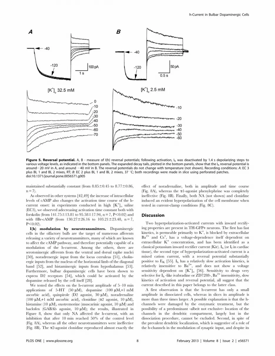
Reversal potential.. Panels A and B of Fig. 5 show current
records during protocols used to determine the voltage at which Ih
reverses. They consist of a series of fixed hyperpolarizing pulses,
followed by repolarizations to various levels. The reversal potential
did not depend upon the temperature (not shown) but only on
sodium and potassium ion concentration. In Figure 5 we show the
dependence on K+ ions; on average, Eh was 220.1861.67 mV
(n = 9) in [K+]o 32.5 mM, and -43.9561.51 mV in standard saline
([K+]o 2.5 mM; n = 7); the reversal potential was not temperature-
dependent (not shown). From the reversal potential and the h-
current amplitude, the maximal conductance could be calculated,
giving a value of 1.37 nS in standard saline.
PharmacologyBlockers. The h-current is sensitive to low concentrations of
Cs+ (1–2 mM) [30] and to a certain number of organic
compounds capable of selectively blocking the h-channels, like
h-Current in Bulbar Dopaminergic Cells
PLOS ONE | www.plosone.org 5 February 2013 | Volume 8 | Issue 2 | e56571

ZD7288 [31] and S-16257 (ivabradine) [32,33]. Cs+ 1 mM
effectively blocks the h-current (Figure 1D, H); however, as
already described for calf Purkinje cells [43], the action of Cs+ is
clearly voltage-dependent: in the negative region of the I-V curve
Cs+ induces a channel blockade, whereas at more positive
potentials Cs+ is ineffective, and sometimes it can even produce
the opposite effect, i.e. a current increase (not shown). More
selective and completely voltage-independent blockages can be
obtained with ivabradine 10 mM (not shown) and ZD7288 30 mM
(Figure 1E–F).
Role of Ih in autorhythmicity. The presence of the h-
current, characteristically associated with the pacemaking process
in a large number of autorhythmic cell (see [17] for a review)
suggests that it could play its archetypal role also in bulbar DA
neurons.
Recording at 37 uC in perforated patches, the block of the h-
current by focal application of any drug blocking the h-current
(Cs+ 1 mM, ZD7288 30 mM or ivabradine 10 mM) induces a
hyperpolarization from 258.7660.9 mV to 265.1761.64 mV
with Cs+ (n = 5), 263.561.31 mV with ZD7288 (n = 5), and
264.6461.81 mV with ivabradine (n = 8; Figure 6A), all signif-
icant at the 0.01 level with Student t-test paired data analysis The
effect was rapid and reversible with Cs+ and ivabradine (tens of
seconds for focal application, 2 min for bath application), slower
(about 5 min) and often irreversible with ZD7288.
We then tested whether this blockade represented the evidence
of a direct role played by the h-current in the pacemaking
mechanism, or just the consequence of the hyperpolarization. In
the presence of ivabradine, following the injection of a depolar-
izing current restoring the resting potential to the value preceding
the Ih block (grey arrowhead in Figure 6B), spontaneous activity
resumed reproducibly and immediately (Figure 6C), proving the
absence of any direct involvement of the h-current in auto-
rhythmicity, but also demonstrating that this current has a relevant
role in determining the resting membrane potential. The blockage
of spontaneous activity for membrane hyperpolarization of a
relatively small amplitude (7 mV on average) is not surprising, as
cell firing is based on a delicate interplay of conductances that can
be easily disrupted by modifications of the resting potential even of
small amplitude [16].
These results are different to those observed by Puopolo et al.
[44], who, blocking the Ih with ZD7288, failed to observe any
Figure 3. Deactivation kinetics. A: Envelope test during deactivation at 240 mV. After current activation at 2130 mV, pulses to 240 mV ofvariable duration were followed by re-activating steps to 2130 mV (see protocol in the top panel). Recording temperature 24 uC. B: The tail at240 mV was also re-plotted after appropriate scaling (red trace) to better compare its time course with that of the re-activation records envelopeshown in panel A. C: Analysis of the deactivation time constant for a group of five cells at 32 (blue) and 22 uC (red); the average time dependence wasfitted with the equation I(t) = 1 - exp(-t/t) (continuous lines); C - inset: box chart of deactivation time constants at 22 and 32 uC; All recordings shown inthis figure were made in slice, perforated patches, EC 3 saline plus BL 1 and BL 2 mixes, at the indicated temperatures.doi:10.1371/journal.pone.0056571.g003
h-Current in Bulbar Dopaminergic Cells
PLOS ONE | www.plosone.org 6 February 2013 | Volume 8 | Issue 2 | e56571

effect on spontaneous firing and resting potential. We believe that
this discrepancy might have several explanations. First, we worked
in perforated patch and not in whole-cell configuration; in
ruptured patches the h-current is barely discernible, probably
because of the known washout of the cytoplasmic compartment,
which evidently removes some factors essential for the mainte-
nance of the current. The second explanation might be in the
blocker used: we used ivabradine, and Puopolo ZD 7288. The
advantage of ivabradine is in its rapidity of action, and in the
reversibility of its effect, whereas ZD is slow, and was applied only
for a minute. Finally, notice that recordings were done at different
temperature (22–24 vs. 34 uC).
I(h) modulation by intracellular cAMP. The h current is
dually regulated by the hyperpolarization and by cyclic AMP,
directly binding to a sequence (cyclic nucleotide binding domain,
CNBD) located in the C-terminal segment [45,46]. We have
therefore analyzed the modulatory effect of cAMP on the h-
current using a recording configuration (perforated patch with
amphotericin B) minimizing the perturbations of the intracellular
medium and using a physiological external potassium concentra-
tion.
Under current clamp conditions and in normal saline, the
addition to the extracellular solution of 10 mM forskolin, a classical
activator of adenylyl cyclase [47] and0.1 mM IBMX, a phospho-
diesterase inhibitor [48], induced a marked depolarization
(Fig. 7A); in six cells the average depolarization was
20.5364.21 mV (n = 6; p,0.005, t-test for paired data), an effect
more evident at more negative membrane potentials (Fig. 7B), as
expected due to the increasing importance of the h-current at
more polarized membrane levels.
Under voltage-clamp conditions and in TEA-normal [K+]o
saline (EC2), the bath application of 10 mM forskolin and 0.1 mM
IBMX, induces a significant increase of Ih amplitude (Figure 7C):
at 2130 mV the current amplitude is 293.863.95 pA in control
conditions (n = 5), and -139.5620.4 pA (n = 5) in the presence of
increased levels of cAMP;for each tested potential, the increase in
current amplitude was statistically significant (p,0.005, ANOVA).
Forskolin promotes a depolarizing shifts of the steady-state
activation curve in the depolarizing direction. This could be
measured in TEA-normal [K+]o saline (EC2), with a variation of
V50 from 282.4461.57 mV to 277.1161.33 (n = 4; significant at
0.005 level; not shown), but it was more evident in high [K+]o
saline (EC3), a condition favoring more precise measurements of
the h-current: in these situations, V50 was shifted from
285.2661.96 to 75.7762.41 mV (n = 7, p,0.002, ANOVA),
and the slope was maintained substantially constant (from
8.7760.34 to 8.7760.69, n = 7; Fig. 7D). The experiment was
repeated in the same testing conditions but increasing the
intracellular cAMP concentration with 10 mM 8Br-cAMP, with
results largely superimposable: from 285.3662.32 to
78.9963.59 mV (n = 7, p,0.05, ANOVA), and the slope was
Figure 4. Analysis of time constants. A: Activation at 22 uC: family of current tracings recorded in a single cell in response to hyperpolarizingpulses ranging from 270 to 2130 mV from a holding potential of 240 mV; the red line represents the best fit with a double exponential;B: Family ofcurrent tracings recorded in a single cell in response to hyperpolarizing pulses from 240 to 2130 mV, repeated at the temperatures indicated. C:Activation at 32 uC, using a different protocol minimizing the stress of the membrane at the more negative potentials; D: Comparison of the 10–90rise time at the two temperatures. All the records shown in this figure were made in slice, perforated patches, EC 3 saline plus BL 1 and BL 2 mixes, atthe indicated temperatures.doi:10.1371/journal.pone.0056571.g004
h-Current in Bulbar Dopaminergic Cells
PLOS ONE | www.plosone.org 7 February 2013 | Volume 8 | Issue 2 | e56571
maintained substantially constant (from 8.8560.45 to 8.7760.86,
n = 7).
As observed in other systems [42,49] the increase of intracellular
levels of cAMP also changes the activation time course of the h-
current onset: in experiments conducted in high [K+]o saline
(EC3), we observed adecreasing activation time constant both with
forskolin (from 141.75613.81 to 95.58617.94, n = 7, P,0.02) and
with 8Br-cAMP (from 130.27626.16 to 103.21623.48, n = 7,
P,0.02).
I(h) modulation by neurotransmitters. Dopaminergic
cells in the olfactory bulb are the target of numerous afferents
releasing a variety of neurotransmitters, many of which are known
to affect the cAMP pathway, and therefore potentially capable of a
modulation of the h-current. Among the others, there are
serotoninergic afferents from the ventral and dorsal raphe nuclei
[50], noradrenergic input from the locus cœruleus [51], cholin-
ergic inputs from the nucleus of the horizontal limb of the diagonal
band [52], and histaminergic inputs from hypothalamus [53].
Furthermore, bulbar dopaminergic cells have been shown to
express D2 receptors [54], which could be activated by the
dopamine released by the cell itself [28].
We tested the effects on the h-current amplitude of 5–10 min
applications of 5-HT (50 mM), dopamine (100 mM,+1 mM
ascorbic acid), quinpirole (D2 agonist, 30 mM), noradrenaline
(100 mM,+1 mM ascorbic acid), clonidine (a2 agonist, 10 mM),
histamine (10 mM), oxotremorine (muscarinic agonist, 10 mM) and
baclofen (GABAb agonist, 10 mM); the results, illustrated in
Figure 8, show that only NA affected the h-current, with an
inhibition that after 10 min reached 50% of the control level
(Fig. 8A), whereas all the other neurotransmitters were ineffective
(Fig. 8B). The a2-agonist clonidine reproduced almost exactly the
effect of noradrenaline, both in amplitude and time course
(Fig. 8A), whereas the a1-agonist phenylephrine was completely
ineffective (Fig. 8B). Finally, both NA (not shown) and clonidine
induced an evident hyperpolarization of the cell membrane when
tested in current-clamp conditions (Fig. 8C).
Discussion
Two hyperpolarization-activated currents with inward rectify-
ing properties are present in TH-GFP+ neurons. The first has fast
kinetics, is permeable primarily to K+, is blocked by extracellular
Ba2+ and Cs+, has a voltage-dependence itself dependent on
extracellular K+ concentration, and has been identified as a
classical potassium inward rectifier current (Kir). Ih (or If in cardiac
tissue), the second type of hyperpolarization-activated current is a
mixed cation current, with a reversal potential substantially
positive to EK [55]. Ih has a relatively slow activation kinetics, is
relatively insensitive to Ba2+, and does not show a voltage
sensitivity dependent on [K+]o [56]. Sensitivity to drugs very
selective for Ih -like ivabradine or ZD7288-, Ba2+ insensitivity, slow
kinetics of activation and reversal potential, all suggest that the
current described in this paper belongs to the latter class.
A first observation is that the h-current has only a small
amplitude in dissociated cells, whereas in slices its amplitude is
more than three times larger. A possible explanation is that the h-
channels were damaged by the enzymatic treatment, but the
possibility of a predominant -albeit not exclusive- location of the
channels in the dendritic compartment, largely lost in the
dissociation procedure, cannot be excluded. Second, in spite of
the prevalent dendritic localization, which is suggestive of a role of
the h-channels in the modulation of synaptic input, and despite its
Figure 5. Reversal potential. A, B - measure of I(h) reversal potentials; following activation, Ih was deactivated by 1.4 s depolarizing steps tovarious voltage levels, as indicated in the bottom panels. The expanded decay tails, plotted in the bottom panels, show that the Ih reversal potential isaround220 mV in A, and around 240 mV in B. The reversal potentials do not change with temperature (not shown). Recording conditions: A: EC 3plus BL 1 and BL 2 mixes, RT; B: EC 2 plus BL 1 and BL 2 mixes, 37 uC; both recordings were made in slice using perforated patches.doi:10.1371/journal.pone.0056571.g005
h-Current in Bulbar Dopaminergic Cells
PLOS ONE | www.plosone.org 8 February 2013 | Volume 8 | Issue 2 | e56571

small amplitude, the h-current also gives a significant contribution
to the resting membrane potential.
Which population of HCN channels?Four channel isoforms exist (HCN1-HCN4) that can form
homo- or heteromers [17]. Searching in the literature, the
expression of TH and of any of the four HCN channels in the
olfactory bulb, the situation appears rather confused. The Allen
Brain Atlas, based on RT-PCR data, describes only HCN4
channels, whereas others find both HCN2 and HCN4 [57].
Authors using in situ hybridization techniques find either type 1, 2,
and 4 [58] or all four of them [59]. The immunohistochemical
localization of the different channel subunits has shown that high
expression levels of HCN3 can be found in the glomerular layer of
the olfactory bulb [60,61], where HCN3-immunopositive fine
dendritic processes and somata are clearly visible;unfortunately the
further identification of the cellular subtypes was beyond the scope
of those studies.
The search in the literature for co-expression of TH and any of
the four HCN channels in the olfactory bulb gives few results. In a
recent paper, using acomprehensive set of antibodies against all
four isoforms, it was found that all four HCN isoforms are
abundantly expressed in the olfactory bulb, where they can be
detected in most cell populations, with at least 17 different
combinations of staining patterns [19]; however, no HCN
channels were detected in TH+ glomerular cells [19], confirming
and extending a previous observation limited to the HCN1
subtype [62]. We are notable to explain the reason for the absence
of any HCN expression in PG TH+ cells, but certainly, insofar as
it is possible to infer from the analysis of the electrophysiological
recordings, we expect low levels of expression in these cells, and
probably a preferential localization in the dendritic compartment,
for which the assignment to a certain cell type or another is not so
straightforward.
KineticsAt 37 uC, and using 1 s hyperpolarizing pulses, we found a
midpoint of activation at -94.161.20 mV with a slope of
9.8860.28 mV (n = 12). In these conditions only 5% of the h-
channels would be open at the restingpotential (265 mV);
however, trying different pulse durations, at 37 uC we calculated
a h-current steady-state activation midpoint equal to 282.73 mV.
Figure 6. Effect of blockers of h-channels on membrane potential and spontaneous firing. A - Box charts showing the effect of h-channelsblockers on resting membrane potential; the recording condition for the experiments represented in this figure were: slice, perforated patch, 37 uC;all differences were significant at the 0.01 level with Student t-test paired data analysis. B – Ivabradine (10 mM, bar) block of spontaneous activity. Atthe times indicated by arrowheads, depolarizing currents of increasing amplitudes (+15, +30, +40, +50, +70 pA, in the order) were delivered. C -Enlargement of the response to the third injection of depolarizing current (grey arrowhead) to show that the block of the h-current does not impairthe spontaneous activity, as indicated by the restoration of firing upon forcing the membrane back to resting values. All the experiments shown inthis figure were performed using standard saline (EC 1) plus BL 1 mix;slice, perforated patch, 37 uC.doi:10.1371/journal.pone.0056571.g006
h-Current in Bulbar Dopaminergic Cells
PLOS ONE | www.plosone.org 9 February 2013 | Volume 8 | Issue 2 | e56571

The functional implication of these values becomes more evident if
one considers that, assuming a slope of 9.2 (the slope shows only a
modest increase with change in conditioning step), at 265 mV
about 12.7% h-channels are open. The fraction is small but, since
the input resistance of these cells is high (915.69 MV, personal
observation), is sufficient to provide a significant depolarizing
contribution to the resting membrane potential. Accordingly,
pharmacological block of the h-current with Cs+, ivabradine or
ZD7288, induces a mean hyperpolarization of 7 mV (Fig. 6A),
thus stopping the spontaneous firing. A severe restriction of
spontaneous activity following a membrane hyperpolarization of
this entity is not surprising, as the cell firing is based on a delicate
interplay of conductancesthat can be easily disrupted by changes
of a few millivolts of the resting membrane potential [16].
PharmacologyI(h) dependence on cAMP. The h-channels are directly
activated by cyclic nucleotides [45], and -accordingly- we observed
a 48.7% increase of Ih amplitude in presence of 10 mM forskolin
and 0.1 mM IBMX (Fig. 7C). This effect was at least in part
sustained by a positive shift of the steady-state activation curve,
whose midpoint was moved in the depolarizing directionby
5.33 mV.
The increase of intracellular cAMP induced a depolarization of
the membrane which was positively correlated with the resting
membrane potential: the larger the membrane polarization, the
larger the depolarization induced by the intracellular increase of
cAMP Fig. 7B), an effect which can be easily explained by the
higher degree of activation (and hence by the greater importance)
of the h-current at more negative potentials.
I(h) dependence on neurotransmitters. Dopaminergic
cells in the olfactory bulb receive numerous afferents releasing a
large variety of neurotransmitters, many of which are known to
affect the cAMP pathway, and therefore potentially capable of a h-
current modulation. We tested several of them, including
monoamine (dopamine, serotonin, histamine), and metabotropic
cholinergic and GABAergic agonists (oxotremorine and baclofen).
All these neurotransmitters do have some effect on the excitability
Figure 7. Effect of forskolin. A: Focal application of forskolin (10 mM) and IBMX (0.1 mM) alone and in the presence of 1 mM Cs+ at the timesindicated by the red bars in current-clamp conditions. Extracellular saline was EC 1 plus BL 1 mix; B:Voltage-dependence of the effect of forskolin onthe membrane potential, showing that the depolarization increases at more negative potentials; the cell membrane was manually hyperpolarizedbefore the application of the drug to circumvent spontaneous activity; recording conditions as in A.C: Modification in h-current amplitude after 10’perfusion with forskolin (10 mM) and 8Br-cAMP (10 mM). Extracellular saline was EC 2 plus BL 1 and BL 2 mixes; D: Shift in h-current activation curvefollowing perfusion with forskolin; following treatment with the drugthe midpoint is depolarized of 4.4 mV, and the slope becomes faster (from 7.6 to5.2 mV); recording conditions as in C. All the experiments illustrated in this figure were performed in slice, perforated patch,37 uC.doi:10.1371/journal.pone.0056571.g007
h-Current in Bulbar Dopaminergic Cells
PLOS ONE | www.plosone.org 10 February 2013 | Volume 8 | Issue 2 | e56571

profile of bulbar dopaminergic cells, but none of them showed any
effect on the amplitude of the h-current.
The only neurotransmitter showing an effect on the h-current
amplitude is NA, which causes inhibition; the effect, that could be
replicated by an a2 agonist, is similar to what has been described
in L4 and L5 rat dorsal root ganglion neurons [63]. Also in
midbrain DA neurons NA inhibits the h-current, either with
cAMP-independent mechanisms (activation of the PKC pathway,
as in VTA [64]) or due to space-clamp effect (as in substantia nigra
[65]). There are, however, also reports of enhancement of Ih by
NA, as in thalamic neurons [66] and in CA1 hippocampal stratum
oriens-alveus interneurons [67], but without a clear indication of the
underlying mechanism.
The OB receives a dense projection from the pontine nucleus
locus cœruleus (LC), the largest collection of NA-containing cells
in the brain, from where an estimated 40% of neurons project to
the OB [68], and which is the exclusive source of NA innervation
of the OB [51,69]. NA fibers preferentially target the internal
plexiform layer and the granule cell layer, and, to a lesser extent,
the mitral cell layer and the external plexiform layer [51]. The
functional role of NA in adult rodent olfactory bulb has been
accurately reviewed recently [70]: converging data from electro-
physiological studies of cellular-membrane properties of OB
neurons and behavioral assays of perception indicate that NA
affects the olfactory bulb network improving odor detection and
discrimination. Most of the available electrophysiological data
concerning the NA effect in different cell types of the olfactory
bulb are limited to the granule and mitral cell layer. As for the
glomerular layer, despite the fact that all NA receptor types have
been localized in this region [71–74], no data concerning possible
modulation roles of NA inputs are available. Our observation,
therefore, would be the first reporting an NA action in a specific
subpopulation of glomerular cells, and is well in line with the
general effect of NA: dopamine released by glomerular dopami-
nergic cells is known to inhibit glutamate release from olfactory
nerve terminals [11]. Therefore, an inhibition of the h-current,
with its hyperpolarizing effect, would decrease the DA-mediated
tonic inhibition of olfactory nerve terminals, in line with the
general positive influenceof NA on odor detection and discrim-
ination.
Figure 8. Effect of different neurotransmitters on the h-current. A: effect of NA (100 mM, +1 mM ascorbic acid); the amplitudes at differenttime points are normalized with respect to the amplitude at time zero; n = 16 (controls) 11 (NA), 6 (clonidine). B: box charts showing the lack of effectof the indicated neurotransmitters;5-HT (50 mM, n = 7),quinpirole (30 mM, n = 14), histamine (10 mM, n = 11), oxotremorine (10 mM, n = 7), baclofen(10 mM, n = 4) and dopamine (100 mM,+1 mM ascorbic acid; n = 8). C: effect of clonidine (a2 agonist, 10 mM) on the resting membrane potential. Themembrane was hyperpolarized to 274 mV (by injecting 245 pA) in order to prevent spontaneous firing. All recordings shown in this figure wereperformed in slice, perforated patch, 37 uC; external saline was EC 3 plus BL 1 and BL 2 mixes for experiments shown in A and B, EC 1 plus BL 1 for theexperiment shown in C.doi:10.1371/journal.pone.0056571.g008
h-Current in Bulbar Dopaminergic Cells
PLOS ONE | www.plosone.org 11 February 2013 | Volume 8 | Issue 2 | e56571

Acknowledgments
We wish to thank Prof. Gaetano Zanghirati for his advices in the use of
numerical methods for the solution of eqn. 1, Dr. Alessio Filippetti for
helpful discussion, and Mrs. Elizabeth Manning for English revision.
Author Contributions
Conceived and designed the experiments: OB AP. Performed the
experiments: AP CG MB AFI. Analyzed the data: OB AP. Contributed
reagents/materials/analysis tools: OB. Wrote the paper: OB.
References
1. Halasz N (1990) The vertebrate olfactory system: chemical neuroanatomy,function and development. Budapest: Academiai Kiado.
2. McLean JH, Shipley MT (1988) Postmitotic, postmigrational expression of
tyrosine hydroxylase in olfactory bulb dopaminergic neurons. J Neurosci 8:
3658–3669.
3. Kratskin I, Belluzzi O (2003) Anatomy and neurochemistry of the olfactory bulb.
In: Doty RL, editors. Handbook of Olfaction and Gustation. New York - Basel:
Marcel Dekker. pp. 139–164.
4. Panzanelli P, Fritschy JM, Yanagawa Y, Obata K, Sassoe-Pognetto M (2007)
GABAergic phenotype of periglomerular cells in the rodent olfactory bulb.
J Comp Neurol 502: 990–1002.
5. Gall CM, Hendry SH, Seroogy KB, Jones EG, Haycock JW (1987) Evidence for
coexistence of GABA and dopamine in neurons of the rat olfactory bulb. J Comp
Neurol 266: 307–318.
6. Kosaka T, Hataguchi Y, Hama K, Nagatsu I, Wu JY (1985) Coexistence ofimmunoreactivities for glutamate decarboxylase and tyrosine hydroxylase in
some neurons in the periglomerular region of the rat main olfactory bulb:
possible coexistence of gamma-aminobutyric acid (GABA) and dopamine. Brain
Res 343: 166–171.
7. Baker H, Kawano T, Margolis FL, Joh TH (1983) Transneuronal regulation of
tyrosine hydroxylase expression in olfactory bulb of mouse and rat. J Neurosci 3:
69–78.
8. Guthrie KM, Pullara JM, Marshall JF, Leon M (1991) Olfactory deprivationincreases dopamine D2 receptor density in the rat olfactory bulb. Synapse 8: 61–
70.
9. Doty RL, Risser JM (1989) Influence of the D-2 dopamine receptor agonist
quinpirole on the odor detection performance of rats before and after spiperoneadministration. Psychopharmacology (Berl) 98: 310–315.
10. Nowycky MC, Halasz N, Shepherd GM (1983) Evoked field potential analysis of
dopaminergic mechanisms in the isolated turtle olfactory bulb. Neuroscience 8:717–722.
11. Ennis M, Zhou FM, Ciombor KJ, Aroniadou-Anderjaska V, Hayar A, et al.
(2001) Dopamine D2 receptor-mediated presynaptic inhibition of olfactory
nerve terminals. J Neurophysiol 86: 2986–2997.
12. Davila NG, Blakemore LJ, Trombley PQ (2003) Dopamine modulates synaptic
transmission between rat olfactory bulb neurons in culture. J Neurophysiol 90:
395–404.
13. Grace AA, Onn SP (1989) Morphology and electrophysiological properties of
immunocytochemically identified rat dopamine neurons recorded in vitro.
J Neurosci 9: 3463–3481.
14. Hainsworth AH, Roper J, Kapoor R, Ashcroft FM (1991) Identification andelectrophysiology of isolated pars compacta neurons from guinea-pig substantia
nigra. Neuroscience 43: 81–93.
15. Feigenspan A, Gustincich S, Bean BP, Raviola E (1998) Spontaneous activity of
solitary dopaminergic cells of the retina. J Neurosci 18: 6776–6789.
16. Pignatelli A, Kobayashi K, Okano H, Belluzzi O (2005) Functional properties of
dopaminergic neurones in the mouse olfactory bulb. J Physiol 564: 501–514.
17. Wahl-Schott C, Biel M (2009) HCN channels: structure, cellular regulation andphysiological function. Cell Mol Life Sci 66: 470–494.
18. Cadetti L, Belluzzi O (2001) Hyperpolarisation-activated current in glomerular
cells of the rat olfactory bulb. Neuroreport 12: 3117–3120.
19. Fried HU, Kaupp UB, Muller F (2010) Hyperpolarization-activated and cyclic
nucleotide-gated channels are differentially expressed in juxtaglomerular cells in
the olfactory bulb of mice. Cell Tissue Res 339: 463–479.
20. Matsushita N, Okada H, Yasoshima Y, Takahashi K, Kiuchi K et al. (2002)Dynamics of tyrosine hydroxylase promoter activity during midbrain dopami-
nergic neuron development. J Neurochem 82: 295–304.
21. Sawamoto K, Nakao N, Kakishita K, Ogawa Y, Toyama Y, et al. (2001)Generation of dopaminergic neurons in the adult brain from mesencephalic
precursor cells labeled with a nestin-GFP transgene. J Neurosci 21: 3895–3903.
22. Gustincich S, Feigenspan A, Wu DK, Koopman LJ, Raviola E (1997) Control of
dopamine release in the retina: a transgenic approach to neural networks.Neuron 18: 723–736.
23. Kamondi A, Reiner PB (1991) Hyperpolarization-activated inward current in
histaminergic tuberomammillary neurons of the rat hypothalamus.
J Neurophysiol 66: 1902–1911.
24. Hayar A, Karnup S, Shipley MT, Ennis M (2004) Olfactory bulb glomeruli:
external tufted cells intrinsically burst at theta frequency and are entrained by
patterned olfactory input. J Neurosci 24: 1190–1199.
25. Shipley MT, Ennis M (1996) Functional organization of olfactory system.
J Neurobiol 30: 123–176.
26. Sawamoto K, Nakao N, Kobayashi K, Matsushita N, Takahashi H, et al. (2001)
Visualization, direct isolation, and transplantation of midbrain dopaminergicneurons. Proc Natl Acad Sci U S A 98: 6423–6428.
27. Saino-Saito S, Sasaki H, Volpe BT, Kobayashi K, Berlin R, et al. (2004)
Differentiation of the dopaminergic phenotype in the olfactory system of
neonatal and adult mice. J Comp Neurol 479: 389–398.
28. Maher BJ, Westbrook GL (2008) Co-transmission of dopamine and GABA in
periglomerular cells. J Neurophysiol 99: 1559–1564.
29. Pignatelli A, Ackman JB, Vigetti D, Beltrami AP, Zucchini S, et al. (2009) A
potential reservoir of immature dopaminergic replacement neurons in the adult
mammalian olfactory bulb. Pflugers Arch 457: 899–915.
30. DiFrancesco D, Ojeda C (1980) Properties of the current if in the sino-atrial
node of the rabbit compared with those of the current iK, in Purkinje fibres.
J Physiol 308: 353–367.
31. BoSmith RE, Briggs I, Sturgess NC (1993) Inhibitory actions of ZENECA
ZD7288 on whole-cell hyperpolarization activated inward current (If) in guinea-
pig dissociated sinoatrial node cells. Br J Pharmacol 110: 343–349.
32. Bucchi A, Baruscotti M, DiFrancesco D (2002) Current-dependent block of
rabbit sino-atrial node I(f) channels by ivabradine. J Gen Physiol 120: 1–13.
33. Bucchi A, Tognati A, Milanesi R, Baruscotti M, DiFrancesco D (2006)
Properties of ivabradine-induced block of HCN1 and HCN4 pacemaker
channels. J Physiol 572: 335–346.
34. Hart G (1983) The kinetics and temperature dependence of the pace-maker
current if in sheep Purkinje fibres. J Physiol 337: 401–416.
35. Yanagida H, Inoue R, Tanaka M, Ito Y (2000) Temperature-sensitive gating of
cation current in guinea pig ileal muscle activated by hyperpolarization.
Am J Physiol Cell Physiol 278: C40–C48.
36. Pena F, Amuzescu B, Neaga E, Flonta ML (2006) Thermodynamic properties of
hyperpolarization-activated current (Ih) in a subgroup of primary sensory
neurons. Exp Brain Res 173: 282–290.
37. Seifert R, Scholten A, Gauss R, Mincheva A, Lichter P, et al. (1999) Molecular
characterization of a slowly gating human hyperpolarization-activated channel
predominantly expressed in thalamus, heart, and testis. Proc Natl Acad Sci U S A
96: 9391–9396.
38. Cuevas J, Harper AA, Trequattrini C, Adams DJ (1997) Passive and active
membrane properties of isolated rat intracardiac neurons: regulation by H- and
M-currents. J Neurophysiol 78: 1890–1902.
39. DiFrancesco D, Ferroni A, Mazzanti M, Tromba C (1986) Properties of the
hyperpolarizing-activated current (if) in cells isolated from the rabbit sino-atrial
node. J Physiol 377: 61–88.
40. Brent R (1973) Algorithms for Minimization Without Derivatives. Prentice-Hall.
41. Forsythe GE, Malcom MA, Moler CB (1976) Computer Methods for
Mathematical Computations. Englewood Cliffs, NJ: Prentice-Hall. 259 p.
42. Gambardella C, Pignatelli A, Belluzzi O (2012) The h-Current in the Substantia
Nigra pars Compacta Neurons: A Re-examination. PLoS ONE 7: e52329.
43. DiFrancesco D (1982) Block and activation of the pace-maker channel in calf
purkinje fibres: effects of potassium, caesium and rubidium. J Physiol 329: 485–
507.
44. Puopolo M, Bean BP, Raviola E (2005) Spontaneous activity of isolated
dopaminergic periglomerular cells of the main olfactory bulb. J Neurophysiol 94:
3618–3627.
45. DiFrancesco D, Tortora P (1991) Direct activation of cardiac pacemaker
channels by intracellular cyclic AMP. Nature 351: 145–147.
46. Wainger BJ, DeGennaro M, Santoro B, Siegelbaum SA, Tibbs GR (2001)
Molecular mechanism of cAMP modulation of HCN pacemaker channels.
Nature 411: 805–810.
47. Seamon KB, Daly JW (1981) Forskolin: a unique diterpene activator of cyclic
AMP-generating systems. J Cyclic Nucleotide Res 7: 201–224.
48. Beavo JA, Rogers NL, Crofford OB, Hardman JG, Sutherland EW, et al. (1970)
Effects of xanthine derivatives on lipolysis and on adenosine 3’,5’-monophos-
phate phosphodiesterase activity. Mol Pharmacol 6: 597–603.
49. Pedarzani P, Storm JF (1995) Protein kinase A-independent modulation of ion
channels in the brain by cyclic AMP. Proc Natl Acad Sci U S A 92: 11716–
11720.
50. Araneda S, Bobillier P, Buda M, Pujol JF (1980) Retrograde axonal transport
following injection of [3H]serotonin in the olfactory bulb. I. Biochemical study.
Brain Res 196: 405–415.
51. McLean JH, Shipley MT, Nickell WT, Aston-Jones G, Reyher CK (1989)
Chemoanatomical organization of the noradrenergic input from locus coeruleus
to the olfactory bulb of the adult rat. J Comp Neurol 285: 339–349.
52. Zaborszky L, Carlsen J, Brashear HR, Heimer L (1986) Cholinergic and
GABAergic afferents to the olfactory bulb in the rat with special emphasis on the
projection neurons in the nucleus of the horizontal limb of the diagonal tract.
J Comp Neurol 243: 488–509.
53. Panula P, Pirvola U, Auvinen S, Airaksinen MS (1989) Histamine-immunore-
active nerve fibers in the rat brain. Neuroscience 28: 585–610.
h-Current in Bulbar Dopaminergic Cells
PLOS ONE | www.plosone.org 12 February 2013 | Volume 8 | Issue 2 | e56571

54. Gutierrez-Mecinas M, Crespo C, Blasco-Ibanez JM, Gracia-Llanes FJ,
Marques-Mari AI, et al. (2005) Distribution of D2 dopamine receptor in theolfactory glomeruli of the rat olfactory bulb. Eur J Neurosci 22: 1357–1367.
55. Hibino H, Inanobe A, Furutani K, Murakami S, Findlay I, et al. (2010) Inwardly
rectifying potassium channels: their structure, function, and physiological roles.Physiol Rev 90: 291–366.
56. Biel M, Wahl-Schott C, Michalakis S, Zong X (2009) Hyperpolarization-activated cation channels: from genes to function. Physiol Rev 89: 847–885.
57. Santoro B, Chen S, Luthi A, Pavlidis P, Shumyatsky GP, et al. (2000) Molecular
and functional heterogeneity of hyperpolarization-activated pacemaker channelsin the mouse CNS. J Neurosci 20: 5264–5275.
58. Moosmang S, Biel M, Hofmann F, Ludwig A (1999) Differential distribution offour hyperpolarization-activated cation channels in mouse brain. Biol Chem
380: 975–980.59. Monteggia LM, Eisch AJ, Tang MD, Kaczmarek LK, Nestler EJ (2000) Cloning
and localization of the hyperpolarization-activated cyclic nucleotide-gated
channel family in rat brain. Brain Res Mol Brain Res 81: 129–139.60. Notomi T, Shigemoto R (2004) Immunohistochemical localization of Ih channel
subunits, HCN1-4, in the rat brain. J Comp Neurol 471: 241–276.61. Mistrik P, Mader R, Michalakis S, Weidinger M, Pfeifer A, et al. (2005) The
murine HCN3 gene encodes a hyperpolarization-activated cation channel with
slow kinetics and unique response to cyclic nucleotides. J Biol Chem 280: 27056–27061.
62. Holderith NB, Shigemoto R, Nusser Z (2003) Cell type-dependent expression ofHCN1 in the main olfactory bulb. Eur J Neurosci 18: 344–354.
63. Yagi J, Sumino R (1998) Inhibition of a hyperpolarization-activated current byclonidine in rat dorsal root ganglion neurons. J Neurophysiol 80: 1094–1104.
64. Inyushin MU, Arencibia-Albite F, Vazquez-Torres R, Velez-Hernandez ME,
Jimenez-Rivera CA (2010) Alpha-2 noradrenergic receptor activation inhibitsthe hyperpolarization-activated cation current (Ih) in neurons of the ventral
tegmental area. Neuroscience 167: 287–297.
65. Cathala L, Paupardin-Tritsch D (1999) Effect of catecholamines on the
hyperpolarization-activated cationic Ih and the inwardly rectifying potassiumI(Kir) currents in the rat substantia nigra pars compacta. Eur J Neurosci 11:
398–406.
66. Pape HC, Mccormick DA (1989) Noradrenaline and serotonin selectivelymodulate thalamic burst firing by enhancing a hyperpolarization-activated
cation current. Nature 340: 715–718.67. Maccaferri G, Mcbain CJ (1996) The hyperpolarization-activated current (Ih)
and its contribution to pacemaker activity in rat CA1 hippocampal stratum
oriens-alveus interneurones. J Physiol 497: 119–130.68. Shipley MT, Halloran FJ, De La Torre J (1985) Surprisingly rich projection
from locus coeruleus to the olfactory bulb. Brain Res 329: 294–299.69. Zaborszky L, Carlsen J, Brashear HR, Heimer L (1986) Cholinergic and
GABAergic afferents to the olfactory bulb in the rat with special emphasis on theprojection neurons in the nucleus of the horizontal limb of the diagonal band.
J Comp Neurol 243: 488–509.
70. Linster C, Nai Q, Ennis M (2011) Non-linear effects of noradrenergicmodulation of olfactory bulb function in adult rodents. J Neurophysiol 105:
1432–1443.71. Young WS III, Kuhar MJ (1980) Noradrenergic alpha 1 and alpha 2 receptors:
light microscopic autoradiographic localization. Proc Natl Acad Sci U S A 77:
1696–1700.72. Day HE, Campeau S, Watson SJ Jr, Akil H (1997) Distribution of alpha 1a-,
alpha 1b- and alpha 1d-adrenergic receptor mRNA in the rat brain and spinalcord. J Chem Neuroanat 13: 115–139.
73. Domyancic AV, Morilak DA (1997) Distribution of alpha1A adrenergic receptormRNA in the rat brain visualized by in situ hybridization. J Comp Neurol 386:
358–378.
74. Winzer-Serhan UH, Raymon HK, Broide RS, Chen Y, Leslie FM (1997)Expression of alpha 2 adrenoceptors during rat brain development--I. Alpha 2A
messenger RNA expression. Neuroscience 76: 241–260.
h-Current in Bulbar Dopaminergic Cells
PLOS ONE | www.plosone.org 13 February 2013 | Volume 8 | Issue 2 | e56571