midbrain dopaminergic neurons generate calcium and sodium currents and release dopamine in the...
TRANSCRIPT
ORIGINAL RESEARCH ARTICLEpublished: 08 March 2012
doi: 10.3389/fncel.2012.00007
Midbrain dopaminergic neurons generate calcium andsodium currents and release dopamine in the striatumof pupsDiana C. Ferrari1,2 , Baya J. Mdzomba1,2†, Nathalie Dehorter1,2†, Catherine Lopez1,2,François J. Michel1,2, Frédéric Libersat1,2† and Constance Hammond1,2*
1 Institut National de la Recherche Médicale et de la Santé Inserm, INMED UMR 901, Marseille, France2 Faculté des Sciences, Aix Marseille Université, Marseille, France
Edited by:
Enrico Cherubini, InternationalSchool for Advanced Studies, Italy
Reviewed by:
Nicola B. Mercuri, University ofRome, ItalyKazuto Kobayashi, FukushimaMedical University, Japan
*Correspondence:
Constance Hammond, InstitutNational de la Recherche Médicaleet de la Santé Inserm, INMED UMR901, 163 route de Luminy, BP13,13273 Marseille Cédex 9, France.e-mail: [email protected]†Present address:
Baya J. Mdzomba, Bernstein FocusNeurotechnology andJohann-Friedrich-BlumenbachInstitute for Zoology andAnthropology, University ofGöttingen, D-37075 Göttingen,Germany.Nathalie Dehorter, Instituto deNeurociencias de Alicante CSIC,Universidad Miguel Hernández03550 Alicante, España.Frédéric Libersat, Department ofLife Sciences, Ben GurionUniversity, POB 653, Beer Sheva,Israel.
Midbrain dopaminergic neurons (mDA neurons) are essential for the control of diversemotor and cognitive behaviors. However, our understanding of the activity of immaturemDA neurons is rudimentary. Rodent mDA neurons migrate and differentiate early inembryonic life and dopaminergic axons enter the striatum and contact striatal neuronsa few days before birth, but when these are functional is not known. Here, we recordedCa2+ transients and Na+ spikes from embryonic (E16–E18) and early postnatal (P0–P7)mDA neurons with dynamic two-photon imaging and patch clamp techniques in slicesfrom tyrosine hydroxylase-GFP mice, and measured evoked dopamine release in thestriatum with amperometry. We show that half of identified E16–P0 mDA neuronsspontaneously generate non-synaptic, intrinsically driven Ca2+ spikes and Ca2+ plateausmediated by N- and L-type voltage-gated Ca2+ channels. Starting from E18–P0, half of themDA neurons also reliably generate overshooting Na+ spikes with an abrupt maturationat birth (P0 = E19). At that stage (E18–P0), dopaminergic terminals release dopamine in acalcium-dependent manner in the striatum in response to local stimulation. This suggeststhat mouse striatal dopaminergic synapses are functional at birth.
Keywords: development, basal ganglia, substantia nigra, dopamine, immature activity, patch clamp, two-photons
imaging
INTRODUCTIONDopaminergic neurons located in the ventral midbrain (mDA)give rise to the mesostriatal, mesocortical, and mesolimbic path-ways. The vast majority (around 80%) of mDA neurons areborn at E12 in rats (Gates et al., 2006) in the ventral aqueduc-tal ventricular zone. Then they become post-mitotic, enter intoa differentiation and specification program, and migrate ventro-laterally and rostrally along radial glia processes to their finallocation in the tegmental mantle to form the A8–A10 subgroups(Kawano et al., 1995; Hall et al., 2003). They start extending pro-cesses at E13 in rats (Moon and Herkenham, 1984; van der Kooyand Fishell, 1987; Voorn et al., 1988; Fishell and van der Kooy,1989; Gates et al., 2006; van den Heuvel and Pasterkamp, 2008).Tyrosine hydroxylase (TH), the rate limiting enzyme for cate-cholamine synthesis, is localized in the growing tips of axons,and TH-positive (TH+) axonal processes are first detected withinthe ventrolateral developing striatum at E14.5 where they form
a few specialized contacts with striatal somas or near the ori-gin of dendrites (Specht et al., 1981a,b). Accordingly, dopamineis first detected in the forebrain at E13 in mice and DA bindingsites (D1-like and D2-like) are present in the embryonic rodentneostriatum from E14 (Ohtani et al., 2003; Goffin et al., 2010). Inaddition, antidromic activation of rat substantia nigra compacta(SNc, A9) neurons from the striatum at P0 in vivo confirms thepresence of the nigro-striatal DA pathway at birth (Tepper et al.,1990; Trent et al., 1991). Collectively, these studies suggest that thenigro-striatal system is ready to operate at late embryonic stagesbut the functionality of this pathway and whether it does releasedopamine has not been established. This information is impor-tant as it conditions our understanding of the operation and roleof this system during development.
Here, we combined electrophysiological and imaging studiesto describe the developmental sequences of neuronal and net-work activity, with dopamine release experiments to detect the
Frontiers in Cellular Neuroscience www.frontiersin.org March 2012 | Volume 6 | Article 7 | 1
CELLULAR NEUROSCIENCE
Ferrari et al. Ontogeny of midbrain dopaminergic neurons activity
earliest evoked release of DA in the striatum. Since perinatal mDAneurons cannot be always identified by their adult electrophysio-logical characteristics (Washio et al., 1999) or their localization,we performed our experiments in brain slices from TH-GFP mice(Sawamoto et al., 2001). Our results show that at birth (P0), a sub-population (20%) of mDA neurons spontaneously generate fullamplitude Na+ spikes, in an intrinsically drive tonic or burstingpattern. At the same age, dopaminergic fibers release dopaminein a calcium-dependent manner in the striatum upon stimula-tion. Therefore, this suggests that mouse striatal dopaminergicsynapses are functional at birth.
METHODSANIMALS AND SLICESWe performed experiments on wild-type or TH-GFP C57BL/6mice (Matsushita et al., 2002) maintained in our institutionalanimal facility. Female mice were examined for the presence ofa vaginal plug the morning after mating. The day of plug dis-covery was designated as embryonic day 0 (E0). Experimentswere performed on mice of either sex. We removed E16 and E18mice from deeply anesthetized dams [subcutaneous injection of amixture of xylazine (Rompun 2%; used at 0.05%) and ketamine(Imalgene 1000 used at 50 g/L) volume injected: 0.1 mL/10 g].The embryos were kept in an ice-cold oxygenated solution con-taining (in mM): 110 choline, 2.5 KCl, 1.25 NaH2PO4, 7 MgCl2,0.5 CaCl2, 25 NaHCO3, 7 glucose. Postnatal mice (P0–P7) werekilled by decapitation under isofluorane anaesthesia. Coronaland parasagittal slices (400 μm thick) were cut in the ice-coldoxygenated choline solution using a vibratome (VT1200 LeicaMicrosystems Germany). During the recovery period, slices wereplaced at room temperature with standard artificial cerebrospinalfluid (ACSF) saturated with 95%O2/5%CO2 and containing thefollowing (in mM): 126 NaCl, 3.5 KCl, 1.2 NaH2PO4, 1.3 MgCl2,2 CaCl2, 25 NaHCO3, 11 glucose.
CALCIUM IMAGINGSlices were incubated in the dark with 25 μL of a fura-2 AMsolution (1 mM in DMSO + 0.8% pluronic acid; MolecularProbes). We performed imaging studies with a multibeam two-photon laser scanning system (Trimscope-LaVision Biotec) cou-pled to an Olympus microscope. Slices were imaged using ahigh numerical aperture objective (20×, NA 0.95, Olympus).Images (4 × 4 binning) were acquired via a CCD camera (LaVision Imager 3QE) with a time resolution of 115–147 ms perframe. Size of the scan field (444 × 336 μm) and duration ofthe movies (1000 frames) were unchanged. We first took imagesof the GFP-expressing neurons located in the mesencephalon(laser at 910 nm) before acquiring spontaneous fura-2 fluores-cence changes (laser at 780 nm). To verify the location of therecorded field, at the end of the imaging session we bleached thefura-2 fluorescence from the field and observed its correspondinglocation on the GFP image. During the analysis, GFP-expressingfura 2-loaded neurons were identified by superposing the twofields. We performed analysis of the calcium activity with custom-made software written in Matlab (MathWorks) (Bonifazi et al.,2009). Active cells were neurons exhibiting any Ca2+ event of atleast 5% DF/F deflection within the period of recording. Ca2+
spike or Ca2+ plateau cells were neurons exhibiting at least oneCa2+ spike or one Ca2+ plateau within the period of recording.A calcium plateau sustained a calcium level for at least 30 framesas opposed to a calcium spike which started decaying at the peak.We computed the activity correlation of cell pairs as previouslydescribed (Crepel et al., 2007; Dehorter et al., 2011).
PATCH CLAMP RECORDINGSWe performed all recordings at 32◦C. Cells were visualizedwith infrared–differential interference optics (Axioskop2; Zeiss).For whole-cell current clamp recordings the pipette (6–10 M�)contained the following (in mM): 128.5 K-gluconate, 11.5 KCl,1 CaCl2, 10 HEPES, 10 EGTA, 2.5 MgATP and 0.3 NaGFP, pH7.2–7.4 (275–285 mOsm). We determined input membrane resis-tance (Rm) by on-line fitting analysis of the transient currents inresponse to a 5–10 mV pulse at VH = –60 mV. Criteria for consid-ering a recording included Rm > 100 M�. The input resistance(Rm) of mDA neurons decreased significantly from 355 ± 39 M�
before birth (E18, n = 8) to 203 ± 31 M� at P5–P7 (n = 5, p <
0.05, Mann–Whitney test). In parallel, the percentage of mDAneurons displaying the hyperpolarization-activated cationic cur-rent Ih increased from 61% at E18 to 100% at P7. Amplitude ofaction potentials was measured from peak to after spike hyperpo-larization (AHP) potential and their duration half-way betweenthreshold and peak (half-width duration).
AMPEROMETRYCoronal slices were placed in a chamber and perfused with O2
saturated ACSF at 32◦C. We measured the evoked and not thespontaneous release of dopamine as performed in P9–14 pri-mary cultures of mDA neurons (Kim et al., 2008) or 30–40 daysorganotypic slices of the striatum (Cragg et al., 1998) becausethe high perfusion rate of ACSF needed to keep slices healthyprevents such a measure. Stimulation was performed with a bipo-lar tungsten electrode with a tip separation of 100 μm (WorldPrecision Instruments TST33C05KT, stereo tungsten electrode,in vitro impedance of 1 M�) inserted into the striatum. We didnot study DA overflow in response to median forebrain bun-dle (MFB) stimulation because medial sagittal slices containingthe MFB cannot be reliably obtained at embryonic stages. Toevoke a reproducible DA release, we used a train of four 100 Hzsquare pulses of 50 V amplitude and 100 μs duration. To moni-tor the electrically evoked dopamine release, we used continuousamperometry with carbon fiber electrodes because it gives simi-lar results as cyclic voltammetry in the striatum (Schmitz et al.,2001). The carbon fiber electrode (active surface 10 μm in diam-eter and 500 μm long; World Precision Instruments, CF10) wasimplanted into the striatum at an angle of 60◦ from vertical sothat the entire length of the active surface was inside the sliceat a depth of about 50 μm from the surface. This was done inthe ventrolateral and dorsomedial striatum where the evokedDA release was maximal and minimal respectively. The carbonfiber electrode was connected to a potentiostat (MicroC, WorldPrecision instruments) to apply voltage and measure current. Tomeasure DA release, the imposed voltage between the carbonfiber electrode and the Ag/AgCl pellet was 0.5 V. In response tothe stimulus train, the current generated by oxidation of evoked
Frontiers in Cellular Neuroscience www.frontiersin.org March 2012 | Volume 6 | Article 7 | 2
Ferrari et al. Ontogeny of midbrain dopaminergic neurons activity
dopamine released was recorded. To separate the evoked cur-rent from an artefact, the same stimulus protocol was done with0V applied between the carbon electrode and the Ag/AgCl refer-ence. At this voltage, no oxidation of DA should occur. Signalswere digitized using a Digidata data acquisition system (Digidata1440A) coupled to a PC running the clampex nine programresponding to the Multiclamp700A amplifier. Results are pre-sented as maximum response obtained per brain hemisphere. Tomeasure DA release during the blockade of dopamine reuptake,we incubated the slices in nomifensine (10 μM) for a mini-mum of 20 min. To test the calcium-dependence of dopaminerelease, we used a modified ACSF containing the following (inmM): 126 NaCl, 3.5 KCl, 1.2 NaH2PO4, 3.3 MgCl2, 25 NaHCO3,11 glucose.
IMMUNOCYTOCHEMISTRYTo visualize the TH-positive fibers in the striatum we performedimmunocytochemistry of TH in embryonic and early postnatalslices, and to identify the recorded cells we revealed the neuro-biotin injected during whole-cell recordings in recorded slices, aspreviously described (Dehorter et al., 2009). Dendritic and axonalfields were reconstructed for morphological analysis using theNeurolucida system (MicroBrightField Inc., Colchester, VT).
DRUGSDrugs were prepared as concentrated stock solutions and dilutedin ACSF for bath application. Gabazine, D-amino pyruvate(D-APV), 6-cyano-7-nitroquinoxaline 2,3-dione (CNQX),Tetrodotoxin (TTX), Nifedipine, Thapsigargin and Nomifensinemaleate were purchased from Sigma (St. Louis, MO, USA).ω–conotoxin GVIA was purchased from Alomone Labs(Jerusalem, Israel).
STATISTICAL ANALYSISStatistical results are given as means ± SEM. We performed sta-tistical analysis using GraphPad Prism (GraphPad Software, Inc.,La Jolla, CA): one-way ANOVA (Tukey’s Test as post hoc test),Mann–Whitney test (non-parametric t-test), and paired t-tests asindicated in the results section. Differences were considered sig-nificant at p ≤ 0.05 (∗∗∗ for p ≤ 0.001, ∗∗ for p ≤ 0.01 and ∗ forp ≤ 0.05). We grouped the P5 and P7 sets of data since they didnot present a statistical difference. In the box plots of Figures 1Aand 5B the bottom and top of the boxes represent the 25th and75th, the band inside the box is the 50th percentile (median) andthe top and bottom vertical bars (whiskers) denote the maximumand minimum values.
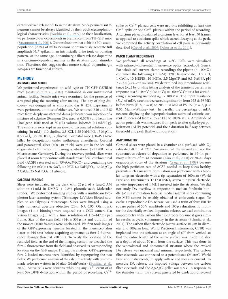
RESULTSEMBRYONIC mDA NEURONS PROJECT TO THE DEVELOPING STRIATUMAxons of E16 neurobiotin-filled mDA neurons project rostrallytoward the striatum even when their somas do not have dendritesyet (Figure 1A). The dendritic length and number of dendriticends of mDA neurons significantly increase from E16 (90 ±31 μm; 1.5 ± 0.4 ends; n = 13) to E18 (245 ± 66 μm; 3.9 ± 0.6ends; n = 7; p < 0.05, Mann–Whitney test), and from E18 to P0(1031 ± 287 μm, 12 ± 3 ends; n = 6; p < 0.05, Mann–Whitneytest) (Figure 1A). Accordingly, a substantial diffuse innervation
FIGURE 1 | Morphology of embryonic TH-positive midbrain neurons
and distribution of TH-positive axons in the developing striatum.
(A) (Top) Reconstructed neurobiotin-filled, GFP-positive, mDA neurons atthe indicated ages. Somas and dendrites are in black, axons in gray.(Bottom) Box plots of dendritic length (left) and number of dendritic ends(right) as a function of age. ∗Compared from E16 to E18 and from E18 toP0, Mann–Whitney test. (B) TH-positive axons (black staining) in thedeveloping striatum (arrow) at the indicated ages.
of the striatum by TH+ fibers is already present at E14–E16in the ventro-lateral part of the striatum (Ohtani et al., 2003)(Figure 1B). Later at E18–P0, TH+ fibers invade the more dor-sal regions of the striatum. Therefore, mDA neurons extend longaxons that reach the striatum already at E16 before developingtheir dendritic tree.
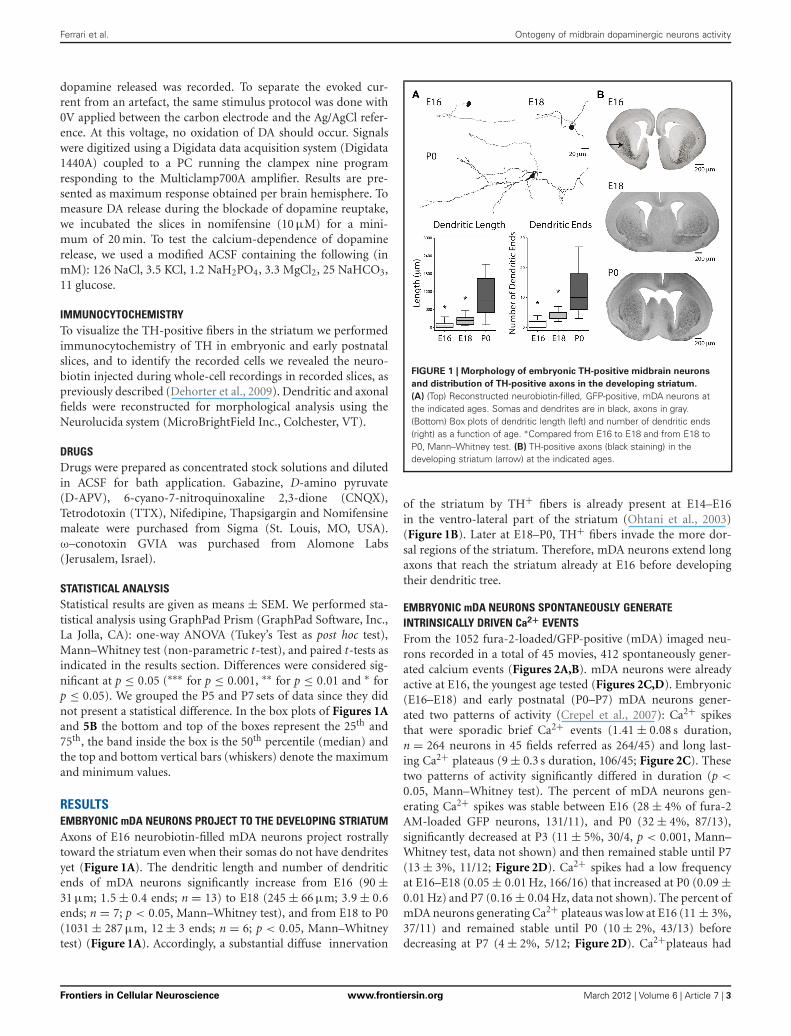
EMBRYONIC mDA NEURONS SPONTANEOUSLY GENERATEINTRINSICALLY DRIVEN Ca2+ EVENTSFrom the 1052 fura-2-loaded/GFP-positive (mDA) imaged neu-rons recorded in a total of 45 movies, 412 spontaneously gener-ated calcium events (Figures 2A,B). mDA neurons were alreadyactive at E16, the youngest age tested (Figures 2C,D). Embryonic(E16–E18) and early postnatal (P0–P7) mDA neurons gener-ated two patterns of activity (Crepel et al., 2007): Ca2+ spikesthat were sporadic brief Ca2+ events (1.41 ± 0.08 s duration,n = 264 neurons in 45 fields referred as 264/45) and long last-ing Ca2+ plateaus (9 ± 0.3 s duration, 106/45; Figure 2C). Thesetwo patterns of activity significantly differed in duration (p <
0.05, Mann–Whitney test). The percent of mDA neurons gen-erating Ca2+ spikes was stable between E16 (28 ± 4% of fura-2AM-loaded GFP neurons, 131/11), and P0 (32 ± 4%, 87/13),significantly decreased at P3 (11 ± 5%, 30/4, p < 0.001, Mann–Whitney test, data not shown) and then remained stable until P7(13 ± 3%, 11/12; Figure 2D). Ca2+ spikes had a low frequencyat E16–E18 (0.05 ± 0.01 Hz, 166/16) that increased at P0 (0.09 ±0.01 Hz) and P7 (0.16 ± 0.04 Hz, data not shown). The percent ofmDA neurons generating Ca2+ plateaus was low at E16 (11 ± 3%,37/11) and remained stable until P0 (10 ± 2%, 43/13) beforedecreasing at P7 (4 ± 2%, 5/12; Figure 2D). Ca2+plateaus had
Frontiers in Cellular Neuroscience www.frontiersin.org March 2012 | Volume 6 | Article 7 | 3
Ferrari et al. Ontogeny of midbrain dopaminergic neurons activity
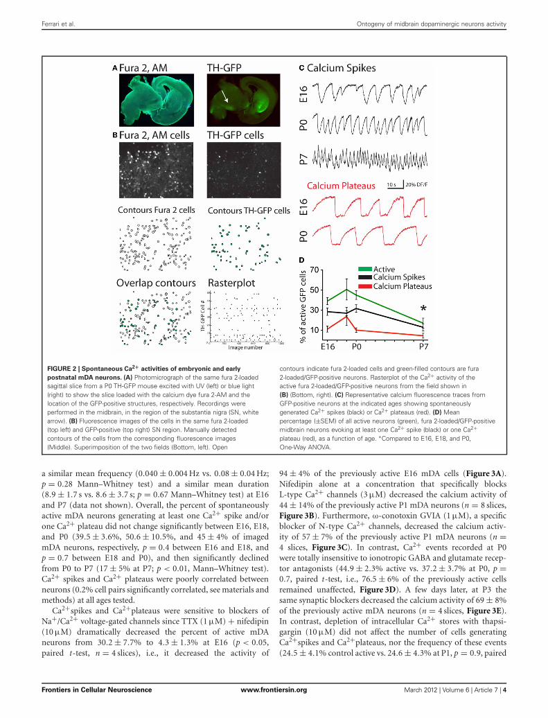
FIGURE 2 | Spontaneous Ca2+ activities of embryonic and early
postnatal mDA neurons. (A) Photomicrograph of the same fura 2-loadedsagittal slice from a P0 TH-GFP mouse excited with UV (left) or blue light(right) to show the slice loaded with the calcium dye fura 2-AM and thelocation of the GFP-positive structures, respectively. Recordings wereperformed in the midbrain, in the region of the substantia nigra (SN, whitearrow). (B) Fluorescence images of the cells in the same fura 2-loaded(top left) and GFP-positive (top right) SN region. Manually detectedcontours of the cells from the corresponding fluorescence images(Middle). Superimposition of the two fields (Bottom, left). Open
contours indicate fura 2-loaded cells and green-filled contours are fura2-loaded/GFP-positive neurons. Rasterplot of the Ca2+ activity of theactive fura 2-loaded/GFP-positive neurons from the field shown in(B) (Bottom, right). (C) Representative calcium fluorescence traces fromGFP-positive neurons at the indicated ages showing spontaneouslygenerated Ca2+ spikes (black) or Ca2+ plateaus (red). (D) Meanpercentage (±SEM) of all active neurons (green), fura 2-loaded/GFP-positivemidbrain neurons evoking at least one Ca2+ spike (black) or one Ca2+plateau (red), as a function of age. ∗Compared to E16, E18, and P0,One-Way ANOVA.
a similar mean frequency (0.040 ± 0.004 Hz vs. 0.08 ± 0.04 Hz;p = 0.28 Mann–Whitney test) and a similar mean duration(8.9 ± 1.7 s vs. 8.6 ± 3.7 s; p = 0.67 Mann–Whitney test) at E16and P7 (data not shown). Overall, the percent of spontaneouslyactive mDA neurons generating at least one Ca2+ spike and/orone Ca2+ plateau did not change significantly between E16, E18,and P0 (39.5 ± 3.6%, 50.6 ± 10.5%, and 45 ± 4% of imagedmDA neurons, respectively, p = 0.4 between E16 and E18, andp = 0.7 between E18 and P0), and then significantly declinedfrom P0 to P7 (17 ± 5% at P7; p < 0.01, Mann–Whitney test).Ca2+ spikes and Ca2+ plateaus were poorly correlated betweenneurons (0.2% cell pairs significantly correlated, see materials andmethods) at all ages tested.
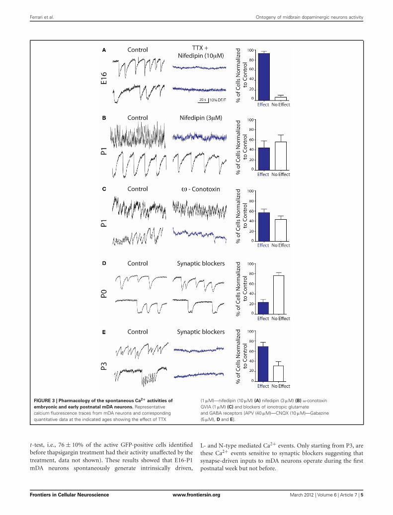
Ca2+spikes and Ca2+plateaus were sensitive to blockers ofNa+/Ca2+ voltage-gated channels since TTX (1 μM) + nifedipin(10 μM) dramatically decreased the percent of active mDAneurons from 30.2 ± 7.7% to 4.3 ± 1.3% at E16 (p < 0.05,paired t-test, n = 4 slices), i.e., it decreased the activity of
94 ± 4% of the previously active E16 mDA cells (Figure 3A).Nifedipin alone at a concentration that specifically blocksL-type Ca2+ channels (3 μM) decreased the calcium activity of44 ± 14% of the previously active P1 mDA neurons (n = 8 slices,Figure 3B). Furthermore, ω-conotoxin GVIA (1 μM), a specificblocker of N-type Ca2+ channels, decreased the calcium activ-ity of 57 ± 7% of the previously active P1 mDA neurons (n =4 slices, Figure 3C). In contrast, Ca2+ events recorded at P0were totally insensitive to ionotropic GABA and glutamate recep-tor antagonists (44.9 ± 2.3% active vs. 37.2 ± 3.7% at P0, p =0.7, paired t-test, i.e., 76.5 ± 6% of the previously active cellsremained unaffected, Figure 3D). A few days later, at P3 thesame synaptic blockers decreased the calcium activity of 69 ± 8%of the previously active mDA neurons (n = 4 slices, Figure 3E).In contrast, depletion of intracellular Ca2+ stores with thapsi-gargin (10 μM) did not affect the number of cells generatingCa2+spikes and Ca2+plateaus, nor the frequency of these events(24.5 ± 4.1% control active vs. 24.6 ± 4.3% at P1, p = 0.9, paired
Frontiers in Cellular Neuroscience www.frontiersin.org March 2012 | Volume 6 | Article 7 | 4
Ferrari et al. Ontogeny of midbrain dopaminergic neurons activity
FIGURE 3 | Pharmacology of the spontaneous Ca2+ activities of
embryonic and early postnatal mDA neurons. Representativecalcium fluorescence traces from mDA neurons and correspondingquantitative data at the indicated ages showing the effect of TTX
(1 μM)—nifedipin (10 μM) (A) nifedipin (3 μM) (B) ω-conotoxinGVIA (1 μM) (C) and blockers of ionotropic glutamateand GABA receptors [APV (40 μM)—CNQX (10 μM)—Gabazine(5 μM), D and E].
t-test, i.e., 76 ± 10% of the active GFP-positive cells identifiedbefore thapsigargin treatment had their activity unaffected by thetreatment, data not shown). These results showed that E16-P1mDA neurons spontaneously generate intrinsically driven,
L- and N-type mediated Ca2+ events. Only starting from P3, arethese Ca2+ events sensitive to synaptic blockers suggesting thatsynapse-driven inputs to mDA neurons operate during the firstpostnatal week but not before.
Frontiers in Cellular Neuroscience www.frontiersin.org March 2012 | Volume 6 | Article 7 | 5
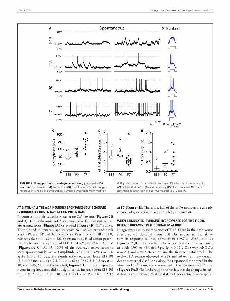
FIGURE 4 | Firing patterns of embryonic and early postnatal mDA
neurons. Spontaneous (A) and evoked (B) membrane potential changesrecorded in whole-cell configuration, current clamp mode from midbrain
GFP-positive neurons at the indicated ages. Distribution of the amplitude(C) half-width duration (D) and frequency (E) of spontaneous Na+ actionpotentials as a function of age. ∗Compared to E18 and P0.
AT BIRTH, HALF THE mDA NEURONS SPONTANEOUSLY GENERATEINTRINSICALLY DRIVEN Na+ ACTION POTENTIALSIn contrast to their capacity to generate Ca2+ events (Figures 2Band 3), E16 embryonic mDA neurons (n = 16) did not gener-ate spontaneous (Figure 4A) or evoked (Figure 4B) Na+ spikes.They started to generate spontaneous Na+ spikes around birthsince 28% and 50% of the recorded mDA neurons at E18 and P0,respectively, (n = 18; n = 12), spontaneously fired action poten-tials with a mean amplitude of 44.8 ± 5.4 mV and 52.6 ± 3.3 mV(Figures 4A–C). At P7, 100% of the recorded mDA neuronswere spontaneously active (amplitude: 73.6 ± 4.5 mV; n = 10).Spike half-width duration significantly decreased from E18–P0(3.8 ± 0.6 ms, n = 5; 4.2 ± 0.6, n = 6) to P7 (2.2 ± 0.2 ms, n =10; p < 0.05, Mann–Whitney test; Figure 4D) but mean sponta-neous firing frequency did not significantly increase from E18–P0to P7 (0.2 ± 0.1 Hz at E18; 0.4 ± 0.2 Hz at P0; 0.6 ± 0.2 Hz
at P7; Figure 4E). Therefore, half of the mDA neurons are alreadycapable of generating spikes at birth (see Figure 2).
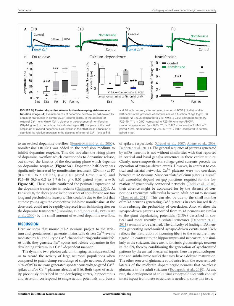
WHEN STIMULATED, TYROSINE-HYDROXYLASE POSITIVE FIBERSRELEASE DOPAMINE IN THE STRIATUM AT BIRTHIn agreement with the presence of TH+ fibers in the embryonicstriatum, we detected from E18 DA release in the stria-tum in response to local stimulation (19.7 ± 1.5 pA, n = 13;Figures 5A,B). This evoked DA release significantly increasedat birth (P0) to 43.1 ± 4.3 pA (p < 0.001, One-way ANOVA;n = 25) and stayed stable during the first postnatal week. Theevoked DA release observed at E18 and P0 was entirely depen-dent on external Ca2+ ions, since the response disappeared in theabsence of Ca2+ ions, and was rescued in the presence of Ca2+ ions(Figures 5A,B) To further support the view that the changes in oxi-dation current evoked by striatal stimulation actually correspond
6
Ferrari et al. Ontogeny of midbrain dopaminergic neurons activity
Frontiers in Cellular Neuroscience www.frontiersin.org March 2012 | Volume 6 | Article 7 |
Ferrari et al. Ontogeny of midbrain dopaminergic neurons activity
FIGURE 5 | Evoked dopamine release in the developing striatum as a
function of age. (A) Example traces of dopamine overflow (in pA) evoked bya train of four pulses in control ACSF (control, black), in the absence ofexternal Ca2+ ions (0 mM Ca2+, blue) or in the presence of nomifensine(10 μM, green) in the bath, at the indicated ages. (B) Box plots of the peakamplitude of evoked dopamine (DA) release in the striatum as a function ofage (left), its relative decrease in the absence of external Ca2+ ions at E18
and P0 with recovery after returning to control ACSF (middle), and itshalf-decay in the presence of nomifensine as a function of age (right). DArelease: ◦p < 0.05 compared to E18; ###p < 0.001 compared to P0, P7,P26–40; ∗∗∗p < 0.001 compared to P26–40; one-way ANOVA.Calcium-dependence: ∗p < 0.05, ∗∗∗p < 0.001 compared to 2 mM Ca2+,paired t-test. Nomifensine: ∗p < 0.05, ∗∗∗p < 0.001 compared to control,paired t-test.
to an evoked dopamine overflow (Benoit-Marand et al., 2000),nomifensine (10 μM) was added to the perfusion medium toinhibit dopamine reuptake. This did not alter the rising phaseof dopamine overflow which corresponds to dopamine release,but slowed the kinetics of the decreasing phase which dependson dopamine reuptake (Figure 5A). Dopamine half-decay wassignificantly increased by nomifensine treatment (20 min) at P7(0.4 ± 0.1 to 3.7 ± 0.3 s, p < 0.001 paired t-test, n = 5), andP25–40 (0.5 ± 0.2 to 3.7 ± 1 s, p < 0.05 paired t-test, n = 5;Figure 5B). These results confirmed the perinatal expression ofthe dopamine transporter in rodents (Galineau et al., 2004). AtE18 and P0, the decay phase in the presence of nomifensine was toolong and precluded its measure. This could be due to the fact thatat these young ages the competitive inhibitor nomifensine, at thedose used, could not be rapidly displaced from its binding sites onthe dopamine transporter (Tuomisto, 1977; Jones et al., 1995; Katzet al., 2000) by the small amount of evoked dopamine overflow.
DISCUSSIONHere we show that mouse mDA neurons project to the stria-tum and spontaneously generate intrinsically driven Ca2+ eventsmediated by N- and L-type Ca2+ channels during embryonic life.At birth, they generate Na+ spikes and release dopamine in thedeveloping striatum in a Ca2+-dependent manner.
The dynamic two-photon calcium imaging technique enabledus to record the activity of large neuronal populations whencompared to patch-clamp recordings of single neurons. Around50% of mDA neurons generated spontaneous voltage-gated Ca2+spikes and/or Ca2+ plateaus already at E16. Both types of activ-ity previously described in the developing cortex, hippocampusand striatum, correspond to single action potentials and bursts
of spikes, respectively, (Crepel et al., 2007; Allene et al., 2008;Dehorter et al., 2011). The general sequence of patterns generatedby mDA neurons is not without similarities with that reportedin cortical and basal ganglia structures in these earlier studies.Clearly, non-synapse-driven, voltage-gated currents precede theoperation of synapse-driven events. However, in contrast to cor-tical and striatal networks, Ca2+ plateaus were not correlatedbetween mDA neurons. Since correlated calcium plateaus in smallcell assemblies depend on gap junctions required for the for-mation of synaptically connected networks (Todd et al., 2010),their absence might be accounted for by the absence of con-nections (recurrent collaterals) between adult A9 mDA neurons(Chen et al., 2011). This can also be due to the small numberof mDA neurons generating Ca2+ plateaus in each imaged field,thus reducing the probability of correlation. Also, whether thesynapse-driven patterns recorded from mDA neurons are similarto the giant depolarizing potentials (GDPs) described in cor-tical and more recently in striatal structures (Dehorter et al.,2011) remains to be clarified. The difficulty of finding mDA neu-rons generating synchronized synapse-driven events most likelyreflects the maturation of incoming fibers to the structure inves-tigated. In contrast to the hippocampus and neocortex, but simi-larly as the striatum, there are no intrinsic glutamatergic neuronsin the SN, thereby conditioning the generation of synchronizedpatterns by the arrival of external inputs: here the pedunculopon-tine and subthalamic nuclei that may have a delayed maturation.The other source of glutamate could arise from the recurrent col-laterals of the midbrain dopaminergic neurons that co-releaseglutamate in the adult striatum (Tecuapetla et al., 2010). At anyrate, the development of an in vitro embryonic slice with enoughintact inputs from these structures is needed to solve this issue.
Frontiers in Cellular Neuroscience www.frontiersin.org March 2012 | Volume 6 | Article 7 | 7
Ferrari et al. Ontogeny of midbrain dopaminergic neurons activity
Although we cannot completely exclude the possibility thata subthreshold calcium-dependent dopamine release is presentbefore E18–P0 this would be without functional consequencesince Na+ spikes required to that effect are not generated bymost mDA neurons before E18. Interestingly, around birth, mDAneurons generate Ca2+ events partly mediated by N-type Ca2+channels, the same channels involved in synaptic DA release in theadult rodent striatum in vivo and in vitro (Herdon and Nahorski,1989; Bergquist et al., 1998).
What could be the functional role of dopamine signals inthe developing striatum? Dopamine has been suggested to mod-ulate multiplication, migration, and wiring of target neurons.The activation of dopamine receptors by exogenous dopamine ordopamine agonists regulates the cell cycle of striatal progenitorsin the lateral ganglionic eminence in explant cultures or in micein vivo from E13 (Ohtani et al., 2003). From E15, dopaminergicagonists, or the invalidation of D1 or D2 receptors, differen-tially modulate the migration of GABAergic interneurons to thecerebral wall in embryonic mouse forebrain organotypic slices(Crandall et al., 2007). In addition, the activation of dopaminereceptors in primary striatal neuronal cultures (7–14 days cul-tures obtained from E16–17 striata) limits the extent of collateralGABAergic synaptogenesis between developing medium spinyneurons (Goffin et al., 2010). Early effects of dopamine beforeE18 could result from activity-independent release of dopaminein the ganglionic eminences as described for glutamate and GABAin the developing hippocampus and shown to be quite efficient
in modulating migration (Demarque et al., 2002; Manent andRepresa, 2007). The possible implications of activity-dependentrelease of DA on striatal maturation remain to be investigated.But, interestingly, the fraction of medium spiny neurons gen-erating glutamate and GABA spontaneous synaptic activity inthe developing striatum also considerably develops during thefirst postnatal week in mice (Dehorter et al., 2011), suggest-ing an important stimulation of the developmental processafter birth.
To conclude, the present work suggests a developmentalsequence of mDA neurons with features that are common andspecific to these neurons. In a previous study, we showed thatstriatal neurons follow an abrupt alteration of their properties intime to start controlling motricity in pups (Dehorter et al., 2011).Future studies will have to interconnect these events and deter-mine the impact of dopaminergic synapses on the operation ofearly striatal neurons.
ACKNOWLEDGMENTSWe thank Dr. Baker (Cornell University) for the gift of TH-GFPmice, F. Gonon for his help with the amperometry techniqueand B. Bloem for his help in preliminary experiments. DianaC. Ferrari was funded by Fondation pour la Recherche Médicale(FRM) and Association France Parkinson. This work was sup-ported by grants from Institut National de la Santé et de laRecherche Médicale (Inserm), and Fédération de Recherche surle Cerveau (CH).
REFERENCESAllene, C., Cattani, A., Ackman, J.
B., Bonifazi, P., Aniksztejn, L.,Ben-Ari, Y., and Cossart, R. (2008).Sequential generation of twodistinct synapse-driven networkpatterns in developing neocortex.J. Neurosci. 28, 12851–12863.
Benoit-Marand, M., Jaber, M., andGonon, F. (2000). Release and elim-ination of dopamine in vivo in micelacking the dopamine transporter:functional consequences. Eur. J.Neurosci. 12, 2985–2992.
Bergquist, F., Jonason, J., Pileblad, E.,and Nissbrandt, H. (1998). Effectsof local administration of L-, N-,and P/Q-type calcium channelblockers on spontaneous dopaminerelease in the striatum and the sub-stantia nigra: a microdialysis studyin rat. J. Neurochem. 70, 1532–1540.
Bonifazi, P., Goldin, M., Picardo, M. A.,Jorquera, I., Cattani, A., Bianconi,G., Represa, A., Ben-Ari, Y., andCossart, R. (2009). GABAergic hubneurons orchestrate synchrony indeveloping hippocampal networks.Science 326, 1419–1424.
Chen, B. T., Patel, J. C., Moran, K. A.,and Rice, M. E. (2011). Differentialcalcium dependence of axonalversus somatodendritic dopaminerelease, with characteristics of
both in the ventral tegmental area.Front. Syst. Neurosci. 5:39. doi:10.3389/fnsys.2011.00039
Cragg, S. J., Holmes, C., Hawkey, C.R., and Greenfield, S. A. (1998).Dopamine is released sponta-neously from developing midbrainneurons in organotypic culture.Neuroscience 84, 325–330.
Crandall, J. E., McCarthy, D. M., Araki,K. Y., Sims, J. R., Ren, J. Q., andBhide, P. G. (2007). Dopaminereceptor activation modulatesGABA neuron migration fromthe basal forebrain to the cerebralcortex. J. Neurosci. 27, 3813–3822.
Crepel, V., Aronov, D., Jorquera,I., Represa, A., Ben-Ari, Y., andCossart, R. (2007). A parturition-associated nonsynaptic coherentactivity pattern in the developinghippocampus. Neuron 54, 105–120.
Dehorter, N., Guigoni, C., Lopez, C.,Hirsch, J., Eusebio, A., Ben-Ari, Y.,and Hammond, C. (2009). Dop-amine-deprived striatal GABAergicinterneurons burst and generaterepetitive gigantic IPSCs in mediumspiny neurons. J. Neurosci. 29,7776–7787.
Dehorter, N., Michel, F. J., Marissal, T.,Rotrou, Y., Matrot, B., Lopez,C., Humphries, M. D., andHammond, C. (2011). Onset of
pup locomotion coincides withloss of NR2C/D-Mediated cortico-striatal EPSCs and dampening ofstriatal network immature activity.Front. Cell. Neurosci. 5:24. doi:10.3389/fncel.2011.00024
Demarque, M., Represa, A., Becq,H., Khalilov, I., Ben Ari, Y., andAniksztejn, L. (2002). Paracrineintercellular communication by aCa2+- and SNARE-independentrelease of GABA and glutamateprior to synapse formation. Neuron36, 1051–1061.
Fishell, G., and van der Kooy, D. (1989).Pattern formation in the striatum:developmental changes in the dis-tribution of striatonigral projec-tions. Brain Res. Dev. Brain Res. 45,239–255.
Galineau, L., Kodas, E., Guilloteau,D., Vilar, M. P., and Chalon, S.(2004). Ontogeny of the dopamineand serotonin transporters in the ratbrain: an autoradiographic study.Neurosci. Lett. 363, 266–271.
Gates, M. A., Torres, E. M., White, A.,Fricker-Gates, R. A., and Dunnett,S. B. (2006). Re-examining theontogeny of substantia nigradopamine neurons. Eur. J. Neurosci.23, 1384–1390.
Goffin, D., Ali, A. B., Rampersaud, N.,Harkavyi, A., Fuchs, C., Whitton, P.
S., Nairn, A. C., and Jovanovic, J. N.(2010). Dopamine-dependent tun-ing of striatal inhibitory synaptoge-nesis. J. Neurosci. 30, 2935–2950.
Hall, A. C., Mira, H., Wagner, J., andArenas, E. (2003). Region-specificeffects of glia on neuronal induc-tion and differentiation with a focuson dopaminergic neurons. Glia 43,47–51.
Herdon, H., and Nahorski, S. R.(1989). Investigations of theroles of dihydropyridine andomega-conotoxin-sensitive cal-cium channels in mediatingdepolarisation-evoked endogenousdopamine release from striatalslices. Naunyn Schmiedebergs Arch.Pharmacol. 340, 36–40.
Jones, S. R., Garris, P. A., Kilts, C.D., and Wightman, R. M. (1995).Comparison of dopamine uptake inthe basolateral amygdaloid nucleus,caudate-putamen, and nucleusaccumbens of the rat. J. Neurochem.64, 2581–2589.
Katz, J. L., Izenwasser, S., and Terry,P. (2000). Relationships amongdopamine transporter affinities andcocaine-likediscriminative-stimuluseffects. Psychopharmacology (Berl)148, 90–98.
Kawano, H., Ohyama, K., Kawamura,K., and Nagatsu, I. (1995).
Frontiers in Cellular Neuroscience www.frontiersin.org March 2012 | Volume 6 | Article 7 | 8
Ferrari et al. Ontogeny of midbrain dopaminergic neurons activity
Migration of dopaminergic neuronsin the embryonic mesencephalon ofmice. Brain Res. Dev. Brain Res. 86,101–113.
Kim, Y., Park, M. K., and Chung,S. (2008). Voltage-operated Ca2+channels regulate dopamine releasefrom somata of dopamine neu-rons in the substantia nigra parscompacta. Biochem. Biophys. Res.Commun. 373, 665–669.
Manent, J. B., and Represa, A. (2007).Neurotransmitters and brain mat-uration: early paracrine actions ofGABA and glutamate modulateneuronal migration. Neuroscientist13, 268–279.
Matsushita, N., Okada, H., Yasoshima,Y., Takahashi, K., Kiuchi, K., andKobayashi, K. (2002). Dynamicsof tyrosine hydroxylase pro-moter activity during midbraindopaminergic neuron development.J. Neurochem. 82, 295–304.
Moon, E. S., and Herkenham, M.(1984). Comparative developmentof striatal opiate receptors anddopamine revealed by autoradiog-raphy and histofluorescence. BrainRes. 305, 27–42.
Ohtani, N., Goto, T., Waeber, C., andBhide, P. G. (2003). Dopaminemodulates cell cycle in the lateralganglionic eminence. J. Neurosci. 23,2840–2850.
Sawamoto, K., Nakao, N., Kobayashi,K., Matsushita, N., Takahashi,
H., Kakishita, K., Yamamoto,A., Yoshizaki, T., Terashima, T.,Murakami, F., Itakura, T., andOkano, H. (2001). Visualization,direct isolation, and transplantationof midbrain dopaminergic neurons.Proc. Natl. Acad. Sci. U.S.A. 98,6423–6428.
Schmitz, Y., Lee, C. J., Schmauss, C.,Gonon, F., and Sulzer, D. (2001).Amphetamine distorts stimulation-dependent dopamine overflow:effects on D-2 autoreceptors, trans-porters, and synaptic vesicle stores.J. Neurosci. 21, 5916–5924.
Specht, L. A., Pickel, V. M., Joh, T.H., and Reis, D. J. (1981a). Light-microscopic immunocytochemicallocalization of tyrosine hydroxy-lase in prenatal rat brain. I. Earlyontogeny. J. Comp. Neurol. 199,233–253.
Specht, L. A., Pickel, V. M., Joh, T.H., and Reis, D. J. (1981b). Light-microscopic immunocytochemicallocalization of tyrosine hydroxy-lase in prenatal rat brain. II. Lateontogeny. J. Comp. Neurol. 199,255–276.
Tecuapetla, F., Patel, J. C., Xenias, H.,English, D., Tadros, I., Shah, F.,Berlin, J., Deisseroth, K., Rice, M.E., Tepper, J. M., and Koos, T.(2010). Glutamatergic signaling bymesolimbic dopamine neurons inthe nucleus accumbens. J. Neurosci.30, 7105–7110.
Tepper, J. M., Trent, F., and Nakamura,S. (1990). Postnatal development ofthe electrical activity of rat nigros-triatal dopaminergic neurons. BrainRes. Dev. Brain Res. 54, 21–33.
Todd, K. L., Kristan, W. B. Jr., andFrench, K. A. (2010). Gap junc-tion expression is required for nor-mal chemical synapse formation.J. Neurosci. 30, 15277–15285.
Trent, F., Nakamura, S., and Tepper,J. M. (1991). Amphetamine exertsanomalous effects on dopamin-ergic neurons in neonatal ratsin vivo. Eur. J. Pharmacol. 204,265–272.
Tuomisto, J. (1977). Nomifensine andits derivatives as possible toolsfor studying amine uptake. Eur. J.Pharmacol. 42, 101–106.
van den Heuvel, D. M., andPasterkamp, R. J. (2008). Gettingconnected in the dopamine system.Prog. Neurobiol. 85, 75–93.
van der Kooy, D., and Fishell, G. (1987).Neuronal birthdate underlies thedevelopment of striatal compart-ments. Brain Res. 401, 155–161.
Voorn, P., Kalsbeek, A., Jorritsma-Byham, B., and Groenewegen,H. J. (1988). The pre- andpostnatal development of thedopaminergic cell groups in theventral mesencephalon andthe dopaminergic innervation ofthe striatum of the rat. Neuroscience25, 857–887.
Washio, H., Takigachi-Hayashi, K., andKonishi, S. (1999). Early postnataldevelopment of substantia nigraneurons in rat midbrain slices:hyperpolarization-activated inwardcurrent and dopamine-activatedcurrent. Neurosci. Res. 34, 91–101.
Conflict of Interest Statement: Theauthors declare that the researchwas conducted in the absence of anycommercial or financial relationshipsthat could be construed as a potentialconflict of interest.
Received: 16 December 2011; accepted:11 February 2012; published online: 08March 2012.Citation: Ferrari DC, Mdzomba BJ,Dehorter N, Lopez C, Michel FJ, LibersatF and Hammond C (2012) Midbraindopaminergic neurons generate cal-cium and sodium currents and releasedopamine in the striatum of pups. Front.Cell. Neurosci. 6:7. doi: 10.3389/fncel.2012.00007Copyright © 2012 Ferrari, Mdzomba,Dehorter, Lopez, Michel, Libersat andHammond. This is an open-accessarticle distributed under the terms ofthe Creative Commons Attribution NonCommercial License, which permitsnon-commercial use, distribution, andreproduction in other forums, providedthe original authors and source arecredited.
Frontiers in Cellular Neuroscience www.frontiersin.org March 2012 | Volume 6 | Article 7 | 9