gdnf: a key player in neuron-glia crosstalk and survival of nigrostriatal dopaminergic neurons
TRANSCRIPT
Emília P. Duarte – [email protected] 1Center for Neuroscience and Cell Biology, University of Coimbra, Coimbra, Portugal. 2Health Sciences Research Center, University of Beira Interior, Covilhã, Portugal.3Department of Zoology, University of Coimbra, Coimbra, Portugal.
8
GDNF: A KEY PLAYER IN NEURON–GLIA CROSSTALK AND SURVIVAL OF NIGROSTRIATAL DOPAMINERGIC
NEURONS
Emília P. Duarte1,3, Ana Saavedra1 and Graça Baltazar2
1. ABSTRACT Glial cell line-derived neurotrophic factor (GDNF) is a potent survival factor for dopaminergic neurons of the nigrostriatal pathway that degenerate in Parkinson’s disease (PD). In animal models of PD, GDNF delivery has been shown to both protect dopaminergic neurons against toxin-induced injury and to rescue damaged neurons, promoting recovery of the motor deficit. GDNF may act both as a target-derived neurotrophic factor in the striatum, and as a local neurotrophic factor at the level of neuronal cell bodies in the substantia nigra. The neuroprotective and regenerative effects of GDNF are mediated by increases in the activity of antioxidant enzymes and induction of antiapoptotic proteins and cell adhesion molecules. In addition, GDNF stimulates dopaminergic neurotransmission by increasing dopamine biosynthesis and neuronal excitability. In the normal adult brain, GDNF is expressed mainly by neuronal cells but, upon injury, both astrocytes and microglial cells express GDNF. How neuronal damage signals glial cells to upregulate GDNF is being uncovered. Dopamine agonists, interleukin-1β (IL-1β) and estrogen have been shown to modulate GDNF expression by astrocytes and/or microglial cells. GDNF produced by activated microglial cells is involved in the axonal growth and sprouting of damaged neurons. Moreover, GDNF can modulate the migration, adhesion and phagocytic activity of microglia. The intercellular messengers involved in the crosstalk between dopaminergic neurons, astrocytes and microglia are potential targets for PD therapies aimed at upregulating the endogenous expression of GDNF. 2. INTRODUCTION Glial cell line-derived neurotrophic factor (GDNF) is widely recognized as a potent survival factor for dopaminergic neurons projecting from the substantia nigra pars compacta in the midbrain to the striatum (reviewed by Hurelbrink and Barker, 2004). The progressive
E.P. Duarte, A. Saavedra and G. Baltazar
degeneration of the nigrostriatal neurons in Parkinson’s disease (PD) leads to dopamine depletion in the striatum, or caudate-putamen in primates, causing the slowness of movement, stiffness, resting tremor and postural instability observed in PD. GDNF was originally identified in conditioned media from a glial cell line based on its ability to promote the survival of embryonic dopaminergic neurons in culture, and to increase cell size, neurite length and the high-affinity uptake of dopamine (Lin et al., 1993, 1994). Subsequently, the neuroprotective effects of GDNF were shown in animal models of PD. The administration of GDNF, either the recombinant protein (reviewed by Grondin et al., 2003) or the delivery of GDNF gene by viral vectors (reviewed by Bjorklund et al., 2000), prevents the degeneration of dopaminergic neurons and the motor impairment induced by a subsequent lesion of the nigrostriatal system. More importantly, GDNF delivery after a toxin-induced lesion, a paradigm that better mimics PD at the time of detection, was shown to increase dopaminergic markers and to restore motor function (Tomac et al., 1995a; Gash et al., 1996, 2005; Mandel et al., 1999; Kordower et al., 2000; Grondin et al., 2002; Wang et al., 2002; Zheng et al., 2005). Therefore, in addition to protecting dopaminergic neurons, GDNF appears to induce neuronal repair, stimulating regenerative growth and axonal sprouting after lesions of the nigrostriatal system. Therefore, GDNF was proposed and tested as a neuroprotective/restorative therapy for PD aimed at slowing down, halting or reversing neurodegeneration. Direct infusion of GDNF into the putamen of PD patients has improved the motor function, decreased the requirements for L-DOPA therapy and induced outgrowth of dopaminergic neurons as assessed by imaging of dopamine transporters (Gill et al., 2003; Love et al., 2005; Patel et al., 2005; Slevin et al., 2005). However, in a double blind, placebo-controlled trial, in a larger number of patients, there were no significant clinical improve-ments and severe side effects were reported (Lang et al., 2006). The clinical trials were discontinued and several problems remain to be solved before GDNF therapy can be used in PD patients (Sherer et al., 2006). Nervous tissue responds to injury upregulating protective and repairing mechanisms (reviewed by Benn and Woolf, 2004; Martino, 2004). Damaged neurons change their own gene expression and stimulate nearby astrocytes and microglial cells to provide support. Activated astrocytes are known to upregulate antioxidant molecules, membrane transporters and trophic factors that support neuronal and glial survival and tissue repair (reviewed by Liberto et al., 2004). Microglia, the resident immune cells of the brain, proliferate and become immunocompetent cells, with the ability of secreting a number of cytotoxic and trophic molecules (reviewed by Block and Hong, 2005). Upregulation of GDNF, by both astrocytes and microglial cells, has been shown in several injury models (Liberatore et al., 1997; Batchelor et al., 1999; Satake et al., 2000; Miyazaki et al., 2001; Ikeda et al., 2002). Therefore, the intercellular signals involved in the communication between damaged neurons and glial cells are potential therapeutic targets aimed at upregulating the endogenous expression of neurotrophic factors. The trophic effects of GDNF are not exclusive of midbrain dopaminergic neurons. GDNF also supports the survival of spinal motoneurons, regulates the differentiation of several peripheral neurons, and has roles outside the nervous system in kidney morphogenesis and in spermatogonial differentiation (reviewed by Sariola and Saarma, 2003). These pleiotropic actions of GDNF are mediated by the activation of a multi-component receptor system. GDNF binds to a glycosylphosphatidylinositol (GPI)-anchored cell surface receptor known as GFRα1 (GDNF family receptor-α1). The GFRα1 acts only as a binding subunit that can activate several signalling receptors. GFRα1 can signal through the transmembrane Ret tyrosine kinase, which in turn activates several intracellular signalling cascades: the
174
GDNF IN THE NIGROSTRIATAL SYSTEM
mitogen-activated protein (MAP) kinase, the phosphoinositide 3-kinase (PI3K) and the phospholipase Cγ (PLC-γ) pathways. In cells not expressing Ret, GDNF triggers Src family kinase activation and the phosphorylation of the extracellular receptor-activated kinase ERK/ MAP kinase, PLC-γ and the transcription factor CREB. More recently, GDNF has been shown to signal through the neural cell adhesion molecule (NCAM; reviewed by Sariola and Saarma, 2003). The present review will focus on GDNF role in the nigrostriatal system, mainly on data gathered in animal and cell culture models of PD, on its neuroprotective effects and mechanisms, and the control of GDNF expression upon nigrostriatal injury. 3. GDNF SUPPORT OF NIGROSTRIATAL DOPAMINERGIC NEURONS:
TARGET-DERIVED AND LOCAL SUPPORT? According to the neurotrophic theory, neuronal survival during development is controlled by target-derived neurotrophic factors (Purves, 1986; Oppenheim, 1991). Only neurons establishing synaptic contacts have access to sufficient amounts of trophic factors. GDNF is highly expressed by striatal neurons during post-natal development, and it was shown to regulate the natural death of substantia nigra dopaminergic neurons (reviewed by Burke, 2004). The levels of GDNF mRNA decrease steeply in the post-natal period (Stromberg et al., 1993; Choi-Lundberg and Bohn, 1995; Oo et al., 2005), and the role of GDNF in the normal adult brain is not well established. However, GDNF (Choi-Lundberg and Bohn, 1995; Pochon et al., 1997; Trupp et al., 1997) and its receptors (Treanor et al., 1996; Trupp et al., 1996, 1997; Kozlowski et al., 2004) are expressed in the adult brain, although some contradictory reports exist (Stromberg et al., 1993). The observation of retrograde axonal transport of GDNF in the adult nigrostriatal neurons also indicates a target-derived function and a neuronal maintenance role for GDNF in the adult nervous system (Tomac et al., 1995b). A deficient neurotrophic support was also suggested as a factor involved in the degeneration of the nigrostriatal neurons in PD (Siegel and Chauhan, 2000). The effects of GDNF in the adult brain and its potential therapeutic value have been extensively studied in animal models of PD. In a widely used rat model, the selective death of substantia nigra dopaminergic neurons is induced by 6-hydroxydopamine (6-OHDA) injected in the substantia nigra, in the medial forebrain bundle or in the striatum (reviewed by Deumens et al., 2002). This analogue of dopamine causes the degeneration of neurons having the dopamine transporter by increasing intracellular oxidative stress. In mice and non-human primates, the degeneration of the nigrostriatal pathway is generally induced by the administration of 1-methyl-4-phenyl-1,2,3,6-tetrahydropyridine (MPTP), a hydrophobic compound capable of permeating the blood–brain barrier. This compound is metabolized in astrocytes to 1-methyl-4-phenyl-2,3-dihydropyridinium (MPDP+), and oxidized to 1-methyl-4-phenylpyridinium (MPP+), the active toxin, which is taken up by dopamine transporters. Once inside neuronal cells, MPP+ inhibits mitochondrial complex I leading to the release of superoxide, decreased ATP cellular content, dopamine leakage from synaptic vesicles and further oxidative stress (reviewed by Dauer and Przedborski, 2003). The administration of GDNF into the striatum, or into the putamen of primates, has consistently shown efficacy in preserving and restoring the structure and function of the nigrostriatal pathway. Striatal delivery of viral vectors encoding for GDNF before the 6-OHDA lesion prevents dopaminergic damage and motor impairment (Bilang-Bleuel et al.,
175
E.P. Duarte, A. Saavedra and G. Baltazar
1997; Mandel et al., 1997; Choi-Lundberg et al., 1998; Connor et al., 1999; Kirik et al., 2000b; Eslamboli et al., 2005). When administered after an established lesion, a condition more relevant to the treatment of PD, GDNF delivery to the striatum was shown to increase the number of tyrosine hydroxylase (TH)-positive cell bodies in the substantia nigra, the density of TH-positive fibres and dopamine levels in the striatum, and to induce recovery of motor impairments (Grondin et al., 2002; Wang et al., 2002; Zheng et al., 2005; Brizard et al., 2006). The use of TH as a marker of dopaminergic cell survival and repair raises some questions. Using fluorgold retrograde tracing, it was shown that the number of surviving dopaminergic neurons is larger than the number of TH-positive cells, suggesting that loss of dopaminergic phenotype precedes neuronal death (Bowenkamp et al., 1996). Therefore, the observed increase in the number of TH-positive cells or fibres upon GDNF treatment might be due to the recovery of TH expression in damaged neurons, rather than to neuronal regeneration and reinnervation of striatum. On the other hand, it was observed that GDNF-induced structural and behavioural recovery occurs only in partial lesions, probably because a number of surviving neurons and fibres are required to reinnervate the striatum by undergoing axonal sprouting, or to function as guidance cues for axonal growth (Rosenblad et al., 1998; Stanic et al., 2003, 2004; Brizard et al., 2006). It is important to stress that functional regeneration of the nigrostriatal system upon GDNF delivery was observed both in young and aged animals, including models of advanced parkinsonism, which more closely mimic the advanced stage and age of PD patients (Grondin et al., 2002; Zheng et al., 2005). In addition to being expressed by the targets of the nigrostriatal neurons, GDNF is expressed in the substantia nigra, in the perikaria area of dopaminergic neurons (Pochon et al., 1997; Sarabi et al., 2001). The GDNF protein detected in substantia nigra might come from the striatum by retrograde transport (Tomac et al., 1995b), but the detection of GDNF mRNA in substantia nigra (Choi-Lundberg and Bohn, 1995; Pochon et al., 1997) shows unequivocally that GDNF is also expressed in the soma area of the nigrostriatal pathway. Furthermore, colocalization of GDNF mRNA and TH immunoreactivity showed that dopaminergic neurons express GDNF (Pochon et al., 1997). The presence of GDNF receptors in the substantia nigra (Trupp et al., 1997), namely on dopaminergic neurons (Sarabi et al., 2001), suggests that locally produced GDNF might also play a role in dopaminergic function and survival. A microarray study also detected the expression of GDNF mRNA in astrocytes from intact adult substantia nigra (Nakagawa and Schwartz, 2004a). GDNF delivery into the substantia nigra before a 6-OHDA lesion in the striatum promotes the survival of nigral dopaminergic neurons (Choi-Lundberg et al., 1997; Connor et al., 1999, 2001; Kirik et al., 2000a, b) but fails to preserve striatal innervation and motor function, whereas the intrastriatal delivery preserves the integrity and function of the nigrostriatal system (Connor et al., 1999, 2001; Kirik et al., 2000a, b). In contrast, most studies on GDNF administration into the substantia nigra several weeks after the lesion, both in rodents and in primates, report marked functional recoveries (Hoffer et al., 1994; Bowenkamp et al., 1995, 1996; Hoffman et al., 1997; Lapchak et al., 1997; Kozlowski et al., 2000; Gash et al., 2005; Smith et al., 2005). Surprisingly, motor improvement was not always accompanied by striatal reinnervation, as assessed by TH immunoreactivity or dopamine levels (Hoffman et al., 1997; Lapchak et al., 1997). These observations support the idea that nigral delivery of GDNF does not promote fibre regeneration and striatal reinnervation, despite neuronal survival. It was proposed that the improvement in motor behaviour was due to the increase in somatodendritic release of dopamine in the substantia nigra (Hoffman et al., 1997; Gerhardt et al., 1999). However, viral delivery of GDNF into the substantia nigra, one week after a striatal 6-OHDA lesion, was shown to increase the number of intact
176
GDNF IN THE NIGROSTRIATAL SYSTEM
projections from the substantia nigra to the striatum as compared to the lesioned animals treated with control vectors (Kozlowski et al., 2000). Moreover, another study, in the same animal model, showed that GDNF treatment increases basal and evoked dopamine release in the striatum as measured by in vivo dialysis (Smith et al., 2005). In summary, the data on rodent and non-human primates, expressing stable parkinsonian symptoms, show that GDNF delivery into the striatum or the substantia nigra can induce marked functional improvements. The regenerative ability of GDNF in the nigrostriatal system depends on the extension of pre-existing damage, as surviving neurons are the substrates for GDNF-induced recovery of dopaminergic phenotype and regenerative sprouting. Moreover, the surviving fibres might produce guidance cues to direct neuronal regeneration towards the targets. It was suggested the possibility that GDNF is activating different mechanisms via axon terminal receptors in the putamen, and somatodendritic receptors in the substantia nigra (Gash et al., 2005). If so, the simultaneous GDNF administration to the substantia nigra and putamen could have additive effects on regene-ration of the nigrostriatal pathway and functional improvements. 4. GDNF AND THE NIGROSTRIATAL RESPONSE TO INJURY Brain injury appears to change the pattern of trophic factor expression. In the intact adult brain, GDNF expression is believed to be largely neuronal as astroglial cells were not found to express detectable GDNF (Blum and Weickert, 1995; Pochon et al., 1997; Bizon et al., 1999; Bresjanac and Antauer, 2000). However, as mentioned before, a microarray study detected GDNF mRNA in astrocytes from normal adult substantia nigra (Nakagawa and Schwartz, 2004a). Upon injury, glial cells appear to become the predominant source of trophic substances. Reactive astrocytes in the nigrostriatal system have been demonstrated to express a number of neurotrophic factors, including GDNF (Bresjanac and Antauer, 2000; Nakagawa and Schwartz, 2004b), basic fibroblast growth factor (bFGF) and nerve growth factor (NGF; Nakagawa and Schwartz, 2004b). The neuronal expression of trophic factors associated with injury is a rapid and transient activity-dependent expression, whereas a delayed and more persistent injury-induced expression is observed in glial cells (Hughes et al., 1999). The selective damage of nigrostriatal dopaminergic neurons in animal models of PD was shown to alter mRNA and/or protein levels for several trophic factors, both in the striatum and the substantia nigra, although some contradictory findings were reported. In the striatum, an increase in GDNF mRNA or protein levels has been observed after 6-OHDA- (Zhou et al., 2000; Nakajima et al., 2001; Yurek and Fletcher-Turner, 2001, 2002) or MPTP-induced injury (Tang et al., 1998). However, other authors did not detect changes in striatal GDNF levels after 6-OHDA lesion (Stromberg et al., 1993; Smith et al., 2003) or MPTP treatment (Inoue et al., 1999; Collier et al., 2005). These conflicting results might arise from differences in the injury models and protocols, or different time points analysed. In general, GDNF protein levels increased in the first 2–3 weeks after the 6-OHDA lesion and decreased thereafter, such that at 6 or 12 weeks no upregulation was detected, or decreased levels were observed (Nakajima et al., 2001; Yurek and Fletcher-Turner, 2002). Moreover, the neuro-trophic response to injury was shown to depend on the age of the animals. In general, young animals showed a greater capacity to upregulate neurotrophic factors (Yurek and Fletcher-Turner, 2001; Collier et al., 2005).
177
E.P. Duarte, A. Saavedra and G. Baltazar
Fewer studies focused on nigral GDNF expression after nigrostriatal lesions, yielding also conflicting results. Some studies found no changes in GDNF expression upon MPTP- (Inoue et al., 1999) or 6-OHDA-induced injury (Yurek and Fletcher-Turner, 2001). Later on, the same authors reported a transient increase of GDNF in midbrain three days post-lesion, whereas at 12 weeks a down-regulation was observed (Yurek and Fletcher-Turner, 2002). We have shown that mild damage to dopaminergic neurons in neuron–glia cultures from substantia nigra, but not extensive damage, induces GDNF up-regulation in astrocytes (Saavedra et al., 2006a). Since GDNF up-regulation was not observed in cultures previously treated with 6-OHDA, we proposed that GDNF induction in astrocytes requires signalling from injured dopaminergic neurons (Saavedra et al., 2006a). Gene expression profiles, obtained with cDNA microarrays, also showed GDNF up-regulation in the substantia nigra and striatum after MPTP intoxication (Grunblatt et al., 2001; Mandel et al., 2002) and in striatal astrocytes from 6-OHDA-lesioned rats (Nakagawa and Schwartz, 2004b). It should be noted that other trophic factors besides GDNF are up-regulated in the injured nigrostriatal system, namely, BDNF, FGF and NGF (Nakajima et al., 2001; Yurek and Fletcher-Turner, 2002; Nakagawa et al., 2005). The effect of nigrostriatal injury on the expression of GDNF receptors was also examined since the neuroprotective effects of GDNF depend not only on the available GDNF protein levels but also on the expression of its receptors. Intrastriatal injection of 6-OHDA was reported to transiently increase GFRα1 and Ret mRNA levels in the substantia nigra 1 day after lesion. Triple-labelling studies indicated that GFRα1 and Ret are expressed by neurons both in control and in 6-OHDA-lesioned animals (Marco et al., 2002). In contrast, marked decreases in GFRα1 and Ret mRNA levels were found in substantia nigra after 6-OHDA lesion of the medial forebrain bundle (Smith et al., 2003). Other study reported no changes in GFRα1 mRNA in the substantia nigra or striatum at 4 weeks after an acute 6-OHDA lesion of the medial forebrain bundle, as assessed by real-time quantitative RT-PCR. In contrast, a progressive 6-OHDA lesion induced by striatal injection resulted in a progressive decrease of GFRα1 mRNA in the striatum, whereas in the substantia nigra GFRα1 mRNA levels were not significantly affected. Since the changes observed in receptor expression did not always parallel the loss of dopamine neurons, the authors suggested that the expression of the receptors was altered in other cells in addition to the nigral dopaminergic neurons (Kozlowski et al., 2004). It is clear from the data reviewed above that a systematic analysis of the effects of nigrostriatal injury on the expression of neurotrophic factors and their receptors is needed, both in the striatum and the substantia nigra, in order to get a detailed knowledge of the temporal pattern of the neurotrophic response to injury, the dependence on the intensity of neuronal damage, the effectiveness of protection, and the effects of aging. It is not known whether the endogenous production of GDNF might be effective in protecting nigrostriatal dopaminergic neurons, contributing for the very slow progression of PD. It should be noticed that the data showing dopaminergic protection and repair involved the delivery of exogenous GDNF, or the transduction of viral encoded GDNF, as detailed in the preceding section. It was proposed that changes in the levels of neurotrophic factors, due to alterations in the synthesis, release or activity, associated with aging or genetic factors, might be involved in the neuronal loss observed in neurodegenerative diseases as PD (reviewed by Siegel and Chauhan, 2000). Post-mortem studies investigating GDNF distribution in the human parkinsonian brain have yielded conflicting results. In situ hybridization studies failed to detect GDNF mRNA in the human midbrain (Hunot et al., 1996), and no significant differences were found in GDNF content in the caudate-putamen and substantia nigra of
178
GDNF IN THE NIGROSTRIATAL SYSTEM
control and PD samples (Mogi et al., 2001). Since the levels of other growth factors were decreased in PD brains, it was suggested that the unchanged levels of GDNF in PD might be due to compensatory production by glial cells (Mogi et al., 2001). However, using immuno-histochemistry, large reductions in GDNF content were reported in surviving PD substantia nigra neurons (Chauhan et al., 2001). More recently, using real-time PCR, modest but significantly increased levels of an isoform of GDNF were found in the putamen of PD patients with marked nigral neuronal loss (Backman et al., 2006). Whatever might be the endogenous changes of neurotrophic factors in PD, it is clear that a therapy with GDNF might prevent the progression of the disease and restore function, at least in early stages of the disease. 5. MICROGLIA IN THE DEATH AND SURVIVAL OF THE NIGROSTRIATAL DOPAMINERGIC NEURONS: A ROLE FOR GDNF? Reactive microglia were observed in post-mortem samples from the substantia nigra of PD patients (McGeer et al., 1988; Langston et al., 1999) and animal models of PD, sometimes long after the initial toxin exposure (Hurley et al., 2003; Barcia et al., 2004). These findings led to the proposal that sustained activation of microglia might perpetuate the initial damage and contribute to the progressive neurodegeneration (reviewed by Block and Hong, 2005). Microglial cells are believed to contribute to dopaminergic cell death due to the production and release of reactive oxygen species (ROS), NO, proteases and inflammatory cytokines (reviewed by Block and Hong, 2005). A large body of evidence supports the role of microglia in the degeneration of dopaminergic neurons. In the MPTP mouse model, inhibition of microglial activation with minocycline decreases dopaminergic death (He et al., 2001; Wu et al., 2002), and neuronal death is greatly diminished in mutant mice deficient in NO synthase (Liberatore et al., 1999; Dehmer et al., 2000), or deficient in NADPH-oxidase that catalyses the production of superoxide (Block and Hong, 2005). Moreover, a model of PD was created by infusing in the substantia nigra lipopolysacharide (LPS), a component of the cell wall of gram-negative bacteria, which activates microglial cells and selectively kills dopaminergic neurons (reviewed by Block and Hong, 2005). In cell culture models, dopaminergic cell death induced by MPP+ is greatly reduced in neuron-enriched cultures, as compared to neuron–glia cultures, and the addition of microglia to neuron-enriched cultures reestablishes MPP+-induced dopaminergic death (reviewed by Block and Hong, 2005). Differences in microglial activation were observed in rodent and primate models of PD. While reactive microglial cells are consistently observed both in the substantia nigra and the striatum of mice treated with MPTP (Breidert et al., 2002; Wu et al., 2002), microgliosis is not observed in the caudate-putamen of monkeys (Hurley et al., 2003; Barcia et al., 2004) or in humans (McGeer et al., 1988; Langston et al., 1999). Moreover, inhibition of microglia activity prevents or decreases MPTP-induced dopaminergic death in the substantia nigra but has no protective effect on dopaminergic terminals (Breidert et al., 2002), suggesting different injury mechanisms in the substantia nigra and the striatum. The greater deleterious effect of microglia in the substantia nigra might be related to the fact that the density of microglial cells is much higher in the substantia nigra compared to other brain areas (Lawson et al., 1990; Kim et al., 2000). Differences were also observed in the time-course of microglial activation in rodents and primates. In mice, the microglial reaction was is generally observed soon after MPTP intoxication and was is relatively transient (Wu et al., 2002), whereas in monkeys the density of activated microglia in the substantia nigra of
179
E.P. Duarte, A. Saavedra and G. Baltazar
MPTP intoxicated animals was still significantly higher than in control animals 1 year after the last MPTP injection (Hurley et al., 2003; McGeer et al., 2003; Barcia et al., 2004). These data suggest that the glial reaction is associated with the loss of neuronal perikarya, as occurs in the midbrain, rather than with the loss of axon terminals in the striatum (Barcia et al., 2004). In addition, these findings raise the hypothesis that the factors regulating microglial activation in the substantia nigra and in the striatum are different. Despite evidences for the negative impact of microglia on dopaminergic cell survival in cell culture and animal models of PD, activated microglial cells have also been shown to have a neuroprotective role and to be involved in brain repair. In the nigrostriatal system, activated microglia and macrophages were shown to promote axonal growth and sprouting of dopaminergic neurons after a mechanical lesion to the striatum (Batchelor et al., 1999). After striatal injury, sprouting dopaminergic fibres grow towards and surround macrophages expressing GDNF and BDNF mRNA (Batchelor et al., 1999). The dopaminergic sprouting after striatal injury was shown to involve the production of GDNF by macrophages at the wound site, since preventing GDNF expression with antisense oligonucleotides resulted in a marked decrease in the intensity of the periwound sprouting as revealed by immuno-histochemistry and activity of the dopamine transporter (Batchelor et al., 2000). Moreover, dopaminergic sprouting was related to a gradient of GDNF (Batchelor et al., 2002). These data clearly show that activated microglia and macrophages induce dopaminergic sprouting through synthesis of neurotrophic factors. Interleukin-1 (IL-1) was also shown to be involved in dopaminergic sprouting since the IL-1 receptor-knockout mice do not show neuronal sprouting after a 6-OHDA lesion (Parish et al., 2002). IL-1, produced by reactive microglia and macrophages, induces astrogliosis. Therefore, activated microglia and macrophages appear to stimulate dopaminergic sprouting both directly, by the secretion of neurotrophic factors, and indirectly by the secretion of IL-1 and the stimulation of reactive astrocytosis (Ho and Blum, 1998; Parish et al., 2002). The production of GDNF by activated microglia and macrophages was observed in the striatum following a mechanical injury (Liberatore et al., 1997; Batchelor et al., 1999) and in other systems: in the injured spinal cord (Satake et al., 2000; Widenfalk et al., 2001), in the cerebral cortex after ischemia (Wei et al., 2001) and in cultured macrophages (Hashimoto et al., 2005a). GDNF also controls the activity of microglial cells. The presence of GDNF receptors GFRα1 and Ret was shown in primary cultures of rat microglia (Honda et al., 1999). In the brain of elderly normal and PD patients, Ret immunoreactivity was observed in ramified microglia having the appearance of resting microglia (Walker et al., 1998). GDNF was shown to promote survival but not the proliferation of cultured microglia (Salimi et al., 2003), to decrease NO production (Nakajima et al., 1998; Salimi et al., 2003), to increase the phagocytic activity of macrophages and microglial cells (Hashimoto et al., 2005b; Chang et al., 2006) and to increase the activity of superoxide dismutase (SOD) and the expression of some adhesion molecules (intercellular adhesion molecule-1 and the integrin α5), which are involved in microglial migration, adhesion and phagocytosis (Chang et al., 2006). GDNF had no effect on the secretion of the pro-inflammatory cytokines tumour necrosis factor-α (TNF-α) and IL-1β (Chang et al., 2006). The effect of GDNF on the phagocytic activity of microglial cells might increase the clearing of death cell debris, which could delay neuronal regeneration after brain damage. In summary, GDNF induced upon injury might, in addition to a neuroprotective/ neurotrophic role, participate in the inflammatory response promoting the removal of cellular debris and improving nervous tissue repair. The net effect of microglia on neuronal survival
180
GDNF IN THE NIGROSTRIATAL SYSTEM
after injury, either neuroprotective or neurotoxic, likely depends on the type and stimulus intensity, on the “cocktail” of molecules secreted by microglia, and the vulnerability of the neuronal populations. Dopaminergic neurons are particularly sensitive to microglial injury due to low levels of antioxidant defences (reviewed by Block and Hong, 2005). 6. NEURON–GLIA CROSSTALK UPON INJURY AND GDNF UPREGULATION As detailed above, a great deal of evidence suggests that nigrostriatal injury may induce GDNF expression both in astrocytes and microglial cells, but how damaged neurons signal glia is not completely understood. Since the expression of neurotrophic factors is regulated by afferent activity (Hughes et al., 1999), dopamine is a likely candidate to control GDNF expression in the nigrostriatal system. This is consistent with the protective effects reported for dopamine in several models of neuronal lesion. Dopamine agonists were shown to protect mouse striatal neurons against 6-OHDA toxicity (Iida et al., 1999), and to protect neuro-blastoma SH-SY5Y cell line and mesencephalic cultures from MPP+ toxicity (Presgraves et al., 2004). Dopamine D2 agonists are also effective against the excitotoxicity evoked by glutamate agonists in mesencephalic neurons (Sawada et al., 1998). The neuroprotection afforded by dopamine or dopamine agonists has been associated with their ability to promote the expression of several neurotrophic factors, although other mechanisms have been proposed like the induction of antioxidant defences (Iida et al., 1999). D1 and D2 agonists increase the synthesis of GDNF, BDNF and NGF in mouse astroglial cultures (Ohta et al., 2004), and of GDNF and BDNF in mesencephalic cultures (Guo et al., 2002). The synthesis and release of FGF-2 is also modulated by activation of D1/D2 receptors by apomorphine in striatal astrocytes. In agreement with these data showing the ability of dopamine agonists to promoting the expression of neurotrophic factors, mice deficient in D2 receptors (D2R−/−) showed reduced levels of both GDNF and neurotrophin-4 (NT-4; Bozzi and Borrelli, 1999). Moreover, the protection of mesencephalic neurons against a MPP+ lesion afforded by D3 receptor preferring agonists was mediated by an increase in the secretion of both GDNF and BDNF (Du et al., 2005). Trophic actions promoted by dopamine agonists appear to be brain region-specific. While cultured foetal striatal cells respond to apomorphine treatment by increasing BDNF and GDNF, hippocampal cells are unable to respond (Guo et al., 2002). Conditioned media from substantia nigra astroglial cultures treated with D3 agonists showed remarkably increased levels of GDNF and BDNF, while no changes were observed in media from striatum or cortex astroglial cultures (Du et al., 2005). Moreover, the DR2 (−/−) knockout mice show a 40%–50% reduction of GDNF mRNA in the major target areas of substantia nigra/ventral tegmental area (VTA) dopaminergic neurons, whereas GDNF mRNA levels are unaltered in other brain areas receiving dopaminergic innervation (Bozzi and Borrelli, 1999). Conversely, the knockout mice presented changes in the expression of NT-4 in other brain areas. There were no changes in the NT-4 mRNA levels in the substantia nigra but the levels in the parietal cortex, an area that receives innervation from the ventral mesencephalon, were substantially reduced (Bozzi and Borrelli, 1999). Collectively, these findings support the idea that dopamine regulates the expression of GDNF and other neurotrophic factors, an effect contributing to the neuroprotective role of dopamine. Therefore, depletion of dopamine upon nigrostriatal injury would lead to a decrease of GDNF expression, unless other factors related to the neuronal injury modulate
181
E.P. Duarte, A. Saavedra and G. Baltazar
GDNF expression. In substantia nigra neuron–glia cultures, we found that selective injury to dopaminergic neurons induces marked increases in IL-1β, and we have shown that this cytokine mediates GDNF upregulation (Saavedra et al., 2006b). We proposed that IL-1β is involved in the neuron–glia crosstalk upon nigrostriatal injury (Saavedra et al., 2006b). Although IL-1β is mostly considered a pro-inflammatory cytokine having harmful effects on neurons, several studies suggest a neuroprotective role for IL-1β. As already mentioned, IL-1 is involved in dopaminergic sprouting after toxin-induced denervation (Parish et al., 2002). Furthermore, intranigral infusion of IL-1β was shown to activate astrocytes and significantly protected nigral dopaminergic cell bodies from a subsequent 6-OHDA lesion (Saura et al., 2003). Our data suggest that these trophic effects of IL-1β might be mediated, at least partially, by GDNF upregulation (Saavedra et al., 2006b). Estrogens have also been shown to regulate the expression of neurotrophic factors, which might mediate their neuroprotective effects (reviewed by Morale et al., 2006). In vivo studies showed beneficial effects of estrogens against the toxicity induced by 6-OHDA or MPTP in the nigrostriatal system (Murray et al., 2003; D’Astous et al., 2004). Several studies suggest that the neuroprotection afforded by estrogens is associated with their regulatory action on the expression of neurotrophic factors. Estrogens stimulate GDNF expression in the developing hypothalamic neurons (Ivanova et al., 2002a) and rescue spinal motoneurons from AMPA (ionotropic glutamate receptor agonist) induced toxicity through an increase in the production and release of GDNF by astrocytes (Platania et al., 2005). However, other mechanisms might mediate a direct neuronal protection by estrogens. Studies using cultured mouse midbrain cells demonstrate that estrogen action involves phosphorylation of Akt in midbrain neurons but not astrocytes (Ivanova et al., 2002b). In addition, estrogens control glial activation and expression of inflammatory mediators, such as cytokines and chemokines implicated in neuroinflammation and neurodegeneration (reviewed by Morale et al., 2006). Estrogens downregulate glial activation promoted by MPTP in the substantia nigra and the striatum (Tripanichkul et al., 2006). Estrogens are also able to prevent the LPS-induced inflammatory response in microglia. Activation of estrogen receptors in microglial cells blocks NO production (Vegeto et al., 2001) and prevents toxicity in primary cultures of rat mesencephalic neurons exposed to conditioned medium from LPS-activated microglia (Block and Hong, 2005). Therefore, estrogens were suggested to switch microglia from a neurotoxic to a neuroprotective state (Morale et al., 2006). 7. THE NEUROPROTECTIVE MECHANISMS SET IN MOTION BY GDNF How GDNF promotes survival and regeneration of dopaminergic neurons is not completely understood. GDNF prevents apoptotic death of midbrain neurons in culture (Clarkson et al., 1997; Burke et al., 1998) and in vivo during development (reviewed by Burke, 2004). In cell cultures, GDNF decreases apoptosis induced by increased oxidative stress (Sawada et al., 2000) or 6-OHDA (Ding et al., 2004). The inhibition of apoptosis might be due to upregulation of anti-apoptotic proteins since GDNF was shown to increase the levels of Bcl-2 and Bcl-xl, and to reduce the subsequent caspase activation in mesencephalic neurons in culture (Sawada et al., 2000) and in other systems (Ghribi et al., 2001; Cheng et al., 2002). Other anti-apoptotic proteins have been shown to mediate the protective effects of GDNF in axotomized motor neurons: the X-linked inhibitor of apoptosis (XIAP) and the neuronal apoptosis inhibitory protein (NAIP; Perrelet et al., 2002). In the mesencephalic cell
182
GDNF IN THE NIGROSTRIATAL SYSTEM
cultures, neuroprotection by GDNF was shown to involve the PI3K/Akt pathway but the involvement of this signalling pathway in the expression of the anti-apoptotic proteins was not addressed (Sawada et al., 2000). GDNF also increases the activities of the antioxidant enzymes SOD, catalase and glutathione peroxidase (Chao and Lee, 1999; Cheng et al., 2004), and the levels of reduced glutathione (Onyango et al., 2005). Accordingly, GDNF was shown to suppress the accu-mulation of oxygen free radicals in mesencephalic cell cultures (Sawada et al., 2000) and in the hippocampus in vivo (Cheng et al., 2004). We have shown that GDNF downregulates an oxidative stress-induced protein, the heme oxygenase-1 (HO-1), in substantia nigra cell cultures (Saavedra et al., 2005). HO-1 is involved in heme catabolism, leading to the generation of biliverdin, free iron and CO. HO-1 is strongly induced by oxidant stimuli and confers protection against oxidative stress-mediated cell death by production of the anti-oxidant bilirubin (Dore et al., 1999; Le et al., 1999; Chen et al., 2000; Yoo et al., 2003). However, HO-1 is considered a “double-edged sword” since the release of haem-derived iron at high levels of HO-1 expression may exacerbate oxidative injury (Suttner and Dennery, 1999). Therefore, we proposed that cells exposed to stimuli mimicking dopamine toxicity upregulate HO-1 in response to increased oxidative stress, as a protective strategy. In a second phase, GDNF, also upregulated in response to injury, downregulates HO-1, keeping its expression at a protective level (Saavedra et al., 2005). In addition to promoting dopaminergic cell survival, GDNF induces axonal outgrowth and sprouting (Batchelor et al., 1999, 2000). This effect might involve signalling through the NCAM, which was shown to function as an alternative signalling receptor for GDNF (Paratcha et al., 2003). It was shown that NCAM and another adhesion molecule, the integrin αv, mediate the effects of GDNF on dopaminergic neuron survival and outgrowth in culture, and on dopamine turnover and motor function in adult rats (Chao et al., 2003). Subchronic administration of GDNF into the rat substantia nigra was found to significantly increase integrin αv and NCAM expression in midbrain dopaminergic neurons, and neutralizing antibodies for NCAM and integrin αv antagonized the effects of GDNF on dopaminergic neuron survival and outgrowth in culture, and on dopamine turnover and locomotor activity in vivo (Chao et al., 2003). It is interesting to note that in the absence of GDNF, GFRα1 interacts with NCAM and decreases cell adhesion, whereas in the presence of GDNF, the complex GFRα–NCAM–GDNF promotes cell adhesion. This signalling pathway has been shown to mediate the effects of GDNF on Schwann cell migration and axonal growth in cortical and hippocampal neurons (Paratcha et al., 2003). On the other hand, GDNF was also implicated in axon guidance. Target-derived GFRα1, in a soluble form after cleavage of the GPI anchor, was suggested to function as a chemoattractant cue (Paratcha et al., 2006) that would promote axonal growth in a process involving the activation of the cyclin-dependent kinase 5 (Cdk5) mediated by the PI3K and the MAPK pathways (Ledda et al., 2002). Thus, this mechanism might be involved in axonal regrowth and target reinnervation upon injury promoted by GDNF. GDNF also exerts direct effects on dopaminergic neurotransmission, not related to the protective/rescue and regenerative actions on dopaminergic neurons. GDNF has been shown to increase quantal release from post-natal dopaminergic neurons in culture (Pothos et al., 1998), an effect likely related to the enhancement of dopamine synthesis since GDNF has been shown to increase TH phosphorylation leading to increased enzyme activity (Salvatore et al., 2004; Kobori et al., 2004). Furthermore, GDNF induces the activity of GTP-cyclohydrolase I generating BH4 (5,6,7,8-tetrahydrobiopterin), which is a cofactor for TH
183
E.P. Duarte, A. Saavedra and G. Baltazar
(Bauer et al., 2002). In addition to increasing dopamine synthesis, GDNF enhances the excitability of midbrain dopaminergic neurons by inhibiting A-type K+ channels, a fast action mediated by MAP kinase activation (Yang et al., 2001). GDNF also potentiates the activation of Ca2+ channels and excitatory transmission in midbrain neurons (Wang et al., 2003). Moreover, GDNF increases the synaptic efficacy of dopaminergic neurons in culture by promoting the establishment of new functional synaptic terminals (Bourque and Trudeau, 2000). These facilitatory mechanisms of dopaminergic transmission are also likely to underlie the improvement of motor function observed in animal models of PD upon administration of GDNF. In conclusion, the effects of GDNF on the nigrostriatal system involve dopamine biosynthesis and neurotransmission, neuronal survival, neurite outgrowth and sprouting, depending on a complex web of signalling mechanisms. 8. CONCLUSIONS AND THE MISSING LINKS OF GDNF
IN THE NIGROSTRIATAL SYSTEM The neuroprotective and repairing actions of GDNF in the nigrostriatal dopaminergic pathway are well established, both as a target-derived and a local trophic factor. Whether different mechanisms mediate protection of nerve terminals and cell bodies is not clear. Dopaminergic injury, at least some levels of injury, can trigger GDNF upregulation by astrocytes and microglial cells but the mechanisms involved are just beginning to uncover (Fig. 1). Dopamine might mediate neurotrophic factor expression in a neuronal activity-dependent way. Cytokines like IL-1β, produced upon injury, induce GDNF upregulation. It is not known whether similar or different mechanisms control GDNF expression in the striatum and the substantia nigra. Microglial cells are both a source and a target for GDNF. This trophic factor was shown to control the phagocytic activity and adhesion properties of microglia, but it is not known whether GDNF might function as a neurotoxic/neurotrophic switch for microglial cells. Knowledge on the intercellular mediators involved in the crosstalk between damaged dopaminergic neurons, astrocytes and microglial cells (Fig. 1) might help to design new therapeutic strategies for PD aimed at upregulating the endogenous expression of GDNF.
184
GDNF IN THE NIGROSTRIATAL SYSTEM
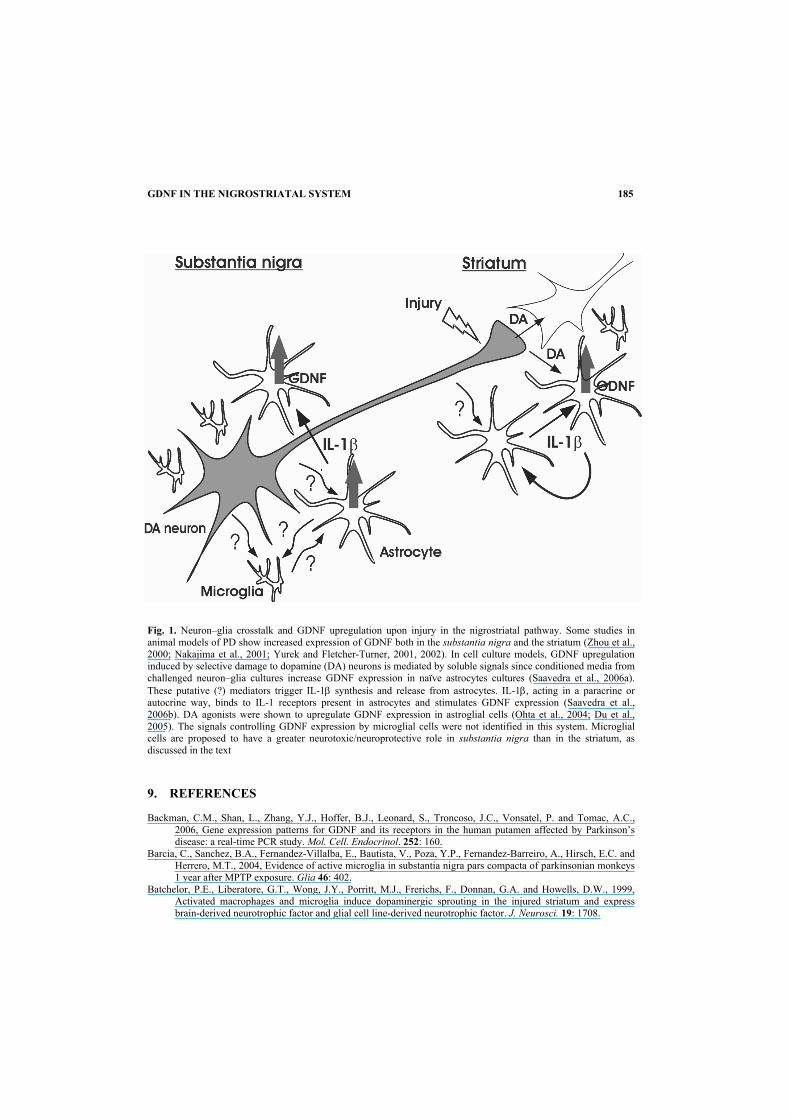
Fig. 1. Neuron–glia crosstalk and GDNF upregulation upon injury in the nigrostriatal pathway. Some studies in animal models of PD show increased expression of GDNF both in the substantia nigra and the striatum (Zhou et al., 2000; Nakajima et al., 2001; Yurek and Fletcher-Turner, 2001, 2002). In cell culture models, GDNF upregulation induced by selective damage to dopamine (DA) neurons is mediated by soluble signals since conditioned media from challenged neuron–glia cultures increase GDNF expression in naïve astrocytes cultures (Saavedra et al., 2006a). These putative (?) mediators trigger IL-1β synthesis and release from astrocytes. IL-1β, acting in a paracrine or autocrine way, binds to IL-1 receptors present in astrocytes and stimulates GDNF expression (Saavedra et al., 2006b). DA agonists were shown to upregulate GDNF expression in astroglial cells (Ohta et al., 2004; Du et al., 2005). The signals controlling GDNF expression by microglial cells were not identified in this system. Microglial cells are proposed to have a greater neurotoxic/neuroprotective role in substantia nigra than in the striatum, as discussed in the text 9. REFERENCES Backman, C.M., Shan, L., Zhang, Y.J., Hoffer, B.J., Leonard, S., Troncoso, J.C., Vonsatel, P. and Tomac, A.C.,
2006, Gene expression patterns for GDNF and its receptors in the human putamen affected by Parkinson’s disease: a real-time PCR study. Mol. Cell. Endocrinol. 252: 160.
Barcia, C., Sanchez, B.A., Fernandez-Villalba, E., Bautista, V., Poza, Y.P., Fernandez-Barreiro, A., Hirsch, E.C. and Herrero, M.T., 2004, Evidence of active microglia in substantia nigra pars compacta of parkinsonian monkeys 1 year after MPTP exposure. Glia 46: 402.
Batchelor, P.E., Liberatore, G.T., Wong, J.Y., Porritt, M.J., Frerichs, F., Donnan, G.A. and Howells, D.W., 1999, Activated macrophages and microglia induce dopaminergic sprouting in the injured striatum and express brain-derived neurotrophic factor and glial cell line-derived neurotrophic factor. J. Neurosci. 19: 1708.
185
E.P. Duarte, A. Saavedra and G. Baltazar
Batchelor, P.E., Liberatore, G.T., Porritt, M.J., Donnan, G.A. and Howells, D.W., 2000, Inhibition of brain-derived neurotrophic factor and glial cell line-derived neurotrophic factor expression reduces dopaminergic sprouting in the injured striatum. Eur. J. Neurosci. 12: 3462.
Batchelor, P.E., Porritt, M.J., Martinello, P., Parish, C.L., Liberatore, G.T., Donnan, G.A. and Howells, D.W., 2002, Macrophages and microglia produce local trophic gradients that stimulate axonal sprouting toward but not beyond the wound edge. Mol. Cell Neurosci. 21: 436.
Bauer, M., Suppmann, S., Meyer, M., Hesslinger, C., Gasser, T., Widmer, H.R. and Ueffing, M., 2002, Glial cell line-derived neurotrophic factor up-regulates GTP-cyclohydrolase I activity and tetrahydrobiopterin levels in primary dopaminergic neurones. J. Neurochem. 82: 1300.
Benn, S.C. and Woolf, C.J., 2004, Adult neuron survival strategies – slamming on the brakes. Nat. Rev. Neurosci. 5: 686.
Bilang-Bleuel, A., Revah, F., Colin, P., Locquet, I., Robert, J.J., Mallet, J. and Horellou, P., 1997, Intrastriatal injection of an adenoviral vector expressing glial-cell-line-derived neurotrophic factor prevents dopaminergic neuron degeneration and behavioral impairment in a rat model of Parkinson disease. Proc. Natl. Acad. Sci. USA 94: 8818.
Bizon, J.L., Lauterborn, J.C. and Gall, C.M., 1999, Subpopulations of striatal interneurons can be distinguished on the basis of neurotrophic factor expression. J. Comp. Neurol. 408: 283.
Bjorklund, A., Kirik, D., Rosenblad, C., Georgievska, B., Lundberg, C. and Mandel, R.J., 2000, Towards a neuroprotective gene therapy for Parkinson’s disease: use of adenovirus, AAV and lentivirus vectors for gene transfer of GDNF to the nigrostriatal system in the rat Parkinson model. Brain Res. 886: 82.
Block, M.L. and Hong, J.S., 2005, Microglia and inflammation-mediated neurodegeneration: multiple triggers with a common mechanism. Prog. Neurobiol. 76: 77.
Blum, M. and Weickert, C.S., 1995, GDNF mRNA expression in normal postnatal development, aging, and in Weaver mutant mice. Neurobiol. Aging 16: 925.
Bourque, M.J. and Trudeau, L.E., 2000, GDNF enhances the synaptic efficacy of dopaminergic neurons in culture. Eur. J. Neurosci. 12: 3172.
Bowenkamp, K.E., Hoffman, A.F., Gerhardt, G.A., Henry, M.A., Biddle, P.T., Hoffer, B.J. and Granholm, A.C., 1995, Glial cell line-derived neurotrophic factor supports survival of injured midbrain dopaminergic neurons. J. Comp. Neurol. 355: 479.
Bowenkamp, K.E., David, D., Lapchak, P.L., Henry, M.A., Granholm, A.C., Hoffer, B.J. and Mahalik, T.J., 1996, 6-Hydroxydopamine induces the loss of the dopaminergic phenotype in substantia nigra neurons of the rat. A possible mechanism for restoration of the nigrostriatal circuit mediated by glial cell line-derived neurotrophic factor. Exp. Brain Res. 111: 1.
Bozzi, Y. and Borrelli, E., 1999, Absence of the dopamine D2 receptor leads to a decreased expression of GDNF and NT-4 mRNAs in restricted brain areas. Eur. J. Neurosci. 11: 1275.
Breidert, T., Callebert, J., Heneka, M.T., Landreth, G., Launay, J.M. and Hirsch, E.C., 2002, Protective action of the peroxisome proliferator-activated receptor-gamma agonist pioglitazone in a mouse model of Parkinson’s disease. J. Neurochem. 82: 615.
Bresjanac, M. and Antauer, G., 2000, Reactive astrocytes of the quinolinic acid-lesioned rat striatum express GFRalpha1 as well as GDNF in vivo. Exp. Neurol. 164: 53.
Brizard, M., Carcenac, C., Bemelmans, A.P., Feuerstein, C., Mallet, J. and Savasta, M., 2006, Functional reinnervation from remaining DA terminals induced by GDNF lentivirus in a rat model of early Parkinson’s disease. Neurobiol. Dis. 21: 90.
Burke, R.E., 2004, Ontogenic cell death in the nigrostriatal system. Cell Tissue Res. 318: 63. Burke, R.E., Antonelli, M. and Sulzer, D., 1998, Glial cell line-derived neurotrophic growth factor inhibits apoptotic
death of postnatal substantia nigra dopamine neurons in primary culture. J. Neurochem. 71: 517. Chang, Y.P., Fang, K.M., Lee, T.I. and Tzeng, S.F., 2006, Regulation of microglial activities by glial cell line
derived neurotrophic factor. J. Cell Biochem. 97: 501. Chao, C.C. and Lee, E.H., 1999, Neuroprotective mechanism of glial cell line-derived neurotrophic factor on
dopamine neurons: role of antioxidation. Neuropharmacology 38: 913. Chao, C.C., Ma, Y.L., Chu, K.Y. and Lee, E.H., 2003, Integrin alpha and NCAM mediate the effects of GDNF on
DA neuron survival, outgrowth, DA turnover and motor activity in rats. Neurobiol. Aging 24: 105. Chauhan, N.B., Siegel, G.J. and Lee, J.M., 2001, Depletion of glial cell line-derived neurotrophic factor in substantia
nigra neurons of Parkinson’s disease brain. J. Chem. Neuroanat. 21: 277. Chen, K., Gunter, K. and Maines, M.D., 2000, Neurons overexpressing heme oxygenase-1 resist oxidative stress-
mediated cell death. J. Neurochem. 75: 304. Cheng, H., Wu, J.P. and Tzeng, S.F. 2002. Neuroprotection of glial cell line-derived neurotrophic factor in damaged
spinal cords following contusive injury. J. Neurosci. Res. 69: 397. Cheng, H., Fu, Y.S. and Guo, J.W., 2004, Ability of GDNF to diminish free radical production leads to protection
against kainate-induced excitotoxicity in hippocampus. Hippocampus 14: 77.
186
GDNF IN THE NIGROSTRIATAL SYSTEM
Choi-Lundberg, D.L. and Bohn, M.C., 1995, Ontogeny and distribution of glial cell line-derived neurotrophic factor (GDNF) mRNA in rat. Brain Res. Dev. Brain Res. 85: 80.
Choi-Lundberg, D.L., Lin, Q., Chang, Y.N., Chiang, Y.L., Hay, C.M., Mohajeri, H., Davidson, B.L. and Bohn, M.C., 1997, Dopaminergic neurons protected from degeneration by GDNF gene therapy. Science 275: 838.
Choi-Lundberg, D.L., Lin, Q., Schallert, T., Crippens, D., Davidson, B.L., Chang, Y.N., Chiang, Y.L., Qian, J., Bardwaj, L. and Bohn, M.C., 1998, Behavioral and cellular protection of rat dopaminergic neurons by an adenoviral vector encoding glial cell line-derived neurotrophic factor. Exp. Neurol. 154: 261.
Clarkson, E.D., Zawada, W.M. and Freed, C.R., 1997, GDNF improves survival and reduces apoptosis in human embryonic dopaminergic neurons in vitro. Cell Tissue Res. 289: 207.
Collier, T.J., Dung, L.Z., Carvey, P.M., Fletcher-Turner, A., Yurek, D.M., Sladek, J.R., Jr. and Kordower, J.H., 2005, Striatal trophic factor activity in aging monkeys with unilateral MPTP-induced parkinsonism. Exp. Neurol. 191: S60.
Connor, B., Kozlowski, D.A., Schallert, T., Tillerson, J.L., Davidson, B.L. and Bohn, M.C., 1999, Differential effects of glial cell line-derived neurotrophic factor (GDNF) in the striatum and substantia nigra of the aged Parkinsonian rat. Gene Ther. 6: 1936.
Connor, B., Kozlowski, D.A., Unnerstall, J.R., Elsworth, J.D., Tillerson, J.L., Schallert, T. and Bohn, M.C., 2001, Glial cell line-derived neurotrophic factor (GDNF) gene delivery protects dopaminergic terminals from degeneration. Exp. Neurol. 169: 83.
D’Astous, M., Morissette, M. and Di Paolo, T., 2004, Effect of estrogen receptor agonists treatment in MPTP mice: evidence of neuroprotection by an ER alpha agonist. Neuropharmacology 47: 1180.
Dauer, W. and Przedborski, S., 2003, Parkinson’s disease: mechanisms and models. Neuron 39: 889. Dehmer, T., Lindenau, J., Haid, S., Dichgans, J. and Schulz, J.B., 2000, Deficiency of inducible nitric oxide synthase
protects against MPTP toxicity in vivo. J. Neurochem. 74: 2213. Deumens, R., Blokland, A. and Prickaerts, J., 2002, Modeling Parkinson’s disease in rats: an evaluation of 6-OHDA
lesions of the nigrostriatal pathway. Exp. Neurol. 175: 303. Ding, Y.M., Jaumotte, J.D., Signore, A.P. and Zigmond, M.J., 2004, Effects of 6-hydroxydopamine on primary
cultures of substantia nigra: specific damage to dopamine neurons and the impact of glial cell line-derived neurotrophic factor. J. Neurochem. 89: 776.
Dore, S., Takahashi, M., Ferris, C.D., Zakhary, R., Hester, L.D., Guastella, D. and Snyder, S.H., 1999, Bilirubin, formed by activation of heme oxygenase-2, protects neurons against oxidative stress injury. Proc. Natl. Acad. Sci. USA 96: 2445.
Du, F., Li, R., Huang, Y., Li, X. and Le, W., 2005, Dopamine D3 receptor-preferring agonists induce neurotrophic effects on mesencephalic dopamine neurons. Eur. J. Neurosci. 22: 2422.
Eslamboli, A., Georgievska, B., Ridley, R.M., Baker, H.F., Muzyczka, N., Burger, C., Mandel, R.J., Annett, L. and Kirik, D., 2005, Continuous low-level glial cell line-derived neurotrophic factor delivery using recombinant adeno-associated viral vectors provides neuroprotection and induces behavioral recovery in a primate model of Parkinson’s disease. J. Neurosci. 25: 769.
Gash, D.M., Zhang, Z., Ovadia, A., Cass, W.A., Yi, A., Simmerman, L., Russell, D., Martin, D., Lapchak, P.A., Collins, F., Hoffer, B.J. and Gerhardt, G.A., 1996, Functional recovery in parkinsonian monkeys treated with GDNF. Nature 380: 252.
Gash, D.M., Zhang, Z., Ai, Y., Grondin, R., Coffey, R. and Gerhardt, G.A., 2005, Trophic factor distribution predicts functional recovery in parkinsonian monkeys. Ann. Neurol. 58: 224.
Gerhardt, G.A., Cass, W.A., Huettl, P., Brock, S., Zhang, Z. and Gash, D.M., 1999, GDNF improves dopamine function in the substantia nigra but not the putamen of unilateral MPTP-lesioned rhesus monkeys. Brain Res. 817: 163.
Ghribi, O., Herman, M.M., Forbes, M.S., DeWitt, D.A. and Savory, J., 2001, GDNF protects against aluminum-induced apoptosis in rabbits by upregulating Bcl-2 and Bcl-XL and inhibiting mitochondrial Bax translocation. Neurobiol. Dis. 8: 764.
Gill, S.S., Patel, N.K., Hotton, G.R., O’Sullivan, K., McCarter, R., Bunnage, M., Brooks, D.J., Svendsen, C.N. and Heywood, P., 2003, Direct brain infusion of glial cell line-derived neurotrophic factor in Parkinson disease. Nat. Med. 9: 589.
Grondin, R., Zhang, Z., Yi, A., Cass, W.A., Maswood, N., Andersen, A.H., Elsberry, D.D., Klein, M.C., Gerhardt, G.A. and Gash, D.M., 2002, Chronic, controlled GDNF infusion promotes structural and functional recovery in advanced parkinsonian monkeys. Brain 125: 2191.
Grondin, R., Zhang, Z., Ai, Y., Gash, D.M. and Gerhardt, G.A., 2003, Intracranial delivery of proteins and peptides as a therapy for neurodegenerative diseases. Prog. Drug Res. 61: 101.
Grunblatt, E., Mandel, S., Maor, G. and Youdim, M.B., 2001, Gene expression analysis in N-methyl-4-phenyl-1,2,3,6-tetrahydropyridine mice model of Parkinson’s disease using cDNA microarray: effect of R-apomorphine. J. Neurochem. 78: 1.
187
E.P. Duarte, A. Saavedra and G. Baltazar
Guo, H., Tang, Z., Yu, Y., Xu, L., Jin, G. and Zhou, J., 2002,. Apomorphine induces trophic factors that support fetal rat mesencephalic dopaminergic neurons in cultures. Eur. J. Neurosci. 16: 1861.
Hashimoto, M., Nitta, A., Fukumitsu, H., Nomoto, H., Shen, L. and Furukawa, S., 2005a, Inflammation-induced GDNF improves locomotor function after spinal cord injury. Neuroreport 16: 99.
Hashimoto, M., Nitta, A., Fukumitsu, H., Nomoto, H., Shen, L. and Furukawa, S., 2005b, Involvement of glial cell line-derived neurotrophic factor in activation processes of rodent macrophages. J. Neurosci. Res. 79: 476.
He, Y., Appel, S. and Le, W., 2001, Minocycline inhibits microglial activation and protects nigral cells after 6-hydroxydopamine injection into mouse striatum. Brain Res. 909: 187.
Ho, A. and Blum, M., 1998, Induction of interleukin-1 associated with compensatory dopaminergic sprouting in the denervated striatum of young mice: model of aging and neurodegenerative disease. J. Neurosci. 18: 5614.
Hoffer, B.J., Hoffman, A., Bowenkamp, K., Huettl, P., Hudson, J., Martin, D., Lin, L.F. and Gerhardt, G.A., 1994, Glial cell line-derived neurotrophic factor reverses toxin-induced injury to midbrain dopaminergic neurons in vivo. Neurosci. Lett. 182: 107.
Hoffman, A.F., van Horne, C.G., Eken, S., Hoffer, B.J. and Gerhardt, G.A., 1997, In vivo microdialysis studies on somatodendritic dopamine release in the rat substantia nigra: effects of unilateral 6-OHDA lesions and GDNF. Exp. Neurol. 147: 130.
Honda, S., Nakajima, K., Nakamura, Y., Imai, Y. and Kohsaka, S., 1999, Rat primary cultured microglia express glial cell line-derived neurotrophic factor receptors. Neurosci. Lett. 275: 203.
Hughes, P.E., Alexi, T., Walton, M., Williams, C.E., Dragunow, M., Clark, R.G. and Gluckman, P.D., 1999, Activity and injury-dependent expression of inducible transcription factors, growth factors and apoptosis-related genes within the central nervous system. Prog. Neurobiol. 57: 421.
Hunot, S., Bernard, V., Faucheux, B., Boissiere, F., Leguern, E., Brana, C., Gautris, P.P., Guerin, J., Bloch, B., Agid, Y. and Hirsch, E.C., 1996, Glial cell line-derived neurotrophic factor (GDNF) gene expression in the human brain: a post mortem in situ hybridization study with special reference to Parkinson’s disease. J. Neural. Transm. 103: 1043.
Hurelbrink, C.B. and Barker, R.A., 2004, The potential of GDNF as a treatment for Parkinson’s disease. Exp. Neurol. 185: 1.
Hurley, S.D., O’Banion, M.K., Song, D.D., Arana, F.S., Olschowka, J.A. and Haber, S.N., 2003, Microglial response is poorly correlated with neurodegeneration following chronic, low-dose MPTP administration in monkeys. Exp. Neurol. 184: 659.
Ikeda, T., Koo, H., Xia, Y.X., Ikenoue, T. and Choi, B.H., 2002, Bimodal upregulation of glial cell line-derived neurotrophic factor (GDNF) in the neonatal rat brain following ischemic/hypoxic injury. Int. J. Dev. Neurosci. 20: 555.
Iida, M., Miyazaki, I., Tanaka, K., Kabuto, H., Iwata-Ichikawa, E. and Ogawa, N., 1999, Dopamine D2 receptor-mediated antioxidant and neuroprotective effects of ropinirole, a dopamine agonist. Brain Res. 838: 51.
Inoue, T., Tsui, J., Wong, N., Wong, S.Y., Suzuki, F. and Kwok, Y.N., 1999, Expression of glial cell line-derived neurotrophic factor and its mRNA in the nigrostriatal pathway following MPTP treatment. Brain Res. 826: 306.
Ivanova, T., Karolczak, M. and Beyer, C., 2002a, Estradiol stimulates GDNF expression in developing hypothalamic neurons. Endocrinology 143: 3175.
Ivanova, T., Mendez, P., Garcia-Segura, L.M. and Beyer, C. (2002b). Rapid stimulation of the PI3-kinase/Akt signalling pathway in developing midbrain neurones by oestrogen. J. Neuroendocrinol. 14: 73.
Kim, W.G., Mohney, R.P., Wilson, B., Jeohn, G.H., Liu, B. and Hong, J.S., 2000, Regional difference in susceptibility to lipopolysaccharide-induced neurotoxicity in the rat brain: role of microglia. J. Neurosci. 20: 6309.
Kirik, D., Rosenblad, C. and Bjorklund, A., 2000a, Preservation of a functional nigrostriatal dopamine pathway by GDNF in the intrastriatal 6-OHDA lesion model depends on the site of administration of the trophic factor. Eur. J. Neurosci. 12: 3871.
Kirik, D., Rosenblad, C., Bjorklund, A. and Mandel, R.J., 2000b, Long-term rAAV-mediated gene transfer of GDNF in the rat Parkinson’s model: intrastriatal but not intranigral transduction promotes functional regeneration in the lesioned nigrostriatal system. J. Neurosci. 20: 4686.
Kobori, N., Waymire, J.C., Haycock, J.W., Clifton, G.L. and Dash, P.K., 2004, Enhancement of tyrosine hydroxylase phosphorylation and activity by glial cell line-derived neurotrophic factor. J. Biol. Chem. 279: 2182.
Kordower, J.H., Emborg, M.E., Bloch, J., Ma, S.Y., Chu, Y., Leventhal, L., McBride, J., Chen, E.Y., Palfi, S., Roitberg, B.Z., Brown, W.D., Holden, J.E., Pyzalski, R., Taylor, M.D., Carvey, P., Ling, Z., Trono, D., Hantraye, P., Deglon, N. and Aebischer, P., 2000, Neurodegeneration prevented by lentiviral vector delivery of GDNF in primate models of Parkinson’s disease. Science 290: 767.
Kozlowski, D.A., Connor, B., Tillerson, J.L., Schallert, T. and Bohn, M.C., 2000, Delivery of a GDNF gene into the substantia nigra after a progressive 6-OHDA lesion maintains functional nigrostriatal connections. Exp. Neurol. 166: 1.
188
GDNF IN THE NIGROSTRIATAL SYSTEM
Kozlowski, D.A., Miljan, E.A., Bremer, E.G., Harrod, C.G., Gerin, C., Connor, B., George, D., Larson, B. and Bohn, M.C., 2004, Quantitative analyses of GFRalpha-1 and GFRalpha-2 mRNAs and tyrosine hydroxylase protein in the nigrostriatal system reveal bilateral compensatory changes following unilateral 6-OHDA lesions in the rat. Brain Res. 1016: 170.
Lang, A.E., Gill, S., Patel, N.K., Lozano, A., Nutt, J.G., Penn, R., Brooks, D.J., Hotton, G., Moro, E., Heywood, P., Brodsky, M.A., Burchiel, K., Kelly, P., Dalvi, A., Scott, B., Stacy, M., Turner, D., Wooten, V.G., Elias, W.J., Laws, E.R., Dhawan,V., Stoessl, A.J., Matcham, J., Coffey, R.J. and Traub, M., 2006, Randomized controlled trial of intraputamenal glial cell line-derived neurotrophic factor infusion in Parkinson disease. Ann. Neurol. 59: 459.
Langston, J.W., Forno, L.S., Tetrud, J., Reeves, A.G., Kaplan, J.A. and Karluk, D., 1999, Evidence of active nerve cell degeneration in the substantia nigra of humans years after 1-methyl-4-phenyl-1,2,3,6-tetrahydropyridine exposure. Ann. Neurol. 46: 598.
Lapchak, P.A., Araujo, D.M., Hilt, D.C., Sheng, J. and Jiao, S., 1997, Adenoviral vector-mediated GDNF gene therapy in a rodent lesion model of late stage Parkinson’s disease. Brain Res. 777: 153.
Lawson, L.J., Perry, V.H., Dri, P. and Gordon, S., 1990, Heterogeneity in the distribution and morphology of microglia in the normal adult mouse brain. Neuroscience 39: 151.
Le, W.D., Xie, W.J. and Appel, S.H., 1999, Protective role of heme oxygenase-1 in oxidative stress-induced neuronal injury. J. Neurosci. Res. 56: 652.
Ledda, F., Paratcha, G. and Ibanez, C.F., 2002, Target-derived GFRalpha1 as an attractive guidance signal for developing sensory and sympathetic axons via activation of Cdk5. Neuron 36: 387.
Liberatore, G.T., Wong, J.Y., Porritt, M.J., Donnan, G.A. and Howells, D.W., 1997, Expression of glial cell line-derived neurotrophic factor (GDNF) mRNA following mechanical injury to mouse striatum. Neuroreport 8: 3097.
Liberatore, G.T., Jackson-Lewis, V., Vukosavic, S., Mandir, A.S., Vila, M., McAuliffe, W.G., Dawson, V.L., Dawson, T.M. and Przedborski, S., 1999, Inducible nitric oxide synthase stimulates dopaminergic neuro-degeneration in the MPTP model of Parkinson disease. Nat. Med. 5: 1403.
Liberto, C.M., Albrecht, P.J., Herx, L.M., Yong, V.W. and Levison, S.W., 2004, Pro-regenerative properties of cytokine-activated astrocytes. J. Neurochem. 89: 1092.
Lin, L.F., Doherty, D.H., Lile, J.D., Bektesh, S. and Collins, F., 1993, GDNF: a glial cell line-derived neurotrophic factor for midbrain dopaminergic neurons. Science 260: 1130.
Lin, L.F., Zhang, T.J., Collins, F. and Armes, L.G., 1994, Purification and initial characterization of rat B49 glial cell line-derived neurotrophic factor. J. Neurochem. 63: 758.
Love, S., Plaha, P., Patel, N.K., Hotton, G.R., Brooks, D.J. and Gill, S.S., 2005, Glial cell line-derived neurotrophic factor induces neuronal sprouting in human brain. Nat. Med. 11: 703.
Mandel, R.J., Spratt, S.K., Snyder, R.O. and Leff, S.E., 1997, Midbrain injection of recombinant adeno-associated virus encoding rat glial cell line-derived neurotrophic factor protects nigral neurons in a progressive 6-hydroxydopamine-induced degeneration model of Parkinson’s disease in rats. Proc. Natl. Acad. Sci. USA 94: 14083.
Mandel, R.J., Snyder, R.O. and Leff, S.E., 1999, Recombinant adeno-associated viral vector-mediated glial cell line-derived neurotrophic factor gene transfer protects nigral dopamine neurons after onset of progressive degeneration in a rat model of Parkinson’s disease. Exp. Neurol. 160: 205.
Mandel, S., Grunblatt, E., Maor, G. and Youdim, M.B., 2002, Early and late gene changes in MPTP mice model of Parkinson’s disease employing cDNA microarray. Neurochem. Res. 27: 1231.
Marco, S., Saura, J., Perez-Navarro, E., Jose, M.M., Tolosa, E. and Alberch, J., 2002, Regulation of c-Ret, GFRalpha1, and GFRalpha2 in the substantia nigra pars compacta in a rat model of Parkinson’s disease. J. Neurobiol. 52: 343.
Martino, G., 2004, How the brain repairs itself: new therapeutic strategies in inflammatory and degenerative CNS disorders. Lancet Neurol. 3: 372.
McGeer, P.L., Itagaki, S., Boyes, B.E. and McGeer, E.G., 1988, Reactive microglia are positive for HLA-DR in the substantia nigra of Parkinson’s and Alzheimer’s disease brains. Neurology 38: 1285.
McGeer, P.L., Schwab, C., Parent, A. and Doudet, D., 2003, Presence of reactive microglia in monkey substantia nigra years after 1-methyl-4-phenyl-1,2,3,6-tetrahydropyridine administration. Ann. Neurol. 54: 599.
Mirza, B., Hadberg, H., Thomsen, P. and Moos, T., 2000, The absence of reactive astrocytosis is indicative of a unique inflammatory process in Parkinson’s disease. Neuroscience 95: 425.
Miyazaki, H., Nagashima, K., Okuma, Y. and Nomura, Y., 2001, Expression of glial cell line-derived neurotrophic factor induced by transient forebrain ischemia in rats. Brain Res. 922: 165.
Mogi, M., Togari, A., Kondo, T., Mizuno, Y., Kogure, O., Kuno, S., Ichinose, H. and Nagatsu, T., 2001, Glial cell line-derived neurotrophic factor in the substantia nigra from control and parkinsonian brains. Neurosci. Lett. 300: 179.
189
E.P. Duarte, A. Saavedra and G. Baltazar
Morale, M.C., Serra, P.A., L’Episcopo, F., Tirolo, C., Caniglia, S., Testa, N., Gennuso, F., Giaquinta, G., Rocchitta, G., Desole, M.S., Miele, E. and Marchetti, B., 2006, Estrogen, neuroinflammation and neuroprotection in Parkinson’s disease: glia dictates resistance versus vulnerability to neurodegeneration. Neuroscience 138: 869.
Murray, H.E., Pillai, A.V., McArthur, S.R., Razvi, N., Datla, K.P., Dexter, D.T. and Gillies, G.E., 2003, Dose- and sex-dependent effects of the neurotoxin 6-hydroxydopamine on the nigrostriatal dopaminergic pathway of adult rats: differential actions of estrogen in males and females. Neuroscience 116: 213.
Nakagawa, T. and Schwartz, J.P., 2004a, Gene expression patterns in in vivo normal adult astrocytes compared with cultured neonatal and normal adult astrocytes. Neurochem. Int. 45: 203.
Nakagawa, T. and Schwartz, J.P., 2004b, Gene expression profiles of reactive astrocytes in dopamine-depleted striatum. Brain Pathol. 14: 275.
Nakagawa, T., Yabe, T. and Schwartz, J.P., 2005, Gene expression profiles of reactive astrocytes cultured from dopamine-depleted striatum. Neurobiol. Dis. 20: 275.
Nakajima, K., Kikuchi, Y., Ikoma, E., Honda, S., Ishikawa, M., Liu, Y. and Kohsaka, S., 1998, Neurotrophins regulate the function of cultured microglia. Glia 24: 272.
Nakajima, K., Hida, H., Shimano, Y., Fujimoto, I., Hashitani, T., Kumazaki, M., Sakurai, T. and Nishino, H., 2001, GDNF is a major component of trophic activity in DA-depleted striatum for survival and neurite extension of DAergic neurons. Brain Res. 916: 76.
Ohta, K., Fujinami, A., Kuno, S., Sakakimoto, A., Matsui, H., Kawahara, Y. and Ohta, M., 2004, Cabergoline stimulates synthesis and secretion of nerve growth factor, brain-derived neurotrophic factor and glial cell line-derived neurotrophic factor by mouse astrocytes in primary culture. Pharmacology 71: 162.
Onyango, I.G., Tuttle, J.B. and Bennett, J.P., Jr., 2005, Brain-derived growth factor and glial cell line-derived growth factor use distinct intracellular signaling pathways to protect PD cybrids from H2O2-induced neuronal death. Neurobiol. Dis. 20: 141.
Oo, T.F., Ries, V., Cho, J., Kholodilov, N. and Burke, R.E., 2005, Anatomical basis of glial cell line-derived neurotrophic factor expression in the striatum and related basal ganglia during postnatal development of the rat. J. Comp. Neurol. 484: 57.
Oppenheim, R.W., 1991, Cell death during development of the nervous system. Ann. Rev. Neurosci. 14: 453. Paratcha, G., Ledda, F. and Ibanez, C.F., 2003, The neural cell adhesion molecule NCAM is an alternative signaling
receptor for GDNF family ligands. Cell 113: 867. Paratcha, G., Ibanez, C.F. and Ledda, F., 2006, GDNF is a chemoattractant factor for neuronal precursor cells in the
rostral migratory stream. Mol. Cell Neurosci. 31: 505. Parish, C.L., Finkelstein, D.I., Tripanichkul, W., Satoskar, A.R., Drago, J. and Horne, M.K., 2002, The role of
interleukin-1, interleukin-6, and glia in inducing growth of neuronal terminal arbors in mice. J. Neurosci. 22: 8034.
Patel, N.K., Bunnage, M., Plaha, P., Svendsen, C.N., Heywood, P. and Gill, S.S., 2005, Intraputamenal infusion of glial cell line-derived neurotrophic factor in PD: a two-year outcome study. Ann. Neurol. 57: 298.
Perrelet, D., Ferri, A., Liston, P., Muzzin, P., Korneluk, R.G. and Kato, A.C., 2002, IAPs are essential for GDNF-mediated neuroprotective effects in injured motor neurons in vivo. Nat. Cell Biol. 4: 175.
Platania, P., Seminara, G., Aronica, E., Troost, D., Catania, V. and Sortino, A., 2005, 17beta-estradiol rescues spinal motoneurons from AMPA-induced toxicity: a role for glial cells. Neurobiol. Dis. 20: 461.
Pochon, N.A., Menoud, A., Tseng, J.L., Zurn, A.D. and Aebischer, P., 1997, Neuronal GDNF expression in the adult rat nervous system identified by in situ hybridization. Eur. J. Neurosci. 9: 463.
Pothos, E.N., Davila, V. and Sulzer, D., 1998, Presynaptic recording of quanta from midbrain dopamine neurons and modulation of the quantal size. J. Neurosci. 18: 4106.
Presgraves, S.P., Borwege, S., Millan, M.J. and Joyce, J.N., 2004, Involvement of dopamine D(2)/D(3) receptors and BDNF in the neuroprotective effects of S32504 and pramipexole against 1-methyl-4-phenylpyridinium in terminally differentiated SH-SY5Y cells. Exp. Neurol. 190: 157.
Purves, D., 1986, The trophic theory of neural connections. Trends Neurosci. 9: 486. Rosenblad, C., Martinez-Serrano, A. and Bjorklund, A., 1998, Intrastriatal glial cell line-derived neurotrophic factor
promotes sprouting of spared nigrostriatal dopaminergic afferents and induces recovery of function in a rat model of Parkinson’s disease. Neuroscience 82: 129.
Saavedra, A., Baltazar, G., Carvalho, C.M. and Duarte, E.P., 2005. GDNF modulates HO-1 expression in substantia nigra postnatal cell cultures. Free Radic. Biol. Med. 39: 1611
Saavedra, A., Baltazar, G., Santos, P., Carvalho, C. and Duarte, E.P., 2006a, Selective injury to dopaminergic neurons up-regulates GDNF in substantia nigra postnatal cell cultures: role of neuron-glia crosstalk. Neurobiol. Dis. 23: 533.
Saavedra, A., Baltazar, G. and Duarte, E.P., 2006b, Interleukin-1β mediates GDNF up-regulation upon dopaminergic injury in substantia nigra cell cultures. Neurobiol. Dis. DOI 10.1016/j.nbd.2006.08.019.
190
GDNF IN THE NIGROSTRIATAL SYSTEM
Salimi, K., Moser, K.V., Marksteiner, J., Reindl, M. and Humpel, C., 2003, GDNF and TGF-beta1 promote cell survival in serum-free cultures of primary rat microglia. Cell Tissue Res. 312: 135.
Salvatore, M.F., Zhang, J.L., Large, D.M., Wilson, P.E., Gash, C.R., Thomas, T.C., Haycock, J.W., Bing, G., Stanford, J.A., Gash, D.M. and Gerhardt, G.A., 2004, Striatal GDNF administration increases tyrosine hydroxylase phosphorylation in the rat striatum and substantia nigra. J. Neurochem. 90: 245.
Sarabi, A., Hoffer, B.J., Olson, L. and Morales, M., 2001, GFRalpha-1 mRNA in dopaminergic and nondopaminergic neurons in the substantia nigra and ventral tegmental area. J. Comp. Neurol. 441: 106.
Sariola, H. and Saarma, M., 2003, Novel functions and signalling pathways for GDNF. J. Cell Sci. 116: 3855. Satake, K., Matsuyama, Y., Kamiya, M., Kawakami, H., Iwata, H., Adachi, K. and Kiuchi, K., 2000, Up-regulation
of glial cell line-derived neurotrophic factor (GDNF) following traumatic spinal cord injur. Neuroreport 11: 3877.
Saura, J., Pares, M., Bove, J., Pezzi, S., Alberch, J., Marin, C., Tolosa, E. and Marti, M.J., 2003, Intranigral infusion of interleukin-1beta activates astrocytes and protects from subsequent 6-hydroxydopamine neurotoxicity. J. Neurochem. 85: 651.
Sawada, H., Ibi, M., Kihara, T., Urushitani, M., Akaike, A. and Shimohama, S., 1998, Estradiol protects mesencephalic dopaminergic neurons from oxidative stress-induced neuronal death. J. Neurosci. Res. 54: 707.
Sawada, H., Ibi, M., Kihara, T., Urushitani, M., Nakanishi, M., Akaike, A. and Shimohama, S., 2000, Neuroprotective mechanism of glial cell line-derived neurotrophic factor in mesencephalic neurons. J. Neurochem. 74: 1175
Sherer, T.B., Fiske, B.K., Svendsen, C.N., Lang, A.E. and Langston, J.W., 2006, Crossroads in GDNF therapy for Parkinson’s disease. Mov. Disord. 21: 136.
Siegel, G.J. and Chauhan, N.B., 2000, Neurotrophic factors in Alzheimer’s and Parkinson’s disease brain. Brain Res. Brain Res. Rev. 33: 199.
Slevin, J.T., Gerhardt, G.A., Smith, C.D., Gash, D.M., Kryscio, R. and Young, B., 2005, Improvement of bilateral motor functions in patients with Parkinson disease through the unilateral intraputaminal infusion of glial cell line-derived neurotrophic factor. J. Neurosurg. 102: 216.
Smith, A.D., Antion, M., Zigmond, M.J. and Austin, M.C., 2003, Effect of 6-hydroxydopamine on striatal GDNF and nigral GFRalpha1 and RET mRNAs in the adult rat. Brain Res. Mol. Brain Res. 117: 129.
Smith, A.D., Kozlowski, D.A., Bohn, M.C. and Zigmond, M.J., 2005, Effect of AdGDNF on dopaminergic neurotransmission in the striatum of 6-OHDA-treated rats. Exp. Neurol. 193: 420.
Stanic, D., Finkelstein, D.I., Bourke, D.W., Drago, J. and Horne, M.K., 2003, Timecourse of striatal re-innervation following lesions of dopaminergic SNpc neurons of the rat. Eur. J. Neurosci. 18: 1175.
Stanic, D., Tripanichkul, W., Drago, J., Finkelstein, D.I. and Horne, M.K., 2004, Glial responses associated with dopaminergic striatal reinnervation following lesions of the rat substantia nigra. Brain Res. 1023: 83.
Stromberg, I., Bjorklund, L., Johansson, M., Tomac, A., Collins, F., Olson, L., Hoffer, B. and Humpel, C., 1993, Glial cell line-derived neurotrophic factor is expressed in the developing but not adult striatum and stimulates developing dopamine neurons in vivo. Exp. Neurol. 124: 401.
Suttner, D.M. and Dennery, P.A., 1999, Reversal of HO-1 related cytoprotection with increased expression is due to reactive iron. FASEB J. 13: 1800.
Tang, Y.P., Ma, Y.L., Chao, C.C., Chen, K.Y. and Lee, E.H., 1998, Enhanced glial cell line-derived neurotrophic factor mRNA expression upon (–)-deprenyl and melatonin treatments. J. Neurosci. Res. 53: 593.
Tomac, A., Lindqvist, E., Lin, L.F., Ogren, S.O., Young, D., Hoffer, B.J. and Olson, L., 1995a, Protection and repair of the nigrostriatal dopaminergic system by GDNF in vivo. Nature 373: 335.
Tomac, A., Widenfalk, J., Lin, L.F., Kohno, T., Ebendal, T., Hoffer, B.J. and Olson, L., 1995b, Retrograde axonal transport of glial cell line-derived neurotrophic factor in the adult nigrostriatal system suggests a trophic role in the adult. Proc. Natl. Acad. Sci. USA 92: 8274.
Treanor, J., Goodman, L., Desauvage, F., Stone, D.M., Poulsen, K.T., Beck, C.D., Gray, C., Armanini, M.P., Pollock, R.A., Hefti, F., Phillips, H.S., Goddard, A., Moore, M.W., Bujbello, A., Davies, A.M., Asai, N., Takahashi, M., Vandlen, R., Henderson, C.E. and Rosenthal, A., 1996, Characterization of a multicomponent receptor for GDNF. Nature 382: 80.
Tripanichkul, W., Sripanichkulchai, K. and Finkelstein, D.I., 2006, Estrogen down-regulates glial activation in male mice following 1-methyl-4-phenyl-1,2,3,6-tetrahydropyridine intoxication. Brain Res. 108: 28.
Trupp, M., Arenas, E., Fainzilber, M., Nilsson, A.S., Sieber, B.A., Grigoriou, M., Kilkenny, C., Salazar-Grueso, E., Pachnis, V. and Arumae, U., 1996, Functional receptor for GDNF encoded by the c-ret proto-oncogene. Nature 381: 785.
Trupp, M., Belluardo, N., Funakoshi, H. and Ibanez, C.F., 1997, Complementary and overlapping expression of glial cell line-derived neurotrophic factor (GDNF), c-ret proto-oncogene, and GDNF receptor-alpha indicates multiple mechanisms of trophic actions in the adult rat CNS. J. Neurosci. 17: 3554.
191
E.P. Duarte, A. Saavedra and G. Baltazar
Vegeto, E., Bonincontro, C., Pollio, G., Sala, A., Viappiani, S., Nardi, F., Brusadelli, A., Viviani, B., Ciana, P. and Maggi, A., 2001, Estrogen prevents the lipopolysaccharide-induced inflammatory response in microglia. J. Neurosci. 21: 1809.
Walker, D.G., Beach, T.G., Xu, R., Lile, J., Beck, K.D., McGeer, E.G. and McGeer, P.L., 1998, Expression of the proto-oncogene Ret, a component of the GDNF receptor complex, persists in human substantia nigra neurons in Parkinson’s disease. Brain Res. 792: 207.
Wang, L., Muramatsu, S., Lu, Y., Ikeguchi, K., Fujimoto, K., Okada, T, Mizukami, H., Hanazono, Y., Kume, A., Urano, F, Ichinose, H., Nagatsu, T., Nakano, I. and Ozawa, K., 2002, Delayed delivery of AAV-GDNF prevents nigral neurodegeneration and promotes functional recovery in a rat model of Parkinson’s disease. Gene Ther. 9: 381.
Wang, J., Chen, G., Lu, B. and Wu, C.P., 2003, GDNF acutely potentiates Ca2+ channels and excitatory synaptic transmission in midbrain dopaminergic neurons. Neurosignals 12: 78.
Wei, G., Wu, G. and Cao, X., 2000, Dynamic expression of glial cell line-derived neurotrophic factor after cerebral ischemia. Neuroreport 11: 1177.
Widenfalk, J., Lundstromer, K., Jubran, M., Brene, S. and Olson, L., 2001, Neurotrophic factors and receptors in the immature and adult spinal cord after mechanical injury or kainic acid. J. Neurosci. 21: 3457.
Wu, D.C., Jackson-Lewis, V., Vila, M., Tieu, K., Teismann, P., Vadseth, C., Choi, D.K., Ischiropoulos, H. and Przedborski, S., 2002, Blockade of microglial activation is neuroprotective in the 1-methyl-4-phenyl-1,2,3, 6-tetrahydropyridine mouse model of Parkinson disease. J. Neurosci. 22: 1763.
Yang, F., Feng, L., Zheng, F., Johnson, S.W., Du, J., Shen, L., Wu, C.P. and Lu, B., 2001, GDNF acutely modulates excitability and A-type K+ channels in midbrain dopaminergic neurons. Nat. Neurosci. 4: 1071.
Yoo, M.S., Chun, H.S., Son, J.J., DeGiorgio, L.A., Kim, D.J., Peng, C. and Son, J.H., 2003, Oxidative stress regulated genes in nigral dopaminergic neuronal cells: correlation with the known pathology in Parkinson’s disease. Brain Res. Mol. Brain Res. 110: 76.
Yurek, D.M. and Fletcher-Turner, A., 2001, Differential expression of GDNF, BDNF, and NT-3 in the aging nigrostriatal system following a neurotoxic lesion. Brain Res. 891: 228.
Yurek, D.M. and Fletcher-Turner, A., 2002, Temporal changes in the neurotrophic environment of the denervated striatum as determined by the survival and outgrowth of grafted fetal dopamine neurons. Brain Res. 931: 126.
Zheng, J.S., Tang, L.L., Zheng, S.S., Zhan, R.Y., Zhou ,Y.Q., Goudreau, J., Kaufman, D. and Chen, A.F., 2005, Delayed gene therapy of glial cell line-derived neurotrophic factor is efficacious in a rat model of Parkinson’s disease. Mol. Brain Res. 134: 155.
Zhou, J., Yu, Y., Tang, Z., Shen, Y. and Xu, L., 2000, Differential expression of mRNAs of GDNF family in the striatum following 6-OHDA-induced lesion. Neuroreport 11: 3289.
192