dpp and hedgehog mediate neuron–glia interactions in drosophila eye development by promoting the...
TRANSCRIPT
Dpp and Hedgehog mediate neuron–glia interactions in Drosophila eyedevelopment by promoting the proliferation and motility of subretinal glia
Radha Rangarajan1, Helene Courvoisier1, Ulrike Gaul*
Laboratory of Developmental Neurogenetics, Rockefeller University, 1230 York Avenue, New York, NY 10021, USA
Received 12 July 2001; accepted 18 July 2001
Abstract
Neuron–glia interactions are crucial for the establishment of normal connectivity in the nervous system during development, but the
molecular signals involved in these interactions are largely unknown. Here we show that differentiating photoreceptors in the developing
Drosophila eye influence the proliferative and migratory behavior of the subretinal glia through the diffusible factors Decapentaplegic (Dpp)
and Hedgehog (Hh). We demonstrate that proliferation and migration of the glia are separable processes, and that Dpp promotes both the
proliferation and motility of the glia, whereas Hh appears to promote only their motility; neither specifies the direction of migration. We
present evidence that Dpp and Hh act on the glia in parallel and through the regulation of transcription. Finally, we show that ectopic
migration of subretinal glia can result in the ectopic projection of photoreceptor axons. Our study suggests a novel function for Hh in
regulating migratory behavior and provides further evidence for a complex mutual dependence between glial and neuronal cells during
development. q 2001 Elsevier Science Ireland Ltd. All rights reserved.
Keywords: Dpp; Hedgehog; Subretinal glia; Glial migration; Visual system development; Drosophila
1. Introduction
Glial cells are major constituents of the nervous system
and play an important role during its development in both
vertebrates and invertebrates (reviewed by Auld, 1999;
Birling and Price, 1995; Silver, 1993; Tear, 1999; Tessier-
Lavigne and Goodman, 1996; Thomas, 1998). They have
been shown to help control the patterning of neuronal differ-
entiation, to provide guidance for migrating neurons and
growing axons, and to prompt fasciculation and defascicu-
lation of axons. Later on, glia are essential for wrapping and
insulating neurons, providing them with trophic and survi-
val factors, and effecting their homeostasis.
In order for glial cells to fulfill their role, they must be
matched in number and positioned correctly with respect to
the neurons they support. For many glial populations, this
means proliferation of a limited pool of precursor cells and
migration over many cell diameters within a specific time
period during development. In vertebrates, this is true for
the oligodendrocytes of the CNS, which arise from a prolif-
erating pool of motile precursor cells, as well as for the
peripheral glia, which originate from the neural crest
(Barres and Raff, 1999). In the Drosophila CNS, most
glial cells arise from a relatively small number of neuro-
glioblasts, then migrate and spread to reach their final desti-
nation (Hartenstein et al., 1998; Ito et al., 1995); most PNS
glia are similarly derived from proliferative pools of motile
precursor cells (Choi and Benzer, 1994; Giangrande et al.,
1993; Perez and Steller, 1996). The regulation of glial cell
proliferation and survival is well studied, in particular in the
vertebrate system (for review see Barres and Raff, 1999),
however, relatively little is known about the molecular
mechanisms involved in glial migration.
The present study investigates the proliferation and
migration of the subretinal glia in the developing Droso-
phila visual system. Subretinal glial precursors originate
in the optic stalk and migrate into the eye imaginal disc
concomitant with the onset of differentiation of photorecep-
tors (Choi and Benzer, 1994). Their presence in the eye disc,
in turn, is necessary for guiding photoreceptor axons from
the eye disc into the optic stalk (Rangarajan et al., 1999).
Here we examine the effects of the diffusible factors Deca-
pentaplegic (Dpp) and Hedgehog (Hh), which have an
important role in photoreceptor differentiation, on the devel-
opment of the glial precursor cells.
Dpp belongs to the TGF-b superfamily of ligands, which
include the bone morphogenetic proteins (BMPs), the
Mechanisms of Development 108 (2001) 93–103
0925-4773/01/$ - see front matter q 2001 Elsevier Science Ireland Ltd. All rights reserved.
PII: S0925-4773(01)00501-9
www.elsevier.com/locate/modo
* Corresponding author. Tel.: 11-212-327 7621; fax: 11-212-327 7923.
E-mail address: [email protected] (U. Gaul).1 These authors contributed equally to this work.
growth and differentiation factors (GDFs), and various
forms of transforming growth factor-b, Activin and Nodal
(for review see Massague, 1998; Massague and Chen,
2000). Produced by diverse cell types, these factors have
been shown to regulate a wide range of processes, including
cell migration, adhesion, proliferation, fate specification,
and survival, although effects on cell migration and adhe-
sion have mostly been studied in culture. In Drosophila,
Dpp regulates the growth and patterning of the imaginal
discs (Burke and Basler, 1996; Capdevila and Guerrero,
1994; Heberlein et al., 1993). In the eye disc, it is one of
the key players in the initiation of the morphogenetic
furrow, which marks the propagating front of photoreceptor
differentiation (Greenwood and Struhl, 1999; Heberlein et
al., 1993).
Dpp transmits its signal by binding to Type I serine threo-
nine kinase receptors, Saxophone and Thick veins (Tkv),
and the Type II receptor Punt. Upon ligand binding, the
Type I receptors become phosphorylated by the Type II
receptor, resulting in the activation of the receptor I kinase.
This kinase then phosphorylates the Receptor-Smad Mad,
which in turn binds the Co-Smad, Medea. The Smad
complex translocates to the nucleus, where it activates target
gene expression (Massague and Chen, 2000).
The secreted protein Hedgehog and its vertebrate homo-
logs have been shown to regulate a wide range of develop-
mental processes, involving growth control, patterning, and
organ morphogenesis (for review see Ingham, 1998). In the
developing Drosophila eye, Hh controls the initiation and
propagation of the morphogenetic furrow, leading to photo-
receptor differentiation (for review see Treisman and Heber-
lein, 1998). In addition, Hh is transported along the
photoreceptor axons into the optic lobe, where it triggers
the proliferation and differentiation of neuronal precursor
cells that form the lamina (Huang and Kunes, 1996,
1998). In the vertebrate nervous system, Sonic Hedgehog
(Shh) acts as a morphogen in the specification of neuronal
cell fates in the ventral spinal cord (Briscoe and Ericson,
1999) and as a mitogen for a variety of cell types, including
astrocytes in the rodent optic nerve (Wallace and Raff,
1999). Effects of Hh on the migratory behavior of cells
have not been reported.
Hh transduces its signal by binding to its receptor Patched
(Ptc), which acts as a negative regulator of the seven-trans-
membrane protein Smoothened (Smo); binding of Ptc by Hh
removes this inhibitory effect and allows Smo to activate the
transcriptional effector Cubitus interruptus (Ci). In the
absence of Hh, Ci is proteolytically cleaved and acts as a
transcriptional repressor of Hh target genes. Smo activity
prevents the proteolysis of Ci, resulting in the accumulation
of full-length Ci, which acts as a transcriptional activator of
Hh target genes (for review see McMahon, 2000).
In this study, we show that Dpp and Hh, in addition to
controlling the propagation of photoreceptor differentiation
in the eye disc, also have a role in regulating glial migration
and proliferation. Both Dpp and Hh promote the overall
motility of the subretinal glia, while only Dpp stimulates
their proliferation. We provide evidence that both factors
exert their effects through transcriptional regulation. Our
findings identify Dpp and Hh as important factors in coor-
dinating neuronal and glial cell development during the
establishment of the Drosophila visual system.
2. Results
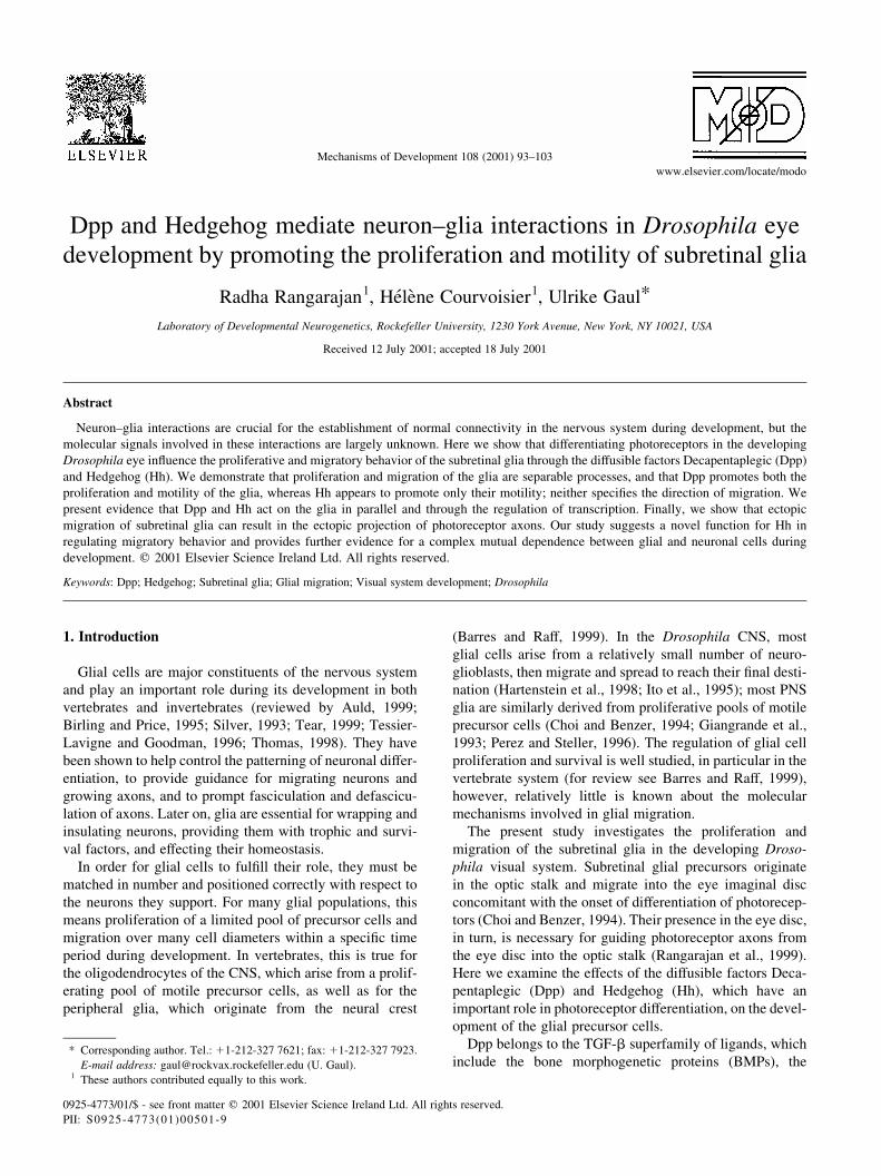
In the developing Drosophila visual system, the eye disc
is connected to the optic lobe by the optic stalk. Photore-
ceptor cells are generated in the eye disc in a posterior-to-
anterior progression; the wave of differentiation is marked at
its front by an indentation of the disc epithelium called the
morphogenetic furrow. The photoreceptors extend axons
into the basal layer of the eye disc, where they turn and
proceed posteriorly to exit the disc through the optic stalk
(Fig. 1B,C). The subretinal glia (also known as retinal basal
glia) originate from precursor cells in the optic stalk; the
glial precursors in the stalk already express the glial-specific
marker Repo (Halter et al., 1995). The glia begin to migrate
into the eye disc with the onset of the differentiation of
photoreceptors (Choi and Benzer, 1994), but do not require
the presence of axons in the stalk to find their way into the
eye disc (Rangarajan et al., 1999). Once in the eye disc, the
glia migrate anteriorly on the basal surface of the eye epithe-
lium (Fig. 1A); the anterior border of their migration lies
typically 2–4 rows of ommatidia posterior to the morpho-
genetic furrow, which is where axons sent out by the differ-
entiating photoreceptors begin to turn posteriorly (Fig.
1B,C). Thus, glia are present only in the axonal portion of
the eye disc. The glial cells associate closely with the photo-
receptor axons and develop extensive processes that
surround the axons and fill the intervening space (Choi
and Benzer, 1994). The development of the subretinal glia
thus involves proliferation, migration, and terminal differ-
entiation events, and it is closely tied, both spatially and
temporally, to the development of the photoreceptors.
2.1. Pattern of glial proliferation and terminal
differentiation
In order to get a better understanding of the sequence of
events in the development of the subretinal glia, we sought
to determine the pattern of glial proliferation and cell shape
change. We marked proliferating cells using antibodies
against phosphorylated histone H3 (Hendzel et al., 1997).
These antibodies recognize cells between late G2 and meta-
phase of the cell cycle and are reliable markers for mitosis.
We observe dividing glial cells in the optic stalks of early
and late third instar larval stages. Interestingly, we also find
dividing glial cells in the eye discs of these animals (Fig.
1D). The number of labeled cells is generally very small,
between one and ten, which is probably due to the relatively
small window of expression of phosphorylated histone H3
and a lack of synchrony in glial cell divisions. The cell
R. Rangarajan et al. / Mechanisms of Development 108 (2001) 93–10394
divisions show no consistent spatial distribution pattern.
These data show that the glial cell population continues to
proliferate throughout the third instar larval stage, both in
the optic stalk and after entering the eye disc. During this
expansion period, there is no significant amount of cell
death in the glial cell population, as determined by using
acridine orange and human anti-caspase antibodies as
markers (data not shown).
To relate the morphology of the subretinal glia to their
position within the eye disc, we generated small clones of
glial cells expressing GFP (see Section 4). We find that in
the anterior, all cells are small and rounded, whereas in the
posterior a range of cell morphologies coexist. In addition to
small cells with simple morphology, which may represent
migrating cells, we find large cells that are elongated to
varying degrees and show a complex morphology (Fig.
1E–J). This indicates that terminal differentiation of the
subretinal glia (e.g. wrapping of axons) does not wait until
the eye disc is completely developed and filled with glia, but
rather takes place concomitantly with the progression of the
morphogenetic furrow and while other glia are still migrat-
ing.
How are the proliferation and migration of the subretinal
glia regulated; and what is the molecular basis for the coor-
dination between photoreceptor and glial cell development?
In an effort to address these questions, we examined the role
of the diffusible factors Dpp and Hh.
2.2. Role of Dpp in subretinal glial development
2.2.1. Loss of Dpp signaling impairs the accumulation of
glia in the eye disc
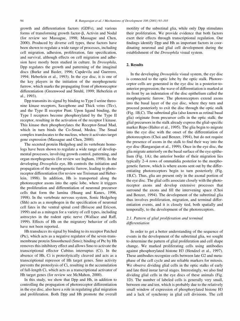
Dpp is an important factor in the initiation and propaga-
tion of the morphogenetic furrow. It is expressed in the
posterior and lateral margins of the eye disc in early third
instar animals (Fig. 2A) and in the morphogenetic furrow in
later stages (Fig. 2B) (Blackman et al., 1991; Heberlein et
al., 1993). It is not significantly expressed anywhere else in
the eye disc or in the glial cells, as confirmed by enhancer
trap expression analysis and RNA in situ hybridization (Fig.
2C).
Since removal of Dpp affects overall eye development,
we sought to use the FLP-FRT system (Duffy et al., 1998;
Xu and Rubin, 1993) to generate mosaic animals in which
mad, an essential component of the Dpp signaling pathway
(Wiersdorff et al., 1996), is removed from the glia, but not
from cells of the overlying photoreceptor epithelium. Since
the glial population is small compared to that of neuronal
cells, it is impossible to use hsFLP to generate glial clones
without also greatly affecting the other relevant tissues. We
therefore decided to specifically target the glial cells for
somatic recombination using ombGAL4 driving a UASFLP
transgene; ombGAL4 is expressed in the glia and in the
margins of the eye disc throughout larval development,
but not in the photoreceptors (see Section 4) (Brand and
Perrimon, 1993; Duffy et al., 1998; Lecuit et al., 1996;
Rangarajan et al., 1999). A UAS nuclear-GFP transgene
serves as a negative marker for the mutant glial population.
To determine the efficiency of recombination, we generated
clones with an FRT chromosome bearing no mutations and
counted the percentage of cells lacking GFP. The average
percentage of GFP-negative glial cells, i.e. cells having lost
the marker through recombination, in such eye discs is
35.2% (^2.7; n ¼ 29) (Fig. 2F), indicating that the effi-
ciency of recombination between the FRT chromosomes is
lower than the theoretical limit. Since glial cells are still able
to divide after arriving in the eye disc (see above), this
R. Rangarajan et al. / Mechanisms of Development 108 (2001) 93–103 95
Fig. 1. Organization and morphology of subretinal glia in third instar larval
eye discs. (A) Wild-type eye disc, showing the distribution of subretinal
glia nuclei, labeled with anti-Repo antibodies (red), in the basal layer of the
eye disc and in the optic stalk. (B) Same disc as in (A), with photoreceptor
cell bodies and axons labeled with anti-HRP antibodies (green) and glial
nuclei labeled with anti-Repo antibodies (red). (C) Schematic drawing
showing the progression of development along the anterior–posterior axis
and the relative positions of the photoreceptor cell bodies and axons (green)
and glia (red) along the apical-basal axis. (D) Eye disc labeled with anti-
pH3-antibodies (green) to reveal mitotic cells and with propidium iodide to
reveal all nuclei (red). Two glial cells in the basal layer of the eye disc are
undergoing mitosis (yellow, arrows); the other two mitotic cells are part of
the eye disc margin. (E,G–J) show the morphologies of individual glial cells
expressing cytoplasmic GFP (green) at different positions along the ante-
rior-posterior axis of the eye disc. The glial nuclei are labeled with anti-
Repo antibodies (red). In the anteriormost row of glia, the cells are small,
round, and have a simple morphology (G); more posteriorly, glial cells are
larger and show a more complex and elongated morphology (E,H,J).
However, cells of simple morphology (I) are also observed in the posterior.
(F) Schematic drawing showing the relative positions of the cells depicted
in (G–J) within the eye disc. In all figures except (C) posterior is down,
dorsal to the right; in (C), posterior is to the left. Scale bars in (B) for
(A,B,D): 30 mm; in (E) and (J) for (G–J): 20 mm.
suggests that at least a fraction of cells migrate into the eye
disc (and possibly reach the anterior boundary) as hetero-
zygotes before undergoing recombination to become homo-
zygous mutant.
In mad mosaic animals, the average percentage of GFP
negative glial cells in the eye disc is significantly lower than
for the wild type clones, at 17.2% (^1.8; n ¼ 29) (Fig.
2G,H). This indicates that the loss of mad function impairs
the accumulation of mutant glia in the eye disc. The total
number of glia in the eye disc, however, appears unchanged,
suggesting that wild-type glia are able to compensate for the
loss of mad mutant cells. The relative distribution of GFP
negative cells within the eye disc is also unaffected: mutant
cells are found at all positions within the differentiating
portion of the eye disc. When a dominant negative form
of the Dpp receptor Tkv is expressed in the entire glial
cell population, again using the UAS/GAL4 system and
ombGAL4 as a driver, the total number of glia in the eye
disc is reduced, but again without effect on their spatial
distribution (Fig. 2E).
These findings show that Dpp signaling is required for the
accumulation of glial cells in the eye disc, but, partly
because of the limitation in the mosaic technique, they do
not allow us to determine whether this results from an effect
on glial proliferation, survival, or on migration. The follow-
ing gain of function experiments address this question.
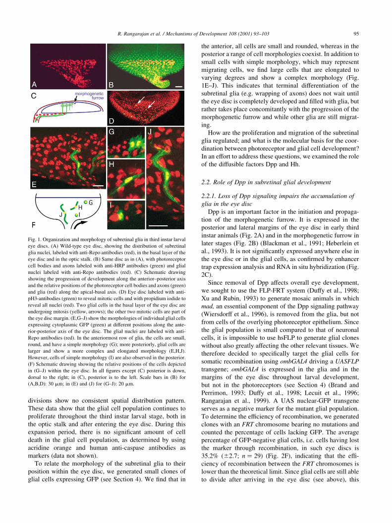
2.2.2. Dpp stimulates proliferation and motility of the
subretinal glia
We created different situations of overactive Dpp signal-
ing, by expressing Dpp in ectopic positions (either with the
GMRGAL4 driver or using Act . FLPout . GAL4) and by
expressing a constitutively active version of the Tkv recep-
tor in the glial cells (Das et al., 1998; Hay et al., 1994;
Nellen et al., 1996; Pignoni and Zipursky, 1997). In all
three situations, we find a pronounced increase in the
density of glial cells in the entire system, that is both in
the optic stalk and in the eye disc (Fig. 3B–E). Given that
there is no significant cell death in the subretinal glia at this
stage in wild type, this increase in cell number has to be the
R. Rangarajan et al. / Mechanisms of Development 108 (2001) 93–10396
Fig. 3. Increased Dpp signaling stimulates the accumulation of glia in the
eye disc. Third instar larval eye discs, glia are labeled with anti-Repo
antibodies (red). (A) Wild type. (B) y,hs FLP122/Act . FLPout . GAL4;
UAS dpp, containing random, unmarked clones of dpp expression. (C)
GMRGAL4; UAS dpp, with dpp expression in the differentiating portion
of the eye disc. (D) ombGAL4; UAS tkvact, with constitutive activation of
the Dpp pathway in all glial cells. (E) Histogram showing the average
number of glial cells per row of ommatidia (mean ^ SEM) in eye discs
of wild type, GMRGal4;UASdpp, ombGAL4;UAStkvact; GMRGal4;UAShh,
and ombGAL4;UASCiact, respectively. Sample sizes are shown in paren-
theses, *P , 0:01 as compared to wild type.
Fig. 2. Loss of Dpp signaling disrupts subretinal glial development. (A) In
early third instar larval eye discs, dpp-lacZ expression is visible in the
posterior and lateral margins. In late third instar larvae, dpp-lacZ (B) and
dpp RNA (C) are expressed in the morphogenetic furrow, with no expres-
sion in the glial layer of the eye disc. (D–G) late third instar larval eye discs,
glial nuclei labeled with anti-Repo antibodies (red). (D) wild type. (E)
ombGAL4; UAS tkvDGSK, showing a significant reduction in the number
of glial cells. (F) ombGAL4; FRT40 UAS GFP/FRT40; UAS FLP, having
undergone FRT-UAS FLP mediated mitotic recombination to generate
clones of glial cells that lack GFP but are otherwise wild type. Cells that
have not undergone mitotic recombination are GFP positive and appear
yellow, whereas those that have appear red. (G) ombGAL4; FRT40 UAS
GFP/FRT40mad12; UAS FLP. Glia lacking mad function are GFP-negative
(red). (H) Histogram showing the average percentage of GFP-minus cells
(mean ^ SEM) in eye discs when clones of wild type, mad12, smo3, and
mad12/smo3, respectively, are generated in the glia. Sample sizes are shown
in parentheses; (a) indicates P , 0:0001 as compared to wild type, (b)
indicates P , 0:01 as compared to the single mutant conditions. Scalebar
in (D) for (D–G): 30 mm.
result of increased proliferation, rather than of a suppression
of apoptosis.
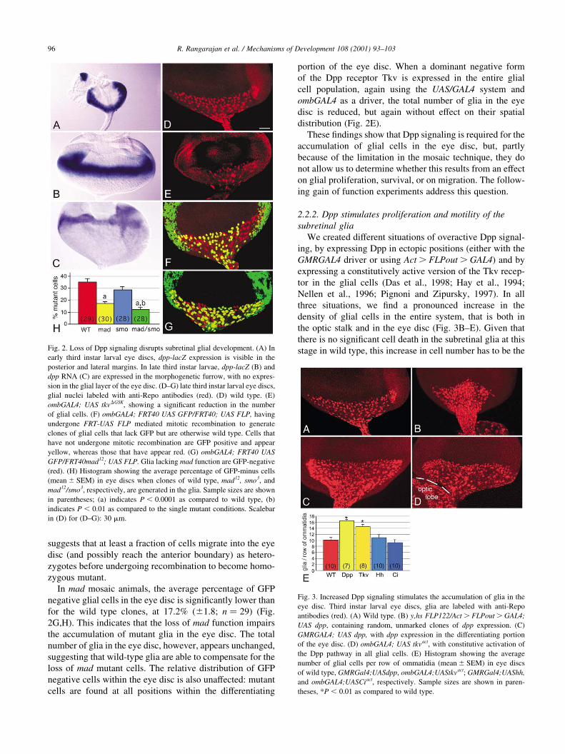
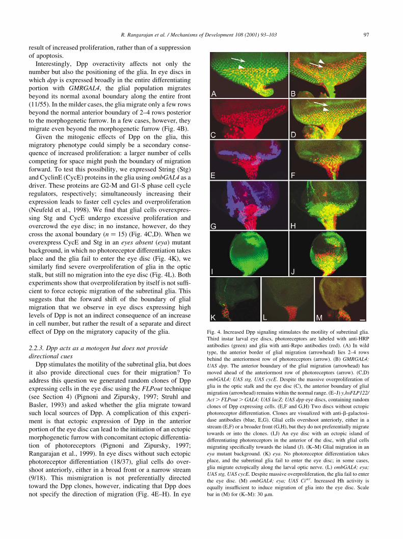
Interestingly, Dpp overactivity affects not only the
number but also the positioning of the glia. In eye discs in
which dpp is expressed broadly in the entire differentiating
portion with GMRGAL4, the glial population migrates
beyond its normal axonal boundary along the entire front
(11/55). In the milder cases, the glia migrate only a few rows
beyond the normal anterior boundary of 2–4 rows posterior
to the morphogenetic furrow. In a few cases, however, they
migrate even beyond the morphogenetic furrow (Fig. 4B).
Given the mitogenic effects of Dpp on the glia, this
migratory phenotype could simply be a secondary conse-
quence of increased proliferation: a larger number of cells
competing for space might push the boundary of migration
forward. To test this possibility, we expressed String (Stg)
and CyclinE (CycE) proteins in the glia using ombGAL4 as a
driver. These proteins are G2-M and G1-S phase cell cycle
regulators, respectively; simultaneously increasing their
expression leads to faster cell cycles and overproliferation
(Neufeld et al., 1998). We find that glial cells overexpres-
sing Stg and CycE undergo excessive proliferation and
overcrowd the eye disc; in no instance, however, do they
cross the axonal boundary (n ¼ 15) (Fig. 4C,D). When we
overexpress CycE and Stg in an eyes absent (eya) mutant
background, in which no photoreceptor differentiation takes
place and the glia fail to enter the eye disc (Fig. 4K), we
similarly find severe overproliferation of glia in the optic
stalk, but still no migration into the eye disc (Fig. 4L). Both
experiments show that overproliferation by itself is not suffi-
cient to force ectopic migration of the subretinal glia. This
suggests that the forward shift of the boundary of glial
migration that we observe in eye discs expressing high
levels of Dpp is not an indirect consequence of an increase
in cell number, but rather the result of a separate and direct
effect of Dpp on the migratory capacity of the glia.
2.2.3. Dpp acts as a motogen but does not provide
directional cues
Dpp stimulates the motility of the subretinal glia, but does
it also provide directional cues for their migration? To
address this question we generated random clones of Dpp
expressing cells in the eye disc using the FLPout technique
(see Section 4) (Pignoni and Zipursky, 1997; Struhl and
Basler, 1993) and asked whether the glia migrate toward
such local sources of Dpp. A complication of this experi-
ment is that ectopic expression of Dpp in the anterior
portion of the eye disc can lead to the initiation of an ectopic
morphogenetic furrow with concomitant ectopic differentia-
tion of photoreceptors (Pignoni and Zipursky, 1997;
Rangarajan et al., 1999). In eye discs without such ectopic
photoreceptor differentiation (18/37), glial cells do over-
shoot anteriorly, either in a broad front or a narrow stream
(9/18). This mismigration is not preferentially directed
toward the Dpp clones, however, indicating that Dpp does
not specify the direction of migration (Fig. 4E–H). In eye
R. Rangarajan et al. / Mechanisms of Development 108 (2001) 93–103 97
Fig. 4. Increased Dpp signaling stimulates the motility of subretinal glia.
Third instar larval eye discs, photoreceptors are labeled with anti-HRP
antibodies (green) and glia with anti-Repo antibodies (red). (A) In wild
type, the anterior border of glial migration (arrowhead) lies 2–4 rows
behind the anteriormost row of photoreceptors (arrow). (B) GMRGAL4;
UAS dpp. The anterior boundary of the glial migration (arrowhead) has
moved ahead of the anteriormost row of photoreceptors (arrow). (C,D)
ombGAL4; UAS stg, UAS cycE. Despite the massive overproliferation of
glia in the optic stalk and the eye disc (C), the anterior boundary of glial
migration (arrowhead) remains within the normal range. (E–J) y,hsFLP122/
Act . FLPout . GAL4; UAS lacZ; UAS dpp eye discs, containing random
clones of Dpp expressing cells. (E,F and G,H) Two discs without ectopic
photoreceptor differentiation. Clones are visualized with anti-b-galactosi-
dase antibodies (blue, E,G). Glial cells overshoot anteriorly, either in a
stream (E,F) or a broader front (G,H), but they do not preferentially migrate
towards or into the clones. (I,J) An eye disc with an ectopic island of
differentiating photoreceptors in the anterior of the disc, with glial cells
migrating specifically towards the island (J). (K–M) Glial migration in an
eya mutant background. (K) eya. No photoreceptor differentiation takes
place, and the subretinal glia fail to enter the eye disc; in some cases,
glia migrate ectopically along the larval optic nerve. (L) ombGAL4; eya;
UAS stg, UAS cycE. Despite massive overproliferation, the glia fail to enter
the eye disc. (M) ombGAL4; eya; UAS Ciact. Increased Hh activity is
equally insufficient to induce migration of glia into the eye disc. Scale
bar in (M) for (K–M): 30 mm.
discs forming ectopic patches of differentiating photorecep-
tors, on the other hand (19/37), glial cells migrate specifi-
cally to such patches (17/19) (Fig. 4I,J). Differentiating
photoreceptors thus attract the subretinal glia, but Dpp by
itself cannot mimic this effect. As is the case for the effects
on proliferation, the effects of Dpp on the migratory beha-
vior of the subretinal glia can be phenocopied by activation
of the Dpp pathway in the glia themselves: in eye discs in
which constitutively active Tkv is expressed in the glia with
the ombGAL4 driver, we also find anterior overshooting of
glia, albeit less pronounced (10/53) (data not shown).
In summary, our analysis of Dpp overactivity shows that
Dpp signaling promotes both the proliferation and the moti-
lity of the subretinal glia, but without specifying the direc-
tion of migration. The motogenic effect is separable from
the effect on proliferation.
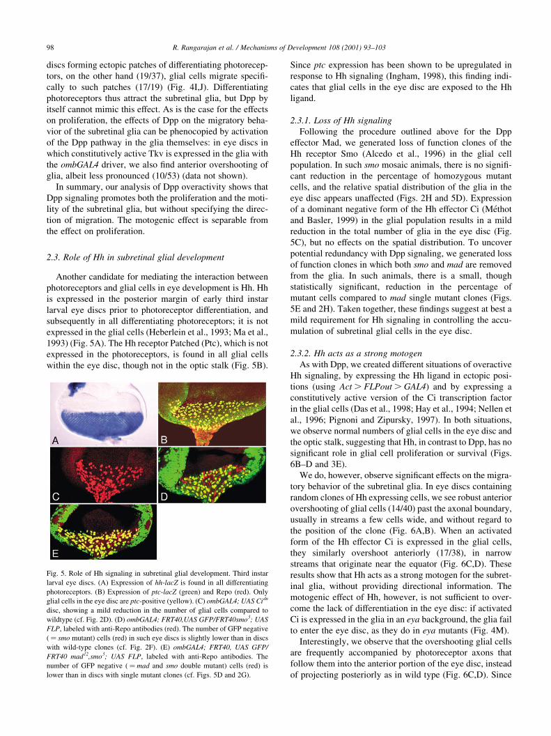
2.3. Role of Hh in subretinal glial development
Another candidate for mediating the interaction between
photoreceptors and glial cells in eye development is Hh. Hh
is expressed in the posterior margin of early third instar
larval eye discs prior to photoreceptor differentiation, and
subsequently in all differentiating photoreceptors; it is not
expressed in the glial cells (Heberlein et al., 1993; Ma et al.,
1993) (Fig. 5A). The Hh receptor Patched (Ptc), which is not
expressed in the photoreceptors, is found in all glial cells
within the eye disc, though not in the optic stalk (Fig. 5B).
Since ptc expression has been shown to be upregulated in
response to Hh signaling (Ingham, 1998), this finding indi-
cates that glial cells in the eye disc are exposed to the Hh
ligand.
2.3.1. Loss of Hh signaling
Following the procedure outlined above for the Dpp
effector Mad, we generated loss of function clones of the
Hh receptor Smo (Alcedo et al., 1996) in the glial cell
population. In such smo mosaic animals, there is no signifi-
cant reduction in the percentage of homozygous mutant
cells, and the relative spatial distribution of the glia in the
eye disc appears unaffected (Figs. 2H and 5D). Expression
of a dominant negative form of the Hh effector Ci (Methot
and Basler, 1999) in the glial population results in a mild
reduction in the total number of glia in the eye disc (Fig.
5C), but no effects on the spatial distribution. To uncover
potential redundancy with Dpp signaling, we generated loss
of function clones in which both smo and mad are removed
from the glia. In such animals, there is a small, though
statistically significant, reduction in the percentage of
mutant cells compared to mad single mutant clones (Figs.
5E and 2H). Taken together, these findings suggest at best a
mild requirement for Hh signaling in controlling the accu-
mulation of subretinal glial cells in the eye disc.
2.3.2. Hh acts as a strong motogen
As with Dpp, we created different situations of overactive
Hh signaling, by expressing the Hh ligand in ectopic posi-
tions (using Act . FLPout . GAL4) and by expressing a
constitutively active version of the Ci transcription factor
in the glial cells (Das et al., 1998; Hay et al., 1994; Nellen et
al., 1996; Pignoni and Zipursky, 1997). In both situations,
we observe normal numbers of glial cells in the eye disc and
the optic stalk, suggesting that Hh, in contrast to Dpp, has no
significant role in glial cell proliferation or survival (Figs.
6B–D and 3E).
We do, however, observe significant effects on the migra-
tory behavior of the subretinal glia. In eye discs containing
random clones of Hh expressing cells, we see robust anterior
overshooting of glial cells (14/40) past the axonal boundary,
usually in streams a few cells wide, and without regard to
the position of the clone (Fig. 6A,B). When an activated
form of the Hh effector Ci is expressed in the glial cells,
they similarly overshoot anteriorly (17/38), in narrow
streams that originate near the equator (Fig. 6C,D). These
results show that Hh acts as a strong motogen for the subret-
inal glia, without providing directional information. The
motogenic effect of Hh, however, is not sufficient to over-
come the lack of differentiation in the eye disc: if activated
Ci is expressed in the glia in an eya background, the glia fail
to enter the eye disc, as they do in eya mutants (Fig. 4M).
Interestingly, we observe that the overshooting glial cells
are frequently accompanied by photoreceptor axons that
follow them into the anterior portion of the eye disc, instead
of projecting posteriorly as in wild type (Fig. 6C,D). Since
R. Rangarajan et al. / Mechanisms of Development 108 (2001) 93–10398
Fig. 5. Role of Hh signaling in subretinal glial development. Third instar
larval eye discs. (A) Expression of hh-lacZ is found in all differentiating
photoreceptors. (B) Expression of ptc-lacZ (green) and Repo (red). Only
glial cells in the eye disc are ptc-positive (yellow). (C) ombGAL4; UAS Cidn
disc, showing a mild reduction in the number of glial cells compared to
wildtype (cf. Fig. 2D). (D) ombGAL4; FRT40,UAS GFP/FRT40smo3; UAS
FLP, labeled with anti-Repo antibodies (red). The number of GFP negative
( ¼ smo mutant) cells (red) in such eye discs is slightly lower than in discs
with wild-type clones (cf. Fig. 2F). (E) ombGAL4; FRT40, UAS GFP/
FRT40 mad12,smo3; UAS FLP, labeled with anti-Repo antibodies. The
number of GFP negative ( ¼ mad and smo double mutant) cells (red) is
lower than in discs with single mutant clones (cf. Figs. 5D and 2G).
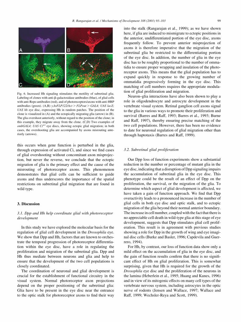
this occurs when gene function is perturbed in the glia,
through expression of activated Ci, and since we find cases
of glial overshooting without concomitant axon misprojec-
tion, but never the reverse, we conclude that the ectopic
migration of glia is the primary effect and the cause of the
misrouting of photoreceptor axons. This phenomenon
demonstrates that glial cells can be sufficient to guide
axons and thus underscores the importance of the spatial
restrictions on subretinal glial migration that are found in
wild type.
3. Discussion
3.1. Dpp and Hh help coordinate glial with photoreceptor
development
In this study we have explored the molecular basis for the
regulation of glial cell development in the Drosophila eye.
We show that Dpp and Hh, factors that are known to orches-
trate the temporal progression of photoreceptor differentia-
tion within the eye disc, have a role in regulating the
proliferation and migration of the subretinal glia. Dpp and
Hh thus mediate between neurons and glia and help to
ensure that the development of the two cell populations is
closely coordinated.
The coordination of neuronal and glial development is
crucial for the establishment of functional circuitry in the
visual system. Normal photoreceptor axon projections
depend on the proper positioning of the subretinal glia:
Glia have to be present in the eye disc near the entrance
to the optic stalk for photoreceptor axons to find their way
into the stalk (Rangarajan et al., 1999); as we have shown
here, if glia are induced to mismigrate to ectopic positions in
the anterior, undifferentiated portion of the eye disc, axons
frequently follow. To prevent anterior misprojection of
axons it is therefore imperative that the migration of the
subretinal glia be restricted to the differentiating portion
of the eye disc. In addition, the number of glia in the eye
disc has to be roughly proportional to the number of omma-
tidia to ensure proper wrapping and insulation of the photo-
receptor axons. This means that the glial population has to
expand quickly in response to the growing number of
ommatidia progressively forming in the eye disc. This
matching of cell numbers requires the appropriate modula-
tion of glial proliferation and migration.
Neuron–glia interactions have also been shown to play a
role in oligodendrocyte and astrocyte development in the
vertebrate visual system. Retinal ganglion cell axons signal
to the glia in various ways to promote their proliferation and
survival (Barres and Raff, 1993; Barres et al., 1993; Burne
and Raff, 1997), thereby ensuring precise matching of the
two cell populations. However, there has been no evidence
to date for neuronal regulation of glial migration other than
through haptotaxis (Barres and Raff, 1999).
3.2. Subretinal glial proliferation
Our Dpp loss of function experiments show a substantial
reduction in the number or percentage of mutant glia in the
eye disc, indicating that a disruption of Dpp signaling impairs
the accumulation of subretinal glia in the eye disc. This
phenotype could be the result of an effect of Dpp on the
proliferation, the survival, or the migration of the glia. To
determine which aspect of glial development is affected, we
have taken a gain of function approach. We find that Dpp
overactivity leads to a pronounced increase in the number of
glial cells in both eye disc and optic stalk, and to ectopic
migration of the glia beyond their normal anterior boundary.
The increase in cell number, coupled with the fact that there is
no appreciable cell death in wild-type glia at this stage of eye
development, suggests that Dpp stimulates glial cell prolif-
eration. This result is in agreement with previous studies
showing a role for Dpp in the growth of wing and eye imagi-
nal disc cells (Burke and Basler, 1996; Capdevila and Guer-
rero, 1994).
For Hh, by contrast, our loss of function data show only a
mild effect on the accumulation of glia in the eye disc, and
the gain of function results confirm that there is no signifi-
cant effect of Hh on glial proliferation. This is somewhat
surprising, given that Hh is required for the growth of the
Drosophila eye disc and the proliferation of the neurons in
the lamina (Heberlein et al., 1995; Huang and Kunes, 1996)
and in view of its mitogenic effects on many cell types of the
vertebrate nervous system, including astrocytes in the optic
nerve of rodents (Jensen and Wallace, 1997; Wallace and
Raff, 1999; Wechsler-Reya and Scott, 1999).
R. Rangarajan et al. / Mechanisms of Development 108 (2001) 93–103 99
Fig. 6. Increased Hh signaling stimulates the motility of subretinal glia.
Labeling of clones with anti-b-galactosidase antibodies (blue), of glial cells
with anti-Repo antibodies (red), and of photoreceptors/axons with anti-HRP
antibodies (green). (A,B) y,hsFLP122/Act . FLPout . GAL4; UAS lacZ;
UAS hh eye disc, expressing Hh in random patches. The position of the
clone is visualized in (A) and the ectopically migrating glia (arrow) in (B).
The glia overshoot anteriorly, without regard to the position of the clone; in
this example, they migrate away from the clone. (C,D) Two examples of
ombGAL4; UAS Ciact eye discs, showing ectopic glial migration; in both
cases, the overshooting glia are accompanied by axons misrouting ante-
riorly (arrows).

3.3. Subretinal glial migration
Subretinal glia migrate from the optic stalk into eye discs
only when photoreceptors have begun to differentiate in the
eye disc (Choi and Benzer, 1994), but they do not require
axons as a substrate for their migration (Rangarajan et al.,
1999). They also migrate towards ectopic islands of differ-
entiating photoreceptors, again without axonal substrate.
These findings suggest that differentiating photoreceptors
regulate the migration of the subretinal glia over a distance,
either by secreting diffusible molecules or by preferentially
stabilizing far-reaching glial filopodia (Rangarajan et al.,
1999). Our present study demonstrates that the diffusible
factors Dpp and Hh are among the signals the differentiating
photoreceptors send to promote subretinal glial motility.
Our gain-of-function experiments show that overactivity
of Dpp as well as of Hh signaling leads subretinal glial cells to
overshoot their normal anterior boundary, either in a rela-
tively narrow stream or in a broad front. In the case of Dpp,
this ectopic migration of the glia is accompanied by severe
overproliferation, and we therefore had to consider the possi-
bility that the changes in migratory behavior are merely a
secondary consequence of the increase in cell number.
However, we find that severe overproliferation of glia in
otherwise wild type eye discs does not lead to ectopic migra-
tion. This is true not only when overproliferation is induced
by acceleration of the cell cycle, but also when it is caused by
Ras overactivity (Rangarajan and Gaul, unpublished obser-
vation), which presumably mimics the effects of a wide range
of growth factors. Similarly, overproliferation of glia in the
optic stalk of eya mutants, which lack photoreceptors, does
not induce migration into the eye disc. Conversely, we do
observe ectopic migration of glia in eye discs with Hh over-
activity, i. e. in discs in which the number of glial cells is
largely normal. These results demonstrate that overprolifera-
tion in itself is not sufficient to induce abnormal migration of
glial cells and that proliferation and migration are in fact
independently regulated processes.
The ectopic migrations of glial cells that we observe are
not preferentially directed towards the sources of Dpp or
Hh. Moreover, similar migratory phenotypes can be induced
by expression of constitutively active components of the
Dpp and Hh pathways within the glial cells. Such cell-
autonomous activation of the pathway precludes the use
of information about ligand distribution as a positional
cue. Both findings therefore argue that Dpp and Hh exert
their effect on the glia not by providing positional informa-
tion, but by stimulating motility. This motogenic effect may
be due to an enhanced ability to migrate or to an impaired
ability to respond to an inhibitory signal (see below). In any
case, both Dpp and Hh appear to act by regulating transcrip-
tion within the glial cells, through their canonical signal
transduction machinery.
One of the best understood motogens is scatter factor (SF)
(for review see Birchmeier and Gherardi, 1998), which
disperses cohesive colonies of epithelial cells by stimulating
random motility. However, it can also act as a mitogen
(hepatocyte growth factor), morphogen, trophic factor, or
chemoattractant depending on the cellular context; in all
cases, its effects are mediated by the receptor tyrosine
kinase c-Met. The actual scattering induced by SF occurs
only 4–6 h after addition of SF and is inhibited by cyclo-
heximide, suggesting that regulation of gene expression is
involved (Ridley et al., 1995). Thus, Dpp and Hh resemble
SF both in terms of phenotypic effect and with regard to
their mechanism of action.
The function of Hh as a motogen is novel. Although the
molecule is being studied in many contexts, a role in cell
motility has not been attributed to it to date. Dpp and its
vertebrate counterparts, on the other hand, have been impli-
cated in the control of migratory processes during develop-
ment. Dpp plays a role in tracheal cell migration along the
dorso-ventral body axis (Vincent et al., 1997) and in the
dorsal migration of the ectoderm during dorsal closure of
the Drosophila embryo (for review see Stronach and Perri-
mon, 1999). During wound healing, the migration of various
cell types, including astrocytes, depends on TGF-b (Martin,
1997).
Why don’t we observe more clear-cut effects on the
migratory behavior of the subretinal glia in our loss of func-
tion experiments? Our mosaic data confirm at least for Dpp
that the molecule is required for the normal accumulation of
glia in the eye disc. But because of the limitation of our
mosaic technique it is difficult to distinguish between effects
on proliferation and on motility and to discern specific
migratory effects: the failure of ombGAL4 to drive recom-
bination to completion prior to entry of the glia into the eye
disc means that at least some glia arrive in the eye disc and
migrate anteriorly as heterozygotes, i.e. before undergoing
recombination to become homozygous mutant.
Another major reason for the lack of a more pronounced
loss of function effect is most likely that the Dpp and Hh
signal transduction pathways are (partially) functionally
redundant with other pathways in regulating glial cell devel-
opment. As we have shown previously, Ras signaling, possi-
bly triggered by diffusible RTK ligands, is very likely to
play a role in regulating both the proliferative and migratory
behavior of the subretinal glia (Rangarajan et al., 1999).
Generally, Dpp and Hh must be part of a more complex
network of signals regulating glial development in the eye
disc. For example, we do not yet know how the sharp ante-
rior boundary of glial migration behind the morphogenetic
furrow is established. Possible mechanisms include an
attractive substrate or signal in the posterior, or an inhibitory
signal in the anterior. Since in wild type the distribution of
both Dpp and Hh extends further to the anterior than the
glial cells migrate, this mechanism also has to be able to
counteract the motogenic effects of Dpp and Hh.
3.4. Signaling by Dpp and Hh: mechanism of action
How do Dpp and Hh exert their effects on subretinal glial
R. Rangarajan et al. / Mechanisms of Development 108 (2001) 93–103100
migration? Since Hh and Dpp are known to regulate each
other’s expression in other tissues (Ingham, 1998), it would
be conceivable that they act in sequence. However, neither
dpp nor hh is expressed in the subretinal glial cells, which
rules out the possibility that they mutually regulate each
other’s transcription and implies that Dpp and Hh both act
in a paracrine fashion. It therefore seems likely that Dpp and
Hh signaling converge to control the transcription of genes
required for cell motility (or possibly for reading the stop
signal). These targets could include genes coding for cytos-
keletal components, proteins regulating cell adhesion, or
enzymes to degrade the extracellular matrix. The idea that
such genes could be transcriptionally controlled by Dpp or
Hh is supported by the recent finding that in the Drosophila
wing compartmental cell sorting at the anterior/posterior
boundary is under the opposing transcriptional control of
Hh and En, suggesting that these molecules regulate the
expression of a single cell adhesion molecule (Dahmann
and Basler, 2000). The identification of transcriptional
targets of Dpp and Hh signaling will provide important
insights into the mechanism by which these signals regulate
glial cell motility.
4. Experimental procedures
4.1. Drosophila stocks
The GMRGAL4 line was kindly provided by M. Freeman,
ombGAL4 by S. Cohen, UAS dpp and UAS hh by J. Treis-
man, Act . FLPout . GAL4 by S.L. Zipursky, UAS tkvact
by R. Padgett (chromosome II) and G. Struhl (chromosome
III), UAS tkvDGSK (dominant negative form) by M. O’Con-
nor, UAS stg and UAS cycE by B. Edgar, UAS Ciact by T.
Orenic, UAS Cidn by K. Basler, UAS FLP by the Blooming-
ton Stock Center, FRT40A mad12 by E. Matunis, and
FRT40A smo3 by S. DiNardo.
4.2. Construction of UAS . FLPout . GFP
A UAS GFP transgene was constructed by cutting out a
0.7 kb Xba fragment containing the GFP coding sequence
from PeGFP (Clontech) and cloning it into the Xba site of
the pUAST vector (Brand and Perrimon, 1993). Next, the
FLPout cassette, consisting of DRaf sequences flanked by
FRT sites, was excised as a 7.0 kb Kpn fragment from
plasmid D237 (gift of G. Struhl) (Struhl and Basler, 1993)
and cloned into the Kpn site of the polylinker of the UAS
GFP construct located between the UAS and GFP
sequences to generate the UAS . DRaf . GFP transgene.
4.3. Tracking morphologies of individual glial cells
expressing GFP
y,hsFLP122; Adv/SM6-TM6B females were crossed to
ombGAL4; UAS . DRaf . GFP males. The progeny of
the cross were heat-shocked at 48–96 h of development at
358C for 30 min and female larvae were dissected the next
day. The morphologies of these cells was visualized by the
cytoplasmic GFP; glial nuclei were labeled with anti-Repo
antibodies.
4.4. Generation of dpp- and hh- expressing FLPout clones
This was done as described in Pignoni and Zipursky,
(1997). y,hsFLP122; UASdpp/TM6B males were crossed
to Act . FLPout . GAL4; UASlacZ females. The progeny
of the cross were heat shocked at 24–36 h of development
for 30 min at 358C. Female larvae were dissected at the late
third instar larval stage and stained with 22C10, FITC anti-
HRP, anti-b-galactosidase, and anti-Repo antibodies; or
with anti-pH3 (phosphorylated histone 3), FITC-coupled
anti-HRP antibodies, and propidium iodide, a DNA dye.
For hh-expressing clones, y,hsFLP122; UAShh/TM6B
females were crossed to Act . CD2 . GAL4; UASlacZ
males. The progeny of the cross were heat shocked at 24–
36 h of development for 30 min at 398C.
4.5. Generation of loss of function clones in the glia
This was done essentially by the FRT-UAS FLP method
described before (Duffy et al., 1998; Xu and Rubin, 1993),
using ombGAL4 as a driver to restrict recombination to the
glial population. A UAS GFP nuclear transgene inserted on
the non-mutant FRT chromosome (Courvoisier and Gaul,
unpublished) served to negatively mark mutant glial cells.
To generate homozygous mutant clones of smo3 and mad12,
and double mutant clones of smo3 and mad12 in the glial
cells, ombGAL4; FRT40A,UAS GFP males were crossed to
FRT40A smo3; UAS FLP/SM6-TM6B females, FRT40A
mad12;UAS FLP/SM6-TM6B females, or FRT 40A mad12,
smo3; UAS FLP/SM6-TM6B females, respectively. Female
third instar larvae were dissected and stained with anti-Repo
antibodies and propidium iodide. The numbers of mutant or
GFP minus cells were counted and are reported as percen-
tages of the total number of glial cells. Average percentages
of GFP minus cells are given as mean ^ SEM. Due to the
non-Gaussian distribution of the data, differences between
groups have been statistically assessed by the Mann–Whit-
ney test (Altman, 1991).
4.6. Histology
For RNA in situ hybridization, antisense digoxigenin
labeled RNA probes for dpp were generated according to
the manufacturer’s specifications (Boehringer). Whole-
mount in situ hybridization was carried out essentially as
described by Tear et al. (1996). For antibody stainings, eye–
brain complexes were dissected and stained as described in
Rangarajan et al. (1999); anti-H3 phosphorylated histones
(Upstate Biotechnology) were used at 1:100 dilution. To
stain samples with propidium iodide, fixed eye brain
complexes were digested with 0.1 mg/ml of RNase A for 1
h, followed by two quick washes with PBS-TX and incuba-
R. Rangarajan et al. / Mechanisms of Development 108 (2001) 93–103 101
tion with propidium iodide (Sigma) at a 1:500 dilution for 1
h. Specimens were imaged on a Zeiss LSM 510 confocal
microscope.
Acknowledgements
We thank K. Basler, S. Cohen, S. DiNardo, B. Edgar, M.
Freeman, E. Matunis, M. O’Connor, T. Orenic, R. Padgett,
G. Struhl, J. Treisman, S. L. Zipursky, and the Bloomington
Stock Center for stocks and reagents, and we thank S.L.
Zipursky for sharing results prior to publication. We are
grateful to S. Cohen, N. Heintz, J. Hudspeth, and members
of the Gaul laboratory for critically reading the manuscript,
and to H.R. Chung for help in preparing Fig. 1. This work
was supported by a graduate fellowship from the Rockefel-
ler University (R.R.), by a postdoctoral fellowship from the
Association pour la Recherche sur le Cancer (H.C.), by a
grant from the National Institutes of Health (U.G.), and by
Klingenstein Fellowship and McKnight Scholar Awards
(U.G.).
References
Alcedo, J., Ayenzon, M., Von Ohlen, T., Noll, M., Hooper, J.E., 1996. The
Drosophila smoothened gene encodes a seven-pass membrane protein,
a putative receptor for the Hedgehog signal. Cell 86, 221–232.
Altman, D.G., 1991. Practical Statistics for Medical Research, Chapman
and Hall, London.
Auld, V., 1999. Glia as mediators of growth cone guidance: studies from
insect nervous systems. Cell. Mol. Life Sci. 55, 1377–1385.
Barres, B.A., Jacobson, M.D., Schmid, R., Sendtner, M., Raff, M.C., 1993.
Does oligodendrocyte survival depend on axons? Curr. Biol. 3, 489–
497.
Barres, B.A., Raff, M.C., 1993. Proliferation of oligodendrocyte precursor
cells depends on electrical activity in axons. Nature 361, 258–260.
Barres, B.A., Raff, M.C., 1999. Axonal control of oligodendrocyte devel-
opment. J. Cell Biol. 147, 1123–1128.
Birchmeier, C., Gherardi, E., 1998. Developmental roles of HGF/SF and its
receptor, the c-Met tyrosine kinase. Trends Cell Biol. 8, 404–410.
Birling, M.C., Price, J., 1995. Influence of growth factors on neuronal
differentiation. Curr. Opin. Cell Biol. 7, 878–884.
Blackman, R.K., Sanicola, M., Raftery, L.A., Gillevet, T., Gelbart, W.M.,
1991. An extensive 3 0 cis-regulatory region directs the imaginal disk
expression of decapentaplegic, a member of the TGF-b family in
Drosophila. Development 111, 657–666.
Brand, A.H., Perrimon, N., 1993. Targeted gene expression as a means of
altering cell fates and generating dominant phenotypes. Development
118, 401–415.
Briscoe, J., Ericson, J., 1999. The specification of neuronal identity by
graded Sonic hedgehog signalling. Cell Dev. Biol. 10, 353–362.
Burke, R., Basler, K., 1996. Hegehog-dependent patterning in the Droso-
phila eye can occur in the absence of Dpp signaling. Dev. Biol. 179,
360–368.
Burne, J.F., Raff, M.C., 1997. Retinal ganglion cell axons drive the prolif-
eration of astrocytes in the developing rodent optic nerve. Neuron 18,
223–230.
Capdevila, J., Guerrero, I., 1994. Targeted expression of the signalling
molecule decapentaplegic induces pattern duplications and growth
alterations in Drosophila wings. EMBO J. 13, 4459–4468.
Choi, K.-W., Benzer, S., 1994. Migration of glia along photoreceptor axons
in the developing Drosophila eye. Neuron 12, 423–431.
Dahmann, C., Basler, K., 2000. Opposing transcriptional outputs of Hedge-
hog signaling and engrailed control compartmental cell sorting at the
Drosophila A/P boundary. Cell 100, 411–422.
Das, P., Madzuia, L.L., Wang, H., Finelli, A.L., Cho, S., Smith, M.M.,
Padgett, R.W., 1998. The Drosophila gene medea demonstrates the
requirement for different Smads in dpp signaling. Development 125,
1519–1528.
Duffy, J.B., Harrison, D.A., Perrimon, N., 1998. Identifying loci required
for follicular patterning using directed mosaics. Development 125,
2263–2271.
Giangrande, A., Murray, M.A., Palka, J., 1993. Development and organiza-
tion of glial cells in the peripheral nervous system of Drosophila mela-
nogaster. Development 117, 895–904.
Greenwood, S., Struhl, G., 1999. Progression of the morphogenetic furrow
in the Drosophila eye: the roles of Hedgehog, Decapentaplegic and the
Raf pathway. Development 126, 5795–5808.
Halter, D.A., Urban, J., Rickert, C., Ner, S.S., Ito, K., Travers, A.A., Tech-
nau, G.M., 1995. The homeobox gene repo is required for the differ-
entiation and maintenance of glia function in the embryonic nervous
system of Drosophila melanogaster. Development 121, 317–332.
Hartenstein, V., Nassif, C., Lekven, A., 1998. Embryonic development of
the Drosophila brain. II. Pattern of glial cells. J. Comp. Neurol. 402, 32–
47.
Hay, B.A., Wolff, T., Rubin, G.M., 1994. Expression of bacculovirus P35
prevents cell death in Drosophila. Development 121, 2121–2129.
Heberlein, U., Wolff, T., Rubin, G.M., 1993. The TGF-b homolog Dpp and
the segment polarity gene Hedgehog are required for the propagation of
a morphogenetic wave in the Drosophila retina. Cell 75, 913–926.
Heberlein, U., Singh, C.M., Luk, A.Y., Donohoe, T.J., 1995. Growth and
differentiation in the Drosophila eye coordinated by Hedgehog. Nature
373, 709–711.
Hendzel, M.J., Wei, Y., Mancini, M.A., Van Hooser, A., Ranalli, T., Brink-
ley, B.R., Bazett-Jones, D.P., Allis, C.D., 1997. Mitosis specific phos-
phorylation of histone H3 initiates primarily within pericentromeric
heterochromatin during G2 and spreads in an ordered fashion coinci-
dent with mitotic chromosome condensation. Chromosoma 106, 348–
360.
Huang, Z., Kunes, S., 1996. Hedgehog, transmitted along retinal axons,
triggers neurogenesis in the developing visual centers of the Drosophila
brain. Cell 86, 411–422.
Huang, Z., Kunes, S., 1998. Signals transmitted along retinal axons in
Drosophila: Hedgehog signal reception and the cell circuitry of lamina
cartridge assembly. Development 125, 3753–3764.
Ingham, P., 1998. Transducing Hedgehog: the story so far. EMBO J. 17,
3505–3511.
Ito, K., Urban, J., Technau, G.M., 1995. Distribution, classification and
development of Drosophila glial cells in the late embryonic and early
larval ventral nerve cord. Roux’s Arch. Dev. Biol. 204, 284–307.
Jensen, A., Wallace, V.A., 1997. Expression of Sonic hedgehog and its
putative role as precursor cell mitogen in the developing mouse retina.
Development 124, 363–371.
Lecuit, T., Brook, W.J., Ng, M., Calleja, M., Sun, H., Cohen, S.M., 1996.
Two distinct mechanisms for long-range patterning by Decapentaplegic
in the Drosophila wing. Nature 381, 387–393.
Ma, C., Zhou, Y., Beachy, P.A., Moses, K., 1993. The segment polarity
gene hedgehog is required for the progression of the morphogenetic
furrow in the developing Drosophila eye. Cell 75, 927–938.
Martin, P., 1997. Wound healing–aiming for perfect skin regeneration.
Science 276, 75–81.
Massague, J., 1998. TGF-beta signal transduction. Annu. Rev. Biochem.
67, 753–791.
Massague, J., Chen, Y.G., 2000. Controlling TGF-beta signaling. Genes
Dev. 14, 627–644.
McMahon, A.P., 2000. More surprises in the Hedgehog signaling pathway.
Cell 100, 185–188.
Methot, N., Basler, K., 1999. Hedgehog controls limb development by
R. Rangarajan et al. / Mechanisms of Development 108 (2001) 93–103102
regulating the activities of distinct transcriptional activator and repres-
sor forms of Cubitus interruptus. Cell 96, 819–831.
Nellen, D., Burke, R., Struhl, G., Basler, K., 1996. Direct and long-range
action of a Dpp morphogen gradient. Cell 85, 357–358.
Neufeld, T.P., de la Cruz, A.F., Johnston, L.A., Edgar, B.A., 1998. Coordi-
nation of growth and cell division in the Drosophila wing. Cell 93,
1183–1193.
Perez, S.E., Steller, H., 1996. Migration of glial cells into retinal axon target
field in Drosophila melanogaster. J. Neurobiol. 30, 359–373.
Pignoni, F., Zipursky, S.L., 1997. Induction of Drosophila eye development
by decapentaplegic. Development 124, 271–278.
Rangarajan, R., Gong, Q., Gaul, U., 1999. Migration and function of glia in
the developing Drosophila eye. Development 126, 3285–3292.
Ridley, A.J., Comoglio, P.M., Hall, A., 1995. Regulation of Scatter Factor/
Hepatocyte Growth Factor responses by Ras, Rac and Rho in MDCK
cells. Mol. Cell. Biol. 15, 1110–1122.
Silver, J., 1993. Glia–neuron interactions at the midline of the developing
mammalian brain and spinal cord. Perspect. Dev. Neurobiol. 1, 227–
236.
Stronach, B.E., Perrimon, N., 1999. Stress signaling in Drosophila. Onco-
gene 18, 6172–6182.
Struhl, G., Basler, K., 1993. Organizing activity of Wingless protein in
Drosophila. Cell 72, 527–540.
Tear, G., 1999. Neuronal guidance. A genetic perspective. Trends Genet.
15, 113–118.
Tear, G., Harris, R., Sutaria, S., Kilomanski, K., Goodman, C.S., Seeger,
M.A., 1996. commissureless controls growth cone guidance across the
CNS midline in Drosophila and encodes a novel membrane protein.
Neuron 16, 501–514.
Tessier-Lavigne, M., Goodman, C.S., 1996. The molecular biology of axon
guidance. Science 274, 1123–1133.
Thomas, J.B., 1998. Axon guidance: crossing the midline. Curr. Biol. 8,
R102–R104.
Treisman, J.E., Heberlein, U., 1998. Eye development in Drosophila:
formation of the eye field and control of differentiation. Curr. Top.
Dev. Biol. 39, 119–158.
Vincent, S.E.R., Greider, N., Chen, C.K., Haerry, T., Schuh, R., Affolter,
M., 1997. Dpp controls tracheal cell migration along the dorsoventral
body axis of the Drosophila embryo. Development 124, 2741–2750.
Wallace, V.A., Raff, M.C., 1999. A role for Sonic hedgehog in axon-to-
astrocyte signaling in the rodent optic nerve. Development 126, 2901–
2909.
Wechsler-Reya, R.J., Scott, M.P., 1999. Control of neuronal precursor
proliferation in the cerebellum by Sonic Hedgehog. Neuron 22, 103–114.
Wiersdorff, V., Lecuit, T., Cohen, S.M., Mlodzik, M., 1996. Mad acts
downstream of Dpp receptors, revealing a differential requirement for
dpp signaling in initiation and propagation of morphogenesis in the
Drosophila eye. Development 122, 2153–2162.
Xu, T., Rubin, G.M., 1993. Analysis of genetic mosaics in developing and
adult Drosophila tissue. Development 117, 1223–1237.
R. Rangarajan et al. / Mechanisms of Development 108 (2001) 93–103 103