the role of gdnf in patterning the excretory system
TRANSCRIPT

www.elsevier.com/locate/ydbio
Developmental Biology
The role of GDNF in patterning the excretory system
Reena Shakyaa, Eek-hoon Jhoa,1, Pille Kotkaa,2, Zaiqi Wua, Nikolai Kholodilovb,
Robert Burkeb,c, Vivette D’Agatic, Frank Costantinia,*
aDepartment of Genetics and Development, Columbia University Medical Center, 630 W. 168th Street, New York, NY 10032, USAbDepartment of Neurology, Columbia University Medical Center, 630 W. 168th Street, New York, NY 10032, USAcDepartment of Pathology, Columbia University Medical Center, 630 W. 168th Street, New York, NY 10032, USA
Received for publication 24 January 2005, revised 28 March 2005, accepted 6 April 2005
Available online 10 May 2005
Abstract
Mesenchymal–epithelial interactions are an important source of information for pattern formation during organogenesis. In the
developing excretory system, one of the secreted mesenchymal factors thought to play a critical role in patterning the growth and branching
of the epithelial ureteric bud is GDNF. We have tested the requirement for GDNF as a paracrine chemoattractive factor by altering its site of
expression during excretory system development. Normally, GDNF is secreted by the metanephric mesenchyme and acts via receptors on the
Wolffian duct and ureteric bud epithelium. Misexpression of GDNF in the Wolffian duct and ureteric buds resulted in formation of multiple,
ectopic buds, which branched independently of the metanephric mesenchyme. This confirmed the ability of GDNF to induce ureter
outgrowth and epithelial branching in vivo. However, in mutant mice lacking endogenous GDNF, kidney development was rescued to a
substantial degree by GDNF supplied only by the Wolffian duct and ureteric bud. These results indicate that mesenchymal GDNF is not
required as a chemoattractive factor to pattern the growth of the ureteric bud within the developing kidney, and that any positional
information provided by the mesenchymal expression of GDNF may provide for renal branching morphogenesis is redundant with other
signals.
D 2005 Elsevier Inc. All rights reserved.
Keywords: Organogenesis; Kidney development; GDNF; Ret receptor tyrosine kinase; Ret; Hoxb7; Metanephric kidney; Wolffian duct; Ureteric bud;
Branching morphogenesis; Metanephric mesenchyme
Introduction
Mesenchymal–epithelial interactions are thought to play
a critical role in the growth and pattern formation of organs
that develop by branching morphogenesis, such as the
kidney and lung. During kidney development, the epithelial
ureteric bud (UB) emerges from the Wolffian duct, in
response to inductive signals from the metanephric mesen-
chyme, a specialized region of the nephrogenic cord (Saxen,
0012-1606/$ - see front matter D 2005 Elsevier Inc. All rights reserved.
doi:10.1016/j.ydbio.2005.04.008
* Corresponding author. Fax: +1 212 923 2090.
E-mail address: [email protected] (F. Costantini).1 Present address: Department of Life Science, University of Seoul, 90
Jeonnong-Dong, Dongdaemun-Gu, Seoul, Korea.2 Present address: National Institute of Chemical Physics and Biophysics
and Tallinn University of Technology, Department of Gene Technology,
Tallinn, Estonia.
1987). As the UB invades the metanephric mesenchyme,
further inductive signals from the mesenchyme and renal
stromal cells (Carroll and McMahon, 2003; Shah et al.,
2004; Vainio and Lin, 2002) induce the UB to undergo a
complex and stereotypic process of branching and elonga-
tion, eventually giving rise to the epithelium of the ureter,
renal pelvis, calyces, and collecting ducts (Al-Awqati and
Goldberg, 1998; Oliver, 1968). The ureteric bud, in turn,
secretes factors that induce the metanephric mesenchymal
cells to condense, convert to epithelia and eventually form
the various segments of the nephron (Carroll and McMahon,
2003; Saxen, 1987).
One of the main signaling pathways that promotes
ureteric bud branching morphogenesis involves the secreted
factor GDNF, which signals through the Ret receptor
tyrosine kinase and the co-receptor GFRa-1 (Sariola and
283 (2005) 70 – 84

R. Shakya et al. / Developmental Biology 283 (2005) 70–84 71
Saarma, 2003). Prior to outgrowth of the UB from the
Wolffian duct, Ret and GFRa-1 are expressed throughout
the Wolffian duct, while GDNF is expressed broadly in the
nephrogenic cord surrounding the posterior duct (Hellmich
et al., 1996; Pachnis et al., 1993; Suvanto et al., 1996). As
the UB evaginates from the caudal end of the Wolffian duct,
the expression of GDNF becomes limited to and upregu-
lated in the adjacent metanephric blastema (Grieshammer
et al., 2004). Concomitantly, Ret and GFRa-1 are down-
regulated in the Wolffian duct and become restricted to the
newly formed ureteric bud. As UB branching progress, Ret
and GFRa-1 soon become restricted to the tips of UB
branches, and GDNF to the surrounding mesenchymal cells
at the periphery of the kidney (Pachnis et al., 1993; Sainio
et al., 1997).
The importance of this pathway for renal development
was established by the finding that null mutations in gdnf,
ret, or gfra-1 result in renal agenesis or severe dysgenesis,
due to the failure of the UB to emerge from the Wolffian
duct or to grow and branch normally (Cacalano et al., 1998;
Enomoto et al., 1998; Moore et al., 1996; Pichel et al., 1996;
Sanchez et al., 1996; Schuchardt et al., 1994, 1996).
However, the specific cellular and developmental processes
induced by GDNF remain unclear. While there were
conflicting reports as to whether GDNF induces UB cell
proliferation (Michael and Davies, 2004; Pepicelli et al.,
1997; Sainio et al., 1997; Vega et al., 1996), a recent
analysis of the behavior of ret�/� cells in mosaic kidneys
supports the conclusion that Ret signaling is particularly
important for the proliferation of cells at the growing UB
tips (Shakya et al., 2005). GDNF may also promote the
survival of UB cells (Towers et al., 1998), as it does for
some neuronal populations (Sariola and Saarma, 2003). A
third potential role, for which there is considerable evidence,
albeit indirect, involves specifying the pattern of branching
morphogenesis. A GDNF-soaked bead can induce the
outgrowth of ectopic buds from the Wolffian duct in organ
culture (Brophy et al., 2001; Pichel et al., 1996; Sainio et al.,
1997), while mutations that alter the distribution of GDNF
along the Wolffian duct can cause ectopic UBs to form in
vivo (Grieshammer et al., 2004; Kume et al., 2000).
Addition of GDNF to kidney cultures can cause increased
or abnormal UB branching (Pepicelli et al., 1997; Pichel et
al., 1996; Sainio et al., 1997; Towers et al., 1998; Vega et
al., 1996), while expression of activated forms of Ret in the
Wolffian duct and ureteric bud also induces ectopic UBs and
abnormal patterns of UB growth and branching (de Graaff et
al., 2001; Srinivas et al., 1999b). These studies have led to
the idea that GDNF/Ret signaling may specifically induce
branching of the UB epithelium (i.e., that it is a ‘‘ramogen’’),
and that it provides a chemoattractive cue, whose concen-
tration gradient is sensed by the UB tips and thus directs
their growth. The latter hypothesis was supported by studies
in which Ret-expressing cells were shown to migrate
towards a source of GDNF (Tang et al., 1998), as well as
by observations that GDNF may serve as a chemoattractant
for migrating enteric neural crest cells (Natarajan et al.,
2002; Young et al., 2001). Thus, a model has emerged in
which the localized expression of GDNF is important for
normal patterning of the excretory system, first in determin-
ing the site of initial outgrowth of the UB, and later in
determining the branching pattern within the developing
kidney (Lechner and Dressler, 1997; Sariola and Saarma,
2003; Sariola and Sainio, 1997).
Another potential role of GDNF involves the proximal–
distal patterning of the ureteric bud. GDNF is expressed
only at the periphery of the growing kidney, and several
genes expressed specifically in the peripheral UB tips,
including Ret and Wnt11, are upregulated by GDNF
(Pepicelli et al., 1997). Thus, it is possible that the pattern
of expression of these (and perhaps other) tip-specific genes
is a direct consequence of the localized expression of
GDNF.
To test these hypotheses in an in vivo situation, we
generated transgenic mice in which GDNF is ectopically
expressed in cells throughout the Wolffian duct and
ureteric bud epithelium. The effects of GDNF misexpres-
sion were examined first on a wild type background, in
which endogenous GDNF is also expressed by the
mesenchyme, and then on a gdnf knockout background,
in which endogenous GDNF is absent, and replaced by
the ectopically-expressed GDNF. We reasoned that if
GDNF were required only for UB cell survival and
proliferation, such alterations in its spatial distribution
might have little impact on the pattern of UB outgrowth
and branching; however, if GDNF served as a critical
chemoattractive factor, they should have profound effects
on UB branching morphogenesis. Our results support the
conclusion that GDNF stimulates bud formation by the
Wolffian duct epithelium and that the localized expression
of GDNF in the posterior nephrogenic cord is important
for the outgrowth of a single, correctly placed ureteric
bud. They also show that growth and branching of the
collecting duct system within the kidney can be perturbed
to some extent by the misexpression of GDNF in the
ureteric bud. Surprisingly, many aspects of kidney devel-
opment and UB morphogenesis were normal in mice in
which GDNF was expressed only in the Wolffian duct and
UB epithelial cells, but not in the mesenchyme. This
observation argues that, if the mesenchymal expression of
GDNF does provide positional cues for ureteric bud
growth and patterning, this information must be redundant
with other signals that can independently pattern the
ureteric bud.
Materials and methods
Generation of Hoxb7/rtTA transgenic mice
The Hoxb7 promoter (�1316 bp to +81 bp of the
Hoxb7 gene) (Kress et al., 1990) was excised from

Fig. 1. Excretory system defects in Hoxb7/GDNF transgenic mice. (A)
Schematic diagram of transgene construct. (B) and (D) Sagittal sections of
two transgenic kidneys at E18.5. Asterisks denote cystic UB tips in the
cortex. (C) and (E) Enlargements of boxed areas in B and D, showing
branching of cyst epithelium (arrows). Inset in C, dolichos biflorus lectin
(brown) staining confirms the UB origin of cystic epithelia; methyl green
counterstain. Inset in E shows hyperplasia of the cystic epithelium, which is
5–6 cell diameters in thickness compared to the simple epithelium of
normal collecting ducts. (F) The male reproductive tract is abnormally
dilated (e, epididymis) and branched ectopic UBs are found nearby
(arrowheads). t, testis; b, bladder. (G) Enlargement of the boxed area in
(F) showing extensive branching of ectopic UBs. H and E staining. Scale
bars, 200 Am.
R. Shakya et al. / Developmental Biology 283 (2005) 70–8472
pGEM-Blue with KpnI and AvaI, the ends blunted, and
subcloned into the EcoRV site of pBluescriptII KS+. The
Hoxb7 promoter in pBluescript was then excised with
XhoI and EcoRI and the EcoRI end blunted. Plasmid
pUHDrtTA2S-M2 (Urlinger et al., 2000) was digested
with XhoI and SacI to remove the CMV promoter,
leaving the rtTA2S-M2 gene followed by SV40 polyA
sequences. The SacI site was blunted, and the Hoxb7
promoter fragment was then cloned into the XhoI–SacI
fragment of the pUHDrtTA2S-M2 vector. The entire 2.7
kb Hoxb7/rtTA2S-M2/SV40-pA transgene (abbreviated
‘‘Hoxb7/rtTA’’) was excised from the vector with XhoI
and HindIII for pronuclear injection into B6CBAF1/J
mouse eggs (Hogan et al., 1994). Three transgenic
founder animals (RS-HTA2, RS-HTA3 and RS-HTA8)
were identified by Southern blot using EcoRI, which cuts
once within the transgene, using a probe generated from
the rtTA2S-M2 cDNA. Offspring of the three founders
were tested for expression of rtTA2S-M2 in kidney by
RT-PCR (data not shown) and by crossing to tetO/h-galreporter mice, inducing with Dox and staining for h-gal(e.g., Figs. 2C, D, 3A and data not shown). All
experiments in this paper were conducted using animals
from line RS-HTA2, on a mixed genetic background.
Inheritance of the transgene was monitored by PCR using
primers rtTA2S-a (5V CATGGCAAGACTTTCTGCGG)
and rtTA2S-b (5VTTGQTCTCAGAAGTGGGGGCA),
which amplified a band of ¨296 bp from the rtTA2S-
M2 sequence under the following conditions: 94-C, 5
min; 30 cycles of (94-C, 30 s; 58-C, 30 s; 72-C, 50 s);
72-C, 7 min.
Generation of Hoxb7/GDNF transgenic mice
A PmeI restriction site was introduced 5Vof the Hoxb7
promoter in pBluescriptII (KS+) (see above) while a
multiple cloning site (MCS) (5V EcoRI–HindIII–PacI–
NheI –KpnI –XhoI –AseI–NotI –BamHI–PstI –PmeI 3V)was introduced downstream of the Hoxb7 fragment. A
BamHI–PstI fragment including an intron and polyadeny-
lation sequences of the human h-globin gene was
subcloned into the MCS to generate plasmid Hoxb7-
MCS-hh-globin-pA. The mouse GDNF cDNA (¨636 bp)
was excised from plasmid GDNFXVL with HindIII and
partial BamHI digestions, HindIII end blunted, and
subcloned into EcoRI (blunted) and BamHI sites, upstream
of an IRES2-EGFP sequence in pBluescriptII (KS+). The
GDNF-IRES2-EGFP fragment was excised with NheI and
NotI, the NotI end blunted, and subcloned into the NheI
and BamHI (blunted) sites in the MCS of Hoxb7-MCS-hh-globin-pA. The final construct, RS#29, was verified by
restriction mapping and partial sequencing. The ¨5 kb
insert was excised with PmeI, gel-purified, and eluted in
injection buffer (Hogan et al., 1994). Transgenic fetuses
were identified by Southern blot using EcoRI and a mouse
GDNF cDNA probe (data not shown).

Fig. 2. Transgene constructs for inducible expression of GDNF and h-gal inthe Wolffian duct and ureteric bud. A–C, transgene constructs. A, Hoxb7/
rtTA2S-M2 (abbreviated Hoxb7/rtTA) or Axin2/rtTA2S-M2 (abbreviated
Axin2/rtTA). B, BiTetO/lacZ/GDNF (Kholodilov et al., 2004). C, tetO/lacZ
(Furth et al., 1994). D, expression of control tetO/lacZ transgene, activated
by Hoxb7/rtTA, in the renal collecting ducts of a 40-day-old bi-transgenic
mouse maintained on Dox. c, cortex; m, medulla; p, papilla.
R. Shakya et al. / Developmental Biology 283 (2005) 70–84 73
Generation of Axin2-rtTA transgenic mice
The 5.7-kb insert of plasmid Axin2P-Luc (Jho et al.,
2002), containing the Axin2 promoter was excised with
MluI and HindIII, and the CMV promoter in pUHDrt-
Fig. 3. Ectopic ureteric buds emerging from the Wolffian duct of Hoxb7/rtTA
rtTA�tetO/lacZ bi-transgenic embryo with a single T-stage UB (arrow) that emerg
rtTA�BiTetO/lacZ/GDNF bi-transgenic embryo with a secondary T-stage UB (red
the more anterior Wolffian duct (yellow arrows). C, a strongly affected Hoxb7/rtTA
length of the Wolffian duct. Ectopic buds in more anterior regions of the Wolffia
TA2S-M2 (a gift of Dr. Wolfgang Hillen) was replaced
with the 5.7-kb Axin2 fragment, using the enzymes
described above for Hoxb7/rtTA. The Axin2-rtTA
plasmid was digested with AflII and HindIII and the
7-kb insert purified for microinjection. Genotyping was
performed with the same PCR primers as for Hoxb7/
rtTA.
BiTetO/lacZ/GDNF and tetO/lacZ transgenic mice and
GDNF mutant mice
Strain BiTetO/lacZ/GDNF has been described (Kholo-
dilov et al., 2004). The tetO/lacZ strain B6;SJL-Tg(tetop-
lacZ)2Mam/J (Furth et al., 1994) was obtained from The
Jackson Laboratory. The gdnflacZ knock-in null allele
(Sanchez et al., 1996) was obtained from M. Barbacid
(Bristol-Meyers Squib).
Transgene induction with Doxycycline
Pregnant females were fed 2 mg/ml Doxycycline
(Sigma D-9891), 5% sugar (w/v) in the water. Dox was
administrated continuously from day 8.5 of gestation in
the experiments of Figs. 3–6, from day 9.5 or 10.5 in
the rescue experiments (Figs. 7 and 8). Full induction
is achieved 1 day after the start of Dox (Kistner et al.,
1996). The rationale for delaying the induction in the
rescue experiments was to wait for a UB to form
naturally (¨E11), as occurs in some gdnf�/� embryos,
and then support its further growth by the induction of
the transgene-encoded GDNF. Whether this delay
contributed to the success of the rescue is not known.
�BiTetO/lacZ/GDNF bi-transgenic embryos at E11.5. A, control Hoxb7/
ed from the posterior end of the Wolffian duct. B, a weakly affected Hoxb7/
arrow) anterior to the normal UB (black arrow), and a few small buds along
�BiTetO/lacZ/GDNF bi-transgenic embryo with multiple UBs all along the
n duct had not yet branched at E11.5. Scale bar, 0.5 mm.

R. Shakya et al. / Developmental Biology 283 (2005) 70–8474
b-gal staining of whole mount tissues and cryosections
Tissues were fixed in fresh, ice-cold 2% PFA (parafor-
maldehye) and 0.2% glutaraldehyde in 1 � PIPES buffer
(0.1 M Pipes, 2 mM MgCl2, 2 mM EGTA, pH 6.9) 4-C for
1 h, washed in PBS, and postfixed in 0.2% glutaraldehyde
in 1 � PIPES buffer for 1 h at room temp. Alternatively,
tissues were fixed in 4% PFA in PBS for 1–3 h at 4-C. After
R. Shakya et al. / Developmental Biology 283 (2005) 70–84 75
several washes in PBS, for sectioning tissues were
permeated with 15% and 30% sucrose in 0.1 M phosphate
buffer, embedded in OCT (Tissue-Tek\) and frozen in dry
ice. 12–15 Am sections were fixed in 2 mM MgCl2 and
0.2% glutaraldehyde in PBS for 5 min before staining with
X-gal. Sections and whole tissues were stained with fresh X-
gal (2 mM MgCl2, 2.12 mg/ml potassium ferrocyanide, 1.64
mg/ml potassium ferricyanide, 1 mg/ml X-gal in PBS).
Tissues were stored in 10% Formalin buffer, while
cryosections were mounted with non-aqueous mounting
medium.
In situ hybridization
In situ hybridization was performed with digoxigenin-
labeled riboprobes as described (Mendelsohn et al., 1999)
and on whole-mount tissues as described (Wilkinson, 1992).
Ret, GDNF, and GFRa1 anti-sense riboprobes were gen-
erated as described (Srinivas et al., 1999b). For Wnt11 anti-
sense riboprobe, the Wnt11 cDNA clone pnk3 was linearized
with XhoI and transcribed with T3 polymerase (Kispert et al.,
1996).
Results
A Hoxb7/GDNF transgene can induce abnormal branching
of ureteric bud-derived epithelia in vivo
The Hoxb7 gene is expressed throughout the Wolffian
duct and ureteric bud epithelium, and its promoter directs a
similar pattern of transgene expression (Kress et al., 1990;
Srinivas et al., 1999a,b). Using this approach, it was
previously found that the expression of constitutively active
forms of Ret could induce abnormalities in UB growth and
branching (de Graaff et al., 2001; Srinivas et al., 1999b).
Here, we sought to determine whether the ectopic expres-
sion of the Ret ligand GDNF would have similar effects. We
first generated transgenic mice that carried a GDNF cDNA
under the direct control of the Hoxb7 promoter (Fig. 1A). Of
23 independently generated Hoxb7/GDNF transgenic
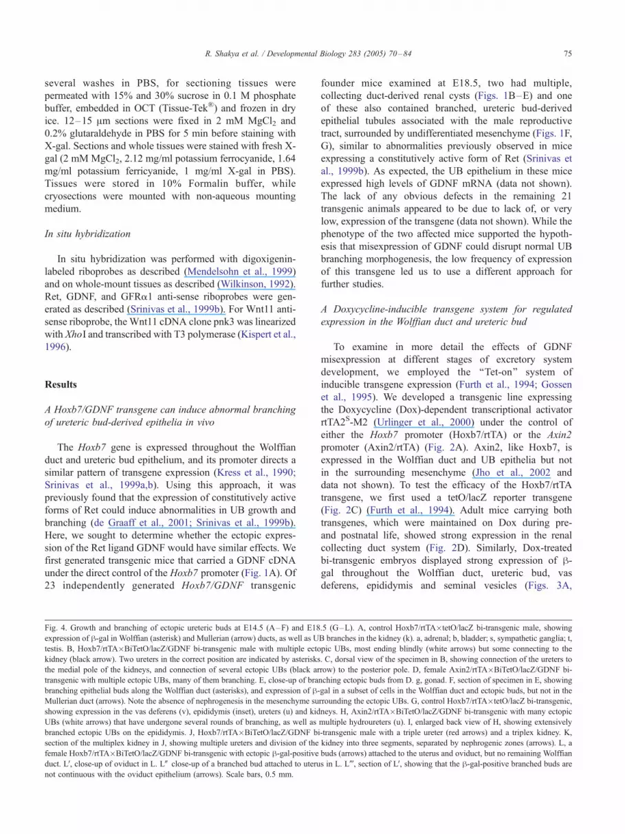
ig. 4. Growth and branching of ectopic ureteric buds at E14.5 (A–F) and E18.5 (G–L). A, control Hoxb7/rtTA�tetO/lacZ bi-transgenic male, showing
xpression of h-gal in Wolffian (asterisk) and Mullerian (arrow) ducts, as well as UB branches in the kidney (k). a, adrenal; b, bladder; s, sympathetic ganglia; t
stis. B, Hoxb7/rtTA�BiTetO/lacZ/GDNF bi-transgenic male with multiple ectopic UBs, most ending blindly (white arrows) but some connecting to the
idney (black arrow). Two ureters in the correct position are indicated by asterisks. C, dorsal view of the specimen in B, showing connection of the ureters to
e medial pole of the kidneys, and connection of several ectopic UBs (black arrow) to the posterior pole. D, female Axin2/rtTA�BiTetO/lacZ/GDNF bi
ansgenic with multiple ectopic UBs, many of them branching. E, close-up of branching ectopic buds from D. g, gonad. F, section of specimen in E, showing
ranching epithelial buds along the Wolffian duct (asterisks), and expression of h-gal in a subset of cells in the Wolffian duct and ectopic buds, but not in the
ullerian duct (arrows). Note the absence of nephrogenesis in the mesenchyme surrounding the ectopic UBs. G, control Hoxb7/rtTA�tetO/lacZ bi-transgenic
howing expression in the vas deferens (v), epididymis (inset), ureters (u) and kidneys. H, Axin2/rtTA�BiTetO/lacZ/GDNF bi-transgenic with many ectopic
Bs (white arrows) that have undergone several rounds of branching, as well as multiple hydroureters (u). I, enlarged back view of H, showing extensively
ranched ectopic UBs on the epididymis. J, Hoxb7/rtTA�BiTetO/lacZ/GDNF bi-transgenic male with a triple ureter (red arrows) and a triplex kidney. K
ection of the multiplex kidney in J, showing multiple ureters and division of the kidney into three segments, separated by nephrogenic zones (arrows). L, a
emale Hoxb7/rtTA�BiTetO/lacZ/GDNF bi-transgenic with ectopic h-gal-positive buds (arrows) attached to the uterus and oviduct, but no remaining Wolffian
uct. LV, close-up of oviduct in L. LW close-up of a branched bud attached to uterus in L. Lj, section of LV, showing that the h-gal-positive branched buds are
ot continuous with the oviduct epithelium (arrows). Scale bars, 0.5 mm.
F
e
te
k
th
tr
b
M
s
U
b
s
f
d
n
founder mice examined at E18.5, two had multiple,
collecting duct-derived renal cysts (Figs. 1B–E) and one
of these also contained branched, ureteric bud-derived
epithelial tubules associated with the male reproductive
tract, surrounded by undifferentiated mesenchyme (Figs. 1F,
G), similar to abnormalities previously observed in mice
expressing a constitutively active form of Ret (Srinivas et
al., 1999b). As expected, the UB epithelium in these mice
expressed high levels of GDNF mRNA (data not shown).
The lack of any obvious defects in the remaining 21
transgenic animals appeared to be due to lack of, or very
low, expression of the transgene (data not shown). While the
phenotype of the two affected mice supported the hypoth-
esis that misexpression of GDNF could disrupt normal UB
branching morphogenesis, the low frequency of expression
of this transgene led us to use a different approach for
further studies.
A Doxycycline-inducible transgene system for regulated
expression in the Wolffian duct and ureteric bud
To examine in more detail the effects of GDNF
misexpression at different stages of excretory system
development, we employed the ‘‘Tet-on’’ system of
inducible transgene expression (Furth et al., 1994; Gossen
et al., 1995). We developed a transgenic line expressing
the Doxycycline (Dox)-dependent transcriptional activator
rtTA2S-M2 (Urlinger et al., 2000) under the control of
either the Hoxb7 promoter (Hoxb7/rtTA) or the Axin2
promoter (Axin2/rtTA) (Fig. 2A). Axin2, like Hoxb7, is
expressed in the Wolffian duct and UB epithelia but not
in the surrounding mesenchyme (Jho et al., 2002 and
data not shown). To test the efficacy of the Hoxb7/rtTA
transgene, we first used a tetO/lacZ reporter transgene
(Fig. 2C) (Furth et al., 1994). Adult mice carrying both
transgenes, which were maintained on Dox during pre-
and postnatal life, showed strong expression in the renal
collecting duct system (Fig. 2D). Similarly, Dox-treated
bi-transgenic embryos displayed strong expression of h-gal throughout the Wolffian duct, ureteric bud, vas
deferens, epididymis and seminal vesicles (Figs. 3A,
,
-
,
,

R. Shakya et al. / Developmental Biology 283 (2005) 70–8476
4A, 4G). In contrast, no h-gal expression was observed
in singly transgenic tetO/lacZ embryos treated with Dox,
or in bi-transgenic embryos in the absence of Dox (data
not shown). Thus, the Hoxb7/rtTA transgenic mice
represent a useful tool for conditional expression of
transgenes in cells of the Wolffian duct and ureteric bud
lineages, during both pre- and postnatal development.
Fig. 5. Tip-specific gene expression patterns are maintained in ectopic
ureteric buds. A and B, in E14.5 Hoxb7/rtTA�BiTetO/lacZ/GDNF bi-
transgenic fetuses that also carry a Hoxb7/GFP transgene (Srinivas et al.,
1999a), GFP is expressed throughout the ectopic UBs that formed along the
Wolffian duct. Arrows point to some of the highly branched ectopic buds,
and arrowheads in B point to an ectopic ureter. AV, whole mount in situ
hybridization for Ret in the same specimen shown in A. Ret is expressed
only at the tips of the ectopic buds (arrows). C, Ret in situ hybridization to a
section of a similar bi-transgenic embryo, showing tip-specific expression
in the ectopic buds (arrows) along the Wolffian duct (arrowheads). BV,whole mount in situ hybridization for Wnt11 in the same specimen shown
in B. Arrows, specific expression at the ectopic UB tips; arrowhead,
specific expression at tip of the ectopic ureter. D, Raldh2 is expressed in
stroma (arrows) surrounding the UBs within the kidney (k), but not in the
mesenchyme surrounding the ectopic UBs (asterisks); a, adrenal; g, gonad;
k, kidney. Scale bars, 0.3 mm.
Expression of GDNF in the Wolffian duct epithelium causes
supernumerary ureteric buds to emerge and branch outside
the kidney
To examine the effects of GDNF misexpression on
urogenital development, we crossed the Hoxb7/rtTA or the
Axin2/rtTA activator strain with a BiTetO-lacZ-GDNF
transgenic ‘‘responder’’ strain, which carries GDNF and
lacZ coding sequences under the control of a Dox-inducible,
bi-directional promoter (Fig. 2B and Kholodilov et al.,
2004), and administered Dox to the mothers beginning at
E8.5. At E11.5, the Hoxb7/rtTA�BiTetO/lacZ/GDNF bi-
transgenic embryos displayed multiple ectopic buds (aver-
age 7, range 0–21, N = 24) along the entire length of the
Wolffian duct (Figs. 3B, C). The variation in bud number
correlated with variability in the level of GDNF transgene
expression, as reflected by the intensity of h-gal staining(Fig. 3 and data not shown). The BiTetO/lacZ/GDNF
transgene was expressed in most or all cells of the Wolffian
duct and early UB, unlike the mosaic expression observed at
later stages (see below).
In bi-transgenic male embryos at E14.5, ectopic buds
were observed along the vas deferens and epididymis,
derivatives of the Wolffian duct. These buds had grown
larger since E11.5, and many had undergone secondary
branching (Figs. 4B–F). Most of the ectopic buds ended
blindly, but some connected to the metanephric kidney (e.g.,
Figs. 4B, C). By E18.5, branching of the ectopic buds was
often even more extensive (Figs. 4H, I), similar to what was
seen in Hoxb7/GDNF transgenics (Figs. 1F, G). In males,
the buds remained connected to the Wolffian duct deriva-
tives. In females, most of the Wolffian duct had degenerated
(as normally occurs by E15.5 in the absence of male
hormones, Kobayashi and Behringer, 2003) but the h-gal-positive buds remained as isolated epithelial structures,
embedded in the mesenchyme of the oviduct and uterus
(Fig. 4L). Presumably, these buds survived while the duct
degenerated, because they had differentiated from Wolffian
duct into ureteric bud epithelium (a conclusion supported by
marker gene expression patterns, as described below), and
were thus refractory to the mechanism that causes degen-
eration of the Wolffian duct epithelium in females.
In addition to the numerous ectopic buds, bi-transgenic
fetuses usually had a correctly-positioned ureter that
connected normally to the bladder and the medial pole of
the kidney (e.g., Figs. 4B, C). In some cases (37%, N = 26),
there were two or three ureters on one side (Figs. 4H, J, K),
each associated with a separate renal pelvis, resulting in a
duplex or triplex kidney (Figs. 4J, K). Frequently (54%, N =
26), one or more of the ureters was an expanded ‘‘hydro-
ureter’’ (Figs. 4H, J, K), which in males could be seen to
terminate on the sex duct instead of the bladder, thus
preventing proper drainage. This observation is consistent
with the model of Mackie and Stephens (1975), in which
abnormal positioning of the ureteric bud along the anterior–
posterior axis of the Wolffian duct is thought to result in an

R. Shakya et al. / Developmental Biology 283 (2005) 70–84 77
ectopic ureter that terminates abnormally either inside, or
outside of the bladder.
Unexpectedly, in many bi-transgenic females (typically
those with the most ectopic UBs), the uterus was missing
in whole or in part (data not shown). As the uterus is
derived from the Mullerian duct (which normally devel-
ops parallel to the Wolffian duct—Fig. 4A), this suggests
that the Mullerian ducts either did not form fully, or
degenerated. As Ret is not expressed in the Mullerian
duct, it is unlikely that the GDNF secreted by the nearby
Wolffian duct and ectopic ureteric buds could have acted
directly on the Mullerian duct epithelium. An alternative
possibility is that the ectopic ureteric buds secreted a
factor(s) that inhibited Mullerian duct formation or
persistence either directly, or indirectly, but this hypoth-
esis remains to be further explored.
Fig. 6. GDNF misexpression in developing collecting ducts of Hoxb7/rtTA�BiTe
E18.5. A, Wild type kidney section. B, enlargement of wild type papilla. C, en
transgenic kidney, showing normal overall organization but a few scattered cyst
collecting ducts. F, enlargement of cortex and nephrogenic zone, showing a cortica
(U), lack of a normal-shaped pelvis (p), disorganization of the medullary region an
abnormally branched and cystic collecting ducts (*). I, enlargement of cortex, sh
detected by in situ hybridization in a bi-transgenic kidney. K, enlargement of the
duct cells. L, enlargement of the cortex of J, showing strong expression of GDNF
GDNF in the peripheral mesenchyme (bracket). A– I, H and E stain. Scale bars
magnification.
Ectopic buds express ureteric bud markers with proper
proximal–distal patterning in the absence of metanephric
mesenchyme or renal stroma
The buds that formed at inappropriate locations were
surrounded by mesenchymal cells, but this mesenchyme did
not condense as it does around the normal UB tips, nor did it
form epithelial vesicles (Figs. 1G, 4F, 4Lj). To determine if
this mesenchyme had properties of metanephric mesen-
chyme or renal stroma, we examined the expression of the
metanephric mesenchyme markers WT1 (Kreidberg et al.,
1993) and GDNF and the stromal markers FoxD1 (Hatini et
al., 1996) and Raldh2 (Niederreither et al., 2002). None of
these markers were expressed by the mesenchymal cells
surrounding the ectopic buds (Fig. 5D and data not shown).
Thus, ectopic ureteric buds do not induce the surrounding
tO/lacZ/GDNF bi-transgenic mice, and its effects of on kidney histology at
largement of wild type cortex and nephrogenic zone. D, Section of a bi-
s (*). E, enlargement of papilla from D, showing several cystic, branched
l cyst (*). G, section of a different bi-transgenic kidney, showing hydroureter
d numerous cysts (*). H, enlargement of medulla from G, showing several
owing several cystic collecting ducts (*). J, expression of GDNF mRNA
medulla of J, showing strong expression of GDNF in a subset of collecting
in a subset of collecting duct cells, and weaker expression of endogenous
, 0.3 mm. A, D, G at same magnification; B, C, E, F, H, I, K, L at same

Fig. 7. Rescue of kidney development in gdnf�/� mice by transgenic
misexpression of GDNF in the ureteric bud. A, lack of ureters or kidneys in
a newborn gdnf�/� mouse (a gdnf lacZ knock-in null allele, Sanchez et al.,
1996), stained for h-gal. g, a segment of gut; a, adrenals; t, testes; b,
bladder. B, a control gdnflacZ heterozygote, with normal kidneys expressing
h-gal in the pattern of gdnf. k, kidney; ur, ureter; ut, uterus; o, ovary. C, a
newborn ‘‘rescued’’ gdnf�/� mouse carrying the Hoxb7/rtTA and BiTetO/
lacZ/GDNF transgenes. On the left is a duplex kidney, and on the right is a
single kidney. h-gal stain in the ureters and ectopic UBs (arrows) is due to
expression of the BiTetO/lacZ/GDNF transgene, while h-gal in the kidney
is from both the gdnflacZ allele and the BiTetO/lacZ/GDNF transgene. D, a
different rescued newborn gdnf�/� mouse, with two kidneys and ureters
(not stained for h-gal). Arrows, ectopic UBs. Scale bar, 0.3 mm.
R. Shakya et al. / Developmental Biology 283 (2005) 70–8478
mesenchymal cells to form normal metanephric mesen-
chyme or renal stroma. This is consistent with the fact that
in normal development, metanephric mesenchyme appears
to be specified before the invasion of the ureteric bud
(Brophy et al., 2001; Grieshammer et al., 2004; Kreidberg
et al., 1993).
To examine patterning of the ectopic buds, we analyzed
expression of three genes, Ret, Wnt11, and GFRa-1, which
are expressed in tips of the normal branching UB epithelium,
but not in the UB trunks or the Wolffian duct at E14.5. The
expression of all three genes was restricted to the tips of the
ectopic buds (Fig. 5 and data not shown). Thus, the ectopic
buds resemble normal ureteric buds not only in their ability to
branch but also in their pattern of tip-specific gene
expression. The expression of UB markers with a normal
proximal–distal pattern appears to be intrinsic to the UB
branching morphogenesis program and does not require
inductive signals from the metanephric mesenchyme or
stroma. This conclusion was further supported by studies in
whichwild type E11.5 ureteric buds were cultured in a system
that allows branching morphogenesis in the absence of
mesenchymal cells (Qiao et al., 1999). The expression of Ret
(Qiao et al., 1999) and Wnt11 (B. Lu and F.C., unpublished
data) was also restricted to the tips of the branched ureteric
bud-derived epithelia in these cultures.
Renal collecting duct abnormalities induced by
misexpression of GDNF
The kidneys of the bi-transgenic mice displayed a range of
phenotypes: many appeared normal, while a few displayed
defects similar to, although less severe than, those observed
in the Hoxb7/GDNF mice shown in Fig. 1. Aside from
multiple ureters and kidneys, described above, 30% had
cortical collecting duct cysts (Figs. 6F, I) and/or dilated
medullary collecting ducts (Figs. 6E, H), 9% had abnormal
branching of medullary collecting ducts (Fig. 6H), and 23%
had a misshapen and dilated pelvis (Fig. 6G). These results
are consistent with our preliminary observations (Fig. 1) that
misexpression of GDNF in the UB epithelium can induce
abnormalities in its growth and branching. The heterogeneity
in phenotype, and the generally mild defects, seemed to be a
consequence of the variable and mosaic expression of the
BiTetO/lacZ/GDNF transgene in the kidney, as revealed by in
situ hybridization for GDNF mRNA (Figs. 6J–L) or staining
for h-gal (e.g., Fig. 4F). This mosaic expression was a
specific property of the BiTetO/lacZ/GDNF responder trans-
genic line, and apparently a consequence of transgene
methylation (data not shown), a well-established phenom-
enon (Allen et al., 1990; Engler et al., 1991). In contrast, the
Hoxb7/rtTA activator was uniformly expressed in the renal
collecting ducts, as shown by control experiments with the
tetO/lacZ responder transgene (Fig. 2D).
Since the GDNF transgene was expressed in a small
proportion of UB cells within the kidney, and endogenous
GDNF was expressed normally in the peripheral mesen-
chyme of these kidneys (Fig. 6L, bracket), we suspected that
the overall distribution of GDNF might be only minimally
altered. Thus, according to a model in which gradients of
secreted GDNF pattern the growth and branching of the UB,
the collecting duct system might be relatively normal in
these mice because, at the periphery of the kidney, GDNF
was still produced mostly in the mesenchyme and only in a
few UB cells. To further test this model, we next generated
animals that expressed GDNF solely in the ureteric bud
epithelium and lacked any mesenchymal GDNF expression.
GDNF expressed by the ureteric bud epithelium supports
kidney development in the absence of mesenchyme-derived
GDNF
To produce mice lacking endogenous GDNF in the
metanephric mesenchyme and expressing it only in the

R. Shakya et al. / Developmental Biology 283 (2005) 70–84 79
Wolffian duct and ureteric bud lineages, we introduced a
gdnf null mutation. Hoxb7/rtTA, gdnf +/� mice were
crossed with BiTetO/lacZ/GDNF, gdnf +/� mice and the
resulting offspring were examined at term. The gdnf �/�
Fig. 8. Histology of ‘‘rescued’’ kidneys and expression of GDNF, Ret and FoxD1
shown in Fig. 7D. Note the similar organization to wild type (Fig. 6A), with we
enlargement of medulla from B, showing a few dilated collecting ducts (*). D a
kidney in B. The arrowhead in D indicates the thickened peripheral stromal layer,
green arrow in D points to a glomerulus, and the double arrow in E to a bifurca
organization comparable to wild type (Fig. 6A), but smaller in size. G, enlargement
enlargement of cortex and nephrogenic zone from F. The asterisks indicate UB tips
layer. I –J, in situ hybridization for GDNF in the rescued kidney shown in B. I, tr
cells in the medulla (black dots). IV, enlargement showing several ducts with GDN
smaller subset of collecting duct cells than in the medulla (small arrows), while end
is absent (compare to Fig. 6L). JVand JW, enlargements showing GDNF-expressing
shaped UB. K, in the same kidney, Ret is expressed primarily in the UB tips at t
mesenchyme. L, enlargement of K. Scale bars in IV, JV, JW are 0.05 mm; all others
offspring that did not inherit both transgenes either had
no kidneys (60%) (Fig. 7A) or tiny kidney rudiments
smaller than the adrenal gland (40%) (Fig. 8A), as
previously observed (Moore et al., 1996; Pichel et al.,
. A, a rudimentary kidney from a gdnf�/� mouse. B, the rescued kidney
ll-shaped pelvis (p), medulla, cortex and nephrogenic zone. a, adrenal. C,
nd E, enlargements of two regions of the nephrogenic zone of the rescued
and inset DVshows that these cells express the stromal marker FoxD1. The
ted UB at the periphery. F, a different rescued kidney, with normal overall
of papilla and pelvis from F, showing several dilated collecting ducts (*). H,
, the green arrow a glomerulus, and the black arrowhead a thickened stromal
ansgene-encoded GDNF is strongly expressed in a subset of collecting duct
F-positive cells. J, in the cortex, transgene-encoded GDNF is expressed in a
ogenous GDNF, usually expressed in the peripheral mesenchyme (bracket),
cells in collecting ducts (small arrows). The double arrow in JVindicates a T-he periphery of the kidney, despite the absence of GDNF expression in the
are 0.3 mm. D, DV, E at same magnification as C.
R. Shakya et al. / Developmental Biology 283 (2005) 70–8480
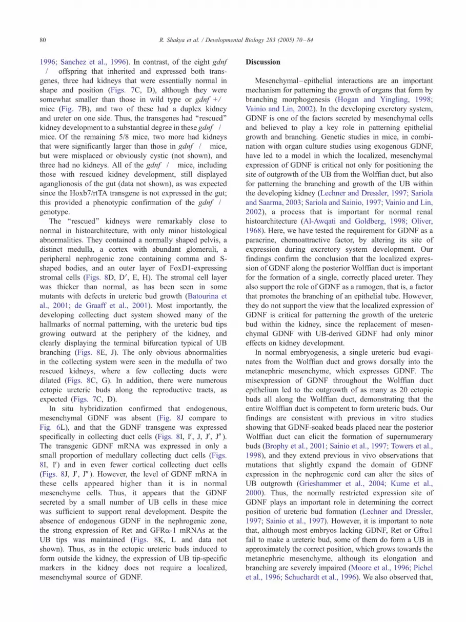
1996; Sanchez et al., 1996). In contrast, of the eight gdnf
�/� offspring that inherited and expressed both trans-
genes, three had kidneys that were essentially normal in
shape and position (Figs. 7C, D), although they were
somewhat smaller than those in wild type or gdnf +/�mice (Fig. 7B), and two of these had a duplex kidney
and ureter on one side. Thus, the transgenes had ‘‘rescued’’
kidney development to a substantial degree in these gdnf�/�mice. Of the remaining 5/8 mice, two more had kidneys
that were significantly larger than those in gdnf�/� mice,
but were misplaced or obviously cystic (not shown), and
three had no kidneys. All of the gdnf�/� mice, including
those with rescued kidney development, still displayed
aganglionosis of the gut (data not shown), as was expected
since the Hoxb7/rtTA transgene is not expressed in the gut;
this provided a phenotypic confirmation of the gdnf�/�genotype.
The ‘‘rescued’’ kidneys were remarkably close to
normal in histoarchitecture, with only minor histological
abnormalities. They contained a normally shaped pelvis, a
distinct medulla, a cortex with abundant glomeruli, a
peripheral nephrogenic zone containing comma and S-
shaped bodies, and an outer layer of FoxD1-expressing
stromal cells (Figs. 8D, DV, E, H). The stromal cell layer
was thicker than normal, as has been seen in some
mutants with defects in ureteric bud growth (Batourina et
al., 2001; de Graaff et al., 2001). Most importantly, the
developing collecting duct system showed many of the
hallmarks of normal patterning, with the ureteric bud tips
growing outward at the periphery of the kidney, and
clearly displaying the terminal bifurcation typical of UB
branching (Figs. 8E, J). The only obvious abnormalities
in the collecting system were seen in the medulla of two
rescued kidneys, where a few collecting ducts were
dilated (Figs. 8C, G). In addition, there were numerous
ectopic ureteric buds along the reproductive tracts, as
expected (Figs. 7C, D).
In situ hybridization confirmed that endogenous,
mesenchymal GDNF was absent (Fig. 8J compare to
Fig. 6L), and that the GDNF transgene was expressed
specifically in collecting duct cells (Figs. 8I, IV, J, JV, JW).The transgenic GDNF mRNA was expressed in only a
small proportion of medullary collecting duct cells (Figs.
8I, IV) and in even fewer cortical collecting duct cells
(Figs. 8J, JV, JW). However, the level of GDNF mRNA in
these cells appeared higher than it is in normal
mesenchyme cells. Thus, it appears that the GDNF
secreted by a small number of UB cells in these mice
was sufficient to support renal development. Despite the
absence of endogenous GDNF in the nephrogenic zone,
the strong expression of Ret and GFRa-1 mRNAs at the
UB tips was maintained (Figs. 8K, L and data not
shown). Thus, as in the ectopic ureteric buds induced to
form outside the kidney, the expression of UB tip-specific
markers in the kidney does not require a localized,
mesenchymal source of GDNF.
Discussion
Mesenchymal–epithelial interactions are an important
mechanism for patterning the growth of organs that form by
branching morphogenesis (Hogan and Yingling, 1998;
Vainio and Lin, 2002). In the developing excretory system,
GDNF is one of the factors secreted by mesenchymal cells
and believed to play a key role in patterning epithelial
growth and branching. Genetic studies in mice, in combi-
nation with organ culture studies using exogenous GDNF,
have led to a model in which the localized, mesenchymal
expression of GDNF is critical not only for positioning the
site of outgrowth of the UB from the Wolffian duct, but also
for patterning the branching and growth of the UB within
the developing kidney (Lechner and Dressler, 1997; Sariola
and Saarma, 2003; Sariola and Sainio, 1997; Vainio and Lin,
2002), a process that is important for normal renal
histoarchitecture (Al-Awqati and Goldberg, 1998; Oliver,
1968). Here, we have tested the requirement for GDNF as a
paracrine, chemoattractive factor, by altering its site of
expression during excretory system development. Our
findings confirm the conclusion that the localized expres-
sion of GDNF along the posterior Wolffian duct is important
for the formation of a single, correctly placed ureter. They
also support the role of GDNF as a ramogen, that is, a factor
that promotes the branching of an epithelial tube. However,
they do not support the view that the localized expression of
GDNF is critical for patterning the growth of the ureteric
bud within the kidney, since the replacement of mesen-
chymal GDNF with UB-derived GDNF had only minor
effects on kidney development.
In normal embryogenesis, a single ureteric bud evagi-
nates from the Wolffian duct and grows dorsally into the
metanephric mesenchyme, which expresses GDNF. The
misexpression of GDNF throughout the Wolffian duct
epithelium led to the outgrowth of as many as 20 ectopic
buds all along the Wolffian duct, demonstrating that the
entire Wolffian duct is competent to form ureteric buds. Our
findings are consistent with previous in vitro studies
showing that GDNF-soaked beads placed near the posterior
Wolffian duct can elicit the formation of supernumerary
buds (Brophy et al., 2001; Sainio et al., 1997; Towers et al.,
1998), and they extend previous in vivo observations that
mutations that slightly expand the domain of GDNF
expression in the nephrogenic cord can alter the sites of
UB outgrowth (Grieshammer et al., 2004; Kume et al.,
2000). Thus, the normally restricted expression site of
GDNF plays an important role in determining the correct
position of ureteric bud formation (Lechner and Dressler,
1997; Sainio et al., 1997). However, it is important to note
that, although most embryos lacking GDNF, Ret or Gfra1
fail to make a ureteric bud, some of them do form a UB in
approximately the correct position, which grows towards the
metanephric mesenchyme, although its elongation and
branching are severely impaired (Moore et al., 1996; Pichel
et al., 1996; Schuchardt et al., 1996). We also observed that,
R. Shakya et al. / Developmental Biology 283 (2005) 70–84 81
even in transgenic fetuses with multiple ectopic buds along
the Wolffian duct, only the bud(s) that formed at or near the
correct position went on to form a ureter with a normal
connection to the kidney. Therefore, even when GDNF is
not correctly localized, other signals limited to the posterior
region, and probably produced by the metanephric mesen-
chyme, appear to contribute to the positioning and out-
growth of the ureter.
One important difference from previous studies is that, in
our experiments, GDNF was produced by the Wolffian duct
itself, that is, by the same epithelial tube that expresses the
GDNF receptors Ret and Gfra1. When the source of GDNF
is a bead or a population of mesenchymal cells outside the
Wolffian duct, it might generate a gradient of GNDF, which
could guide the ureteric bud towards the source of GDNF.
However, GDNF secreted by the Wolffian duct is unlikely
to form the same gradient—it would more likely form an
inverted gradient, with the highest levels of GDNF close to
the duct. Thus, the growth of buds away from the Wolffian
duct does not require a localized external source of GDNF,
but merely that the Wolffian duct cells be exposed to GDNF.
It is also intriguing that, despite the uniform expression of
the BiTetO/lacZ/GDNF transgene along the Wolffian duct
epithelium (Fig. 3C), the buds formed as discrete tubular
outgrowths, rather than as a continuous swelling of the
entire Wolffian duct. This suggests that a region of the
Wolffian duct epithelium that begins to bud in response to
GDNF sends out inhibitory signals that repress budding in
the adjacent region of the duct.
Unlike the ectopic buds induced by GDNF beads in
vitro, which do not branch again after their initial
evagination from the Wolffian duct (Brophy et al., 2001;
Sainio et al., 1997), many ectopic buds in the transgenic
mice continued to grow and branch repeatedly. This is
likely due to the continued expression of GDNF by the
epithelium of the ectopic buds. Many of these buds were
quite far from the developing kidney (and therefore not
exposed to factors secreted by the metanephric mesen-
chyme), and the mesenchymal cells surrounding them did
not display features of metanephric mesenchyme or renal
stroma. Therefore, it appears that the GDNF expressed by
the buds themselves is inducing their continued branching.
In contrast, when FGF-7 (which also signals through a
tyrosine kinase receptor) was expressed in the Wolffian
duct epithelium, it induced uniform swelling of the duct but
not budding or branching (unpublished data). This supports
the view that GDNF has the specific effect of inducing the
Wolffian duct and UB epithelium to branch (Davies et al.,
1999; Pepicelli et al., 1997; Towers et al., 1998; Vega et al.,
1996). The branching of the ectopic UBs did not follow the
stereotypical pattern that occurs within a normal developing
kidney. Atypical patterns of branching morphogenesis is
also seen in other situations in which the UB is induced to
branch outside of its normal relationship with metanephric
mesenchyme, for example, when cultured in an artificial
matrix (Qiao et al., 1999) or recombined with lung
mesenchyme lung (Kispert et al., 1996; Lin et al., 2003).
Therefore, the metanephric mesenchyme imposes pattern on
the growth and branching of the ureteric bud. Whether the
localized expression of GDNF within the metanephric
mesenchyme is a critical determinant of this pattern is a
question we address below.
One aspect of ureteric bud patterning that we found to be
independent of interaction with the metanephric mesen-
chyme was proximal–distal differentiation into tip vs.
trunk. It was previously observed that tip-specific expres-
sion of Ret and Wnt11 was maintained in E11.5 UBs
recombined with lung mesenchyme (Kispert et al., 1996),
although it was possible that localized expression of GDNF
in the lung mesenchyme accounted for this pattern, as both
Ret and Wnt11 are GDNF target genes (Pepicelli et al.,
1997). In our experiments, the normal tip-specific expres-
sion of Ret, Gfra1 and Wnt11 in ectopic buds from the
Wolffian duct, as well as in isolated wild type UBs cultured
without mesenchyme, showed that this pattern is an
intrinsic property of the developing UB. It does not depend
on a localized source of GDNF, as GDNF was expressed
throughout the epithelium of the ectopic buds, or through-
out the medium surrounding the cultured UBs. Nor do
ureteric bud tip and trunk cells represent two entirely
separate cell lineages with different patterns of gene
expression, as we have found that some tip cells give rise
to trunk cells during normal UB growth (Shakya et al.,
2005). How ureteric bud cells ‘‘know’’ whether they are in
the trunk or tip is an interesting problem that remains to be
solved.
To investigate the role of GDNF in patterning ureteric
bud branching morphogenesis within the developing kidney,
we first examined kidneys from newborn transgenic mice on
a wild type background, which misexpressed GDNF in the
UB epithelium while still expressing endogenous GDNF in
the metanephric mesenchyme. The extent of renal defects
varied considerably, from a few severely affected cases with
multiple branched, UB-derived cysts (e.g., Fig. 1), to others
with fewer and smaller cysts (Fig. 6), to many apparently
unaffected cases. This wide range of severity was apparently
a consequence of the variable levels of GDNF transgene
expression. The defects observed in the more severely
affected cases were similar to those previously observed in
transgenic mice that expressed ligand-independent forms of
Ret throughout the UB (de Graaff et al., 2001). This
confirms that the misexpression of GDNF can indeed
perturb the pattern of UB branching morphogenesis,
although this may be due to the elevated overall level of
secreted GDNF rather than to the altered spatial pattern of
expression. While these observations were consistent with
ability of GDNF to stimulate epithelial branching and
growth, they did not provide a clear answer regarding its
importance as a chemoattractive factor. The relatively
normal patterning of the collecting system in most of the
transgenic kidneys might imply that the site of GDNF
expression is not important for UB patterning, or it might

R. Shakya et al. / Developmental Biology 283 (2005) 70–8482
simply be a consequence of relatively weak expression of
the transgene, together with the normal expression of
endogenous GDNF in the peripheral metanephric mesen-
chyme (Fig. 6L). To better address this question, we
generated mice in which the endogenous, mesenchymal
expression of GDNF was eliminated.
In newborn gdnf�/� mice, we found that transgene-
encoded GDNF expressed only in the Wolffian duct and
UB epithelium was sufficient to support the development of
kidneys with many normal features, including a normal
overall shape and histoarchitecture, and a normally-shaped
pelvis, medulla, cortex and nephrogenic zone. Although the
difficulty in obtaining such animals precluded a devel-
opmental time course, the histoarchitecture of the kidney is
thought to reflect the earlier pattern of UB branching and
elongation, suggesting that these processes had occurred in
a largely normal manner. Furthermore, the nephrogenic
zone contained many ureteric bud tips, apparently growing
in the normal peripheral direction, and typical terminal
bifurcations were observed. The abnormal features of these
rescued kidneys were their reduced size compared to the
wild type, thickened peripheral stromal layer, and dilation
of a few collecting ducts. These features have been
previously observed in mice with a hypomorphic Ret
mutation (de Graaff et al., 2001), or mutations in other
genes that reduce Ret expression (Batourina et al., 2001).
We suggest that the level of transgene-encoded GDNF
expression was high enough to support kidney development
throughout most of fetal life, but at later stages, it decreased
to suboptimal levels, leading to the observed minor
abnormalities.
The only GDNF in the rescued kidneys was expressed
(and presumably secreted) by ureteric bud epithelial cells.
Therefore, it is difficult to imagine that the GDNF could
have formed the same concentration gradients as might
occur in a wild type kidney, where GDNF is secreted by the
metanephric mesenchymal cells. While it is conceivable that
binding of secreted GDNF to localized extracellular matrix
components could cause it to concentrate at specific sites
outside the UB, regardless of its site of synthesis, the
available experimental evidence argues against this possi-
bility: when fetal kidneys were incubated with soluble 125I-
labeled GDNF, it bound primarily to the UB tips (Sainio et
al., 1997). Thus, our findings argue against a model in
which gradients of GDNF are required to guide the
centrifugal growth of the UB tips, or one in which the
bifurcation of UB tips is determined by a double gradient of
GDNF in the surrounding mesenchyme (Sariola and
Saarma, 2003). We do not exclude the possibility that
GDNF is one of several factors that together pattern the
growth and branching of the ureteric bud, but if so, it
appears to be redundant with other patterning factors. Other
proteins that are expressed by the metanephric mesenchyme,
and may contribute to this process, include FGFs, HGF,
members of the TGF-h family such as BMP4 and BMP7,
and extracellular matrix components such as Glypican-3, to
name just a few (Carroll and McMahon, 2003; Davies,
2002; Davies and Fisher, 2002; Piscione and Rosenblum,
2002; Shah et al., 2004). While neurturin, another GDNF
family ligand, is an obvious candidate to be redundant with
GDNF, it is expressed only in the UB and thus is unlikely to
serve as a paracrine factor (Davies et al., 1999).
If GDNF is not required as a paracrine factor to pattern
UB branching, for what aspects of kidney development is it
important? GDNF promotes the outgrowth of the UB, and
also provides positional information that helps to determine
the site at which the UB will emerge from the Wolffian duct.
It stimulates the rapid proliferation of ureteric bud tip cells
(Michael and Davies, 2004; Pepicelli et al., 1997), a process
required to form the terminal ampullae, as shown by the
inability of ret�/� cells to contribute to the ampullae in
mosaic kidneys (Shakya et al., 2005). Formation of the
ampulla is an intermediate step in terminal branching by the
UB (Saxen, 1987; Watanabe and Costantini, 2004), and thus
the importance of GDNF/Ret signaling for formation of the
ampulla is consistent with its ability to promote UB
branching. The ability of GDNF expressed in the Wolffian
duct and ureteric bud epithelium to promote the repeated
branching of ectopic ureteric buds, independent of any
interaction with metanephric mesenchyme, also supports its
role as a ramogen. Finally, GDNF plays a continued role in
renal growth, as suggested by the reduced kidney size in
some GDNF heterozygous mutant mice (Cullen-McEwen et
al., 2001) or homozygotes for Ret hypomorphic alleles (de
Graaff et al., 2001; Jijiwa et al., 2004), as well as by studies
with GDNF antibodies (Towers et al., 1998; Vega et al.,
1996) or a Ret–Ig fusion protein (Ehrenfels et al., 1999) in
organ culture.
Acknowledgments
We thank Andreas Zimmer for the GDNF cDNA,
Wolfgang Hillen for the rtTA2S-M2 gene, Mariano Barbacid
for the gdnf mutant mice, and Cathy Mendelsohn and Doris
Herzlinger for critical comments on the manuscript. This
work was supported by a grant to F.C. from the NIDDK.
References
Al-Awqati, Q., Goldberg, M.R., 1998. Architectural patterns in branching
morphogenesis in the kidney. Kidney Int. 54, 1832–1842.
Allen, N.D., Norris, M.L., Surani, M.A., 1990. Epigenetic control of
transgene expression and imprinting by genotype-specific modifiers.
Cell 61, 853–861.
Batourina, E., Gim, S., Bello, N., Shy, M., Clagett-Dame, M., Srinivas,
S., Costantini, F., Mendelsohn, C., 2001. Vitamin A controls
epithelial/mesenchymal interactions through Ret expression. Nat.
Genet. 27, 74–78.
Brophy, P.D., Ostrom, L., Lang, K.M., Dressler, G.R., 2001. Regulation of
ureteric bud outgrowth by Pax2-dependent activation of the glial
derived neurotrophic factor gene. Development 128, 4747–4756.

R. Shakya et al. / Developmental Biology 283 (2005) 70–84 83
Cacalano, G., Farinas, I., Wang, L.C., Hagler, K., Forgie, A., Moore, M.,
Armanini, M., Phillips, H., Ryan, A.M., Reichardt, L.F., Hynes, M.,
Davies, A., Rosenthal, A., 1998. GFRalpha1 is an essential receptor
component for GDNF in the developing nervous system and kidney.
Neuron 21, 53–62.
Carroll, T.J., McMahon, A.P., 2003. Overview: the molecular basis of
kidney development. In: Vize, P.D., Woolf, A.S., Bard, J.B.L. (Eds.),
The Kidney. From Normal Development to Congenital Disease.
Academic Press, Amsterdam, pp. 343–376.
Cullen-McEwen, L.A., Drago, J., Bertram, J.F., 2001. Nephron endowment
in glial cell line-derived neurotrophic factor (GDNF) heterozygous
mice. Kidney Int. 60, 31–36.
Davies, J.A., 2002. Do different branching epithelia use a conserved
developmental mechanism? Bioessays 24, 937–948.
Davies, J.A., Fisher, C.E., 2002. Genes and proteins in renal development.
Exp. Nephrol. 10, 102–113.
Davies, J.A., Millar, C.B., Johnson Jr., E.M., Milbrandt, J., 1999. Neurturin:
an autocrine regulator of renal collecting duct development. Dev. Genet.
24, 284–292.
de Graaff, E., Srinivas, S., Kilkenny, C., D’Agati, V., Mankoo, B.S.,
Costantini, F., Pachnis, V., 2001. Differential activities of the RET
tyrosine kinase receptor isoforms during mammalian embryogenesis.
Genes Dev. 15, 2433–2444.
Ehrenfels, C.W., Carmillo, P.J., Orozco, O., Cate, R.L., Sanicola, M., 1999.
Perturbation of RET signaling in the embryonic kidney. Dev. Genet. 24,
263–272.
Engler, P., Haasch, D., Pinkert, C.A., Doglio, L., Glymour, M., Brinster,
R., Storb, U., 1991. A strain-specific modifier on mouse chromosome
4 controls the methylation of independent transgene loci. Cell 65,
939–947.
Enomoto, H., Araki, T., Jackman, A., Heuckeroth, R.O., Snider, W.D.,
Johnson, E.M.J., Milbrandt, J., 1998. GFRa1-deficient mice have
deficits in the enteric nervous system and kidneys. Neuron 21, 317–324.
Furth, P.A., St Onge, L., Boger, H., Gruss, P., Gossen, M., Kistner, A.,
Bujard, H., Hennighausen, L., 1994. Temporal control of gene
expression in transgenic mice by a tetracycline-responsive promoter.
Proc. Natl. Acad. Sci. U. S. A. 91, 9302–9306.
Gossen, M., Freundlieb, S., Bender, G., Muller, G., Hillen, W., Bujard, H.,
1995. Transcriptional activation by tetracyclines in mammalian cells.
Science 268, 1766–1769.
Grieshammer, U., Le, M., Plump, A.S., Wang, F., Tessier-Lavigne, M.,
Martin, G.R., 2004. SLIT2-mediated ROBO2 signaling restricts kidney
induction to a single site. Dev. Cell 6, 709–717.
Hatini, V., Huh, S.O., Herzlinger, D., Soares, V.C., Lai, E., 1996. Essential
role of stromal mesenchyme in kidney morphogenesis revealed by
targeted disruption of Winged Helix transcription factor BF-2. Genes
Dev. 10, 1467–1478.
Hellmich, H.L., Kos, L., Cho, E.S., Mahon, K.A., Zimmer, A., 1996.
Embryonic expression of glial cell-line derived neurotrophic factor
(GDNF) suggests multiple developmental roles in neural differentiation
and epithelial–mesenchymal interactions. Mech. Dev. 54, 95–105.
Hogan, B.L., Yingling, J.M., 1998. Epithelial/mesenchymal interactions
and branching morphogenesis of the lung. Curr. Opin. Genet. Dev. 8,
481–486.
Hogan, B., Bedington, R., Costantini, F., Lacy, E., 1994. Manipulating the
Mouse Embryo: A Laboratory Manual. Cold Spring Harbor Laboratory,
Cold Spring Harbor.
Jho, E.H., Zhang, T., Domon, C., Joo, C.K., Freund, J.N., Costantini, F.,
2002. Wnt/beta-catenin/Tcf signaling induces the transcription of
Axin2, a negative regulator of the signaling pathway. Mol. Cell. Biol.
22, 1172–1183.
Jijiwa, M., Fukuda, T., Kawai, K., Nakamura, A., Kurokawa, K.,
Murakumo, Y., Ichihara, M., Takahashi, M., 2004. A targeting mutation
of tyrosine 1062 in Ret causes a marked decrease of enteric neurons and
renal hypoplasia. Mol. Cell. Biol. 24, 8026–8036.
Kholodilov, N., Yarygina, O., Oo, T.F., Zhang, H., Sulzer, D., Dauer, W.,
Burke, R.E., 2004. Regulation of the development of mesencephalic
dopaminergic systems by the selective expression of glial cell line-
derived neurotrophic factor in their targets. J. Neurosci. 24, 3136–3146.
Kispert, A., Vainio, S., Shen, L., Rowitch, D.H., McMahon, A.P., 1996.
Proteoglycans are required for maintenance of Wnt-11 expression in the
ureter tips. Development 122, 3627–3637.
Kistner, A., Gossen, M., Zimmermann, F., Jerecic, J., Ullmer, C., Lubbert,
H., Bujard, H., 1996. Doxycycline-mediated quantitative and tissue-
specific control of gene expression in transgenic mice. Proc. Natl. Acad.
Sci. U. S. A. 93, 10933–10938.
Kobayashi, A., Behringer, R.R., 2003. Developmental genetics of the
female reproductive tract in mammals. Nat. Rev., Genet. 4, 969–980.
Kreidberg, J.A., Sariola, H., Loring, J.M., Maeda, M., Pelletier, J.,
Housman, D., Jaenisch, R., 1993. WT-1 is required for early kidney
development. Cell 74, 679–691.
Kress, C., Vogels, R., De, G.W., Bonnerot, C., Meijlink, F., Nicolas, J.F.,
Deschamps, J., 1990. Hox-2.3 upstream sequences mediate lacZ
expression in intermediate mesoderm derivatives of transgenic mice.
Development 109, 775–786.
Kume, T., Deng, K., Hogan, B.L., 2000. Murine forkhead/winged helix
genes Foxc1 (Mf1) and Foxc2 (Mfh1) are required for the early
organogenesis of the kidney and urinary tract. Development 127,
1387–1395.
Lechner, M.S., Dressler, G.R., 1997. The molecular basis of embryonic
kidney development. Mech. Dev. 62, 105–120.
Lin, Y., Zhang, S., Tuukkanen, J., Peltoketo, H., Pihlajaniemi, T., Vainio, S.,
2003. Patterning parameters associated with the branching of the
ureteric bud regulated by epithelial–mesenchymal interactions. Int. J.
Dev. Biol. 47, 3–13.
Mackie, G.G., Stephens, F.D., 1975. Duplex kidneys: a correlation of
renal dysplasia with position of the ureteral orifice. J. Urol. 114,
274–280.
Mendelsohn, C., Batourina, E., Fung, S., Gilbert, T., Dodd, J., 1999.
Stromal cells mediate retinoid-dependent functions essential for renal
development. Development 126, 1139–1148.
Michael, L., Davies, J.A., 2004. Pattern and regulation of cell proliferation
during murine ureteric bud development. J. Anat. 204, 241–255.
Moore, M.W., Klein, R.D., Farinas, I., Sauer, H., Armanini, M., Phillips,
H., Reichardt, L.F., Ryan, A.M., Carver-Moore, K., Rosenthal, A.,
1996. Renal and neuronal abnormalities in mice lacking GDNF. Nature
382, 76–79.
Natarajan, D., Marcos-Gutierrez, C., Pachnis, V., de Graaff, E., 2002.
Requirement of signalling by receptor tyrosine kinase RET for the
directed migration of enteric nervous system progenitor cells during
mammalian embryogenesis. Development 129, 5151–5160.
Niederreither, K., Fraulob, V., Garnier, J.M., Chambon, P., Dolle, P., 2002.
Differential expression of retinoic acid-synthesizing (RALDH) enzymes
during fetal development and organ differentiation in the mouse. Mech.
Dev. 110, 165–171.
Oliver, J., 1968. Nephrons and Kidneys: A Quantitative Study of
Developmental and Evolutionary Mammalian Renal Architectonics.
Hoeber Medical Division Harper and Row, New York.
Pachnis, V., Mankoo, B.S., Costantini, F., 1993. Expression of the c-ret
proto-oncogene during mouse embryogenesis. Development 119,
1005–1017.
Pepicelli, C.V., Kispert, A., Rowitch, D.H., McMahon, A.P., 1997. GDNF
induces branching and increased cell proliferation in the ureter of the
mouse. Dev. Biol. 192, 193–198.
Pichel, J.G., Shen, L., Sheng, H.Z., Granholm, A.C., Drago, J., Grinberg,
A., Lee, E.J., Huang, S.P., Saarma, M., Hoffer, B.J., Sariola, H.,
Westphal, H., 1996. Defects in enteric innervation and kidney develop-
ment in mice lacking GDNF. Nature 382, 73–76.
Piscione, T.D., Rosenblum, N.D., 2002. The molecular control of renal
branching morphogenesis: current knowledge and emerging insights.
Differentiation 70, 227–246.
Qiao, J., Sakurai, H., Nigam, S.K., 1999. Branching morphogenesis
independent of mesenchymal–epithelial contact in the developing
kidney. Proc. Natl. Acad. Sci. U. S. A. 96, 7330–7335.

R. Shakya et al. / Developmental Biology 283 (2005) 70–8484
Sainio, K., Suvanto, P., Davies, J., Wartiovaara, J., Wartiovaara, K., Saarma,
M., Arumae, U., Meng, X., Lindahl, M., Pachnis, V., Sariola, H., 1997.
Glial-cell-line-derived neurotrophic factor is required for bud initiation
from ureteric epithelium. Development 124, 4077–4087.
Sanchez, M.P., Silos-Santiago, I., Frisen, J., He, B., Lira, S.A., Barbacid,
M., 1996. Renal agenesis and the absence of enteric neurons in mice
lacking GDNF. Nature 382, 70–73.
Sariola, H., Saarma, M., 2003. Novel functions and signalling pathways for
GDNF. J. Cell Sci. 116, 3855–3862.
Sariola, H., Sainio, K., 1997. The tip-top branching ureter. Curr. Opin. Cell
Biol. 9, 877–884.
Saxen, L., 1987. Organogenesis of the Kidney. Cambridge Univ. Press,
Cambridge.
Schuchardt, A., D’Agati, V., Larsson-Blomberg, L., Costantini, F., Pachnis,
V., 1994. Defects in the kidney and enteric nervous system of mice
lacking the tyrosine kinase receptor Ret. Nature 367, 380–383.
Schuchardt, A., D’Agati, V., Pachnis, V., Costantini, F., 1996. Renal
agenesis and hypodysplasia in ret-k-mutant mice result from defects in
ureteric bud development. Development 122, 1919–1929.
Shah, M.M., Sampogna, R.V., Sakurai, H., Bush, K.T., Nigam, S.K., 2004.
Branching morphogenesis and kidney disease. Development 131,
1449–1462.
Shakya, R., Watanabe, T., Costantini, F., 2005. The role of GDNF/Ret
signaling in ureteric bud cell fate and branching morphogenesis. Dev.
Cell 8, 65–74.
Srinivas, S., Goldberg, M.R., Watanabe, T., D’Agati, V., al-Awqati, Q.,
Costantini, F., 1999a. Expression of green fluorescent protein in the
ureteric bud of transgenic mice: a new tool for the analysis of ureteric
bud morphogenesis. Dev. Genet. 24, 241–251.
Srinivas, S., Wu, Z., Chen, C.M., D’Agati, V., Costantini, F., 1999b.
Dominant effects of RET receptor misexpression and ligand-independ-
ent RET signaling on ureteric bud development. Development 126,
1375–1386.
Suvanto, P., Hiltunen, J.O., Arumae, U., Moshnyakov, M., Sariola, H.,
Sainio, K., Saarma, M., 1996. Localization of glial cell line-derived
neurotrophic factor (GDNF) mRNA in embryonic rat by in situ
hybridization. Eur. J. Neurosci. 8, 816–822.
Tang, M.J., Worley, D., Sanicola, M., Dressler, G.R., 1998. The RET-
glial cell-derived neurotrophic factor (GDNF) pathway stimulates
migration and chemoattraction of epithelial cells. J. Cell Biol. 142,
1337–1345.
Towers, P.R., Woolf, A.S., Hardman, P., 1998. Glial cell line-derived
neurotrophic factor stimulates ureteric bud outgrowth and enhances
survival of ureteric bud cells in vitro. Exp. Nephrol. 6, 337–351.
Urlinger, S., Baron, U., Thellmann, M., Hasan, M.T., Bujard, H., Hillen,
W., 2000. Exploring the sequence space for tetracycline-dependent
transcriptional activators: novel mutations yield expanded range and
sensitivity. Proc. Natl. Acad. Sci. U. S. A. 97, 7963–7968.
Vainio, S., Lin, Y., 2002. Coordinating early kidney development: lessons
from gene targeting. Nat. Rev., Genet. 3, 533–543.
Vega, Q.C., Worby, C.A., Lechner, M.S., Dixon, J.E., Dressler, G.R., 1996.
Glial cell line-derived neurotrophic factor activates the receptor tyrosine
kinase RET and promotes kidney morphogenesis. Proc. Natl. Acad. Sci.
U. S. A. 93, 10657–10661.
Watanabe, T., Costantini, F., 2004. Real-time analysis of ureteric bud
branching morphogenesis in vitro. Dev. Biol. 271, 98–108.
Wilkinson, D.G., 1992. Whole mount in situ hybridization of vertebrate
embryos. In: Wilkinson, D.G. (Ed.), In Situ Hybridization: A Practical
Approach. IRL Press, Oxford, pp. 75–83.
Young, H.M., Hearn, C.J., Farlie, P.G., Canty, A.J., Thomas, P.Q.,
Newgreen, D.F., 2001. GDNF is a chemoattractant for enteric neural
cells. Dev. Biol. 229, 503–516.