invitro and in vivo study of sustained nitric oxide release coating using diazeniumdiolate-doped...
TRANSCRIPT

Journal ofMaterials Chemistry B
PAPER
Publ
ishe
d on
03
June
201
3. D
ownl
oade
d by
Uni
vers
ity o
f M
ichi
gan
Lib
rary
on
19/0
6/20
13 1
5:55
:51.
View Article OnlineView Journal
aDepartment of Surgery, University of MichibDepartment of Chemistry, University of Mic
MI 48109, USA. E-mail: [email protected] of Pediatrics and Communi
Medical Center, Ann Arbor, MI, USA
† Electronic supplementary informa10.1039/c3tb20277a
Cite this: DOI: 10.1039/c3tb20277a
Received 27th February 2013Accepted 30th May 2013
DOI: 10.1039/c3tb20277a
www.rsc.org/MaterialsB
This journal is ª The Royal Society of
In vitro and in vivo study of sustained nitric oxide releasecoating using diazeniumdiolate-doped poly(vinylchloride) matrix with poly(lactide-co-glycolide)additive†
Hitesh Handa,a Elizabeth J. Brisbois,b Terry C. Major,a Lahdan Refahiyat,b
Kagya A. Amoako,a Gail M. Annich,c Robert H. Bartletta and Mark E. Meyerhoff*b
Nitric oxide (NO) is an endogenous vasodilator as well as natural inhibitor of platelet adhesion
and activation that can be released from a NO donor species, such as diazeniumdiolated dibutyl-
hexanediamine (DBHD/N2O2) within a polymer coating. In this study, various Food and Drug
Administration approved poly(lactic-co-glycolic acid) (PLGA) species were evaluated as additives to
promote a prolonged NO release from DBHD/N2O2 within a plasticized poly(vinyl chloride) (PVC) matrix.
When using an ester-capped PLGA additive with a slow hydrolysis time, the resulting coatings
continuously release between 7 and 18 � 10�10 mol cm�2 min�1 NO for 14 days at 37 �C in PBS buffer.
The corresponding pH changes within the polymer films were visualized using pH sensitive indicators
and are shown to correlate with the extended NO release pattern. The optimal combined
diazeniumdiolate/PLGA-doped NO release (NOrel) PVC coating was evaluated in vitro and its effect on
the hemodynamics was also studied within a 4 h in vivo extracorporeal circulation (ECC) rabbit model
of thrombogenicity. Four out of 7 control circuits clotted within 3 h, whereas all the NOrel coated
circuits were patent after 4 h. Platelet counts on the NOrel ECC were preserved (79 � 11% compared to
54 � 6% controls). The NOrel coatings showed a significant decrease in the thrombus area as compared
to the controls. Results suggest that by using ester-capped PLGAs as additives to a conventional
plasticized PVC material containing lipophilic diazeniumdiolates, the NO release can be prolonged for
up to 2 weeks by controlling the pH within the organic phase of the coating.
1 Introduction
Blood/material interaction is critical to the success of implant-able medical devices, ranging from simple catheters, stents andgras, to complex extracorporeal articial organs that are usedin thousands of patients every day.1 Thrombosis is one of theprimary problems associated with clinical application of bloodcontacting materials. Despite a thorough understanding of themechanisms of blood–surface interactions and decades ofbioengineering research effort, the ideal non-thrombogenicprosthetic surface remains an unsolved problem.2 Over the last50 years much has been learned about foreign surface-inducedthrombosis and attempts to prevent it with systemic anti-coagulation and surface modications. Surface modications
gan Medical Center, Ann Arbor, MI, USA
higan, 930 N. University Ave, Ann Arbor,
edu; Tel: +1 734 764 2169
cable Diseases, University of Michigan
tion (ESI) available. See DOI:
Chemistry 2013
have included using pure, very smooth silicone rubber3 orpolyurethane,4 pre-exposure of the surfaces to albumin5 andother coating proteins,6 and surface binding of heparin in anionic7 as well as a covalent fashion.8 Despite extensive researchto develop a non-thrombogenic surface that mimics the endo-thelium, none of these modications have been successful.
In 1993 Radomski and Moncada9 described nitric oxide (NO)as one of two potent vasodilators secreted by normal endothe-lium that has the ability to inhibit platelet adhesion andaggregation to the blood vessel wall. The amount of NO releasedfrom the surface of a normal and stimulated endothelium hasbeen estimated to be in the range of 0.5–4 � 10�10 mol cm�2
min�1.10 Nitric oxide has been extensively studied for itsinhibitory effects on circulating platelet and monocyteactivation that leads to aggregation and ultimately initiationof thrombosis.11–14 A wide range of NO donors such asS-nitrosothiols,15,16 N-hydroxy-N-nitrosoamines,17 N-dia-zeniumdiolates18,19 and nitrosyl metal complexes20 have beenstudied over the past decade, as a means to release NO either bysystemic infusion21 or local release from a polymer surface.22
Despite the promising potential of NO releasing materials, their
J. Mater. Chem. B

Journal of Materials Chemistry B Paper
Publ
ishe
d on
03
June
201
3. D
ownl
oade
d by
Uni
vers
ity o
f M
ichi
gan
Lib
rary
on
19/0
6/20
13 1
5:55
:51.
View Article Online
development has been hindered due to challenges in prolong-ing the NO release beyond a few days.
Diazeniumdiolates have been one of the most widely studiedNO donors, which release NO through proton23 or thermal24
driven mechanisms. Prior work has shown that NO can bereleased from the NO donor compound, diazeniumdiolateddibutylhexanediamine (DBHD/N2O2), when this species isincorporated into hydrophobic polymer lms.22 While DBHD/N2O2 is an excellent donor for incorporation into polymers tocreate NO release coatings, the loss of NO from this moleculecreates free lipophilic amine species within the polymer thatreact with water, thereby increasing the pH within the organicpolymer phase which effectively turns off the NO productionbefore a signicant fraction of the total NO payload has beenreleased.25 In earlier work, to overcome this complication, tet-rakis-( p-chlorophenyl)-borate was employed as an additive tomaintain a low enough pH within the organic polymer phaseand to promote a sustained NO ux.22,25 However, in a recentcell proliferation study it was shown that tetrakis-( p-chloro-phenyl)-borate was not successful in prolonging the NO releasebeyond few days and, further, this compound was found to becytotoxic towards endothelial and smooth muscle cells.26
The work presented herein focuses on a completely differentapproach to address this pH control problem and hence togreatly prolong the NO release, one of the key challenges withNO delivery materials. The method involves the use of poly-(lactide-co-glycolide) (PLGA) species as additives to help stabi-lize the pH within the organic phase polymeric coatings. Theaddition of PLGA can be used to control the ux of NO emittedfrom polymers containing diazeniumdiolate species by helpingto control the pH within the polymer phase.27,28 PLGA is abiodegradable and biocompatible polymer29–31 that can be usedas a small additive to sustain the NO ux for a prolonged periodby the slow formation of lactic and glycolic acid within the basepolymer layer of the coating. Ester linkages of the PLGA areslowly hydrolyzed as small amounts of water penetrate thepolymer from the surrounding aqueous environment togenerate lactic and glycolic acid within the polymer matrix.32,33
The presence of this continuous acid production reactioncompensates for the increase in pH from generation of organo-ammonium hydroxide (reaction of liberated free amines fromDBHD with water in the polymer lm) from the NO releasereaction, thereby maintaining a greater rate of NO release forlonger periods of time.
In the literature there are reports of two main strategies thatutilize PLGA to deliver NO from diazeniumdiolate species. Therst strategy is dispersing the NO donor compound within thePLGA matrix, creating a completely biodegradable NO releasematerial. Yoo et al. employed PLGA microparticles with an NOdonor (diethylenetriamine diazeniumdiolate (DETA NONOate),a low molecular weight diazeniumdiolate) within to deliver NOfor a very short 6 h period.34 In another study, Cai et al. studiedin vitro effects of NO releasing PLGA lms on biolm forma-tion.35 In this study the authors dispersed the NO donorcompound in a completely hydrolysable PLGA matrix, howeverdue to the toxicity concerns of the product amine, leaching is apotential limitation of these lms. In the second strategy, Zhou
J. Mater. Chem. B
and Meyerhoff have shown that PLGA has the potential to act asa proton donor to enhance the release of NO from a polymermaterial that had covalently linked diazeniumdiolate groups fora relatively short 20 h period.27 Although these approaches havehad some limited success, prolonging the NO release stillremains one of the great challenges preventing use of NOreleasing materials in clinical application.
Herein, we report the use of PLGA as an additive in apoly(vinyl chloride) (PVC)/dioctyl sebacate (DOS) matrix tocontrol NO release from a lipophilic diazeniumdiolate (DBHD/N2O2) species added to the organic phase of the coating toaddress the need to prolong the NO release beyond a few days.A comparison was made between PLGAs that have differentend group chemistries; either a free carboxylic acid or an esterend group. The hydrolysis rate of the PLGA is primarilydetermined by the copolymer ratio, the nature of the endgroup (e.g., free acid or ester), and molecular weight. It isshown that the hydrolysis rate and acid content of the givenPLGA species used greatly inuences the NO release prole.Optimized lm compositions prepared with ester end-cappedPLGA additives can sustain high uxes of NO for up to 14 days.Further, incorporation of pH indicators in the coatingsprovides a means to correlate the NO release with the observedpH changes within the PVC/DOS matrix. The newly formulatedPLGA/diazeniumdiolate-doped PVC/DOS coatings were furthertested in an in vivo rabbit extracorporeal circulation (ECC)model to assess platelet count and function preservation, inaddition to reduction in the thrombus coverage area. The newPLGA-doped NOrel PVC coatings could provide a breakthroughfor achieving sustained preservation of circulating platelets, animportant goal for longer-term ECC situations, such as ECMO,or other blood-contacting devices (e.g., catheters, vasculargras, etc.).
2 Materials and methods2.1 Materials
Tygon� poly(vinyl chloride) (PVC) tubing was purchased fromFisher Healthcare (Houston, TX). High molecular weightpoly(vinyl chloride) (PVC), dioctyl sebacate (DOS), anhydroustetrahydrofuran (THF), anhydrous acetonitrile, bromothymolblue, bromocresol green, sodium chloride, potassium chloride,sodium phosphate dibasic, and potassium phosphate mono-basic were purchased from Sigma-Aldrich Chemical Company(St. Louis, MO). Various poly(D,L-lactide-co-glycolide) materials,including 5050DLG1A and 5050DLG7E, were obtained fromSurModics Pharmaceuticals Inc. (Birmingham, AL). N,N0-Dibu-tyl-1,6-hexanediamine (DBHD) was purchased from Alfa Aesar(Ward Hill, MA). DBHD/N2O2 was synthesized by treating DBHDwith 80 psi NO gas purchased from Cryogenic Gases (Detroit,MI) at room temperature for 48 h, as previously described.25
2.2 Preparation of NOrel lms for NO release and pH studies
As mentioned earlier, the main focus of this work is to use PLGAas an additive in a PVC/DOS polymer matrix to prolong the NOrelease from a lipophilic DBHD/N2O2 species (Fig. 1). In this
This journal is ª The Royal Society of Chemistry 2013
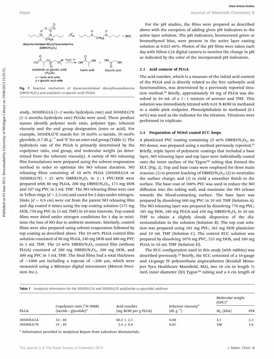
Fig. 1 Reaction mechanism of diazeniumdiolated dibutylhexanediamine(DBHD/N2O2) and poly(lactic-co-glycolic acid) (PLGA).
Paper Journal of Materials Chemistry B
Publ
ishe
d on
03
June
201
3. D
ownl
oade
d by
Uni
vers
ity o
f M
ichi
gan
Lib
rary
on
19/0
6/20
13 1
5:55
:51.
View Article Online
study, 5050DLG1A (1–2 weeks hydrolysis rate) and 5050DLG7E(1–2 months hydrolysis rate) PLGAs were used. These productnames identify polymer mole ratio, polymer type, inherentviscosity and the end group designation (ester or acid). Forexample, 5050DLG7E stands for: 50 mol% DL-lactide, 50 mol%glycolide, 0.7 dL g�1 and ‘E’ for an ester end group (Table 1). Thehydrolysis rate of the PLGA is primarily determined by thecopolymer ratio, end group, and molecular weight (as deter-mined from the inherent viscosity). A variety of NO releasinglm formulations were prepared using the solvent evaporationmethod in order to optimize the NO release duration. NOreleasing lms consisting of 10 wt% PLGA (5050DLG1A or5050DLG7E) + 25 wt% DBHD/N2O2 in 2 : 1 PVC/DOS wereprepared with 80 mg PLGA, 200 mg DBHD/N2O2, 173 mg DOSand 347 mg PVC in 5 mL THF. The NO releasing lms were castin Teon rings (d¼ 2.5 cm) and cured for 2 days under nitrogen.Disks (d ¼ 0.9 cm) were cut from the parent NO releasing lmand dip coated 8 times using the top coating solution (375 mgDOS, 750 mg PVC in 15mL THF) in 10 min intervals. Top coatedlms were dried under nitrogen conditions for 1 day to mini-mize the loss of NO due to ambient moisture. Similarly, controllms were also prepared using solvent evaporation followed bytop coating as described above. The 10 wt% PLGA control lmsolution consisted of 80 mg PLGA, 240 mg DOS and 480 mg PVCin 5 mL THF. The 25 wt% DBHD/N2O2 control lm (withoutPLGA) consisted of 200 mg DBHD/N2O2, 200 mg DOS, and400 mg PVC in 5 mL THF. The nal lms had a total thicknessof �1000 mm including a topcoat of �200 mm, which weremeasured using a Mitutoyo digital micrometer (Metron Preci-sion Inc.).
Table 1 Analytical information for the 5050DLG1A and 5050DLG7E poly(lacide-co
PLGACopolymer ratio (1H NMR)(lactide : glycolide)a
Acid number(mg KOH per
5050DLG1A 52 : 48 60.4 � 2.55050DLG7E 51 : 49 2.4 � 0.8
a Information provided in Analytical Report from Lakeshore Biomaterials
This journal is ª The Royal Society of Chemistry 2013
For the pH studies, the lms were prepared as describedabove with the exception of adding given pH indicators to theactive layer solution. The pH indicators, bromocresol green orbromothymol blue, were present in the active layer castingsolution at 0.025 wt%. Photos of the pH lms were taken eachday with Nikon L24 digital camera to monitor the change in pHas indicated by the color of the incorporated pH indicators.
2.3 Acid content of PLGA
The acid number, which is a measure of the initial acid contentof the PLGA and is directly related to the free carboxylic acidfunctionalities, was determined by a previously reported titra-tion method.36 Briey, approximately 50 mg of PLGA was dis-solved in 10 mL of a 1 : 1 mixture of acetone and THF. Thissolution was immediately titrated with 0.01 N KOH in methanolto a stable pink endpoint. Phenolphthalein in methanol (0.1wt%) was used as the indicator for the titration. Titrations wereperformed in triplicate.
2.4 Preparation of NOrel coated ECC loops
A plasticized PVC coating containing 25 wt% DBHD/N2O2, anNO donor, was prepared using a method previously reported.22
Briey, triple layers of polymeric coatings that included a baselayer, NO releasing layer and top layer were individually coatedonto the inner surface of the Tygon� tubing that formed theECC (Fig. 2). Top and base coats were employed for three mainreasons: (1) to prevent leaching of DBHD/N2O2; (2) to neutralizethe surface charge; and (3) to yield a smoother nish to thesurface. The base coat of 100% PVC was used to reduce the NOdiffusion into the tubing wall, and maximize the NO releasetowards the blood-contacting surface. The base layer wasprepared by dissolving 600 mg PVC in 10 mL THF (Solution A).The NO releasing layer was prepared by dissolving 770 mg PVC,385 mg DOS, 180 mg PLGA and 450 mg DBHD/N2O2 in 10 mLTHF to obtain a slightly cloudy dispersion of the dia-zeniumdiolate in the solution (Solution B). The top coat solu-tion was prepared using 181 mg PVC, 362 mg DOS plasticizerand 10 mL THF (Solution C). The control ECC solution wasprepared by dissolving 1070 mg PVC, 535 mg DOS, and 180 mgPLGA in 10 mL THF (Solution D).
The ECC conguration used in this study (with rabbits) wasdescribed previously.22 Briey, the ECC consisted of a 16-gaugeand 14-gauge IV polyurethane angiocatheters (Kendall Mono-ject Tyco Healthcare Manseld, MA), two 16 cm in length ¼inch inner diameter (ID) Tygon� tubing and a 8 cm length of
-glycolide) additives
g PLGA)Inherent viscositya
(dL g�1)
Molecular weight(GPC)a
Mw (kDa) PDI
0.08 4.1 2.10.65 106 1.6
.
J. Mater. Chem. B
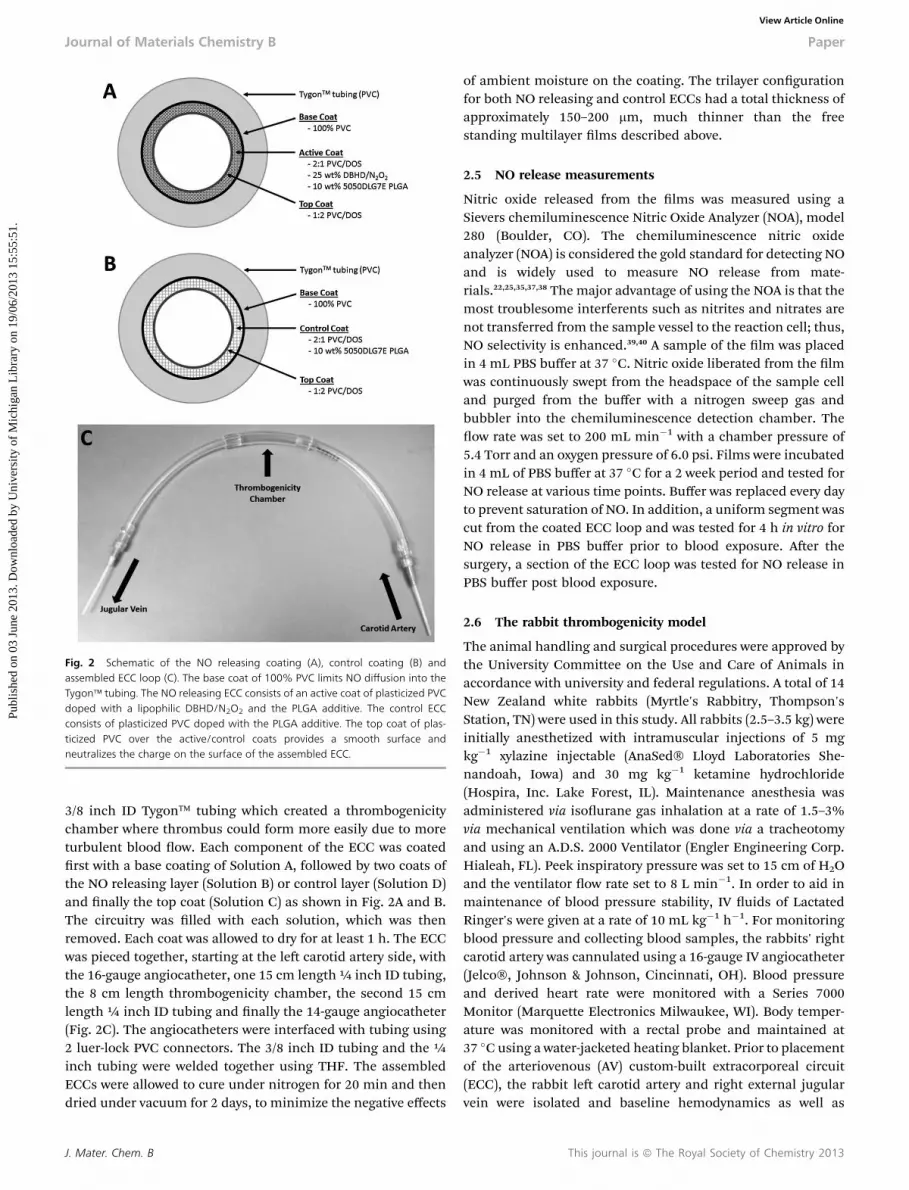
Fig. 2 Schematic of the NO releasing coating (A), control coating (B) andassembled ECC loop (C). The base coat of 100% PVC limits NO diffusion into theTygon� tubing. The NO releasing ECC consists of an active coat of plasticized PVCdoped with a lipophilic DBHD/N2O2 and the PLGA additive. The control ECCconsists of plasticized PVC doped with the PLGA additive. The top coat of plas-ticized PVC over the active/control coats provides a smooth surface andneutralizes the charge on the surface of the assembled ECC.
Journal of Materials Chemistry B Paper
Publ
ishe
d on
03
June
201
3. D
ownl
oade
d by
Uni
vers
ity o
f M
ichi
gan
Lib
rary
on
19/0
6/20
13 1
5:55
:51.
View Article Online
3/8 inch ID Tygon� tubing which created a thrombogenicitychamber where thrombus could form more easily due to moreturbulent blood ow. Each component of the ECC was coatedrst with a base coating of Solution A, followed by two coats ofthe NO releasing layer (Solution B) or control layer (Solution D)and nally the top coat (Solution C) as shown in Fig. 2A and B.The circuitry was lled with each solution, which was thenremoved. Each coat was allowed to dry for at least 1 h. The ECCwas pieced together, starting at the le carotid artery side, withthe 16-gauge angiocatheter, one 15 cm length ¼ inch ID tubing,the 8 cm length thrombogenicity chamber, the second 15 cmlength ¼ inch ID tubing and nally the 14-gauge angiocatheter(Fig. 2C). The angiocatheters were interfaced with tubing using2 luer-lock PVC connectors. The 3/8 inch ID tubing and the ¼inch tubing were welded together using THF. The assembledECCs were allowed to cure under nitrogen for 20 min and thendried under vacuum for 2 days, to minimize the negative effects
J. Mater. Chem. B
of ambient moisture on the coating. The trilayer congurationfor both NO releasing and control ECCs had a total thickness ofapproximately 150–200 mm, much thinner than the freestanding multilayer lms described above.
2.5 NO release measurements
Nitric oxide released from the lms was measured using aSievers chemiluminescence Nitric Oxide Analyzer (NOA), model280 (Boulder, CO). The chemiluminescence nitric oxideanalyzer (NOA) is considered the gold standard for detecting NOand is widely used to measure NO release from mate-rials.22,25,35,37,38 The major advantage of using the NOA is that themost troublesome interferents such as nitrites and nitrates arenot transferred from the sample vessel to the reaction cell; thus,NO selectivity is enhanced.39,40 A sample of the lm was placedin 4 mL PBS buffer at 37 �C. Nitric oxide liberated from the lmwas continuously swept from the headspace of the sample celland purged from the buffer with a nitrogen sweep gas andbubbler into the chemiluminescence detection chamber. Theow rate was set to 200 mL min�1 with a chamber pressure of5.4 Torr and an oxygen pressure of 6.0 psi. Films were incubatedin 4 mL of PBS buffer at 37 �C for a 2 week period and tested forNO release at various time points. Buffer was replaced every dayto prevent saturation of NO. In addition, a uniform segment wascut from the coated ECC loop and was tested for 4 h in vitro forNO release in PBS buffer prior to blood exposure. Aer thesurgery, a section of the ECC loop was tested for NO release inPBS buffer post blood exposure.
2.6 The rabbit thrombogenicity model
The animal handling and surgical procedures were approved bythe University Committee on the Use and Care of Animals inaccordance with university and federal regulations. A total of 14New Zealand white rabbits (Myrtle's Rabbitry, Thompson'sStation, TN) were used in this study. All rabbits (2.5–3.5 kg) wereinitially anesthetized with intramuscular injections of 5 mgkg�1 xylazine injectable (AnaSed� Lloyd Laboratories She-nandoah, Iowa) and 30 mg kg�1 ketamine hydrochloride(Hospira, Inc. Lake Forest, IL). Maintenance anesthesia wasadministered via isourane gas inhalation at a rate of 1.5–3%via mechanical ventilation which was done via a tracheotomyand using an A.D.S. 2000 Ventilator (Engler Engineering Corp.Hialeah, FL). Peek inspiratory pressure was set to 15 cm of H2Oand the ventilator ow rate set to 8 L min�1. In order to aid inmaintenance of blood pressure stability, IV uids of LactatedRinger's were given at a rate of 10 mL kg�1 h�1. For monitoringblood pressure and collecting blood samples, the rabbits' rightcarotid artery was cannulated using a 16-gauge IV angiocatheter(Jelco�, Johnson & Johnson, Cincinnati, OH). Blood pressureand derived heart rate were monitored with a Series 7000Monitor (Marquette Electronics Milwaukee, WI). Body temper-ature was monitored with a rectal probe and maintained at37 �C using a water-jacketed heating blanket. Prior to placementof the arteriovenous (AV) custom-built extracorporeal circuit(ECC), the rabbit le carotid artery and right external jugularvein were isolated and baseline hemodynamics as well as
This journal is ª The Royal Society of Chemistry 2013

Paper Journal of Materials Chemistry B
Publ
ishe
d on
03
June
201
3. D
ownl
oade
d by
Uni
vers
ity o
f M
ichi
gan
Lib
rary
on
19/0
6/20
13 1
5:55
:51.
View Article Online
arterial blood pH, pCO2, pO2, and total hemoglobin weremeasured using an ABL 825 blood-gas analyzer. In addition,baseline blood samples were collected for platelet and totalwhite blood cell (WBC) counts which were measured on aCoulter Counter Z1 (Coulter Electronics Hialeah, FL). Plasmabrinogen levels were determined using a Dade Behring BCSCoagulation Analyzer (Siemans Deereld, IL), activated clottingtimes (ACT) were monitored using a Hemochron Blood Coag-ulation System Model 801 (International Technidyne Corp.Edison, NJ), platelet function was assessed using a Chrono-Logoptical aggregometer model 490 (Havertown, PA).
Aer baseline blood measurements, the custom-built ECCwas placed into position by cannulating the le carotid arteryfor ECC inow and the right external jugular vein for ECCoutow. The ow through the ECC was initiated by unclampingthe arterial and venous sides of ECC and blood ow in circuitwas monitored with an ultrasonic ow probe and ow meter(Transonic HT207 Ithaca, NY). Animals were not systemicallyanticoagulated during the experiments.
Aer 4 h on ECC, the circuits were clamped, removed fromanimal, rinsed with 60 mL of saline and drained. Any residualthrombus in the larger tubing of ECC (i.e., thrombogenicitychamber) was photographed and the degree of thrombus imagewas quantitated using ImageJ imaging soware from NationalInstitutes of Health (Bethesda, MD). Prior to euthanasia, allanimals were given a dose of 400 U kg�1 sodium heparin toprevent necrotic thrombosis. The animals were euthanizedusing a dose of Fatal Plus (130 mg kg�1 sodium pentobarbital)(Vortech Pharmaceuticals Dearborn, MI). All animals under-went gross necropsy aer being euthanized, including exami-nation of the lungs, heart, liver and spleen for any signs ofthromboembolic events.
2.7 Blood sampling
Rabbit whole blood samples were collected in non-anti-coagulated 1 cm3 syringes for ACT, 3.2% sodium citrate vacu-tainers (Becton, Dickinson. Franklin Lakes, NJ) in 3 cm3
volumes for cell counts, aggregometry, and 1 cm3 syringescontaining 40 U per mL of sodium heparin (APP Pharmaceuti-cals, LLC Schaumburg, IL) for blood-gas analysis. Following theinitiation of ECC blood ow, blood samples were collected everyhour for 4 h for ex vivo measurements. Samples were usedwithin 2 h of collection to avoid any activation of platelets,monocytes or plasma brinogen.
2.8 Platelet aggregometry
Rabbit platelet aggregation was assayed based on the Born'sturbidimetric method using a Chrono-Log optical aggreg-ometer. Briey, citrated blood (1 : 10 blood to ACD) wascollected (6 mL) and platelet-rich plasma (PRP) was obtained bycentrifugation at 110 � g for 15 min. Platelet-poor plasma (PPP)was obtained by another centrifugation of the PRP-removedblood sample at 2730 � g for 15 min and was used to normalizethe PRP for aggregation. Normalized PRP was incubated for10 min at 37 �C and then 25 mg mL�1 collagen (Chrono-PAR#385 Havertown, PA) was added. As shown by Major et al., the
This journal is ª The Royal Society of Chemistry 2013
percentage of aggregation was determined 3 min aer theaddition of collagen using Chrono-Log Aggrolink soware.22,41
2.9 Statistical analysis
Data are expressed as mean � SEM (standard error of themean). Comparison of ECC results between the various NOreland control polymer groups were analyzed by a one-way ANOVAwith a multiple comparison of means using Student's t-test.All statistical analyses were performed using the statisticalprogram SAS JMP (SAS Institute Cary, NC). Values of p < 0.05were considered statistically signicant for all tests.
3 Results and discussions3.1 In vitro NO release from lms containing DBHD/N2O2 inPVC/DOS with various PLGA additives
The diazeniumdiolate species investigated here, DBHD/N2O2,decomposes to generate NO primarily by a proton-drivenmechanism.25 A tetrakis-(p-chlorophenyl)-borate derivative wasused previously as a lipophilic additive counteranion to stabi-lize the pH within NO releasing polymers prepared with DBHD/N2O2.25 However, the borate derivative is not an ideal additivebecause of its toxicity.26 In this study, PLGA additives withvarying hydrolysis rates were used as a replacement to theborate derivative to act as a proton donor source to control theNO release from DBHD/N2O2-doped PVC coatings. It is wellknown that, in the presence of water, the ester bonds in PLGAhydrolyze to yield lactic and glycolic acids, and that PLGA is awidely used biodegradable/biocompatible polymer that hasbeen approved by FDA for numerous products.42
The lms used in this study had a three layer conguration:base coat, active coat and top coat. The base and top-coat con-sisted of PVC/DOS in 2 : 1 ratio and the active coat consisted ofPVC/DOS with 25 wt% DBHD/N2O2 and 10 wt% PLGA additive.As demonstrated by Batchelor et al.,25 PVC lms containingDBHD/N2O2 with a 2 : 1 ratio of PVC/DOS had a more prolongedNO release when compared to 1 : 1 or 1 : 2 ratio of PVC/DOS.Increasing the DOS content of the polymer increases the wateruptake, resulting in a higher initial burst of NO release.Therefore, in this study, a 2 : 1 ratio of PVC/DOS was used. Topand base coats were employed for three main reasons indicatedabove in the Experimental section. In this study, 5050DLG1A(1–2 weeks hydrolysis rate) and 5050DLG7E (1–2 monthshydrolysis rate) PLGAs were compared.
It has been previously reported that DBHD/N2O2 within PVClms without an additive releases NO, producing the corre-sponding diamine, DBHD, that raises the pH within the poly-mer lm slowing and eventually stopping the NO release in 1–2days.25 Use of a PLGA additive promotes a more sustained NOrelease. As shown in Fig. 3, DBHD/N2O2 in plasticized PVC with5050DLG1A as the additive had an initial burst of NO due tohigh proton activity, but the NO release quickly diminished overa 10 day period. In contrast, the PVC lms prepared with the5050DLG7E additive had a more consistent NO ux with noinitial burst of NO, and this enabled the NO release to be pro-longed for a 14 day period. Not only does the 5050DLG1A
J. Mater. Chem. B

Fig. 3 NO release profiles of 25 wt% DBHD/N2O2 films containing 10 wt%5050DLG1A (1–2 weeks hydrolysis rate) and 5050DLG7E (1–2 months hydrolysisrate) PLGA additives in PVC/DOS polymer matrix. The data are means � SEM.
Fig. 4 NO release profiles of plasticized PVC doped with 25 wt% DBHD/N2O2
and 5, 10, or 30 wt% 5050DLG7E (A). NO release profiles of plasticized PVC dopedwith 30 wt% 5050DLG7E and 15, 25, or 35 wt% DBHD/N2O2 (B). The data aremeans � SEM.
Journal of Materials Chemistry B Paper
Publ
ishe
d on
03
June
201
3. D
ownl
oade
d by
Uni
vers
ity o
f M
ichi
gan
Lib
rary
on
19/0
6/20
13 1
5:55
:51.
View Article Online
hydrolyze and produce acid monomers more quickly than the5050DLG7E, but it has a higher initial acid content (comparedto the ester capped PLGA) (Table 1). The higher acid content andfaster hydrolysis rate of the 5050DLG1A directly correlates to thehigh initial burst and greater initial NO uxes which quicklydepletes the DBHD/N2O2 reservoir.
From Fig. 3 it is apparent that the 5050DLG7E additive lmsexhibit little or no initial burst of NO and release the NO for aprolonged time period. In order to optimize the lm formula-tion containing 5050DLG7E, the amounts of PLGA (5–30 wt%)and DBHD/N2O2 (15–35 wt%) were varied. In Fig. 4A, theamount of DBHD/N2O2 is kept constant at 25 wt%, while the5050DLG7E PLGA amount is varied from 5–30 wt%. The 5 wt%5050DLG7E lms are shown to release NO for 14 days; however,the uxes were quite low for the rst few days of soaking. Theselow uxes indicate that the 5 wt% PLGA is not adequate tocompensate for the pH increase due to production of free DBHDdiamine within the lm. Increasing the 5050DLG7E to 30 wt%yields lms that exhibit high uxes from days 7–10 due to theincreased amount of acid monomers being produced, resultingin complete depletion of the NO reservoir before day 14. Theideal NO release coating should have a consistent NO release,with little variation in the NO ux from day-to-day underphysiological conditions. The lms with 10 wt% 5050DLG7Ehad little variation in the NO ux until days 10–14, while the 5%and 30% lms gave uxes that were either low or very high.From this data one can conclude that using 10 wt% 5050DLG7EPLGA gives the more constant and sustained NO release prole.Based on our calculations, approximately 85% of the theoreticalNO is recovered from plasticized PVC doped with 10 wt%5050DLG7E and 25 wt% DBHD/N2O2 coatings. The 15% of thetheoretical NO is lost during coating preparation and curing,which is likely due to the residual acid monomers present in thePLGA and thermal NO release at room temperature. This loss ofNO is difficult to avoid during the coating and curing process.
In Fig. 4B the amount of 5050DLG7E is kept constant at 10wt%, while the amount of DBHD/N2O2 is varied from 15 to 35
J. Mater. Chem. B
wt%. Decreasing the amount of DBHD/N2O2 to 15 wt% limitsthe available NO load and the lms only release NO for 7 days.Increasing the DBHD/N2O2 content of the lms to 35 wt% yieldsa lower initial NO ux, but overall did not show any gain in thenumber of days of NO release when compared to the 25 wt%DBHD/N2O2 lms (Fig. 4B). Based on the data shown in Fig. 4B,25 wt% DBHD/N2O2 with 10 wt% 5050DLG7E appears to beoptimal lm composition in terms of sustained NO release for14 days. Hence, this formulation was used for subsequentantithrombosis evaluation in an ECC rabbit model.
3.2 Correlating NO release and pH change in the lms
At 37 �C, incubation of DBHD/N2O2 lms in PBS enables NO tobe released through a proton driven mechanism and thediamine DBHD product formed increases the pH within thePVC lm. The pH increase causes the NO release rate todecrease and eventually cease completely, without deliveringthe entire NO payload. In contrast, using PLGA as an additive inappropriate proportion helps ensure that the DBHD/N2O2 is thelimiting reagent and the entire NO payload is eventually
This journal is ª The Royal Society of Chemistry 2013
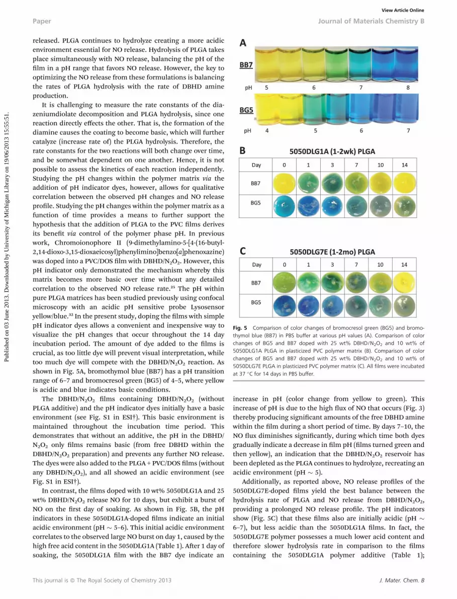
Fig. 5 Comparison of color changes of bromocresol green (BG5) and bromo-thymol blue (BB7) in PBS buffer at various pH values (A). Comparison of colorchanges of BG5 and BB7 doped with 25 wt% DBHD/N2O2 and 10 wt% of5050DLG1A PLGA in plasticized PVC polymer matrix (B). Comparison of colorchanges of BG5 and BB7 doped with 25 wt% DBHD/N2O2 and 10 wt% of5050DLG7E PLGA in plasticized PVC polymer matrix (C). All films were incubatedat 37 �C for 14 days in PBS buffer.
Paper Journal of Materials Chemistry B
Publ
ishe
d on
03
June
201
3. D
ownl
oade
d by
Uni
vers
ity o
f M
ichi
gan
Lib
rary
on
19/0
6/20
13 1
5:55
:51.
View Article Online
released. PLGA continues to hydrolyze creating a more acidicenvironment essential for NO release. Hydrolysis of PLGA takesplace simultaneously with NO release, balancing the pH of thelm in a pH range that favors NO release. However, the key tooptimizing the NO release from these formulations is balancingthe rates of PLGA hydrolysis with the rate of DBHD amineproduction.
It is challenging to measure the rate constants of the dia-zeniumdiolate decomposition and PLGA hydrolysis, since onereaction directly effects the other. That is, the formation of thediamine causes the coating to become basic, which will furthercatalyze (increase rate of) the PLGA hydrolysis. Therefore, therate constants for the two reactions will both change over time,and be somewhat dependent on one another. Hence, it is notpossible to assess the kinetics of each reaction independently.Studying the pH changes within the polymer matrix via theaddition of pH indicator dyes, however, allows for qualitativecorrelation between the observed pH changes and NO releaseprole. Studying the pH changes within the polymer matrix as afunction of time provides a means to further support thehypothesis that the addition of PLGA to the PVC lms derivesits benet via control of the polymer phase pH. In previouswork, Chromoionophore II (9-dimethylamino-5-[4-(16-butyl-2,14-dioxo-3,15-dioxaeicosyl)phenylimino]benzo[a]phenoxazine)was doped into a PVC/DOS lm with DBHD/N2O2. However, thispH indicator only demonstrated the mechanism whereby thismatrix becomes more basic over time without any detailedcorrelation to the observed NO release rate.25 The pH withinpure PLGA matrices has been studied previously using confocalmicroscopy with an acidic pH sensitive probe Lysosensoryellow/blue.32 In the present study, doping the lms with simplepH indicator dyes allows a convenient and inexpensive way tovisualize the pH changes that occur throughout the 14 dayincubation period. The amount of dye added to the lms iscrucial, as too little dye will prevent visual interpretation, whiletoo much dye will compete with the DBHD/N2O2 reaction. Asshown in Fig. 5A, bromothymol blue (BB7) has a pH transitionrange of 6–7 and bromocresol green (BG5) of 4–5, where yellowis acidic and blue indicates basic conditions.
The DBHD/N2O2 lms containing DBHD/N2O2 (withoutPLGA additive) and the pH indicator dyes initially have a basicenvironment (see Fig. S1 in ESI†). This basic environment ismaintained throughout the incubation time period. Thisdemonstrates that without an additive, the pH in the DBHD/N2O2 only lms remains basic (from free DBHD within theDBHD/N2O2 preparation) and prevents any further NO release.The dyes were also added to the PLGA + PVC/DOS lms (withoutany DBHD/N2O2), and all showed an acidic environment (seeFig. S1 in ESI†).
In contrast, the lms doped with 10 wt% 5050DLG1A and 25wt% DBHD/N2O2 release NO for 10 days, but exhibit a burst ofNO on the rst day of soaking. As shown in Fig. 5B, the pHindicators in these 5050DLG1A-doped lms indicate an initialacidic environment (pH � 5–6). This initial acidic environmentcorrelates to the observed large NO burst on day 1, caused by thehigh free acid content in the 5050DLG1A (Table 1). Aer 1 day ofsoaking, the 5050DLG1A lm with the BB7 dye indicate an
This journal is ª The Royal Society of Chemistry 2013
increase in pH (color change from yellow to green). Thisincrease of pH is due to the high ux of NO that occurs (Fig. 3)thereby producing signicant amounts of the free DBHD aminewithin the lm during a short period of time. By days 7–10, theNO ux diminishes signicantly, during which time both dyesgradually indicate a decrease in lm pH (lms turned green andthen yellow), an indication that the DBHD/N2O2 reservoir hasbeen depleted as the PLGA continues to hydrolyze, recreating anacidic environment (pH � 5).
Additionally, as reported above, NO release proles of the5050DLG7E-doped lms yield the best balance between thehydrolysis rate of PLGA and NO release from DBHD/N2O2,providing a prolonged NO release prole. The pH indicatorsshow (Fig. 5C) that these lms also are initially acidic (pH �6–7), but less acidic than the 5050DLG1A lms. In fact, the5050DLG7E polymer possesses a much lower acid content andtherefore slower hydrolysis rate in comparison to the lmscontaining the 5050DLG1A polymer additive (Table 1);
J. Mater. Chem. B

Journal of Materials Chemistry B Paper
Publ
ishe
d on
03
June
201
3. D
ownl
oade
d by
Uni
vers
ity o
f M
ichi
gan
Lib
rary
on
19/0
6/20
13 1
5:55
:51.
View Article Online
therefore, no initial burst of NO is observed. This lower initialacid content is crucial to prolonging the NO release from theselms. The lms containing 5050DLG7E PLGA exhibit little colorchange until days 10–14, when they begin to become moreacidic. This demonstrates that the acid production rate (fromthe PLGA hydrolysis) and DBHD amine production rate isclosely balanced within these lms, explaining the consistencyof the pH and NO release from day-to-day. These lms alsoturned yellow by day 14, indicating the depletion of the NOreservoir. In short, the use of pH indicators within the lmsprovides further evidence that the 5050DLG7E PLGA hydrolysisrate balances the decomposition rate of the DBHD/N2O2,producing the optimum pH and concomitant prolonged NOrelease/ux prole.
Fig. 6 Time dependent effects of NOrel ECC (25 wt% DBHD/N2O2 + 10 wt%5050DLG7E PLGA) as compared to control ECC on rabbit platelet count (i.e.consumption) as measured via Coulter counter. The data are means � SEM. * ¼ p< 0.05, baseline vs. control ECC circuits.
3.3 PLGA-doped NOrel lms in ECC and effects on rabbithemodynamics
ECC circuits coated (Fig. 2) (on inner walls) with PVC containingthe a 5050DLG7E PLGA/DBHD/N2O2 NO release formulationwere tested for NO release ux, pre- and post-4 h rabbit bloodexposure. The optimized PVC coating material continuouslyreleases NO under physiological conditions at levels thatexceeds the physiological NO release from endothelial cells(0.5–4 � 10�10 mol cm�2 min�1).10 The NO release as measuredusing chemiluminescence NO analyzer shows a sustained NOux of approximately 11 � 10�10 mol cm�2 min�1 for 4 h whenmeasuring the pre-blood exposure NO release prole. The NOrelease from the ECC circuit does not decrease signicantlyaer exposure to the owing blood. Indeed, aer 4 h of bloodow, the NO ux was found to be 10 � 10�10 mol cm�2 min�1.The NO release proles of the NOrel coatings were very similarwhen tested in PBS or plasma (see Fig. S2, ESI†). The fact thatthe blood environment does not alter the kinetics of the NOrelease from the coating agrees well with the previously reporteddata for various NO release circuits.22 Further, given the pro-longed NO release capability of the new coatings being tested,applications in much longer-term extracorporeal or otherbiomedical applications would be possible. Our goal here,however, is to demonstrate that the presence of PLGA does notalter the physiological effectiveness of the NO release, and thisis most easily examined using the 4 h ECC test model.
The ECC blood ow was maintained at approximately105 mL min�1 for the NOrel circuits over the 4 h animal testperiod. However, the blood ow typically drops from the initial105 mL min�1 to approximately 80 mL min�1 in the rst onehour for the control circuits, and then remains at 80 mL min�1
for the rest of the 4 h period. This maintenance of blood ow inthe control circuits is due to the addition of intravascular uidsto the animal over the test period. The NOrel and control ECCloops have the same inner diameter (which remains static dueto the rigidity of the tubing) and therefore maintains theintegrity of the coating, hence having no effect on the blood owrates. Monitoring the ow rate is a means to measure the timeat which the ECC circuits has completely clotted. Since thecontrol ECC loops oen clotted, this blocked the blood owthrough the tubing, thus the ow rates decreased. No
J. Mater. Chem. B
signicant difference in the mean arterial pressure of theanimals on the NOrel vs. control circuits was noted, with pres-sures averaging 46 � 4 mmHg for both types of circuits. Theactivation clotting time for blood obtained from the testanimals increases over the 4 h period for both NOrel and controlcoated circuits. As noted in previous studies,22 this behavior canbe attributed to the increase in intravascular uids andconcomitant hemodilution effect.
3.4 Effects of PLGA-doped NOrel PVC polymer coatings onrabbit platelet function and thrombus formation
Platelet function during exposure to the NOrel and controlpolymer-coated ECCs was assessed by observing platelet count(Fig. 6) and percent of platelet aggregation, as determined by exvivo collagen (25 mg mL�1) stimulation of PRP. Platelet countwas corrected for any hemodilution due to added IV uids intothe rabbits. Four out of 7 control circuits clotted within 3 h,whereas all the 7 NOrel coated circuits remained patent aer 4h. The animals tested with the NOrel polymer coated ECCsshowed 79 � 11% preservation of the platelet count over thecourse of the 4 h blood contact period, whereas animals testedwith the control polymer ECCs exhibited a time-dependent lossin platelet count (54 � 6%). The blood from animals subjectedto the NOrel and control ECCs exhibited similar response tocollagen-stimulated platelet aggregation over the course of 4 hblood exposure. The percent of platelet aggregation was deter-mined by ex vivo collagen stimulation of PRP, measured byoptical turbidity.22 NOrel and control coated circuits were ableto maintain 88% and 91% aggregation, respectively, comparedto their baseline values. This indicates that NO did not adverselyaffect the platelets ability to aggregate and the platelets main-tained their functionality even aer 4 h of exposure to the NOreleasing surface. The level of plasma brinogen to which theactivated platelets bind during the 4 h ECC blood exposure wasalso assessed. The plasma brinogen levels were corrected forany hemodilution due to added IV uids into the rabbits. The
This journal is ª The Royal Society of Chemistry 2013

Fig. 7 Evaluation of thrombus formation on NOrel and control polymer ECCafter 4 h blood exposure in rabbit thrombogenicity model. (A) Image of thrombusarea in 3/8 inch ID tubing in the control ECC. (B) Image of thrombus area in 3/8inch ID NOrel polymer ECC. (C) Quantitation of thrombus area as calculatedwith NIH ImageJ software using a 2D representation of thrombus. The data aremeans � SEM. * ¼ p < 0.05, control ECC vs. NOrel ECC after 4 h ECC flow.
Paper Journal of Materials Chemistry B
Publ
ishe
d on
03
June
201
3. D
ownl
oade
d by
Uni
vers
ity o
f M
ichi
gan
Lib
rary
on
19/0
6/20
13 1
5:55
:51.
View Article Online
plasma brinogen levels decreased by approximately 10% forthe control ECCs, whereas the brinogen levels for NOrel ECCexperiments dropped by 18% in 4 h (see Fig. S3 in ESI†). Thiswas found to be consistent with the previously reported resultswhere the decrease in plasma brinogen levels was attributed tothe binding of brinogen to the ECC surfaces.22,37
To ascertain the differential formation of thrombus in thethrombogenicity chamber (i.e., the 3/8 inch ID Tygon� tubing 8cm in length within the ECC loop) of the NOrel vs. controlpolymer-coated ECCs, 2-dimensional (2D) image analysis wasperformed aer 4 h of blood exposure. Fig. 7A and B, showrepresentative images of the control and NOrel circuits,respectively, aer being run for 4 h in the rabbit ECC model.ImageJ imaging soware was used to calculate the representa-tive 2D thrombus area (pixels per cm2) in each tubing chamber.These thrombi area measurements were quantitated and, asshown in Fig. 7C, the thrombus area of the NOrel polymer ECCwas signicantly reduced compared to the control polymerECCs, 1.5� 0.5 and 6.5� 0.4 pixels per cm2, respectively. For anA-V shunt procedure, the lungs are the rst main lter for theblood. Aer the 4 h experimental period, the lungs were eval-uated for accumulated thromboemboli. The control ECCsrabbit lungs appeared to have more emboli accumulated in thelower lobes of both lungs then any of the NOrel ECC rabbitlungs.
4 Conclusions
This study demonstrates, for the rst time, that an ester-cappedPLGAmaterial can be used as an additive within plasticized PVClms containing lipophilic diazeniumdiolate species, and thepresence of the PLGA species sustains the NO release for muchlonger time periods than possible without the additive. By usingvarious pH indicators it was shown that the hydrolysis rates of
This journal is ª The Royal Society of Chemistry 2013
specic PLGA species employed can control the NO releaseproperties by inuencing the steady-state pH within the poly-mer lms. Nitric oxide release from optimal PLGA-doped coat-ings used on the inner walls of ECC circuits was able toattenuate the activation of the platelets while maintaining theirfunctionality. A signicant reduction in the clot area was alsoseen with the new coatings vs. that observed on control ECCcircuits. The new PLGA-doped NOrel PVC coatings are nowunder investigation in a 2–3 week rabbit model and couldprovide a breakthrough for achieving long-term preservation ofcirculating platelets, an important goal for longer-term ECCsituations, such as ECMO.43
Acknowledgements
The authors declare this work is supported by the NationalInstitutes of Health, Grants HL015434, EB000783 andK25HL111213.
References
1 B. D. Ratner, Biomaterials, 2007, 28, 5144–5147.2 B. D. Ratner, J. Biomater. Sci., Polym. Ed., 2000, 11, 1107–1119.
3 T. Kolobow, E. W. Stool, P. K. Weathersby, J. Pierce,F. Hayano and J. Suaudeau, Trans. – Am. Soc. Artif. Intern.Organs, 1974, 20, 269–276.
4 M. Szycher, J. Biomater. Appl., 1988, 3, 297–402.5 L. H. Edmunds, Jr, ASAIO J., 1995, 41, 824–830.6 P. Didisheim, ASAIO J., 1994, 40, 230–237.7 W. S. Kim and H. Jacobs, Blood Purif., 1996, 14, 357–372.8 O. Larm, R. Larsson and P. Olsson, Biomater., Med. Devices,Artif. Organs, 1983, 11, 161–173.
9 M. W. Radomski and S. Moncada, Adv. Exp. Med. Biol., 1993,344, 251–264.
10 M. W. Vaughn, L. Kuo and J. C. Liao, Am. J. Physiol., 1998,274, H2163–H2176.
11 J. S. Isenberg, M. J. Romeo, C. Yu, C. K. Yu, K. Nghiem,J. Monsale, M. E. Rick, D. A. Wink, W. A. Frazier andD. D. Roberts, Blood, 2008, 111, 613–623.
12 B. L. Nguyen, M. Saitoh and J. A. Ware, Am. J. Physiol., 1991,261, H1043–H1052.
13 Y. Sato, Y.Hiramatsu, S.Homma,M. Sato, S. Sato, S. EndoandY. Sohara, J. Thorac. Cardiovasc. Surg., 2005, 130, 346–350.
14 A. K. Zimmermann, H. Aebert, A. Reiz, M. Freitag,M. Husseini, G. Ziemer and H. P. Wendel, ASAIO J., 2004,50, 193–199.
15 G. F. P. de Souza, J. K. U. Yokoyama-Yasunaka, A. B. Seabra,D. C. Miguel, M. G. de Oliveira and S. R. B. Uliana, NitricOxide, 2006, 15, 209–216.
16 A. B. Seabra, G. F. P. de Souza, L. L. da Rocha, M. N. Eberlinand M. G. de Oliveira, Nitric Oxide, 2004, 11, 263–272.
17 T. A. Alston, D. J. Porter and H. J. Bright, J. Biol. Chem., 1985,260, 4069–4074.
18 G. M. Annich, J. P. Meinhardt, K. A. Mowery, B. A. Ashton,S. I. Merz, R. B. Hirschl, M. E. Meyerhoff andR. H. Bartlett, Crit. Care Med., 2000, 28, 915–920.
J. Mater. Chem. B

Journal of Materials Chemistry B Paper
Publ
ishe
d on
03
June
201
3. D
ownl
oade
d by
Uni
vers
ity o
f M
ichi
gan
Lib
rary
on
19/0
6/20
13 1
5:55
:51.
View Article Online
19 M. H. Schoensch, K. A. Mowery, M. V. Rader, N. Baliga,J. A. Wahr and M. E. Meyerhoff, Anal. Chem., 2000, 72,1119–1126.
20 G. B. Richter-Addo and P. Legzdins, Metal Nitrosyls, OxfordUniversity Press, New York, 1992.
21 W. J. Paulus, P. J. Vantrimpont and A. M. Shah, Circulation,1994, 89, 2070–2078.
22 T. C. Major, D. O. Brant, M. M. Reynolds, R. H. Bartlett,M. E. Meyerhoff, H. Handa and G. M. Annich, Biomaterials,2010, 31, 2736–2745.
23 K. M. Davies, D. A. Wink, J. E. Saavedra and L. K. Keefer,J. Am. Chem. Soc., 2001, 123, 5473–5481.
24 H. Zhang, G. M. Annich, J. Miskulin, K. Osterholzer,S. I. Merz, R. H. Bartlett and M. E. Meyerhoff, Biomaterials,2002, 23, 1485–1494.
25 M. M. Batchelor, S. L. Reoma, P. S. Fleser, V. K. Nuthakki,R. E. Callahan, C. J. Shanley, J. K. Politis, J. Elmore, S. I. Merzand M. E. Meyerhoff, J. Med. Chem., 2003, 46, 5153–5161.
26 B. Wu, PhD Dissertation, University of Michigan, Ann Arbor,2009.
27 Z. Zhou and M. E. Meyerhoff, Biomacromolecules, 2005, 6,780–789.
28 S. Kaul, B. Cercek, J. Rengstrom, X.-P. Xu, M. D. Molloy,P. Dimayuga, A. K. Parikh, M. C. Fishbein, J. Nilsson,T. B. Rajavashisth and P. K. Shah, J. Am. Coll. Cardiol.,2000, 35, 493–501.
29 C. E. Holy, S. M. Dang, J. E. Davies and M. S. Shoichet,Biomaterials, 1999, 20, 1177–1185.
30 L. Lu, S. J. Peter, M. D. Lyman, H. L. Lai, S. M. Leite,J. A. Tamada, S. Uyama, J. P. Vacanti, R. Langer andA. G. Mikos, Biomaterials, 2000, 21, 1837–1845.
J. Mater. Chem. B
31 S. J. Siegel, J. B. Kahn, K. Metzger, K. I. Winey, K. Werner andN. Dan, Eur. J. Pharm. Biopharm., 2006, 64, 287–293.
32 A. Ding and S. Schwendeman, Pharm. Res., 2008, 25, 2041–2052.
33 A. Shenderova, A. G. Ding and S. P. Schwendeman,Macromolecules, 2004, 37, 10052–10058.
34 J. W. Yoo, J. S. Lee and C. H. Lee, J. Biomed. Mater. Res., PartA, 2010, 92, 1233–1243.
35 W. Cai, J. Wu, C. Xi and M. E. Meyerhoff, Biomaterials, 2012,33, 7933–7944.
36 D. H. Na and P. P. DeLuca, Pharm. Res., 2005, 22, 736–742.
37 S. M. Lantvit, B. J. Barrett and M. M. Reynolds, J. Biomed.Mater. Res., Part A, 2013, DOI: 10.1002/jbm.a.34627.
38 K. M. Miranda, T. Katori, C. L. Torres de Holding,L. Thomas, L. A. Ridnour, W. J. McLendon, S. M. Cologna,A. S. Dutton, H. C. Champion, D. Mancardi,C. G. Tocchetti, J. E. Saavedra, L. K. Keefer, K. N. Houk,J. M. Fukuto, D. A. Kass, N. Paolocci and D. A. Wink,J. Med. Chem., 2005, 48, 8220–8228.
39 E. M. Hetrick and M. H. Schoensch, in Annual Review ofAnalytical Chemistry, 2009, pp. 409–433.
40 P. N. Coneski and M. H. Schoensch, Chem. Soc. Rev., 2012,41, 3753–3758.
41 T. C. Major, D. O. Brant, C. P. Burney, K. A. Amoako,G. M. Annich, M. E. Meyerhoff, H. Handa andR. H. Bartlett, Biomaterials, 2011, 32, 5957–5969.
42 S. P. Schwendeman, Crit. Rev. Ther. Drug Carrier Syst., 2002,19, 73–98.
43 W. C. Oliver, Semin. Cardiothorac. Vasc. Anesth., 2009, 13,154–175.
This journal is ª The Royal Society of Chemistry 2013