technique paper for wet-spinning poly(l-lactic acid) and poly(dl-lactide-co-glycolide) monofilament...
TRANSCRIPT
1323
INTRODUCTION
POLY(L-LACTIC ACID) (PLLA) poly(glycolic acid)(PGA), and their copolymers and blends have been
used as bioresorbable polymers in medical applicationssince the 1960s.1 They have been used as dissolvable su-tures,2 in orthopedic applications,3,4 and more recentlyhave become important synthetic scaffoldings for tissue-engineering applications.5–11 They were chosen becausethey have FDA approval in many applications, they werefound to have good strength, were readily processed, andeasy to obtain, purify, and use in bulk quantities.
The fiber format was nearly always obtained by con-ventional melt-extrusion techniques; however, because of
the size and cost of melt-extrusion equipment, and thelarge amount of raw material required, it has not beenwell suited to bench-top, laboratory quantities. Therefore,we sought other processing methods to obtain similarfibers. This article describes simple, inexpensive, bench-top techniques for wet-spinning PLLA and poly(DL-lac-tide-co-glycolide) (PLGA) monofilament fibers suitablefor scaffoldings for tissue-engineering applications. Theconcept of wet-spinning is not new; Kulkarni et al. wet-spun PLLA fibers as far back as 1966.1 However, wet-spinning has generally produced fibers not as mechani-cally strong as fibers produced by melt-extruding, andtherefore has not been investigated as thoroughly. There-fore, we felt that a technical report to teach the concepts
TISSUE ENGINEERINGVolume 9, Number 6, 2003© Mary Ann Liebert, Inc.
Technical Report
Technique Paper for Wet-Spinning Poly(L-lactic acid) andPoly(DL-lactide-co-glycolide) Monofilament Fibers
KEVIN D. NELSON, Ph.D.,1,2 ANDRES ROMERO, M.S.,1 PAULA WAGGONER, M.S.,2
BRENT CROW, B.S.,1 ANGELA BORNEMAN, M.S.,1 and GEORGE M. SMITH, Ph.D.2,3
ABSTRACT
A simple and repeatable method is described for wet-spinning poly(L-lactic acid) (PLLA) andpoly(DL-lactic-co-glycolic acid) (PLGA) monofilament fibers. These fibers are strong, elastic, andsuitable for many applications, including use as tissue-engineering scaffolds. The PLLA wet-extrudedfibers do not show additional strain-induced crystallization as a result of drawing the fibers duringfabrication; however, there is an apparent increase in crystallinity late in the degradation processin saline at 37°C. We have measured the molecular weight degradation in saline at 37°C for fibersof both PLLA and PLGA. Changing solvent systems, polymer blends, and winding rates alters me-chanical and morphological properties of these fibers for specific applications. The authors discussa possible theoretical explanation for these observed changes due to changes in polymer concen-tration, solvent system, and coagulation bath properties. This wet-extrusion process is simple andinexpensive enough to be carried out in almost any laboratory interested in tissue engineering.
1Joint Program in Biomedical Engineering, University of Texas Southwestern Medical Center at Dallas, Dallas, Texas; andUniversity of Texas at Arlington, Arlington, Texas.
2TissueGen, Arlington, Texas.3Department of Physiology, and Spinal Cord and Brain Injury Research Center, University of Kentucky, Lexington, Kentucky.
and techniques of wet-spinning would be of great bene-fit to the majority of tissue engineers who are currentlyusing PLLA or PLGA fibers in their research. The tech-niques described herein avoid the large capital, space, andraw material requirements of conventional melt-extrusionof these polymers; and further, they demonstrate that fiberproperties are controllable and tunable for specific ap-plications, thus making this technique more versatile thanmelt-extruding. For example, wet-spinning into PEG 600creates an interpenetrating network of PEG with thePLLA, which results in a highly hydrophilic fiber, al-though with good mechanical properties; which would beimpossible to obtain by conventional melt-extrusiontechniques. This technique may also lend itself to the in-corporation of heat-sensitive drugs into the fiber, as theentire process takes place at room temperature.
We have been using these techniques successfully forapproximately 6 years to culture a number of cell types,such as human fetal foreskin fibroblasts, rabbit cornealfibroblasts, human umbilical vein endothelial cells, rat fi-broblasts, mouse immature myoblasts, and rat pup dor-sal root ganglion cells in vitro. We have also used thesefibers in vivo to create a peripheral nerve graft, whichwas capable of successfully inducing axonal elongationup to 18 mm in a rat sciatic nerve resection model.12,13
These fibers have caused no apparent problems in vitrowhen used without further modification. In vivo unmod-ified fibers have evoked a mild to moderate inflamma-tory response in rat spinal cord and rat sciatic nerve im-plantation, but not with intraocular implantation. Wehave also used simple dip-coating techniques of laminin14
and collagen to improve cellular adhesion in vitro.
MATERIALS AND METHODS
Fiber fabrication by a wet-spinning technique
Poly(L-lactic acid) (50–200 kDa; Polysciences, War-rington, PA) was dissolved in varying concentrationsfrom 7.5 to 15 wt/v% in a variety of solvent systems, in-cluding chloroform (Aldrich Chemical, St. Louis, MO),1,4-dioxane, a 5:1 mixture of chloroform–toluene, and a4:1 mixture of chloroform–hexane.
PLGA (intrinsic viscosity, 0.66–0.80; Polysciences)was dissolved in methylene chloride or chloroform at aconcentration of 20 wt/v%. We allowed a minimum of90 min for complete dissolution of all polymer solutions.
The polymer solution was loaded into a glass syringe(gas-tight syringes; Hamilton, Reno, NV) and placed ina syringe pump (model 945 [Harvard Apparatus, Hollis-ton, MA] or model KDS200 [KD Scientific, New Hope,PA]). The polymer flow rate was typically between 0.02and 0.1 mL/min. A Viton tube (Cole-Parmer, VernonHills, IL) connected the syringe to the needle dispensers.We used only blunt-tipped needles (Small Parts, Miami
NELSON ET AL.
Lakes, FL), because the bevel cut on sharp needles wouldcause problems during extrusion. We immersed the tipof the needle in a coagulation bath, which was a poor sol-vent for the polymer, and yet was highly miscible withthe solvent used to dissolve the polymer. The coagulat-ing baths we have used include isopropyl alcohol, shortalkanes (heptane, cyclohexane, hexane, and pentane) andpoly(ethylene glycol) (PEG) 200 and 600 Mw, and am-phiphilic polymers including several of the Pluronicsfamily (BASF). The fiber mechanical and physical prop-erties such as strength, diameter, surface tension, and soon are strongly dependent on the choice of coagulationbath. If the precipitation of the polymer fiber was toorapid, up to 20 v/v% of the polymer solvent may havebeen added to the coagulation bath.
The original extrusion setup was a narrow aluminumtank with overall dimensions of approximately 60 330 3 3 cm with two movable rollers. Lowering the rollersinto the tank after the fiber began to form provided a fixedpath length for the fiber.15 This setup was used to gen-erate much of the data in this article; however, we havesince simplified this setup to a simple glass tube (i.e., 25mm i.d., length, 20 cm) with a rubber septum at the topthrough which the dispensing needle was pierced. Theglass tube was immersed in a small container below, asshown in Fig. 1, and filled with the coagulation bath fluid.When the fiber exited the coagulating bath (from eithersetup) it was wound on a 8.25-cm-diameter bobbin at-tached to the jaws of a modified 5-in. garden lathe (Sears-Craftsman model 549-289000; Sears and Roebuck,Chicago, IL). A 24-V power supply (model 5005R;Power Designs, NY) drove a 0.03-horsepower DC mo-tor (Pittman Motors, Harleysville, PA) that had replacedthe original motor on the lathe. We first wrapped the bob-bin with paper so that on completion of the extrusion run,we would pull the paper from the bobbin to remove thefiber intact. The angular velocity of the lathe was mea-sured with an optical tachometer. The draw ratio was cal-culated as the ratio of the linear velocity of the fiber mea-
1324
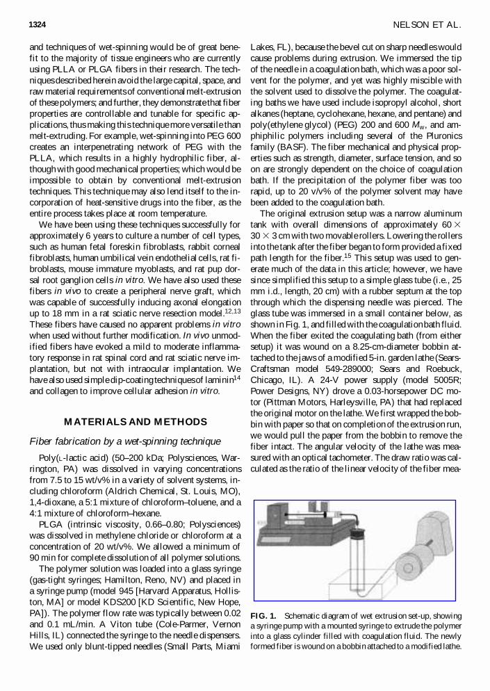
FIG. 1. Schematic diagram of wet extrusion set-up, showinga syringe pump with a mounted syringe to extrude the polymerinto a glass cylinder filled with coagulation fluid. The newlyformed fiber is wound on a bobbin attached to a modified lathe.
sured at the take-up bobbin to the calculated mean linearvelocity of the polymer solution within the dispensingneedle. We typically observed no die-swell during theseextrusions; therefore, we used the mean linear velocitywithin the dispensing needle for draw ratio calculations.16
These techniques readily produced draw ratios as high as40:1.
At the end of the extrusion run, fibers were removedfrom the lathe, left on the paper roll, and placed undervacuum at room temperature for at least 4 h at room tem-perature to help remove remaining solvents. The fiberwas stored in a desiccator or in a 220°C freezer untilneeded.
To generate fibers that were not wound, it was possi-ble to simply extrude into an Erlenmeyer flask or even a50-mL conical Falcon tube that was filled with the co-agulation fluid. At the end of extrusion, these fibers werecollected, placed under vacuum, and stored as describedabove.
Tensile test apparatus
A motorized platform was constructed to fit into a cab-inet, on top of which an electric balance (model AE240;Mettler Toledo, Worthington, OH) was placed. The fiberwas tied to a metal fixture approximately 1 mm in di-ameter, which was secured to the underscale attachmenton the electronic balance through a hole in the top of thecabinet. Similarly, the other end of the fiber was tiedaround a similar holder, which was attached to the mov-able platform. The initial, unstressed fiber length was 5cm. After taring the balance, the platform descended ata steady rate of 2.78 mm/min. We converted the elec-tronic balance measurements of the induced load alongwith the position of the platform into stress strain data.If the fiber failure occurred at the knot, or if any slippingof the knot occurred, the data were discarded. Ultimatestress and strain were calculated for each fiber sample,using the initial diameter of the fiber (obtained from scan-ning electron micrographs taken before any mechanicaltesting). Our experience has shown that the diameter wasrelatively constant over the length of the fiber.
PLLA sample preparation for DSC measurements
We calculated percent crystallinity on the basis of ther-mograms produced by differential scanning calorimetry(model 2010; TA Instruments). The samples were vac-uum dried at 70°C for 2 h and then weighed, pressed toform a disc, and placed inside aluminum pans. The ref-erence was an empty aluminum pan. The temperatureprofile runs from 30 to 250°C at a constant rate of10°C/min under a nitrogen purge. Integration of the melt-ing and crystallization peaks produced the heat of fusion(DHf) and heat of crystallization (DHc) for each samplein joules per gram. We calculated percent crystallinity by
WET-SPINNING PLLA AND PLGA MONOFILAMENT FIBERS 1325
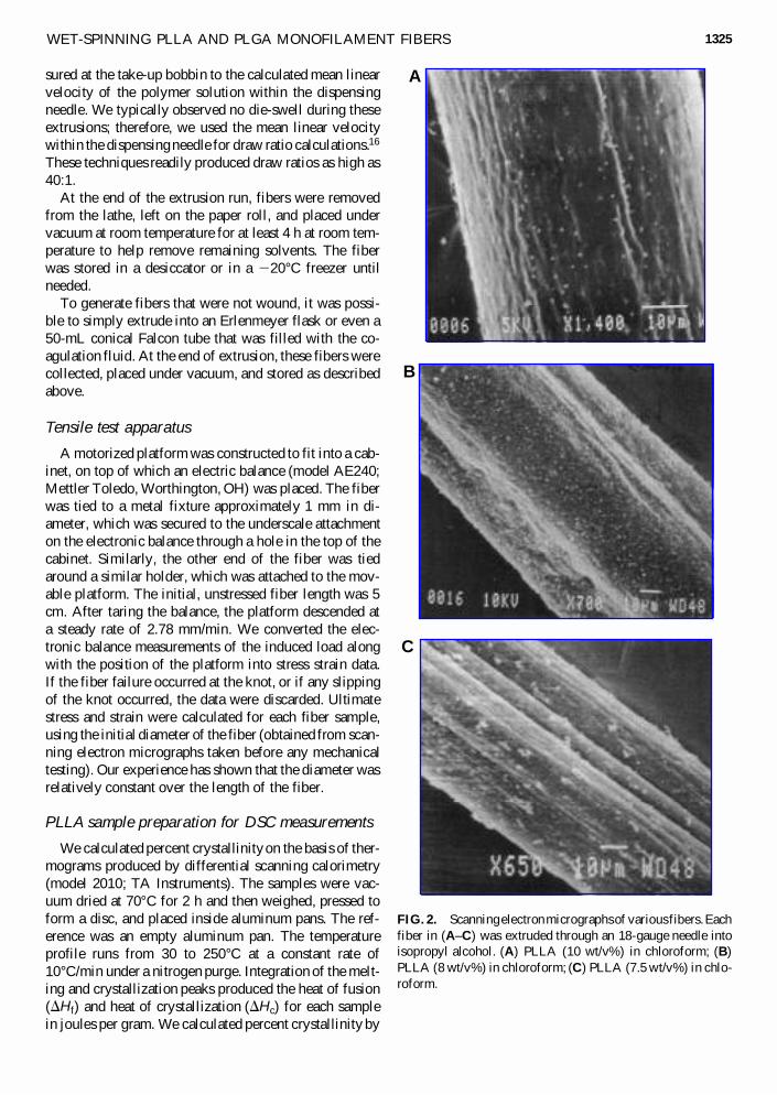
FIG. 2. Scanning electron micrographs of various fibers. Eachfiber in (A–C) was extruded through an 18-gauge needle intoisopropyl alcohol. (A) PLLA (10 wt/v%) in chloroform; (B)PLLA (8 wt/v%) in chloroform; (C) PLLA (7.5 wt/v%) in chlo-roform.
C
B
A
subtracting DHc from DHf and dividing by 93.6 J/g,which is DHf for a pure PLLA crystal.17
Molecular weight degradation studies
The fibers were cut into pieces (typically 33 cm),carefully weighed, dipped in 70% ethanol for 15 s tominimize bacterial contamination, placed in sterile Ep-pendorf tubes, and covered with 1.0 mL of phosphate-buffered saline (pH 7.4) with 0.01 wt%. Thimerasol asa broad-spectrum antibiotic and incubated at 37°C. Atweekly intervals, the spent PBS was replaced with freshPBS in each sample, and three fibers were removedfrom the PBS, dried with a Kimwipe, and stored in thefreezer (220°C) until analyzed. The pH of the spentmedium was measured. The molecular weight was de-termined by dissolving each sample in 3.0 mL of meth-ylene chloride and injecting it into an HPLC (Akta pu-rifier 10; Amersham Biosciences, Piscataway, NJ) andanalyzing using UV at 236 nm and RI detectors(Shodex R-71; Showa Denko, Tokyo, Japan). We useda GPC column (Asahipak GPC guard column modelGF-1G7B [Phenomenex] and TSK gel column modelG2000-HHR [Supelco]) (10-mL injection loop) withmethylene chloride as the mobile phase at flow rate of1.0 mL/min. Mn and Mw were calculated from the chro-matograms on the basis of polystyrene molecularweight standards (Supelco).
NELSON ET AL.
RESULTS
Physical and mechanical characterization
Over the past 6 years, hundreds of fibers have beenproduced with a wide range of physical and mechanicalproperties; for example, we have produced fibers with di-ameters as small as 28 mm, and as large as 550 mm, onlya small fraction of which are reported here. The chosensolvent system and polymer composition greatly affectedthe external morphology of the filaments as shown byscanning electron microscopy (SEM) of various PLLAfibers (Fig. 2). We also noted morphological changessimply by varying the concentration of the polymer so-lution under otherwise identical extrusion conditions(Fig. 2). In all these cases, the dispensing tip was an 18-gauge needle and the coagulating bath was isopropyl al-cohol. These different surface morphologies and texturesmay be advantageous for tissue-engineering applications.For example, longitudinal grooves may provide contactguidance and increased surface area for cell attachment.
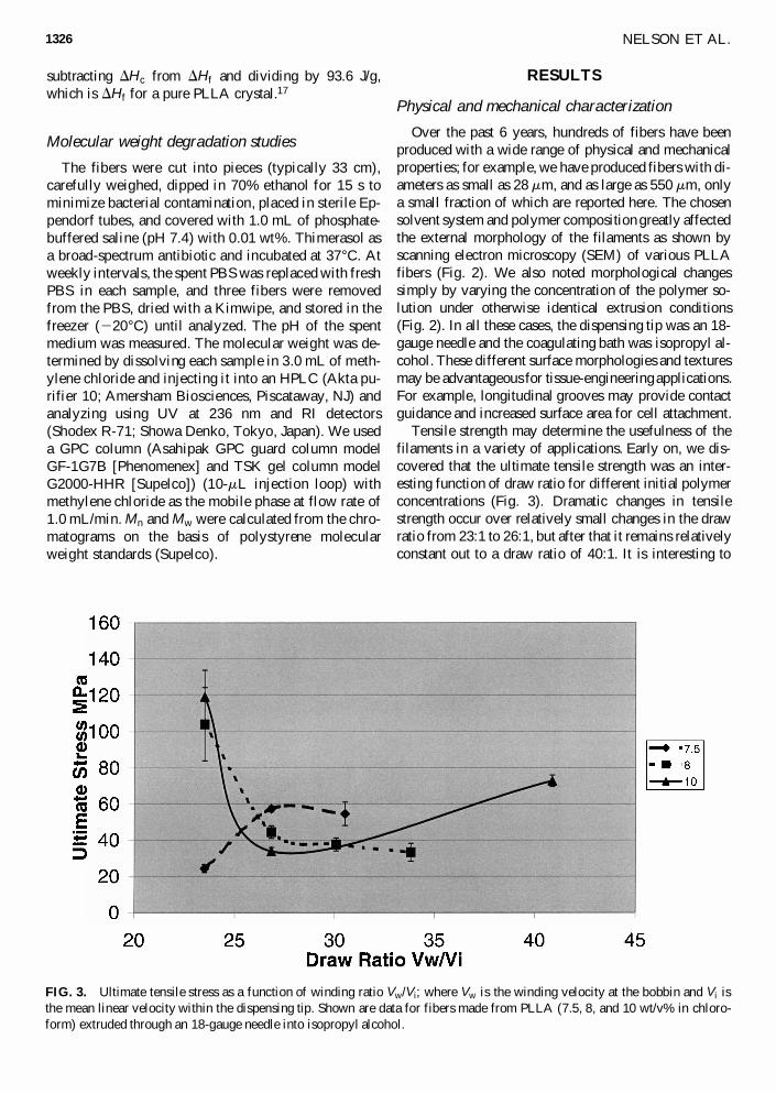
Tensile strength may determine the usefulness of thefilaments in a variety of applications. Early on, we dis-covered that the ultimate tensile strength was an inter-esting function of draw ratio for different initial polymerconcentrations (Fig. 3). Dramatic changes in tensilestrength occur over relatively small changes in the drawratio from 23:1 to 26:1, but after that it remains relativelyconstant out to a draw ratio of 40:1. It is interesting to
1326
FIG. 3. Ultimate tensile stress as a function of winding ratio Vw/Vi; where Vw is the winding velocity at the bobbin and Vi isthe mean linear velocity within the dispensing tip. Shown are data for fibers made from PLLA (7.5, 8, and 10 wt/v% in chloro-form) extruded through an 18-gauge needle into isopropyl alcohol.
compare the similarity in behavior of the 8 and 10 wt/v%polymer concentration fibers, and to contrast it with thebehavior of 7.5 wt/v% polymer concentration fibers; the8 and 10 wt/v% polymer concentration fibers showed acounterintuitive precipitous drop in ultimate tensile stressas the draw ratio increased from 23:1 to 26:1. This dropin ultimate tensile stress occurred despite a decrease incross-sectional area, thus indicating that there was a sub-stantial decrease in the load-carrying ability in thesefibers. For the 7.5 wt/v% concentration fibers, however,an increase in tensile stress was observed over these samedraw ratios. Unfortunately, high draw ratios are more dif-ficult to achieve as the polymer concentration of the spin-ning solution decreases, which made comparison athigher draw ratios impossible across all polymer con-centrations. These tensile strengths are for PLLA fibersdissolved in chloroform, and extruded into isopropyl al-cohol through an 18-gauge dispensing tip.
Molecular weight and crystalline characterization
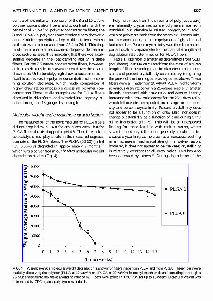
The measured pH of the spent medium for PLLA fibersdid not drop below pH 6.8 for any given week, but forPLGA fibers the pH dropped to pH 4.8. Therefore, acidicautocatalysis may play a role in the measured degrada-tion rate of the PLGA fibers. The PLGA (50:50) (initiali.v., 0.66–0.8) degraded in approximately 2 months,18
which was also verified in our in vitro molecular weightdegradation studies (Fig. 4).
WET-SPINNING PLLA AND PLGA MONOFILAMENT FIBERS
Polymers made from the L-isomer of poly(lactic acid)are inherently crystalline, as are polymers made fromnonchiral but chemically related poly(glycolic acid),whereas polymers made from the racemic DL isomer mix-ture are amorphous, as are copolymers of glycolic andlactic acids.19 Percent crystallinity was therefore an im-portant qualitative parameter for mechanical strength anddegradation rate determination for PLLA fibers.
Table 1 lists fiber diameter as determined from SEM(not shown), density calculated from the mass of a givenlength of fiber assuming that the diameter remains con-stant, and percent crystallinity calculated by integratingthe peaks of the thermograms as explained above. Thesefibers were all made from 10 wt/v% PLLA in chloroformat various draw ratios with a 21-gauge needle. Diameterlinearly decreased with draw ratio, and density linearlyincreased with draw ratio except for the 26:1 draw ratio,which fell outside the expected linear range for both den-sity and percent crystallinity. Percent crystallinity doesnot appear to be a function of draw ratio, nor does itchange substantially as a function of time during 37°Csaline incubation (Fig. 5). This will be an unexpectedfinding for those familiar with melt-extrusion, wherestrain-induced crystallization generally results in in-creased crystallinity as the draw ratio increases, resultingin an increase in mechanical strength. In wet-extrusion,however, it does not appear to be the case; crystallinityis relatively constant for all draw ratios. This has alsobeen observed by others.16 During degradation of the
1327
FIG. 4. Weight average molecular weight degradation is shown for fibers made from PLLA and from PLGA. These fibers weremade by dissolving the polymer (PLLA at 10 wt/v%; and PLGA at 20 wt/v%) in methylene chloride and extruding it through a22-gauge needle into hexane at a winding ratio of 42. Fibers were stored in 37°C PBS for up to 15 weeks. Molecular weight wasdetermined by GPC against polystyrene standards.
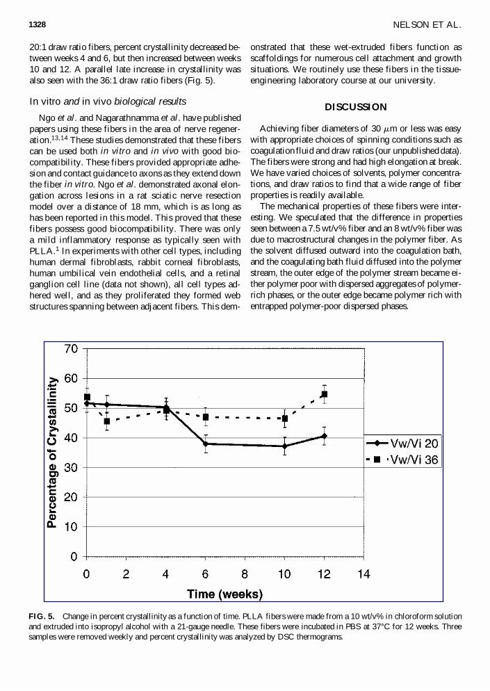
20:1 draw ratio fibers, percent crystallinity decreased be-tween weeks 4 and 6, but then increased between weeks10 and 12. A parallel late increase in crystallinity wasalso seen with the 36:1 draw ratio fibers (Fig. 5).
In vitro and in vivo biological results
Ngo et al. and Nagarathnamma et al. have publishedpapers using these fibers in the area of nerve regener-ation.13,14 These studies demonstrated that these fiberscan be used both in vitro and in vivo with good bio-compatibility. These fibers provided appropriate adhe-sion and contact guidance to axons as they extend downthe fiber in vitro. Ngo et al. demonstrated axonal elon-gation across lesions in a rat sciatic nerve resectionmodel over a distance of 18 mm, which is as long ashas been reported in this model. This proved that thesefibers possess good biocompatibility. There was onlya mild inflammatory response as typically seen withPLLA.1 In experiments with other cell types, includinghuman dermal fibroblasts, rabbit corneal fibroblasts,human umbilical vein endothelial cells, and a retinalganglion cell line (data not shown), all cell types ad-hered well, and as they proliferated they formed webstructures spanning between adjacent fibers. This dem-
NELSON ET AL.
onstrated that these wet-extruded fibers function asscaffoldings for numerous cell attachment and growthsituations. We routinely use these fibers in the tissue-engineering laboratory course at our university.
DISCUSSION
Achieving fiber diameters of 30 mm or less was easywith appropriate choices of spinning conditions such ascoagulation fluid and draw ratios (our unpublished data).The fibers were strong and had high elongation at break.We have varied choices of solvents, polymer concentra-tions, and draw ratios to find that a wide range of fiberproperties is readily available.
The mechanical properties of these fibers were inter-esting. We speculated that the difference in propertiesseen between a 7.5 wt/v% fiber and an 8 wt/v% fiber wasdue to macrostructural changes in the polymer fiber. Asthe solvent diffused outward into the coagulation bath,and the coagulating bath fluid diffused into the polymerstream, the outer edge of the polymer stream became ei-ther polymer poor with dispersed aggregates of polymer-rich phases, or the outer edge became polymer rich withentrapped polymer-poor dispersed phases.
1328
FIG. 5. Change in percent crystallinity as a function of time. PLLA fibers were made from a 10 wt/v% in chloroform solutionand extruded into isopropyl alcohol with a 21-gauge needle. These fibers were incubated in PBS at 37°C for 12 weeks. Threesamples were removed weekly and percent crystallinity was analyzed by DSC thermograms.
Case 1: polymer-poor continuous outer phase
If the outer edge became a polymer-poor continuousphase, there would be little barrier to diffusion of the co-agulation bath fluid into the forming fiber. The dispersedpolymer-rich phases would continue to contract and losesolvent. These polymer-rich dispersed phases would co-alesce and form fibrils as the fiber fabrication proceeds.The result is noncircular cross-sectional fibers composedof compacted fibrils.
Case 2: polymer-rich continuous outer phase
In this case, the outside of the forming fiber would be-come polymer rich, forming a skin, which would provideincreased resistance to diffusion of coagulation bath fluid,which is a nonsolvent, through this skin into or out of thepolymer stream. The coagulation bath fluid that initiallyenters the polymer stream is entrapped and forms a poly-mer-lean dispersed phase, which would tend to coalesceduring fiber formation into macrovoids trapped under thepolymer-rich skin; however, the cross-section would re-main substantially circular.
The data from our system with PLLA, chloroform, andisopropyl alcohol indicated that the initial polymer con-centration of 7.5 wt/v% was described by case 1; whereasat 8 to 10 wt/v%, it appeared that case 2 is dominant.This is supported by SEMs of 7.5, 8, and 10 wt/v% fibersall extruded with an 18-gauge tip into isopropyl alcohol(Fig. 2C, B, and A, respectively). Note the coarse, non-circular appearance of the 7.5 wt/v% fiber (Fig. 2C), theintermediate texture of the 8 wt/v% fiber (Fig. 2B), andthe relatively smooth, circular appearance of the 10wt/v% fiber (Fig. 2A).
We also speculated that at draw ratios of 26:1 or higher(in our system), the polymer chains within the fiber wouldbecome essentially aligned. Therefore, in the absence ofadditional strain-induced crystallization (Table 1), therewas little or no increase in mechanical strength with fur-ther increasing draw ratio.
ACKNOWLEDGMENTS
We acknowledge University of Texas at ArlingtonREP grant 1430004, Texas Higher Education Coordinat-ing Board ATP grant 010019-0036-1997, and NIH grantR01 NS 40592-01 for providing funding for this work.
REFERENCES
1. Kulkarni, R., Pani, K., Neuman, C., and Leonard, F.Polylactic acid for surgical implants. Arch. Surg. 93, 839,1966.
WET-SPINNING PLLA AND PLGA MONOFILAMENT FIBERS
2. Makela, P., Pohjonen, T., Tormala, P., Waris, T., andAshammakhi, N. Strength retention properties of self-reinforced poly-lactide (SR-PLLA) sutures comparedwith polyglyconate (Maxon) and polydioxanone (PDS)sutures: An in vitro study. Biomaterials 23, 2587, 2002.
3. Athanasiou, K.A., Agrawal, C.M., Barber, F.A., andBurkhart, S.S. Orthopaedic applications for PLA-PGAbiodegradable polymers. Arthroscopy 14, 726, 1998.
4. Agrawal, C.M., Best, J., Heckman, J.D., and Boyan, B.D.Protein release kinetics of a biodegradable implant for frac-ture non-unions. Biomaterials 16, 1255, 1995.
5. Evans, G.R.D., Brandt, K., Widmer, M.S., Lu, L., Mes-zlenyi, R.K., Gupta, P.K., Mikos, A.G., Hodges, J.,Williams, J., and Gurlek, A. In vivo evaluation of poly(L-lactic acid) porous conduits for peripheral nerve regenera-tion. Biomaterials 20, 1109, 1999.
6. Evans, G.R.D., Brandt, K., Katz, S., Chauvin, P., Otto, L.,Bogle, M., Wang, B., Meszlenyi, R.K., Lu, L., and Mikos,A.G. Bioactive poly(L-lactic acid) conduits seeded withschwann cells for peripheral nerve regeneration. Biomate-rials 23, 841, 2002.
7. Hattori, K., Tomita, N., Yoshikawa, T., and Takakura, Y.Prospects for bone fixation—development of new cerclagefixation techniques. Mater. Sci. Eng. C 17, 27, 2001.
8. Ignjatovic, N., Savic, V., Najman, S., Plavsic, M., andUskokovic, D. A study of HAP/PLLA composite as a sub-stitute for bone powder, using FT-IR spectroscopy. Bio-materials 22, 571, 2001.
9. Mooney, D.J., Mazzoni, C.L., Breuer, C., McNamara K.,Hern, D., Vacanti, J.P., and Langer, R. Stabilized polygly-colic acid fibre-based tubes for tissue engineering. Bioma-terials 17, 115, 1996.
10. Shikinami, Y., and Okuno, M. Bioresorbable devices madeof forged composites of hydroxyapatite (HA) particles andpoly-L-lactide (PLLA). I. Basic characteristics. Biomateri-als 20, 859, 1999.
11. Kohn, J., and Langer, R. Bioresorbable and bioerodible ma-terials. In: Ratner, B., Hoffman, A., Schoen, F., andLemons, J., eds. Biomaterials Science: An Introduction toMaterials in Medicine. San Diego, CA: Academic Press,1996, p. 64.
12. Ngo, T. Enhancing peripheral nerve regeneration with bun-dled bioresorbable filaments. A neural stent [M.S. thesis].Biomedical Engineering, University of Texas Southwest-ern Medical Center at Dallas, Dallas, TX, 2000.
13. Ngo, T., Waggoner, P.J., Romero, A.A., Nelson, K.D.,Eberhart, R.C., Smith, G.M. Poly (L-lactide) microfila-ments enhances peripheral nerve regeneration across ex-tended nerve lesions. J. Neurosci. Res. 72, 227, 2003.
14. Nagarathnamma, R., Romero, A.A., Nelson, K.D., Eber-hart, R.C., and Smith, G.M. Laminin coated poly(L-lac-tide) filaments support robust neurite growth while pro-viding directional orientation. J. Biomed. Mater. Res. 51,625, 2000.
15. Romero-Sanchez, A. Biodegradable fibers fabricated by awet-spinning technique [M.S. thesis]. Biomedical Engi-neering, University of Texas at Arlington, Arlington, TX,1998.
1329
16. Ziabicki, A. Fundamentals of Fiber Formation. New York:John Wiley & Sons, 1976.
17. Migliaresi, C., Fambri, L., and Cohn, D. A study on the in vitrodegradation of polylactic acid. In: Cooper S.B.C., and TsurataT., eds. Polymer Biomaterials in Solution, as Interfaces and asSolids. Zeist, The Netherlands: VSP, 1995, p. 709.
18. Saltzman, W.M. Drug Delivery: Engineering Principles forDrug Therapy. New York: Oxford University Press, 2001.
19. Lu, L., and Mikos, A. Polymer Data Handbook. New York:Oxford University Press, 1999.
NELSON ET AL.
Address reprint requests to:Kevin D. Nelson, Ph.D.
University of Texas at ArlingtonBiomedical Engineering, Box 19138
Engineering Laboratory Building, Room 220501 W. First Street
Arlington, TX 76019-0138
E-mail: [email protected]
1330