antitumoral activity of l-ascorbic acid-poly- d,l-(lactide-co-glycolide) nanoparticles containing...
TRANSCRIPT
© 2010 Martins et al, publisher and licensee Dove Medical Press Ltd. This is an Open Access article which permits unrestricted noncommercial use, provided the original work is properly cited.
International Journal of Nanomedicine
International Journal of Nanomedicine 2010:5 77–85 77
Dovepressopen access to scientific and medical research
Open Access Full Text Article
submit your manuscript | www.dovepress.com
Dovepress
O R I g I N A L R e s e A R c h
Antitumoral activity of L-ascorbic acid-poly- D,L-(lactide-co-glycolide) nanoparticles containing violacein
Dorival Martins1
Lucas Frungillo2
Maristela c Anazzetti2
Patrícia s Melo3
Nelson Durán1
1Institute of chemistry, Biological chemistry Laboratory, Universidade estadual de campinas-UNIcAMP, c.P. 6154, ceP 13083-970, campinas, sP, Brazil; 2Institute of Biology, cell cultures and Biopharmaceutical Laboratory, Universidade estadual de campinas, UNIcAMP, campinas, sP, Brazil; 3campinas Integrated Metropolitan Faculties-MeTROcAMP, campinas, sP, Brazil
correspondence: Nelson Durán Laboratório de Química Biológica, Universidade estadual de campinas, c.P. 6154, campinas, ceP 13083-970, sP, Brazil Tel +55 19 3521 3149 Fax +55 19 3521 3023 email [email protected]
Abstract: It has been demonstrated that tumoral cells have a higher uptake of ascorbic acid
compared to normal cells. This differential characteristic can be used as a way to improve the
specificity of antitumoral compounds if combined with polymeric drug delivery systems. The
aim of this study was to prepare, characterize and evaluate the antitumoral activity of poly-
D,L-(lactide-co-glycolide) 50:50 loading the antitumoral compound violacein and capped
with L-ascorbic acid. Nanoparticles were prepared using the nanoprecipitation method and
morphologically characterized by scanning electron microscopy (SEM). The average diameter
and Zeta potential were determined by photon correlation spectroscopy method (PCS), and
assays were carried out to determine the content of ascorbic acid and in vitro drug release
kinetics. The antitumoral activity of this system was also evaluated against HL-60 cells by
tetrazolium reduction assay. Nanoparticles with size distribution between 300–400 nm and
strong negative outer surface (-40 mV) were obtained by this method. Analysis of ascorbic
acid content showed that this compound was mainly localized on the external surface of
nanoparticles. Violacein loading efficiency was determined as 32% ± 1% and this drug was
gradually released from nanoparticles at different rates depending on the composition of the
release media. In addition, this system was observed to be 2 × more efficient as an antitumoral
compared with free violacein.
Keywords: violacein, ascorbic acid, nanoparticles, PLGA
IntroductionResearch on antitumoral therapies have been broadly studied in the last few years in
order to create systems targeted to tumoral cells.1,2 In this context, differential charac-
teristics between tumoral and normal cells have been exploited as a promising way to
improve antitumoral activities of new and currently used drugs. Some examples are
antiangiogenic compounds,3 drugs that block activity of abnormal expressed signal-
ing proteins such as Ras and Akt,4 cytokines-based therapies,5 DNA- or protein-based
vaccines against specific tumoral markers,6 and tyrosine-kinase inhibitors.2
Another strategy that can be employed in this context is the obtention of drug car-
riers containing antitumoral drugs and with their external surface recovered (capped)
with a specific substrate or antibody.7 In this approach, the complex–drug vehicle can
reach specifically tumoral cells using differential characteristics between malignant and
normal cells as targeting marker. Such capping can be obtained by covalent binding
of substrates to the external surface moieties of the drug carriers, self-assembly based
on hydrophobicity or hydrophilicity, electrostatic interactions or simple deposition/
adsorption of the capping agent on the external surface of the system.
International Journal of Nanomedicine 2010:578
Martins et al Dovepress
submit your manuscript | www.dovepress.com
Dovepress
One remarkable characteristic and common to several
tumoral lineages is their high uptake and accumulation
of ascorbic acid due to a high expression and activity of
glucose transporter proteins (GLUTs), which are mem-
brane or cytosolic proteins involved with internalization
of both glucose and ascorbic acid.8 The pathway by which
tumoral cells obtain ascorbic acid is summarized by the
stromal oxidation mechanism, in which reactive oxygen
species (ROS) generated by cell metabolism promote oxi-
dation of extracellular ascorbic acid to dehydroascorbate
anion. This oxidized form is transportable via GLUTs
and, once in the intracellular environment, the dehydro-
ascorbate anion is further reduced to ascorbic acid. Since
the reduced form of ascorbic acid is not transportable
through the cell membrane, this compound stays trapped
into the cell.9 Although this characteristic gives tumoral
cells a high resistance against oxidative stress induced by
chemotherapeutic compounds, it can be used as a way to
improve the specificity of drugs if combined with drug
delivery systems.
Drug-delivery systems have been broadly studied in
nanomedicine as a way to improve the effects of several
drugs against different biological models.10,11 Between
the examples already described, there are first and second
generation liposomes,12 metallic nanoparticles,11 magnetic
nanoparticles,13 dendrimers14,15 and polymeric nano and
microparticles.16,17 Those systems have many advantages
when compared with conventional drug therapies, such as
increased bioavailability of drugs via protection of drugs
against oxido-reduction and enzymatic reactions, improve-
ment of solubility of hydrophobic drugs in biological
fluids, biocompatibility of matrixes and sustained release
of compounds for long-term therapies.16 The possibility
of capping the drug carriers with important molecules is
also an important feature of those systems. Among the
examples already described, there are capping of nanopar-
ticles with stealth agents to avoid capture of the system
by phagocytes and probing molecules for diagnostic aims.
In addition, cellular substrates or antibodies can be used
to target the system specifically to a type of cell, thereby
reducing harmful effects of drugs on nontargeted cells.16,18
The combination of targeting properties of polymeric drug
delivery systems with knowledge of differential biochemi-
cal features between normal and tumoral cells can be an
interesting way to improve the pharmacological effects of
already known or new antitumoral compounds.
One antitumoral compound has been isolated from
several environmental bacteria such as Pseudoalteromonas
luteoviolacea and Chromobacterium violaceum.19,20 This
compound, violacein, has been shown to have several bio-
logical activities such as antitumoral, antibacterial, antiviral,
antiparasitic, antioxidant, fungicidal and leishmanicide
activities.20 Its antitumoral activity is marked by multitar-
get cytotoxicity mechanisms. It is known that violacein is
a potent apoptosis inducer in leukemic HL-60 lineage by
tumoral necrosis factor 1, caspase-8 and MAP-kinase p38
activation.21 This drug is also capable of inducing cell death
by generalized oxidative stress in colorectal tumoral lineage
HT-2922 and direct enzyme modulation in lymphocytes.23
Despite its therapeutic potential, violacein exhibits poor
water-solubility, which restricts its use in vivo. Also, due to
its broad spectra of biological activities, this compound has
significant toxicity against the host. Thus, systems that allow
to improve the hydrosolubility of violacein and to target this
compound to a specific kind of cell can be useful to further
exploit its pharmacological potential.
The aim of this work was the preparation, characteriza-
tion and determination of antitumoral activity of poly-D,L-
(lactide-co-glycolide) nanoparticles containing violacein and
capped with ascorbic acid in order to improve the antitumoral
activity of this drug. Specifically, the antitumoral activity of
drug carriers capped with ascorbic acid and loading violacein
as compared with the free form of violacein and its complex
with drug carriers noncapped with ascorbic acid.
ExperimentMaterialsSurfactants polyoxyethylenesorbitan monopalmitate
(Tween 40, M.W. = 1,277 kDa), polyoxyethylene-
polyoxypropylene (Pluronic F68, M.W. = 8,400 kDa),
polymer poly-D,L-(lactide-co-glycolide) 50:50 (PLGA
50:50, M.W. = 40,000–75,000 kDa), dimethylsulfoxide
(DMSO), 3-(4,5-dimethylthiazole-2-yl)-2,5-biphenyl tetra-
zolium bromide (MTT) and antibiotics were purchased from
Sigma-Aldrich (St. Louis, MO, USA). Violacein ([3-(1,2-
dihydro-5-(5-hydroxy-1H-indol-3-yl)-2-oxo-3H-pyrrol-3-
ilydene)-1,3-dihydro-2H-indol-2-one]) was extracted and
purified from Chromobacterium violaceum CCT 3468, as
previously described.24 The purity of violacein, assessed by
nuclear magnetic resonance (NMR) and ultraviolet-visible
spectroscopy (UV-vis), was greater than 99%. All organic
solvents in PA grade used to extract and purify violacein were
obtained from Synth (Sao Paulo, Brazil). Cell culture media
RPMI 1640, heat-inactivated fetal bovine serum (FBS) and
L-glutamine were obtained from Cultilab (Campinas, Brazil)
and used as received.

International Journal of Nanomedicine 2010:5 79
Antitumoral L-ascorbic acid-PLgA-violacein nanoparticles Dovepress
submit your manuscript | www.dovepress.com
Dovepress
MethodsNanoparticles preparationNanoparticles were prepared by following the nanoprecipita-
tion method.25,26 Briefly, PLGA 50:50 and violacein were dis-
solved in acetone at concentrations of 1.2% and 0.5% (W/V).
respectively. This solution was added into an aqueous phase
containing surfactant Tween 40 and L-ascorbic acid at 1.2%
(w/v) of each. The mixture was magnetically stirred until the
complete evaporation of the organic solvent and centrifuged
two times (10,000 rpm for 30 min) in Mili-Q water. The final
pellet was freeze-dried and submitted to further analyses. Four
groups of preparations were analyzed: NP (nanoparticles pre-
pared without violacein and without ascorbic acid), NP-AA
(nanoparticles prepared without violacein and with ascorbic
acid), NP-violacein (nanoparticles prepared with violacein
and without ascorbic acid) and NP-AA-violacein (nanopar-
ticles prepared with violacein and with ascorbic acid).
characterization of nanoparticlesAverage diameter and Zeta potential analysisThe average size and charge of the external surface (Zeta
potential) were determined by photon correlation spectroscopy
method in Mili-Q water using a Malvern ZetaSizer Nano Series
(Malvern, United Kingdom), according to manufacturer’s
specifications. The samples were serially diluted to avoid the
interference of Tyndall effect in the measurements and all the
presented data had polydispersity indexes lower than 0.05.
Morphological analysisThe final powder was submitted to morphological analysis
using a JSM-6360LV JEOL scanning electron microscope.
All samples were analyzed at an electron tension of 20 kV.
Drug loading efficiency and polymer recoveryTotal amount of violacein (ε
575 in ethanol = 3.13 × 10⁻² mL.µg-1.cm-1)
was determined by UV-Vis spectroscopy (Hitachi U-200)
at wavelength of 575 nm.27 The particles were dispersed in
absolute ethanol, incubated for one hour at room temperature
to extract the violacein and centrifuged at 10,000 rpm for
30 min to separate the drug from the reminiscent matrix.
Polymer recovery was calculated by the expression: (total
mass – total mass of recovered violacein)/(amount of polymer
used in the preparation).
Ascorbic acid content analysisTo verify the final amount of ascorbic acid in the nanoparticles
and whether this compound was localized on external surface
of nanoparticles, the system was submitted to an ascorbic
acid release kinetics assay. The samples were dissolved
in phosphate-buffered saline (PBS; pH 7.4), incubated at
200 rpm and 37 °C. At different times, the solution was
centrifuged for five minutes at 10,000 rpm and the superna-
tant was submitted to titration by the iodimetric method28 to
determine the amount of ascorbic acid released in the respec-
tive time. As a control for dissolution of free ascorbic acid,
a mass of this compound in powder (equivalent to the total
content of this substance recovered from NP-AA system)
was put into the release solution and submitted to the same
analyses described above. Data was normalized as percent-
age of total ascorbic acid released after an interval of time
(amount of ascorbic acid detected in a determined time/total
content of ascorbic acid recovered from NP-AA).
Violacein release kinetics assayIn vitro release kinetics of violacein loaded in the final powder
was carried out in a solution of PBS and 10% Pluronic F68
(w/v) or ethanol (v/v) at final pH of 7.4. These solutions,
containing the nanoparticles, were incubated at 200 rpm
and 37 °C. Periodically, the solutions were centrifuged and
the supernatant was analyzed by UV-Vis spectroscopy at
575 nm to determine the total amount of violacein released.
After each measurement, 1 ml of fresh release media was
added to the experiments to avoid violacein saturation of
the solution. As a control, free violacein solubilization rates
were measured. Data was normalized as percentage of total
violacein released after an interval of time (amount of vio-
lacein detected in a determined time/total violacein loaded
in nanoparticles).
cytotoxicity assayscell culturesHuman promyelocytic leukemia HL-60 cells, obtained from
the Cell Culture Collection of the Universidade Federal do
Rio de Janeiro (UFRJ), were cultured in RPMI 1640 medium
supplemented with 10% FBS, 100 U.mL-1 of penicillin,
100 µg.mL-1 of streptomycin, and L-glutamine at 37 °C in a
humidified atmosphere containing 5% CO2. Cells were used
in the exponential growth phase at passage 15–30.
To assess cell viability, the cells were seeded (3.105 cells.mL-1)
in 96 well plates and incubated with different concentrations of
free or nanoparticle-loaded violacein for 72 hours, as previously
described.29 In all experiments with encapsulated violacein, the
final concentration of nanoparticles was maintained constant by
addition of a complementary amount of blank. Cell viability
was determined by MTT reduction assay and the inhibitory
concentration for 50% of the cells (IC50
) was standardized as a
International Journal of Nanomedicine 2010:580
Martins et al Dovepress
submit your manuscript | www.dovepress.com
Dovepress
toxicity measurement. Final concentrations of DMSO, needed
to dissolve free violacein, never exceeded 0.2% (v/v). Controls
were exposed to an equivalent concentration of DMSO and
considered as 100% of viability.26 In order to verify whether free
ascorbic acid has any buffering effect in free violacein-mediated
toxicity, cells were exposed to free violacein at IC50
values and
a concentration of ascorbic acid equivalent to the total amount
of this compound recovered from NP-AA. Also, in line with
previous literature,30 no decrease in the viability of HL-60 cells
was observed when the cells were exposed to free ascorbic acid
in concentrations below 10 µM (data not shown).
Tetrazolium reduction assay (MTT reduction)The growth rate of HL-60 cells following treatments with free
or encapsulated violacein was determined by MTT reduction
assay.26 Briefly, 0.1 ml of serum-free medium containing
MTT (1 mg.mL-1) was added to each well. After four hours
of incubation, the supernatant was removed and the blue
formazan product obtained was dissolved with stirring in
0.1 ml of ethanol for 15 minutes on a microplate shaker. The
absorbance at wavelength 570 nm was then obtained with
a UV-vis spectrophotometer. No bleed-through effect (sum
of absorbance values of two compounds caused by overlap-
ping absorption wavelengths) was observed in the range of
treatment concentrations analyzed.
statistical analysesAll experiments were performed at least three times with
four replicates. The IC50
values were calculated after
expressing the results as a percentage of the controls and
were determined graphically from the dose-response curves
using the computer software package, Origin (version 6.0;
OriginLab Corporation, Northampton, MA, USA). Prob-
abilities of P 0.05 were considered significant.
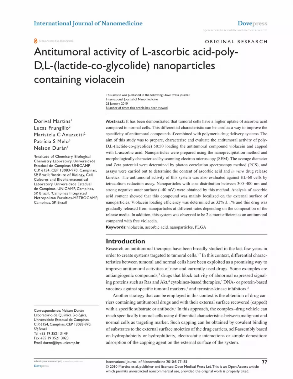
ResultsMorphological analysesThe system obtained by this methodology presented a regular
and smooth spherical shape, as observed in scanning electron
microscopy (SEM) analyses (Figure 1). No significant dif-
ferences were observed between nanoparticles prepared with
or without ascorbic acid (data not shown).
Drug loading efficiency and polymer recoveryTotal loaded violacein rates for NP-violacein and NP-AA-
violacein were determined to be 32% ± 1%, while polymer
recovery varied between 75%–78%.
Analysis of ascorbic acid contentThe total content of ascorbic acid reminescent in NP-AA
and NP-AA-violacein were determined as 1.29 ± 0.2 and
1.31 ± 0.3 µmol per mg of particles. To further characterize
the localization of this compound, a release kinetics assay
was performed. As seen in Figure 2, ascorbic acid in PLGA
nanoparticles exhibited a burst releasing behavior at five
minutes of incubation, which is very similar to the dissolution
profile of free ascorbic acid under the described conditions.
Average diameter and Zeta potentialBy this approach, particles with a nanometric size varying
between 300 and 550 nm and a strong negative Zeta potential
A B
Figure 1 Scanning electron analyses of nanoparticles preparation in magnifications of 5,000 (A) and 10,000 (B) folds. No significant differences were observed among the four obtained systems.
International Journal of Nanomedicine 2010:5 81
Antitumoral L-ascorbic acid-PLgA-violacein nanoparticles Dovepress
submit your manuscript | www.dovepress.com
Dovepress
were obtained (Table 1). Also, the addition of ascorbic acid
to the preparation increased significantly the negative charge
of the external surface by 25–30 mV.
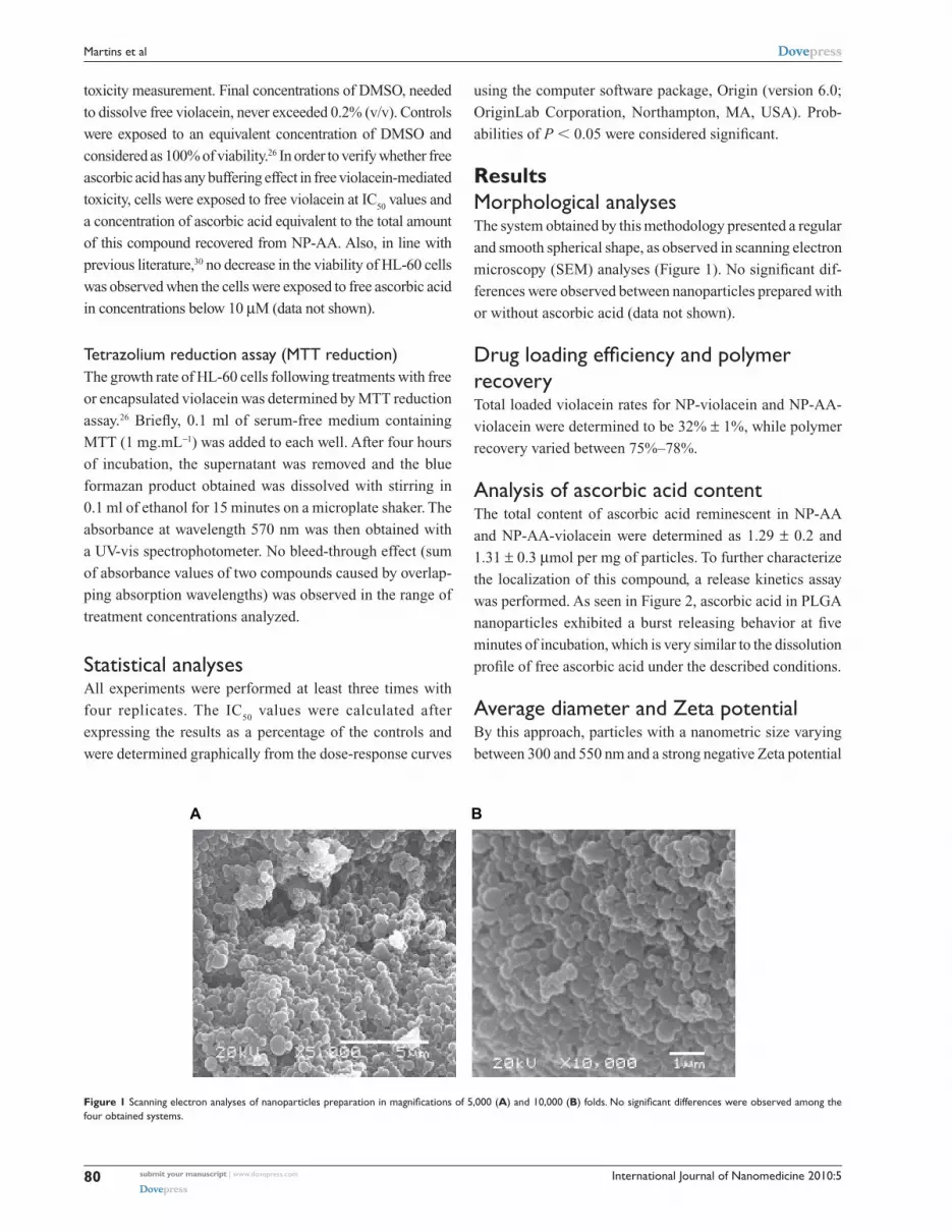
Violacein release kineticsNo differences of the dissolution profile of free violacein in
surfactant-buffer or ethanol-buffer solutions were observed
(Figure 3). In terms of violacein release kinetics of nanopar-
ticles, an initial burst of release in three hours of analysis was
observed (about 40% and 60% for surfactant-buffer and etha-
nol-buffer solutions, respectively), followed by a gradual release
until 36 hours and reaching a cumulative release of 80% of the
initial content at 72 hours of analysis (Figure 3). Moreover,
a significant difference was observed in the release kinetics pro-
file of this system depending on the composition of the analysis
solution. The release rates in ethanol-buffer media was observed
to be faster compared to the surfactant-buffer solution.
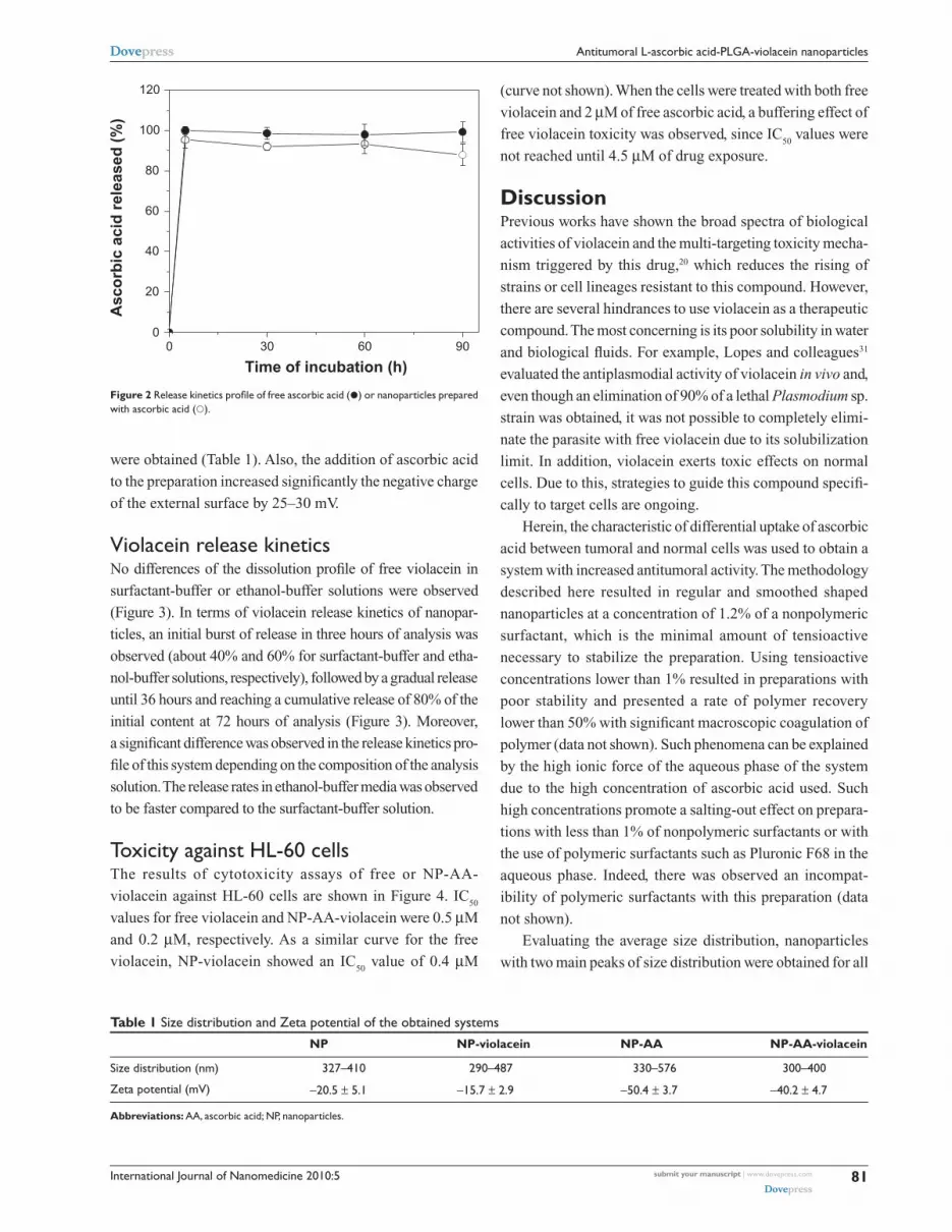
Toxicity against hL-60 cellsThe results of cytotoxicity assays of free or NP-AA-
violacein against HL-60 cells are shown in Figure 4. IC50
values for free violacein and NP-AA-violacein were 0.5 µM
and 0.2 µM, respectively. As a similar curve for the free
violacein, NP-violacein showed an IC50
value of 0.4 µM
(curve not shown). When the cells were treated with both free
violacein and 2 µM of free ascorbic acid, a buffering effect of
free violacein toxicity was observed, since IC50
values were
not reached until 4.5 µM of drug exposure.
DiscussionPrevious works have shown the broad spectra of biological
activities of violacein and the multi-targeting toxicity mecha-
nism triggered by this drug,20 which reduces the rising of
strains or cell lineages resistant to this compound. However,
there are several hindrances to use violacein as a therapeutic
compound. The most concerning is its poor solubility in water
and biological fluids. For example, Lopes and colleagues31
evaluated the antiplasmodial activity of violacein in vivo and,
even though an elimination of 90% of a lethal Plasmodium sp.
strain was obtained, it was not possible to completely elimi-
nate the parasite with free violacein due to its solubilization
limit. In addition, violacein exerts toxic effects on normal
cells. Due to this, strategies to guide this compound specifi-
cally to target cells are ongoing.
Herein, the characteristic of differential uptake of ascorbic
acid between tumoral and normal cells was used to obtain a
system with increased antitumoral activity. The methodology
described here resulted in regular and smoothed shaped
nanoparticles at a concentration of 1.2% of a nonpolymeric
surfactant, which is the minimal amount of tensioactive
necessary to stabilize the preparation. Using tensioactive
concentrations lower than 1% resulted in preparations with
poor stability and presented a rate of polymer recovery
lower than 50% with significant macroscopic coagulation of
polymer (data not shown). Such phenomena can be explained
by the high ionic force of the aqueous phase of the system
due to the high concentration of ascorbic acid used. Such
high concentrations promote a salting-out effect on prepara-
tions with less than 1% of nonpolymeric surfactants or with
the use of polymeric surfactants such as Pluronic F68 in the
aqueous phase. Indeed, there was observed an incompat-
ibility of polymeric surfactants with this preparation (data
not shown).
Evaluating the average size distribution, nanoparticles
with two main peaks of size distribution were obtained for all
Figure 2 Release kinetics profile of free ascorbic acid () or nanoparticles prepared with ascorbic acid ().
00
20
40
60
80
100
120
Asc
orb
ic a
cid
rel
ease
d (
%)
Time of incubation (h)906030
Table 1 size distribution and Zeta potential of the obtained systems
NP NP-violacein NP-AA NP-AA-violacein
size distribution (nm) 327–410 290–487 330–576 300–400
Zeta potential (mV) -20.5 ± 5.1 -15.7 ± 2.9 -50.4 ± 3.7 -40.2 ± 4.7
Abbreviations: AA, ascorbic acid; NP, nanoparticles.

International Journal of Nanomedicine 2010:582
Martins et al Dovepress
submit your manuscript | www.dovepress.com
Dovepress
systems described as follows: 330 and 410 for NP, 290 and
487 for NP-violacein, 330 and 576 for NP-AA, 300 and
400 for NP-AA-violacein. This effect could be explained
by the freeze-drying process, which promotes aggregation
of vicinal nanoparticles. This consequently increases the
average diameter of nanoparticle preparations and frequency
of aggregates.32–34 However, the fact that nanoparticles with
strong negative Zeta potential were obtained implies a better
Figure 4 Toxicity of free or NP-AA-violacein against hL60 cells.Notes: (■) NP-AA; () free violacein + 2 µM of ascorbic acid () free violacein; and () NP-AA-violacein.Abbreviation: NP-AA, nanopatide-asconbic acid.
0
20
40
60
80
100
Via
bili
ty (
% o
f co
ntr
ol)
Treatment concentration (µM)
0.0 0.5 1.0 1.5 2.0 2.5 3.0 3.5 4.0
0
20
40
60
80
100
Vio
lace
in r
elea
sed
(%
)
Time of incubation (h)
BA
8070605040302010807060504030200 010
Figure 3 Release kinetics profile free or NP-AA-violacein in ethanol-buffer ethanol-buffer (A) or surfactant-buffer (B) solutions.Notes: () free violacein in ethanol-PBs solution; () NP-AA-violacein in ethanol-PBs solution; (■) free violacein in Tween-PBs solution () NP-AA-violacein in Tween-PBs solution.Abbreviations: NP-AA, nanopatide-asconbic acid; PBs, Phosphate buffered saline.
International Journal of Nanomedicine 2010:5 83
Antitumoral L-ascorbic acid-PLgA-violacein nanoparticles Dovepress
submit your manuscript | www.dovepress.com
Dovepress
stability in aqueous solution due to the electrostatic repulsion
between particles. This facilitates the dispersion in water
of those systems. Although nanoparticles based in PLGA
often have a negatively charged surface as consequence
of ionizable carboxyl-terminal moieties of this polymer, a
significant difference was observed between particles pre-
pared with or without ascorbic acid. Since Zeta potential
is affected by adsorption or binding of compounds to the
external surface of colloidal particles, this difference indi-
cates the presence of a reminiscent amount of ascorbic acid
on the external surface of the system. Ascorbic acid has two
ionizable hydroxyl groups at carbons 3 and 4 with pKa 4.2
and 11.6, respectively. Once the Zeta potential measurements
were carried out at pH 7, the ionization of carbon 3 (lead-
ing to ascorbate anion formation) can explain the increased
negative charge observed. The analysis of ascorbic acid
content reinforces this observation since the release profile of
ascorbic acid from the nanoparticles is marked by a massive
release burst with five minutes of incubation. This suggests
a weak adsorption of this compound on the external surface
of the system.
In terms of violacein release kinetics of nanoparticles, it
can be observed an initial burst of release at three hours of
analysis can be observed followed by a gradual release until
72 hours. Such behavior suggests the system has violacein
either in its external surface or mixed with the outer surface
of the matrix (being, therefore, easily released in short time
incubations) and an internal content of drug that is released
gradually, reaching a release of 80% of the initial content
at 72 hours of analysis. Also, a significant difference in
release rates was observed between surfactant-buffer and
ethanol-buffer solutions, with a faster drug release rate in the
second media. This can be explained by the difference in the
molecular weight of both compounds since ethanol is a low
molecular weight compound and highly diffusible through
the polymeric matrix, allowing it to bring out the violacein
trapped in the inner side of the nanoparticles, while the
surfactant with high molecular weight is unable to reach the
inner side of nanoparticles. Thus, the media surfactant-buffer
promotes diffusion but not arrest of the drug, mimicking the
physiological situation where the particles are surrounded by
high molecular weight compounds such as proteins.
In cytotoxicity assays, a significant difference of activ-
ity between nanoparticles-loaded and free violacein, being
NP-violacein and NP-AA-violacein 1.25 and 2.5 times more
efficient than free violacein respectively was observed.
The increased biological activity of violacein loaded in
nanoparticles has been demonstrated in previous works
either with tumoral cells26 or Staphylococci strains27 and the
addition of derivatives of ascorbic acid on external surface
of nanoparticles had been shown to increase the efficiency
of dehydrocrotonin loaded in PLGA nanoparticles with a
protein mediated uptake mechanism and a shunt of cell death
mechanism to receptor-mediated pathways.29 However, an
unexpected observation was reached, since free ascorbic acid
exhibits a buffering effect in violacein-mediated toxicity,
while the addition of ascorbic acid on the external surface of
PLGA nanoparticles loading violacein actually promotes an
improvement of the toxicity of this compound. This suggests
a differential cell death mechanism between free violacein
and NP-AA-violacein. Chen and colleagues35 demonstrated
that, although ascorbic acid has mainly antioxidant proper-
ties in cell environment, it can act as a generator of reactive
species of oxygen if it gets concentrated in the external
tumor microenvironment. Therefore, one explanation for the
observed fact is that the increasing in external concentration
of ascorbic acid bound to NP-AA-violacein promotes gen-
eration of ROS in the external environment, while the high
uptake of NP-AA-violacein complex promotes significant
internal oxidative damage, leading to internal and external
cell damage. Since ROS are necessary to promote the inter-
nalization of ascorbic acid, a positive feedback mechanism
of internalization of NP-AA-violacein could be involved
in the increased toxicity observed. Further studies should
provide insights about the death mechanism triggered by
the presented system.
Current drug delivery systems targeted to tumoral cells
often use either peptides or proteins, such as transferrin and
epithelial growth factor receptor (EGFR), or small molecules
such as folate.16,36 All the cited molecules have been used
in vitro or in vivo with promising results and high specificity.
However, the efficiency of targeting depends on the lineage
analyzed, since uptake of tranferrin, EGFR and folate broadly
varies between tumoral cells. Specifically, transferrin and
EGFR are useful for solid tumors,36 whereas folate can be
used for both solid and nonsolid tumors. It has been shown that
uptake of folate-targeted drug delivery systems is decreased in
tumoral models which have lower density of folate receptors,
such as HeLa and MCF-7 cells.37 Thus, the spectra of uses of
those molecules are restricted depending on the cell model
studied. On the other hand, the role of ascorbic acid in cancer
metabolism is well described for several tumoral lineages and
its high uptake is a characteristic common to most solid or
nonsolid tumors analyzed,38–40 which makes it an interesting
molecule to target a broader spectra of tumors. In addition,
it has been reported that overexpression of GLUT proteins
International Journal of Nanomedicine 2010:584
Martins et al Dovepress
submit your manuscript | www.dovepress.com
Dovepress
is a marker of very aggressive hepatocellular carcinomas.41
In this context, the proposed system can be used to target
drugs with multi-toxicity mechanisms to tumors refractory
to current chemotherapies.
Although ascorbic acid is a molecule susceptible to
oxidation, the oxidized form of this molecule (dehydro-
ascorbate anion) can also target drug carriers, since the
internalization of ascorbic acid requires its oxidation to dehy-
droascorbate by cellular ROS. However, efforts to create drug
carriers with derivatives of ascorbic acid to avoid undesirable
oxidation processes and to improve the shelf-life of ascorbic
acid on nanoparticles are being carried out and showed prom-
ising results with polymeric nanoparticles carrying another
antitumoral compound, dehydrocrotonin.29 Complementary
studies on toxicity of such complexes against normal cells and
in vivo evaluations should be carried out to further character-
ize the pharmacological potential of this system.
ConclusionsThe presented system could successfully load violacein and
be capped with ascorbic acid, with good physical-chemical
parameters of stability and a significant improvement of anti-
tumoral activity of the complex. No buffering or antagonist
effects on violacein activity were observed when ascorbic
acid was used as a capping agent in the system. However,
further characterization of the cell mechanism induced by this
system and in vivo assays should be carried out. This prepa-
ration could be used as a selective way to administer higher
doses of violacein in vitro and in vivo in order to improve its
biological activity and to reduce side effects.
AcknowledgmentsFinancial support from CNPq, FAPESP and Brazilian
Nanobiotechnology Network and Brazilian Nanocosmetics
Network (MCT/CNPq), the suggestions of Dr. Marcelo M M
de Azevedo and language reviewing by Ms Meena Kathiresan
and Ms Zornitsa Stoyanova are acknowledged. The authors
report no conflicts of interest in this work.
References 1. Sofou S. Surface-active liposomes for targeted cancer therapy.
Nanomedicine. 2007;2:711–724. 2. Savona M, Talpaz M. Getting to the stem chronic myeloid leukaemia.
Nature Rev Cancer. 2008;8:341–350. 3. Narayana A, Kelly P, Golfinos J, et al. Antiangiogenic therapy using
bevacizumab in recurrent high-grade glioma: impact on local control and patient survival. J Neurosurg. 2009;110:173–180.
4. Paternot S, Roger PP. Combined inhibition of MEK and mammalian target of rapamycin abolishes phosphorylation of cyclin-dependent kinase 4 in glioblastoma cell lines and prevents their proliferation. Cancer Res. 2009;69:4577–4581.
5. Egilmez NK, Kilinc MO, Gu T, et al. Controlled-release particulate cytokine adjuvants for cancer therapy. Endocr Metab Immune Disord Drug Targets. 2007;7:266–270.
6. Nemunaitis JJ. Gene immunotherapy for non-small cell lung cancer. Methods Mol Biol. 2009;542:499–514.
7. Douziech-Eyrolles L, Marchais H, Hervé K, et al. Nanovectors for anticancer agents based on superparamagnetic iron oxide nanoparticles. Int J Nanomedicine. 2007;2:541–550.
8. Ciampi R, Vivaldi A, Romei C, et al. Expression analysis of facilitative glucose transporters (GLUTs) in human thyroid carcinoma cell lines and primary tumors. Mol Cell Endocrinol. 2008;291:57–62.
9. Agus DB, Vera JC, Golde DW. Stromal cell oxidation: a mechanism by which tumors obtain vitamin C. Cancer Res. 1999;18:4555–4558.
10. Torchilin VP. Micellar nanocarriers: pharmaceutical perspectives. Pharm Res. 2007;24:1–16.
11. Marcato PD, Durán N. New aspects of nanopharmaceutical delivery systems. J Nanosci Nanotechnol. 2008;8:2216–2229.
12. Har-el YE, Kato Y. Intracellular delivery of nanocarriers for cancer therapy. Curr Nanosci. 2007;3:329–338.
13. Arruebo M, Fernández-Pacheco R, Ibarra MR, Santamaría J. Magnetic nanoparticles for drug delivery. Nanotoday. 2007;2:22–32.
14. Bharali DJ, Khalil M, Gurbuz M, Simone TM, Mousa SA. Nanoparticles and cancer therapy: A concise review with emphasis on dendrimers. Int J Nanomedicine. 2009;4:1–7.
15. Tekade RK, Kumar PV, Jain NK. Dendrimers in oncology: An expand-ing horizon. Chem Rev. 2009;109:49–87.
16. Nair LS, Laurencin CT. Biodegradable polymers as biomaterials. Progr Polym Sci. 2006;32:762–798.
17. Jain KK. Recent advances in nanoocology. Technol Cancer Res Treat. 2008;7:1–13.
18. Mundargi RC, Babu VR, Rangaswamy V, Patel P, Aminabhavi TM. Nano/micro technologies for delivering macromolecular therapeutics using poly(D,L-lactide-co-glycolide) and its derivatives. J Control Release. 2008;125:193–209.
19. Durán N, Menck CF. Chromobacterium violaceum: a review of pharmacological and industrial perspectives. Crit Rev Microbiol. 2001;27:201–222.
20. Durán N, Justo GZ, Ferreira CV, Melo PS, Cordi L, Martins D, et al. Violacein: properties and biological activities. Biotechnol Appl Biochem. 2007;48:127–133.
21. Ferreira CV, Bos CL, Versteeg HH, Justo GZ, Durán N, Peppelenbosch MP. Molecular mechanism of violacein-mediated human leukemia cell death. Blood. 2004;104:1459–1464.
22. de Carvalho DD, Costa FTM, Durán N, Haun M. Cytotoxic activity of violacein in human colon cancer cells. Toxicol In Vitro. 2006;20:1514–1521.
23. Bromberg N, Justo GZ, Haun M, Durán N, Ferreira CV. Violacein cytotoxicity on human blood lymphocytes and effect on phosphatases. J Enzyme Inhib Med Chem. 2005;20:449–454.
24. Rettori D, Durán N. Production, extraction and purification of violacein: an antibiotic pigment produced by Chromobacterium violaceum. World J Microb Biotechnol. 1998;14:685–688.
25. Italia JL, Yahya MM, Singh D, Ravi Kumar MN. Biodegradable nanoparticles improve oral bioavailability of amphotericin B and show reduced nephrotoxicity compared to intravenous Fungizone. Pharm Res. 2009;26:1324–1331.
26. Melo PS, De Azevedo MM, Frugillo L, Anazetti MC, Marcato PD, Durán N. Nanocytotoxicity: violacein and violacein-loaded poly(D,L-lactide-co-glycolide) nanoparticles acting on human leukemic cells. J Biomed Nanotechnol. 2009;5:192–201.
27. Martins D, Costa FTM, Brocchi M, et al. Evaluation of the anti-bacterial activity of poly-(D,L-lactide-co-glycolide) nanoparticles containing violacein. J Nanoparticle Res. 2009;Submission number:NANO2280.
28. Suntornsuk L, Gritsanapun W, Nilkamhank S, Paochom A. Quantita-tion of vitamin C content in herbal juice using direct titration. J Pharm Biomed Anal. 2002;28:849–55.

International Journal of Nanomedicine 2010:5
International Journal of Nanomedicine
Publish your work in this journal
Submit your manuscript here: http://www.dovepress.com/international-journal-of-nanomedicine-journal
The International Journal of Nanomedicine is an international, peer-reviewed journal focusing on the application of nanotechnology in diagnostics, therapeutics, and drug delivery systems throughout the biomedical field. This journal is indexed on PubMed Central, MedLine, CAS, SciSearch®, Current Contents®/Clinical Medicine,
Journal Citation Reports/Science Edition, EMBase, Scopus and the Elsevier Bibliographic databases. The manuscript management system is completely online and includes a very quick and fair peer-review system, which is all easy to use. Visit http://www.dovepress.com/ testimonials.php to read real quotes from published authors.
85
Antitumoral L-ascorbic acid-PLgA-violacein nanoparticles Dovepress
submit your manuscript | www.dovepress.com
Dovepress
Dovepress
29. Frungillo L, Martins D, Teixeira S, Anazetti MC, Melo Pda S, Durán N. Targeted antitumoral dehydrocrotonin nanoparticls with L-ascorbic acid 6-stearate. J Pharm Sci. 2009;98:4796–4807.
30. Arranz N, Haza AI, García A, Delgado ME, Rafter J, Morales P. Inhibi-tion by vitamin C of apoptosis induced by N-nitrosamines in HepG2 and HL-60 cells. J Appl Toxicol. 2008;28:788–796.
31. Lopes SC, Blanco YC, Justo GZ, et al. Violacein extracted from Chromobacterium violaceum inhibits Plasmodium growth in vitro and in vivo. Antimicrob Agents Chemother. 2009;53:2149–2152.
32. Cabane B, Blanchon S, Neves C. Recombination of nanometric vesicles during freeze-drying. Langmuir. 2006;22:1982–1990.
33. Lee J, Cheng Y. Critical freezing rate in freeze drying nanocrystal dispersions. J Control Release. 2006;111:185–192.
34. Holzer M, Vogel V, Mäntele W, Schwartz D, Haase W, Langer K. Physico-chemical characterisation of PLGA nanoparticles after freeze-drying and storage. Eur J Pharm Biopharm. 2009;72:428–437.
35. Chen Q, Espey MG, Sun AY, et al. Ascorbate in pharmacologic concentrations selectively generates ascorbate radical and hydrogen peroxide in extracellular fluid in vivo. Proc Natl Acad Sci U S A. 2007;104:8749–8754.
36. Yu B, Zhao X, Lee LJ, Lee RJ. Targeted delivery systems for oligo-nucleotide therapeutics. AAPS J. 2009;11:95–203.
37. Chen H, Ahn R, Van den Bossche J, Thompson DH, O’Halloran TV. Folate-mediated intracellular drug delivery increases the anticancer efficacy of nanoparticulate formulation of arsenic trioxide. Mol Cancer Ther. 2009;8:1955–1963.
38. Macheda ML, Rogers S, Best JD. Molecular and cellular regulation of glucose transporter (GLUT) proteins in cancer. J Cell Physiol. 2005;202:654–662.
39. Airley RE, Mobasheri A. Hypoxic regulation of glucose transport, anaerobic metabolism and angiogenesis in cancer: novel pathways and targets for anticancer therapeutics. Chemotherapy. 2007;53:233–256.
40. Fonteyne P, Casneuf V, Pauwels P, et al. Expression of hexokinases and glucose transporters in treated and untreated oesophageal adenocarci-noma. Histol Histopathol. 2009 ; 24:971–977.
41. Daskalow K, Pfander D, Weichert W, et al. Distinct temporospatial expression patterns of glycolysis-related proteins in human hepatocel-lular carcinoma. Histochem Cell Biol. 2009;132:21–31.