pectin/poly(lactide-co-glycolide) composite matrices for biomedical applications
TRANSCRIPT
Biomaterials 25 (2004) 3201–3210
ARTICLE IN PRESS
*Correspondin
6406.
E-mail addres
0142-9612/$ - see
doi:10.1016/j.bio
Pectin/poly(lactide-co-glycolide) composite matrices forbiomedical applications
LinShu Liu*, Young Jun Won, Peter H. Cooke, David R. Coffin, Marshal L. Fishman,Kevin B. Hicks, Peter X. Ma
ERRC, US Department of Agriculture, E 600 Mermaid Lane, Wyndmoor, PA 19038, USA
Received 17 July 2003; accepted 29 September 2003
Abstract
The aim of the research was to develop matrices for the delivery of biologically active substances for tissue regeneration. To this
end, a new biodegradable matrix composed of a hydrophobic porous poly(lactide-co-glycolide), p(LGA), network entangled with
another network of hydrophilic pectin was fabricated in the presence of calcium chloride. The calcium salts function as both a pore
forming reagent and cross-linker for the formation of pectin networks; the method combines creating pores and cross-linking
polymers in one step. Microscopic imaging and dynamic mechanical analysis revealed a double-network structure of the composite
matrices. The pectin enables the composite to carry signal molecules. This is accomplished by linking signal molecules to pectin by
physical adsorption or by chemical reaction. The p(LGA) networks in the composite impart mechanical properties comparable to
p(LGA) alone. The mechanical properties of the composite are far superior to matrices containing only pectin. Furthermore, the
pectin-containing matrices improved cell adhesion and proliferation when compared to plain p(LGA) matrices, as determined
in vitro by osteoblast culture.
Published by Elsevier Ltd.
Keywords: Pectin; Poly(lactide-co-glycolide); Matrix; Biomaterials; Tissue engineering
1. Introduction
Pectins are polysaccharides enriched in galacturonicacid and galacturonic acid methyl ester units. Combinedwith proteins and other polysaccharides, pectins formskeletal tissues of plants, which are chemically stable andphysically strong [1,2]. With high molecular weight and apolyanionic nature, pectins react to their environmentsthrough a continuum of physical states, ranging fromdense gels to dilute solutions. These properties enablepectin polymers to carry signal molecules and supportvarious biologically active substances. In addition, pectinsclosely imitate the structure of polysaccharides found inthe extracellular matrices of mammals. Thus, theirproperties and general availability make pectins viableto consider when engineering new biomedical materials.Pectins have shown promise in engineering drug
carriers for oral drug delivery [3]. The combination of
g author. Tel.: +1-215-233-6386; fax: +1-215-233-
s: [email protected] (L.S. Liu).
front matter Published by Elsevier Ltd.
materials.2003.10.036
pectin and a second polymer into a composite may alterdegree of swelling [4,5], and change mechanical proper-ties [6]. New uses of pectins in biomedical applicationsinclude facilitating the delivery of specific sequences ofamino acids, anti-inflammatory agents, anti-coagulants,and wound healing substances to tissue sites. To be usedas such, pectin based composites can be formed intomembranes, microspheres, scaffolds, or injectable gels.Here, we describe the development and characterizationof composite matrices of pectin and poly(lactide-co-glycolide), pectin/p(LGA).P(LGA) has been used clinically for tissue repair and
organ regeneration for decades. This hydrophobicpolymer is biocompatible, biodegradable, and easilyprocessed into a variety of sizes and shapes which havegood mechanical properties [7–10]. Although p(LGA)will support cell attachment and cell growth, it does notimpart signals to the cells [11]. The inability to conveysignals limits the application of p(LGA). This deficiencyis currently overcome by synthesizing block or graftcopolymers of lactic acid and lysine or other segmentscarrying side chain functional groups [11,12]. Through

ARTICLE IN PRESSL.S. Liu et al. / Biomaterials 25 (2004) 3201–32103202
the functional groups, specific amino acid sequences canbe attached. By this strategy, a number of new chemicalentities have been provided. The preparation of copo-lymers involves a series of isolation, purification andidentification procedures.In this study, we present a method to prepare porous
matrices from pectins and p(LGA) that retain thebiomechanical strength of p(LGA) yet provide accessfor hydrophilic, bioactive substances. The matrices werecharacterized for structural, physical and mechanicalproperties, and the capacity to store signal molecules.Furthermore, these matrices were tested in vitro for theability to support cell attachment and growth. Theseproperties are required in tissue engineering applica-tions.
2. Experimental
2.1. Materials
Sodium salts of pectin from citrus fruits (degree ofesterification, DE, 93%), bovine serum albumin (BSA),fluoresceinamine, p(LGA) [50:50; average Mw; 50,000–75,000; Tg 45–50�C], pectinase 3XL, neutral-bufferedformalin, trypan blue, DNA Quantitation Kit, 2,2,2,-trifluoroethanesulfonyl chloride (tresyl chloride), anddimethylsulfoxide (DMSO) were purchased from Sig-ma-Aldrich (St. Louis, MO). Fetal bovine serum, a-minimum essential medium (a-MEM), ascorbic acid-freea-MEM (Formula 94-5049EL), penicillin–streptomycin,Dulbecco’s phosphate-buffered saline, and trypsin-EDTA were purchased from Gibco BRL Products, LifeTechnologies (Grand Island, NY). Ascorbic acid waspurchased from Fisher Scientific (Pittsburgh, PA).Micro BCA� reagent was from Pierce (Rockford, IL).Ethylene oxide was purchased from H.W. AndersonProducts (Chapel Hill, NC). All other chemicals wereA.C.S. grade, and used without further purification.
2.2. De-esterification of pectins
Gentle alkaline de-esterification was performed byadjusting the pH of a pectin solution (1%, w/v) to 8.0with 0.1n NaOH and stirring at 4�C over 48 h [13]. Thereaction solution was dialyzed against a large volume ofdistilled water (DI water). Pectins were recovered byspreading the pectin solution into ethanol containing0.1% CaCl2, the resultant microparticles were filtered,washed with DI water, and lyophilized. Pectin particleswith the size ranging from 15 to 125 mm were collected.The extent of de-esterification was determined bycomparing the DE values of the de-esterified pectinswith those before the reaction. The DE values of pectinswere measured by high-performance liquid chromato-graphy (HPLC) as previously reported [14]. Other
molecular properties of the de-esterified pectins, suchas the weight average Mw; root mean square radius ofgyration (RgzÞ; and intrinsic viscosity (½Z�Þ; were eval-uated by HPSEC with on-line multi-angle laser lightscattering and viscometric detection as described pre-viously [15].
2.3. Preparation of pectin/p(LGA) composites
Pectin/p(LGA) composite matrices were prepared bya multi-step procedure. In step I, 1.0 g of p(LGA) wasdissolved in 8.0ml of chloroform, into which 0.10 g ofde-esterified pectin, 2.0 g of calcium chloride, and 6.9 gof sodium chloride were dispersed and blended to form aslurry. The size of the inorganic salt particles rangedfrom 50 to 200 mm. In step II, the slurry was cast intodisks in a mold with dimensions of 6mm in diameterand 3mm in thickness, and the solvent was evaporatedto form a solid matrix. In step III, the matrix wasimmersed in 1 l of deionized water (DI water), wherepectin particles started to swell and hydrate, salts beganto dissolve and diffuse. Meanwhile, dissolved calciumions reacted with and bound to the hydrated pectinparticles via inter- and intra-chain chelation. Dissolvedsodium chloride and excessive calcium salts diffused tocreate spaces for water migration. The process in step IIIwas continued for 48 h. In that time the water waschanged several times to complete cross-linking ofpectin and leaching of residual salts. Lastly, freezedrying the matrices created a channeled porous struc-ture.Porous p(LGA) matrices were prepared by the same
method as described above, except for the substitutionof pectin with sodium chloride. Porous pectin matriceswere prepared by casting pectin solution (2.0%, w/v) ina mold (6� 3mm (d � h)) lyophilizing the solution tocreate a solid structure, which thereafter was treatedwith calcium chloride solution (0.1m) and lyophilized.The p(LGA) and pectin matrices were used as controls.
2.4. Recovery of p(LGA) and pectin from pectin/
p(LGA) matrices
Samples were analyzed to determine the efficiencywith which calcium chloride cross-linked pectin particlesand the amounts of pectin and p(LGA) in the finalcomposite matrices. Samples of pectin/p(LGA) matriceswere vacuum-dried for 24 h prior to experimentation.Each dried sample was placed in 2.0ml tetrahydrofuran(THF) in a volumetric flask equipped with a pennyheadstopper to prevent solvent evaporation. The mixture wasgently shaken at room temperature for 2 h to completethe extraction of p(LGA) polymers. The extractionsolution was removed and analyzed for p(LGA) contentusing a Shimadzu HPLC equipped with an RID-10Arefractive index detector and an SCL-10A data station
ARTICLE IN PRESSL.S. Liu et al. / Biomaterials 25 (2004) 3201–3210 3203
(Model LC-10AD, Kyoto, Japan). An aliquot of thesolution (10 ml) was injected and eluted by THF on aphenogel guard column (model 22824G, 50� 7.8mm,Phenomenex, Torrance, CA) and a phenogel column(model GP/4446, 300� 7.8mm, Phenomenex) at theflow rate of 0.5ml/min. p(LGA)/THF solutions ofknown concentrations were run under the same condi-tions and used to prepare a standard curve.After the removal of p(LGA) polymers, the solid
residues, calcium cross-linked pectins, were washed withfresh THF (2� 2ml), dry ethanol (3� 2ml), and air-dried. Sodium phosphate solution (1.0m, 2.0ml, pH 6.5)was added to the flask and sonicated to solubilize thepectin. Pectin content was analyzed by total sugar assay[16,17].
2.5. Chemical modification of pectin/p(LGA) matrices
The chemical modification of pectin/p(LGA) compo-site matrices was performed by grafting the matriceswith fluoresceinamine using tresyl chloride as a couplingreagent as previously reported [18,19]. Samples ofpectin/p(LGA) matrix were immersed in dry acetone(pre-dried over molecular sieve 4A; Acros, Pittsburgh,PA) for 24 h with three changes. To a glass vialcontaining 2.0ml dry acetone and one piece of the drysample, pyridine (200 ml) and tresyl chloride (100 ml)were added, and gently shaken for 10min at roomtemperature. The sample was removed and rinsed withdry acetone (3� 5ml), phosphate-buffered saline (PBS)(pH 7.0, 2� 5ml), and placed in 2.0ml of PBS (pH 7.8)containing fluoresceinamine (20mm) and incubated for20 h at room temperature. To completely remove thefluoreceinamine which was physically adsorbed ratherthan chemically conjugated, the sample was washed with1mm HCl and 0.2m NaHCO3 repeatedly, and 1.0mNaCl containing 1mm HCl, finally with DI water [19].Samples thus treated were examined by confocal laserfluorescence microscopy as described in the followingsection.P(LGA) matrices treated with both tresyl chloride and
fluorescienamine under the same conditions were usedas controls.
2.6. Microscopic imaging
2.6.1. Scanning electron microscopy (SEM)
For SEM examinations, samples of pectin particles,NaCl–CaCl2 crystal mixtures, pectin/p(LGA), andp(LGA) matrices were mounted on specimen stubs,coated with a thin layer of gold in a sputter coatingapparatus (Edwards High Vacuum, Wilmington, MA),and examined in a model JSM 840A scanning electronmicroscope (Jeol USA, Peabody, MA) operating at10 kV in the secondary electron imaging mode. Imageswere collected at 25� and 250� using an Imix-1 digital
image workstation (Princeton Gamma-tech, Princeton,NJ).
2.6.2. Confocal laser microscopy
Samples of fluorescently labeled pectin/p(LGA) com-posite matrices were glued to 1� 3 inch microscopeslides and placed in the sample stage of an IRBE opticalmicroscope with a 10� lens integrated with a modelTCS-SP laser scanning confocal microscope (LeicaMicrosystems, Exton, PA). The parameters for theimage acquisition were set for confocal reflection(633 nm) and confocal fluorescence (488/500–530 nm)in two channels.
2.7. Dynamic mechanical analysis (DMA)
Compressive mechanical testing of the matrices wasperformed on a Rheometric Scientific RSA II SolidsAnalyzer (Rheometric Scientific, Piscataway, NJ) fittedwith 25mm parallel plates. Temperature control wasmaintained using a liquid nitrogen environmentalcontroller. Each sample matrix was placed on the lowerplate, the upper plate was lowered onto the sample togive a slight compressive force, and then locked in theplace. The samples were tested using a compressivestrain of 0.25–1.0%, depending on the stiffness of thesample. Storage modulus, loss modulus, and losstangent were determined over a temperature range of�100�C to +200�C at the heating rate of 10�C/min.The data were analyzed using Rheometric ScientificOrchestrator software, version 6.5.7.
2.8. Determination of equilibrium water content and
protein adsorption
Samples of pectin/p(LGA) and p(LGA) matrices weredried under vacuum at room temperature for 24 h. priorto the experiment. Each dried sample was incubatedwith a large volume of PBS (pH 7.0) at roomtemperature under gentle shaking. Samples were re-moved from the incubation solutions at intervals of 5,15, 30, 45min, and 1, 2, 4, 8 h., rinsed three times withDI water, wiped with tissue paper to remove the wateradsorbed on the surfaces, and weighed ðWwÞ: Thesamples were then re-dried under vacuum for 24 h. andweighed ðWrdÞ: The water content of matrices at eachtime point was calculated: water content=ðWw � WrdÞ=Ww � 100%.The kinetics of protein adsorption in pectin/p(LGA)
and p(LGA) matrices was studied by a proceduresimilar to that used for the determination of equilibriumwater content, except for the addition of BSA (0.1%, w/v) in PBS. After rinsing with DI water, the samples wereanalyzed for the amount of protein adsorbed by proteinBCA assay [20]. A series of BSA solutions with knownconcentrations were used to prepare a standard curve.

ARTICLE IN PRESSL.S. Liu et al. / Biomaterials 25 (2004) 3201–32103204
2.9. In vitro cell culture and bioassays
The potential for application of composite matrices intissue engineering was evaluated in vitro by seeding andculturing osteoblast cells on the matrices. Osteoblasts(MC3T3-E1, clone 26) were thawed, cultured in asupplemented ascorbic acid-free a-MEM and 10% fetalcalf serum (FBS) containing 100U/ml penicillin and100 mg/ml streptomycin in a humidified incubator at37�C with 5% CO2. The medium was changed everyother day. The cells of passages 3 and 4 were harvested,pelleted by centrifugation and re-suspended at theconcentration of 2� 106 cell/ml in a-MEM containingFBS (10%), antibiotics (1%), and l-ascorbic acid(50mg/l) (complete medium). The viability of the cellswas higher than 90% as determined with the trypan blueexclusion assay.The pectin/p(LGA) and p(LGA) matrices were sliced
into disks with dimensions of 6mm in diameter and1.5mm in thickness, and sterilized in culture flasks withethylene oxide for 2 days. The matrices were soaked inethanol for 30min, exchanged with PBS for three timesfor 30min each time, then washed with the completemedium twice for 2 h each time.For the cell attachment test, each of the matrices were
placed in a teflon plate containing 0.5ml of the cellsuspension, cultured on an orbital shaker (Model 3520;Lab-Line Instrument, Melrose Park, IL) at 75 rpmunder standard conditions. At day 3, the cell-loadedmatrices were transferred into six-well tissue cultureplates, 4ml of complete medium were added into eachwell, cultured under standard conditions for 1 day. Thematrices were removed from the medium, washed withPBS, fixed in 10% neutral-buffered formalin, dehy-drated, and embedded in paraffin using standardprocedures. Paraffin-embedded specimens were sec-tioned into 5-mm thick through the center, stained with
Table 1
Molecular properties of pectins
Properties De-esterification
Before After
Mw� 10�5 0.87 (0.02) 0.81 (0.01)
Rgz (nm) 24.7 (2.06) 21.4 (0.06)
½Z�w (dl/g) 1.25 (0.02) 1.22 (0.02)
DE (%) 93 10.2 (0.77)
Table 2
Physical characterization of pectin/p(LGA) and p(LGA) matrices
Matrices Density (g/ml) Pectin content (m
Calculated
Pectin/p(LGA) 0.190 1.28
P(LGA) 0.306 N/A
hematoxylin and eosin, and examined under a lightmicroscope [8,21].For cell proliferation studies, the cell culture was
continued for additional 7 and 14 days. The mediumwas changed every other day. At the conclusion of cellculture, the matrices were removed, washed with PBS,homogenized using a polytron homogenizer (Brink-mann Easycare Generator; Polytron-Aggregate, Swit-zerland) for 30 s at top speed (VI) for three times, thensubjected to DNA assay for cell number quantitation[8,9]. DNA assays were performed using DNA Quanti-tation Kit with Hoechst 33258 dye. The concentration ofDNA in solution was converted to a cell number using aconversion factor of 7.8 pg of DNA per MC3T3-E1 cell.This conversion factor was determined by measuring theamount of DNA from a known cell number.
2.10. Statistical analysis
The data presented here are mean7standard devia-tion. To test the significance of observed differencesbetween the study groups, a paired Student’s t-test wasapplied.
3. Results and discussion
3.1. De-esterification of pectin
Pectins were de-esterified prior to use for preparationof pectin/p(LGA) composite matrices. An almostcomplete de-esterification was accomplished (Table 1)to enable the insolubilization of pectin macromoleculesby calcium ions [22]. The Mw and intrinsic viscosity ofthe pectin were slightly reduced after de-esterification incomparison with the original polymer (Table 1). Inaddition, Rgz of pectin was reduced slightly after its de-esterification (Table 1). This indicated that somedisaggregation and/or degradation of pectin macromo-lecules occurred during its de-esterification. Neverthe-less, this seems to not significantly effect the bindingefficiency of pectin to calcium ions, since more than 80%of the pectin suspended in the p(LGA)/chloroformsolution was recovered from the resulting pectin/p(LGA) matrices. Also the ratio of pectin to p(LGA)polymers was reduced only slightly after matrix fabrica-tion (Table 2).
g) P(LGA) content (mg)
Determined Calculated Determined
1.0670.2 12.8 11.473.2
N/A 26.0 24.872.4
ARTICLE IN PRESSL.S. Liu et al. / Biomaterials 25 (2004) 3201–3210 3205
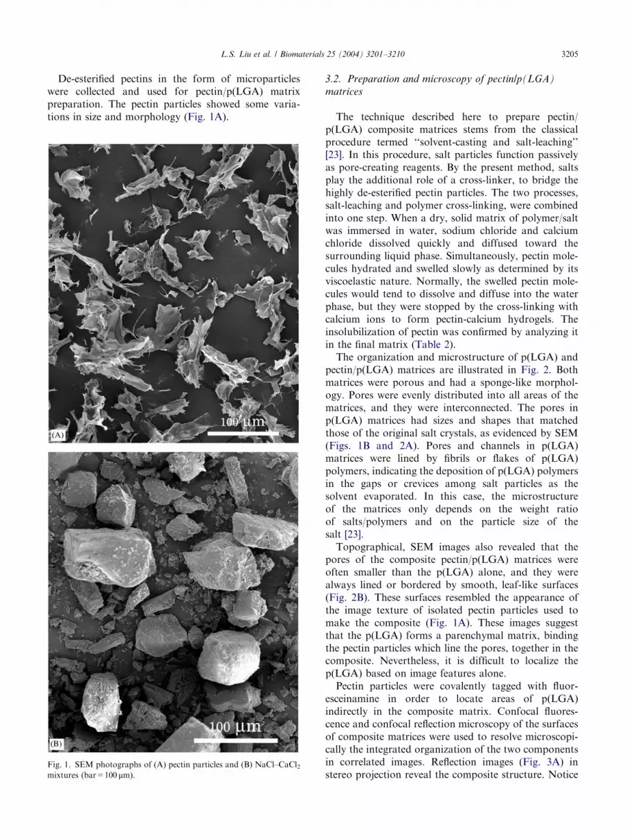
De-esterified pectins in the form of microparticleswere collected and used for pectin/p(LGA) matrixpreparation. The pectin particles showed some varia-tions in size and morphology (Fig. 1A).
Fig. 1. SEM photographs of (A) pectin particles and (B) NaCl–CaCl2mixtures (bar=100 mm).
3.2. Preparation and microscopy of pectin/p(LGA)
matrices
The technique described here to prepare pectin/p(LGA) composite matrices stems from the classicalprocedure termed ‘‘solvent-casting and salt-leaching’’[23]. In this procedure, salt particles function passivelyas pore-creating reagents. By the present method, saltsplay the additional role of a cross-linker, to bridge thehighly de-esterified pectin particles. The two processes,salt-leaching and polymer cross-linking, were combinedinto one step. When a dry, solid matrix of polymer/saltwas immersed in water, sodium chloride and calciumchloride dissolved quickly and diffused toward thesurrounding liquid phase. Simultaneously, pectin mole-cules hydrated and swelled slowly as determined by itsviscoelastic nature. Normally, the swelled pectin mole-cules would tend to dissolve and diffuse into the waterphase, but they were stopped by the cross-linking withcalcium ions to form pectin-calcium hydrogels. Theinsolubilization of pectin was confirmed by analyzing itin the final matrix (Table 2).The organization and microstructure of p(LGA) and
pectin/p(LGA) matrices are illustrated in Fig. 2. Bothmatrices were porous and had a sponge-like morphol-ogy. Pores were evenly distributed into all areas of thematrices, and they were interconnected. The pores inp(LGA) matrices had sizes and shapes that matchedthose of the original salt crystals, as evidenced by SEM(Figs. 1B and 2A). Pores and channels in p(LGA)matrices were lined by fibrils or flakes of p(LGA)polymers, indicating the deposition of p(LGA) polymersin the gaps or crevices among salt particles as thesolvent evaporated. In this case, the microstructureof the matrices only depends on the weight ratioof salts/polymers and on the particle size of thesalt [23].Topographical, SEM images also revealed that the
pores of the composite pectin/p(LGA) matrices wereoften smaller than the p(LGA) alone, and they werealways lined or bordered by smooth, leaf-like surfaces(Fig. 2B). These surfaces resembled the appearance ofthe image texture of isolated pectin particles used tomake the composite (Fig. 1A). These images suggestthat the p(LGA) forms a parenchymal matrix, bindingthe pectin particles which line the pores, together in thecomposite. Nevertheless, it is difficult to localize thep(LGA) based on image features alone.Pectin particles were covalently tagged with fluor-
esceinamine in order to locate areas of p(LGA)indirectly in the composite matrix. Confocal fluores-cence and confocal reflection microscopy of the surfacesof composite matrices were used to resolve microscopi-cally the integrated organization of the two componentsin correlated images. Reflection images (Fig. 3A) instereo projection reveal the composite structure. Notice

ARTICLE IN PRESS
Fig. 2. (A) SEM images of p(LGA) matrix showing a continuous network of salt cavities with the size of 50–200mm (bar=100mm). (B) Pectin/p(LGA) composite matrix showing the leaf- or sheet-like pectin structures which stretched over all space (bar=100 mm).
Fig. 3. (A) Averaged confocal reflection images in stereo-projection of pectin/p(LGA) indicating pectin domains constructed with irregular flat
sheets with mid-line ridges or small flat patches (1), p(LGA) domains of fine network of anastomosing fibers (2), and the areas of both (3). (B) Laser
confocal micrograph of fluorescently labeled pectin/p(LGA) showing the fluorescence located in pectin areas, not in p(LGA) areas (bar=100mm).
L.S. Liu et al. / Biomaterials 25 (2004) 3201–32103206
that a few flat areas of reflection coincide with areas ofgreen fluorescence or pectin (Fig. 3B). This resultindicated that whether or not areas of pectin particlesreflected light depended upon their orientation. Otherareas of reflection, containing irregular tubes andanastomoses do not fluoresce, indicating that theseareas contain p(LGA) (Fig. 3A).
From the above observations, it appears that thepectin domains not only filled in the pore spaces createdby the deposition of p(LGA) polymers in gaps amongparticles, but also covered or wrapped most of thep(LGA) domains. In general, pectin/p(LGA) matricespresent a complex structure of connected porous pectinnetworks which are reinforced by p(LGA) networks.
ARTICLE IN PRESSL.S. Liu et al. / Biomaterials 25 (2004) 3201–3210 3207
3.3. Dynamic mechanical properties of pectin/p(LGA)
matrices
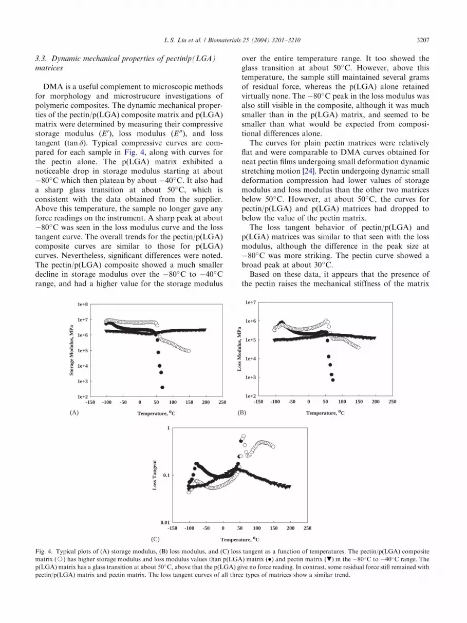
DMA is a useful complement to microscopic methodsfor morphology and microstrucure investigations ofpolymeric composites. The dynamic mechanical proper-ties of the pectin/p(LGA) composite matrix and p(LGA)matrix were determined by measuring their compressivestorage modulus ðE0Þ; loss modulus ðE 00Þ; and losstangent (tan d). Typical compressive curves are com-pared for each sample in Fig. 4, along with curves forthe pectin alone. The p(LGA) matrix exhibited anoticeable drop in storage modulus starting at about�80�C which then plateau by about �40�C. It also hada sharp glass transition at about 50�C, which isconsistent with the data obtained from the supplier.Above this temperature, the sample no longer gave anyforce readings on the instrument. A sharp peak at about�80�C was seen in the loss modulus curve and the losstangent curve. The overall trends for the pectin/p(LGA)composite curves are similar to those for p(LGA)curves. Nevertheless, significant differences were noted.The pectin/p(LGA) composite showed a much smallerdecline in storage modulus over the �80�C to �40�Crange, and had a higher value for the storage modulus
Temperature, oC
-150 -100 -50 0 50 100 150 200 250
Stor
age
Mod
ulus
, MP
a
1e+2
1e+3
1e+4
1e+5
1e+6
1e+7
1e+8
Tempera
-150 -100 -50 0 5
Los
s T
ange
nt
0.01
0.1
1
(A)
(C)
(
Fig. 4. Typical plots of (A) storage modulus, (B) loss modulus, and (C) loss
matrix (J) has higher storage modulus and loss modulus values than p(LGA
p(LGA) matrix has a glass transition at about 50�C, above that the p(LGA) g
pectin/p(LGA) matrix and pectin matrix. The loss tangent curves of all thre
over the entire temperature range. It too showed theglass transition at about 50�C. However, above thistemperature, the sample still maintained several gramsof residual force, whereas the p(LGA) alone retainedvirtually none. The �80�C peak in the loss modulus wasalso still visible in the composite, although it was muchsmaller than in the p(LGA) matrix, and seemed to besmaller than what would be expected from composi-tional differences alone.The curves for plain pectin matrices were relatively
flat and were comparable to DMA curves obtained forneat pectin films undergoing small deformation dynamicstretching motion [24]. Pectin undergoing dynamic smalldeformation compression had lower values of storagemodulus and loss modulus than the other two matricesbelow 50�C. However, at about 50�C, the curves forpectin/p(LGA) and p(LGA) matrices had dropped tobelow the value of the pectin matrix.The loss tangent behavior of pectin/p(LGA) and
p(LGA) matrices was similar to that seen with the lossmodulus, although the difference in the peak size at�80�C was more striking. The pectin curve showed abroad peak at about 30�C.Based on these data, it appears that the presence of
the pectin raises the mechanical stiffness of the matrix
Temperature, oC
-150 -100 -50 0 50 100 150 200 250
Los
s M
odul
us, M
Pa
1e+2
1e+3
1e+4
1e+5
1e+6
1e+7
ture, oC
0 100 150 200 250
B)
tangent as a function of temperatures. The pectin/p(LGA) composite
) matrix () and pectin matrix (.) in the �80�C to �40�C range. The
ive no force reading. In contrast, some residual force still remained with
e types of matrices show a similar trend.

ARTICLE IN PRESSL.S. Liu et al. / Biomaterials 25 (2004) 3201–32103208
above that of the p(LGA) matrix by itself at tempera-tures below the glass transition. Above the glasstransition, the pectin seems to enable the matrix tomaintain some level of physical integrity, although thisis at a much lower level than for the matrix attemperature below the glass transition. The incorpora-tion of the pectin network structure seems to beprimarily responsible for the increase in the values forE0 and for the decrease in the tan d values for the pectin/p(LGA) composite matrix compared to the p(LGA)matrix. These differences are considered to be thecontribution of the well-organized double-networkstructure of the composite matrices, where the thermo-plastic p(LGA) networks were reinforced by the non-thermoplastic pectin networks. The presence of thepectin in the matrix was instrumental in limitingmolecular motion of p(LGA) polymers with increasingtemperature.
3.4. Characterization of pectin/p(LGA) matrices as
carriers of signal molecules
Pectin/p(LGA) matrices were evaluated as carriers ofsignal molecules by conjugating the matrix withfluoresceinamine (Fig. 3). Green fluorescence waslocalized in irregular sheets and patches (Fig. 3B). Thesefluorescent structures are similar to those revealed bySEM for pectin/p(LGA) composite matrices (Fig. 2B),indicating the graft of the fluoresceinamine in pectinareas. Fluorescence was absent from the p(LGA) areasof fibrillar networks in the composite matrices (cf. Figs.3A and B). This is consistent with the lack of fluorescentemission observed for p(LGA) matrices (data notshown), indicating the inert nature of p(LGA) to theimmobilization reaction. The signal molecules of fluor-esceinamine were conjugated directly to the sugar ringsof pectin via the activation of the hydroxyl groups of thepectins. The hydroxyl groups of carbohydrate moleculesare only mildly nucleophilic, approximately equal towater in their relative nucleophilicity. Thus, the activa-
(A) (BIncubation time (min)
0 200 400 600
(Ww -
Wrd
)/W
w X
100
%
-40
0
40
80
120
Fig. 5. Time curves of water adsorption (A) and protein adsorption (B) in pe
conducted at room temperature using PBS as an incubation media. The pro
tion of pectins was performed in dry acetone to formintermediate reactive derivatives containing good leav-ing groups for nucleophilic substitution. The reaction ofactivated hydroxyls with nucleophiles was conducted inPBS (pH 7.8) at room temperature, which resulted instable covalent bonds between the carbohydrate and theamine-containing molecules [18,19]. Tresyl chloride hasbeen demonstrated to be a useful tool to conjugatevarious peptides and proteins with synthetic polymers ornatural polymers [19,25]. Nevertheless, we observedsome loss in matrix integrity in the current experiment,especially when the matrices were treated with dryacetone and during the repeated washing process. It maybe due to the differences in swellability between the twonetworks with medium changes.Since most signal molecules are environmentally
sensitive, the incorporation of signal molecules intobiomedical devices is often done under very mildconditions such as in aqueous media, at neutral solutionpH, and at 37�C or lower. We evaluated the potentialfor composite matrices to adsorb signal molecules fromaqueous solution by measuring the equilibrium watercontent and the amount of adsorbed protein. The totalwater content of matrices was determined by swellingsamples of each matrix in PBS and measuring theincrease in weight at each incubation time point(Fig. 5A). There was an increase in the water contentwith the incubation time for both types of matrices. Dueto the inclusion of a hydrophilic network, pectin/p(LGA) matrices facilitated water diffusion and uptakeinto the matrices, as demonstrated by a quick increase inmatrix weight at the beginning of incubation. Less timeis required to reach equilibrium, and a higher percentageof water adsorbed over the entire time of incubation incomparison with p(LGA) matrices. At steady state, thewater content of pectin/p(LGA) composite matrices wasabout eight-fold of that of p(LGA) matrices (Fig. 6).For protein adsorption, there was a trend similar towater uptake for both pectin/p(LGA) and p(LGA)matrices (Fig. 5B). As in the case of water, the pectin/
) Incubation time (min)
0 200 400 600
Abs
. at
562
nm
0.0
0.3
0.6
0.9
1.2
1.5
1.8
ctin/p(LGA) matrix (J) and p(LGA) matrix (). The experiments weretein concentration in PBS was 0.1%, w/v.
ARTICLE IN PRESSL.S. Liu et al. / Biomaterials 25 (2004) 3201–3210 3209
p(LGA) matrix adsorbed more protein than the p(LGA)matrix (Fig. 6). However, the adsorbed BSA found inpectin/p(LGA) matrices was only 1.5-fold of thatdetected in p(LGA) matrix (Fig. 6). For p(LGA)matrices, both water and BSA are only able to diffuseto and remain in pore spaces of the matrices. In pectin/p(LGA) matrices, small molecules of water not onlydiffused and remained in the pore spaces, but alsopenetrated into the pectin gel domains. Compared towater penetration, only a small fraction of protein BSAwas capable of penetrating to the pectin domains.These results demonstrated the capability of
pectin/p(LGA) composite matrices to carry signal
Protein water0
5
10
A1/
A2
Fig. 6. Comparison of pectin/p(LGA) to p(LGA) matrices in water/
protein adsorption. A1 and A2: the amount of adsorbates detected at
equilibrium in pectin/p(LGA) and p(LGA) matrices, respectively.
Fig. 7. Osteoblast distribution in pectin/p(LGA) matrices (top panel) and p(L
stained using hematoxylin and eosin. There were more osteoblasts (arrow hea
A (40� ) and B (200� ).
molecules either by chemical conjugation or by physicaladsorption.
3.5. In vitro cell culture
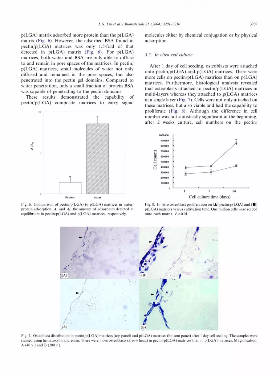
After 1 day of cell seeding, osteoblasts were attachedonto pectin/p(LGA) and p(LGA) matrices. There weremore cells on pectin/p(LGA) matrices than on p(LGA)matrices. Furthermore, histological analysis revealedthat osteoblasts attached to pectin/p(LGA) matrices inmulti-layers whereas they attached to p(LGA) matricesin a single layer (Fig. 7). Cells were not only attached onthese matrices, but also viable and had the capability toproliferate (Fig. 8). Although the difference in cellnumber was not statistically significant at the beginning,after 2 weeks culture, cell numbers on the pectin/
GA) matrices (bottom panel) after 1 day cell seeding. The samples were
d) in pectin/p(LGA) matrices than in p(LGA) matrices. Magnification:
Fig. 8. In vitro osteoblast proliferation on (m) pectin/p(LGA) and (’)
p(LGA) matrices versus cultivation time. One million cells were seeded
onto each matrix. Po0:01:

ARTICLE IN PRESSL.S. Liu et al. / Biomaterials 25 (2004) 3201–32103210
p(LGA) matrices were two-fold of that on the p(LGA)matrices (Fig. 8).
4. Conclusions
We present a method to effectively combine syntheticpolymers and natural polymers in one matrix. Byincluding dry particles of pectins and calcium chloridein p(LGA)/chloroform solution, composite matriceswere created with an interconnected porous morphol-ogy. The composite matrices consist of a pectin networkreinforced by a p(LGA) network. The compositematrices combine the best features of both polymers.Typically, the mechanical properties of the compositeare comparable to p(LGA) whereas their capacity tohold water and accessibility to proteins are comparableto pectin. In addition, the composite matrices provideside chain functional groups for further chemicalmodifications, which could be used in various biomedi-cal applications. As demonstrated by in vitro cellculture, the composite matrices show promise for tissueengineering applications.Thus, by selecting a group of synthetic polymers with
appropriate pairs of inorganic salts and polysaccharides,many polymeric composite matrices can be created bythis simple and environmentally friendly method.
Acknowledgements
The authors gratefully acknowledge Ms. PamelaRockwell-Warner, Mr. Andre White, Mr. TungNguyen, and Ms. Laura Smith for their excellenttechnical assistance. A special thanks to Ms. WendyH. Kramer, M.L.S., for technical editing. PX Ma andYJ Won acknowledge the financial support from Centerfor Biomedical Engineering Research (U. Michigan) andDuPont Young Prof. Award (PXM) and from MyongjiHospital of Kwandong University, College of Medicine(YJW).
References
[1] Schols HA, Voragen AGJ. Complex pectins: structure elucidation
using enzymes. In: Visser J, Vorangen AGJ, editors. Pectin and
pectinases. Amsterdam: Elsevier Science; 1996. p. 3–19.
[2] Hwang J, Kokini JL. Structure and rheological function of side
branches of carbohydrate polymers. J Texture Stud 1991;22:
123–67.
[3] Liu LS, Fishman ML, Kost J, Hicks KB. Pectin based systems for
colon-specific drug delivery via oral route. Biomaterials
2003;24:3333–43.
[4] Semd"e R, Amighi A, Devleeschouwer MJ, Mo.es AJ. Studies of
pectin HM/Eudragits RL/Eudragits NE film coating formula-
tions for colonic drug delivery. Int J Pharm 2000;197:181–92.
[5] Kim JH, Fassihi R. Application of a binary polymer system in
drug release rate modulation. J Pharm Sci 1997;86(3):316–28.
[6] Bodmeier R, Paeratakul O. Mechanical properties of dry and wet
cellulosic and acrylic films prepared from aqueous colloidal
polymer dispersions used in the coating of solid dosage forms.
Pharm Res 1994;11:882–8.
[7] Ma PX, Langer R. Fabrication of biodegradable polymer foams
for cell transplantation and tissue engineering. In: Morgan J,
Yarmush M, editors. Tissue engineering methods and protocols.
Totowa, NJ: Humana Press Inc; 1999. p. 47–56.
[8] Lanza RP, Langer R, Vacanti J. Principles of tissue engineering.
San Diego, CA: Academic Press; 1997.
[9] Patrick Jr CW, Mikos AG, McIntire LV, editors. Frontiers in
tissue engineering. New York: Pergamon; 1998.
[10] Ma PX, Zhang R, Xiao G, Franceschi F. Engineering new bone
tissue in vitro on highly porous poly(a-hydroxyl acids)/hydro-xyapatite composite scaffolds. J Biomed Mater Res 2001;
54(2):284–93.
[11] Langer R, Vacanti JP. Tissue engineering. Science 1993;260:
920–6.
[12] Ouchi T, Toyohara M, Arimura H, Ohya Y. Preparation of
poly(l-lactide)-based microsphere having a cationic or anionic
surface using biodegradable surfactants. Macromolecules
2002;3(5):885–8.
[13] Rubinstein A, Radai R, Ezra M, Pathak S, Rokem S. In vitro
evaluation of calcium pectinate: a potential colon-specific drug
delivery carrier. Pharm Res 1993;10(2):258–63.
[14] Voragen AGJ, Schols HA, Pilnik W. Determination of the degree
of methylation and acetylation of pectin by HPLC. Food
Hydrocolloids 1986;1:65–70.
[15] Fishman ML, Chau HK, Hoagland P, Ayyad K. Characterization
of pectin, flash-extracted from orange albedo by microwave
heating, under pressure. Carbohydrate Res 2000;5:359–79.
[16] Dubois M, Gilles KA, Hamilton JK, Rebers PA, Smith F.
Colorimetric method for determination of sugars and related
substances. Anal Chem 1956;28:350–6.
[17] Liu LS, Berg RA. Adhesion barriers of carboxymethylcellulose
and polyethylene oxide composite gels. J Biomed Mater Res
(Appl Biomater) 2002;63:326–32.
[18] Nilsson K, Mosbach K. Immobilization of enzymes and affinity
ligands to various hydroxyl group carrying supports using highly
reactive sulfonayl chlorides. Biochem Biophys Res Comm
1981;102(1):449–57.
[19] Dickerson KT, Glass JR, Liu LS, Craig WS, Mullen DG, Cheng
S. Immobilization of peptides to hyaluronate. US Patent No.
5,677,276. 1997.
[20] Smith PK, Krohn RI, Hermanson GT, Mallia AK, Gartner HF,
Provenzano MD, Fujimoto EK, Goeke NM, Olson BJ, Klenk
DC. Measurement of protein using bicinchoninic acid. Anal
Biochem 1985;150:76–85.
[21] Carson FL. Histotechnology: a self instructional text. Chicago,
IL: ASCP Press; 1990.
[22] BeMiller JM. An introduction to pectins: structure and proper-
ties. In: Fishman ML, Jen JJ, editors. Chemistry and function of
pectins, ACS series 310. Washington, DC: American Chemical
Society; 1986. p. 2–13.
[23] Mikos AG, Thorsen AJ, Czerwonka LA, Bao Y, Langer R,
Winslow DN, Vacanti JP. Preparation and characterization of
poly(l-lactic acid) foams. Polymer 1994;35(5):1068–77.
[24] Coffin DR, Fishman ML, Ly TV. Thermomechanical properties
of blends of pectin and poly(vinyl alcohol). J Appl Polym Sci
1996;61:71–9.
[25] Massia SP, Hubbell JA. Covalent surface immobilization of Arg–
Gly–Asp- and Tyr–Ile–Gly–Arg-containing peptides to obtain
well-defined cell-adhesive substrates. Anal Biochem
1990;187:292–301.