in vivo biocompatibility and in vitro characterization of poly-lactide-co-glycolide structures...
TRANSCRIPT

In vivo biocompatibility and in vitro characterization ofpoly-lactide-co-glycolide structures containing Levetiracetam,for the treatment of epilepsy
Amy J. Halliday,1,2 Toni E. Campbell,3 Joselito M. Razal,3 Karen J. McLean,2 Timothy S. Nelson,4
Mark J. Cook,1,2 Gordon G. Wallace3
1Department of Medicine, University of Melbourne, St. Vincent’s Hospital, 35 Victoria Parade, Fitzroy, Victoria 3065, Australia2Clinical Neurosciences, 5th Floor, Daly Wing, St. Vincent’s Hospital, 35 Victoria Parade, Fitzroy, Victoria 3065, Australia3Intelligent Polymer Research Institute and ARC Centre of Excellence for Electromaterials Science, University of Wollongong,
AIIM Facility, Innovation Campus, Wollongong, New South Wales 2522, Australia4The Bionic Ear Institute, 384-388 Albert Street, East Melbourne, Victoria 3002, Australia
Received 17 January 2011; revised 3 June 2011; accepted 10 June 2011
Published online 21 November 2011 in Wiley Online Library (wileyonlinelibrary.com). DOI: 10.1002/jbm.a.33208
Abstract: Epilepsy is a chronic neurological disorder charac-
terized by recurrent seizures, and is highly resistant to medi-
cation with up to 40% of patients continuing to experience
seizures whilst taking oral antiepileptic drugs. Recent
research suggests that this may be due to abnormalities in
the blood–brain barrier, which prevent the passage of thera-
peutic substances into the brain. We sought to develop a
drug delivery material that could be implanted within the
brain at the origin of the seizures to release antiepileptic
drugs locally and avoid the blood brain barrier. We produced
poly-lactide-co-glycolide drop-cast films and wet-spun fibers
loaded with the novel antiepileptic drug Levetiracetam, and
investigated their morphology, in vitro drug release charac-
teristics, and brain biocompatibility in adult rats. The best
performing structures released Levetiracetam constantly for
at least 5 months in vitro, and were found to be highly brain
biocompatible following month-long implantations in the
motor cortex of adult rats. These results demonstrate the
potential of polymer-based drug delivery devices in the treat-
ment of epilepsy and warrant their investigation in animal
models of focal epilepsy. VC 2011 Wiley Periodicals, Inc. J Biomed
Mater Res Part A: 100A: 424–431, 2012.
How to cite this article: Halliday AJ, Campbell TE, Razal JM, McLean KJ, Nelson TS, Cook MJ, Wallace GG. 2012. In vivobiocompatibility and in vitro characterization of poly-lactide-co-glycolide structures containing Levetiracetam, for the treatment ofepilepsy. J Biomed Mater Res Part A 2012:100A:424–431.
INTRODUCTION
Epilepsy is a neurological disorder characterized by recur-rent seizures that affects approximately 1% of the popula-tion, making it the world’s most common serious neurologiccondition.1 It is a particularly challenging condition to man-age medically, since over one-third of patients continue toexperience seizures despite taking multiple antiepilepticdrugs (AEDs).1 The mechanisms underlying this drug resist-ance are not well understood; however, one theory with agrowing body of supporting evidence is that the blood–brain barrier plays a significant role. There are many fea-tures of the normal blood–brain barrier that make it partic-ularly impervious to substances in the plasma, and there isalso evidence that repeated seizures further decrease itspermeability in patients with epilepsy.2 Administeringhigher oral doses of AEDs can overcome this decreased per-meability and usually has a beneficial effect on seizures;
however, this also produces side effects that prohibit themaintenance of this treatment regimen and is therefore notan appropriate solution to refractory seizures.
Administration of therapeutic substances directly to thepathological region of the brain is a concept that has gar-nered increasing research attention in recent years. Thisroute of administration has been shown in animal modelsto permit high drug concentrations in the brain whilstreducing the incidence of side effects.3–6 These findings ini-tiated interest in implantable drug delivery devices fordrug-resistant neurological disorders with focal pathologies,including epilepsy.7 Such devices permit long-term deliveryof AEDs directly to the region of the brain responsible forproducing the seizures, whilst drastically reducing the con-centration of AED in unrelated areas of the body.
Previous efforts to design drug-eluting implants for neu-rological disorders have achieved some success using
Correspondence to: M. J. Cook; e-mail: [email protected]
Contract grant sponsors: Victorian Government through its Science Technology and Innovation Initiative from the Department of Industry,
Innovation, and Regional Development; ARC Federation Fellowship (to G.G.W.); Australian Research Council for the APD Fellowship (to J.M.R.)
424 VC 2011 WILEY PERIODICALS, INC.
biodegradable polymers. These polymers slowly degradewithin the body into biocompatible metabolites, and gradu-ally release therapeutic substances into the local microenvir-onment as the polymer scaffold degrades. Biodegradablepolymers also alleviate the need for a second surgery toremove the implant. The most commonly used biodegrad-able polymer has been poly(D,L-lactide-co-glycolide) (PLGA),due to the fact that it is highly biocompatible, its chemistryis easily engineered to alter its biodegradation characteris-tics, and it has been approved for drug delivery purposesby the United States Food and Drug Administration.
A range of the more conventional AEDs have been incor-porated into biodegradable drug delivery devices. One novelAED that is used orally for the treatment of focal-onset seiz-ures in patients with refractory epilepsy is Levetiracetam(LEV). Major side effects of oral LEV treatment include som-nolence, headache, and mood changes related to the distri-bution of the drug throughout the brain following oraladministration, and often lead to withdrawal of treatment. Itwould be of significant benefit to patients to formulate aLEV delivery vehicle that can supply the LEV dosage directlyto the seizure focus to eliminate these unwanted sideeffects. Here, we seek to develop an intracranially implant-able drug delivery device capable of releasing LEV directlyinto the brain parenchyma over an extended time period.
In this investigation, we assessed the response of thebrain to a 5-week subdural implantation of thin dropcastPLGA-based films with a maximum loading of LEV to con-firm the biocompatibility of the implant materials anddesign. We then investigated a range of LEV loaded-PLGAdropcast films and wet-spun fibers, to determine the formu-lation that provided optimal long-term LEV release.
MATERIALS AND METHODS
MaterialsLevetiracetam [(S)-2-(2-oxopyrrolidin-1-yl)butanamide] wasa gift from UCB Pharma (Brussels, Belgium). PLGA with twodifferent mole ratios of lactide:glycolide were investigated:PLGA 85:15 (films in vivo: MW 50,000; Sigma–Aldrich, Aus-tralia; and in vitro MW 135,000; Purac Asia-Pacific, Singa-pore) and PLGA 75:25 [(75:25, MW 110,000; Purac Asia-Pa-cific, Singapore]. Solvents were acetone (Aldrich Sigma) anddichloromethane (DCM) (Chem-Supply Pty). Artificial cere-brospinal fluid (aCSF) was prepared containing sodiumchloride (NaCl; 0.866% w/v), potassium chloride (KCl;0.224% w/v), calcium dichloride (CaCl2.2H2O; 0.0206% w/v), and magnesium dichloride (MgCl2.6H2O; 0.0164% w/v)in 1 mM phosphate buffer (pH 7.4).
Preparation of LEV PLGA films/fibersFilms for in vivo biocompatibility testing were produced bydrop casting 1 mL of a solution of PLGA 85:15 (2% w/v)and LEV (4% w/v) in DCM into a Teflon mold (2 � 2 cm)and dried overnight. The theoretical LEV loading was 67%w/w. Blank films were produced by drop casting the samesolution without the inclusion of LEV.
Films and fibers were then produced for morphologicaland in vitro LEV release analysis. PLGA films were dropcast
in the same manner as described above, by combining PLGA75:25 or PLGA 85:15 with LEV in DCM at the concentra-tions given in Table I. LEV-loaded PLGA fibers were madeby wet spinning the PLGA-LEV solution through a 1:4 iso-propanol:hexane (v/v) coagulation bath using previouslydescribed methods.8,9 Blank films and fibers were producedas described except that LEV was omitted from the polymersolution. Images of these films and fibers were taken usinga scanning electron microscope (Jeoul SEM). Samples wereprepared for SEM by sputter coating with a thin layer ofgold (current 30 mA for 10 s), using a Dynavac MagnetronSC100MS.
Evaluation of the in vivo biocompatibility of LEV-loadedPLGA filmsTo investigate biocompatibility, rats were implanted subdur-ally with blank or LEV-releasing PLGA sheets for 5 weeks, andcompared with rats that underwent craniotomies alone. After5 weeks, the brains were sectioned and examined for evi-dence of toxic or immune-mediated injury. This involved look-ing at the morphology of neurons in thionin-stained sections,and examining the activation of macrophages/monocytes andforeign body giant cells (anti-ED1) and the activation andmorphology of astrocytes and glial scarring (anti-glial fibril-lary acidic protein (GFAP)) using immunohistochemistry.
Animals. Male Sprague–Dawley rats weighing 250–400 gwere obtained from Flinders University (Adelaide, Aus-tralia). All experiments were carried out under license ofthe St. Vincent’s Hospital (Melbourne) Animal Ethics Com-mittee in accordance with the Australian Prevention of Cru-elty to Animals Act (1986), which includes the AustralianCode of Practice for the Care and Use of Animals for Scien-tific Purposes (2004).
Surgical implantation. Rats were anaesthetized with keta-mine (70 mg/kg, i.p.) and xylazine (10 mg/kg, i.p.) andadministered carprofen (Rimadyl; 5 mg/kg, s.c.) for painrelief, and received maintenance isoflurane anesthesiathroughout the surgery (2% in oxygen). The head of the ratwas placed in a stereotaxic frame (Kopf) and a craniotomycreated above the right motor cortex (5 � 5 mm, centeredover AP: 0 mm, ML: þ4.0 mm from Bregma). The centralsection of bone was removed and a flap cut in the exposeddura. A sheet of polymer (3 � 3 mm) either with (n ¼ 8)or without (n ¼ 6) LEV was placed on the brain, and the
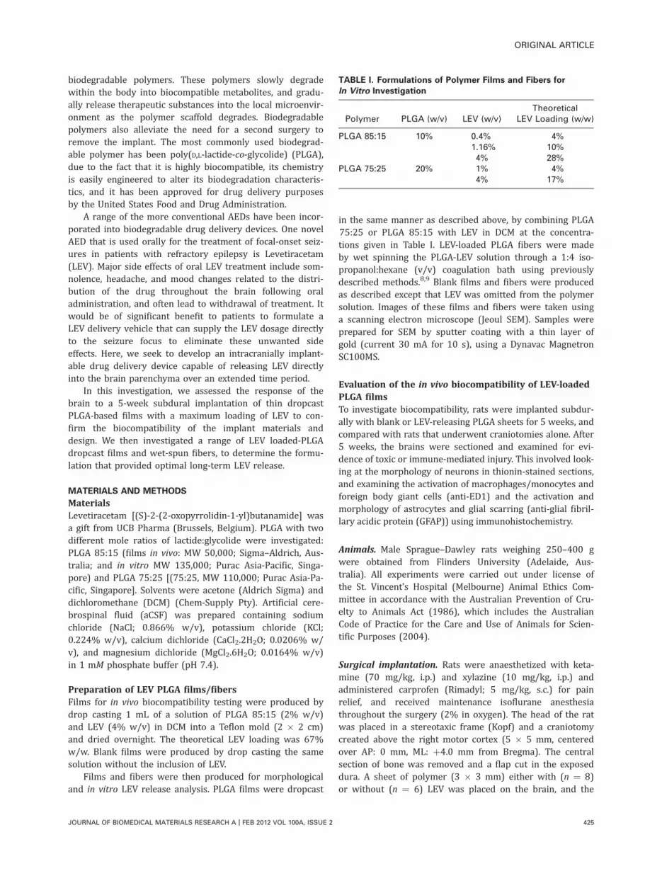
TABLE I. Formulations of Polymer Films and Fibers for
In Vitro Investigation
Polymer PLGA (w/v) LEV (w/v)Theoretical
LEV Loading (w/w)
PLGA 85:15 10% 0.4% 4%1.16% 10%4% 28%
PLGA 75:25 20% 1% 4%4% 17%
ORIGINAL ARTICLE
JOURNAL OF BIOMEDICAL MATERIALS RESEARCH A | FEB 2012 VOL 100A, ISSUE 2 425

dural flap and bone piece replaced. The craniotomy site wassealed with dental cement and the scalp sutured closed.Sham surgeries were performed as described, without theplacement of a polymer sheet on the brain (n ¼ 3).
Tissue preparation. Five weeks after surgery the rats weredeeply anaesthetized with sodium pentobarbitone (Virbac,Australia; 200 mg/kg, i.p.) and perfused transcardially with100 mL of 0.1M phosphate-buffered saline (PBS, pH 7.4) fol-lowed by 500 mL of 10% neutral-buffered formalin (NBF,Sigma–Aldrich, Australia). The brain was immediately dis-sected and post-fixed in 10% NBF at 4�C then cryoprotected in30% sucrose in 0.1M PBS (pH 7.4). The brain was then embed-ded in Tissue-Tek OCT Compound, frozen in liquid nitrogen-cooled isopentane, and stored at �80�C. Brains were sectionedusing a Leica CM1850 cryostat. Parallel series of coronal sec-tions were cut at 20 lm throughout the implant site, mountedonto chrome-gel-alum subbed microscope slides, air-driedthen stored at �80�C until further processing.
Histology. One series of sections from each brain wasstained with thionin. Slides were stained with 0.25% thionin(Sigma–Aldrich, Australia) for 3 min, washed in distilledwater (1 min), dehydrated in ascending concentrations ofethanol (50%, 70%, and 95%; 1 min each), 95% ethanol/1% acetic acid (1 min), and 100% ethanol (2 � 1 min),then cleared in xylene (2 � 3 min; Ajax Finechem, Aus-tralia), mounted in DPX (Sigma–Aldrich, Australia), andcoverslipped.
Immunohistochemistry. To ensure that immunohistochem-istry conditions remained identical, sections from all brainswere processed simultaneously for each of the antibodiesused. Slides were placed into trisodium citrate buffer (10mM, pH 6.0). Antigen retrieval was performed in a micro-wave (750W) on full power until the solution started toboil, then 2 � 5 min at 10% power. Slides were left to coolin solution (20 min). Sections were washed in PBS (3 � 5min), incubated in 0.5% H2O2 in methanol (15 min), andwashed again in PBS (3 � 5 min). Slides were then incu-bated in 10% normal goat serum [1 h, room temperature(RT) in a humidified chamber]; washed again in PBS (3 � 5min), before overnight incubations in either anti-GFAP(1:1000; rabbit polyclonal from Chemicon) or anti-ED1(1:800; mouse monoclonal from Serotec) primary antibodiesat 4�C. The following day slides were washed in PBS (3 � 5min), incubated (30 min, humid chamber, RT) in secondaryantibody (Envisionþ Dual Link System which contains horseradish peroxidase-labeled polymer conjugated to both goat-anti mouse and goat-anti rabbit immunoglobulins; DakoCy-tomation, Australia), and washed again in PBS (3 � 5 min).Immunoreactivity was visualized using 0.05% DAB intensi-fied with ammonium nickel sulfate (0.05%) and cobalt chlo-ride (0.05%) in the presence of 0.015% H2O2 in PBS (pH7.2) for 8 min. Subsequently, sections were washed in PBS(3 � 5 min), lightly counterstained in methyl green solution(0.5%) in 0.1M sodium acetate buffer (pH 4.2) for 5 min,rinsed in dH2O, dehydrated quickly through ethanol (95%,
100%, and 100%, 10 dips each), cleared in xylene (2 � 3min), and coverslipped using DPX. Primary antibody wasomitted on the negative control sections. Rat spleen, liver,and infected brain were used as positive control tissue forthe anti-ED1 antibody.
Histological and immunohistochemical analysis. Bright-field images were obtained using a SPOT RT-Slider digitalcamera attached to an Olympus IX70 microscope. Imageswere captured using SPOT software version 4.0.9 (Diagnos-tic Instruments).
Thionin-stained sections were examined to assess themacroscopic consequences of polymer implantation. Thedegree of damage was rated as follows: minor—incompletedisruption of the meninges and disruption of molecularcortical layer; moderate—complete disruption of themeninges and molecular cortical layer; severe—damageextending beyond the meninges and molecular cortical layer.Anti-GFAP and anti-ED1 immunoreactivity was examined inthe brain tissue beneath the centre of the implant and com-pared with immunoreactivity in the contralateral hemi-sphere, and rated as a mild, moderate, or severe increase inimmunoreactivity. A severe increase was defined as a levelof immunoreactivity similar to that in the regions of brainthat suffered mechanical damage during implantation. Todisplay the results visually, each of the rankings was given anumerical score (minor ¼ 1; moderate ¼ 2; severe ¼ 3)and the mean score and standard deviation was determinedfor each group (sham, blank polymer, and LEV polymer).
In vitro investigationsTo prepare the films for in vitro drug release investigations,discs of 2.5-mm diameter were cut using a hole punch,weighed, and placed in separate vials of 300 lL aCSF. Toprepare the fibers, bunches of fibers (2-cm length) werecut, weighed then placed in vials of 750 lL aCSF. Vials offilms and fibers were incubated in a water bath (37�C). Atset time points, three samples were removed and the con-centration of LEV in the aCSF was then determined usinghigh-performance liquid chromatography (HPLC). The con-centration of LEV was then related to the mass of the filmor fibers incubated and the drug release was reported asmass LEV (lg) released per mg of film or fiber.
The HPLC system comprised of a Shimadzu LC-10ATpump, a Shimadzu SIL-10AXL autosampler, a UV ActivonLinear 200 detector (set to 220 nm), with Shimadzu LCReal Time Analysis Software. The AtlantisTM T3 (C18) 5-lmcolumn (250 mm � 4.6 mm, Waters) was at RT. The injec-tion volume was 50 lL; the mobile phase was 15% (v/v)methanol and 85% (v/v) Milli-q water (pH 2.8) and theflow rate set at 0.8 mL/min.
RESULTS
In vivo biocompatibility of LEV-loaded filmsExamination of thionin-stained sections of sham brainsrevealed a mild disruption of the meninges, with only 33%showing disruption of deep cortical layers (see Fig. 1). Thisdemonstrated that some damage was caused during opening
426 HALLIDAY ET AL. INTRACRANIAL POLYMER IMPLANTS FOR THE TREATMENT OF EPILEPSY
of the dura during surgery. The mean thionin score for thesham group was 2 (Fig. 2). Rats implanted with a polymersheet showed varying degrees of disruption of the meninges.Fifty percent of rats with blank polymers and 13% of ratswith LEV polymers showed moderate damage, and one ratimplanted with a LEV polymer showed severe damage (TableII). The mean thionin score for the blank group was 1.5, whilethat for the LEV-loaded group was 1.4 (Fig. 2).
Examination of anti-GFAP labeled sections from sham ratsshowed that the surgical procedure alone produced a moder-ate increase in GFAP-like immunoreactivity at the surgical site(Fig. 1). Reactive astrocytes were observed in the region ofthe craniotomy; however, no glial scarring was observed atthe polymer–brain interface in any of the brains examined.
The mean GFAP score for the sham group was 2 (Fig. 2). Theincrease in GFAP-like immunoreactivity in polymer-implantedrats was not greater than that seen in these sham rats (TableII). The mean GFAP score for the blank group was 1.3, andthat for the LEV-loaded group 1.25 (Fig. 2).
Examination of anti-ED1 labeled sections from sham ratsshowed that the craniotomy and durotomy alone produceda mild expression of ED1-like immunoreactivity, with la-beled cells seen in the meninges at the centre of the crani-otomy site of all brains (Fig. 1). No multinucleate giant cellswere observed in any sections; however, it is worth notingthat the polymer material is dissolved from the sectionsduring histological processing and multinucleate giant cellsat the tissue–polymer interface may have been removed
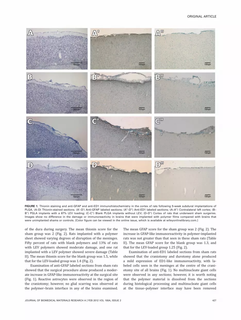
FIGURE 1. Thionin staining and anti-GFAP and anti-ED1 immunohistochemistry in the cortex of rats following 5-week subdural implantations of
PLGA. (A–D) Thionin-stained sections. (A0–D0) Anti-GFAP labeled sections; (A00–D00) Anti-ED1 labeled sections; (A–A00) Contralateral left cortex; (B–B00) PGLA implants with a 67% LEV loading; (C–C00) Blank PLGA implants without LEV; (D–D00) Cortex of rats that underwent sham surgeries.
Images show no difference in the damage or immunoreactivity in brains that were implanted with polymer films compared with brains that
were unimplanted shams or controls. [Color figure can be viewed in the online issue, which is available at wileyonlinelibrary.com.]
ORIGINAL ARTICLE
JOURNAL OF BIOMEDICAL MATERIALS RESEARCH A | FEB 2012 VOL 100A, ISSUE 2 427
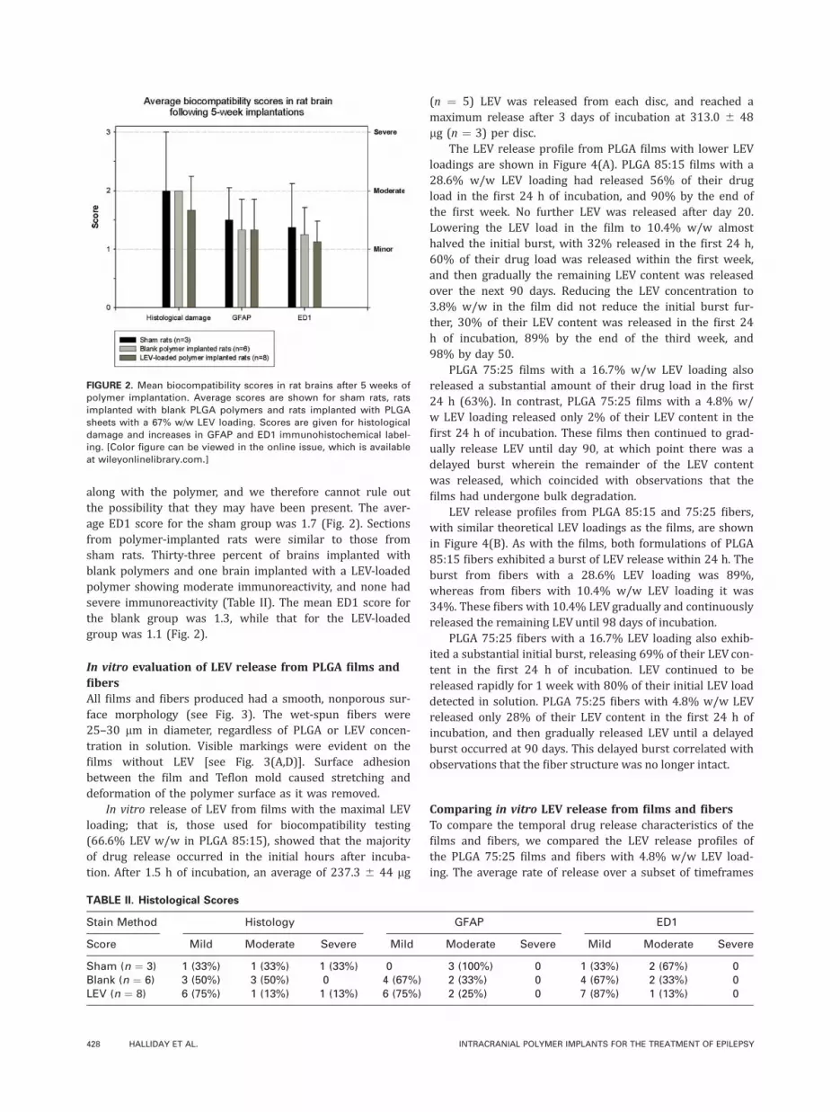
along with the polymer, and we therefore cannot rule outthe possibility that they may have been present. The aver-age ED1 score for the sham group was 1.7 (Fig. 2). Sectionsfrom polymer-implanted rats were similar to those fromsham rats. Thirty-three percent of brains implanted withblank polymers and one brain implanted with a LEV-loadedpolymer showing moderate immunoreactivity, and none hadsevere immunoreactivity (Table II). The mean ED1 score forthe blank group was 1.3, while that for the LEV-loadedgroup was 1.1 (Fig. 2).
In vitro evaluation of LEV release from PLGA films andfibersAll films and fibers produced had a smooth, nonporous sur-face morphology (see Fig. 3). The wet-spun fibers were25–30 lm in diameter, regardless of PLGA or LEV concen-tration in solution. Visible markings were evident on thefilms without LEV [see Fig. 3(A,D)]. Surface adhesionbetween the film and Teflon mold caused stretching anddeformation of the polymer surface as it was removed.
In vitro release of LEV from films with the maximal LEVloading; that is, those used for biocompatibility testing(66.6% LEV w/w in PLGA 85:15), showed that the majorityof drug release occurred in the initial hours after incuba-tion. After 1.5 h of incubation, an average of 237.3 6 44 lg
(n ¼ 5) LEV was released from each disc, and reached amaximum release after 3 days of incubation at 313.0 6 48lg (n ¼ 3) per disc.
The LEV release profile from PLGA films with lower LEVloadings are shown in Figure 4(A). PLGA 85:15 films with a28.6% w/w LEV loading had released 56% of their drugload in the first 24 h of incubation, and 90% by the end ofthe first week. No further LEV was released after day 20.Lowering the LEV load in the film to 10.4% w/w almosthalved the initial burst, with 32% released in the first 24 h,60% of their drug load was released within the first week,and then gradually the remaining LEV content was releasedover the next 90 days. Reducing the LEV concentration to3.8% w/w in the film did not reduce the initial burst fur-ther, 30% of their LEV content was released in the first 24h of incubation, 89% by the end of the third week, and98% by day 50.
PLGA 75:25 films with a 16.7% w/w LEV loading alsoreleased a substantial amount of their drug load in the first24 h (63%). In contrast, PLGA 75:25 films with a 4.8% w/w LEV loading released only 2% of their LEV content in thefirst 24 h of incubation. These films then continued to grad-ually release LEV until day 90, at which point there was adelayed burst wherein the remainder of the LEV contentwas released, which coincided with observations that thefilms had undergone bulk degradation.
LEV release profiles from PLGA 85:15 and 75:25 fibers,with similar theoretical LEV loadings as the films, are shownin Figure 4(B). As with the films, both formulations of PLGA85:15 fibers exhibited a burst of LEV release within 24 h. Theburst from fibers with a 28.6% LEV loading was 89%,whereas from fibers with 10.4% w/w LEV loading it was34%. These fibers with 10.4% LEV gradually and continuouslyreleased the remaining LEV until 98 days of incubation.
PLGA 75:25 fibers with a 16.7% LEV loading also exhib-ited a substantial initial burst, releasing 69% of their LEV con-tent in the first 24 h of incubation. LEV continued to bereleased rapidly for 1 week with 80% of their initial LEV loaddetected in solution. PLGA 75:25 fibers with 4.8% w/w LEVreleased only 28% of their LEV content in the first 24 h ofincubation, and then gradually released LEV until a delayedburst occurred at 90 days. This delayed burst correlated withobservations that the fiber structure was no longer intact.
Comparing in vitro LEV release from films and fibersTo compare the temporal drug release characteristics of thefilms and fibers, we compared the LEV release profiles ofthe PLGA 75:25 films and fibers with 4.8% w/w LEV load-ing. The average rate of release over a subset of timeframes
FIGURE 2. Mean biocompatibility scores in rat brains after 5 weeks of
polymer implantation. Average scores are shown for sham rats, rats
implanted with blank PLGA polymers and rats implanted with PLGA
sheets with a 67% w/w LEV loading. Scores are given for histological
damage and increases in GFAP and ED1 immunohistochemical label-
ing. [Color figure can be viewed in the online issue, which is available
at wileyonlinelibrary.com.]
TABLE II. Histological Scores
Stain Method Histology GFAP ED1
Score Mild Moderate Severe Mild Moderate Severe Mild Moderate Severe
Sham (n ¼ 3) 1 (33%) 1 (33%) 1 (33%) 0 3 (100%) 0 1 (33%) 2 (67%) 0Blank (n ¼ 6) 3 (50%) 3 (50%) 0 4 (67%) 2 (33%) 0 4 (67%) 2 (33%) 0LEV (n ¼ 8) 6 (75%) 1 (13%) 1 (13%) 6 (75%) 2 (25%) 0 7 (87%) 1 (13%) 0
428 HALLIDAY ET AL. INTRACRANIAL POLYMER IMPLANTS FOR THE TREATMENT OF EPILEPSY

within 90 days was calculated (Table III) and the resultsdemonstrated that the fibers released many times more LEVthan the films during the first week of incubation (32.2 lg/mg polymer/day in fibers compared with films 5.1). After 7days, both films and fibers released similar quantities ofLEV (0.2 lg/mg polymer/day) until the last 10 days of incu-bation when the final burst release from fibers was 1.5times less than films.
DISCUSSION
We investigated the drug-release characteristics of LEV-loaded PLGA films and fibers in vivo, and examined the bio-
compatibility of LEV-loaded PLGA films following 5-weeksubdural implantations in adult rats.
The results of this study demonstrate that lower initialdrug loadings lead to more controlled patterns of drugrelease, irrespective of the polymer’s glycolide content orphysical structure. Formulations with theoretical drug load-ings of =10% had the smallest initial bursts and thereafterdemonstrated gradual, continuous release of LEV, consistentwith the published literature.10–12 We hypothesize that athigh drug loadings, there is unlikely to be sufficient polymerpresent to completely entrap the entire drug load withinthe polymer matrix, leading to unregulated drug release assoon as the material is immersed in fluid. In contrast, atlow drug loadings, the polymer entirely and homogeneouslyencapsulates the drug such that drug release is primarilydictated by polymer degradation.
FIGURE 3. SEM images of the surface of PLGA films and fibers, with
and without LEV. The column to the left shows films, while the col-
umn to the right shows fibers. (A, A0) PLGA 85:15 (10%w/v) without
LEV; (B, B0) PLGA 85:15 (10% w/v) with 3.8% (w/w) LEV loading; (C, C0)PLGA 85:15 (10% w/v) with 10.4% (w/w) LEV loading; (D, D0) PLGA
75:25 (20% w/v) without LEV; (E, E0) PLGA 75:25 (20% w/v) with 4.8%
w/w LEV loading.
FIGURE 4. Release of Levetiracetam from PLGA films and fibers in
vitro. (A) Release from PLGA films; (B) Release from PLGA fibers.
TABLE III. Comparison of Mean LEV Release Over Set Time
Periods From the Films and Fibers Made From a Solution of
PLGA 75:25 (10% w/v) and Levetiracetam (4% w/v)
Structure
Mean Release LEV (lg/mg Polymer/day 6 SD)
0–4 h 1–7 days 7–80 days 80–90 days
Films 4.46 (61.4) 0.58 (60.2) 0.24 (60.06) 0.37 (60.07)Fibers 29.73 (618.1) 2.52 (61.6) 0.25 (60.1) 0.22 (60.09)
ORIGINAL ARTICLE
JOURNAL OF BIOMEDICAL MATERIALS RESEARCH A | FEB 2012 VOL 100A, ISSUE 2 429

The films and fibers described in this report were capa-ble of delivering LEV in a controlled manner for at least 3months in vitro. A previous investigation described a poly-mer-based device that released a conventional AED in acontrolled manner for over 250 days in vitro.13 The produc-tion of implants that release AEDs over a clinically applica-ble time frame was an important milestone in the field ofpolymer-based drug delivery; however, the bulky physicalstructure of the implant necessitated the removal of largeportions of the cortex to implant the device. The thin filmimplants developed in this investigation were implantedonto the surface of the brain without displacing brain tissueand therefore significantly reduced the risk of unwantedneurological deficits. We anticipate that as more and morepotent drugs are discovered and made available to research-ers, slower rates of degradation can be utilized so that theperiod of drug release can be significantly extended. Inaddition, intermediate-term drug delivery systems such asthose described in this report may still have several clinicalapplications, in particular in the post-surgical setting.
This investigation also compared drug release from filmand fiber structures. The results demonstrated that the rateof drug release was greater in the fibers during the initialdays of incubation, but greater in the films during the finalweeks of the study. Fibers may therefore be the preferredstructure when drugs with long half lives are chosen, whichrequire a loading dose.
Our study also investigated the biocompatibility of PLGAsheets containing a maximal load of LEV following implanta-tion subdurally in rats. There are two major ways in which animplanted material may be non-biocompatible. The first isthat the material or its biodegradation products may be toxicto the cells of the central nervous system (CNS). Toxic injurycan be identified by a characteristic set of histological fea-tures.14 In the acute setting, neurons appear pyknotic withdisappearance of the nucleolus and loss of Nissl substance inthionin-stained sections. In the chronic setting, neuronsundergo necrosis and the necrotic cellular debris activatesmacrophages/monocytes and astrocytes. Fibroblasts are notpresent within the CNS and therefore the injury is not healedby a fibrotic scar; rather, a gliotic scar is formed by reactiveastrocytes.14 In this investigation, the polymers were stillpresent subdurally after 5 weeks of implantation and werestill undergoing biodegradation; therefore, any acute injurydue to toxicity of biodegradation products should have beenvisible in the brain sections. On examination of the brain slicesin our investigation, we found only minimal disruption of thesuperficial cortical layers of the brain surface, and observedthat the degree of astrocyte activation following implantationof LEV-loaded PLGA sheets was no greater than that seen afterimplantation of blank PLGA sheets or after the sham surgicalprocedure.
The second way that an implant may be non-biocompati-ble is if the material is recognized as foreign by the innateimmune defenses of the CNS and an inflammatory orimmune response is initiated.15 Microglia and monocytes/macrophages are activated, and initiate a localized inflam-matory response in an attempt to destroy the foreign mate-
rial. Frustrated attempts by the macrophages to engulf anddestroy the material result in the fusion of multiple macro-phages into more aggressive multinucleate giant cells, a his-tological hallmark of chronic inflammation.15 The immuneresponse activates astrocytes which subsequently undergogliosis and form a glial scar, as for toxic injury. There wasno evidence of multinucleate giant cells or glial scarring inany of the brains examined in this investigation. In addition,the degree of microglial activation observed after implanta-tion of LEV-loaded PLGA sheets was no greater than thatobserved after implantation of blank sheets or after shamsurgical procedures.
In summary, there was no evidence of toxic injury orimmune-mediated inflammation observed using the meth-ods of investigation employed in this study, indicating thatthe LEV-releasing PLGA sheets demonstrated excellent brainbiocompatibility.
CONCLUSIONS
This study describes the production of biodegradable poly-mer-based materials with LEV that are (i) very well toler-ated by brain tissue after 5 weeks of subdural implantationand (ii) capable of releasing LEV for at least 90 days invitro. The results of this investigation indicate that thesematerials warrant further investigation into the longevity,long-term biocompatibility, and efficacy of such devices andsuggest that biodegradable polymer-based materials areappealing candidates for intracranial drug delivery systemsfor the treatment of epilepsy.
ACKNOWLEDGMENTS
The authors acknowledge the donation of Levetiracetam fromUCB Pharma, and the sponsorships of Victorian Governmentand ARC. The Bionic Ear Institute acknowledges the support itreceives from the Victorian Government through its Opera-tional Infrastructure Support Program and the HelenMacpher-son Smith Trust.
REFERENCES1. Shorvon SD. The epidemiology and treatment of chronic and re-
fractory epilepsy. Epilepsia 1996;37:S1–S3.
2. Loscher W, Potschka H. Role of multidrug transporters in pharma-
coresistance to antiepileptic drugs. J Pharmacol Exp Ther 2002;
301:7–14.
3. Klitgaard H, Matagne A, Grimee R, Vanneste-Goemaere J, Mar-
gineanu DG. Electrophysiological, neurochemical and regional
effects of levetiracetam in the rat pilocarpine model of temporal
lobe epilepsy. Seizure 2003;12:92–100.
4. Serralta A, Barcia JA, Ortiz P, Duran C, Hernandez ME, Alos M.
Effect of intracerebroventricular continuous infusion of valproic
acid versus single i.p. and i.c.v. injections in the amygdala kin-
dling epilepsy model. Epilepsy Res 2006;70:15–26.
5. Barcia JA, Rubio P, Alos M, Serralta A, Belda V. Anticonvulsant
and neurotoxic effects of intracerebroventricular injection of
phenytoin, phenobarbital and carbamazepine in an amygdala-
kindling model of epilepsy in the rat. Epilepsy Res 1999;33:
159–167.
6. Gonzalez-Darder JM, Garcia-Teno M. Anticonvulsant effect of in-
traventricular antiepileptic drugs. Experimental study. Neurol Res
1995;17:190–192.
7. Halliday AJ, Cook MJ. Polymer-based drug delivery devices for neu-
rological disorders. CNSNeurol Disord Drug Targets 2009;8:205–221.
430 HALLIDAY ET AL. INTRACRANIAL POLYMER IMPLANTS FOR THE TREATMENT OF EPILEPSY

8. Razal J, Kita M, Quigley A, Kennedy E, Moulton S, Kapsa R, Clark
G, Wallace G. Wet-spun biodegradable fibers on conducting plat-
forms: Novel architectures for muscle regeneration. Adv Funct
Mater 2009;19:3381–3388.
9. Quigley A, Razal J, Thompson B, Moulton S, Kita M, Kennedy E,
Clark G, Wallace G, Kapsa R. A conducting-polymer platform with
biodegradable fibers for stimulation and guidance of axonal
growth. Adv Mater 2009;21:4393–4397.
10. Wilz A, Pritchard EM, Li T, Lan JQ, Kaplan DL, Boison D. Silk poly-
mer-based adenosine release: Therapeutic potential for epilepsy.
Biomaterials 2008;29:3609–3616.
11. Benelli P, Conti B, Genta I, Costantini M, Montanari L. Clonaze-
pam microencapsulation in poly-D,L-lactide-co-glycolide micro-
spheres. J Microencapsul 1998;15:431–443.
12. Barakat NS, Radwan MA. In vitro performance of carbamazepine
loaded to various molecular weights of poly (D,L-lactide-co-glyco-
lide). Drug Deliv 2006;13:9–18.
13. Cho CS, Han SY, Ha JH, Kim SH, Lim DY. Clonazepam release
from bioerodible hydrogels based on semi-interpenetrating
polymer networks composed of poly(epsilon-caprolactone)
and poly(ethylene glycol) macromer. Int J Pharm 1999;181:
235–242.
14. Kumar V, Abbas AK, Fausto N, Robbins SL, Cotran RS. Robbins
and Cotran Pathologic Basis of Disease: Elsevier Saunders Phil-
adelphia, Pennsylvania; 2005.
15. Fournier E, Passirani C, Montero-Menei CN, Benoit JP. Biocom-
patibility of implantable synthetic polymeric drug carriers: Focus
on brain biocompatibility. Biomaterials 2003;24:3311–3331.
ORIGINAL ARTICLE
JOURNAL OF BIOMEDICAL MATERIALS RESEARCH A | FEB 2012 VOL 100A, ISSUE 2 431