expansion of human bone marrow stromal cells on poly-( dl-lactide- co-glycolide) (p dllga) hollow...
TRANSCRIPT
ARTICLE IN PRESS
0142-9612/$ - se
doi:10.1016/j.bi
�CorrespondE-mail addr
URL: http:/
Biomaterials 28 (2007) 5332–5343
www.elsevier.com/locate/biomaterials
Expansion of human bone marrow stromal cells onpoly-(DL-lactide-co-glycolide) (PDLLGA) hollow fibres designed
for use in skeletal tissue engineering
Suzanne M. Morgana, Simon Tilleya, Semali Pererab, Marianne J. Ellisc, Janos Kanczlera,Julian B. Chaudhuric, Richard O.C. Oreffoa,�
aBone and Joint Research Group, Centre for Human Development, Stem Cells and Regeneration, Institute of Developmental Sciences,
Developmental Origins of Health and Disease, University of Southampton, Southampton SO16 6YD, UKbDepartment of Chemical Engineering, University of Bath, Bath BA2 7AY, UK
cCentre for Regenerative Medicine, Department of Chemical Engineering, University of Bath, Bath BA2 7AY, UK
Received 18 April 2007; accepted 13 August 2007
Available online 5 September 2007
Abstract
Strategies to expand human bone marrow stromal cells (HBMSC) for bone tissue engineering are a key to revolutionising the processes
involved in three-dimensional skeletal tissue reconstruction. To facilitate this process we believe the use of biodegradable porous poly
(DL-lactide-co-glycolide) (PDLLGA) hollow fibres as a scaffold used in combination with HBMSC to initiate natural bone repair and
regeneration offers a potential solution. In this study, the biocompatibility of 75:25 PDLLGA fibres with HBMSC and the capacity of a
PDLLGA fibre-associated HBMSC-monolayer to establish an osteogenic phenotype in vivo was examined. A high proportion of
HBMSC survived when expanded on PDLLGA fibres for 6 days, with only 10% of the propidium iodide (pI)-labelled population
represented in the sub-G1 DNA peak on analysis by flow cytometry. Tracking carboxy-fluorescein diacetate, succinimidyl ester (CFSE)-
labelled HBMSC by flow cytometry indicated that HBMSC attachment to the PDLLGA fibres does not interfere with their rate of
proliferation. Furthermore, in response to osteogenic stimuli, HBMSC expanded on PDLLGA fibres can differentiate, as expected, along
the osteogenic lineage with associated alkaline phosphatase activity. Following implantation into SCID mice, osteogenic-conditioned
PDLLGA fibre–HBMSC graft resulted in type I collagen deposition and associated bone mineralisation and osteoid formation, as
evidenced by immunohistochemistry and histology. These studies provide evidence that porous PDLLGA hollow fibre–HBMSC graft is
an innovative biomaterial that offers new approaches to mesenchymal cell expansion, which could be utilised as a scaffold for skeletal
tissue generation.
r 2007 Elsevier Ltd. All rights reserved.
Keywords: Bone tissue engineering; Poly-(DL-lactide-co-glycolide); Hollow fibre; Human bone marrow stromal cells; Osteogenesis; Wet-spin phase-
inversion technique and biodegradable scaffolds
1. Introduction
The multi-potential of human bone marrow stromal cells(HBMSC), in particular their ability to differentiate intoosteoblasts, or bone producing cells, has generated intenseinterest as to their use in bone tissue engineering [1–4].Thus, lineage-specific differentiation of HBMSC into
e front matter r 2007 Elsevier Ltd. All rights reserved.
omaterials.2007.08.029
ing author. Fax: +44 23 80796141.
ess: [email protected] (R.O.C. Oreffo).
/www.mesenchymalstemcells.org (R.O.C. Oreffo).
osteoblasts can be achieved with relative ease by culturingin osteogenic inducing agents and the judicious selectionof appropriate osteo-inductive growth factors [2,5,6].Furthermore, HBMSC can be obtained from autologoussources and display a tremendous capacity for proliferation[5,7,8]. Reliability in harnessing these cell properties withan appropriate scaffold is under intense investigationto create HBMSC scaffold composites that not onlyact as bulking material but also support cell expansionand differentiation and, ultimately, new bone formation[1,9–13].
ARTICLE IN PRESS
Nomenclature
J gas permeability (mol/m2 Pa s)r pore radius (m)P pressure (Pa)n mean molar flow rate (mol/s)R universal gas constant (8.3145) (J/Kmol)T temperature (K)p Pi (3.14) (dimensionless)MN2
molecular mass of nitrogen (28) (g/mol)e porosity (dimensionless)m viscosity of nitrogen (1.8� 10�2) (g/m s) (Pa s)
Lp effective pore length (M)s stress (N/m2 (Pa))F force (N)A surface area (m2)M mass (kg)g acceleration due to gravity (9.81) (m/s2)FI inner diameter of hollow fibre (M)Fo outer diameter of hollow fibre (M)L extension (M)L original length (M)E Young’s Modulus (N/m2) (Pa)
S.M. Morgan et al. / Biomaterials 28 (2007) 5332–5343 5333
The clinical need for simple and abundant osteopro-genitor cell sources in bone repair is evidenced by the factthat tissue loss as a result of bone injury or disease providesreduced quality of life for many at significant socio-economic cost. Each year in the UK there are some 150,000fractures (wrist, vertebral and hip) due to osteoporosis [14].Healthcare costs in treating patients with these bonefractures, in the UK alone, have been estimated at £1.7billion [15]. In particular, hip fracture is associated withsignificant morbidity and approximately 20% of patientswill die as a consequence of hip surgery [16]. Convention-ally, mesenchymal cell numbers are increased through asimple expansion protocol involving the use of largemonolayer flasks [7,8,11]. This is an uneconomical methodof cell scale up and provides inefficient surface areaplatforms for cell culture. An attractive alternative is theuse of hollow-fibre membranes typically affording surfacearea to volume ratios of 100–200 cm2/cm3 (depending onfibre diameter) [17]. Thus, theoretically, the extendedsurface area of a hollow-fibre reactor has the potential toprovide greatly increased cell expansion in equivalent tissueculture (TC) volumes.
There has long been an interest in developing biodegrad-able scaffolds for use in orthopaedic applications, andbiodegradable materials under consideration include poly(lactide-co-glycolide (PLGA), tricalcium phosphate andglass ceramics [18–23]. Additional features promote PLGAas a candidate scaffold. PLGA shows excellent biocompat-ibility with a safe history of use in the clinic, ismechanically strong and can be processed into almostany shape or size [24,25]. There has been significant drive toformulate PLGA for cell delivery in skeletal tissueengineering [26–31], although to date, a Phase I clinicaltrial has not been conducted. PLGA is not inherentlyosteo-conductive unlike other synthetic materials consid-ered for bone tissue engineering [19,21,32–38]. CombiningHBMSC with PLGA, however, has no detrimental effecton their ability to differentiate appropriately in response toosteogenic stimuli using the osteogenic growth factor bonemorphogenetic protein-2 (BMP2) [31,32]. Previous studies,including work carried out in our own laboratory have alsofound a similar phenotype associated with in vivo
implantation of HBMSC or rodent-derived MSC whencombined with various forms of PLGA [31,32,39–41].Using a wet-spinning phase-inversion process, we have
fabricated PDLLGA hollow fibres, which can be fashionedwith different dimensions and membrane characteristics[17]. In this study, we assess their biocompatibility withprimary HBMSC and the potential of PDLLGAfibre–HBMSC scaffolds for use as a vehicle to aid cellexpansion and as a possible material to augment bonegraft. Unique to their design is the ability to fine-tune theirphysical properties, with the phase-inversion techniqueused allowing controllable fabrication of porous fibres witha uniform hollow core approximately 1mm in diameter andrough surface area (surface area to volume ratio of100–200 cm2/cm3) [17,42]. PLGA has a dual transportmechanism, namely the solution/sorption diffusion anddegradation diffusion and permeation, a property likely tobe critical for sustained viability of a PLGA-cell scaffold.The main mechanism of transport in PLGA prior todegradation is sorption diffusion where molecules aresoluble in polymer and diffuse across dense polymer phase[43]. Such properties allow the perfusion of media and gasexchange and potentially the extensive expansion ofadherent cells. Previously, we have shown that a bonemarrow-derived cell line, pZip, can attach to and expand onthese PDLLGA fibre scaffolds [17]. We have extended ourinitial observations, and in this report describe a detailedstudy on the biocompatibility of 75:25 PDLLGA fibres withprimary HBMSC. The survival and proliferation ofHBMSC is not altered by expansion on these PDLLGAfibres and, in addition, these studies demonstrate thatosteogenic-conditioned PDLLGA fibre–HBMSC graft isassociated with type I collagen deposition and bonemineralisation following in vivo implantation in SCID mice.
2. Materials and methods
2.1. Materials
Foetal calf serum (FCS) was obtained from Gibco BRL (Paisley,
Scotland). Reagents were of analytical grade and obtained from Sigma
(Poole, Dorset) unless otherwise stated. Poly(lactide-co-glycolide)
ARTICLE IN PRESSS.M. Morgan et al. / Biomaterials 28 (2007) 5332–53435334
(PDLLGA) with a lactide:glycolide ratio of 75:25 (GMP grade)
was purchased from Alkermes, Inc. (Cincinnati, OH). 1-Methyl-
2-pyrrolidinone (NMP) was obtained from Fisher Scientific UK Ltd.,
Loughborough.
2.2. Fabrication of PDLLGA hollow fibres
Hollow fibres were prepared using a method described previously [44].
Briefly, hollow fibres were prepared from a 20% (w/w) polymer solution of
75:25 PDLLGA in NMP at 20 1C. The polymer solution was passed
through a spinneret (needle 0.3mm outer diameter, bore 0.5mm inner
diameter) into the water coagulation bath (20 1C), with a take-up rate of
20 rpm (4.84m/min). Fibres were left in distilled water for 3 days with
regular changes of water to allow any remaining solvent to diffuse out of
the fibres. Flat sheet membranes, were prepared as described in (17).
Briefly, polymer solution was spread on a glass plate in a 200mm deep
layer, controlled by a rod and 200mm diameter wire, then submerged in a
deionised water bath. Water was changed twice daily for 3 days to remove
residual solvent.
2.3. HBMSC cultures
Bone marrow samples were obtained from haematologically normal
patients undergoing routine hip replacement surgery at Southampton
General Hospital with the approval of the Southampton and South West
Hampshire ethics committee (LREC194/99). Cultures of HBMSC were
established as previously described [45]. Briefly, cancellous bone marrow
samples were washed with alpha-modified minimal essential medium
(a-MEM), filtered through a 70-mm nylon mesh to remove debris, and
harvested cells cultured in a-MEM supplemented with 10% FCS,
100 units/ml of penicillin and 100mg/ml of streptomycin (culture medium).
Twenty-four hours later, cultures were washed with phosphate buffered
saline (PBS) to remove red blood cells and other non-adherent cell
populations. On reaching confluence (5–7 days), the monolayer of
adherent HBMSC was harvested by trypsinization and cultured further
by seeding on TC plastic or PLGA fibres at a typical concentration of
5� 103 cells/cm2 in culture medium or culture medium supplemented with
10 nM dexamethasone, 100mM ascorbate-2-phosphate and BMP2 (200 ng/
ml) (osteogenic conditions). PDLLGA fibres were incubated with cells as
described previously [17] after sterilising with 70% ethanol for 30min and
rinsing with PBS. Briefly, PLGA fibres were treated with FCS, and cells
added to bundles of fibres in a volume of medium sufficient to cover the
bundles. After seeding, fibres were transferred to a fresh dish. Media
changes were carried out every 3–4 days.
2.4. Labelling of cells with pI and determination of cell viability by
FACS
HBMSC were harvested by trypsinisation, washed in PBS and fixed in
cold ethanol (70%) for 30min at 4 1C. Cells were vortexed during the
addition of ethanol to maintain a single cell suspension. TC supernatants
was saved and pooled with the harvested adherent cells for processing.
Fixed cells were washed with PBS, followed by treatment with
ribonuclease A (50 mg/ml) for 1 h at 20 1C. Next, propidium iodide
(20mg/ml) was added and immediately the cells were analysed by flow
cytometry.
2.5. Labelling cells with 5-chloromethylfluorescein diacetate (Cell
Tracker green) and ethidium homodimer-1
PDLLGA fibre–HBMSC was washed with PBS and incubated with Cell
Tracker green (Molecular Probes, Leiden, The Netherlands) and ethidium
homodimer for 45min to label viable and dead cells, respectively.
PDLLGA fibre–HBMSC was washed to remove unincorporated dye.
Images of fluorescently labelled cells were taken using an inverted
microscope (Zeiss Axiovert 200) and captured using the Carl Zeiss
Axiovision-3.1 software package or taken with a confocal microscope
(Leica Leitz DM RBE ) and imaged using Leica Confocal 2.5 software.
2.6. DNA quantitation
HBMSC (2� 105 in 1ml) were seeded onto fibres (seeding efficiency of
20%) or plastic (4� 104), and cultures were maintained in culture medium.
Cells were harvested after 1, 120 and 168h, lysed in 0.05% Triton X-100
and sonicated. DNA was quantified using the ultra-sensitive fluorescent
picoGreen dsDNA Quantitation Assay (Invitrogen, Paisley, UK) and an
FLx cytofluor microplate reader for detection. An increase in DNA
quantity was taken to directly represent an increase in cell number.
2.7. CFSE dilution analysis
HBMSC (2� 107/ml) were incubated for 10min at 37 1C with 10mMCFSE (Molecular Probes, Leiden, The Netherlands) in PBS containing
0.1% BSA, washed once with DMEM and 10% FCS and then twice with
PBS. CFSE-labelled cells were maintained on plastic or PDLLGA fibres in
culture medium. To monitor cell proliferation rate, cells were harvested by
trypsinization and the levels of CFSE in cells analysed by flow cytometry.
During each round of cell division, the relative intensity of CFSE
decreases by half.
2.8. Alkaline phosphatase expression
PDLLGA fibre–HBMSC were rinsed with PBS, fixed with 95% Ethanol
for 10min at room temperature and stained using a napthol AS-MX
phosphate alkaline substrate solution (No. 85) according to the
manufacturer’s instructions. After staining, fibres were immersed in PBS
and photographed with a digital camera (Canon Powershot G2).
2.9. In vivo studies
HBMSC were expanded on PDLLGA fibres for 9 days for trial 1 or 42
days for trial 2, at which point PDLLGA fibre–HBMSC were impacted
into perforated acrylic graft chambers (1 cm3) using an impactor device as
previously described [46]. Perforated acrylic graft chambers were used to
prevent displacement of the PDLLGA fibre–HBMSC graft from the site of
implantation. The chambers were perforated to ensure adequate
vascularisation. Control graft chambers contained impacted PDLLGA
fibres alone. To track HBMSC, a group was included where PDLLGA
fibre–HBMSC was labelled prior to implantation with the cell linker
PKH26 according to the manufacturer’s instructions (Sigma). In trial 1,
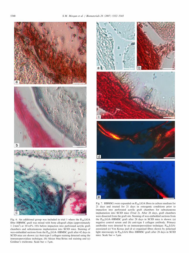
we also included a group where bone allograft chips (approximately
1–2mm2) were mixed at 10 vol% with PDLLGA–HBMSC 18 h before
impaction. We sought to establish whether such a mixture would advance
progression of the PDLLGA fibre–HBMSC graft itself towards the
osteogenic lineage. Bone allograft was processed and stringently washed
as described previously. Graft chambers were implanted subcutaneously in
the flanks of SCID mice. At the end of the trials, the mice were killed and
implants were dissected. Specimens were collected from the graft chambers
and fixed in 4% paraformaldehyde, 85% ethanol or formalin and
embedded in paraffin wax for immuno-histochemistry and histochemistry.
In the first trial, implants were photographed with a digital camera (Canon
Powershot G2) and PKH26-labelled PDLLGA fibre–HBMSC graft was
examined immediately ex vivo by fluorescence microscopy using a
confocal microscope (Leica Leitz DM RBE) and imaged using Leica
Confocal 2.5 software.
2.10. Immunohistochemistry
To detect type I collagen, sections (10 mm) of wax-embedded ex-vivo
specimens fixed with 4% paraformaldehyde were incubated in 3%H2O2 to
block endogenous peroxidase activity. A rabbit polyclonal antibody
ARTICLE IN PRESSS.M. Morgan et al. / Biomaterials 28 (2007) 5332–5343 5335
specific for type I collagen (LF 67, Dr. Larry Fisher, NIH, Bethesda, MD)
was added followed by a biotinylated goat anti-rabbit Ig (DAKO UK
Ltd., Cambridge) and Extravidin Peroxidase in PBS. Peroxidase activity
was detected using 3 amino-9-ethyl-carbazole in N,N-dimethylformamide
containing 0.015% H2O2. For counterstaining, Gills haematoxylin was
used. The primary antibody was replaced with normal rabbit serum as a
negative control. To detect vimentin, formalin-fixed sections (10 mm) of
wax-embedded ex-vivo specimens were incubated in 3% H2O2 to block
endogenous peroxidase activity. A goat polyclonal antibody specific for
vimentin (Sigma) was added followed by a biotinylated rabbit anti-goat Ig
(DAKO UK Ltd., Cambridge) and Extravidin Peroxidase in PBS.
Peroxidase activity was detected using 3 amino-9-ethyl-carbazole in
N,N-dimethylformamide containing 0.015% H2O2. The primary antibody
was replaced with normal goat serum as a negative control. Images of
sections were taken using an inverted microscope (Zeiss Axiovert 200) and
captured using the Carl Zeiss Axiovision-3.1 software package.
2.11. Histology
Staining with Alcian blue/Sirius red, Goldner’s trichrome and Von
Kossa methods was performed on sections (10mm) from ethanol-fixed
blocks. For Alcian blue/Sirius red, sections were stained using Weigert’s
haematoxylin solutions and 5% alcian blue, treated with 1% molybdo-
phosphoric acid and stained with 0.1% Sirius Red. For the Goldner’s
trichrome method, sections were stained with Weigert’s haematoxylin,
Ponceau–fuchsin–azophloxin, orange G and light green solutions and
treated with 1% acetic acid. For Von Kossa, samples were stained with
1% AgNO3 under ultraviolet (UV) light for 30min, fixed with 2.5%
sodium thiosulphate and counterstained with Alcian blue and van Giesen.
Images of sections were taken using an inverted microscope (Zeiss
Axiovert 200 or Zeiss Axioskop MOT) and captured using the Carl Zeiss
Axiovision-3.0 software package.
2.12. PDLLGA hollow fibre membrane characterisation
Membrane morphology and dimensions were analysed by scanning
electron microscopy (SEM) (JSM6310, JEOL) after coating with gold
using an Edwards Sputter Coater (S310B). Mean pore size and effective
surface porosity were calculated using measurements obtained by gas
permeation. A bubble metre was used to measure the volumetric flow rate
of nitrogen through the fibre for a range of pressures. Mean pore size and
effective surface porosity was calculated as described previously [47]. Gas
permeability was calculated using the flux equation J ¼ nA/P and plotted
against mean pressure. The gradient P0 and intercept K0 were calculated
and used in the following equations to find the mean pore radius, r (m),
and the effective porosity e/Lp (m�1):
r ¼16
3
� �P0
K0
� �8RT
pMN2
� �0:5
m,
�
Lp¼
8mRTP0
r2.
Tensile strength testing was measured using an Instron 1122 for a single
fibre. A load was applied at a cross-head speed of 20mm/min until the
ultimate load was reached. The ultimate load and elongation were used to
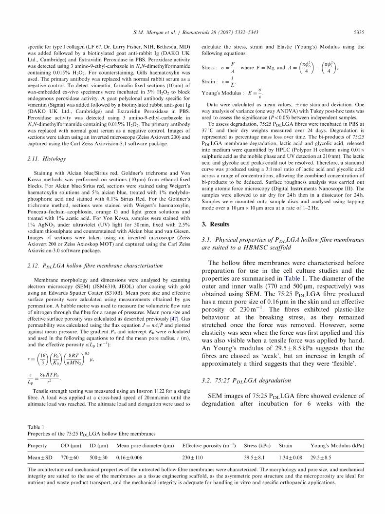
Table 1
Properties of the 75:25 PDLLGA hollow fibre membranes
Property OD (mm) ID (mm) Mean pore diameter (mm) Effective
Mean7SD 770760 500730 0.1670.006 230711
The architecture and mechanical properties of the untreated hollow fibre memb
integrity are suited to the use of the membranes as a tissue engineering scaffo
nutrient and waste product transport, and the mechanical integrity is adequat
calculate the stress, strain and Elastic (Young’s) Modulus using the
following equations:
Stress : s ¼F
Awhere F ¼Mg and A ¼
pf2o
4
� ��
pf2i
4
� �,
Strain : � ¼l
L,
Young0s Modulus : E ¼s�.
Data were calculated as mean values, 7one standard deviation. One
way analysis of variance (one way ANOVA) with Tukey post-hoc tests was
used to assess the significance (Po0.05) between independent samples.
To assess degradation, 75:25 PDLLGA fibres were incubated in PBS at
37 1C and their dry weights measured over 24 days. Degradation is
represented as percentage mass loss over time. The bi-products of 75:25
PDLLGA membrane degradation, lactic acid and glycolic acid, released
into medium were quantified by HPLC (Polypor H column using 0.01 N
sulphuric acid as the mobile phase and UV detection at 210 nm). The lactic
acid and glycolic acid peaks could not be resolved. Therefore, a standard
curve was produced using a 3:1mol ratio of lactic acid and glycolic acid
across a range of concentrations, allowing the combined concentration of
bi-products to be deduced. Surface roughness analysis was carried out
using atomic force microscopy (Digital Instruments Nanoscope III). The
samples were allowed to air dry for 24 h then in a dissicator for 24 h.
Samples were mounted onto sample discs and analysed using tapping
mode over a 10mm� 10 mm area at a rate of 1–2Hz.
3. Results
3.1. Physical properties of PDLLGA hollow fibre membranes
are suited to a HBMSC scaffold
The hollow fibre membranes were characterised beforepreparation for use in the cell culture studies and theproperties are summarised in Table 1. The diameter of theouter and inner walls (770 and 500 mm, respectively) wasobtained using SEM. The 75:25 PDLLGA fibre producedhas a mean pore size of 0.16 mm in the skin and an effectiveporosity of 230m�1. The fibres exhibited plastic-likebehaviour at the breaking stress, as they remainedstretched once the force was removed. However, someelasticity was seen when the force was first applied and thiswas also visible when a tensile force was applied by hand.An Young’s modulus of 29.578.5 kPa suggests that thefibres are classed as ‘weak’, but an increase in length ofapproximately a third suggests that they were ‘flexible’.
3.2. 75:25 PDLLGA degradation
SEM images of 75:25 PDLLGA fibre showed evidence ofdegradation after incubation for 6 weeks with the
porosity (m�1) Stress (kPa) Strain Young’s Modulus (kPa)
0 39.578.1 1.3470.08 29.578.5
ranes were characterized. The morphology and pore size, and mechanical
ld, as the asymmetric pore structure and the microporosity are ideal for
e for handling in vitro and specific orthopaedic applications.
ARTICLE IN PRESSS.M. Morgan et al. / Biomaterials 28 (2007) 5332–53435336
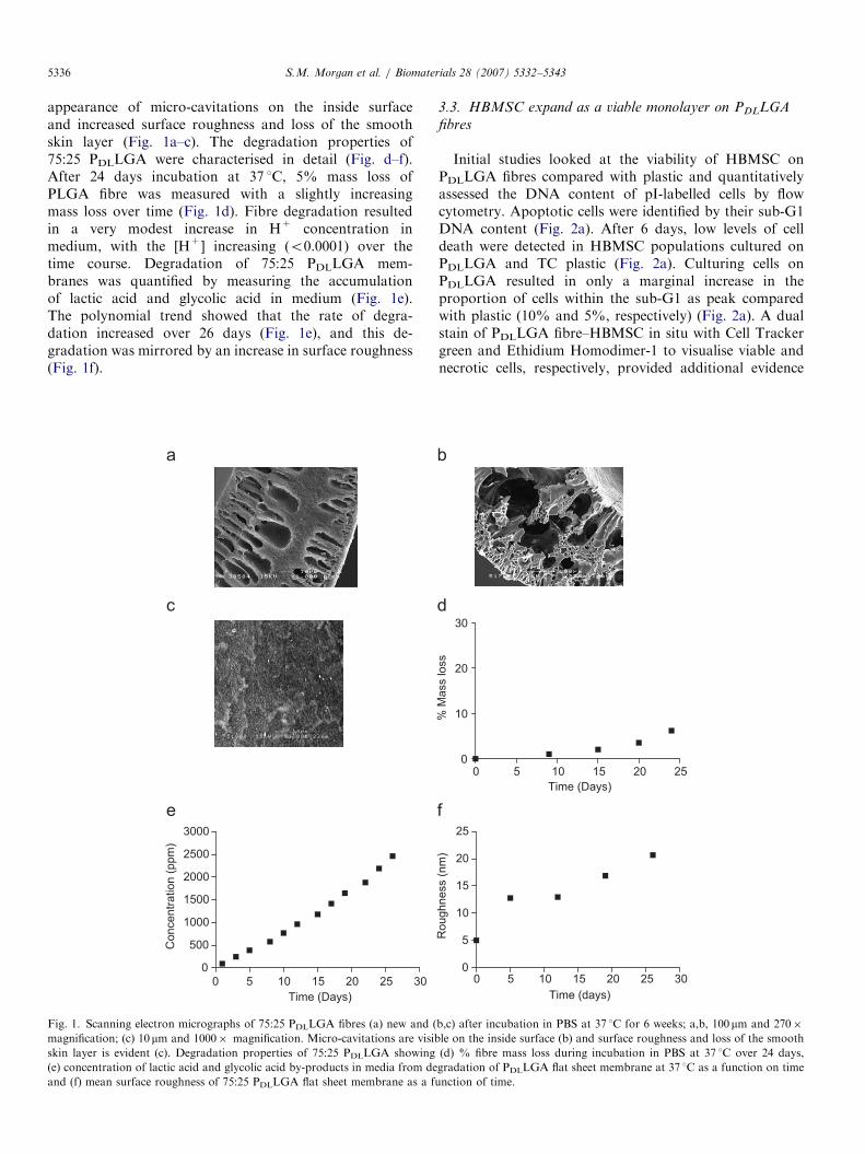
appearance of micro-cavitations on the inside surfaceand increased surface roughness and loss of the smoothskin layer (Fig. 1a–c). The degradation properties of75:25 PDLLGA were characterised in detail (Fig. d–f).After 24 days incubation at 37 1C, 5% mass loss ofPLGA fibre was measured with a slightly increasingmass loss over time (Fig. 1d). Fibre degradation resultedin a very modest increase in H+ concentration inmedium, with the [H+] increasing (o0.0001) over thetime course. Degradation of 75:25 PDLLGA mem-branes was quantified by measuring the accumulationof lactic acid and glycolic acid in medium (Fig. 1e).The polynomial trend showed that the rate of degra-dation increased over 26 days (Fig. 1e), and this de-gradation was mirrored by an increase in surface roughness(Fig. 1f).
0 5 10 15 20 25 30
0
500
1000
1500
2000
2500
3000
Time (Days)
Concentr
ation (
ppm
)
c
e
Fig. 1. Scanning electron micrographs of 75:25 PDLLGA fibres (a) new and (
magnification; (c) 10 mm and 1000� magnification. Micro-cavitations are visib
skin layer is evident (c). Degradation properties of 75:25 PDLLGA showing
(e) concentration of lactic acid and glycolic acid by-products in media from de
and (f) mean surface roughness of 75:25 PDLLGA flat sheet membrane as a fu
3.3. HBMSC expand as a viable monolayer on PDLLGA
fibres
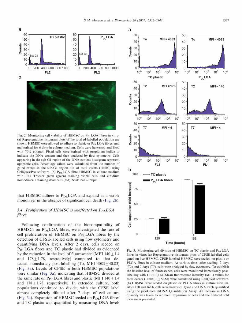
Initial studies looked at the viability of HBMSC onPDLLGA fibres compared with plastic and quantitativelyassessed the DNA content of pI-labelled cells by flowcytometry. Apoptotic cells were identified by their sub-G1DNA content (Fig. 2a). After 6 days, low levels of celldeath were detected in HBMSC populations cultured onPDLLGA and TC plastic (Fig. 2a). Culturing cells onPDLLGA resulted in only a marginal increase in theproportion of cells within the sub-G1 as peak comparedwith plastic (10% and 5%, respectively) (Fig. 2a). A dualstain of PDLLGA fibre–HBMSC in situ with Cell Trackergreen and Ethidium Homodimer-1 to visualise viable andnecrotic cells, respectively, provided additional evidence
0 5 10 15 20 25 300
5
10
15
20
25
Time (days)
Roughness (
nm
)
0 5 10 15 20 250
10
20
30
Time (Days)
% M
ass loss
d
f
b,c) after incubation in PBS at 37 1C for 6 weeks; a,b, 100mm and 270�
le on the inside surface (b) and surface roughness and loss of the smooth
(d) % fibre mass loss during incubation in PBS at 37 1C over 24 days,
gradation of PDLLGA flat sheet membrane at 37 1C as a function on time
nction of time.
ARTICLE IN PRESS
FL2 FL2
Co
un
ts
0 200 400 600 800 10000
10
20
30
40
50
60
0 200 400 600 800 10000
10
20
30
40
50
60
Sub-G1
TC plastic PDL
LGA
Sub-G1
50
b
Fig. 2. Monitoring cell viability of HBMSC on PDLLGA fibres in vitro:
(a) Representative histogram plots of the total pI-labelled population are
shown. HBMSC were allowed to adhere to plastic or PDLLGA fibres, and
maintained for 6 days in culture medium. Cells were harvested and fixed
with 70% ethanol. Fixed cells were stained with propidium iodide to
indicate the DNA content and then analysed by flow cytometry. Cells
appearing in the sub-G1 region of the DNA content histogram represent
apoptotic cells. Percentage values were calculated from the number of
gated events in the sub-G1 region out of total events (10,000) using
CellQuestPro software. (b) PDLLGA fibre–HBMSC in culture medium
with Cell Tracker green (green) staining viable cells and ethidium
homodimer-1 staining dead cells (red). Scale bar ¼ 20 mm.
Co
un
ts
Co
un
ts
Co
un
ts
100 101 102 103 104
0
10
20
30
40
50
100 101 102 103 104
100 101 102 103 104 100 101 102 103 104
100 101 102 103 104 100 101 102 103 104
0
10
20
30
40
50
0
10
20
30
40
50
0
10
20
30
40
50
0
10
20
30
40
50
TC plastic
To MFI= 4083
T2 MFI = 178
T7 MFI = 4
To MFI = 4083
PDL
LGA
0
10
20
30
40
50
T2 MFI = 140
T7 MFI = 4
120 168
0
25
50
75
100 TC plastic
PDL
LGA fibres
Time (hours)
Cell e
xp
an
sio
n (
fold
in
cre
ase)
FL1 FL1
b
Fig. 3. Monitoring cell division of HBMSC on TC plastic and PDLLGA
fibres in vitro: (a) Representative histogram plots of CFSE-labelled cells
gated on live HBMSC. CFSE-labelled HBMSC were seeded on plastic or
PLGA fibres in culture medium. At various times after seeding, 2 days
(T2) and 7 days (T7), cells were analysed by flow cytometry. To establish
the baseline level of fluorescence, cells were monitored immediately post-
labelling with CFSE (To). Mean fluorescence intensity (MFI) values for
total events (10,000) (7SEM) were calculated using CellQuest software.
(b) HBMSC were seeded on plastic or PLGA fibres in culture medium.
After 120 and 168 h, cells were harvested, lysed and DNA levels quantified
using the picoGreen dsDNA Quantitation Assay. An increase in DNA
quantity was taken to represent expansion of cells and the deduced fold
increase is presented.
S.M. Morgan et al. / Biomaterials 28 (2007) 5332–5343 5337
that HBMSC adhere to PDLLGA and expand as a viablemonolayer in the absence of significant cell death (Fig. 2b).
3.4. Proliferation of HBMSC is unaffected on PDLLGA
fibres
Following confirmation of the biocompatibility ofHBMSCs on PDLLGA fibres, we investigated the rate ofcell proliferation of HBMSC on PDLLGA fibres by thedetection of CFSE-labelled cells using flow cytometry andquantifying DNA levels. After 2 days, cells seeded onPDLLGA fibres and TC plastic had divided as evidencedby the reduction in the level of fluorescence (MFI 14071.4and 17871.78, respectively) compared to that de-tected immediately post-labelling (To, MFI 4083740.83)(Fig. 3a). Levels of CFSE in both HBMSC populationswere similar (Fig. 3a), indicating that HBMSC divided atthe same rate on PDLLGA fibres and plastic (MFI 14071.4and 17871.78, respectively). In extended culture, bothpopulations continued to divide, with the CFSE labelalmost completely diluted after 7 days of cell culture(Fig. 3a). Expansion of HBMSC seeded on PDLLGA fibresand TC plastic was quantified by measuring DNA levels
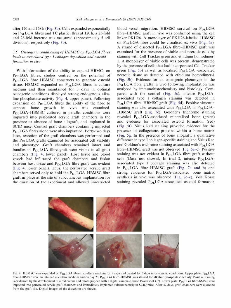
ARTICLE IN PRESSS.M. Morgan et al. / Biomaterials 28 (2007) 5332–53435338
after 120 and 168 h (Fig. 3b). Cells expanded exponentiallyon PDLLGA fibres and TC plastic, thus at 120 h, a 25-foldand 26-fold increase was measured (approximately 5 celldivisions), respectively (Fig. 3b).
3.5. Osteogenic conditioning of HBMSC on PDLLGA fibres
leads to associated type I collagen deposition and osteoid
formation in vivo
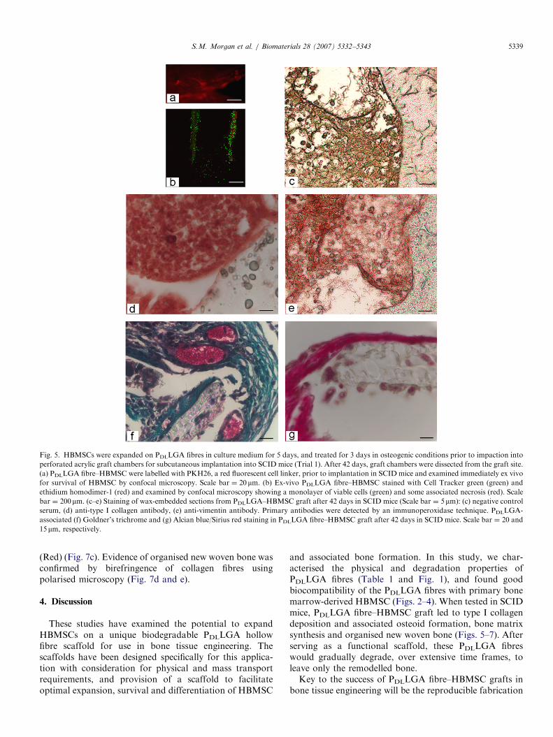
With information of the ability to expand HBMCs onPDLLGA fibres, studies centred on the potential ofPDLLGA fibre–HBMSC constructs to generate osteoidtissue. HBMSC expanded on PDLLGA fibres in culturemedium and then maintained for 3 days in optimalosteogenic conditions displayed strong endogenous alka-line phosphatase activity (Fig. 4, upper panel). Followingexpansion on PDLLGA fibres the ability of the fibre tosupport bone growth in vivo was examined.PDLLGA–HBMSC cultured in parallel conditions wereimpacted into perforated acrylic graft chambers in thepresence or absence of bone allograft, and implanted inSCID mice. Control graft chambers containing impactedPDLLGA fibres alone were also implanted. Forty-two dayslater, resection of the graft chambers was performed andthe PDLLGA grafts examined for associated cell viabilityand phenotype. Graft chambers remained intact andbundles of PDLLGA fibre graft were visible in all graftchambers (Fig. 4, lower panel). Host tissue and bloodvessels had infiltrated the graft chambers and fusionbetween host tissue and PDLLGA fibre graft was evident(Fig. 4, lower panel). Thus, the perforated acrylic graftchambers served only to hold the PDLLGA–HBMSC fibregraft in place at the site of subcutaneous implantation forthe duration of the experiment and allowed unrestricted
Fig. 4. HBMSC were expanded on PDLLGA fibres in culture medium for 5 da
fibre–HBMSC were maintained in culture medium and on day 20, PDLLGA fib
is evidenced by the development of a red colour and photographed with a digita
impacted into perforated acrylic graft chambers and immediately implanted su
from the graft site. Digital images of the dissection are shown.
blood vessel migration. HBMSC survival on PDLLGAfibre–HBMSC graft in vivo was confirmed using the celllinker PKH26. A monolayer of PKH26-labelled HBMSCon PDLLGA fibre could be visualised ex vivo (Fig. 5a).A strand of dissected PDLLGA fibre–HBMSC graft wasexamined for the presence of viable and necrotic cells bystaining with Cell Tracker green and ethidium homodimer-1. A monolayer of viable cells was present, demonstratedby the presence of cells that had incorporated Cell Trackergreen (Fig. 5b) as well as localised PDLLGA -associatednecrotic tissue as detected with ethidium homodimer-1(Fig. 5b). Evidence for an osteogenic phenotype in thePDLLGA fibre grafts in vivo following implantation wasanalysed by immunohistochemistry and histology. Com-pared with the control (Fig. 5c), intense PDLLGA-associated type I collagen staining was detected inPDLLGA fibre–HBMSC graft (Fig. 5d). Positive vimentinstaining was also associated with PDLLGA in PDLLGA–HBMSC graft (Fig. 5e). Goldner’s trichrome stainingrevealed PDLLGA-associated mineralised bone (green)and evidence for associated osteoid formation (red)(Fig. 5f). Sirius Red staining provided evidence for thepresence of collagenous proteins within a bone matrix(Fig. 5g. In the presence of bone allograft, a qualitativedifference in type I collagen-specific staining and Sirius Redand Goldner’s trichrome staining associated with PDLLGAfibre–HBMSC graft was not observed (Fig. 6a–c). Positivestaining was not evident in PDLLGA fibre graft withoutcells (Data not shown). In trial 2, intense PDLLGA-associated type I collagen staining was also detectedin PDLLGA fibre–HBMSC graft (Fig. 7a and b) andstrong evidence for PDLLGA-associated bone matrixsynthesis in vivo was observed (Fig. 7c–e). Von Kossastaining revealed PDLLGA-associated osteoid formation
ys and treated for 3 days in osteogenic conditions. Upper plate; PDLLGA
re–HBMSC was stained for alkaline phosphatase activity. Positive staining
l camera (Canon Powershot G2). Lower plate: PDLLGA fibre-hMSC were
bcutaneously in SCID mice. After 42 days, graft chambers were dissected
ARTICLE IN PRESS
Fig. 5. HBMSCs were expanded on PDLLGA fibres in culture medium for 5 days, and treated for 3 days in osteogenic conditions prior to impaction into
perforated acrylic graft chambers for subcutaneous implantation into SCID mice (Trial 1). After 42 days, graft chambers were dissected from the graft site.
(a) PDLLGA fibre–HBMSC were labelled with PKH26, a red fluorescent cell linker, prior to implantation in SCID mice and examined immediately ex vivo
for survival of HBMSC by confocal microscopy. Scale bar ¼ 20 mm. (b) Ex-vivo PDLLGA fibre–HBMSC stained with Cell Tracker green (green) and
ethidium homodimer-1 (red) and examined by confocal microscopy showing a monolayer of viable cells (green) and some associated necrosis (red). Scale
bar ¼ 200mm. (c–e) Staining of wax-embedded sections from PDLLGA–HBMSC graft after 42 days in SCID mice (Scale bar ¼ 5mm): (c) negative control
serum, (d) anti-type I collagen antibody, (e) anti-vimentin antibody. Primary antibodies were detected by an immunoperoxidase technique. PDLLGA-
associated (f) Goldner’s trichrome and (g) Alcian blue/Sirius red staining in PDLLGA fibre–HBMSC graft after 42 days in SCID mice. Scale bar ¼ 20 and
15 mm, respectively.
S.M. Morgan et al. / Biomaterials 28 (2007) 5332–5343 5339
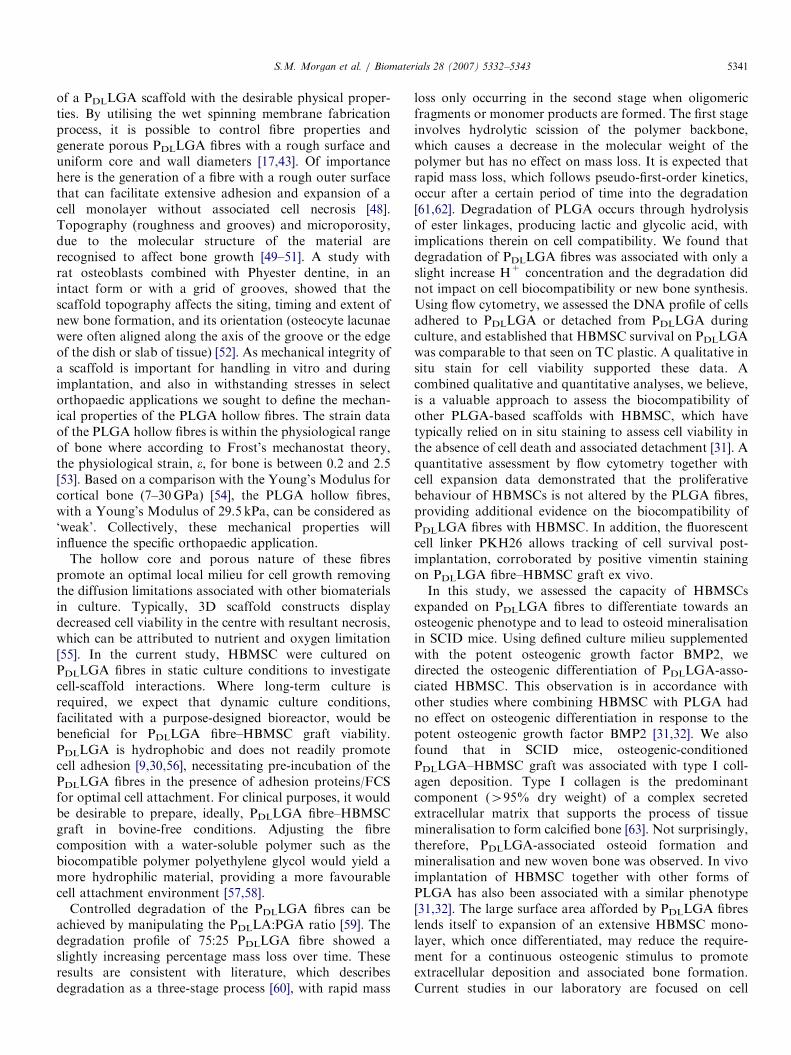
(Red) (Fig. 7c). Evidence of organised new woven bone wasconfirmed by birefringence of collagen fibres usingpolarised microscopy (Fig. 7d and e).
4. Discussion
These studies have examined the potential to expandHBMSCs on a unique biodegradable PDLLGA hollowfibre scaffold for use in bone tissue engineering. Thescaffolds have been designed specifically for this applica-tion with consideration for physical and mass transportrequirements, and provision of a scaffold to facilitateoptimal expansion, survival and differentiation of HBMSC
and associated bone formation. In this study, we char-acterised the physical and degradation properties ofPDLLGA fibres (Table 1 and Fig. 1), and found goodbiocompatibility of the PDLLGA fibres with primary bonemarrow-derived HBMSC (Figs. 2–4). When tested in SCIDmice, PDLLGA fibre–HBMSC graft led to type I collagendeposition and associated osteoid formation, bone matrixsynthesis and organised new woven bone (Figs. 5–7). Afterserving as a functional scaffold, these PDLLGA fibreswould gradually degrade, over extensive time frames, toleave only the remodelled bone.Key to the success of PDLLGA fibre–HBMSC grafts in
bone tissue engineering will be the reproducible fabrication
ARTICLE IN PRESS
Fig. 6. An additional group was included in trial 1 where the PDLLGA
fibre–HBMSC graft was mixed with bone allograft chips (approximately
1–2mm2) at 10 vol% 18 h before impaction into perforated acrylic graft
chambers and subcutaneous implantation into SCID mice. Staining of
wax-embedded sections from the PDLLGA–HBMSC graft after 42 days in
SCID mice are shown: (a) Anti-type I collagen staining detected using the
immunoperoxidase technique, (b) Alcian blue/Sirius red staining and (c)
Goldner’s trichrome. Scale bar ¼ 5 mm.
Fig. 7. HBMSCs were expanded on PDLLGA fibres in culture medium for
21 days and treated for 21 days in osteogenic conditions prior to
impaction into perforated acrylic graft chambers for subcutaneous
implantation into SCID mice (Trial 2). After 28 days, graft chambers
were dissected from the graft site. Staining of wax-embedded sections from
the PDLLGA–HBMSC graft after 28 days in SCID mice is shown: (a)
negative control serum and (b) anti-type I collagen antibody. Primary
antibodies were detected by an immunoperoxidase technique. PDLLGA-
associated (c) Von Kossa and (d–e) organised fibres shown by polarised
light microscopy in PDLLGA fibre–HBMSC graft after 28 days in SCID
mice. Scale bar ¼ 5 mm.
S.M. Morgan et al. / Biomaterials 28 (2007) 5332–53435340
ARTICLE IN PRESSS.M. Morgan et al. / Biomaterials 28 (2007) 5332–5343 5341
of a PDLLGA scaffold with the desirable physical proper-ties. By utilising the wet spinning membrane fabricationprocess, it is possible to control fibre properties andgenerate porous PDLLGA fibres with a rough surface anduniform core and wall diameters [17,43]. Of importancehere is the generation of a fibre with a rough outer surfacethat can facilitate extensive adhesion and expansion of acell monolayer without associated cell necrosis [48].Topography (roughness and grooves) and microporosity,due to the molecular structure of the material arerecognised to affect bone growth [49–51]. A study withrat osteoblasts combined with Phyester dentine, in anintact form or with a grid of grooves, showed that thescaffold topography affects the siting, timing and extent ofnew bone formation, and its orientation (osteocyte lacunaewere often aligned along the axis of the groove or the edgeof the dish or slab of tissue) [52]. As mechanical integrity ofa scaffold is important for handling in vitro and duringimplantation, and also in withstanding stresses in selectorthopaedic applications we sought to define the mechan-ical properties of the PLGA hollow fibres. The strain dataof the PLGA hollow fibres is within the physiological rangeof bone where according to Frost’s mechanostat theory,the physiological strain, e, for bone is between 0.2 and 2.5[53]. Based on a comparison with the Young’s Modulus forcortical bone (7–30GPa) [54], the PLGA hollow fibres,with a Young’s Modulus of 29.5 kPa, can be considered as‘weak’. Collectively, these mechanical properties willinfluence the specific orthopaedic application.
The hollow core and porous nature of these fibrespromote an optimal local milieu for cell growth removingthe diffusion limitations associated with other biomaterialsin culture. Typically, 3D scaffold constructs displaydecreased cell viability in the centre with resultant necrosis,which can be attributed to nutrient and oxygen limitation[55]. In the current study, HBMSC were cultured onPDLLGA fibres in static culture conditions to investigatecell-scaffold interactions. Where long-term culture isrequired, we expect that dynamic culture conditions,facilitated with a purpose-designed bioreactor, would bebeneficial for PDLLGA fibre–HBMSC graft viability.PDLLGA is hydrophobic and does not readily promotecell adhesion [9,30,56], necessitating pre-incubation of thePDLLGA fibres in the presence of adhesion proteins/FCSfor optimal cell attachment. For clinical purposes, it wouldbe desirable to prepare, ideally, PDLLGA fibre–HBMSCgraft in bovine-free conditions. Adjusting the fibrecomposition with a water-soluble polymer such as thebiocompatible polymer polyethylene glycol would yield amore hydrophilic material, providing a more favourablecell attachment environment [57,58].
Controlled degradation of the PDLLGA fibres can beachieved by manipulating the PDLLA:PGA ratio [59]. Thedegradation profile of 75:25 PDLLGA fibre showed aslightly increasing percentage mass loss over time. Theseresults are consistent with literature, which describesdegradation as a three-stage process [60], with rapid mass
loss only occurring in the second stage when oligomericfragments or monomer products are formed. The first stageinvolves hydrolytic scission of the polymer backbone,which causes a decrease in the molecular weight of thepolymer but has no effect on mass loss. It is expected thatrapid mass loss, which follows pseudo-first-order kinetics,occur after a certain period of time into the degradation[61,62]. Degradation of PLGA occurs through hydrolysisof ester linkages, producing lactic and glycolic acid, withimplications therein on cell compatibility. We found thatdegradation of PDLLGA fibres was associated with only aslight increase H+ concentration and the degradation didnot impact on cell biocompatibility or new bone synthesis.Using flow cytometry, we assessed the DNA profile of cellsadhered to PDLLGA or detached from PDLLGA duringculture, and established that HBMSC survival on PDLLGAwas comparable to that seen on TC plastic. A qualitative insitu stain for cell viability supported these data. Acombined qualitative and quantitative analyses, we believe,is a valuable approach to assess the biocompatibility ofother PLGA-based scaffolds with HBMSC, which havetypically relied on in situ staining to assess cell viability inthe absence of cell death and associated detachment [31]. Aquantitative assessment by flow cytometry together withcell expansion data demonstrated that the proliferativebehaviour of HBMSCs is not altered by the PLGA fibres,providing additional evidence on the biocompatibility ofPDLLGA fibres with HBMSC. In addition, the fluorescentcell linker PKH26 allows tracking of cell survival post-implantation, corroborated by positive vimentin stainingon PDLLGA fibre–HBMSC graft ex vivo.In this study, we assessed the capacity of HBMSCs
expanded on PDLLGA fibres to differentiate towards anosteogenic phenotype and to lead to osteoid mineralisationin SCID mice. Using defined culture milieu supplementedwith the potent osteogenic growth factor BMP2, wedirected the osteogenic differentiation of PDLLGA-asso-ciated HBMSC. This observation is in accordance withother studies where combining HBMSC with PLGA hadno effect on osteogenic differentiation in response to thepotent osteogenic growth factor BMP2 [31,32]. We alsofound that in SCID mice, osteogenic-conditionedPDLLGA–HBMSC graft was associated with type I coll-agen deposition. Type I collagen is the predominantcomponent (495% dry weight) of a complex secretedextracellular matrix that supports the process of tissuemineralisation to form calcified bone [63]. Not surprisingly,therefore, PDLLGA-associated osteoid formation andmineralisation and new woven bone was observed. In vivoimplantation of HBMSC together with other forms ofPLGA has also been associated with a similar phenotype[31,32]. The large surface area afforded by PDLLGA fibreslends itself to expansion of an extensive HBMSC mono-layer, which once differentiated, may reduce the require-ment for a continuous osteogenic stimulus to promoteextracellular deposition and associated bone formation.Current studies in our laboratory are focused on cell
ARTICLE IN PRESSS.M. Morgan et al. / Biomaterials 28 (2007) 5332–53435342
expansion protocols where ethanol is substituted withantibiotics for sterilisation of the PLGA hollow fibres toachieve minimum alteration of polymer topography [64].
5. Conclusion
In conclusion we have established that HBMSC canexpand on PDLLGA fibres and survive, and once differ-entiated can lead to an associated osteogenic phenotype.Current studies are focused on the examination of differentcell differentiation populations for bone repair andregeneration where for example osteo-progenitor cells arecombined with differentiated osteoblasts. Such a cell-basedsynthetic graft could be a desirable biomaterial for use inorthopaedic applications where the natural compliment ofcells and physiologically appropriate balance of osteoin-ductive factors intrinsic to bone healing and regenerationare provided.
Acknowledgements
We thank Margaret Edkins and Joanna Hajdukiewiczfor technical support and Ben Bolland for helpfuldiscussions. We thank the orthopaedic surgical staff atSouthampton General Hospital for tissue provision andStryker UK for their kind support to Simon Tilley duringhis time in research. This work is supported by a grantfrom the Biotechnology and Biological Sciences ResearchCouncil.
References
[1] Bianco P, Robey PG. Stem cells in tissue engineering. Nature
2001;414(6859):118–21.
[2] Jaiswal N, Haynesworth SE, Caplan AI, Bruder SP. Osteogenic
differentiation of purified, culture-expanded human mesenchymal
stem cells in vitro. J Cell Biochem 1997;64(2):295–312.
[3] Prockop DJ. Stem cell research has only just begun. Science
2001;293(5528):211–2.
[4] Horwitz EM, Prockop DJ, Fitzpatrick LA, Koo WW, Gordon PL,
Neel M, et al. Transplantability and therapeutic effects of bone
marrow-derived mesenchymal cells in children with osteogenesis
imperfecta. Nat Med 1999;5(3):309–13.
[5] Bruder SP, Jaiswal N, Haynesworth SE. Growth kinetics, self-
renewal, and the osteogenic potential of purified human mesenchymal
stem cells during extensive subcultivation and following cryopreser-
vation. J Cell Biochem 1997;64(2):278–94.
[6] Lecanda F, Avioli LV, Cheng SL. Regulation of bone matrix protein
expression and induction of differentiation of human osteoblasts and
human bone marrow stromal cells by bone morphogenetic protein-2.
J Cell Biochem 1997;67(3):386–96.
[7] Haynesworth SE, Goshima J, Goldberg VM, Caplan AI. Character-
ization of cells with osteogenic potential from human marrow. Bone
1992;13(1):81–8.
[8] Pittenger MF, Mackay AM, Beck SC, Jaiswal RK, Douglas R,
Mosca JD, et al. Multilineage potential of adult human mesenchymal
stem cells. Science 1999;284(5411):143–7.
[9] Drury JL, Mooney DJ. Hydrogels for tissue engineering: scaffold
design variables and applications. Biomaterials 2003;24(24):4337–51.
[10] Sittinger M, Hutmacher DW, Risbud MV. Current strategies for cell
delivery in cartilage and bone regeneration. Curr Opin Biotechnol
2004;15(5):411–8.
[11] Rose FR, Oreffo RO. Bone tissue engineering: hope vs. hype.
Biochem Biophys Res Commun 2002;292(1):1–7.
[12] Green D, Walsh D, Mann S, Oreffo RO. The potential of biomimesis
in bone tissue engineering: lessons from the design and synthesis of
invertebrate skeletons. Bone 2002;30(6):810–5.
[13] Bianco P, Riminucci M, Gronthos S, Robey PG. Bone marrow
stromal stem cells: nature, biology, and potential applications. Stem
Cells 2001;19(3):180–92.
[14] Jordan KM, Cooper C. Epidemiology of osteoporosis. Best Pract Res
Clin Rheumatol 2002;16(5):795–806.
[15] Christodoulou C, Cooper C. What is osteoporosis? Postgrad Med
J 2003;79(929):133–8.
[16] Royal College of Physicians. In: Royal College of Physicians of
London. Osteoporesis: clinical guidelines for prevention and treat-
ment. London: 1999.
[17] Ellis MJ, Chaudhuri JB. Poly(lactic-co-glycolic acid) hollow fibre
membranes for use as a tissue engineering scaffold. Biotechnol Bioeng
2007;96(1):177–87.
[18] Yang X, Tare RS, Partridge KA, Roach HI, Clarke NM, Howdle
SM, et al. Induction of human osteoprogenitor chemotaxis,
proliferation, differentiation, and bone formation by osteoblast
stimulating factor-1/pleiotrophin: osteoconductive biomimetic scaf-
folds for tissue engineering. J Bone Miner Res 2003;18(1):47–57.
[19] Cancedda R, Dozin B, Giannoni P, Quarto R. Tissue engineering and
cell therapy of cartilage and bone. Matrix Biol 2003;22(1):81–91.
[20] Sittinger M, Hutmacher DW, Risbud MV. Current strategies for cell
delivery in cartilage and bone regeneration. Curr Opin Biotechnol
2004;15(5):411–8.
[21] Yang XB, Webb D, Blaker J, Boccaccini AR, Maquet V, Cooper C,
et al. Evaluation of human bone marrow stromal cell growth on
biodegradable polymer/bioglass composites. Biochem Biophys Res
Commun 2006;342(4):1098–107.
[22] Howard D, Partridge K, Yang X, Clarke NM, Okubo Y, Bessho K,
et al. Immunoselection and adenoviral genetic modulation of human
osteoprogenitors: in vivo bone formation on PLA scaffold. Biochem
Biophys Res Commun 2002;299(2):208–15.
[23] Yaszemski MJ, Payne RG, Hayes WC, Langer R, Mikos AG.
Evolution of bone transplantation: molecular, cellular and tissue
strategies to engineer human bone. Biomaterials 1996;17(2):175–85.
[24] Thomas RC, Wake MC, Yaszemski MJ, Mikos AG. Biodegradable
polymer scaffolds to regenerate organs. In: Peppas NA, Langer RS,
editors. Advances in polymer science. New York: 1995. p. 245–74.
[25] Wong WH, Mooney DJ. Synthesis and properties of biodegradable
polymers used as synthetic matrices for tissue engineering. In: Atala
A, Mooney DJ, editors, Langer R, Vacanti JP, associate, editors.
Synthetic biodegradable polymer scaffolds. Boston: 1997. p. 58–85.
[26] Wake MC, Gerecht PD, Lu L, Mikos AG. Effects of biodegradable
polymer particles on rat marrow-derived stromal osteoblasts in vitro.
Biomaterials 1998;19(14):1255–68.
[27] Ishaug-Riley SL, Crane-Kruger GM, Yaszemski MJ, Mikos AG.
Three-dimensional culture of rat calvarial osteoblasts in porous
biodegradable polymers. Biomaterials 1998;19(15):1405–12.
[28] Mikos AG, Bao Y, Cima LG, Ingber DE, Vacanti JP, Langer R.
Preparation of poly(glycolic acid) bonded fiber structures for cell
attachment and transplantation. J Biomed Mater Res 1993;27(2):
183–9.
[29] Owen GR, Jackson J, Chehroudi B, Burt H, Brunette DM. A PLGA
membrane controlling cell behaviour for promoting tissue regenera-
tion. Biomaterials 2005;26(35):7447–56.
[30] Wu YC, Shaw SY, Lin HR, Lee TM, Yang CY. Bone tissue
engineering evaluation based on rat calvaria stromal cells cultured on
modified PLGA scaffolds. Biomaterials 2006;27(6):896–904.
[31] Partridge K, Yang X, Clarke NM, Okubo Y, Bessho K, Sebald W,
et al. Adenoviral BMP-2 gene transfer in mesenchymal stem cells:
in vitro and in vivo bone formation on biodegradable polymer
scaffolds. Biochem Biophys Res Commun 2002;292(1):144–52.
[32] Yang XB, Whitaker MJ, Sebald W, Clarke N, Howdle SM,
Shakesheff KM, et al. Human osteoprogenitor bone formation using
ARTICLE IN PRESSS.M. Morgan et al. / Biomaterials 28 (2007) 5332–5343 5343
encapsulated bone morphogenetic protein 2 in porous polymer
scaffolds. Tissue Eng 2004;10:1037–45.
[33] Blaker JJ, Gough JE, Maquet V, Notingher I, Boccaccini AR.
In vitro evaluation of novel bioactive composites based on bioglass-
filled polylactide foams for bone tissue engineering scaffolds.
J Biomed Mater Res A 2003;67(4):1401–11.
[34] Boccaccini AR, Notingher I, Maquet V, Jerome R. Bioresorbable
and bioactive composite materials based on polylactide foams filled
with and coated by bioglass particles for tissue engineering
applications. J Mater Sci Mater Med 2003;14(5):443–50.
[35] Petricca SE, Marra KG, Kumta PN. Chemical synthesis of
poly(lactic-co-glycolic acid)/hydroxyapatite composites for orthopae-
dic applications. Acta Biomater 2006;2(3):277–86.
[36] Ignjatovic N, Tomic S, Dakic M, Miljkovic M, Plavsic M, Uskokovic
D. Synthesis and properties of hydroxyapatite/poly-L-lactide compo-
site biomaterials. Biomaterials 1999;20(9):809–16.
[37] Kadiyala S, Young RG, Thiede MA, Bruder SP. Culture expanded
canine mesenchymal stem cells possess osteochondrogenic potential
in vivo and in vitro. Cell Transplant 1997;6(2):125–34.
[38] Bruder SP, Kraus KH, Goldberg VM, Kadiyala S. The effect of
implants loaded with autologous mesenchymal stem cells on the
healing of canine segmental bone defects. J Bone Joint Surg Am 1998;
80(7):985–96.
[39] Cowan CM, Shi YY, Aalami OO, Chou YF, Mari C, Thomas R, et
al. Adipose-derived adult stromal cells heal critical-size mouse
calvarial defects. Nat Biotechnol 2004;22(5):560–7.
[40] Ishaug-Riley SL, Crane GM, Gurlek A, Miller MJ, Yasko AW,
Yaszemski MJ, et al. Ectopic bone formation by marrow stromal
osteoblast transplantation using poly(DL-lactic-co-glycolic acid)
foams implanted into the rat mesentery. J Biomed Mater Res 1997;
36(1):1–8.
[41] Holy CE, Fialkov JA, Davies JE, Shoichet MS. Use of a biomimetic
strategy to engineer bone. J Biomed Mater Res A 2003;65(4):447–53.
[42] Wen X, Tresco PA. Fabrication and characterization of permeable
degradable poly(DL-lactide-co-glycolide) (PLGA) hollow fiber phase
inversion membranes for use as nerve tract guidance channels.
Biomaterials 2006;27(20):3800–9.
[43] Eser H, Tihminlioglu F. Determination of thermodynamic and
transport properties of solvents and non solvents in PLGA. J Appl
Polym Sci 2006;102:2426–32.
[44] Wang DL, Li K, Teo WK. Polyethersulfone hollow fiber gas
separation membranes prepared from NMP/alcohol solvent systems.
J Membr Sci 1996;115(1):85–108.
[45] Yang XB, Roach HI, Clarke NM, Howdle SM, Quirk R, Shakesheff
KM, et al. Human osteoprogenitor growth and differentiation on
synthetic biodegradable structures after surface modification. Bone
2001;29(6):523–31.
[46] Tilley S, Bolland BJRF, Partridge K, New AMR, Latham JM,
Dunlop DG, et al. Taking tissue engineering principles into theatre:
augmentation of impacted allograft with human bone marrow
stromal cells. Regen Med 2006;1(5):1–8.
[47] Li K, Kong JF, Wang DL, Teo WK. Tailor-made asymmetric PVDF
hollow fibers for soluble gas removal. AICHE J 1999;45(6):1211–9.
[48] Yang S, Leong K-F, Du Z, Chua C-K. Review: the design of
scaffolds for use in tissue engineering. Part I. traditional factors.
Tissue Eng 2001;7(6):679–89.
[49] Boyan BD, Hummert TW, Dean DD, Schwartz Z. Role of material
surfaces in regulating bone and cartilage cell response. Biomaterials
1996;17(2):137–46.
[50] Schwartz Z, Lohmann CH, Oefinger J, Bonewald LF, Dean DD,
Boyan BD. Implant surface characteristics modulate differentiation
behaviour of cells in the osteoblastic lineage. Adv Dental Res
1999;13:38–48.
[51] Schwartz Z, Lohmann CH, Sisk M, Cochran DL, Sylvia VL,
Simpson J, et al. Local factor production by MG63 osteoblast-like
cells in response to surface roughness and 1,25-(OH)(2)D-3 is
mediated via protein kinase C- and protein kinase A-dependent
pathways. Biomaterials 2001;22(7):731–41.
[52] Gray C, Boyde A, Jones S. Topographically induced bone formation
in vitro: implications for bone implants and bone grafts. Bone
1996;18(2):115–23.
[53] Basso N, Heersche J. Review: characteristics of in vitro osteoblastic
cell loading models. Bone 2002;30(2):347–51.
[54] Dalby MJ, Kayser MV, Bonfield W, Silvio LD. Initial attachment of
osteoblasts to an optimised HAPEX topography. Biomaterials
2002;23:681–90.
[55] Sengers BG, Taylor M, Please CP, Oreffo RO. Computational
modelling of cell spreading and tissue regeneration in porous
scaffolds. Biomaterials 2007;28(10):1926–40.
[56] Chen G, Ushida T, Tateishi T. A biodegradable hybrid sponge
nested with collagen microsponges. J Biomed Mater Res 2000;51(2):
273–9.
[57] Saito N, Okada T, Horiuchi H, Murakami N, Takahashi J, Nawata
M, et al. A biodegradable polymer as a cytokine delivery system for
inducing bone formation. Nat Biotechnol 2001;19(4):332–5.
[58] Wan Y, Chen W, Yang J, Bei J, Wang S. Biodegradable poly(L-
lactide)-poly(ethylene glycol) multiblock copolymer: synthesis and
evaluation of cell affinity. Biomaterials 2003;24(13):2195–203.
[59] Shive MS, Anderson JM. Biodegradation and biocompatibility of
PLA and PLGA microspheres. Adv Drug Deliv Rev 1997;28(1):5–24.
[60] Middleton JC, Tipton AJ. Synthetic biodegradable polymers as
orthopaedic devices. Biomaterials 2000;21:2335–46.
[61] Gunatillake PA, Adhikari R. Biodegradable synthetic polymers for
tissue engineering. Eur Cells Mater 2003;5:1–16.
[62] Hasirci V, Lewandrowski K, Gresser JD, Wise DL, Trantolo DJ.
Versatility of biodegradable biopolymers: degradability and an
in vivo application. J Biotechnol 2001;86:135–50.
[63] Olsen BR. Morphogenesis: collagen it takes and bone it makes. Curr
Biol 1996;6(6):645–7.
[64] Shearer H, Ellis MJ, Perera SP, Chaudhuri JB. Effects of four
common sterilization methods on the structure of poly(lactic-co-
glycolic acid) scaffolds. Tissue Eng 2006;12(10):2717–27.