poly (d,l-lactide-co-glycolide) nanoparticles:uptake by epithelial cells and cytotoxicity
TRANSCRIPT
1. IntroductionNanoparticles have received a lot of interest as car-riers for drug molecules via the oral route of drugadministration because they are capable to overcomecertain drug delivery challenges. Nanoparticles canencapsulate a large variety of hydrophilic and hydro -phobic drugs [1, 2], enhance the bioavailability ofcertain drugs, increase the residence time and cantarget specific tissues [3]. Nanoparticles also pro-vide protection to drug molecules against enzy-
matic and hydrolytic degradation in the gastrointesti-nal tract [4] and can be directly taken up by entero-cytes [5, 6]. Biomaterials that have been used as drugcarriers include natural polymers (e.g. chitosan, cel-lulose, hydroxyapatite) and synthetic polymers (e.g.!-hydroxy acids such as poly(glycolic acid), poly(L-lactic acid) and poly(D,L-lactide-co-glycolide)) [7,8]. The physical properties of nano particles pre-pared from natural polymers are less predictableand concerns were raised regarding their stability
197
Poly (D,L-lactide-co-glycolide) nanoparticles: Uptake by epithelial cells and cytotoxicityL. A. Nkabinde1, L. N. N. Shoba-Zikhali1, B. Semete-Makokotlela2, L. Kalombo2, H. Swai2,A. Grobler3, J. H. Hamman4,5*
1Council for Scientific and Industrial Research, Biosciences, P.O. Box 395, 0001 Pretoria, South Africa2Council for Scientific and Industrial Research, Material Science and Manufacturing, Polymers and Bioceramics,P.O. Box 395, 0001 Pretoria, South Africa
3Pre-clinical Platform for Drug Development, North-West University, Private Bag X6001, 2520 Potchefstroom,South Africa
4Centre of Excellence for Pharmaceutical Sciences, North-West University, Private Bag X6001, 2520 Potchefstroom,South Africa
5Tshwane University of Technology, Department of Pharmaceutical Sciences, Private Bag X680, 0001 Pretoria,South Africa
Received 7 August 2013; accepted in revised form 29 October 2013
Abstract. Nanoparticles as drug delivery systems offer benefits such as protection of the encapsulated drug against degra-dation, site-specific targeting and prolonged blood circulation times. The aim of this study was to investigate nanoparticleuptake into Caco-2 cell monolayers, their co-localization within the lysosomal compartment and their cytotoxicity in differ-ent cell lines. Rhodamine-6G labelled poly(D,L-lactide-co-glycolide) (PLGA) nanoparticles were prepared by a doubleemulsion solvent evaporation freeze-drying method. Uptake and co-localisation of PLGA nanoparticles in lysosomes werevisualized by confocal laser scanning microscopy. The cytotoxicity of the nanoparticles was evaluated on different mam-malian cells lines by means of Trypan blue exclusion and the MTS assay. The PLGA nanoparticles accumulated in the inter-cellular spaces of Caco-2 cell monolayers, but were also taken up transcellularly into the Caco-2 cells and partiallyco-localized within the lysosomal compartment indicating involvement of endocytosis during uptake. PLGA nanoparticlesdid not show cytotoxic effects in all three cell lines. Intact PLGA nanoparticles are therefore capable of moving acrossepithelial cell membranes partly by means of endocytosis without causing cytotoxic effects.
Keywords: biocompatible polymers, Caco-2 cells, cellular uptake, cytotoxicity, PLGA nanoparticles
eXPRESS Polymer Letters Vol.8, No.3 (2014) 197–206Available online at www.expresspolymlett.comDOI: 10.3144/expresspolymlett.2014.23
*Corresponding author, e-mail: [email protected]© BME-PT
and immunogenicity. Synthetic polymers are there-fore more favoured in some cases for drug deliverysystem development [9].Poly(D,L-lactide-co-glycolide) (PLGA) is one ofthe most suitable polymers for bio-applicationsamongst the synthetic polymers owing to its favor-able biodegradable and biocompatibility character-istics which has been proven over the past threedecades [10]. This co-polymer degrades to com-pounds that are found in the human body (i.e. lacticand glycolic acid) and is safe for human consump-tion [11], which has been approved by the Food andDrug Administration (FDA) for use in drug deliverysystems [12–14]. PLGA based nanoparticles aremainly produced by the double-emulsion, solventevaporation or the spray drying techniques [15].Since PLGA particles are hydrophobic in nature, thebody recognizes them as foreign particles and elim-inates them from the blood stream through the retic-ulo-endothelial system. This is probably one of thegreatest disadvantages of particle-based controlleddrug delivery systems [16] since long circulationtimes is key to optimised therapeutic outcomes forsome drugs [17]. Researchers have attempted to over-come this limitation by modifying the surface prop-erties of PLGA nanoparticles. This was achieved bycoating with molecules that hide the hydrophobicnature of these nanoparticles by providing a hydro -philic layer at the surface and thereby increasing theblood circulation half-life of PLGA nanoparticlespronouncedly [18]. The most commonly used com-pounds for coating of PLGA micro- and nanoparti-cles are polyethylene glycol [19] and chitosan [20].Although PLGA nanoparticle surface modification isdone to improve its formulation properties [15],uncoated PLGA nanoparticles have been widelyinvestigated to obtain fundamental information [21].PLGA nanoparticles have been extensively investi-gated and have demonstrated good potential as car-riers for several classes of drugs such as anticanceragents, antihypertensive agents, immunomodulators,hormones, nucleic acids, proteins, peptides and anti-bodies [22]. For purposes of oral drug delivery whereprolonged blood circulation may be beneficial tothe patient [23], it is important to investigate themovement of intact nanoparticles across epithelialcell monolayers and to identify the mechanisms bywhich these nanoparticles are taken up.Since it is known that the interaction of nanoparticlesvaries from one cell line to another [24], it is impor-
tant to test toxicity on the type of cells of interest. Inthis study, the toxicity of PLGA nanoparticles wasinvestigated specifically in two epithelial cell lines(viz. Caco-2 and HeLa) and in a hepatic cell line (viz.HepG2). The epithelial cells were selected to repre-sent the tissue type through which the nanoparticlesare taken up after administration and the hepaticcells were selected to represent tissue from an organto which the nanoparticles are exposed after uptakeinto the systemic circulation. The Caco-2 cell lineoriginated from colorectal epithelial cells and understandard culturing conditions they spontaneouslydifferentiate into columnar cells that resemble thecharacteristics of small intestinal enterocytes [25].The Caco-2 cell line is an established in vitro modelfor predicting human intestinal drug permeability[26]. Both HeLa (human epithelial cells from cervi-cal carcinoma) and HepG2 (human hepatocellularcarcinoma cells) cells have been used successfullyfor in vitro toxicity studies [27]. Furthermore, bothHeLa and HepG2 cell lines are commonly used tostudy three main cytotoxicity indicators (i.e Reac-tive oxygen species, intracellular glutathione deple-tion and calcein uptake) [28].Although different in vitro methods are available tomeasure toxicity of compounds, the Trypan Blueexclusion method and MTS technique were utilisedin this study to evaluate the toxicity effects of PLGAnanoparticles on the selected cell lines. Applicationof Trypan Blue dye to cells result in the selectivestaining of cells with compromised cell mem-branes. During the MTS technique, the compound3-(4,5-dimethylthiazol-2-yl)-5-(3-carboxymethoxy -phenyl)-2-(4-sulfophenyl)-2H-tetrazolium, is con-verted into a blue formazan dye by metabolicallyactive mitochondria of viable cells [29–31].The aim of this study was to determine the uptakeand co-localisation of PLGA nanoparticles in theCaco-2 cell model by means of confocal laser scan-ning microscopy as well as to test the in vitro cyto-toxicity of PLGA nanoparticles by means of TrypanBlue exclusion and MTS assays in the three selectedcell lines (i.e. Caco-2, HeLa and HepG2).
2. Experimental2.1. Materials and cell culturesAll mammalian cell cultures (i.e. Caco-2, HeLa andHepG2) were purchased from Highveld Biologicals(Pty) Ltd (Johannesburg, South Africa). The chemi-cals and growth media used to maintain cell growth
Nkabinde et al. – eXPRESS Polymer Letters Vol.8, No.3 (2014) 197–206
198
were purchased from Sigma-Aldrich (St. Louis, Mo,United State of America). These materials includeDulbecco’s Modified Eagle’s Medium (DMEM),Fetal Bovine Serum, penicillin/streptomycin solutionand trypsin/EDTA. Hanks Balanced Salt Solution(HBSS), D-glucose, 4-(2-hydroxyethyl)-1-piperazi-neethanesulfonic acid (HEPES).The following materials were employed to formulatepoly(D,L-lactide-co-glycolide) (PLGA) nanoparti-cles and were purchased from Sigma-Aldrich (St.Louis, Mo, United State of America): 50:50 PLGAMw = 40–75 kDa, with an inherent viscosity of0.57 dL/g), polyvinyl alcohol (PVA, Mw = 13–23 kDa; 87–89% hydrolyzed) ethyl acetate, Rho-damine 6G fluorophore and phosphate buffer saline(PBS).Tissue culture flasks and Transwell™ permeablesupports were supplied by Corning-Costar® (Corn-ing, New York, USA), while the LysoTraker GreenDNP-26 dye used in the confocal laser scanningmicroscopy (CLSM) study was purchased fromCeltic Molecular Diagnostic (Mowbray, SouthAfrica). For toxicity studies, Trypan Blue dye andemetine were also purchased from Sigma-Aldrich (St.Louis, Mo, United State of America). CellTiter 96®
AQueous Non-Radioactive Cell Proliferation Assaykit used in MTS method was purchased fromPromega Corporation (Madison, United State ofAmerica).
2.2. Formulation of poly(D,L-lactide-co-glycolide) nanoparticles
The PLGA nanoparticles were prepared using a dou-ble emulsion solvent evaporation method as previ-ously described [32]. In brief, 100 mg of PLGA wasdissolved in 8 mL ethyl acetate. For nanoparticle flu-orescent labeling purposes, 1 mg of Rhodamine 6Gwas dissolved in 2 mL PBS at pH 7.4. The PLGA andRhodamine 6G solutions were mixed and placed inan ice bath and then homogenized at 5000 rpm for3 min using a high speed homogenizer (SilversonL4R, Silverson Machines Ltd, UK) to form the firstoil-in-water (o/w) emulsion. This emulsion waspoured into 40 mL of a 1% (w/v) PVA solution, whichwas homogenized at 8000 rpm for 3 min to form awater-in-oil-in-water (w/o/w) emulsion. This emul-sion was stirred overnight on a magnetic stirring plateat 500 rpm to remove the organic solvent throughevaporation under aseptic conditions. The pelletcollected from the centrifugation step was placed at
–72°C (for a minimum period of 2 h) prior to freezedrying. The particles were lyophilised using a Gen-esis 12, 25, 35 freeze-dryer (Virtis Co., New York,USA) for 24–48 h to obtain dry powder.
2.3. Characterization of the physicalproperties of the PLGA nanoparticles
The particle size, polydispersity index (PDI) as wellas the zeta potential were determined by means ofphoton correlation spectroscopy using a MalvernZetasizer Nano ZS apparatus (Malvern InstrumentsLtd, Worcestershire, UK). A quantity of 2 mg of thelyophilized PLGA nanoparticles was suspended in1 mL of distilled water and vortexed for 2 min andthen introduced into the cell of the Zetasizer appara-tus for analysis. The analysis of the nanoparticle sam-ple was performed at 25°C in triplicate. The surfacemorphology of the PLGA nanoparticles was analyzedusing a scanning electron microscope (LEO 1525Field Emission scanning electron microscope, Zeiss,Oberkochen, Germany).
2.4. Caco-2 cell monolayer integrityTransepithelial electrical resistance (TEER) meas-urements have become universally established asthe most convenient method to evaluate and moni-tor the development of confluent epithelial cell cul-ture monolayers. TEER was measured with a Milli-cell®-ERS meter (Microsep (Pty) Ltd, Johannes-burg, SA) for 21 days until an acceptable readinghas been obtained. A TEER value of "250 #/cm2 wasused as a reference point to indicate the formation ofan intact monolayer in order to perform cellularuptake studies [33, 34]. TEER measurements werealso used to check if the PLGA nanoparticles did notaffect the monolayer integrity during treatment. Atthe end of the uptake experiment, the cell monolay-ers were washed with PBS and culture medium(DMEM) was added to both apical and basolateralchambers and incubated in the incubator for 48 hand the TEER was then measured to determinerecovery. The following Equations (1) and (2) wereused to calculate TEER and TEER difference [%]:
TEER [$·cm] = (Twc – Tnc)%·%A (1)
where Twc is the TEER readings across filters withcells, Tnc is the TEER readings across filters with-out cells and A is the membrane surface area.
Nkabinde et al. – eXPRESS Polymer Letters Vol.8, No.3 (2014) 197–206
199
TEER difference [%] = (Tbt – Tat)%·%100 (2)
where Tbt is the TEER readings before treatment at21 days, Tat is the TEER readings after treatmentwith PLGA nanoparticles.
2.5. Cellular uptake and lysosomal co-localization of the PLGA nanoparticles
Caco-2 cells at passage between 28–35 were seededat a density of 1.5·105 cells/cm2 on polycarbonate-treated filter membranes in Transwell plates (6-wellplates, 0.4 µm pores, 4.7 cm2 area) and monitoredfor at least 21 days until confluence was obtained.The culture medium (2 mL in the filter and 3 mL inthe well) was replaced at every 48 h during monitor-ing of the cell monolayers. The culture medium wasdecanted off from the Caco-2 cells and the cellswere then washed three times with phosphate buffersaline and equilibrated for 1 h in the incubator withthe assay medium HBSS supplemented with 10 mMD-glucose and 10 mM HEPES (pH 7.4).The Caco-2 cell monolayers were treated with100 &g/mL of Rhodamine 6G-labelled PLGA nano -particles and also stained with 1 µg/mL of Lyso-Traker Green DNP-26 dye in order to visualize thecompartmentalization of nanoparticles within lyso-somes. The cells were incubated over a 2 h period andthe cells were visualized with the CLSM at 0.5, 1 and2 h. At each time point, the medium was removedand the cells were washed with PBS to removeexcess Rhodamine 6G-labelled PLGA nanoparti-cles. The filter membrane with attached cell mono-layer was cut using a sterile blade and mounted to amicroscope slide and the cover slip was put inplace. The images were acquired with a filter that isappropriate for each fluorescent dye as describedbelow.These experiments were performed with aPCM2000 CLSM with a pinhole setting of 1/4 ArrayUnits used for optimal sample viewing. The fluores-cence of LysoTraker Green DNP-26 (488 nm line ofArgon Ion laser with 515 nm emission filter) andRhodamine 6G (525 nm line of Helium-Neon laserwith 550 nm emission filter) was monitored in dif-ferent optical sections. Z-series of optical sectionswere acquired at spacing steps of 0.6 &m from thesurface through the vertical axis of the specimen bya computer-controlled motor drive. Images werecaptured with EZ2000 Software and converted toTag Image File Format.
2.6. Cytotoxicity of PLGA nanoparticles2.6.1. Trypan blue exclusionCaco-2, HeLa and HepG2 cells were seeded in100 mm tissue culture dishes at a density of1·105 cells/cm2 and grown for 24 h in DMEM. Thecells were washed three times with PBS and treatedwith 1.2 mg/mL of PLGA nanoparticles for 24 h. Asa control, selected dishes containing cells from eachcell line were not treated with nanoparticles. After the24 h period, the cells were detached from the sur-face area with 1 mL trypsin/EDTA and neutralizedwith DMEM. Stained cells (i.e. non-viable cells)and non-stained cells (i.e. viable cells) were countedwith a haemocytometer under an inverted micro-scope (Axiovert, Zeiss) to calculate the percentageviability of the cells.
2.6.2. MTS assayCaco-2 cells were seeded in 96-well plates in 100 µLof culture medium at a density of 1·104 cells/welland grown for 24 h in culturing medium. The Caco-2cells were then washed three times with PBS. Thecells were then treated with 100 µL of PLGA nano -particles suspensions prepared in DMEM with thefollowing concentrations: 0.01, 0.07, 0.64 and5.8 mg/mL. The positive control group consisted ofemetine solutions with the following concentra-tions: 0.01, 0.07, 0.64 and 5.8 &g/mL. The plate wasthen incubated at 37°C, 5% CO2 and 90% humidityconditions for 24 h after which 20 µL of a mixture ofMTS-based solution were added directly into eachwell. The plate was then incubated for additional 3 hunder same atmospheric conditions and theabsorbance measured at 490 nm in a spectrophoto-metric microtitre plate reader (Tecan Infinite F500,Männedorf, Switzerland) against blank wells (withonly DMEM) to subtract background absorbance at690 nm. Cells incubated with DMEM without PLGAnanoparticles were used as a negative control. Thecell viability was expressed as a percentage relativeto the control as calculated by Equation (3):
Cell viability [%] (3)
where ODsample is the optical density of the testcompound, ODcontrol is the optical density of the con-trol group (untreated cells).
5ODsample
ODcontrol
5ODsample
ODcontrol
Nkabinde et al. – eXPRESS Polymer Letters Vol.8, No.3 (2014) 197–206
200
2.7. Data analysis and statisticsAll results reported in this article are expressed asmean±standard deviation (SD) of three replicates(n = 3), unless otherwise stated. Statistical evaluationwas performed with Student’s t test using MicrosoftOffice Excel (2007) merged with GraphPad Prism4.0 (2008) (Microsoft Corporation, Redmond, Wash-ington, USA). A probability (p) value of less than orequal to 0.05 was considered statistically significant.
3. Results and discussion3.1. Physical properties of the PLGA
nanoparticlesThe Rhodamine 6G-labelled PLGA nanoparticleshad an average size of 266.8±10.5 nm with a PDIvalue of 0.061±0.005, indicating a relatively nar-row particle size distribution. The zeta potential ofRhodamine 6G-labelled PLGA nanoparticles wasfound to be –16.1±1.7 mV.
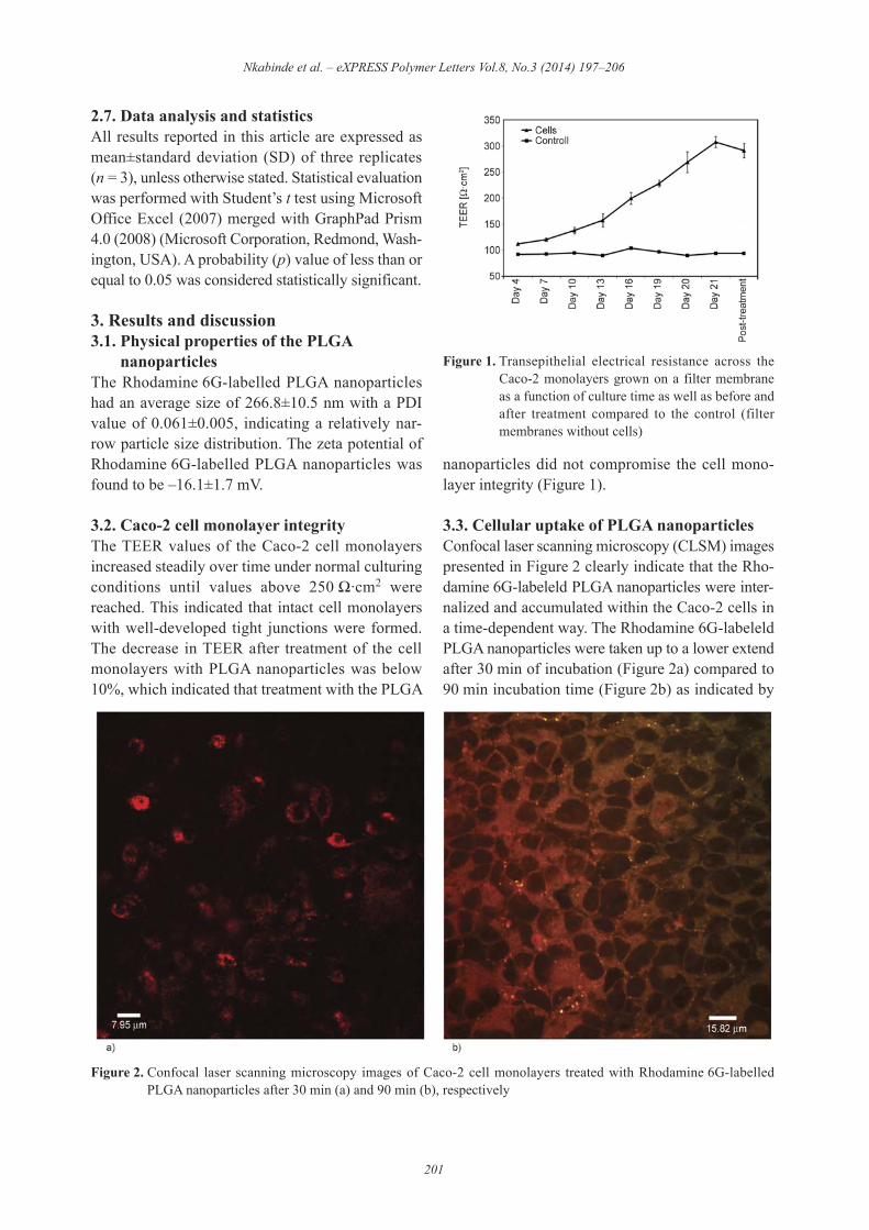
3.2. Caco-2 cell monolayer integrityThe TEER values of the Caco-2 cell monolayersincreased steadily over time under normal culturingconditions until values above 250 $·cm2 werereached. This indicated that intact cell monolayerswith well-developed tight junctions were formed.The decrease in TEER after treatment of the cellmonolayers with PLGA nanoparticles was below10%, which indicated that treatment with the PLGA
nanoparticles did not compromise the cell mono-layer integrity (Figure 1).
3.3. Cellular uptake of PLGA nanoparticlesConfocal laser scanning microscopy (CLSM) imagespresented in Figure 2 clearly indicate that the Rho-damine 6G-labeleld PLGA nanoparticles were inter-nalized and accumulated within the Caco-2 cells ina time-dependent way. The Rhodamine 6G-labeleldPLGA nanoparticles were taken up to a lower extendafter 30 min of incubation (Figure 2a) compared to90 min incubation time (Figure 2b) as indicated by
Nkabinde et al. – eXPRESS Polymer Letters Vol.8, No.3 (2014) 197–206
201
Figure 1. Transepithelial electrical resistance across theCaco-2 monolayers grown on a filter membraneas a function of culture time as well as before andafter treatment compared to the control (filtermembranes without cells)
Figure 2. Confocal laser scanning microscopy images of Caco-2 cell monolayers treated with Rhodamine 6G-labelledPLGA nanoparticles after 30 min (a) and 90 min (b), respectively
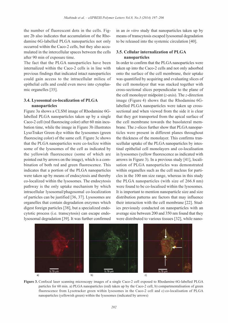
the number of fluorescent dots in the cells. Fig-ure 2b also indicates that accumulation of the Rho-damine 6G-labelled PLGA nanoparticles not onlyoccurred within the Caco-2 cells, but they also accu-mulated in the intercellular spaces between the cellsafter 90 min of exposure time.The fact that the PLGA nanoparticles have beeninternalized within the Caco-2 cells is in line withprevious findings that indicated intact nanoparticlescould gain access to the intracellular milieu ofepithelial cells and could even move into cytoplas-mic organelles [35].
3.4. Lysosomal co-localization of PLGAnanoparticles
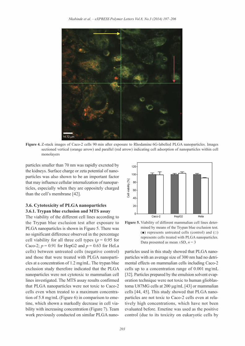
Figure 3a shows a CLSM image of Rhodamine 6G-labelled PLGA nanoparticles taken up by a singleCaco-2 cell (red fluorescing color) after 60 min incu-bation time, while the image in Figure 3b illustratesLysoTraker Green dye within the lysosomes (greenfluorescing color) of the same cell. Figure 3c showsthat the PLGA nanoparticles were co-loclize withinsome of the lysosomes of the cell as indicated bythe yellowish fluorescence (some of which arepointed out by arrows on the image), which is a com-bination of both red and green fluorescence. Thisindicates that a portion of the PLGA nanoparticleswere taken up by means of endocytosis and therebyco-localized within the lysosomes. The endocytosispathway is the only uptake mechanism by whichintracellular lysosomal/phagosomal co-localizationof particles can be justified [36, 37]. Lysosomes areorganelles that contain degradation enzymes whichdigest foreign particles [38], but a specialized endo-cytotic process (i.e. transcytosis) can escape endo-lysosomal degradation [39]. It was further confirmed
in an in vitro study that nanoparticles taken up bymeans of transcytosis escaped lysosomal degradationto be released into the systemic circulation [40].
3.5. Cellular internalization of PLGAnanoparticles
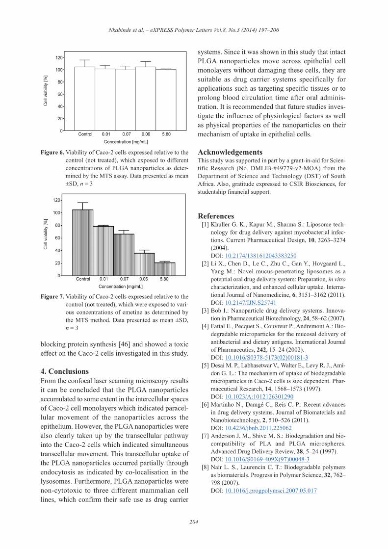
In order to confirm that the PLGA nanoparticles weretaken up into the Caco-2 cells and not only adsorbedonto the surface of the cell membrane, their uptakewas quantified by acquiring and evaluating slices ofthe cell monolayer that was stacked together withcross-sectional slices perpendicular to the plane ofthe cell monolayer midpoint (z-axis). The z-directionimage (Figure 4) shows that the Rhodamine 6G-labelled PLGA nanoparticles were taken up cross-sectional and when viewed from the side it is clearthat they got transported from the apical surface ofthe cell membrane towards the basolateral mem-brane. The z-slices further show that PLGA nanopar-ticles were present in different planes throughoutthe thickness of the monolayer. This confirms tran-scellular uptake of the PLGA nanoparticles by intes-tinal epithelial cell monolayers and co-localisationin lysosomes (yellow fluorescence as indicated witharrows in Figure 3). In a previous study [41], locali-sation of PLGA nanoparticles was demonstratedwithin organelles such as the cell nucleus for parti-cles in the 100 nm size range, whereas in this studythe PLGA nanoparticles (with size of 266.8 nm)were found to be co-localised within the lysosomes.It is important to mention nanoparticle size and sizedistribution patterns are factors that may influencetheir interaction with the cell membrane [22]. Stud-ies previously conducted on nanoparticles with anaverage size between 200 and 350 nm found that theywere distributed to various tissues [32], while nano -
Nkabinde et al. – eXPRESS Polymer Letters Vol.8, No.3 (2014) 197–206
202
Figure 3. Confocal laser scanning microscopy images of a single Caco-2 cell exposed to Rhodamine 6G-labelled PLGAparticles for 60 min. a) PLGA nanoparticles (red) taken up by the Caco-2 cell, b) compartmentalisation of greenfluorescence from Lysotracker green within lysosomes in the Caco-2 cell and c) co-localisation of PLGAnanoparticles (yellowish green) within the lysosomes (indicated by arrows)
particles smaller than 70 nm was rapidly excreted bythe kidneys. Surface charge or zeta potential of nano -particles was also shown to be an important factorthat may influence cellular internalization of nanopar-ticles, especially when they are oppositely chargedthan the cell’s membrane [42].
3.6. Cytotoxicity of PLGA nanoparticles3.6.1. Trypan blue exclusion and MTS assayThe viability of the different cell lines according tothe Trypan blue exclusion test after exposure toPLGA nanoparticles is shown in Figure 5. There wasno significant difference observed in the percentagecell viability for all three cell types (p = 0.95 forCaco-2; p = 0.91 for HepG2 and p = 0.63 for HeLacells) between untreated cells (negative control)and those that were treated with PLGA nanoparti-cles at a concentration of 1.2 mg/mL. The trypan blueexclusion study therefore indicated that the PLGAnanoparticles were not cytotoxic to mammalian celllines investigated. The MTS assay results confirmedthat PLGA nanoparticles were not toxic to Caco-2cells even when treated to a maximum concentra-tion of 5.8 mg/mL (Figure 6) in comparison to eme-tine, which shown a markedly decrease in cell via-bility with increasing concentration (Figure 7). Teamwork previously conducted on similar PLGA nano -
particles used in this study showed that PLGA nano -particles with an average size of 300 nm had no detri-mental effects on mammalian cells including Caco-2cells up to a concentration range of 0.001 mg/mL[32]. Particles prepared by the emulsion solvent evap-oration technique were not toxic to human glioblas-toma U87MG cells at 200 &g/mL [43] or mammaliancells [44, 45]. This study showed that PLGA nano -particles are not toxic to Caco-2 cells even at rela-tively high concentrations, which have not beenevaluated before. Emetine was used as the positivecontrol (due to its toxicity on eukaryotic cells by
Nkabinde et al. – eXPRESS Polymer Letters Vol.8, No.3 (2014) 197–206
203
Figure 4. Z-stack images of Caco-2 cells 90 min after exposure to Rhodamine 6G-labelled PLGA nanoparticles. Imagessectioned vertical (orange arrow) and parallel (red arrow) indicating cell adsorption of nanoparticles within cellmonolayers
Figure 5. Viability of different mammalian cell lines deter-mined by means of the Trypan blue exclusion test.(') represents untreated cells (control) and (()represents cells treated with PLGA nanoparticles.Data presented as mean ±SD, n = 3
blocking protein synthesis [46] and showed a toxiceffect on the Caco-2 cells investigated in this study.
4. ConclusionsFrom the confocal laser scanning microscopy resultsit can be concluded that the PLGA nanoparticlesaccumulated to some extent in the intercellular spacesof Caco-2 cell monolayers which indicated paracel-lular movement of the nanoparticles across theepithelium. However, the PLGA nanoparticles werealso clearly taken up by the transcellular pathwayinto the Caco-2 cells which indicated simultaneoustranscellular movement. This transcellular uptake ofthe PLGA nanoparticles occurred partially throughendocytosis as indicated by co-localisation in thelysosomes. Furthermore, PLGA nanoparticles werenon-cytotoxic to three different mammalian celllines, which confirm their safe use as drug carrier
systems. Since it was shown in this study that intactPLGA nanoparticles move across epithelial cellmonolayers without damaging these cells, they aresuitable as drug carrier systems specifically forapplications such as targeting specific tissues or toprolong blood circulation time after oral adminis-tration. It is recommended that future studies inves-tigate the influence of physiological factors as wellas physical properties of the nanoparticles on theirmechanism of uptake in epithelial cells.
AcknowledgementsThis study was supported in part by a grant-in-aid for Scien-tific Research (No. DMLIB-#49779-v2-MOA) from theDepartment of Science and Technology (DST) of SouthAfrica. Also, gratitude expressed to CSIR Biosciences, forstudentship financial support.
References [1] Khuller G. K., Kapur M., Sharma S.: Liposome tech-
nology for drug delivery against mycobacterial infec-tions. Current Pharmaceutical Design, 10, 3263–3274(2004).DOI: 10.2174/1381612043383250
[2] Li X., Chen D., Le C., Zhu C., Gan Y., Hovgaard L.,Yang M.: Novel mucus-penetrating liposomes as apotential oral drug delivery system: Preparation, in vitrocharacterization, and enhanced cellular uptake. Interna-tional Journal of Nanomedicine, 6, 3151–3162 (2011).DOI: 10.2147/IJN.S25741
[3] Bob I.: Nanoparticle drug delivery systems. Innova-tion in Pharmaceutical Biotechnology, 24, 58–62 (2007).
[4] Fattal E., Pecquet S., Couvreur P., Andremont A.: Bio -degradable microparticles for the mucosal delivery ofantibacterial and dietary antigens. International Journalof Pharmaceutics, 242, 15–24 (2002).DOI: 10.1016/S0378-5173(02)00181-3
[5] Desai M. P., Labhasetwar V., Walter E., Levy R. J., Ami-don G. L.: The mechanism of uptake of biodegradablemicroparticles in Caco-2 cells is size dependent. Phar-maceutical Research, 14, 1568–1573 (1997).DOI: 10.1023/A:1012126301290
[6] Martinho N., Damgé C., Reis C. P.: Recent advancesin drug delivery systems. Journal of Biomaterials andNanobiotechnology, 2, 510–526 (2011).DOI: 10.4236/jbnb.2011.225062
[7] Anderson J. M., Shive M. S.: Biodegradation and bio-compatibility of PLA and PLGA microspheres.Advanced Drug Delivery Review, 28, 5–24 (1997).DOI: 10.1016/S0169-409X(97)00048-3
[8] Nair L. S., Laurencin C. T.: Biodegradable polymersas biomaterials. Progress in Polymer Science, 32, 762–798 (2007).DOI: 10.1016/j.progpolymsci.2007.05.017
Nkabinde et al. – eXPRESS Polymer Letters Vol.8, No.3 (2014) 197–206
204
Figure 6. Viability of Caco-2 cells expressed relative to thecontrol (not treated), which exposed to differentconcentrations of PLGA nanoparticles as deter-mined by the MTS assay. Data presented as mean±SD, n = 3
Figure 7. Viability of Caco-2 cells expressed relative to thecontrol (not treated), which were exposed to vari-ous concentrations of emetine as determined bythe MTS method. Data presented as mean ±SD,n = 3
[9] Makadia H. K., Siegel S. J.: Poly lactic-co-glycolicacid (PLGA) as biodegradable controlled drug deliv-ery carrier. Polymers, 3, 1377–1397 (2011).DOI: 10.3390/polym3031377
[10] Jain R. A.: The manufacturing techniques of variousdrug loaded biodegradable poly(lactide-co-glycolide)(PLGA) devices. Biomaterials, 21, 2475–2490 (2000).DOI: 10.1016/S0142-9612(00)00115-0
[11] Athanasiou K. A., Niederauer G. G., Agrawal C. M.:Sterilization, toxicity, biocompatibility and clinicalapplications of polylactic acid/polyglycolic acid copoly-mers. Biomaterials, 17, 93–102 (1996).DOI: 10.1016/0142-9612(96)85754-1
[12] Chakravarthi S. S., Robinson D. H., De S.: Nanopar-ticulate drug delivery systems. in ‘Nanoparticles pre-pared using natural and synthetic polymers’ (eds.:Thassu D., Deleers M., Pathak Y.) Informa Healthcare,New York, 51–60 (2007).
[13] Lewis D. H.: Controlled release of bioactive agentsfrom lactide/glycolide polymers. in ‘Biodegradablepolymers as drug delivery systems’ (eds.: Chasin M.,Langer R.) Marcel Dekker, New York, 1–41 (1990).
[14] Ishihara T., Kubota T., Choi T., Takahashi M., AyanoE., Kanazawa H., Higaki M.: Polymeric nanoparticlesencapsulating betamethasone phosphate with differentrelease profiles and stealthiness. International Journalof Pharmaceutics, 375, 148–154 (2009).DOI: 10.1016/j.ijpharm.2009.04.001
[15] Mundargi R. C., Babu V. R., Rangaswamy V., Patel P.,Aminabhavi T. M.: Nano/micro technologies for deliv-ering macromolecular therapeutics using poly(D,L-lactide-co-glycolide) and its derivatives. Journal ofControlled Release, 125, 193–209 (2008).DOI: 10.1016/j.jconrel.2007.09.013
[16] Kumari A., Yadav S. K., Yadav S. C.: Biodegradablepolymeric nanoparticles based drug delivery systems.Colloids and Surfaces B: Biointerfaces, 75, 1–18(2010).DOI: 10.1016/j.colsurfb.2009.09.001
[17] Soppimath K. S., Aminabhavi T. M., Kulkarni A. R.,Rudzinski W. E.: Biodegradable polymeric nanoparti-cles as drug delivery devices. Journal of ControlledRelease, 70, 1–20 (2001).DOI: 10.1016/S0168-3659(00)00339-4
[18] Owens D. E., Peppas N. A.: Opsonization, biodistribu-tion, and pharmacokinetics of polymeric nanoparti-cles. International Journal of Pharmaceutics, 307, 93–102 (2006).DOI: 10.1016/j.ijpharm.2005.10.010
[19] Danhier F., Feron O., Préat V.: To exploit the tumormicroenvironment: Passive and active tumor targetingof nanocarriers for anti-cancer drug delivery. Journalof Controlled Release, 148, 135–146 (2010).DOI: 10.1016/j.jconrel.2010.08.027
[20] Tahara K., Sakai T., Yamamoto H., Takeuchi H.,Hirashima N., Kawashima Y.: Improved cellular uptakeof chitosan-modified PLGA nanospheres by A549 cells.International Journal of Pharmaceutics, 382, 198–204(2009).DOI: 10.1016/j.ijpharm.2009.07.023
[21] Shenoy D. B., Amiji M. M.: Poly(ethylene oxide)-modified poly()-caprolactone) nanoparticles for tar-geted delivery of tamoxifen in breast cancer. Interna-tional Journal of Pharmaceutics, 293, 261–270 (2005).DOI: 10.1016/j.ijpharm.2004.12.010
[22] Danhier F., Ansorena E., Silva J. M., Coco R., Le Bre-ton A., Préat V.: PLGA-based nanoparticles: An over -view of biomedical applications. Journal of ControlledRelease, 161, 505–522 (2012).DOI: 10.1016/j.jconrel.2012.01.043
[23] Letendre L., Scott M., Dobson G., Hidalgo I., AungstB.: Evaluating barriers to bioavailability in vivo: Vali-dation of a technique for separately assessing gastroin-testinal absorption and hepatic extraction. Pharmaceu-tical Research, 21, 1457–1462 (2004).DOI: 10.1023/B:PHAM.0000036921.87928.72
[24] Vasir J. K., Labhasetwar V.: Quantification of the forceof nanoparticle-cell membrane interactions and itsinfluence on intracellular trafficking of nanoparticles.Biomaterials, 29, 4244–4252 (2008).DOI: 10.1016/j.biomaterials.2008.07.020
[25] Hidalgo I. J., Raub T. J., Borchardt R. T.: Characteriza-tion of the human colon carcinoma cell line (Caco-2)as a model system for intestinal epithelial permeabil-ity. Gastroenterology, 96, 736–749 (1989).
[26] Li A. P.: Screening for human ADME/Tox drug prop-erties in drug discovery. Drug Discovery Today, 6,357–366 (2001).DOI: 10.1016/S1359-6446(01)01712-3
[27] du Plessis L. H., Hamman J. H.: In vitro evaluation ofthe cytotoxic and apoptogenic properties of aloe wholeleaf and gel materials. Drug and Chemical Toxicology,in press, p.9 (2013).DOI: 10.3109/01480545.2013.834356
[28] Schoonen W. G. E. J., Westerink W. M. A., de Roos J.A. D. M., Débiton E.: Cytotoxic effects of 100 refer-ence compounds on Hep G2 and HeLa cells and of 60compounds on ECC-1 and CHO cells. I Mechanisticassays on ROS, glutathione depletion and calceinuptake. Toxicology in Vitro, 19, 505–516 (2005).DOI: 10.1016/j.tiv.2005.01.003
[29] Mosmann T.: Rapid colorimetric assay for cellulargrowth and survival: Application to proliferation andcytotoxicity assays. Journal of Immunological Meth-ods, 65, 55–63 (1983).DOI: 10.1016/0022-1759(83)90303-4
[30] Vistica D. T., Skehan P., Scudiero D., Monks A.,Pittman A., Boyd M. R.: Tetrazolium based assay forcellular viability: A critical examination of selectedparameters affecting formazan production. CancerResearch, 51, 2515–2520 (1991).
Nkabinde et al. – eXPRESS Polymer Letters Vol.8, No.3 (2014) 197–206
205
[31] Wrobel K., Claudio E., Segade F., Ramos S., Lazo K.:Measurement of cytotoxicity by propidium iodidestaining of target cell DNA: Application to the quan-tification of murine TNF-!. Journal of ImmunologicalMethods, 189, 243–249 (1996).DOI: 10.1016/0022-1759(95)00253-7
[32] Semete B., Booysen L., Lemmer Y., Kalombo L.,Katata L., Verschoor J., Swai H. S.: In vivo evaluationof the biodistribution and safety of PLGA nanoparti-cles as drug delivery systems. Nanomedicine: Nano -technology, Biology and Medicine, 6, 662–671 (2010).DOI: 10.1016/j.nano.2010.02.002
[33] Artursson P., Palm K., Luthman K.: Caco-2 monolay-ers in experimental and theoretical predictions of drugtransport. Advanced Drug Delivery Reviews, 46, 27–43 (2001).DOI: 10.1016/S0169-409X(00)00128-9
[34] Hubatsch I., Ragnarsson E. E. G., Artursson P.: Deter-mination of drug permeability and prediction of drugabsorption in Caco-2 monolayers. Nature Protocols, 2,2111–2119 (2007).DOI: 10.1038/nprot.2007.303
[35] Qaddoumi M. G., Ueda H., Yang J., Davda J., Lab-hasetwar V., Lee V. H.: The characteristics and mecha-nisms of uptake of PLGA nanoparticles in rabbit con-junctival epithelial cell layers. Pharmaceutical Research,21, 641–648 (2004).DOI: 10.1023/B:PHAM.0000022411.47059.76
[36] Panyam J., Labhasetwar V.: Targeting intracellular tar-gets. Current Drug Delivery, 1, 235–247 (2004).DOI: 10.2174/1567201043334768
[37] Cartiera M. S., Ferreira E. C., Caputo C., Egan M. E.,Caplan M. J., Saltzman W. M.: Partial correction ofcystic fibrosis defects with PLGA nanoparticles encap-sulating curcumin. Molecular Pharmaceutics, 7, 86–93(2009).DOI: 10.1021/mp900138a
[38] Ayala P., Lin L., Hopper S., Fukuda M., So M.: Infec-tion of epithelial cells by pathogenic neisseriae reducesthe levels of multiple lysosomal constituents. Infectionand Immunity, 66, 5001–5007 (1998).
[39] Panyam J., Zhou W-Z., Prabha S., Sahoo S. K., Lab-hasetwar V.: Rapid endo-lysosomal escape of poly(DL-lactide-co-glycolide) nanoparticles: Implications fordrug and gene delivery. The FASEB Journal, 16, 1217–1226 (2002).DOI: 10.1096/fj.02-0088com
[40] Sanderson I. R., Walker W. A.: Uptake and transport ofmacromolecules by the intestine: Possible role in clin-ical disorder (an update). Gastroenterology, 104, 622–629 (1993).
[41] Gaumet M., Gurny R., Delie F.: Localization andquantification of biodegradable particles in an intes-tinal cell model: The influence of particle size. Euro-pean Journal of Pharmaceutical Sciences, 36, 465–473(2009).DOI: 10.1016/j.ejps.2008.11.015
[42] He C., Hu Y., Yin L., Tang C., Yin C.: Effects of parti-cle size and surface charge on cellular uptake and bio -distribution of polymeric nanoparticles. Biomaterials,31, 3657–3666 (2010).DOI: 10.1016/j.biomaterials.2010.01.065
[43] Mo L., Hou L., Guo D., Xiao X., Mao P., Yang X.:Preparation and characterization of teniposide PLGAnanoparticles and their uptake in human glioblastomaU87MG cells. International Journal of Pharmaceutics,436, 815–824 (2012).DOI: 10.1016/j.ijpharm.2012.07.050
[44] Csaba N., Caamaño P., Sanchez A., Domınguez F.,Alonso M. J.: PLGA:Poloxamer and PLGA:Polox-amine blend nanoparticles: New carriers for gene deliv-ery. Biomacromolecules, 6, 271–278 (2005).DOI: 10.1021/bm049577p
[45] Mura S., Hillaireau H., Nicolas J., Le Droumaguet B.,Gueutin C., Zanna S., Tsapis N., Fattal E.: Influence ofsurface charge on the potential toxicity of PLGAnanoparticles towards Calu-3 cells. International Jour-nal of Nanomedicine, 6, 2591–2605 (2011).DOI: 10.2147/IJN.S24552
[46] Dimitrijevic S., Duncan R.: Synthesis and characteriza-tion of N-(2-Hydroxypropyl)-methacrylamide (HPMA)copolymer-emetine conjugates. Journal of Bioactiveand Compatible Polymers, 13, 165–178 (1998).DOI: 10.1177/088391159801300301
Nkabinde et al. – eXPRESS Polymer Letters Vol.8, No.3 (2014) 197–206
206