peroxynitrite-induced cytotoxicity: mechanism and opportunities for intervention
TRANSCRIPT
Short communication
Peroxynitrite-induced cytotoxicity: mechanism andopportunities for intervention
Laszlo Virag a,*, Eva Szabo b, Pal Gergely a, Csaba Szabo c,d
a Department of Medical Chemistry, Medical and Health Science Center, University of Debrecen, Bem ter 18/B, 4026 Debrecen, Hungaryb Department of Dermatology, Medical and Health Science Center, University of Debrecen, Debrecen, Hungary
c Inotek Pharmaceutical Corporation, Beverly, MA, USAd Institute of Human Physiology and Clinical Experimental Research, Semmelweis University, Budapest, Hungary
Received 15 September 2002; accepted 12 December 2002
Abstract
Peroxynitrite is formed in biological systems when superoxide and nitric oxide are produced at near equimolar ratio.
Although not a free radical by chemical nature (as it has no unpaired electron), peroxynitrite is a powerful oxidant
exhibiting a wide array of tissue damaging effects ranging from lipid peroxidation, inactivation of enzymes and ion
channels via protein oxidation and nitration to inhibition of mitochondrial respiration. Low concentrations of
peroxynitrite trigger apoptotic death, whereas higher concentrations induce necrosis with cellular energetics (ATP and
NAD) serving as switch between the two modes of cell death. Peroxynitrite also damages DNA and thus triggers the
activation of DNA repair systems. A DNA nick sensor enzyme, poly(ADP-ribose) polymerase-1 (PARP-1) also
becomes activated upon sensing DNA breakage. Activated PARP-1 cleaves NAD� into nicotinamide and ADP-ribose
and polymerizes the latter on nuclear acceptor proteins. Peroxynitrite-induced overactivation of PARP consumes
NAD� and consequently ATP culminating in cell dysfunction, apoptosis or necrosis. This cellular suicide mechanism
has been implicated among others in the pathomechanism of stroke, myocardial ischemia, diabetes and diabetes-
associated cardiovascular dysfunction. Here, we review the cytotoxic effects (apoptosis and necrosis) of peroxynitrite
focusing on the role of accelerated ADP-ribose turnover. Regulatory mechanisms of peroxynitrite-induced cytotoxicity
such as antioxidant status, calcium signalling, NFkB activation, protein phosphorylation, cellular adaptation are also
discussed.
# 2003 Published by Elsevier Science Ireland Ltd.
Keywords: Peroxynitrite; Cytotoxicity; Apoptosis; Necrosis; Poly(ADP-ribose) polymerase; Poly(ADP-ribose) glycohydrolase
1. Peroxynitrite production in biological systems
Nitric oxide (NO+) is a unique diffusible mole-
cular messenger in the vascular and nervous
system. NO+
is produced by a family of enzymes
called nitric oxide synthetases (NOS) through
enzymatic oxidation of the guanidino group of L-
* Corresponding author. Tel.: �/36-52-412-345; fax: �/36-52-
412-566.
E-mail address: [email protected] (L.v.v. Virag).
Toxicology Letters 140�/141 (2003) 113�/124
www.elsevier.com/locate/toxlet
0378-4274/03/$ - see front matter # 2003 Published by Elsevier Science Ireland Ltd.
doi:10.1016/S0378-4274(02)00508-8
arginine (Marletta et al., 1998). This occurs in twosequential monooxygenase reactions utilizing
NADPH as cosubstrate and involving the utiliza-
tion of molecular oxygen (Marletta et al., 1998).
Constitutive expression of two NOS isoforms is
responsible for a low basal level of NO+
synthesis
in neural cells (nNOS or NOS1) and endothelial
cells (eNOS or NOS3). These constitutively ex-
pressed NOS isoforms require calcium for theirenzymatic activity. Induction of the inducible
isoform (iNOS or NOS2) by cytokines and/or
bacterial products (endotoxin/LPS) has been ob-
served in virtually all cell types tested including
macrophages, dendritic cells, fibroblasts, chondro-
cytes, osteoclasts, astrocytes, epithelial cells and
results in the production of large amounts of NO
(Nathan, 1997). Mitochondria also contain aunique NO producing enzyme (mtNOS) (Ghafour-
ifar and Richter, 1997). Although enzymatic NO+
production accounts for the bulk of NO+
formed
in biological systems, alternative pathways also
exist. In the skin, for example, non-enzymatic
reduction of sweat nitrite has been shown to give
rise to NO+
production (Weller et al., 1996).
Whereas the physiological effects (e.g. vasore-laxation, neuronal signalling) of NO
+are mostly
mediated by the activation of guanylate cyclase
(Arnold et al., 1977), the mechanism of pathophy-
siological effects is much more complex. A heavily
debated feature of NO+
is its cytotoxic effect. The
controversy arises from observations reporting
both cytotoxic and cytoprotective effects of NO+
depending on variables of the assay systems used.In cases where NO
+was found cytotoxic, it was
questioned whether NO+
directly or indirectly,
through the formation of more reactive oxidative
species such as peroxynitrite exerted its cytotoxic
effects (Beckman and Koppenol, 1996). Peroxyni-
trite (ONOO�) is formed when NO+
and super-
oxide anion react in a near diffusion-limited
reaction (Beckman and Koppenol, 1996). Sourcesof superoxide include the mitochondrial respira-
tory chain where there is a constant leak of
superoxide, NADPH oxidases, xanthine oxidase
and autoxidation of several biomolecules such as
catecholamines or myoglobin. The most powerful
cellular antioxidant system protecting against the
harmful effects of superoxide is embodied by
superoxide dismutases (SOD) (CuZnSOD in thecytosol and MnSOD in the mitochondria). How-
ever, it was shown that NO+
efficiently competes
with SOD for superoxide (Beckman and Koppe-
nol, 1996). Joseph Beckman has therefore pro-
posed (Beckman and Koppenol, 1996) that under
conditions of increased NO+
production, NO+
can
outcompete SOD for superoxide resulting in
peroxynitrite (ONOO�) formation.
NO��O��2 0 ONOO�
As both excess NO+
or excess superoxidedecreases the bioavailability of peroxynitrite, equi-
molar concentrations of the radicals are ideal for
peroxynitrite formation (Radi et al., 2001). Perox-
ynitrite anion (ONOO�) is in a pH-dependent
protonation equilibrium with peroxynitrous acid
(ONOOH). Homolysis of ONOOH gives rise to
formation of the highly reactive hydroxyl radical
(+OH) mediating molecular and tissue damage
associated with peroxynitrite production (Radi et
al., 2001).
2. Peroxynitrite-induced apoptosis
Despite of its non-radical nature, peroxynitrite
is more reactive than its parent molecules. Perox-
ynitrite initiates lipid peroxidation (Radi et al.,
1991b), causes DNA breakage (Salgo et al., 1995)
and reacts with thiols (Radi et al., 1991a). Perox-
ynitrite-induced protein modifications include pro-
tein oxidation (on methionine, cysteine,
tryptophane or tyrosine residues) and nitration(of tyrosine or tryptophane residues). However,
enzymes containing a redox active transition metal
center are the prime targets of the oxidant (Beck-
man and Koppenol, 1996). Reactions of peroxyni-
trite are affected by the local pH and the
microenvironment with hydrophobic membrane
compartments favoring nitration and aqueous
environments favoring oxidation. Moreover, car-bon dioxide reacts with peroxynitrite resulting in
the formation of nitroso-peroxocarbonates (Radi
et al., 2001). The ubiquitous presence of CO2 at
high concentration may favor this reaction route.
As nitroso-peroxocarbonates divert peroxynitrite-
induced protein modifications toward nitration,
L.v.v. Virag et al. / Toxicology Letters 140�/141 (2003) 113�/124114
CO2 is now considered as key determinant ofperoxynitrite chemistry.
When peroxynitrite-induced cellular damage
reaches a level where it cannot be handled by the
repair mechanisms, cells undergo one of the basic
cell death pathways, apoptosis or necrosis. Apop-
tosis is the ‘default’ death pathway characterized,
among other parameters, by a compact morphol-
ogy, maintenance of plasma membrane integrity,mitochondrial depolarization, secondary oxidant
production, activation of caspases (cysteinyl as-
partate specific proteases ) and oligonucleosomal
DNA fragmentation (Green and Kroemer, 1998).
The first report indicating that peroxynitrite can
trigger apoptotic death came from Pryor’s labora-
tory. They have detected DNA fragmentation in
peroxynitrite treated thymocytes (Salgo et al.,1995). Later, activation of caspase-3, a key player
in the caspase cascade has also been detected in
thymocytes (Virag et al., 1998b) and HL-60 cells
(Virag and Szabo, 1998c; Lin et al., 1998). These
early reports have later been followed by a series of
publications on the cytotoxic effects of peroxyni-
trite in lymphoblastoid cells (Li et al., 2002),
dopaminergic SH-SY5Y cells (Saeki et al., 2000;Yamamoto et al., 2002), human aortic endothelial
cells (Foresti et al., 1999), osteoblasts (Reiff et al.,
2001), HaCaT keratinocytes (Szabo et al., 2001),
cardiac myocytes (Arstall et al., 1999) human and
rat islet cells (Hadjivassiliou et al., 1998). Proto-
typical apoptosis models utilize apoptosis inducers
such as tumor necrosis factor or FAS ligand acting
upon cell surface death receptors. Channeling thedeath signal from these receptors to apoptotic
effector machineries is well described (Green and
Kroemer, 1998). However, it is not quite clear,
how peroxynitrite triggers the apoptotic machin-
ery. Mitochondria are likely sites for peroxynitrite-
induced apoptosis initiation. Mitochondria are
now recognized as central organizators of apop-
tosis (Green and Kroemer, 1998). A characteristicsequence of events including opening of mitochon-
drial permeability transition pore, mitochondrial
depolarization, secondary superoxide production,
release of apoptotic mediators from the intermem-
brane space to the cytoplasm, takes place in
apoptosing cells (Green and Kroemer, 1998).
Some of the mitochondria-derived apoptogenic
factors act as nucleases (e.g. endonuclease G),nuclease activators (e.g. apoptosis-inducing factor,
cytochrome c ), or serine proteases (e.g. Omi/
HtrA2) (Fig. 1). Peroxynitrite was found to inhibit
the mitochondrial respiratory chain by inactivat-
ing complexes I�/III (Lizasoain et al., 1996).
Furthermore, adenosine nucleotide translocator,
a member of the permeability pore is also targeted
by peroxynitrite (Vieira et al., 2001). The char-acteristic mitochondrial perturbations have also
been described in peroxynitrite treated cells (Virag
et al., 1998b). The role of mitochondria in
peroxynitrite-induced apoptosis is also supported
by findings that bcl-2, a mitochondrial antiapop-
totic protein inhibits peroxynitrite-induced apop-
tosis (Virag and Szabo, 2000; Spear et al., 1998).
The cellular energetics may become compromisedby peroxynitrite also via alternative mechanisms
(e.g. inactivation of creatine kinase in cardiomyo-
cytes) which may also contribute to peroxynitrite
cytotoxicity (Mihm et al., 2001a).
Recent reports from Bauer’s laboratory indicate
a possible role for free 3-nitrotyrosine, in perox-
ynitrite-induced apoptosis (Mihm et al., 2000).
They found that preincubation of rat thoracicaorta segments with 3-nitrotyrosine resulted in
selective, concentration-dependent impairment of
acetylcholine-induced vasorelaxation indicative of
endothelial dysfunction. Moreover, nitrotyrosine
triggered DNA damage in the endothelial cells
(Mihm et al., 2000) and proved to be neurotoxic in
vivo (Mihm et al., 2001b). These data suggest that
nitrotyrosine, released from proteins nitrated byperoxynitrite, is more than a benign biomarker in
vivo, and may contribute to vascular endothelial
dysfunction and neurotoxicity through promotion
of DNA damage and/or apoptosis (Mihm et al.,
2000). Nonetheless, the exact mechanism of per-
oxynitrite-induced apoptosis initiation remains to
be investigated. The executional phase of apopto-
sis is carried out by caspases which are likely to beactivated by mitochondria-derived apoptogenic
factors in peroxynitrite treated cells (Green and
Kroemer, 1998). Caspase activation has also been
reported to occur during the course of peroxyni-
trite-induced apoptosis (Virag et al., 1998b; Virag
and Szabo, 1998c; Lin et al., 1998). A detailed
analysis of caspase activation has revealed that
L.v.v. Virag et al. / Toxicology Letters 140�/141 (2003) 113�/124 115
caspase 3-like proteases and (to a lesser extent)caspase 2, but not caspase 1 or caspase 6, are
required for peroxynitrite-induced apoptosis
(Zhuang and Simon, 2000).
3. Peroxynitrite-induced necrosis: role of
poly(ADP-ribose) polymerase-1
Whilst low concentrations of peroxynitrite trig-
ger apoptosis, higher concentrations of the oxidantcompromise the apoptotic machinery forcing the
cells to die by necrosis (Virag et al., 1998a,b;
Bonfoco et al., 1995). For a long time, necrosis was
thought to be a passive process resulting from the
inability of the cells to cope with high degree of
oxidative stress. Recently, a new paradigm has
emerged identifying an active element in oxidative
stress-induced necrosis. According to this concept,
degree of the activation of poly(ADP-ribose)
polymerase-1 (PARP-1) determines the fate of
the oxidatively-injured cells (Virag and Szabo,
2002). PARP-1 is activated by DNA strand break.
Activated PARP-1 catalyzes the cleavage of
NAD� into nicotinamide and ADP-ribose and
uses the latter to synthesize branched nucleic acid-
like polymers poly(ADP-ribose) covalently at-
tached to nuclear acceptor proteins. The branched
polymer, the size of which varies from a few to 200
ADP-ribose units, may facilitate recruitment of
DNA repair enzymes to the sites of DNA injury
(Virag and Szabo, 2002). In vivo the most abun-
dantly poly-ADP-ribosylated protein is PARP-1
itself and auto-poly(ADP-ribosylation) represents
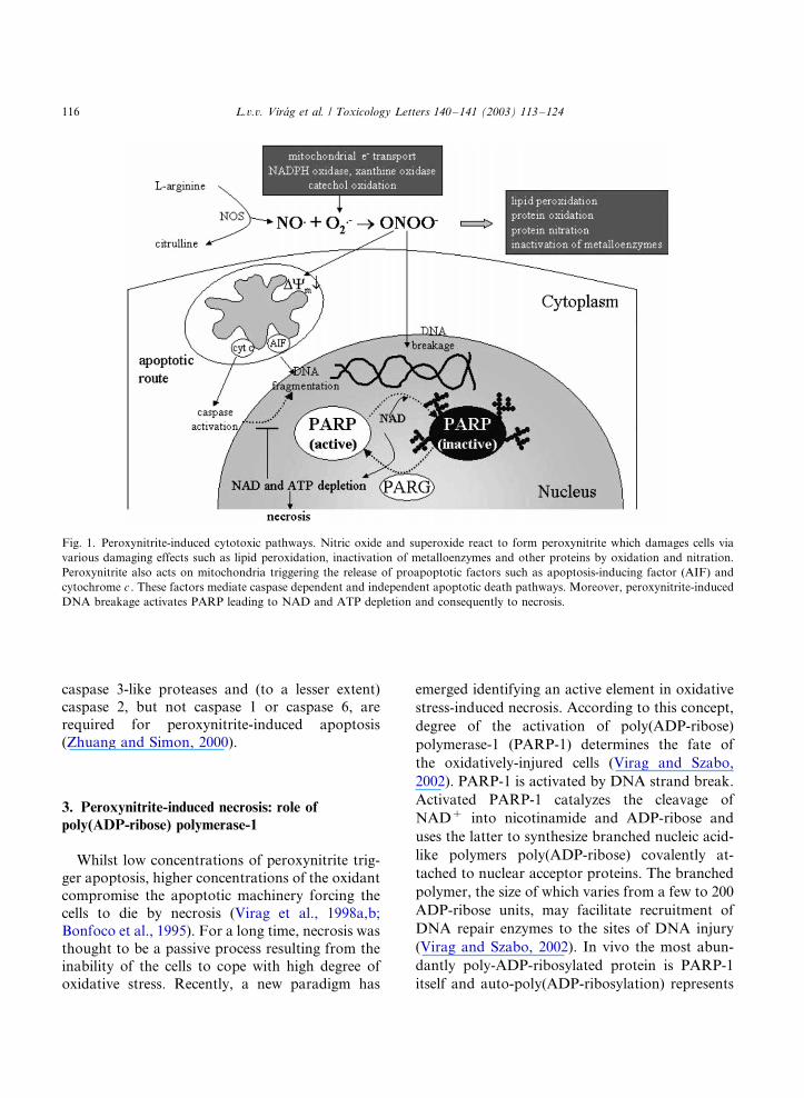
Fig. 1. Peroxynitrite-induced cytotoxic pathways. Nitric oxide and superoxide react to form peroxynitrite which damages cells via
various damaging effects such as lipid peroxidation, inactivation of metalloenzymes and other proteins by oxidation and nitration.
Peroxynitrite also acts on mitochondria triggering the release of proapoptotic factors such as apoptosis-inducing factor (AIF) and
cytochrome c . These factors mediate caspase dependent and independent apoptotic death pathways. Moreover, peroxynitrite-induced
DNA breakage activates PARP leading to NAD and ATP depletion and consequently to necrosis.
L.v.v. Virag et al. / Toxicology Letters 140�/141 (2003) 113�/124116
a major regulatory mechanism for PARP-1 result-
ing in the downregulation of the enzyme activity.
The polymer is degraded by poly(ADP-ribose)
glycohydrolase (PARG) and ADP-ribosyl protein
lyase with the latter enzyme removing the protein
proximal ADP-ribose residue (Virag and Szabo,
2002). The concerted action of PARP-1 and
PARG maintains a highly accelerated ADP-ribose
turnover in peroxynitrite treated cells. As a result,
NAD becomes depleted in the cells leading to
malfunctioning glycolysis, Krebs cycle, mitochon-
drial electron transport and eventually to ATP
depletion (Berger et al., 1986). Moreover, shortage
on ATP is exaggerated by attempts of the cells to
resynthesize NAD from ATP and nicotinamide.
The net result of this pathway is a dramatic drop
in cellular ATP (Berger et al., 1983). As the
apoptotic machinery is known to depend on
ATP (Volbracht et al., 1999; Nicotera et al.,
1998, 2000), apoptosis is incapacitated and necro-
sis takes predominance. This cellular suicide
hypothesis described by Nathan Berger (Berger
et al., 1983) has been applied by our group to
peroxynitrite cytotoxicity. We have used mouse
thymocytes (and later several other cell types, too)
to validate the role of PARP-1 in peroxynitrite-
induced cytotoxicity. Low concentrations (B/20
mM) of peroxynitrite caused apoptotic thymocyte
death characterized by phosphatidylserine expo-
sure, caspase activation and DNA fragmentation
(Virag et al., 1998b). At higher concentrations of
peroxynitrite, however, PARP activation and ATP
depletion occurred, apoptotic parameters (DNA
fragmentation, caspase activation) declined and
necrotic death occurred as indicated by the break-
down of plasma membrane integrity (Virag et al.,
1998b). Inhibition of PARP-1 by 3-aminobenza-
mide or the absence of PARP-1 in PARP-1
deficient thymocytes resulted in dramatic protec-
tion against the loss of plasma membrane integrity
(necrosis). In the same time, output of apoptotic
parameters (DNA fragmentation and caspase
activation) increased in cells treated with PARP
inhibitors or in PARP-1 deficient cells (Virag et al.,
1998b). These findings indicate that PARP-1
activation diverts the default apoptotic process
toward necrosis.
An interesting new finding of our work was toestablish that similarly to apoptosis, peroxynitri-
trite-induced, PARP-1 mediated necrotic death is
also accompanied by mitochondrial alterations
(collapse of mitochondrial membrane potential,
overproduction of superoxide and mitochondrial
membrane damage) and calcium mobilization
(Virag et al., 1998a). Inhibition or the absence of
PARP-1 provided remarkable protection fromderailment of mitochondrial functions indicating
the central role of PARP-1 in peroxynitrite-in-
duced mitochondrial perturbation (Virag et al.,
1998a).
The deterioration of cellular energetic status
may play a central role in the ‘cell death switch
role’ of PARP-1. This hypothesis is supported by
findings that cellular ATP levels determine themode of cell death (apoptosis versus necrosis)
(Bonfoco et al., 1995; Leist et al., 1997; Nicotera
et al., 1998, 2000). Moreover, a recent report from
Swanson’s laboratory supports a central role of
cellular energy homeostasis in PARP-1 mediated
cell death (Ying et al., 2002). In these experiments,
mouse cortical astrocyte and astrocyte-neuron
cocultures were treated with the DNA alkylatingagent, N -methyl-N ?-nitro-N -nitrosoguanidine
(MNNG) in order to activate PARP-1. Studies
using the 2-deoxyglucose method confirmed that
glycolytic flux was reduced by more than 90% in
MNNG-treated cultures. The addition of 5 mmol/l
of alpha-ketoglutarate, 5 mmol/l pyruvate, or
other mitochondrial substrates to the cultures after
MNNG treatment reduced cell death from ap-proximately 70% to near basal levels (Ying et al.,
2002).
4. Regulation of peroxynitrite-induced cytotoxicity
The mechanism of peroxynitrite-induced cyto-
toxicity is cell type dependent. The ratio of PARP
dependent and PARP independent pathways var-ies between different cell types with thymocytes
and other primary lymphoid cells representing one
end of the spectrum (mostly PARP independent)
and HL-60 myeloid cell line standing at the other
end (no PARP dependence). Of note, out of the
many cell lines tested in our laboratory, HL-60
L.v.v. Virag et al. / Toxicology Letters 140�/141 (2003) 113�/124 117
cells were the only ones not protected by PARPinhibitors from peroxynitrite-induced cytotoxicity.
The question arises, what the underlying principle
behind the differential peroxynitrite sensitivity of
the various cell types may be? These factors may or
maynot be linked to PARP activation.
4.1. PARP dependent factors
Thymocytes represent an ideal cellular modelfor the analysis of PARP dependent resistance
factors as in this cell type, peroxynitrite-induced
cell death is mainly PARP dependent (Virag et al.,
1998b). We have reported that TPEN, a zinc
chelator, inhibits peroxynitrite-induced PARP ac-
tivation and necrosis (Virag et al., 1999a). The
mechanism of cytoprotection is not known. How-
ever, given that two zinc finger motives areresponsible for DNA binding of PARP-1, TPEN
may interfere with this process. Furthermore,
peroxynitrite induces calcium mobilization both
from intra- and from extracellular sources and
intracellular calcium chelation protects from per-
oxynitrite-induced PARP activation and necrosis
(Virag et al., 1999b). In these experiments, calcium
chelators abolished peroxynitrite-induced DNAbreakage indicating that they may act upstream
to PARP activation. In a cell-free system, calcium
chelators did not inhibit peroxynitrite induced
DNA breakage (Virag et al., 1999b). These find-
ings support the hypothesis that calcium signalling
triggers secondary events leading to DNA damage
and subsequently PARP activation. These second-
ary events may include production of secondaryROI (reactive oxygen intermediates) in the mito-
chondria. This novel concept implies that although
peroxynitrite can directly break DNA in cell-free
system, the mechanism of DNA breakage may be
fundamentally different in a cellular environment.
A calcium-dependent, mitochondrial production
of secondary oxygen radicals has been reported in
other cellular models of oxidative stress (Guidar-elli et al., 2000a,c).
We have also identified purines
(hypoxanthine�/inosine�/adenosine) as potential
endogenous PARP inhibitors (Virag and Szabo,
2001). This observation may have implication for
ischemia reperfusion injury where these substances
reach high enough concentrations during ischemiato modulate PARP activation by ROI overpro-
duced during the reperfusion phase. Recently, we
have identified cell density signalling as a new
factor regulating peroxynitrite sensitivity of Ha-
CaT keratinocytes. We have showed that subcon-
fluent (10�/95%) cultures are more sensitive to
peroxynitrite or hydrogen peroxide-induced cell
death than confluent monolayers. The resistanceto oxidative stress provided by high cell density
involved both inhibition of caspase activation and
PARP activation but not protein kinase C signal-
ling. Our data may explain the resistance to
oxidative stress of superficial, highly differentiated
keratinocytes and may indicate that basal prolif-
erative keratinocytes are possible sensitive in vivo
targets of oxidative stress injury. By virtue of theepidermal calcium gradient (increasing calcium
concentration in baso-superficial direction), our
data also raise the possibility that calcium signal-
ling and density-dependent signalling are interre-
lated.
Recently a new regulatory element of PARP
activation has been identified. It has long been
known that PARP-1 auto-poly-ADP-ribosylatesitself leading to downregulation of enzyme activ-
ity. By removing inhibitory ADP-ribose residues
from PARP-1, PARG may reactivate PARP-1 and
thus may help maintain a high NAD/ADP-ribose
turnover. This hypothesis has recently been tested
by Swanson’s group reporting that PARG inhibi-
tion by gallotannin and nobotanin B protected
astrocytes and neuronal cells from oxidative stress(Ying and Swanson, 2000; Ying et al., 2001). We
have confirmed these data in HaCaT keratinocytes
and A549 pulmonary epithelial cells and found
similar cytoprotection by gallotannin (submitted
for publication).
Two studies have reported that peroxynitrite
induces the expression of heat shock proteins. One
of the studies has shown that the cytoprotectiveeffect of the heat shock response is related to
inhibition of PARP-1 activation (Szabo et al.,
1996b). It is worthwhile that nitric oxide and
peroxynitrite were found to have different effect
on heat shock protein 70 expression in human
monocytes with peroxynitrite inducing and nitric
oxide not affecting HSP 70 expression (Adrie et
L.v.v. Virag et al. / Toxicology Letters 140�/141 (2003) 113�/124118
al., 2000). However, the mechanism by which heatshock inhibits PARP activation and other cyto-
toxic pathways remains to be elucidated.
4.2. PARP independent factors
The overall antioxidant status obviously deter-
mines the sensitivity of cells toward peroxynitrite
toxicity. The importance of glutathione is sup-
ported by several observations. Increased glu-tathione levels, as achieved by administration of
gamma-glu-cys-ethyl ester, has been shown to
protect cortical synaptosomes from peroxynitrite-
induced damage (Drake et al., 2002). Further-
more, depletion of cellular glutathione pools by
buthionine sulfoximide sensitizes cells and animals
to peroxynitrite toxicity or peroxynitrite-mediated
inflammatory tissue injury, respectively (Cuzzo-crea et al., 1998). In an in vivo model of
myocardial ischemia, coadministration of ascorbic
acid with glutathione methyl ester (GSHme)
markedly enhanced the protective effects of
GSHme, although ascorbic acid alone had no
effect (Gao et al., 2002). The protection exerted
by the combination of GSHme and ascorbic acid
was significantly greater than that observed with 1mM GSHme alone. Moreover, treatment with
GSHme alone or GSHme plus ascorbic acid
markedly reduced myocardial nitrotyrosine levels,
suggesting that these treatments attenuated myo-
cardial peroxynitrite formation (Gao et al., 2002).
Manganese- (Szabo et al., 1996a; Ferrer-Sueta et
al., 1999) or ferrous porphyrine compounds (Shi-
manovich and Groves, 2001; Salvemini et al.,1998) often sold as superoxide dismutase mimetics
or peroxynitrite decomposition catalysts also pro-
tect from peroxynitrite. Several other antioxidants
such as ebselen (Roussyn et al., 1996; Masumoto
and Sies, 1996; Sata et al., 1997) or melatonin
(Gilad et al., 1997; Cuzzocrea et al., 1997) as well
as phytopharmacons (plant-derived antioxidants)
(Choi et al., 2002a,b; Valdez et al., 2002) have alsobeen shown to protect from the deleterious effects
of peroxynitrite.
Peroxynitrite-induced apoptotic death can be
prevented by classical apoptosis inhibitors such as
caspase inhibitors (Virag et al., 1998b; Zhuang and
Simon, 2000) or bcl-2 overexpression (Virag and
Szabo, 2000; Spear et al., 1998). Although bcl-2has been reported to provide protection against
both apoptotic and necrotic stimuli, we have
shown that it protected thymocytes from perox-
ynitrite-induced apoptotic but not against PARP-1
mediated necrotic death (Virag and Szabo, 2000).
In addition to antioxidants and direct antiapop-
totic interventions other alternatives may also exist
to modulate peroxynitrite-triggered cytotoxicpathways. The most intensively studied pathway
is the inhibition of tyrosine kinase cascades by
peroxynitrite-mediated tyrosine nitration (Beck-
man, 1996). Nitration of critical tyrosine residues
by peroxynitrite in tyrosine kinase substrates
interferes with the phosphorylation of the proteins
and may inhibit downstream signalling events
(Gow et al., 1996; Kong et al., 1996). Peroxynitritehas been shown to activate several signal transduc-
tion pathways. For example, peroxynitrite triggers
the activation of various types of kinases, the G
protein-phosphatidylinositol 3 kinase (PI3 kinase)
pathway and phospholipase A2 (Klotz et al., 2000;
Kaji et al., 2002; Guidarelli et al., 2000b). Many of
these signalling pathways have been implicated in
the regulation of cell death (Cross et al., 2000;Holmstrom and Eriksson, 2000; Sarmay, 2002).
The G receptor-coupled PI3 kinase activation
pathway for example was proposed to counteract
peroxynitrite toxicity in primary rat astrocytes, a
cell type expressing opioid receptors. Treatment of
cells with morphine significantly protected astro-
cytes from apoptosis mediated by the peroxynitrite
donor SIN-1, whereas it did not in other types ofcells including C6 glioma, RAW 264.7, and HL-60
cells (Kim et al., 2001). The effects of morphine on
SIN-1-induced cytotoxicity were inhibited by pre-
treatment with the G(i) protein inhibitor, pertussis
toxin, and the PI3 kinase inhibitors, wortmannin
and LY294002 (Kim et al., 2001). These results
suggest that morphine may protect primary rat
astrocytes from peroxynitrite-induced cytotoxicityvia the signalling cascades that involve both G
protein and PI3 kinase.
Maeda’s group has investigated the activation of
mitogen-activated protein kinase (MAP kinase) in
relation to cell death induced by peroxynitrite in
human neuroblastoma SH-SY5Y cells (Saeki et
al., 2000). Exposure of the cells to peroxynitrite
L.v.v. Virag et al. / Toxicology Letters 140�/141 (2003) 113�/124 119
caused transient increase in MAP kinase activity,and resulted in cell death. PD98059, a selective
inhibitor of MAP kinase kinase, reduced perox-
ynitrite-induced cell death suggesting that activa-
tion of MAP kinase may be involved in cell death
induced by peroxynitrite (Saeki et al., 2000).
Furthermore, stimulation of several growth factor
receptors for example by insulin like growth factor
(Saeki et al., 2002), acidic fibroblast growth factor(Reiff et al., 2001), fibroblast growth factor-1
(Spear et al., 1998) or nerve growth factor (Spear
et al., 1997, 1998) has also been found to modulate
(in most cases to protect from) peroxynitrite
toxicity. In the case of epidermal growth factor
receptor (EGFR), peroxynitrite was found to
crosslink the receptors in A431 epidermoid carci-
noma cells resulting in dimer formation. CovalentEGFR dimerization by peroxynitrite probably
involved intermolecular dityrosine cross-linking
and was enhanced after receptor activation with
epidermal growth factor. Furthermore, irreversi-
bly cross-linked EGFR was more extensively
tyrosine-phosphorylated compared with the
monomeric form. However, exposure of A431 cells
to peroxynitrite markedly reduced the kinetics oftyrosine phosphorylation of a downstream EGFR
substrate, phospholipase C-gamma1. This study
indicates that peroxynitrite may also interfere with
tyrosine kinase pathways with a mechanism dif-
ferent from tyrosine nitration.
An interesting issue is the potential regulatory
role of NFkB in peroxynitrite cytotoxicity. NFkB
is a redox-sensitive transcription factor regulatingthe expression of various inflammatory mediators
(Janssen-Heininger et al., 2000). The antiapoptotic
effects of NFkB activation are also well documen-
ted (Aggarwal, 2000; Mattson et al., 1997), how-
ever, the outcome (death promotion or
prevention) is cell type and stimulus dependent
(Bours et al., 2000). The parent molecules of
peroxynitrite appear to regulate NFkB activationin an opposing manner with superoxide (likely via
hydrogen peroxide formation) activating and ni-
tric oxide inhibiting NFkB activation (Schmidt et
al., 1995; von Knethen et al., 1999; Marshall and
Stamler, 2002). Inhibition of NFkB activation by
NO-mediated nitrosylation has*/at least in
part*/been made responsible for the proapoptotic
effect of NO (Marshall and Stamler, 2002).Furthermore, immunoprecipitation studies
showed that NO stabilized the NF-kappa B
inhibitor, I kappa B alpha, by preventing its
degradation from NF-kappa B (Peng et al.,
1995). NO also increased the mRNA expression
of I kappa B alpha (Peng et al., 1995). The
contrasting roles of superoxide and NO on
NFkB activation point toward the importance ofunderstanding conditions regulating peroxynitrite
formation. Peroxynitrite itself has also been found
to activate NFkB (Matata and Galinanes, 2002).
However, the role of NFkB in the regulation of
peroxynitrite-induced cell death has not yet been
investigated in detail. Only one study addressed
the issue and found that peroxynitrite treatment
did not activate NFkB in IEC-6 enterocytes butinhibition of NFkB by transfection with AdIkB, a
mutated IkB functioning as a superrepressor of
NFkB activation, significantly enhanced peroxy-
nitrite-induced apoptosis in IEC-6 cells (Potoka et
al., 2002).
Chronic exposure of cells to sublethal peroxyni-
trite concentrations (e.g. in inflammation) may
also allow cells to develop adaptive responses tooxidative stress. This kind of regulation may
involve upregulation of defense proteins/antioxi-
dant enzymes. In A549 cells, peroxynitrite has
been shown to trigger expression of mRNA for
MnSOD, a key antioxidant enzyme (Jackson et al.,
1998). However, MnSOD protein and enzyme
activity were not changed. In Dr. Ischiropoulos’s
lab, a peroxynitrite resistant cell line has beengenerated by repeated exposure of the cells to
sublethal concentrations of peroxynitrite. DNA
chip technology will permit large-scale analysis of
changes in gene expression pattern of peroxynitrite
resistant cultures and may provide insight into the
mechanism of cellular adaptation to oxidative
stress situations.
5. Conclusions
Peroxynitrite has been implicated in the patho-
mechanism of various diseases. However, the lack
of specific tools to unequivocally verify whether
peroxynitrite is indeed produced in all of these
L.v.v. Virag et al. / Toxicology Letters 140�/141 (2003) 113�/124120
conditions and is responsible for at least part ofthe tissue injury has put peroxynitrite in the center
of heated debates. Nonetheless, as evidence sup-
porting the pathophysiological role of peroxyni-
trite is constantly increasing, targeting
peroxynitrite-induced cytotoxic pathways is now
accepted as a viable strategy to alleviate disease
signs in numerous diseases. Which one of the
above listed interventions or combinations of themprovides the highest therapeutic benefit with the
least risk remains to be seen.
Acknowledgements
The work in the authors’ laboratory is sup-
ported by grants from the Hungarian NationalScience Research Fund (OTKA T035182,
T037210), from the Hungarian Ministry of Health
(ETT-046/2001), from the National Institutes of
Health (RO1GM60915) and from the Hungarian
Ministry of Education (BIO-00002/2002). L.V. is
supported by a Bolyai Fellowship from the Hun-
garian Academy of Sciences.
References
Adrie, C., Richter, C., Bachelet, M., Banzet, N., Francois, D.,
Dinh-Xuan, A.T., Dhainaut, J.F., Polla, B.S., Richard,
M.J., 2000. Contrasting effects of NO and peroxynitrites on
HSP70 expression and apoptosis in human monocytes. Am.
J. Physiol. Cell Physiol. 279, C452�/C460.
Aggarwal, B.B., 2000. Apoptosis and nuclear factor-kappa B: a
tale of association and dissociation. Biochem. Pharmacol.
60, 1033�/1039.
Arnold, W.P., Mittal, C.K., Katsuki, S., Murad, F., 1977.
Nitric oxide activates guanylate cyclase and increases
guanosine 3?:5?-cyclic monophosphate levels in various
tissue preparations. Proc. Natl. Acad. Sci. USA 74, 3203�/
3207.
Arstall, M.A., Sawyer, D.B., Fukazawa, R., Kelly, R.A., 1999.
Cytokine-mediated apoptosis in cardiac myocytes: the role
of inducible nitric oxide synthase induction and peroxyni-
trite generation. Circ. Res. 85, 829�/840.
Beckman, J.S., 1996. Oxidative damage and tyrosine nitration
from peroxynitrite. Chem. Res. Toxicol. 9, 836�/844.
Beckman, J.S., Koppenol, W.H., 1996. Nitric oxide, super-
oxide, and peroxynitrite: the good, the bad, and ugly. Am. J.
Physiol. 271, C1424�/C1437.
Berger, N.A., Sims, J.L., Catino, D.M., Berger, S.J., 1983.
Poly(ADP-ribose) polymerase mediates the suicide response
to massive DNA damage: studies in normal and DNA-
repair defective cells. Princess Takamatsu Symp. 13, 219�/
226.
Berger, S.J., Sudar, D.C., Berger, N.A., 1986. Metabolic
consequences of DNA damage: DNA damage induces
alterations in glucose metabolism by activation of poly
(ADP-Ribose) polymerase. Biochem. Biophys. Res. Com-
mun. 134, 227�/232.
Bonfoco, E., Krainc, D., Ankarcrona, M., Nicotera, P., Lipton,
S.A., 1995. Apoptosis and necrosis: two distinct events
induced, respectively, by mild and intense insults with N-
methyl-D-aspartate or nitric oxide/superoxide in cortical cell
cultures. Proc. Natl. Acad. Sci. USA 92, 7162�/7166.
Bours, V., Bentires-Alj, M., Hellin, A.C., Viatour, P., Robe, P.,
Delhalle, S., Benoit, V., Merville, M.P., 2000. Nuclear
factor-kappa B, cancer, and apoptosis. Biochem. Pharma-
col. 60, 1085�/1089.
Choi, H.R., Choi, J.S., Han, Y.N., Bae, S.J., Chung, H.Y.,
2002a. Peroxynitrite scavenging activity of herb extracts.
Phytother. Res. 16, 364�/367.
Choi, J.J., Oh, Y.K., Kim, H.S., Kim, H.C., Ko, K.H., Kim,
W.K., 2002b. Mimosine prevents the death of glucose-
deprived immunostimulated astrocytes by scavenging per-
oxynitrite. Glia 39, 37�/46.
Cross, T.G., Scheel-Toellner, D., Henriquez, N.V., Deacon, E.,
Salmon, M., Lord, J.M., 2000. Serine/threonine protein
kinases and apoptosis. Exp. Cell Res. 256, 34�/41.
Cuzzocrea, S., Zingarelli, B., Gilad, E., Hake, P., Salzman,
A.L., Szabo, C., 1997. Protective effect of melatonin in
carrageenan-induced models of local inflammation: rela-
tionship to its inhibitory effect on nitric oxide production
and its peroxynitrite scavenging activity. J. Pineal Res. 23,
106�/116.
Cuzzocrea, S., Zingarelli, B., O?Connor, M., Salzman, A.L.,
Szabo, C., 1998. Effect of L-buthionine-(S,R)-sulphoximine,
an inhibitor of gamma-glutamylcysteine synthetase on
peroxynitrite- and endotoxic shock-induced vascular failure.
Br. J. Pharmacol. 123, 525�/537.
Drake, J., Kanski, J., Varadarajan, S., Tsoras, M., Butterfield,
D.A., 2002. Elevation of brain glutathione by gamma-
glutamylcysteine ethyl ester protects against peroxynitrite-
induced oxidative stress. J. Neurosci. Res. 68, 776�/784.
Ferrer-Sueta, G., Batinic-Haberle, I., Spasojevic, I., Fridovich,
I., Radi, R., 1999. Catalytic scavenging of peroxynitrite by
isomeric Mn(III) N-methylpyridylporphyrins in the pre-
sence of reductants. Chem. Res. Toxicol. 12, 442�/449.
Foresti, R., Sarathchandra, P., Clark, J.E., Green, C.J.,
Motterlini, R., 1999. Peroxynitrite induces haem oxyge-
nase-1 in vascular endothelial cells: a link to apoptosis.
Biochem. J. 339 (Pt 3), 729�/736.
Gao, F., Yao, C.L., Gao, E., Mo, Q.Z., Yan, W.L., McLaugh-
lin, R., Lopez, B.L., Christopher, T.A., Ma, X.L., 2002.
Enhancement of glutathione cardioprotection by ascorbic
acid in myocardial reperfusion injury. J. Pharmacol. Exp.
Ther. 301, 543�/550.
Ghafourifar, P., Richter, C., 1997. Nitric oxide synthase
activity in mitochondria. FEBS Lett. 418, 291�/296.
L.v.v. Virag et al. / Toxicology Letters 140�/141 (2003) 113�/124 121
Gilad, E., Cuzzocrea, S., Zingarelli, B., Salzman, A.L., Szabo,
C., 1997. Melatonin is a scavenger of peroxynitrite. Life Sci.
60, PL169�/PL174.
Gow, A.J., Duran, D., Malcolm, S., Ischiropoulos, H., 1996.
Effects of peroxynitrite-induced protein modifications on
tyrosine phosphorylation and degradation. FEBS Lett. 385,
63�/66.
Green, D., Kroemer, G., 1998. The central executioners of
apoptosis: caspases or mitochondria? Trends Cell Biol. 8,
267�/271.
Guidarelli, A., Fiorani, M., Cantoni, O., 2000a. Calcium-
dependent mitochondrial formation of species promoting
strand scission of genomic DNA in U937 cells exposed to
tert-butylhydroperoxide: the role of arachidonic acid. Free
Radical Res. 33, 477�/487.
Guidarelli, A., Palomba, L., Cantoni, O., 2000b. Peroxynitrite-
mediated release of arachidonic acid from PC12 cells. Br. J.
Pharmacol. 129, 1539�/1541.
Guidarelli, A., Tommasini, I., Fiorani, M., Cantoni, O., 2000c.
Essential role of the mitochondrial respiratory chain in
peroxynitrite-induced strand scission of genomic DNA.
IUBMB Life 50, 195�/201.
Hadjivassiliou, V., Green, M.H., James, R.F., Swift, S.M.,
Clayton, H.A., Green, I.C., 1998. Insulin secretion, DNA
damage, and apoptosis in human and rat islets of Langer-
hans following exposure to nitric oxide, peroxynitrite, and
cytokines. Nitric Oxide 2, 429�/441.
Holmstrom, T.H., Eriksson, J.E., 2000. Phosphorylation-based
signaling in Fas receptor-mediated apoptosis. Crit. Rev.
Immunol. 20, 121�/152.
Jackson, R.M., Parish, G., Helton, E.S., 1998. Peroxynitrite
modulates MnSOD gene expression in lung epithelial cells.
Free Radical Biol. Med. 25, 463�/472.
Janssen-Heininger, Y.M., Poynter, M.E., Baeuerle, P.A., 2000.
Recent advances towards understanding redox mechanisms
in the activation of nuclear factor kappaB. Free Radical
Biol. Med. 28, 1317�/1327.
Kaji, T., Kaieda, I., Hisatsune, T., Kaminogawa, S., 2002. 3-
Morpholinosydnonimine hydrochloride induces p53-depen-
dent apoptosis in murine primary neural cells: a critical role
for p21(ras)-MAPK-p19(ARF) pathway. Nitric Oxide 6,
125�/134.
Kim, M.S., Cheong, Y.P., So, H.S., Lee, K.M., Kim, T.Y., Oh,
J., Chung, Y.T., Son, Y., Kim, B.R., Park, R., 2001.
Protective effects of morphine in peroxynitrite-induced
apoptosis of primary rat neonatal astrocytes: potential
involvement of G protein and phosphatidylinositol 3-kinase
(PI3 kinase). Biochem. Pharmacol. 61, 779�/786.
Klotz, L.O., Schieke, S.M., Sies, H., Holbrook, N.J., 2000.
Peroxynitrite activates the phosphoinositide 3-kinase/Akt
pathway in human skin primary fibroblasts. Biochem. J. 352
(Pt 1), 219�/225.
Kong, S.K., Yim, M.B., Stadtman, E.R., Chock, P.B., 1996.
Peroxynitrite disables the tyrosine phosphorylation regula-
tory mechanism: lymphocyte-specific tyrosine kinase fails to
phosphorylate nitrated cdc2(6-20)NH2 peptide. Proc. Natl.
Acad. Sci. USA 93, 3377�/3382.
Leist, M., Single, B., Castoldi, A.F., Kuhnle, S., Nicotera, P.,
1997. Intracellular adenosine triphosphate (ATP) concen-
tration: a switch in the decision between apoptosis and
necrosis. J. Exp. Med. 185, 1481�/1486.
Li, C.Q., Trudel, L.J., Wogan, G.N., 2002. Genotoxicity,
mitochondrial damage, and apoptosis in human lympho-
blastoid cells exposed to peroxynitrite generated from SIN-
1. Chem. Res. Toxicol. 15, 527�/535.
Lin, K.T., Xue, J.Y., Lin, M.C., Spokas, E.G., Sun, F.F.,
Wong, P.Y., 1998. Peroxynitrite induces apoptosis of HL-60
cells by activation of a caspase-3 family protease. Am. J.
Physiol. 274, C855�/C860.
Lizasoain, I., Moro, M.A., Knowles, R.G., Darley-Usmar, V.,
Moncada, S., 1996. Nitric oxide and peroxynitrite exert
distinct effects on mitochondrial respiration which are
differentially blocked by glutathione or glucose. Biochem.
J. 314, 877�/880.
Marletta, M.A., Hurshman, A.R., Rusche, K.M., 1998. Cata-
lysis by nitric oxide synthase. Curr. Opin. Chem. Biol. 2,
656�/663.
Marshall, H.E., Stamler, J.S., 2002. Nitrosative stress-induced
apoptosis through inhibition of NF-kappa B. J. Biol. Chem.
277, 34233�/34238.
Masumoto, H., Sies, H., 1996. The reaction of ebselen with
peroxynitrite. Chem. Res. Toxicol. 9, 262�/267.
Matata, B.M., Galinanes, M., 2002. Peroxynitrite is an essential
component of cytokines production mechanism in human
monocytes through modulation of nuclear factor-kappa B
DNA binding activity. J. Biol. Chem. 277, 2330�/2335.
Mattson, M.P., Goodman, Y., Luo, H., Fu, W., Furukawa, K.,
1997. Activation of NF-kappaB protects hippocampal
neurons against oxidative stress-induced apoptosis: evidence
for induction of manganese superoxide dismutase and
suppression of peroxynitrite production and protein tyro-
sine nitration. J. Neurosci. Res. 49, 681�/697.
Mihm, M.J., Coyle, C.M., Schanbacher, B.L., Weinstein, D.M.,
Bauer, J.A., 2001a. Peroxynitrite induced nitration and
inactivation of myofibrillar creatine kinase in experimental
heart failure. Cardiovasc. Res. 49, 798�/807.
Mihm, M.J., Jing, L., Bauer, J.A., 2000. Nitrotyrosine causes
selective vascular endothelial dysfunction and DNA da-
mage. J. Cardiovasc. Pharmacol. 36, 182�/187.
Mihm, M.J., Schanbacher, B.L., Wallace, B.L., Wallace, L.J.,
Uretsky, N.J., Bauer, J.A., 2001b. Free 3-nitrotyrosine
causes striatal neurodegeneration in vivo. J. Neurosci. 21,
RC149.
Nathan, C., 1997. Inducible nitric oxide synthase: what
difference does it make? J. Clin. Invest. 100, 2417�/2423.
Nicotera, P., Leist, M., Ferrando-May, E., 1998. Intracellular
ATP, a switch in the decision between apoptosis and
necrosis. Toxicol. Lett. 102�/103, 139�/142.
Nicotera, P., Leist, M., Fava, E., Berliocchi, L., Volbracht, C.,
2000. Energy requirement for caspase activation and
neuronal cell death. Brain Pathol. 10, 276�/282.
Peng, H.B., Libby, P., Liao, J.K., 1995. Induction and
stabilization of I kappa B alpha by nitric oxide mediates
L.v.v. Virag et al. / Toxicology Letters 140�/141 (2003) 113�/124122
inhibition of NF-kappa B. J. Biol. Chem. 270, 14214�/
14219.
Potoka, D.A., Upperman, J.S., Nadler, E.P., Wong, C.T.,
Zhou, X., Zhang, X.R., Ford, H.R., 2002. NF-kappaB
inhibition enhances peroxynitrite-induced enterocyte apop-
tosis. J. Surg. Res. 106, 7�/14.
Radi, R., Beckman, J.S., Bush, K.M., Freeman, B.A., 1991a.
Peroxynitrite oxidation of sulfhydryls. The cytotoxic poten-
tial of superoxide and nitric oxide. J. Biol. Chem. 266,
4244�/4250.
Radi, R., Beckman, J.S., Bush, K.M., Freeman, B.A., 1991b.
Peroxynitrite-induced membrane lipid peroxidation: the
cytotoxic potential of superoxide and nitric oxide. Arch.
Biochem. Biophys. 288, 481�/487.
Radi, R., Peluffo, G., Alvarez, M.N., Naviliat, M., Cayota, A.,
2001. Unraveling peroxynitrite formation in biological
systems. Free Radical Biol. Med. 30, 463�/488.
Reiff, D.A., Kelpke, S., Rue, L., III, Thompson, J.A., 2001.
Acidic fibroblast growth factor attenuates the cytotoxic
effects of peroxynitrite in primary human osteoblast pre-
cursors. J. Trauma 50, 433�/438.
Roussyn, I., Briviba, K., Masumoto, H., Sies, H., 1996.
Selenium-containing compounds protect DNA from sin-
gle-strand breaks caused by peroxynitrite. Arch. Biochem.
Biophys. 330, 216�/218.
Saeki, M., Kamisaki, Y., Maeda, S., 2000. Involvement of
mitogen-activated protein kinase in peroxynitrite-induced
cell death of human neuroblastoma SH-SY5Y cells. Neu-
rosci. Res. 38, 213�/216.
Saeki, M., Maeda, S., Wada, K., Kamisaki, Y., 2002. Insulin-
like growth factor-1 protects peroxynitrite-induced cell
death by preventing cytochrome c -induced caspase-3 acti-
vation. J. Cell Biochem. 84, 708�/716.
Salgo, M.G., Squadrito, G.L., Pryor, W.A., 1995. Peroxynitrite
causes apoptosis in rat thymocytes. Biochem. Biophys. Res.
Commun. 215, 1111�/1118.
Salvemini, D., Wang, Z.Q., Stern, M.K., Currie, M.G., Misko,
T.P., 1998. Peroxynitrite decomposition catalysts: therapeu-
tics for peroxynitrite-mediated pathology. Proc. Natl. Acad.
Sci. USA 95, 2659�/2663.
Sarmay, G., 2002. Phosphatidylinositol 3-kinase, phosphoino-
sitides and apoptosis. Polyphosphoinositol phosphatase and
apoptosis. Subcell. Biochem. 36, 309�/333.
Sata, N., Klonowski-Stumpe, H., Han, B., Haussinger, D.,
Niederau, C., 1997. Cytotoxicity of peroxynitrite in rat
pancreatic acinar AR4-2J cells. Pancreas 15, 278�/284.
Schmidt, K.N., Amstad, P., Cerutti, P., Baeuerle, P.A., 1995.
The roles of hydrogen peroxide and superoxide as messen-
gers in the activation of transcription factor NF-kappa B.
Chem. Biol. 2, 13�/22.
Shimanovich, R., Groves, J.T., 2001. Mechanisms of peroxyni-
trite decomposition catalyzed by FeTMPS, a bioactive
sulfonated iron porphyrin. Arch. Biochem. Biophys. 387,
307�/317.
Spear, N., Estevez, A.G., Barbeito, L., Beckman, J.S., Johnson,
G.V., 1997. Nerve growth factor protects PC12 cells against
peroxynitrite-induced apoptosis via a mechanism dependent
on phosphatidylinositol 3-kinase. J. Neurochem. 69, 53�/59.
Spear, N., Estevez, A.G., Johnson, G.V., Bredesen, D.E.,
Thompson, J.A., Beckman, J.S., 1998. Enhancement of
peroxynitrite-induced apoptosis in PC12 cells by fibroblast
growth factor-1 and nerve growth factor requires p21Ras
activation and is suppressed by Bcl-2. Arch. Biochem.
Biophys. 356, 41�/45.
Szabo, C., Day, B.J., Salzman, A.L., 1996a. Evaluation of the
relative contribution of nitric oxide and peroxynitrite to the
suppression of mitochondrial respiration in immunostimu-
lated macrophages using a manganese mesoporphyrin
superoxide dismutase mimetic and peroxynitrite scavenger.
FEBS Lett. 381, 82�/86.
Szabo, C., Wong, H.R., Salzman, A.L., 1996b. Pre-exposure to
heat shock inhibits peroxynitrite-induced activation of
poly(ADP) ribosyltransferase and protects against perox-
ynitrite cytotoxicity in J774 macrophages. Eur. J. Pharma-
col. 315, 221�/226.
Szabo, E., Virag, L., Bakondi, E., Gyure, L., Hasko, G., Bai,
P., Hunyadi, J., Gergely, P., Szabo, C., 2001. Peroxynitrite
production, DNA breakage and poly(ADP-ribose) poly-
merase activation in a mouse model of oxazolone-induced
contact hypersensitivity. J. Invest. Dermatol. 117, 74�/80.
Valdez, L.B., Actis-Goretta, L., Boveris, A., 2002. Polyphenols
in red wines prevent NADH oxidation induced by perox-
ynitrite. Ann. NY Acad. Sci. 957, 274�/278.
Vieira, H.L., Belzacq, A.S., Haouzi, D., Bernassola, F., Cohen,
I., Jacotot, E., Ferri, K.F., El Hamel, C., Bartle, L.M.,
Melino, G., Brenner, C., Goldmacher, V., Kroemer, G.,
2001. The adenine nucleotide translocator: a target of nitric
oxide, peroxynitrite, and 4-hydroxynonenal. Oncogene 20,
4305�/4316.
Virag, L., Salzman, A.L., Szabo, C., 1998a. Poly(ADP-ribose)
synthetase activation mediates mitochondrial injury during
oxidant-induced cell death. J. Immunol. 161, 3753�/3759.
Virag, L., Scott, G.S., Cuzzocrea, S., Marmer, D., Salzman,
A.L., Szabo, C., 1998b. Peroxynitrite-induced thymocyte
apoptosis: the role of caspases and poly (ADP-ribose)
synthetase (PARS) activation. Immunology 94, 345�/355.
Virag, L., Szabo, C., 1998c. The crucial role of apopain in the
peroxynitrite-induced apoptotic DNA fragmentation. Free
Radical Biol. Med. 25, 1075�/1082.
Virag, L., Salzman, A.L., Szabo, C., 1999a. Inhibition of
poly(ADP-ribose) synthetase (PARS) and protection
against peroxynitrite-induced cytotoxicity by zinc chelation.
Br. J. Pharmacol. 126, 769�/777.
Virag, L., Scott, G.S., Antal-Szalmas, P., O’Connor, M.,
Ohshima, H., Szabo, C., 1999b. Requirement of intracel-
lular calcium mobilization for peroxynitrite-induced
poly(ADP-ribose) synthetase activation and cytotoxicity.
Mol. Pharmacol. 56, 824�/833.
Virag, L., Szabo, C., 2000. Bcl-2 protects peroxynitrite-treated
thymocytes from poly(ADP-ribose) synthase (PARS) inde-
pendent apoptotic but not from PARS-mediated necrotic
cell death. Free Radical Biol. Med. 29, 704�/713.
L.v.v. Virag et al. / Toxicology Letters 140�/141 (2003) 113�/124 123
Virag, L., Szabo, C., 2001. Purines inhibit poly(ADP-ribose)
polymerase activation and modulate oxidant-induced cell
death. FASEB J. 15, 99�/107.
Virag, L., Szabo, C., 2002. The therapeutic potential of
poly(ADP-ribose) polymerase inhibitors. Pharm. Rev. 54,
375�/429.
Volbracht, C., Leist, M., Nicotera, P., 1999. ATP controls
neuronal apoptosis triggered by microtubule breakdown or
potassium deprivation. Mol. Med. 5, 477�/489.
von Knethen, A., Callsen, D., Brune, B., 1999. Superoxide
attenuates macrophage apoptosis by NF-kappa B and AP-1
activation that promotes cyclooxygenase-2 expression. J.
Immunol. 163, 2858�/2866.
Weller, R., Pattullo, S., Smith, L., Golden, M., Ormerod, A.,
Benjamin, N., 1996. Nitric oxide is generated on the skin
surface by reduction of sweat nitrate. J. Invest. Dermatol.
107, 327�/331.
Yamamoto, T., Maruyama, W., Kato, Y., Yi, H., Shamoto-
Nagai, M., Tanaka, M., Sato, Y., Naoi, M., 2002. Selective
nitration of mitochondrial complex I by peroxynitrite:
involvement in mitochondria dysfunction and cell death
of dopaminergic SH-SY5Y cells. J. Neural Transm. 109, 1�/
13.
Ying, W., Swanson, R.A., 2000. The poly(ADP-ribose) glyco-
hydrolase inhibitor gallotannin blocks oxidative astrocyte
death. Neuroreport 11, 1385�/1388.
Ying, W., Sevigny, M.B., Chen, Y., Swanson, R.A., 2001.
Poly(ADP-ribose) glycohydrolase mediates oxidative and
excitotoxic neuronal death. Proc. Natl. Acad. Sci. USA 98,
12227�/12232.
Ying, W., Chen, Y., Alano, C.C., Swanson, R.A., 2002.
Tricarboxylic acid cycle substrates prevent PARP-mediated
death of neurons and astrocytes. J. Cereb. Blood Flow
Metab. 22, 774�/779.
Zhuang, S., Simon, G., 2000. Peroxynitrite-induced apoptosis
involves activation of multiple caspases in HL-60 cells. Am.
J. Physiol. Cell Physiol. 279, C341�/C351.
L.v.v. Virag et al. / Toxicology Letters 140�/141 (2003) 113�/124124