original article: suppression of natural killer cell cytotoxicity in postpartum women: postpartum nk...
TRANSCRIPT
For Peer Review
Suppression of Natural Killer Cell Cytotoxicity in Postpartum
Women: Time Course and Potential Mechanisms
Journal: Biological Research for Nursing
Manuscript ID: BRN-ROR-12-08-0055.R1
Manuscript Type: Report of Original Research
Keywords: Natural Killer Cells, Cytotoxicity, Postpartum women
http://mc.manuscriptcentral.com/brn
Biological Research for Nursing
For Peer Review
NK SUPPRESSION IN THE POSTPARTUM
1
Suppression of Natural Killer Cell Cytotoxicity in Postpartum Women: Time Course and
Potential Mechanisms
Maureen W. Groer, PhD, RN, FAAN, Nagwa El-Badri, MD, PhD, Julie Djeu, MD, S. Nicole
Williams, MS, MPH, Bradley Kane, BS, Karoly Szekeres, PhD
Author Note: Maureen Groer, S. Nicole Williams, Bradley Kane: College of Nursing,
University of South Florida. Nagwa El-Badri, College of Medicine, University of South
Florida. Julie Djeu, Moffitt Cancer Center, Tampa, Florida.
This research was supported by an NIH R01-ARRA supplement awarded to
Maureen Groer, Nagwa El-Badri, and Julie Djeu.
Contact: Maureen Groer, USF Colleges of Nursing and Medicine, 12910 Bruce B.
Downs Blvd., Tampa, FL, 33612; [email protected]; 813-974-2703 (o); 813-974-1537
(fax)
Coauthor emails: S. Nicole Williams: [email protected]; Bradley
Kane:[email protected],edu; Nagwa El-Badri:[email protected]; Julie Djeu:
Page 1 of 26
http://mc.manuscriptcentral.com/brn
Biological Research for Nursing
123456789101112131415161718192021222324252627282930313233343536373839404142434445464748495051525354555657585960
For Peer Review
NK SUPPRESSION IN THE POSTPARTUM
2
Abstract
Little is known about the recovery of the immune system from normal pregnancy
and whether the postpartum period is a uniquely adapted immune state. This report
extends previous observations from our group of decreased natural killer (NK) cell
cytotoxicity in the postpartum period. NK cytotoxicity was measured from 1 week
through 9 months postpartum. In addition, NK cytotoxicity was assayed in the presence
or absence of pooled plasmas collected from either postpartum or nonpostpartum
women. Samples of cells were stained for inhibitory receptors and analyzed by flow
cytometry.
NK cytotoxicity remained decreased in postpartum women compared to controls
through the first 6 postpartum months, returned to normal levels by 9 months and
remained normal at 12 months. NK cytotoxicity during the first 6 months was further
inhibited by the addition of pooled plasma to NK cultures from postpartum women, but
the addition of pooled plasma from the control group did not affect that group’s NK
cultures. There were differences in inhibitory receptor staining between the two groups,
with decreased CD158a and CD158b and increased NKG2A expression on postpartum
NK cells during the first 3 postpartum months. These data suggest that NK cytotoxicity
postpartum inhibition lasts 6 months and is influenced by unidentified postpartum
plasma components. The effect may also involve receptors on NK cells.
Keywords. NK cytotoxicity, postpartum inhibition, plasma effects, flow cytometry,
human leukocyte antigen-G
Page 2 of 26
http://mc.manuscriptcentral.com/brn
Biological Research for Nursing
123456789101112131415161718192021222324252627282930313233343536373839404142434445464748495051525354555657585960
For Peer Review
NK SUPPRESSION IN THE POSTPARTUM
3
Natural killer (NK) cells are large granular lymphocytes normally comprising 5-
15% of peripheral blood lymphocytes. Peripheral NK cells are identified as CD3
negative and CD16 and CD56 positive (Cooper, Fehniger, & Caligiuri, 2001). The
majority (around 90%) of peripheral NK cells are classified as CD56dim (Caligiuri, 2008).
NK cells are considered part of the innate immune system and lyse transformed,
infected, and allogeneic cells without requiring prior sensitization (Konjevic et al., 1995).
They also release type 1 cytokines such as interferon (IFN)-γ and tumor necrosis factor
(TNF)-α and regulate the adaptive immune system. Regulation of NK cytotoxicity is
complex and involves a balance between activating and inhibitory receptors and
interaction with cytokines and major histocompatibility (MHC) receptors on target-cell
surfaces (Bryceson & Long, 2008). Normal levels of MHC molecules on target cells
produce inhibition of NK effector action by engaging as ligands to the NK inhibitory
receptors. When cells have decreased MHC or lack self-MHC, the activating receptors
produce killing through release of cytotoxic molecules such as perforin.
Peripheral CD56dim cell numbers are reduced in pregnancy (Kraus et al., 2012)
and show diminished function (cytotoxicity and cytokine secretion; Borzychowski, Croy,
Chan, Redman, & Sargent, 2005; Gregory, Lee, et al., 1985; Gregory, Shah, Lee, Scott,
& Golding, 1985; Kraus et al., 2012). Kraus et al. followed 50 women prospectively
across pregnancy and found both numbers and functions of NK cells to be reduced. It is
presumed that the peripheral NK cell decrease in pregnancy is one of the multiple
mechanisms that allow fetal semi-allogeneic cells to escape attack by the maternal
immune system. One of several mechanisms for NK cell downregulation in pregnancy is
interaction of NK cells with cells bearing human leukocyte antigen (HLA)-G or with
Page 3 of 26
http://mc.manuscriptcentral.com/brn
Biological Research for Nursing
123456789101112131415161718192021222324252627282930313233343536373839404142434445464748495051525354555657585960
For Peer Review
NK SUPPRESSION IN THE POSTPARTUM
4
soluble HLA-G (sHLA-G). HLA-G, which is present on fetal and cytotrophoblast cells
and is shed in a soluble form, acts through receptors on NK cells to induce apoptosis
and inhibit proliferation and cytotoxicity (LeMaoult, Zafaranloo, Le Danff, & Carosella,
2005).
The postpartum period may reflect the physiological alterations that occur in
pregnancy for a period of time. Through upregulated innate immune mechanisms, a
state of controlled sterile inflammation is required in early and late pregnancy for
successful implantation and labor and delivery. This state may be a response to
circulating trophoblastic debris, which is common in pregnancy (Chamley, Chen, Ding,
Stone, & Abumaree, 2011). The orchestration of immune balance during these periods
is toward a downregulated type 1 (cellular) and upregulated type 2 (humoral) immune
state, along with aspects of exaggerated inflammation (Challis et al., 2009; Mor,
Cardenas, Abrahams, & Guller, 2011).
NK cells are thought to play a role in regulating the immune system in pregnancy
(Borzychowski et al., 2005). Nevertheless, peripheral NK cells (in contrast to uterine NK
cells) do seem to be diminished in number and less active in pregnancy. In an earlier
cross-sectional study of 200 women at 4–6 weeks postpartum, we found that the NK
cell number was reduced compared to controls (Groer et al., 2005). With regard to NK
functional activity, we previously reported that NK cytotoxicity was reduced through 6
postpartum months in a small sample of 39 women being measured in a larger study of
postpartum immunity (the parent study; Groer, El-Badri, Djeu, Harrington, & Van
Eepoel, 2010). We conceptualized this reduced NK cytotoxicity in the postpartum period
to be a continuation of pregnancy-induced NK cytotoxicity suppression, proposing that it
Page 4 of 26
http://mc.manuscriptcentral.com/brn
Biological Research for Nursing
123456789101112131415161718192021222324252627282930313233343536373839404142434445464748495051525354555657585960
For Peer Review
NK SUPPRESSION IN THE POSTPARTUM
5
could possibly facilitate tolerization of fetal microchimeric cells to survive and establish
themselves in tissue niches (Groer, Manion, Szekeres, & El-Badri, 2011).
The need for further investigation of the role of NK cells during the postpartum
period is related to the essential function of these cells in immune surveillance. The
suppression of function must have a significant benefit for maternal health that overrides
the need for the normal NK cytotoxic balance. Clarifying the length of time that NK cells
are suppressed and the molecular nature of the suppression will illuminate this
phenomenon and provide for potential clinical interpretations. When we received
additional funding in the parent study, we extended the follow-up period to 9 and 12
months in participants we were continuing to recruit into the parent study and were able
to design experiments to investigate potential mechanisms of NK suppression.
In this continuation of work we specifically asked the following questions:
1. What is the length of postpartum NK suppression?
2. Is the source of suppression one or more soluble plasma components such as
cytokines or unique biomarkers of this reproductive phase such as hormones or sHLA-
G, or is suppression accomplished through NK inhibitory receptor expression?
Materials and Method
Participants
The original sample of 39 women, about whom we previously published findings,
were early participants in the parent study. We have now completed data collection in
that study and can report on 107 women followed across 6 months, with subsets
followed for 9 (n = 24) and 12 (n = 10) months. At 1 week postpartum there were 93
participants, and at 1 month, 107. Thereafter we had data on 103 at 2 months, 89 at 3
Page 5 of 26
http://mc.manuscriptcentral.com/brn
Biological Research for Nursing
123456789101112131415161718192021222324252627282930313233343536373839404142434445464748495051525354555657585960
For Peer Review
NK SUPPRESSION IN THE POSTPARTUM
6
months, 89 at 4 months, 84 at 5 months, and 79 at 6 months. The drop-off in
participants was related mostly to women moving to different locations and becoming
unavailable, with a few being lost to follow-up because their contact information
changed and we could not locate them. Occasionally participants were too anemic for
blood draw (hematocrit less than 34%) or there was no or an inadequate blood sample
provided. Very occasionally we lost data due to laboratory errors.
All women gave informed consent to participate, and the university’s Institutional
Review Board approved the study. We recruited the women for the postpartum study
during prenatal visits in several large private and public prenatal clinics between 16 and
25 weeks of pregnancy. Exclusion criteria included HIV diagnosis, drug addiction,
autoimmune disease, thyroid disease, medications that could affect immunity, extreme
thinness and in vitro fertilization. The parent study investigated postpartum thyroiditis
(PPT), and 37 women were positive for the thyroid peroxidase (TPO) antibody but never
developed PPT. Any women who developed PPT at any time in the postpartum period
were not included in the current analyses as the inflammatory nature of this disease
might have confounded the findings. Repeated-measures, mixed-model analyses of
variation (ANOVAS) of lytic units (LUs) comparing TPO-positive and -negative women
showed no statistically significant difference in LUs by group over time, so we included
both the TPO-negative and TPO-positive women without PPT in the present analyses.
Nonpostpartum, age-matched women (n = 16) provided blood samples that we used in
several of the assays (controls).
Assessments and Measures
Page 6 of 26
http://mc.manuscriptcentral.com/brn
Biological Research for Nursing
123456789101112131415161718192021222324252627282930313233343536373839404142434445464748495051525354555657585960
For Peer Review
NK SUPPRESSION IN THE POSTPARTUM
7
Isolation of peripheral blood mononuclear cells (PBMCs) and NK cell
cytotoxicity assay (NKCA). A research nurse drew 15 ml of blood from participants at
monthly home visits. The blood was collected in heparinized tubes and brought to the
lab in a cooler within 2 hr for immediate processing. PBMCs were isolated by Ficoll-
Histopaque (Sigma-Aldrich) density gradient centrifugation, washed twice in RPMI-1640
media (Fisher) with 10% fetal calf serum and 0.05 mg/ml gentamycin (Fisher). Plasma
was separated and frozen in aliquots at -80°C for cytokine, hormone, and sHLA-G
measurement. Aliquots of PBMCs were preserved in 10% dimethyl sulfoxide (DMSO)
and stored in aliquots in liquid nitrogen for later phenotyping by flow cytometry. For the
NKCA, additional aliquots of fresh, washed PBMCs (4 X 10⁶) were pre-incubated in
RPMI-1640 media overnight with 5 units/ml of recombinant interleukin-2 (rIL-2) in an
incubator set at 37°C with 5% CO2 added to the incubator’s atmosphere. Without this
IL-2 pre-incubation, the cytotoxicity of these postpartum NK cell cultures was extremely
low or non-existent. The PBMCs were then co-cultured the next day with 100 uCi Cr51-
labeled K562 cells for 5 hr for the NKCA, as previously reported (Groer et al., 2010). NK
cytotoxicity data are reported as LUs or as percent cytotoxicity at the 50:1 effector:target
(E:T) ratio, depending on the experiment. Not all participants’ samples had an NKCA
done at each monthly time point because of the volume of blood needed for multiple
other assays in the parent research study, lack of a sample, or viability issues. Viability
of 80% or higher in the NKCA cultures was determined to be optimal. The participants
who had samples available for NKCAs at each time interval ranged from 58 women at
Week 1 to 79 at Month 3 and 63 at Month 6. In addition four samples were extreme
Page 7 of 26
http://mc.manuscriptcentral.com/brn
Biological Research for Nursing
123456789101112131415161718192021222324252627282930313233343536373839404142434445464748495051525354555657585960
For Peer Review
NK SUPPRESSION IN THE POSTPARTUM
8
outliers with LUs 3–16 times the standard deviation, and they were removed from
analysis.
Pooled plasma experiments. Equal aliquots of randomly selected plasma
samples collected at postpartum Month 1, 2 or 3 were pooled (total number of samples
= 34). These aliquots were all from different participants and were therefore a
representative pooled sample. Pooled plasma was used rather than autologous plasma
as the PBMCs had been washed several times before incubation and the goal of the
experiment was to determine if a uniform set of postpartum plasma components would
influence lytic activity of these cells. Pooling multiple plasmas provided the best
opportunity to measure this effect. Experiments were then done in which 50 µLs of this
pooled postpartum plasma were added to NKCAs at each postpartum time to compare
percent cytotoxicity at the 50:1 E:T ratio in cultures with and without added pooled
plasma. NK cells from the same participant (N = 20) were analyzed prospectively in
these experiments (total number of experiments = 74) at the separate postpartum times.
To ensure that any effect was not due to volume or osmotic influences, the experiments
were volume controlled and experiments to test the effect of heat-inactivated plasma
were done to ensure that any plasma effect was due to biological molecules rather than
other (e.g., pH, osmotic) effects. Experiments were also done in the same manner on
NK cultures from nonpostpartum participants (n = 16), adding either postpartum or
control pooled plasma to the cultures and measuring cytotoxicity at 50:1, in order to
determine if any effect observed was unique to postpartum cultures.
Flow cytometry. PBMCs from 33 postpartum women collected at Months 1, 2 or
3 and 16 age-matched controls were labeled with a series of fluorescent antibodies to
Page 8 of 26
http://mc.manuscriptcentral.com/brn
Biological Research for Nursing
123456789101112131415161718192021222324252627282930313233343536373839404142434445464748495051525354555657585960

For Peer Review
NK SUPPRESSION IN THE POSTPARTUM
9
NK-cell inhibitory receptors. NK cells were labeled with monoclonal antibodies to CD16
and CD 56 (Biolegend), and then the following fluorescent monoclonal antibodies were
used to label the inhibitory receptors: CD158a and CD158b, CD94, NKG2A (Miltenyi
Biotec). Freshly isolated 1 X 10⁶ PBMCs were washed three times in RPMI and then
incubated for 30 min at 4ο C with appropriate conjugates of the monoclonal antibodies,
combined to allow for eight-color flow cytometry (FACS) analysis. These cells were then
fixed in paraformaldehyde prior to analysis and kept in the cold until they were read
within 24 hr. CD3- cells were gated on expression of both CD16 and CD56 markers.
The percentages of cells with the various surface markers were measured on a BD
LSRII flow cytometer and analyzed with Flow Jo software. They were expressed as
percentages of the parent population of CD3-, CD56+ and CD16+ cells.
Plasma cytokines and hormones. Cytokines (IFN-γ, IL6, IL10, IL4 and TNF-α)
were measured in the plasma samples at each monthly time point by multiplexing with
Millipore kits (St. Louis) and analysis in a Luminex-200. Estradiol, progesterone, and
prolactin (ALPCO) and cortisol (DRG) were all measured by ELISA at Week 1 and then
again at Months 1, 3, and 6 postpartum. Soluble HLA-G1 and G5 were also measured
by ELISA (BioVendor) at these time points (n = 53 at Week 1, n = 79 at Month 1, n = 69
at Month 3, n = 44 at Month 6). Not all participants had ELISAs done at every time point
for a variety of reasons such as no blood sample because of anemia, inadequate blood
sample for all the measures in the study, or missed visit. These ELISAs were performed
according to kit directions and had intra-assay coefficients of variation less than 10%.
Statistical Analyses
Page 9 of 26
http://mc.manuscriptcentral.com/brn
Biological Research for Nursing
123456789101112131415161718192021222324252627282930313233343536373839404142434445464748495051525354555657585960
For Peer Review
NK SUPPRESSION IN THE POSTPARTUM
10
We assumed that 30 postpartum women would contribute an average of six
blood samples over time to the analyses coupled with 16 single measurements from the
normal controls. With a two-sided type error rate of 0.05, the study would provide 80%
power to detect modest-to-large effect sizes of 0.58, 0.66, and 0.72 assuming within-
subject (postpartum women) correlations of 0.2, 0.4, and 0.6, respectively.
CD158a, CD158b and sHLA-G data were logarithmically transformed, as they
were positively skewed. T-tests were done to compare the postpartum sample LUs to
controls for every time point. T-tests were also done to compare groups on inhibitory
membrane markers. Pearson correlation coefficients were computed to analyze
relationships between sHLA-G, LUs, and membrane markers. Paired t-tests were
computed to compare cytotoxicity in the absence and presence of pooled plasma.
Results
Participants
The mean age of women in the postpartum group was 28.7 years and their mean
parity was two. The majority were married (62%); 60% had annual incomes greater than
$40,000, while 10% had incomes under $5000; 40% were college educated. The
racial/ethnic composition of the postpartum group was as follows: 43% Caucasian, 29%
Hispanic, and 14% African American, with the remainder comprising other ethnicities or
undisclosed. Approximately one-third (33%) had delivered by Caesarean section, with
the remainder delivering vaginally. The majority had breastfed at least some of the time
in the postpartum period. None of these demographic variables influenced NK
cytotoxicity across the postpartum period. In other words, there were no significant
Page 10 of 26
http://mc.manuscriptcentral.com/brn
Biological Research for Nursing
123456789101112131415161718192021222324252627282930313233343536373839404142434445464748495051525354555657585960
For Peer Review
NK SUPPRESSION IN THE POSTPARTUM
11
correlations between socioeconomic variables and LUs, and t-tests comparing LUs
between groups based on type of delivery or breastfeeding were nonsignificant.
The controls were volunteers (mainly students and staff from the college) who
were between the ages of 18 and 45 years. We collected very little information from
them, other than determining their age and race and that they were not experiencing
stressful life situations.
Time Course
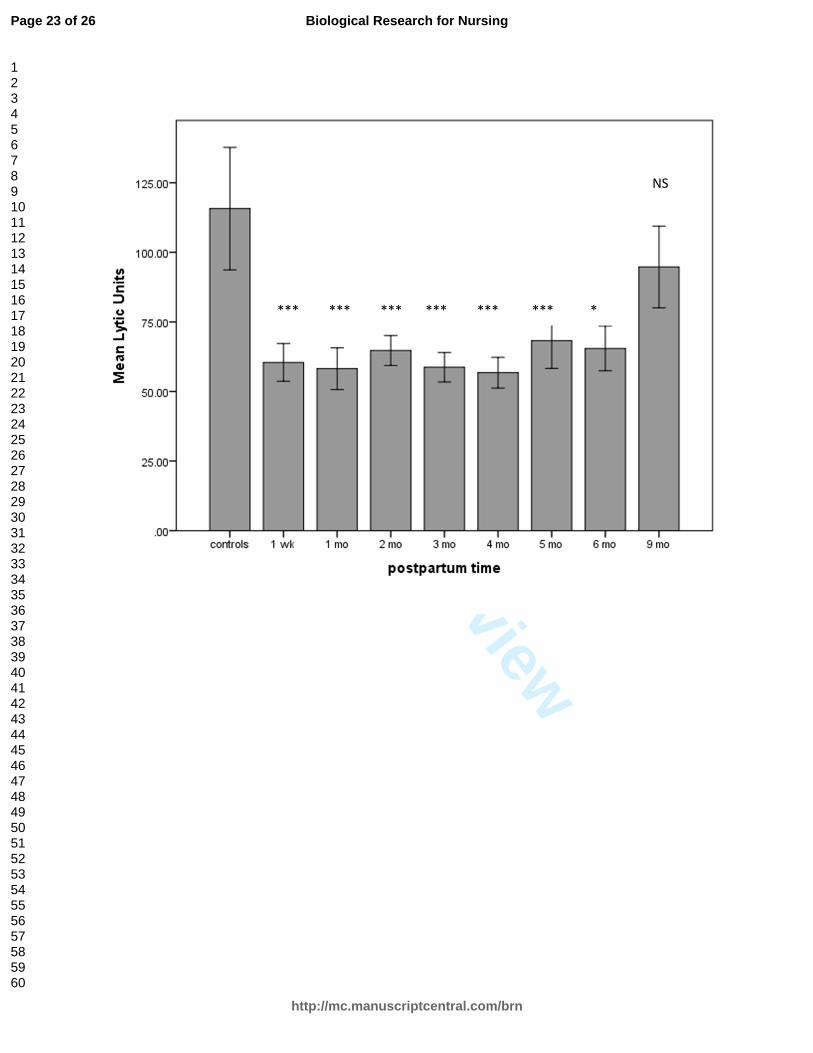
Figure 1 depicts the comparison between NK cell cytotoxicity in postpartum
women across 9 months of the postpartum period and in control women. There was a
range in the number of assays done for each time point, from 58 to 79, except for 21 at
Week 1 and 24 at Month 9. The findings at 9 and 12 months were identical, though we
had a smaller sample at 12 months (n = 10). The difference between the LUs for the
control and postpartum groups was significant at all time points through 6 postpartum
months, with t-values ranging from 2.9 to 4.1 and p-values all < .001 for the first 5
months; t = 1.0, p = .05 at 6 months; t = .87, p = .39 at 9 months; and t = 1.27, p =.12 at
12 months.
<Figure 1 near here.>
Postpartum Plasma Effects
The addition of heat-inactivated plasma had no effect on NK cytotoxicity,
suggesting that any effect we observed with the addition of pooled plasma would be
attributable to biological molecules in that plasma. For these experiments we used the
percent lysis at the 50:1 E:T ratio in the NKCA rather than LUs due to constraints of cell
numbers. The 50:1 E:T ratio produced the greatest cytotoxicity in our assay and was
Page 11 of 26
http://mc.manuscriptcentral.com/brn
Biological Research for Nursing
123456789101112131415161718192021222324252627282930313233343536373839404142434445464748495051525354555657585960
For Peer Review
NK SUPPRESSION IN THE POSTPARTUM
12
highly correlated with total LUs (r = .70, p < .001). Comparisons of cytotoxicity in the
presence and absence of pooled postpartum plasma across the postpartum period
showed a significant suppression of cytotoxicity with the addition of pooled plasma. The
average percent cytotoxicity across 6 months for the samples with added pooled
plasma was 15.4 +/-1.6 S.E.M. compared to the control cytotoxicity of 26.6 +/-1.4
S.E.M. (t = 10.3, df = 73, p < .001; Figure 2). Addition of pooled postpartum plasma to
nonpostpartum age-matched control women’s NK cell cultures (n = 16) did not affect
cytotoxicity. The cytotoxicity at an E:T ratio of 50:1 was 10.7 in control wells (no added
plasma) and 10.01 when pooled postpartum plasma was added (nonsignificant). These
data suggest a that postpartum NK cells are uniquely responsive to plasma
components.
<Figure 2 near here.>
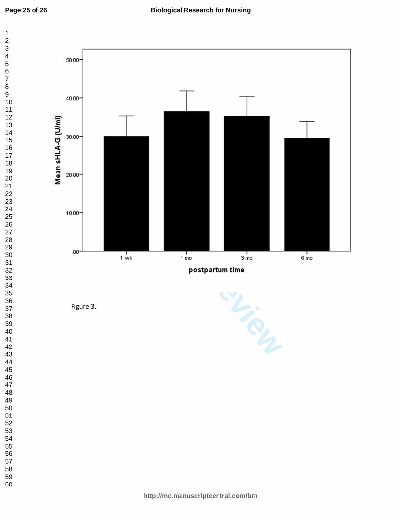
Plasma cytokines were not correlated with NK cytotoxicity at any time point. The
sHLA-G values across the postpartum period remained stable (29–36 U/ml), dropping
slightly at Month 6 (Figure 3). There was a significant inverse correlation between LUs
and sHLA-G levels at Week 1 (r = -.34, p = .02) and Month 3 (r=-.32, p=.02), but the
correlation was not statistically significant at the two other measurement times. There
was a significant correlation of sHLA-G concentration with NKG2A expression on
postpartum NK cells (r = .57, p < .02).
<Figure 3 near here.>
Plasma concentrations of estradiol, progesterone, prolactin and cortisol
measured at Week 1 and Months 1, 3 and 6 showed no correlations with LUs at any
Page 12 of 26
http://mc.manuscriptcentral.com/brn
Biological Research for Nursing
123456789101112131415161718192021222324252627282930313233343536373839404142434445464748495051525354555657585960
For Peer Review
NK SUPPRESSION IN THE POSTPARTUM
13
time point. Breastfeeding women did not differ from formula-feeding women in LUs
across the postpartum period.
Flow Cytometry
Phenotyping of cells by flow cytometry showed that the percentage of NK cells
did not change significantly over the first 3 postpartum months (data not shown). The
average percentage of NK cells in the total lymphocyte population in 33 postpartum
samples measured at Months 1, 2 and 3 was 9.3% (range 0.9–39; SD = 4.7). The
measures at Months 1 and 2 were nearly identical (8.9% and 8.2%), but the NK
percentage did rise to over 11% at 3 months. Control NK percentage (n = 16) was 7.2
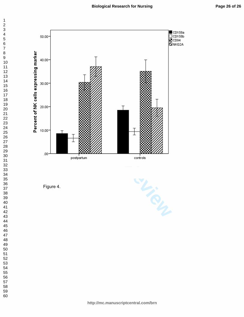
(range 1.5–21.2). We analyzed flow cytometric phenotyping of inhibitory receptors of NK
cells from postpartum women compared to those from nonpostpartum age-matched
women who served as controls (Figure 4). These cells were preserved from the initial
washed PBMC sample in liquid nitrogen and had not undergone any manipulation or
culturing with rIL2. Inhibitory receptors measured were the lectin-type receptors CD94
and NKG2A and the killer immunoglobulin-like receptors (KIRs) CD158a and CD158b.
There was a significantly lower percent staining of CD158a (t = -5.3, p < .001) and
CD158b (t = -2.5, p < .017) receptors in the postpartum NK cells compared to the
control NK cells. The percent NKG2A staining of NK cells was significantly higher in
postpartum women compared to controls (t = -2.55, p = .01). CD94 expression was not
significantly different (t = .8, p = .4) between the groups, nor were the percentages of
total NK cells (t = .99, p = .3) in this sample.
<Figure 4 near here.>
Discussion
Page 13 of 26
http://mc.manuscriptcentral.com/brn
Biological Research for Nursing
123456789101112131415161718192021222324252627282930313233343536373839404142434445464748495051525354555657585960

For Peer Review
NK SUPPRESSION IN THE POSTPARTUM
14
There are many potential pathways through which NK cytotoxicity could be
suppressed in postpartum women. Plasma components including cytokines and
hormones might conceivably play a role during this reproductive phase, but in this study
there were no relationships between these plasma components and cytotoxicity. Other
mechanisms could include activation of the KIR receptors, inhibition or reduced
expression of activation markers, blocking of lysis, inhibition of cytokine secretion, or
interruption of NK–target conjugate formation (Gan, Zhang, Solomon, & Bonavida,
2002).
Data from the present study clearly show that NK suppression extends for
several months postpregnancy and that plasma components and possibly NKG2A
receptors are involved in this suppression. The experimental results suggest that only
postpartum, and not nonpostpartum control, NK cells were uniquely responsive to the
addition of pooled postpartum plasma components. Plasma levels of sHLA-G were
inversely correlated with LUs in the early postpartum period, suggesting that it may be
an NK-cell suppressive molecule. This hypothesis is further supported by the
relationship between NKG2A expression and sHLA-G levels in the plasma. sHLA-G
may still be present in postpartum plasma as the result of the shedding of
cytotrophoblastic cells in pregnancy or as a marker of fetal microchimerism. The HLA-G
gene is low polymorphic, and its expression in fetal cells is accompanied by a lack of
classical Class I HLAs and Class II antigens. This state leads to an inability of maternal
T and NK cells to recognize fetal cells as foreign (Rebmann, Regel, Stolke, & Grosse-
Wilde, 2003). sHLA-G can be detected in healthy women (mean of 20.7 +/-2.1 ng/ml;
Pistoia, Morandi, Wang, & Ferrone, 2007) and is increased in pregnancy, as we also
Page 14 of 26
http://mc.manuscriptcentral.com/brn
Biological Research for Nursing
123456789101112131415161718192021222324252627282930313233343536373839404142434445464748495051525354555657585960
For Peer Review
NK SUPPRESSION IN THE POSTPARTUM
15
found in this postpartum sample. Other sources of HLA-G include the thymus, cornea,
nail matrix, pancreas, erythroid and endothelial precursor cells, and monocytes
(Fainardi et al., 2011). The NK receptors thought to bind to HLA-G include KIR2DL4
(which is difficult to measure) and possibly CD94/NKG2A (which may respond to
peptides derived from HLA-G and presented by HLA-E; Apps, Gardner, & Moffett,
2008). There was not a consistent relationship between LUs and sHLA-G across the
postpartum period, so it is unlikely that sHLA-G was the sole source of plasma
suppression of cytoxicity.
NKG2A forms a dimer with CD94 and acts as an inhibitor of NK cytotoxicity. We
found that NG2A was markedly increased in postpartum women compared to control
women. CD94 was not upregulated in postpartum women, but this may have been due
to the fact that CD94 also dimerizes with other molecules, some of which are activating
receptors. Recently investigators reported that the NKG2A–CD94 complexes potently
suppress uterine NK cell cytotoxicity (Le Bouteiller et al., 2011). NKG2A–CD94 binds to
HLA-E on cells, and HLA-E, like HLA-G, is a nonclassical, low polymorphic Class I MHC
molecule. Like HLA-G, it is also highly expressed by cytotrophoblast cells in pregnancy
(Ishitani, Sageshima, & Hatake, 2006).
Expression of CD158a and CD158b was decreased in NK cells from postpartum
women compared to those from controls in the present study. These KIRs react with
classical Class I HLA molecules (HLA-A,HLA-B, and HLA-C). There may be differential
patterns of receptor expression on postpartum NK cells that respond to inhibitory or
activating signals. The decreased expression of CD158a and CD158b we observed in
the freshly stained postpartum NK cells may be related to an absence of IL-2 in the
Page 15 of 26
http://mc.manuscriptcentral.com/brn
Biological Research for Nursing
123456789101112131415161718192021222324252627282930313233343536373839404142434445464748495051525354555657585960
For Peer Review
NK SUPPRESSION IN THE POSTPARTUM
16
media. IL-2 is needed to increase the expression of these inhibitory receptors (Marras,
Bozzano, & De Maria, 2011). We did note that postpartum cells had an extreme
dependence on IL-2 and were essentially nonfunctional in the absence of IL-2.
Remarkably, while pooled postpartum plasma consistently and significantly
increased inhibition of NK cytotoxicity in the NK cultures of postpartum women, it did not
have any effect on nonostpartum control women’s NK cultures. Therefore it seems likely
that both plasma factors and NK-receptor functions are involved in the NK cytotoxicity
suppression of the postpartum period. This proposition suggests that postpartum NK
cells have unique properties compared to NK cells from nonpostpartum women, set
apart by as-yet-unknown mechanisms.
It would be instructive to compare our findings in the present study with studies of
NK cytotoxicity in pregnant women, since it is reasonable to assume that the
postpartum period may extend pregnancy effects. We do know that cytotoxicity and
number of peripheral CD56 cells are decreased in normal pregnancy, that these cells
play a role in orchestrating pregnancy-associated type 2 shifting, and that increases in
the number of NK cells are associated with spontaneous abortion (Lashley et al., 2011;
Lu et al., 2011). The mechanism by which this decline in number and cytotoxic activity
of peripheral NK cells occurs is not well described. Research is now focused on uterine
NK cells, which differ in multiple ways from peripheral NK cells. Peripheral NK cells are
more cytotoxic and less secretory than the CD56bright uterine NK population. The low
cytotoxicity and the interaction of plasma components with possibly upregulated
inhibitory receptor NKG2A suggests that ligands, including sHLA-G, in plasma may be
involved in NK suppression in the postpartum period through inhibitory receptors.
Page 16 of 26
http://mc.manuscriptcentral.com/brn
Biological Research for Nursing
123456789101112131415161718192021222324252627282930313233343536373839404142434445464748495051525354555657585960

For Peer Review
NK SUPPRESSION IN THE POSTPARTUM
17
Researchers have previously identified several plasma components as inhibitors of NK
cytotoxicity against K562 cells (Rolin, Sand, Knudsen, & Maghazachi, 2010). Further
work is needed to identify unique components of postpartum plasma.
Limitations and Implications for Future Research
The research presented here is preliminary and exploratory. We were
constrained by availability of adequate cell numbers and available samples. Other
inhibitory as well as activating receptor phenotyping is warranted in future work. We
acknowledge that the staining results from unmanipulated cells may be very different
than those from cells after rIL-2 pre-incubation. It was difficult to obtain enough cells to
stain after IL-2 incubation and still have enough for the NKCA, as there was always
some cell loss with each incubation. Additional aspects of NK cytotoxicity such as
perforin and granzyme activity would be of interest. Isolation of the unique components
of plasma that increase the suppression of NK cytotoxicity and further investigation of
the role of sHLA-G in NK function are also warranted.
Identification and isolation of these components, along with an increased
understanding of their pathways of interaction with NK cells, may prove useful in
translational medicine. Understanding the immune system of healthy postpartum
women may reveal mechanisms that can ultimately be informative about or lead to new
treatments in disease states. It seems obvious that healthy postpartum women are not
particularly at risk even in the presence of significant NK cytotoxicity suppression, and
the physiology associated with this surprising finding bears additional scrutiny.
Page 17 of 26
http://mc.manuscriptcentral.com/brn
Biological Research for Nursing
123456789101112131415161718192021222324252627282930313233343536373839404142434445464748495051525354555657585960
For Peer Review
NK SUPPRESSION IN THE POSTPARTUM
18
Figure Captions
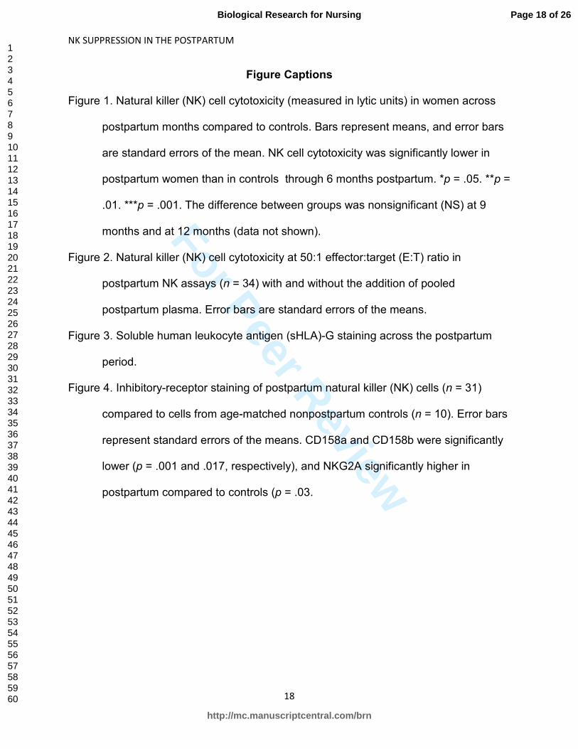
Figure 1. Natural killer (NK) cell cytotoxicity (measured in lytic units) in women across
postpartum months compared to controls. Bars represent means, and error bars
are standard errors of the mean. NK cell cytotoxicity was significantly lower in
postpartum women than in controls through 6 months postpartum. *p = .05. **p =
.01. ***p = .001. The difference between groups was nonsignificant (NS) at 9
months and at 12 months (data not shown).
Figure 2. Natural killer (NK) cell cytotoxicity at 50:1 effector:target (E:T) ratio in
postpartum NK assays (n = 34) with and without the addition of pooled
postpartum plasma. Error bars are standard errors of the means.
Figure 3. Soluble human leukocyte antigen (sHLA)-G staining across the postpartum
period.
Figure 4. Inhibitory-receptor staining of postpartum natural killer (NK) cells (n = 31)
compared to cells from age-matched nonpostpartum controls (n = 10). Error bars
represent standard errors of the means. CD158a and CD158b were significantly
lower (p = .001 and .017, respectively), and NKG2A significantly higher in
postpartum compared to controls (p = .03.
Page 18 of 26
http://mc.manuscriptcentral.com/brn
Biological Research for Nursing
123456789101112131415161718192021222324252627282930313233343536373839404142434445464748495051525354555657585960

For Peer Review
References
Apps, R., Gardner, L., & Moffett, A. (2008). A critical look at HLA-G. Trends in
Immunology, 29, 313-321. doi:10.1016/j.it.2008.02.012
Borzychowski, A. M., Croy, B. A., Chan, W. L., Redman, C. W., & Sargent, I. L. (2005).
Changes in systemic type 1 and type 2 immunity in normal pregnancy and pre-
eclampsia may be mediated by natural killer cells. European Journal of
Immunology, 35, 3054-3063. doi:10.1002/eji.200425929
Bryceson, Y. T., & Long, E. O. (2008). Line of attack: NK cell specificity and integration
of signals. Current Opinions in Immunology, 20, 344-352.
doi:10.1016/j.coi.2008.03.005
Caligiuri, M. A. (2008). Human natural killer cells. Blood, 112, 461-469.
doi:10.1182/blood-2007-09-077438
Challis, J. R., Lockwood, C. J., Myatt, L., Norman, J. E., Strauss, J. F., 3rd, & Petraglia,
F. (2009). Inflammation and pregnancy. Reproductive Science, 16, 206-215.
doi:10.1177/1933719108329095
Chamley, L. W., Chen, Q., Ding, J., Stone, P. R., & Abumaree, M. (2011). Trophoblast
deportation: Just a waste disposal system or antigen sharing? Journal of
Reproductive Immunology, 88(2), 99-105. doi:10.1016/j.jri.2011.01.002
Cooper, M. A., Fehniger, T. A., & Caligiuri, M. A. (2001). The biology of human natural
killer-cell subsets. Trends in Immunology, 22, 633-640.
Fainardi, E., Castellazzi, M., Stignani, M., Morandi, F., Sana, G., Gonzalez, R., . . .
Pena, J. (2011). Emerging topics and new perspectives on HLA-G. Cell and
Molecular Life Sciences, 68, 433-451. doi:10.1007/s00018-010-0584-3
Page 19 of 26
http://mc.manuscriptcentral.com/brn
Biological Research for Nursing
123456789101112131415161718192021222324252627282930313233343536373839404142434445464748495051525354555657585960
For Peer Review
Gan, X., Zhang, L., Solomon, G. F., & Bonavida, B. (2002). Mechanism of
norepinephrine-mediated inhibition of human NK cytotoxic functions: Inhibition of
cytokine secretion, target binding, and programming for cytotoxicity. Brain
Behavior and Immunity, 16, 227-246. doi:10.1006/brbi.2000.0615
Gregory, C. D., Lee, H., Rees, G. B., Scott, I. V., Shah, L. P., & Golding, P. R. (1985).
Natural killer cells in normal pregnancy: Analysis using monoclonal antibodies
and single-cell cytotoxicity assays. Clinical and Experimental Immunology, 62,
121-127.
Gregory, C. D., Shah, L. P., Lee, H., Scott, I. V., & Golding, P. R. (1985). Cytotoxic
reactivity of human natural killer (NK) cells during normal pregnancy: A
longitudinal study. Journal of Clinical and Laboratory Immunology, 18(4), 175-
181.
Groer, M., El-Badri, N., Djeu, J., Harrington, M., & Van Eepoel, J. (2010). Suppression
of natural killer cell cytotoxicity in postpartum women. American Jourmal of
Reproductive Immunologyl, 63, 209-213. doi:10.1111/j.1600-0897.2009.00788.x
Groer, M. W., Davis, M. W., Smith, K., Casey, K., Kramer, V., & Bukovsky, E. (2005).
Immunity, inflammation and infection in post-partum breast and formula feeders.
American Journal of Reproductive Immunology, 54, 222-231. doi:10.1111/j.1600-
0897.2005.00301.x
Groer, M. W., Manion, M., Szekeres, C., & El-Badri, N. S. (2011). Fetal microchimerism
and women's health: A new paradigm. Biological Research for Nursing, 13, 346-
350. doi:10.1177/1099800410385840
Page 20 of 26
http://mc.manuscriptcentral.com/brn
Biological Research for Nursing
123456789101112131415161718192021222324252627282930313233343536373839404142434445464748495051525354555657585960
For Peer Review
Ishitani, A., Sageshima, N., & Hatake, K. (2006). The involvement of HLA-E and -F in
pregnancy. Journal of Reproductive Immunology, 69, 101-113.
doi:10.1016/j.jri.2005.10.004
Konjevic, G., Schlesinger, B., Cheng, L., Olsen, K. J., Podack, E. R., & Spuzic, I.
(1995). Analysis of perforin expression in human peripheral blood lymphocytes,
CD56+ natural killer cell subsets and its induction by interleukin-2. Immunological
Investigations, 24, 499-507.
Kraus, T. A., Engel, S. M., Sperling, R. S., Kellerman, L., Lo, Y., Wallenstein, S., . . .
Moran, T. M. (2012). Characterizing the pregnancy immune phenotype: Results
of the viral immunity and pregnancy (VIP) study. Journal of Clinical Immunology,
32, 300-311. doi:10.1007/s10875-011-9627-2
Lashley, L. E., van der Hoorn, M. L., van der Mast, B. J., Tilburgs, T., van der Lee, N.,
van der Keur, C., . . . Scherjon, S. A. (2011). Changes in cytokine production and
composition of peripheral blood leukocytes during pregnancy are not associated
with a difference in the proliferative immune response to the fetus. Human
Immunology, 72, 805-811. doi:10.1016/j.humimm.2011.05.027
Le Bouteiller, P., Siewiera, J., Casart, Y., Aguerre-Girr, M., El Costa, H., Berrebi, A., . . .
Jabrane-Ferrat, N. (2011). The human decidual NK-cell response to virus
infection: What can we learn from circulating NK lymphocytes? Journal of
Reproductive Immunology, 88, 170-175. doi:10.1016/j.jri.2010.12.005
LeMaoult, J., Zafaranloo, K., Le Danff, C., & Carosella, E. D. (2005). HLA-G up-
regulates ILT2, ILT3, ILT4, and KIR2DL4 in antigen presenting cells, NK cells,
and T cells. FASEB Journal, 19, 662-664. doi:10.1096/fj.04-1617fje
Page 21 of 26
http://mc.manuscriptcentral.com/brn
Biological Research for Nursing
123456789101112131415161718192021222324252627282930313233343536373839404142434445464748495051525354555657585960
For Peer Review
Lu, Y., Zeng, B., Zhang, Y., Xiang, W., Hu, L., & Liao, A. (2011). Quantitative and
functional changes in peripheral natural killer cells in women with reproductive
failure after artificial insemination with donor sperm. Journal of Reproductive
Immunology, 91(1-2), 83-89. doi:10.1016/j.jri.2011.05.009
Marras, F., Bozzano, F., & De Maria, A. (2011). Involvement of activating NK cell
receptors and their modulation in pathogen immunity. Journal of Biomedicine and
Biotechnology 2011, 152430. doi:10.1155/2011/152430
Mor, G., Cardenas, I., Abrahams, V., & Guller, S. (2011). Inflammation and pregnancy:
The role of the immune system at the implantation site. Annals of the New York
Academy of Science, 1221, 80-87. doi:10.1111/j.1749-6632.2010.05938.x
Pistoia, V., Morandi, F., Wang, X., & Ferrone, S. (2007). Soluble HLA-G: Are they
clinically relevant? Seminars in Cancer Biology, 17, 469-479.
doi:10.1016/j.semcancer.2007.07.004
Rebmann, V., Regel, J., Stolke, D., & Grosse-Wilde, H. (2003). Secretion of sHLA-G
molecules in malignancies. Seminars in Cancer Biology, 13, 371-377.
Rolin, J., Sand, K. L., Knudsen, E., & Maghazachi, A. A. (2010). FTY720 and SEW2871
reverse the inhibitory effect of S1P on natural killer cell mediated lysis of K562
tumor cells and dendritic cells but not on cytokine release. Cancer Immunology,
Immunotheapeutics 59, 575-586. doi:10.1007/s00262-009-0775-7
Page 22 of 26
http://mc.manuscriptcentral.com/brn
Biological Research for Nursing
123456789101112131415161718192021222324252627282930313233343536373839404142434445464748495051525354555657585960
For Peer Review
*** *** *** *** *** *** *
NS
Page 23 of 26
http://mc.manuscriptcentral.com/brn
Biological Research for Nursing
123456789101112131415161718192021222324252627282930313233343536373839404142434445464748495051525354555657585960
For Peer Review
Figure 2.
This image cannot currently be displayed.
Page 24 of 26
http://mc.manuscriptcentral.com/brn
Biological Research for Nursing
123456789101112131415161718192021222324252627282930313233343536373839404142434445464748495051525354555657585960
For Peer Review
Figure 3.
Page 25 of 26
http://mc.manuscriptcentral.com/brn
Biological Research for Nursing
123456789101112131415161718192021222324252627282930313233343536373839404142434445464748495051525354555657585960
For Peer Review
Figure 4.
Page 26 of 26
http://mc.manuscriptcentral.com/brn
Biological Research for Nursing
123456789101112131415161718192021222324252627282930313233343536373839404142434445464748495051525354555657585960