dual-functional capability of cd3+cd56+ cik cells, a t-cell subset that acquires nk function and...
TRANSCRIPT

IMMUNOBIOLOGY
Dual-functional capability of CD3�CD56� CIK cells, a T-cell subset that acquiresNK function and retains TCR-mediated specific cytotoxicityAlice Pievani,1 Gianmaria Borleri,1 Daniela Pende,2 Lorenzo Moretta,3 Alessandro Rambaldi,1 Josee Golay,1 andMartino Introna1
1Laboratory of Cellular Therapy “G. Lanzani,” USC Hematology, Ospedali Riuniti di Bergamo, Bergamo, Italy; 2Istituto Nazionale per la Ricerca sul Cancro,Genoa, Italy; and 3Istituto Giannina Gaslini, Genoa, Italy
CD3�CD56� cytokine-induced killer (CIK)cells display a potent cytolytic activity.The adhesion molecule lymphocytefunction-associated antigen-1 plays a cru-cial role in binding as well as in cytolyticactivity of CIK cells against tumor targetcells expressing the corresponding ligands.CIK cells express activating natural killer(NK) receptors, including NKG2D, DNAX ac-cessory molecule-1 (DNAM-1), and low lev-els of NKp30. Cell signaling not only throughTCR/CD3 but also through NKG2D, DNAM-1,
and NKp30 leads to CIK cell activation result-ing in granule exocytosis, cytokine secre-tion, and cytotoxicity. Antibody blocking ex-periments showed that DNAM-1, NKG2D,and NKp30 are involved in the TCR-indepen-dent tumor cell recognition and killing.Anti–CMV-specific CIK cells could be expandedin standard CIK cultures and mediate bothspecific, MHC-restricted recognition andTCR-independent NK-like cytolytic activityagainst leukemic cell lines or fresh leukemicblasts. Antibody blocking of lymphocyte
function-associated antigen-1 and DNAM-1led to significant reduction of both CTL andNK-cell functions, whereas blocking ofNKG2D and NKp30 only inhibited NK-likecytotoxicity. Their dual-effector function sug-gests that CIK cells, when used in a clinicalsetting, may control both neoplastic re-lapsesandviral infections,2 frequentlyasso-ciated complications in patients whoreceived a transplant. (Blood. 2011;118(12):3301-3310)
Introduction
Cytokine-induced killer (CIK) cells are ex vivo–activated lympho-cytes that can be obtained in large numbers within 3 weeks ofculture from either human peripheral blood or BM, or cord bloodmononuclear cells by the sequential addition of IFN-�, anti-CD3Ab (OKT3), and high doses of recombinant human IL-2 (rhIL-2).1-4 CIK cells represent an heterogeneous cell population, includ-ing a large majority of CD3�CD56� cells and minor fractions oftypical T cells (CD3�CD56�) and NK cells (CD3�CD56�).5,6 CIKcells can lyse a broad array of tumor targets, including acutemyeloid leukemia, chronic myelogenous leukemia, and chroniclymphocytic leukemia by a non–MHC-restricted, natural killer(NK)–like mechanism.3,6 Interestingly, in mice, CIK cells displaynegligible alloreactivity and cause minimal GVHD compared withallogeneic splenocytes, when infused after allogeneic BM transplan-tation in murine models.7,8 CIK cells express several chemokinereceptors and can migrate to the site of tumors after intravenousadministration, as shown by models in vivo.9-13 Because CIK cellscould be produced by a simple approach and displayed antitumoractivity in vitro, they appeared to be suitable candidates for celltherapy in solid and hematopoietic tumor treatment. Indeed, bothautologous and allogeneic CIK cells have been used in phase 1 and2 clinical trials for the treatment of different tumor types. In thesetrials, they displayed limited toxicity, whereas evidence has beenobtained that they exert antitumor activity.14-20
The molecular structures that account for tumor recognition andkilling by CIK cells are only partially understood. Previousobservations suggested a possible involvement of the NKG2D andlymphocyte function-associated antigen-1 (LFA-1) molecules21-23;however, little is known on the role of other activating NK recep-
tors, including DNAX accessory molecule-1 (DNAM-1), NKp30,NKp44, NKp46, as well as on the involvement of TCR/CD3complex in the cytolytic activity of CIK cells.
Our previous studies clarified that CD3�CD56� CIK cells havephenotypic characteristics typical of terminally differentiated CD8�
effector memory T cells (TEMRA; CCR7�, CD45RA�, CD62Llow,CD11a�, CD27�, CD28�). We also showed that CIK cells origi-nate in vitro from CD56�CD8� T-cell progenitors that stronglyexpand on culture in the presence of IL-2 and acquire CD56 antigen.5 Inaddition, CIK cells share several characteristics with NK cells, such asthe large granular lymphocyte structure, the capacity to kill the HLAclass I–negative cell line K562, and the surface expression of highdensities of CD56 and NKG2D. Different from NK cells, however, CIKcells express low densities of NKp30, whereas they do not expressNKp44 and NKp46, and the inhibitory killer immunoglobulin-likereceptors, NKG2A and CD94.5
In the present study, we investigated in detail the receptorsinvolved in the NK-like cytolytic activity of CIK cells and analyzedwhether they have retained also their ability to elicit specificcytolytic effector T-cell function.
Methods
Cells
The human cell lines BJAB (B lymphoma), KARPAS422 (B lymphoma),MOLT4 (T-cell leukemia), RAJI (B-cell leukemia), and K562 (erythroleu-kemia) were maintained in RPMI-1640 medium (Cambrex Bio Science)
Submitted February 11, 2011; accepted July 20, 2011. Prepublished online asBlood First Edition paper, August 5, 2011; DOI 10.1182/blood-2011-02-336321.
The online version of this article contains a data supplement.
The publication costs of this article were defrayed in part by page chargepayment. Therefore, and solely to indicate this fact, this article is herebymarked ‘‘advertisement’’ in accordance with 18 USC section 1734.
© 2011 by The American Society of Hematology
3301BLOOD, 22 SEPTEMBER 2011 � VOLUME 118, NUMBER 12
For personal use only.on April 3, 2016. by guest www.bloodjournal.orgFrom

supplemented with 10% FBS (Euroclone), 2mM L-glutamine (Euroclone),and 110�M gentamycin (PHT Pharma), hereafter called complete medium.
Leukemic cells, obtained by Ficoll-Hypaque density centrifugation ofperipheral blood samples, were used for cytolytic assays after thawing.
PHA-induced blasts were obtained by stimulating peripheral bloodlymphocytes with 1 �g/mL PHA (Sigma-Aldrich) and 120 U/mL rhIL-2(Proleukin; Chiron).
Generation of CIK cells
CIK cells were prepared as previously described.5 Briefly, the CD56-depleted fractions separated from PBMCs by negative selection with theuse of MACS (Miltenyi Biotec) with anti-CD56 microbeads (MiltenyiBiotec) were cultured in serum-free X-VIVO-15 medium (BioWhittaker)with 1000 U/mL IFN-� (Gammakine; Boehringer Ingelheim) added on day0, 50 ng/mL anti-CD3 (OKT-3; Janssen-Cilag S.p.a.) added on day 1, and500 U/mL rhIL-2 included in the medium from day 1 onward. Expansionwas performed for 21-28 days. At the end of the expansion, CD56� CIKcells were immunoselected with anti-CD56 microbeads according to themanufacturer’s instructions.
Anti–CMV-specific CIK cells were generated, as described earlier,starting from peripheral blood lymphocytes obtained from selected HLA-A*0201 CMV-seropositive healthy donors. Anti–CMV-specific lympho-cytes were identified by flow cytometry after staining with PE-conjugatedHLA-A*0201/pp65495-503 tetramer (Immunotech Laboratories, BeckmanCoulter) and FITC-conjugated anti-CD8 Ab (Becton Dickinson). Anti–CMV-specific cells were isolated at the end of the expansion, using cognatePE-conjugated tetramers followed by separation with anti-PE Ab-coatedmicrobeads (Miltenyi Biotec) and MACS (Miltenyi Biotec). After sorting,tetramer-positive cells were resuspended in X-VIVO-15 medium contain-ing 500 U/mL rhIL-2 and irradiated allogeneic feeder cells and werecultured for 2 weeks. At the end of culture, CD56� CMV-specific CIK cellswere immunoselected.
Anti–EBV-specific CIK cells were generated from peripheral bloodlymphocytes from HLA-A*0201-positive persons with recent onset ofacute infectious mononucleosis. Anti–EBV-specific lymphocytes wereidentified by staining with PE-conjugated HLA-A*0201 EBV (GLCTL-VAML) tetramer (Beckman Coulter).
mAbs and flow cytometric analysis
To characterize the surface expression of the activating receptor ligands onthe tumor cell lines, the following panel of mAbs was used: anti–ULBP-1(UL16 binding protein-1; 170818 clone), anti–ULBP-2 (165903 clone),anti–ULBP-3 (166510 clone), and anti-MICA/B (MHC class I–relatedchain molecules A/B; 159207 clone; R&D Systems). Anti–Nectin-2 (L14clone) and anti-PVR (poliovirus receptor; L95 clone) were previouslydescribed.24 The adhesion molecules CD54 (ICAM-1), CD102 (ICAM-2),and CD50 (ICAM-3) were detected with PE or FITC-conjugated Ab clonesHA58, CBR-1C2/2, and TU41, respectively (BD Biosciences PharMingen).
CIK cells were characterized with FITC-conjugated anti-CD3 (SK7clone) and PE-conjugated anti-CD56 (NCAM16.2 clone) mAbs (BectonDickinson). To analyze the expression of activating receptors on CIK cells,the following mAbs were used: anti-NKG2D (BAT221 clone), anti-NKp30(A76 clone), anti-NKp46 (BAB281 clone), anti-NKp44 (KS38 clone), andanti–DNAM-1 (F22 clone).24,25 CD11a (LFA-1) was detected with FITC-conjugated Ab clone G-25.2 (BD Biosciences). mAb24 that recognizesLFA-1 in high-affinity conformation was also used (Abcam).
For indirect immunofluorescence staining, cells were stained with theprimary mAb, followed by PE-conjugated goat anti–mouse IgG1 (Invitro-gen) or FITC-conjugated goat anti–mouse Ig second reagent (BD Biosci-ences). A FACScan flow cytometer device (BD Biosciences) was used toanalyze the samples.
CFSE and PKH26 staining
Target cells were washed and resuspended in 1 mL of PBS with 1% BSA ata final concentration of 5 � 106/mL, then labeled for 10 minutes at 37°Cwith green fluorescent dye CFSE (2�M; Sigma-Aldrich). Quench-staining
was performed on ice for 5 minutes by adding 5 volumes of ice-coldcomplete RPMI. Cells were then washed 3 times with ice-cold PBS with1% BSA and cultured under appropriate conditions.
For PKH-26 staining of CIK cells, 10 � 106 cells were incubated withlipophilic red fluorescent dye PKH-26 (Sigma-Aldrich) according to themanufacturer’s instructions.
Cell conjugation assay
A total of 5 � 105 PKH26-labeled CIK cells and 5 � 105 CFSE-labeledtarget cells were resuspended in 200 �L of complete medium containing orlacking 1mM EDTA (Sigma-Aldrich) and mixed in a 12 � 75-mm polysty-rene tube (BD Biosciences Labware). The effector-target cell mixture wascentrifuged at 1000 rpm for 1 minute and incubated at 37°C for theindicated time points. The cells were then gently resuspended and analyzedby flow cytometry. The conjugation ratio was calculated as the portion ofFITC/PE double-positive events within the PE-positive events. For Ab-blocking experiments, 5 � 105 PKH26-labeled CIK cells were preincu-bated with 10 �g/mL blocking anti–LFA-1 mAb for 30 minutes, and thenthe conjugation ratio was detected by flow cytometry as described.
Calcein release cytotoxicity assay
Cell lysis was evaluated with the calcein release assay as previouslydescribed.2 In some experiment, the cytotoxicity assay was performed in thepresence of 1mM EDTA.
In some experiments, autologous or recipient PHA-induced blastspulsed with pp65495-503 or a HLA-A*0201–binding control peptide (Primm)were used as target cells. Peptide-pulsed cells were incubated with10 �g/mL peptide for 16 hours at 37°C before labeling with calcein-AM.
For masking experiments, anti-NKp30 (F252 clone), anti–DNAM-1 (F5clone), anti-NKG2D (BAT221 clone), and anti–HLA-I (W6/32 clone) wereused.26 Masking of LFA-1 was performed with TS1-22 hybridoma superna-tant (a kind gift of Dr P. Allavena, Istituto Clinico Humanitas). CIK cellswere preincubated 15 minutes at room temperature either in the absence orin the presence of 10 �g/mL of specific or isotype-matched control mAbs orsupernatant and, after washing, used in the cytolytic assay tested.
Redirected cytotoxicity assay
For redirected killing assay, Fc�R-positive cell line P815 (murine mastocy-toma) was labeled with calcein-AM as described earlier. Anti-CD3 (OKT3clone), anti-NKG2D (BAT221 clone), anti-NKp30 (A76 clone), anti–DNAM-1 (F22 clone), anti-NKp46 (BAB281 clone), isotype-matchedcontrol or no mAbs were added at saturating concentration and incubatedfor 15 minutes. The P815 target cells were then used as target in the calceinrelease cytotoxicity assay as described.
Degranulation assay
Degranulation assay was performed as previously described.27,28 Briefly,CIK and P815 cells loaded with anti-CD3, anti-NKG2D, anti-NKp30,anti–DNAM-1, anti-NKp46, isotype-matched control or no mAbs wereplated at E/T ratio of 1:1 and incubated for 2 hours at 37°C in the presenceof CD107a-PE mAb (BD Pharmingen). CIK cell degranulation wasassessed by cell surface staining for the lysosomal markers CD107a by flowcytometry.
Statistical analysis
The data were analyzed with paired or unpaired Student t tests, asappropriate (*P � .05, **P � .01, and ***P � .001).
Results
Natural cytotoxicity of CD3�CD56� CIK cells correlates with thebinding rate to tumor targets and is LFA dependent
In the standard protocol for CIK cell expansion, cultures are initiatedstarting from unselected PBMCs and performed as previously described
3302 PIEVANI et al BLOOD, 22 SEPTEMBER 2011 � VOLUME 118, NUMBER 12
For personal use only.on April 3, 2016. by guest www.bloodjournal.orgFrom

by us5 and others.1 In the resulting bulk cultures 3 subpopulation can bedistinguished: CD3�CD56� terminally differentiated CIK cells, respon-sible for the HLA-unrestricted antitumor activity, CD3�CD56� earlyeffector T cells, and a minor fraction of CD3�CD56� NK cells.5 Todirectly assess the functional activity of CD3�CD56� CIK cells derivedfrom purified CD3�CD56� cells (ie, the putative peripheral bloodprecursors of CD3�CD56� CIK cells), PBMCs were depleted ofCD56� cells and then cultured by applying the standard expansionprotocol (supplemental Figure 1A, available on the Blood Web site; seethe Supplemental Materials link at the top of the online article). Finalyields and expansion folds were not changed compared with CIKcultures started from unmanipulated PBMCs (data not shown). At theend of the culture, CD3�CD56� CIK cells were purified by CD56�
selection (mean purity, 95%; range, 88%-99% in 5 independentexperiments; supplemental Figure 1A; data not shown). The surfacephenotype of such CIK cells did not differ from that previouslydescribed for CIK cells obtained from unselected PBMCs and isconsistent with �� CD8� TEMRA cells (supplemental Figure 1B).5 All
the following experiments were performed with the use of such purifiedCD3�CD56� CIK cells to directly test their antitumor function.
To investigate whether cytotoxicity of CIK cells requires effector/target cell contact and involves conjugate formation, PKH26-labeledCIK and CFSE-labeled B lymphoma cell line BJAB was mixed andincubated at 37°C for different time intervals. The cells were thenanalyzed by flow cytometry. Figure 1A shows a representative experi-ment of 3 experiments performed. CIK cells (34% 3%) were able tobind target after 5 minutes whereas a slow progressive decline wasobserved at later intervals (24% 2% after 1 hour; Figure 1A). Toinvestigate whether binding to target cells is necessary to CIK-mediatedcytotoxicity, experiments were performed in which conjugate formationwas blocked by adding EDTA, a calcium chelator that prevents celladhesion. Binding of CIK to BJAB cells was significantly inhibited byEDTA at all time points (average reduction of binding rate, 77% 3%;Figure 1A). Similarly, the cytolytic activity of CIK cells pretreated withEDTA against BJAB cells was significantly reduced by 79% 7% atall E/T ratios tested (P � .05; Figure 1B).
Figure 1. Natural cytotoxicity of CD3�CD56� CIK correlates with binding to tumor targets and is LFA-1 dependent. (A) PKH26-labeled CIK and CFSE-labeled BJABcells were mixed and incubated at 37°C for the indicated times with or without 1�M EDTA and analyzed by flow cytometry. Percentage of binding (values indicated in the topright quadrants) is calculated as the portion of CFSE/PKH-26 double-positive events within the PKH-26–positive events. One representative example is shown. (B) Cytotoxicityof CIK cells against BJAB in the presence (white bars) or absence (gray bars) of 1�M EDTA was evaluated at the indicated E/T ratios. Data were mean SD collected from3 independent experiments and were analyzed by Student t test; *P � .05, **P � .01, and ***P � .005. (C) CIK cells binding to BJAB or KARPAS 422 at different times wasanalyzed by flow cytometry. Data were obtained from � 3 independent experiments and analyzed by Student t test; *P � .05, **P � .01. (D) Cytotoxicity of CIK cells againstBJAB or KARPAS 422 was evaluated by calcein-release assay. Data were mean SD obtained from � 3 independent experiments and were analyzed by Student t test;*P � .05. (E) Expression of LFA-1 and their ligands (ICAM-1, -2, and -3) was determined, respectively, on CIK cells and BJAB target by flow cytometry. Background stainingwith the use of an isotype control Ab is shown in each histogram (gray curve). (F) The functional role of LFA-1 in CIK binding to BJAB (left) and in cytotoxicity (right) wasevaluated. For binding assays, PKH-26–labeled CIK cells were exposed to saturating concentrations of blocking Ab anti–LFA-1 or medium alone (CTR) for 30 minutes and thenincubated with CFSE-labeled BJAB for 15 minutes. For cytotoxicity assays, CIK cells were preincubated with saturating concentration of anti–LFA-1 or medium alone (CTR) for15 minutes and then tested in a calcein-release assay against BJAB. Data are the mean percentage of lysis obtained with respect to untreated controls. Data were mean SDobtained from � 3 independent experiments and were analyzed by Student t test; *P � .05, **P � .01, and ***P � .005 compared with control in the absence of mAbs.
CIK CELLS ARE T MEMORY WITH NK FUNCTIONS 3303BLOOD, 22 SEPTEMBER 2011 � VOLUME 118, NUMBER 12
For personal use only.on April 3, 2016. by guest www.bloodjournal.orgFrom

The BJAB and KARPAS422 human B lymphoma cell linesdisplayed different sensitivities to CIK-mediated cytotoxicity, of36% 6% and 23% 6% specific lysis at 10:1 E/T ratio,respectively (n 3; P � .05; Figure 1D). Notably, CIK-BJABconjugates were significantly more abundant than those formedwith KARPAS422 cells during the 60-minute incubation period(n 3; P � .05; Figure 1C). These results suggest that CIK cellbinding to target and formation of the cellular conjugates areprerequisite to and may correlate with CIK-mediated cytotoxicityagainst at least 2 cell lines.
Cell surface adhesion molecules, such as LFA-1, are known toparticipate in effector/target recognition and stable conjugateformation. CIK cells expressed LFA-1 (Figure 1E), and in particu-lar a significant percentage constitutively express LFA-1 in itshigh-affinity conformation, as identified by mAb24(67.5% 12.4%; n 3; data not shown). Moreover, the mostsusceptible target BJAB expressed all the major LFA-1 ligands(ICAM-1, -2, and -3; Figure 1E). In contrast, KARPAS422 did notexpress ICAM-1 and only low levels of ICAM-2 and -3 (supplemen-tal Figure 2A). In addition, although CIK-target cell conjugateformation and cytotoxicity against BJAB were strongly inhibitedby anti–LFA-1 mAb (respectively, 76% 11% and 78% 7%;n 3; P � .05; Figure 1F), binding and cytotoxicity againstKARPAS422 were only marginally affected (supplemental Figure2B-C). These results confirm that LFA-1 has a crucial role in
binding as well as in cytotoxicity of CIK cells and that additionalmolecules may also be involved in target cell recognition by CIK cells.
Functional role of CD3 and activating NK receptors in CIK cells
We next attempted to identify the receptors mediating non–HLA-restricted tumor cell killing by CIK cells. To investigate this point,we analyzed the main receptors involved in NK-cell triggering inthe process of tumor cell lysis. These include NKp46, NKp30,NKp44, NKG2D, and DNAM-1. As previously reported by ourgroup5 and others,21 CIK cells express NKG2D at high density. TheNCRs NKp44 and NKp46 were not or were very poorly expressed,whereas NKp30 was present at low densities5 (Figure 2A).However, DNAM-1 was found to be highly expressed (Figure 2A).
We investigated the functional status of the various NK recep-tors expressed on CIK cells by a reverse Ab-dependent cellularcytotoxicity assay. As shown in Figure 2B, robust redirected lysiswas obtained by crosslinking of NKG2D and DNAM-1, althoughless robust than one observed by anti-CD3 mAb. NKp30 crosslink-ing also led to redirected cytolysis, albeit to a lesser extent. On thecontrary, an isotype control Ab or an anti-NKp46 mAb did notinduce target cell lysis (Figure 2B; data not shown). The concomi-tant presence of anti-NKp30, anti–DNAM-1, or both of them withanti-NKG2D did not significantly change the activity of anti-NKG2D alone (Figure 2B). We also tested cytotoxic granule
Figure 2. Functional role of CD3/TCR and NK cytotoxic activating receptors on CIK cells. (A) Expression of activating receptors NKG2D, DNAM-1, NKp30, NKp46, andNKp44 on CIK cells was analyzed by flow cytometry. Gray profiles represent isotype controls. (B) Redirected killing assay was performed with the use of CIK cells treated withindicated agonist mAbs or without mAbs and calcein-labeled P815 cells. The data were mean SD obtained from 3 independent experiments. (C) CIK cells were triggered withP815 cells loaded with different mAbs and then assayed for degranulation by cell surface staining for the lysosomal markers CD107a. The data were mean SD obtained from� 3 independent experiments and were analyzed by Student t test; *P � .05, ***P � .005 compared with control in the absence of mAbs.
3304 PIEVANI et al BLOOD, 22 SEPTEMBER 2011 � VOLUME 118, NUMBER 12
For personal use only.on April 3, 2016. by guest www.bloodjournal.orgFrom

release after receptor engagement, using a mAb directed against thelysosomal-associated membrane protein-1 (CD107a).27,28 Consis-tently with cytotoxicity data, CD3 ligation resulted in an increase inthe percentage of cells undergoing degranulation (P � .005; Figure2C). Ligation of DNAM-1, NKG2D, and partially NKp30 alsoresulted in some degranulation (Figure 2C). Finally, also theproduction of IFN-� and TNF-� was investigated after receptorengagement. Crosslinking of CD3 resulted in IFN-� and TNF-�production (P � .05). A weaker cytokine secretion was detectedalso after stimulation of NKp30 and NKG2D (supplementalFigure 3A-B).
Altogether, these results show that signals delivered not onlythrough TCR but also through activating NK receptors can lead toCIK cell activation, resulting in granule exocytosis, cytokinesecretion, and cytotoxicity.
Effect of mAb-mediated blocking of NKG2D, DNAM-1, andNKp30 on CIK-mediated target cell lysis
To identify the triggering molecules that are actually involved inthe induction of CIK-mediated lysis of tumor targets, we firstanalyzed the ligands of activating NK receptors expressed ontarget cells.
Thus, we assessed the expression of ligands of NKG2D(MICA/B and ULBPs) and DNAM-1 (Nectin-2 and PVR) by theuse of specific mAb and FACS analysis. Figure 3 shows 2 targetssimilarly susceptible to CIK-mediated lysis that differ for the
expression of these ligands. Although RAJI cells were virtuallynegative for all ligands tested, MOLT4 cells were positive for PVRand ULBP-1 and -2 (Figure 3A-B).
Analysis of the effect of Ab blocking of the activating receptorsexpressed by CIK cells was consistent with data on ligandexpression. Indeed, masking of DNAM-1 had no effect on CIK-mediated lysis of RAJI cells, whereas lysis of MOLT4 cells waspartially inhibited. Similarly, NKG2D-masking resulted in a sharpinhibition of lysis of MOLT4 cells only. Interestingly, mAb-mediated blocking of NKp30 consistently resulted in a significantinhibition of killing of both targets implying that RAJI andMOLT4 cells express NKp30 ligand(s).29 An isotype control Abhas no effect on cytotoxicity (data not shown). The concomitantaddition of more than 1 Ab does not significantly increase theinhibitory effect detected by NKG2D, DNAM-1, or NKp30 alone(Figure 3C-D).
Taken together, these data indicate that NKG2D, DNAM-1, andNKp30 may be involved in CIK-mediated killing of target cellsupon recognition of their specific ligands.
CIK cells retain TCR-mediated Ag specificity
The data described in the previous subsection suggested that CIKcells may maintain a functional TCR/CD3 complex and acquireNK-like cytolytic functions through the expression of activatingNK receptors, including NKG2D, NKp30, and DNAM-1. There-fore, we asked whether TCR expressed by CIK cells could mediate
Figure 3. Direct role of activating receptors in CIK-mediated lysis of tumor targets. (A-B) RAJI (A) and MOLT4 (B) cells were analyzed for expression of MICA/B andULBP-1, -2, and -3 (NKG2D ligands), and PVR and Nectin-2 (DNAM-1 ligands) by flow cytometry. Gray profiles represent isotype control. (C-D) Blocking of activating receptorsNKG2D, NKp30, and DNAM-1 in CIK cells cytotoxicity against RAJI (C) and MOLT4 (D) targets. CIK cells were preincubated with saturating concentrations of anti-NKG2D,anti-NKp30, anti–DNAM-1, and cytotoxic activity was measured in calcein-release assays. The data were mean SD obtained from 3 independent experiments and wereanalyzed by Student t test, *P � .05, compared with control in the absence of mAbs.
CIK CELLS ARE T MEMORY WITH NK FUNCTIONS 3305BLOOD, 22 SEPTEMBER 2011 � VOLUME 118, NUMBER 12
For personal use only.on April 3, 2016. by guest www.bloodjournal.orgFrom
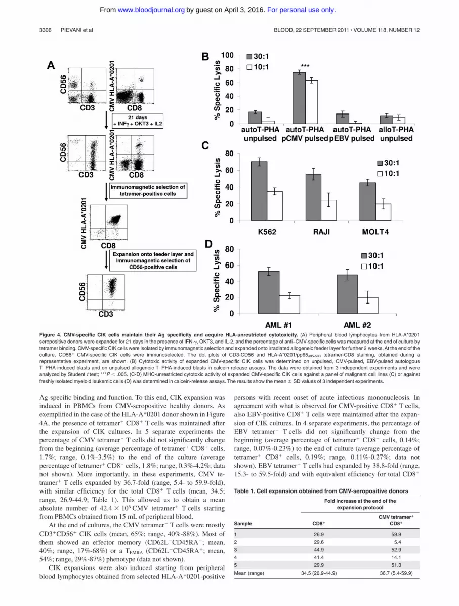
Ag-specific binding and function. To this end, CIK expansion wasinduced in PBMCs from CMV-seropositive healthy donors. Asexemplified in the case of the HLA-A*0201 donor shown in Figure4A, the presence of tetramer� CD8� T cells was maintained afterthe expansion of CIK cultures. In 5 separate experiments thepercentage of CMV tetramer� T cells did not significantly changefrom the beginning (average percentage of tetramer� CD8� cells,1.7%; range, 0.1%-3.5%) to the end of the culture (averagepercentage of tetramer� CD8� cells, 1.8%; range, 0.3%-4.2%; datanot shown). More importantly, in these experiments, CMV te-tramer� T cells expanded by 36.7-fold (range, 5.4- to 59.9-fold),with similar efficiency for the total CD8� T cells (mean, 34.5;range, 26.9-44.9; Table 1). This allowed us to obtain a meanabsolute number of 42.4 � 106 CMV tetramer� T cells startingfrom PBMCs obtained from 15 mL of peripheral blood.
At the end of cultures, the CMV tetramer� T cells were mostlyCD3�CD56� CIK cells (mean, 65%; range, 40%-88%). Most ofthem showed an effector memory (CD62L�CD45RA�; mean,40%; range, 17%-68%) or a TEMRA (CD62L�CD45RA�; mean,54%; range, 29%-87%) phenotype (data not shown).
CIK expansions were also induced starting from peripheralblood lymphocytes obtained from selected HLA-A*0201-positive
persons with recent onset of acute infectious mononucleosis. Inagreement with what is observed for CMV-positive CD8� T cells,also EBV-positive CD8� T cells were maintained after the expan-sion of CIK cultures. In 4 separate experiments, the percentage ofEBV tetramer� T cells did not significantly change from thebeginning (average percentage of tetramer� CD8� cells, 0.14%;range, 0.07%-0.23%) to the end of culture (average percentage oftetramer� CD8� cells, 0.19%; range, 0.11%-0.27%; data notshown). EBV tetramer� T cells had expanded by 38.8-fold (range,15.3- to 59.5-fold) and with equivalent efficiency for total CD8�
Figure 4. CMV-specific CIK cells maintain their Ag specificity and acquire HLA-unrestricted cytotoxicity. (A) Peripheral blood lymphocytes from HLA-A*0201seropositive donors were expanded for 21 days in the presence of IFN-�, OKT3, and IL-2, and the percentage of anti–CMV-specific cells was measured at the end of culture bytetramer binding. CMV-specific CIK cells were isolated by immunomagnetic selection and expanded onto irradiated allogeneic feeder layer for further 2 weeks. At the end of theculture, CD56� CMV-specific CIK cells were immunoselected. The dot plots of CD3-CD56 and HLA-A*0201/pp65495-503 tetramer-CD8 staining, obtained during arepresentative experiment, are shown. (B) Cytotoxic activity of expanded CMV-specific CIK cells was determined on unpulsed, CMV-pulsed, EBV-pulsed autologousT–PHA-induced blasts and on unpulsed allogeneic T–PHA-induced blasts in calcein-release assays. The data were obtained from 3 independent experiments and wereanalyzed by Student t test; ***P � .005. (C-D) MHC-unrestricted cytotoxic activity of expanded CMV-specific CIK cells against a panel of malignant cell lines (C) or againstfreshly isolated myeloid leukemic cells (D) was determined in calcein-release assays. The results show the mean SD values of 3 independent experiments.
Table 1. Cell expansion obtained from CMV-seropositive donors
Fold increase at the end of theexpansion protocol
Sample CD8�
CMV tetramer�
CD8�
1 26.9 59.9
2 29.6 5.4
3 44.9 52.9
4 41.4 14.1
5 29.9 51.3
Mean (range) 34.5 (26.9-44.9) 36.7 (5.4-59.9)
3306 PIEVANI et al BLOOD, 22 SEPTEMBER 2011 � VOLUME 118, NUMBER 12
For personal use only.on April 3, 2016. by guest www.bloodjournal.orgFrom

T cells that showed a mean fold increase of 34.5 (range, 11.7- to67.2-fold increase; supplemental Table 1).
To directly test if indeed Ag-specific CD8� T cells expandedin CIK culture conditions had maintained TCR-dependentcytolytic activity, we analyzed purified, Ag-specific CD3�CD56�
CIK cells. In some experiments, as exemplified in Figure 4A,after 21 days of culture, CMV tetramer� T cells were positivelyselected with the use of anti-PE paramagnetic beads (averagepercentage of purity, 85%; range, 75%-94%; n 3; data notshown) and then cultured for 2 weeks in the presence ofirradiated allogeneic feeder and rhIL-2. At the end of the culture,CD3�CD56� CIK cells were purified by immunomagneticselection (average percentage, 85%; range, 78%-91%; n 3;data not shown) and used for functional experiments.
As shown in Figure 4B, CMV-specific CIK cells showed astrong cytolytic activity against CMV-pulsed autologous T–PHA-induced blasts (average lysis, 63% 3%; n 3) at E/T 10:1. Nolysis was observed against PHA-induced blasts pulsed with EBVpeptides (average lysis, 1% 4%). No killing was detected againstautologous unpulsed (average lysis, 4% 2%) or allogeneicT–PHA-induced blasts (average lysis, 8% 3%).
We next analyzed whether CMV tetramer� CIK cells couldmediate NK-like MHC-unrestricted cytotoxicity against a varietyof malignant cell lines, including K562 (eritroleukemia), RAJI
(B-cell leukemia), and MOLT4 (T-cell leukemia). As shown inFigure 4C, CMV tetramer� CIK cells efficiently killed all malig-nant cell lines tested with 58%-82% specific lysis at 30:1 E/T ratio.Perhaps more importantly, CMV tetramer� CIK cells were able tolyse freshly isolated (allogeneic) leukemic blasts (Figure 4D).
Along this line, CMV tetramer� CIK cells displayed anexpression profile of NKG2D, NKp30, DNAM-1, and LFA-1similar to that of unselected CIK cells (data not shown).Therefore, we further tested the functional capability of all thesereceptors in the context of both TCR-dependent (Ag-specific)and TCR-independent cytotoxicity. K562 and CMV-pulsedT–PHA-induced blasts were assessed for the expression ofligands recognized by NKG2D, DNAM-1, and LFA-1. Asshown in Figure 5, the K562 cells expressed MICA/B, ULBP-2,ULBP-3, PVR, Nectin-2, ICAM-1, and ICAM-2 but not ICAM-3(Figure 5B), whereas CMV-pulsed autologous T–PHA-inducedblasts expressed ULBP-2 and -3 and low levels of PVR andNectin-2 (Figure 5A). Calcein-release assays were performed inthe presence and absence of anti–LFA-1, anti–DNAM-1, anti-NKG2D, and anti-NKp30 blocking mAbs. Importantly, theaddition of blocking mAb directed against LFA-1 and DNAM-1induced a strong inhibition of lysis of both targets, possiblyinterfering with binding of the effector to the target cells (Figure
Figure 5. NKp30, NKG2D, LFA-1, and DNAM-1 are differently involved in Ag-specific and HLA-unrestricted cytotoxicity exerted by CMV-specific CIK cells.(A-B) CMV-pulsed autologous T–PHA-induced blasts (A) and K562 cell line (B) were analyzed for expression of MICA/B and ULBP-1, -2, and -3 (NKG2D ligands) and PVR andNectin-2 (DNAM-1 ligands) by flow cytometry. Gray profiles represent isotype control. (C-D) Blocking of activating receptors NKG2D, NKp30, and DNAM-1 in CIK cellscytotoxicity against CMV-pulsed autologous T–PHA-induced blasts (C) and K562 cell line (D) targets. CIK cells preincubated with saturating concentrations of the indicatedmAbs were used in calcein-release assays. The results shown are the mean percentage of cytotoxic activity of treated compared with untreated CIK cells. The data were SDobtained from 3 independent experiments and were analyzed by Student t test. *P � .05, **P � .01 compared with control in the absence of mAbs.
CIK CELLS ARE T MEMORY WITH NK FUNCTIONS 3307BLOOD, 22 SEPTEMBER 2011 � VOLUME 118, NUMBER 12
For personal use only.on April 3, 2016. by guest www.bloodjournal.orgFrom

5B,D). More importantly, anti-NKG2D and anti-NKp30 block-ing mAbs affected the non–MHC-restricted cytotoxicity but hadno effect on TCR-dependent killing of CMV-pulsed autologousT–PHA-induced blasts (Figure 5B,D), suggesting a role inNK-like cytotoxicity only. It is of note that mAb-mediatedmasking of HLA-class I molecules inhibited lysis of CMV-pulsed autologous T–PHA-induced blasts but not K562(Figure 5C).
Discussion
In this report we analyzed CD3�CD56� CIK cells obtained fromPBMCs after in vitro expansion for 3 weeks by the sequentialaddition of IFN-�, OKT3, and IL-2. We show that CIK cells arecharacterized by a dual function, acting both as CD8�-specificeffector T and NK-like cells. Thus, they represent a particularlyuseful tool, in adoptive immunotherapy, to treat not only cancer butalso associated life-threatening viral infections.
A number of reports on CIK cells, including clinical trials ofadoptively transferred CIK-cell infusion, used bulk-expandedCIK cultures. Within bulk cultures 2 main subpopulations can bedistinguished, one coexpressing the CD3 and CD56 molecules,characterized by a phenotype typical of terminally differentiatedeffector cells, a potent non–MHC-restricted cytotoxicity andlow proliferative capacity. The other cell fraction, displaying aCD3�CD56� phenotype, is composed of effector T cells withreduced cytotoxicity but higher capability of proliferating. Thebulk culture also comprises a small CD3�CD56� classicNK-cell subset.5 In the present study, we characterized in detailthe CD3�CD56� CIK cell fraction, in terms of their molecularmechanism of activation and cytolytic function.
CD3�CD56� CIK cells are capable of rapid binding to lym-phoma target cells, such as BJAB. Preventing cell adhesion withEDTA sharply reduces cytolytic activity of CIK cells againstBJAB. Nonetheless, we cannot rule out also a direct role of calciumchelation induced by EDTA on the cytolytic activity of CIK cells. Adirect correlation seems to exist between degree of binding andsensitivity to CIK-mediated cytotoxicity in different targets, even ifthis is, at the moment, just a hypothesis based only on 2 cell lines.In this context, a significant inhibition of CIK-mediated cytotoxic-ity was obtained by blocking the cell surface adhesion moleculeLFA-1.23,30,31 Although it is clear the crucial role of LFA-1 in CIKcells for functional binding and cytotoxicity against target cellssuch as BJAB, which express high amounts of ICAM-1, -2, and-3 (the main LFA-1 ligands) in the case of other target cells such asKARPAS422, it is probable that other surface molecules areinvolved in binding. The surface expression of high-affinityconformation of LFA-1 does not correlate with the cytotoxicactivity of the different subpopulations of CIK cultures, being thatthis integrin is equally present on CD3�CD56� and on the lesscytotoxic CD3�CD56� cells (data not shown). This confirms thatLFA-1 may not be sufficient for target cell recognition and killingmediated by CD3�CD56� CIK cells.
CIK cells express high levels of CD3 and respond toTCR/CD3-mediated stimulation by degranulating, killing P815cells loaded with anti-CD3 mAb and producing cytokines suchas TNF-� and IFN-�. This is in agreement with our previousobservations that CIK cells are characterized by a phenotypicprofile typical of terminally differentiated effector CD8� T cellsthat originate in vitro from CD56�CD8� T-cell progenitors. Inaddition, CIK cells express NKG2D, DNAM-1, and, at lower
surface densities, NKp30. In contrast, NKp44 and NKp46 arevirtually absent. With the use of redirected killing, degranula-tion assays, and cytokine secretion we could show that NKG2D,DNAM-1, and NKp30 expressed by CIK cells are functional.Remarkably, CD3 triggering induced higher cytotoxicity inredirected killing and higher degranulation and cytokine produc-tion after receptor triggering by activating mAb, compared withNKG2D, DNAM-1, and NKp30, even when used in combina-tion. These results suggest that NKp30 and DNAM-1 do notprovide costimulation to NKG2D signaling.
Despite various studies on CIK cells, the molecular mecha-nism by which they kill tumor cells was not clearly identified.Although the sensitivity of MOLT4 and RAJI cell lines to thecytolytic activity of CIK cells did not appear to correlate withthe surface expression of the ligands of DNAM-1 and NKG2D,the inhibitory effect exerted by anti–DNAM-1 or -NKG2Dblocking antibodies in CIK-mediated lysis of MOLT4 cellssuggested a role of these receptors at least in the case of targets(such as MOLT4) expressing the specific ligands. A partialinhibition of lysis of both targets could be achieved bymAb-mediated blocking of NKp30, which was not significantlyincreased by the addition of a combination of antibodies.
NKG2D has a significant role in triggering IL-2–activatedNK cells, and its ligation induces calcium flux, cytokine release,and cytotoxicity. NKG2D is also expressed on �� T cells andCD8� TCR �� T cells.32,33 In contrast to NK cells, crosslinkingof NKG2D in Ag-specific CTL clones did not induce calciumflux or cytokine production or cytotoxicity. However, NKG2Dsignaling has been shown to augment cytotoxic and proliferativeresponses of T cells to Ag stimulation, thus qualifying NKG2Das a T-cell costimulatory molecule.33-35 A main role for NKG2Dmolecule in CIK-cell function has already been proposed.21,22
Studies of Abs blocking NKG2D molecules, RNA interfering,and redirected cytolysis indicated that the antitumor cytotoxicactivity of CIK cells is exerted through NKG2D rather than TCRengagement.21,22
In the present study we provide evidence that also DNAM-1 andNKp30 play a role in CIK cell–mediated antitumor cytotoxicity.We show that NKp30, although expressed at relatively low density,is involved in the recognition and killing of lymphoma targets.Notably, a functional role of NKp30 in T cells has been describedso far only in the case of IL-15 long-term cultured cord bloodlymphocytes.36 The expression of NKp30 in CIK cells is intriguingalso in light of the reciprocal activation between CIK and dendriticcells37,38 and of the crucial role of NKp30 in the interactionbetween NK cells and dendritic cells.39-41
Other relevant information provided by our study is thedemonstration that Ag-specific CD8� T cells (displaying theCD3�CD56� CIK phenotype) can be expanded in CIK cultures,as shown here for both CMV- and EBV-specific memory cells.We show that these cells are characterized by a dual function.Indeed, they can both specifically kill autologous cells loadedwith CMV peptides and lyse tumor cell lines and freshly isolatedleukemic blasts. Cytotoxicity mediated by CIK cells is non-MHC restricted, and the addition of anti-MHC class I Ab totarget cells had no inhibitory effect.31 Moreover, CIK cells killedwith similar efficiency allogeneic and autologous leukemiccells.4 Killer inhibitory receptors have not been detected in CIKcells.5 Interestingly, anti-NKG2D and anti-NKp30 blockingmAbs inhibit the CIK-mediated killing of the HLA classI–negative K562, whereas no effect was detected on TCR-dependent killing of CMV-pulsed autologous T–PHA-induced
3308 PIEVANI et al BLOOD, 22 SEPTEMBER 2011 � VOLUME 118, NUMBER 12
For personal use only.on April 3, 2016. by guest www.bloodjournal.orgFrom
blasts. These data suggest that these receptors are only involvedin NK-like cytotoxicity. Notably, previous reports suggestedthat, in T cells, NKG2D plays a role as costimulating moleculerather than as a true receptor. In contrast blocking mAbs againstLFA-1 and DNAM-1 induced a significant inhibition of lysis ofboth target types, most probable by interfering with bindingbetween effector and target cells.
On the basis of the data presented, we can hypothesize thatAg-specific T cells may, in particular inflammatory conditions(here mimicked by IFN-� and IL-2 stimulation), acquire addi-tional NK-like functions, similar to those already suggested fora population of T cells expanded in the presence of IL-15.42-44
Indeed, if this was the case, Ag-specific T “CIK” cells may hometo Ag sites (such as the lung in case of CMV infection) and thereexploit also their NK-like potential by contributing to killinfected cells and release cytokines.
The information that CIK cells retain the CTL function (heretested only for anti–CMV-specific memory) adds further interestto their potential clinical application. In patients who received aBM transplant the recovery of functional specific T cells isparticularly delayed, and most patients experience severe infec-tions.45,46 In this respect, CIK cells may thus represent a “onecoin-2 sides” chances of treating leukemia relapses and transfer-ring the donor T-cell memory to cope with most frequent andpossibly fatal infections (ie, CMV, EBV, Aspergillus, etc).Indeed, the absolute numbers of CMV-specific CIK cells, whichcan be obtained from a small amount of peripheral blood, arefully compatible with the idea of obtaining a clinically effectiveanti-CMV CTL (� 1 � 105 in most reports47,48) in standard CIKcultures approved with good manufacturing practices.
Acknowledgments
The authors thank Dr C. Passerini Tosi (Microbiology andVirology Division, Ospedali Riuniti di Bergamo) for providingsamples.
This work was supported in part by research funding fromthe Italian Association for Cancer Research (AIRC; regionalgrant Lombardia and Special Program in Molecular ClinicalOncology 5x1000 “Innate Immunity in Cancer”), by the ItalianMinistry of Health (progetto ordinario, Provincia autonoma diBolzano), and the Associazione Italiana contro le Leucemie,Linfomi e Mieloma (AIL), Bergamo-Sezione Paolo Belli (Ber-gamo, Italy).
Authorship
Contribution: A.P. designed research, performed experiments,analyzed data, and wrote the paper; G.B. performed experimentsand analyzed data; D.P. and L.M. provided reagents, analyzed data,and edited the paper; A.R. and L.M. edited the paper; J.G. designedresearch, analyzed data, and wrote the paper; and M.I. designedresearch, analyzed data, and wrote the paper.
Conflict-of-interest disclosure: The authors declare no compet-ing financial interests.
Correspondence: Martino Introna, Laboratory of CellularTherapy “G. Lanzani,” Ospedali Riuniti, c/o Presidio Matteo Rota,Via Garibaldi 11-13, 24128 Bergamo, Italy; e-mail:[email protected].
References
1. Schmidt-Wolf IG, Negrin RS, Kiem HP, BlumeKG, Weissman IL. Use of a SCID mouse/humanlymphoma model to evaluate cytokine-inducedkiller cells with potent antitumor cell activity. J ExpMed. 1991;174(1):139-149.
2. Introna M, Franceschetti M, Ciocca A, et al. Rapidand massive expansion of cord blood-derivedcytokine-induced killer cells: an innovative pro-posal for the treatment of leukemia relapse aftercord blood transplantation. Bone Marrow Trans-plant. 2006;38(9):621-627.
3. Hoyle C, Bangs CD, Chang P, Kamel O, Mehta B,Negrin RS. Expansion of Philadelphia chromo-some-negative CD3(�)CD56(�) cytotoxic cellsfrom chronic myeloid leukemia patients: in vitroand in vivo efficacy in severe combined immuno-deficiency disease mice. Blood. 1998;92(9):3318-3327.
4. Linn YC, Lau LC, Hui KM. Generation of cytokine-induced killer cells from leukaemic samples within vitro cytotoxicity against autologous and alloge-neic leukaemic blasts. Br J Haematol. 2002;116(1):78-86.
5. Franceschetti M, Pievani A, Borleri G, et al. Cyto-kine-induced killer cells are terminally differenti-ated activated CD8 cytotoxic T-EMRA lympho-cytes. Exp Hematol. 2009;37(5):616-628.e2.
6. Lu PH, Negrin RS. A novel population of ex-panded human CD3�CD56� cells derived fromT cells with potent in vivo antitumor activity inmice with severe combined immunodeficiency.J Immunol. 1994;153(4):1687-1696.
7. Baker J, Verneris MR, Ito M, Shizuru JA,Negrin RS. Expansion of cytolytic CD8(�) naturalkiller T cells with limited capacity for graft-versus-host disease induction due to interferon gammaproduction. Blood. 2001;97(10):2923-2931.
8. Verneris MR, Ito M, Baker J, Arshi A, Negrin RS,Shizuru JA. Engineering hematopoietic grafts:
purified allogeneic hematopoietic stem cells plusexpanded CD8� NK-T cells in the treatment oflymphoma. Biol Blood Marrow Transplant. 2001;7(10):532-542.
9. Marin V, Dander E, Biagi E, et al. Characteriza-tion of in vitro migratory properties of anti-CD19chimeric receptor-redirected CIK cells for theirpotential use in B-ALL immunotherapy. Exp He-matol. 2006;34(9):1219-1229.
10. Sweeney TJ, Mailander V, Tucker AA, et al. Visu-alizing the kinetics of tumor-cell clearance in liv-ing animals. Proc Natl Acad Sci U S A. 1999;96(21):12044-12049.
11. Nishimura R, Baker J, Beilhack A, et al. In vivotrafficking and survival of cytokine-induced killercells resulting in minimal GVHD with retention ofantitumor activity. Blood. 2008;112(6):2563-2574.
12. Edinger M, Cao YA, Verneris MR, Bachmann MH,Contag CH, Negrin RS. Revealing lymphomagrowth and the efficacy of immune cell therapiesusing in vivo bioluminescence imaging. Blood.2003;101(2):640-648.
13. Thorne SH, Negrin RS, Contag CH. Synergisticantitumor effects of immune cell-viral biotherapy.Science. 2006;311(5768):1780-1784.
14. Introna M, Borleri G, Conti E, et al. Repeated in-fusions of donor-derived cytokine-induced killercells in patients relapsing after allogeneic stemcell transplantation: a phase I study. Haemato-logica. 2007;92(7):952-959.
15. Leemhuis T, Wells S, Scheffold C, Edinger M,Negrin RS. A phase I trial of autologous cytokine-induced killer cells for the treatment of relapsedHodgkin disease and non-Hodgkin lymphoma.Biol Blood Marrow Transplant. 2005;11(3):181-187.
16. Schmidt-Wolf IG, Finke S, Trojaneck B, et al.Phase I clinical study applying autologous immu-
nological effector cells transfected with the inter-leukin-2 gene in patients with metastatic renalcancer, colorectal cancer and lymphoma. Br JCancer. 1999;81(6):1009-1016.
17. Olioso P, Giancola R, Di Riti M, Contento A,Accorsi P, Iacone A. Immunotherapy with cyto-kine induced killer cells in solid and hematopoietictumours: a pilot clinical trial. Hematol Oncol.2009;27(3):130-139.
18. Shi M, Zhang B, Tang ZR, et al. Autologous cyto-kine-induced killer cell therapy in clinical trialphase I is safe in patients with primary hepatocel-lular carcinoma. World J Gastroenterol. 2004;10(8):1146-1151.
19. Wang FS, Liu MX, Zhang B, et al. Antitumor ac-tivities of human autologous cytokine-inducedkiller (CIK) cells against hepatocellular carcinomacells in vitro and in vivo. World J Gastroenterol.2002;8(3):464-468.
20. Verneris MR, Baker J, Edinger M, Negrin RS.Studies of ex vivo activated and expanded CD8�NK-T cells in humans and mice. J Clin Immunol.2002;22(3):131-136.
21. Verneris MR, Karami M, Baker J, Jayaswal A,Negrin RS. Role of NKG2D signaling in the cyto-toxicity of activated and expanded CD8� T cells.Blood. 2004;103(8):3065-3072.
22. Karimi M, Cao TM, Baker JA, Verneris MR,Soares L, Negrin RS. Silencing human NKG2D,DAP10, and DAP12 reduces cytotoxicity of acti-vated CD8� T cells and NK cells. J Immunol.2005;175(12):7819-7828.
23. Mehta BA, Schmidt-Wolf IG, Weissman IL,Negrin RS. Two pathways of exocytosis of cyto-plasmic granule contents and target cell killing bycytokine-induced CD3� CD56� killer cells.Blood. 1995;86(9):3493-3499.
24. Bottino C, Castriconi R, Pende D, et al. Identifica-tion of PVR (CD155) and Nectin-2 (CD112) as
CIK CELLS ARE T MEMORY WITH NK FUNCTIONS 3309BLOOD, 22 SEPTEMBER 2011 � VOLUME 118, NUMBER 12
For personal use only.on April 3, 2016. by guest www.bloodjournal.orgFrom

cell surface ligands for the human DNAM-1(CD226) activating molecule. J Exp Med. 2003;198(4):557-567.
25. Pende D, Rivera P, Marcenaro S, et al. Major his-tocompatibility complex class I-related chain Aand UL16-binding protein expression on tumorcell lines of different histotypes: analysis of tumorsusceptibility to NKG2D-dependent natural killercell cytotoxicity. Cancer Res. 2002;62(21):6178-6186.
26. Pende D, Spaggiari GM, Marcenaro S, et al.Analysis of the receptor-ligand interactions in thenatural killer-mediated lysis of freshly isolatedmyeloid or lymphoblastic leukemias: evidence forthe involvement of the Poliovirus receptor(CD155) and Nectin-2 (CD112). Blood. 2005;105(5):2066-2073.
27. Alter G, Malenfant JM, Altfeld M. CD107a as afunctional marker for the identification of naturalkiller cell activity. J Immunol Methods. 2004;294(1-2):15-22.
28. Sabouri AH, Usuku K, Hayashi D, et al. Impairedfunction of human T-lymphotropic virus type 1(HTLV-1)-specific CD8� T cells in HTLV-1-asso-ciated neurologic disease. Blood. 2008;112(6):2411-2420.
29. Brandt CS, Baratin M, Yi EC, et al. The B7 familymember B7-H6 is a tumor cell ligand for the acti-vating natural killer cell receptor NKp30 in hu-mans. J Exp Med. 2009;206(7):1495-1503.
30. Schmidt-Wolf IG, Lefterova P, Johnston V, et al.Sensitivity of multidrug-resistant tumor cell linesto immunologic effector cells. Cell Immunol.1996;169(1):85-90.
31. Schmidt-Wolf IG, Lefterova P, Mehta BA, et al.Phenotypic characterization and identification ofeffector cells involved in tumor cell recognition ofcytokine-induced killer cells. Exp Hematol. 1993;21(13):1673-1679.
32. Bauer S, Groh V, Wu J, et al. Activation of NKcells and T cells by NKG2D, a receptor for stress-inducible MICA. Science. 1999;285(5428):727-729.
33. Groh V, Rhinehart R, Randolph-Habecker J, ToppMS, Riddell SR, Spies T. Costimulation ofCD8alphabeta T cells by NKG2D via engagementby MIC induced on virus-infected cells. Nat Im-munol. 2001;2(3):255-260.
34. Jamieson AM, Diefenbach A, McMahon CW,Xiong N, Carlyle JR, Raulet DH. The role of theNKG2D immunoreceptor in immune cell activa-tion and natural killing. Immunity. 2002;17(1):19-29.
35. Diefenbach A, Tomasello E, Lucas M, et al. Se-lective associations with signaling proteins deter-mine stimulatory versus costimulatory activity ofNKG2D. Nat Immunol. 2002;3(12):1142-1149.
36. Tang Q, Grzywacz B, Wang H, et al. Umbilicalcord blood T cells express multiple natural cyto-toxicity receptors after IL-15 stimulation, but onlyNKp30 is functional. J Immunol. 2008;181(7):4507-4515.
37. Marten A, Ziske C, Schottker B, et al. Interactionsbetween dendritic cells and cytokine-inducedkiller cells lead to an activation of both popula-tions. J Immunother. 2001;24(6):502-510.
38. Li H, Wang C, Yu J, et al. Dendritic cell-activatedcytokine-induced killer cells enhance the anti-tumor effect of chemotherapy on non-small celllung cancer in patients after surgery. Cytotherapy.2009:1-8.
39. Ferlazzo G, Tsang ML, Moretta L, Melioli G,Steinman RM, Munz C. Human dendritic cellsactivate resting natural killer (NK) cells and arerecognized via the NKp30 receptor by activatedNK cells. J Exp Med. 2002;195(3):343-351.
40. Vitale M, Della Chiesa M, Carlomagno S, et al.
NK-dependent DC maturation is mediated byTNFalpha and IFNgamma released upon en-gagement of the NKp30 triggering receptor.Blood. 2005;106(2):566-571.
41. Walzer T, Dalod M, Robbins SH, Zitvogel L, VivierE. Natural-killer cells and dendritic cells: “l’unionfait la force.” Blood. 2005;106(7):2252-2258.
42. Meresse B, Chen Z, Ciszewski C, et al. Coordi-nated induction by IL15 of a TCR-independentNKG2D signaling pathway converts CTL into lym-phokine-activated killer cells in celiac disease.Immunity. 2004;21(3):357-366.
43. Meresse B, Curran SA, Ciszewski C, et al. Repro-gramming of CTLs into natural killer-like cells inceliac disease. J Exp Med. 2006;203(5):1343-1355.
44. Tang F, Chen Z, Ciszewski C, et al. CytosolicPLA2 is required for CTL-mediated immunopa-thology of celiac disease via NKG2D and IL-15.J Exp Med. 2009;206(3):707-719.
45. Boeckh M, Nichols WG. The impact of cytomega-lovirus serostatus of donor and recipient beforehematopoietic stem cell transplantation in the eraof antiviral prophylaxis and preemptive therapy.Blood. 2004;103(6):2003-2008.
46. Leen AM, Rooney CM. Adenovirus as an emerg-ing pathogen in immunocompromised patients.Br J Haematol. 2005;128(2):135-144.
47. Einsele H, Roosnek E, Rufer N, et al. Infusion ofcytomegalovirus (CMV)-specific T cells for thetreatment of CMV infection not responding to an-tiviral chemotherapy. Blood. 2002;99(11):3916-3922.
48. Micklethwaite K, Hansen A, Foster A, et al. Exvivo expansion and prophylactic infusion of CMV-pp65 peptide-specific cytotoxic T-lymphocytesfollowing allogeneic hematopoietic stem celltransplantation. Biol Blood Marrow Transplant.2007;13(6):707-714.
3310 PIEVANI et al BLOOD, 22 SEPTEMBER 2011 � VOLUME 118, NUMBER 12
For personal use only.on April 3, 2016. by guest www.bloodjournal.orgFrom

online August 5, 2011 originally publisheddoi:10.1182/blood-2011-02-336321
2011 118: 3301-3310
Golay and Martino IntronaAlice Pievani, Gianmaria Borleri, Daniela Pende, Lorenzo Moretta, Alessandro Rambaldi, Josée acquires NK function and retains TCR-mediated specific cytotoxicity
CIK cells, a T-cell subset that+CD56+Dual-functional capability of CD3
http://www.bloodjournal.org/content/118/12/3301.full.htmlUpdated information and services can be found at:
(5373 articles)Immunobiology Articles on similar topics can be found in the following Blood collections
http://www.bloodjournal.org/site/misc/rights.xhtml#repub_requestsInformation about reproducing this article in parts or in its entirety may be found online at:
http://www.bloodjournal.org/site/misc/rights.xhtml#reprintsInformation about ordering reprints may be found online at:
http://www.bloodjournal.org/site/subscriptions/index.xhtmlInformation about subscriptions and ASH membership may be found online at:
Copyright 2011 by The American Society of Hematology; all rights reserved.of Hematology, 2021 L St, NW, Suite 900, Washington DC 20036.Blood (print ISSN 0006-4971, online ISSN 1528-0020), is published weekly by the American Society
For personal use only.on April 3, 2016. by guest www.bloodjournal.orgFrom