nondestructive covalent “grafting-from” of poly(lactide) particles of different geometries
TRANSCRIPT

Nondestructive Covalent “Grafting-from” of Poly(lactide) Particles ofDifferent GeometriesRobertus Wahyu N. Nugroho, Karin Odelius, Anders Hoglund, and Ann-Christine Albertsson*
Department of Fibre and Polymer Technology, KTH Royal Institute of Technology, SE-10044, Stockholm, Sweden
*S Supporting Information
ABSTRACT: A nondestructive “grafting-from” method has been developed using poly(lactide) (PLA) particles of differentshapes as substrates and three hydrophilic monomers as grafts. Irregularly shaped particles and spheres of PLA were covalentlysurface functionalized using a versatile method of photoinduced free radical polymerization. The preservation of the molecularweight of the PLA particle bulk and the retention of the original particle shape confirmed the negligible effect of the graftingmethod. The changes in surface composition were determined by FTIR for both spherical and irregular particles and by XPS forthe irregular particles showing the versatility of the method. Changes in the surface morphology of the PLA spherical particleswere observed using microscopy techniques showing a full surface coverage of one of the grafted monomers. The method isapplicable to a wide set of grafting monomers and provides a permanent alteration of the surface chemistry of the PLA particlescreating hydrophilic PLA surfaces in addition to creating sites for further modification and drug delivery in the biomedical fields.
KEYWORDS: surface grafting, PLA, hydrophilic, particles, geometry
■ INTRODUCTIONParticles for use in biomedical applications require a controlled3D-structure and a controlled chemical composition of theparticle surface.1 Hence, there is a need to be able to createparticles with attributes such as a predetermined narrowparticle size range, anisotropic properties, a well-defined surfacemicrostructure, topography, and shape factor, i.e., symmetricity,while simultaneously retaining or creating an appropriateparticle surface composition. Spherical particles are createdthrough techniques such as oil-in-water emulsion, precipitation,solvent evaporation, and mini-emulsion polymerization.2,3
More complex particle shapes have been demonstrated usingprogressive techniques such as electrospraying,4 particlereplication in nonwetting templates,5 and recently by themanipulation of spherical particles.6 These techniques createparticles of different geometries, and their shape can directlyinfluence the performance in an intended application. Knowingthe shape factor is therefore necessary to determine the primaryrole of the particles in a wide range of applications.7−9
In surface modification, in contrast to bulk modification, onlythe surface of the object should be physically or chemicallymodified. Physical modification entails the physical adsorption
of a polymer onto the substrate surface, whereas in chemicalmodification grafted chains are covalently attached to thesubstrate surface. Numerous chemical modification techniqueshave been established including wet-chemical reactions, ozonetreatment, UV-treatment and photografting, plasma treatment,high-energy irradiation, and vapor-phase grafting.10−12 Vapor-phase grafting employs a solvent-free system that underreduced pressure creates a vapor of monomer and initiatorthat upon UV-irradiation polymerizes from the substratesurface. This procedure has been proven viable for biostableand biodegradable substrates and for simpler and morecomplex structures, creating covalently attached grafted chainswithout degrading the substrate.13−16 Unfortunately, many ofthese methods are not easily translated from polymer films topolymer particles with different geometries if complete surfacecoverage of new functionality is desired. Thus, there are todayfew viable routes for the covalent attachment of functionalmoieties on particle surfaces. One method that has been proven
Received: February 27, 2012Accepted: May 16, 2012Published: May 16, 2012
Research Article
www.acsami.org
© 2012 American Chemical Society 2978 dx.doi.org/10.1021/am3003507 | ACS Appl. Mater. Interfaces 2012, 4, 2978−2984

to work in the case of particles is the use of wet-chemicalreactions where chemical compounds in solution react with thesurface of the objects. For biodegradable polymers such aspolylactide, this is normally performed through aminolysis,17
hydrogen peroxide treatment,18,19 and alkaline or acidichydrolysis.20,21 An increase in the wettability and surfaceroughness are benefits commonly seen, but there are also somelimitations, including nonspecificity giving a range of functionalgroups, a lack of reproducibility, and sometimes accelerateddegradation rates of the modified substrates. Another methodthat can be used with particles is UV-treatment andphotografting. The UV treatment of films has been extensivelystudied22−25 mainly because it is easily performed under mildreaction conditions and because in the presence of aphotoinitiator, such as benzophenone and hydrogen peroxide,the UV-radiation selectively influences the sample surface.23
However, less attention has been paid to the surfacemodification of particles and, perhaps more importantly, tothe introduction of covalent functionalization without deteri-oration in the properties of the substrate.Our main objective in the present work was to design a
nondestructive surface “grafting-from” technique that can beapplied to sensitive biodegradable polymers particles. Thetechnique should be valid for simple to complex geometries andallow the substrates to maintain their original shape withoutdegradation of the substrate particle. In addition, it should beapplicable to a wide range of grafting monomers and shouldprovide permanent alteration of the surface chemistry of thesubstrate creating not only hydrophilic poly(lactide) (PLA)surfaces but also sites for further modification. The surfacegrafted particles can be used in biomedical applications wheresecondary interactions are important such as drug delivery.
■ EXPERIMENTAL SECTIONMaterials. Poly(lactide) (PLA) was obtained from Nature Works
Co. Ltd. USA (5200D) and used as received. Benzophenone (BP)
(99%, SigmaAldrich), acrylamide (AAm) (98.5%, Acros), and maleicanhydride (MAH) (>99%, Fluka) were used as received. Acrylic acid(AA) (90%, Alfa Aesar) was purified by vacuum distillation at 40 °Cprior to use. Dichloromethane (DCM) (>99%, Fisher Scientific),ethanol (96% v/v, VWR), and polysorbate 80 (commonly known asTween 80) (Fluka) were used as received.
Particle Fabrication. Oil-in-Water Emulsion. PLA sphericalparticles were fabricated by an oil-in-water emulsion technique (O/W). PLA (0.2 g) was dissolved in 100 mL of DCM (organic phase),and 10 mL of this organic phase was thereafter poured into an aqueoussolution containing 0.05% w/v of Tween 80. The two immisciblephases were continuously stirred at room temperature overnight untilthe organic solvent had evaporated, and PLA spherical particles withdiameters in the range of 10−60 μm were then recovered from theaqueous phase. The spheres were filtered, rinsed with deionized waterto remove surfactant, and finally dried overnight.
Cryogenic Milling. Ten grams of PLA pellets was frozen in acylindrical thermos filled with liquid nitrogen in order to minimizeoverheating during the grinding process. The grinding process wasconducted using a ZM 200 Retsch mill, equipped with a stainless steelbath to collect the irregular particles, and adjusted at a fixed rotationalvelocity of 12 000 rpm. PLA irregular particles (for SEM images seeFigure S1 in the Supporting Information) with Feret’s diameter in therange of 600−1200 μm were thereafter recovered from their bath. Themill was run for a maximum of 1 min in order to avoid thermaldegradation during the process.
“Grafting-from” Approach. Photoactivation. The PLA particleswere activated by soaking them in 5% w/v BP in ethanol in a Pyrexglass tube. The particle-containing solution was then placed on astirring plate and UV-irradiated for 20 min with a UV lamp (OhsramUltra Vitalux, 300 W) having a wavelength range of 280−320 nm andan output intensity of 38 mW/cm2. After activation, the particles werecentrifuged using a Hettich Universal 30 F at a fixed speed of 5000rpm for 3 min to remove all ungrafted initiator. The particles wererinsed with ethanol several times, placed in a closed Petri dish, driedovernight, and finally characterized by FTIR spectroscopy. Controlparticles without BP were also prepared using the same procedures.
Photoinduced Polymerization from the Activated Particles. TheBP-activated particles were placed in Pyrex glass tubes using ethanol assolvent and 20% w/v acrylamide (AAm), 20% v/v acrylic acid (AA), or
Scheme 1. Two-Step “Grafting-from” Technique to Produce Spherical Particles Indicated with Acrylamide, Acrylic Acid, andMaleic Anhydride
ACS Applied Materials & Interfaces Research Article
dx.doi.org/10.1021/am3003507 | ACS Appl. Mater. Interfaces 2012, 4, 2978−29842979

20% w/v maleic anhydride (MAH). The particle-containing solutionswere exposed to UV-radiation for periods of 15 min, 30 min, 45 min, 1h, and 1.5 h. The surface-grafted particles were thereafter centrifugedat a fixed speed of 5000 rpm for 3 min to eliminate any free polymerchains, washed with ethanol, dried overnight, and characterized byFTIR spectroscopy. Control particles without added monomer werealso prepared using the same procedure.Characterization. Size Exclusion Chromatography (SEC). The
molecular weights of the pristine, control, activated, and graftedparticles were determined using a Waters 717 plus autosampler and aWaters model 510 apparatus equipped with two PLgel 10 μm mixed Bcolumns, 300 × 7.5 mm (Polymer Laboratories, U.K). Chloroform wasused as eluent at a flow rate of 0.1 mL/min. The instrument wascalibrated with polystyrene standards with narrow molecular weightdistributions in the range from 580 to 400 000 g/mol.Fourier Transform Infrared (FTIR) Spectroscopy. The success of
the surface grafting technique was verified using FTIR. Spectra wererecorded in the range of 4000−600 cm−1 on a Spectrum 2000 Perkin-Elmer spectrometer equipped with an attenuated total reflectance(ATR) accessory (Golden Gate) that makes measurements to a depthof approximately 1 μm on the sample surface. All FTIR spectra wereobtained as means of 5 samples and 16 individual scans at 4 cm−1
resolution.X-ray Photoelectron Spectroscopy (XPS). The success of the
surface grafting on the PLA irregular particles was verified using XPS.The XPS measurements were run on a Phi Electronics Quantum 2000,using a monochromatic Al X-ray source (hυ = 1486.86 eV). For X-raysurface analysis, a 45° angle monochromatic Al Kα X-ray sourceoperating at 21.1 W and a pass energy of 58.7 eV was used.Scanning Electron Microscope (SEM). The PLA spherical particles
produced by O/W emulsion were examined using a Hitachi S-4800scanning electron microscope (SEM) at an accelerating voltage of 0.7kV. The particles were mounted on adhesive carbon black and sputter-coated with a 10 nm gold/palladium layer.Atomic Force Microscopy (AFM). The PLA spherical particles were
topographically characterized using a nanoscope IIIa multimode AFM(Digital Instruments, Santa Barbara, CA) via 5346 EV scanner. Asilicon-etched probe tip (NSC14/noAl, Mikromasch, Estonia), with anormal spring constant (k) of 5 N/m and a resonant frequency (fo) of110−220 kHz, was used to scan the image in the tapping mode. Thesurface of the emulsified PLA particles was scanned in the range of 1−2 Hz. The very slow scan rate was chosen to avoid sampledeformation. The maximum sample size (512 × 512 pixels) wasselected.
■ RESULTS AND DISCUSSION
We here demonstrate a surface “grafting-from” method of PLAparticles having two different shapes with three differentmonomers: acrylamide (AAm), acrylic acid (AA), and maleicanhydride (MAH). The “grafting-from” approach is a two-stepprocess where the PLA particle surfaces are first activated withbenzophenone, BP, one of the three chosen monomers is then“grafted-from” the surface of the PLA particles through aphotoinduced free-radical polymerization method. A schematicview of the two-step “grafting-from” technique onto sphericalparticles is shown in Scheme 1.23 PLA pellets were used tocreate particles with two different geometries; irregularlyshaped particles made by cryogenic milling and sphericalparticles from an oil/water emulsion. Due to their higher yieldand more complex structure, the grinded particles were used todevelop the surface-grafting method and the nondestructivecharacter was thereafter confirmed using the spherical particles.Alterations in surface chemistry, morphology, and topographywere characterized to verify that the “grafting-from” techniqueis an applicable nondestructive method and that the three-dimensional shape of the particles was retained aftermodification. Control experiments with acrylamide in the
absence of benzophenone under UV irradiation source for 1.5 hwere performed to verify that the grafted layers were not aresult of physisorbed monomer.
Nondestructive Nature of the “Grafting-From” Tech-nique. It is well-known that the surface modification ofbiodegradable polymers tends to cause polymer degradation bychain scission, unzipping, or cross-linking reactions. High-energy techniques such as electron-beam26,27 and gamma-rayirradiation,28 and in many cases also UV-irradiation,29 aredetrimental to biodegradable aliphatic polyesters such as
Table 1. Number-Average Molecular Weights andPolydispersity Indices of the PLA Particles before, during,and after Surface Grafting
sample Mnb PDIb
PLA pellet 166 000 ± 3800 1.46 ± 0.00cryogenic milled PLA particles 145 000 ± 200 1.62 ± 0.01
emulsified PLA particles 146 000 ± 10 300 1.62 ± 0.05irregular PLA particles with BPa 144 000 ± 3300 1.64 ± 0.02spherical PLA particles with BPa 142 000 ± 13 000 1.66 ± 0.17
BP activated irregular particles and UVirradiated for 1.5 h
158 000 ± 3600 1.54 ± 0.02
BP activated spherical particles and UVirradiated for 1.5 h
131 000 ± 5423 1.72 ± 0.14
MAH grafted PLA irregular particlesfor 1.5 h
126 000 ± 3600 1.46 ± 0.02
MAH grafted PLA spherical particlesfor 1.5 hc
aThe PLA particles were activated in ethanol for 20 min. bDeterminedusing SEC. cImmiscible in chloroform.
Figure 1. ATR-FTIR spectra of pristine PLA particles and acrylamide-grafted PLA particles at different times. The amide I band is indicatedby a solid arrow, and the amide II band is indicated by a dotted arrow.The asterisk (∗) shows the absorption peak of CO stretching.
Figure 2. ATR-FTIR spectra of pristine PLA particles and acrylic acid-grafted PLA particles at different times. The asterisk (∗) shows theabsorption peak of CO stretching.
ACS Applied Materials & Interfaces Research Article
dx.doi.org/10.1021/am3003507 | ACS Appl. Mater. Interfaces 2012, 4, 2978−29842980
poly(lactide). However, it has been shown that the reduction inmolecular weight due to UV-irradiation is reduced if the PLAsample is placed in a pyrex tube that prevents wavelengthsshorter than 300 nm from reaching the sample.25,29 To confirmthe nondestructive nature of our approach, the molecularweights of the PLA particles before and after surface activationand grafting were determined, Table 1. The molecular weightsof the PLA particles were not significantly affected either duringthe activation or during the surface modification initiated byUV-irradiation. Unfortunately, the molecular weights of theparticles after “grafting-from” of the three different monomerscould only be determined for MAH due to the limited solubilityof the grafted chains in common SEC solvents. The molecularweight of the MAH-grafted PLA particles was slightly lowerthan that of the pristine PLA particles. An increase or decreasein molecular weight is commonly seen in co- and graft-
polymers when the two polymers have different hydrodynamicvolumes in the SEC solvent and an artificial change in themolecular weight can be measured. Depending on the graftthickness, the molecular weights of films have in certain casespreviously been measured by SEC.15,25 A low grafting yieldresults in a thin grafted film that is soluble, whereas a highgrafting yield results in thicker grafted films that are insoluble.The same reasoning can be applied to particles that have amuch higher surface area-to-volume than that of the films andexplain the insolubility of the grafted particles with a low graftthickness.Since the benzophenone initiator is a hydrogen abstractor, it
could lead to cross-linking reactions between PLA chains in theparticles or between the growing chains resulting in gelformation. This was, however, no major side reaction in oursystem since the PLA particles activated by BP showed nochange in molecular weight. In addition, the BP-activatedchains that were subsequently UV-irradiated for 1.5 h showedonly a very a slight increase in molecular weight and a smalldecrease in PDI, indicating that a small fraction of the formedradicals had combined, Table 1. Further, the PAA-grafted PLAparticles were soluble in 1,4-dioxane, a common solvent for thetwo polymers, which showed that the grafted chains had notformed networks.
Covalent Surface Modification. The surface chemistriesof the PLA irregular particles after “grafting-from” wereevaluated using FTIR and compared to that of the pristinePLA irregular particles, Figures 1, 2, and 3. As a control, PLAparticles UV-irradiated for 1.5 h in ethanol were used and nochange in the surface chemistry was observed. The PLAparticles showed the expected FTIR spectra with a character-istic ester CO band at 1745 cm−1 (asterisk). Upon AAmgrafting, Figure 1, this band was shifted down and showed amarked change in area and height. With increasing graftingtime, a shoulder formed near 1653 cm−1, which is consistentwith the characteristic CO band which primary amidestypically portray in the 1670−1650 cm−1 region (amide Iband). Primary amides also typically show a band around 1620cm−1 (amide II band), observed here at 1615 cm−1,15,25,30
confirming the successful grafting of the AAm onto the PLA.The C−N stretching bands that should appear in the 1440−1200 cm−1 region were difficult to identify because they overlapwith bands originating from the PLA substrate.For AA-grafted PLA, Figure 2, the CO stretching bands
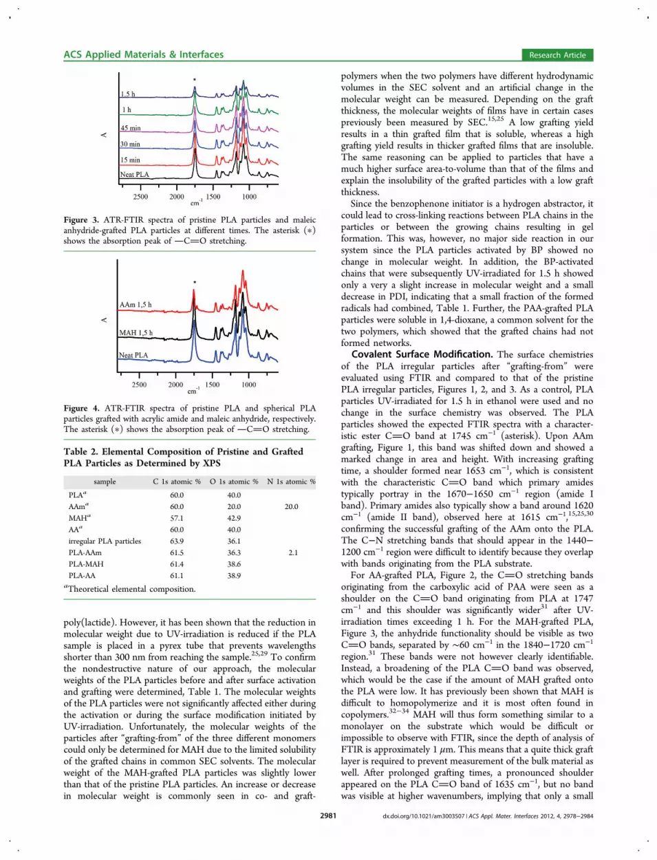
originating from the carboxylic acid of PAA were seen as ashoulder on the CO band originating from PLA at 1747cm−1 and this shoulder was significantly wider31 after UV-irradiation times exceeding 1 h. For the MAH-grafted PLA,Figure 3, the anhydride functionality should be visible as twoCO bands, separated by ∼60 cm−1 in the 1840−1720 cm−1
region.31 These bands were not however clearly identifiable.Instead, a broadening of the PLA CO band was observed,which would be the case if the amount of MAH grafted ontothe PLA were low. It has previously been shown that MAH isdifficult to homopolymerize and it is most often found incopolymers.32−34 MAH will thus form something similar to amonolayer on the substrate which would be difficult orimpossible to observe with FTIR, since the depth of analysis ofFTIR is approximately 1 μm. This means that a quite thick graftlayer is required to prevent measurement of the bulk material aswell. After prolonged grafting times, a pronounced shoulderappeared on the PLA CO band of 1635 cm−1, but no bandwas visible at higher wavenumbers, implying that only a small
Figure 3. ATR-FTIR spectra of pristine PLA particles and maleicanhydride-grafted PLA particles at different times. The asterisk (∗)shows the absorption peak of CO stretching.
Figure 4. ATR-FTIR spectra of pristine PLA and spherical PLAparticles grafted with acrylic amide and maleic anhydride, respectively.The asterisk (∗) shows the absorption peak of CO stretching.
Table 2. Elemental Composition of Pristine and GraftedPLA Particles as Determined by XPS
sample C 1s atomic % O 1s atomic % N 1s atomic %
PLAa 60.0 40.0AAma 60.0 20.0 20.0MAHa 57.1 42.9AAa 60.0 40.0irregular PLA particles 63.9 36.1PLA-AAm 61.5 36.3 2.1PLA-MAH 61.4 38.6PLA-AA 61.1 38.9
aTheoretical elemental composition.
ACS Applied Materials & Interfaces Research Article
dx.doi.org/10.1021/am3003507 | ACS Appl. Mater. Interfaces 2012, 4, 2978−29842981

amount of anhydride groups were present on the PLA particles.However, considering the probability of monolayer formationand the instability of anhydride functionalities toward water,this could be expected.To assess the versatility of the surface grafting technique to
different substrate shapes and two monomers, AAm and MAHwere chosen as model monomers for grafting onto sphericallyshaped particles. The FTIR spectra of the grafted PLA particlesafter 1.5 h of grafting were similar to those found for theirregularly shaped particles. However, weaker absorption bandsoriginating from the grafted AAm and MAH were observed,Figure 4. This effect is explained by the different shapes of theparticles, where the spherical particles have a larger surface areathan the irregular particles and hence need longer reactiontimes to reach the same grafting degree. This effect was evenmore pronounced after longer reaction times.To confirm that the “grafting-from” technique resulted in the
covalent attachment of grafts to the particle surface and notonly adsorption of the polymers onto the particle surface, apostsurface grafting test was conducted. AAm-grafted particleswere selected as test specimens since both the monomer andthe polymer are soluble in water. Particles were soaked in waterfor 24 h; samples were regularly withdrawn, and the covalentattachment of AAm was assessed with FTIR. No change in thesurface characteristics of the PLA-g-AAm was seen.The surface compositions of the grafted films were also
analyzed by XPS, permitting the elemental analysis of the
outermost surface of the irregular particles, Table 2. The surfacecomposition measured by XPS (as atomic % of the elementspresent, H excluded) of the AAm-grafted PLA particlesrevealed, as expected, that the AAm-grafted particles containednitrogen, confirming the FTIR results. The amount of nitrogenwas however lower than the theoretical value for pure PAAm.This could indicate that the substrate was not completelycovered or more likely that the XPS penetrates deeper into thesubstrate than the thickness of the AAm layer and hence thatthe PLA bulk material of the substrate was included in the layerinvestigated by this analysis. For the AA- and MAH-graftedPLA particles, there was no significant difference between thetheoretical composition of PLA and PMAH and PAA, makinginterpretation of the XPS results difficult. It can be seen that theoxygen amount was higher in the grafted samples than in thepristine PLA particles, indicating that a change in the surfacecomposition had occurred. This is interpreted as confirming thesuccessful surface grafting of AA and MAH onto the PLAparticle surface. We can also conclude that carbon and oxygenwere the only elements present on the surface and that thesurface contained no nitrogen. The discrepancy betweentheoretical atomic composition of PLA (60% carbon and 40%oxygen) and measured atomic composition of PLA (63.9%carbon and 36.1% oxygen) is probably due to a combination ofsmall amounts of impurities attached on the PLA surface andcontamination of the XPS instrument.
Figure 5. Scanning electron microscopy images of (A) pristine PLA spheres, (B) control PLA spheres UV-irradiated for 1.5 h without the presenceof any monomer, (C) PLA spheres grafted with acrylamide for 1.5 h, and (D) PLA spheres grafted with maleic anhydride for 1.5 h.
ACS Applied Materials & Interfaces Research Article
dx.doi.org/10.1021/am3003507 | ACS Appl. Mater. Interfaces 2012, 4, 2978−29842982

Morphological and Topographical Changes of theGrafted PLA Particles. To elucidate the influence of thesurface grafting technique on the morphology of the PLAspheres, SEM images were captured before and after surfacegrafting, Figure 5. The pristine PLA spheres had an averagediameter of 36 ± 13 μm and in some cases showed a surfacemorphology with small pores formed during the fabricationprocess,35 probably due to a very rapid evaporation of theorganic solvent (DCM) and the air humidity, Figure 5.Although they were imperfect, the particles were chosen forsurface morphology and topography measurements using thepores as indicators of complete or partial surface coverage. Thecontrol PLA spheres UV-irradiated for 1.5 h in ethanol withoutmonomer showed that the ethanol does not harm the PLAparticles since no change in the surface morphology wasobserved. A surface morphology similar to that of the pristinePLA spheres was observed for the MAH-grafted PLA,confirming the low degree of grafting indicated by FTIR.This again demonstrated the nondetrimental surface graftingapproach. However, the surface morphology of the acrylamide-grafted spheres, with an average diameter of 63 ± 28 μm, wasdifferent from that of the pristine PLA spheres. A smoothsurface and an increase in the diameter size were obtained,confirming the FTIR results and showing a complete but thinsurface coverage. The diameters of the spheres weredetermined by ImageJ software measuring approximately 100randomly chosen spheres.Atomic force microscopy was used to evaluate the top-
ographies of pristine PLA spheres, PLA spheres UV-irradiatedin ethanol only for 1.5 h, and acrylamide-grafted and maleicanhydride-grafted PLA spheres, Figure 6. The pristine PLAspheres had a relatively smooth surface with a surface asperityof 5.33 nm, calculated as the root-mean-square (rms)roughness. UV-irradiation of the PLA spheres in ethanol gave
a surface asperity with a rms roughness of 37.2 nm, showing theeffect of ethanol on the surface topography of the sphericalparticles. The surface roughness of maleic anhydride-graftedPLA spheres was slightly greater at 42.8 nm, while theacrylamide-grafted PLA spheres had a rms roughness of 16.9nm. This again indicates that a monolayer of maleic anhydridewas grafted-from the PLA backbone but that the grafting layerof the polyacrylamide was thicker and smoother and coveredthe PLA surface evenly as shown by SEM and FTIR. The AFMphase images thus show that the monomer-carrier solventinfluenced the rms roughness, even though there was nosignificant change in molecular weight before and aftertreatment.
■ CONCLUSIONS
A nondestructive surface “grafting-from” technique wasdeveloped and shown to be successful in covalently graftingthree different monomers (acrylamide, acrylic acid, and maleicanhydride) onto particle substrates of different geometries. Themolecular weight of the PLA particles did not change after thesurface activation and grafting process, and the particlesretained their 3D-shape, proving the nondestructivity of themethod. Alterations in the surface chemistry of the two PLAsubstrates were confirmed by FTIR and XPS, for the irregularparticles. Additionally, an increase in the diameter size ofacrylamide-g-PLA spheres, as compared to pristine PLAspheres, has exhibited the success of the nondestructive“grafting-from” technique. The root-mean-square roughnessdetermined by AFM decreased upon grafting of acrylamidecompared to the PLA control and showed that the graftinglayer was evenly distributed over the spherical particle surface.Maleic anhydride formed a monolayer of grafted monomer onthe PLA sphere surface, as confirmed by FTIR, SEM, and AFM.
Figure 6. Atomic force microscopy phase images of the surfaces of (A) pristine PLA sphere, (B) PLA sphere UV-irradiated in ethanol only for 1.5 h,(C) acrylamide-grafted PLA sphere, and (D) maleic anhydride-grafted PLA spheres. All AFM pictures were scanned over an area of 5 μm × 5 μm.
ACS Applied Materials & Interfaces Research Article
dx.doi.org/10.1021/am3003507 | ACS Appl. Mater. Interfaces 2012, 4, 2978−29842983

We emphasize that the designated “grafting-from” techniquedescribed here is nondestructive, achieves covalently attachedgrafts, is applicable to PLA particles of different geometries, andinduces hydrophilicity to the PLA surface and a functionalitythat can be used for further coupling reactions and colloidalstability in the medical applications.
■ ASSOCIATED CONTENT*S Supporting InformationSEM images of irregular PLA particles. This material is availablefree of charge via the Internet at http://pubs.acs.org
■ AUTHOR INFORMATIONCorresponding Author*Tel. +46-8-790 82 74. Fax: +46-8-20 84 77. E-mail: [email protected] authors declare no competing financial interest.
■ ACKNOWLEDGMENTSThe authors gratefully acknowledge Bjorn Atthoff for his kindhelp in running and interpreting the XPS measurements andthe ERC Advanced Grant, PARADIGM, (Grant AgreementNo.: 246776) and The Swedish Research Council, VR, (GrantID: 621-2010-3478) for their financial support for this work.
■ REFERENCES(1) Wang, J.; Byrne, J. D.; Napier, M. E.; DeSimone, J. M. Small2011, 7, 1919−1931.(2) Landfester, K. Angew. Chem., Int. Ed. 2009, 48, 4488−4507.(3) Edlund, U.; Albertsson, A.-C. Adv. Polym. Sci. 2002, 157, 67−112.(4) Bhaskar, S.; Pollock, K. M.; Yoshida, M.; Lahann, J. Small 2010,6, 404−411.(5) Gratton, S. E. A.; Ropp, P. A.; Pohlhaus, P. D.; Luft, J. C.;Madden, V. J. N., M. E.; DeSimone, J. M. Proc. Natl. Acad. Sci. U.S.A.2008, 105, 11613.(6) Champion, J. A.; Katare, Y. K.; Mitragotri, S. Proc. Natl. Acad. Sci.2007, 104, 11901−11904.(7) Hassan, M.; Lau, R. AAPS PharmSciTech 2009, 10, 1252−1262.(8) Qi, W.; Wang, M.; Liu, Q. J. Mater. Sci. 2005, 40, 2737−2739.(9) Casal, J.; Lucas, A.; Arnaldos, J. Chem. Eng. J. 1985, 30, 155−158.(10) Desmet, T.; Morent, R.; Geyter, N. D.; Leys, C.; Schacht, E.;Dubruel, P. Biomacromolecules 2009, 10, 2351−2378.(11) Li, H.; Xia, Y.; Wu, J.; He, Q.; Zhou, X.; Lu, G.; Shang, L.; Boey,F.; Venkatraman, S. S.; Zhang, H. ACS Appl. Mat. Interfaces 2012, 4,687−693.(12) Kim, D.; Andou, Y.; Shirai, Y.; Nishida, H. ACS Appl. Mat.Interfaces 2010, 3, 385−391.(13) Wirsen, A.; Sun, H.; Emilsson, L.; Albertsson, A. C.Biomacromolecules 2005, 6, 2281−2289.(14) Wirsen, A.; Sun, H.; Albertsson, A.-C. Biomacromolecules 2005,6, 2697−2702.(15) Edlund, U.; Kallrot, M.; Albertsson, A.-C. J. Am. Chem. Soc.2005, 127, 8865−8871.(16) Kallrot, M.; Edlund, U.; Albertsson, A.-C. Macromol. Biosci.2008, 8, 645−654.(17) Zhu, Y.; Gao, C.; Liu, X.; Shen, J. Biomacromolecules 2002, 3,1312−1319.(18) De Feng, X.; Voong; Sun, Y. H.; Qiu, K. Y. Makromol. Chem.1985, 186, 1533−1541.(19) Guan, J.; Gao, C.; Feng, L.; Shen, J. J. Mater. Sci.: Mater. Med.2001, 12, 447−452.(20) Gao, J.; Niklason, L.; Langer, R. J. Biomed. Mater. Res. 1998, 42,417−424.(21) Sun, S.-P.; Wei, M.; Olson, J. R.; Shaw, M. T. ACS Appl. Mat.Interfaces 2009, 1, 1572−1578.
(22) Chan, C. M.; Ko, T. M.; Hiraoka, H. Surf. Sci. Rep. 1996, 24, 1−54.(23) Ma, H.; Davis, R. H.; Bowman, C. N. Macromolecules 1999, 33,331−335.(24) He, D.; Ulbricht, M. Macromol. Chem. Phys. 2007, 208, 1582−1591.(25) Janorkar, A. V.; Metters, A. T.; Hirt, D. E. Macromolecules 2004,37, 9151−9159.(26) Loo, S. C. J.; Ooi, C. P.; Boey, Y. C. F. Polym. Degrad. Stab.2004, 83, 259−265.(27) Plikk, P.; Odelius, K.; Hakkarainen, M.; Albertsson, A. C.Biomaterials 2006, 27, 5335−5347.(28) Gupta, M. C.; Deshmukh, V. G. Polymer 1983, 24, 827−830.(29) Janorkar, A. V.; Metters, A. T.; Hirt, D. E. J. Appl. Polym. Sci.2007, 106, 1042−1047.(30) Rasal, R. M.; Bohannon, B. G.; Hirt, D. E. J. Biomed. Mater. Res.,Part B: Appl. Biomater. 2008, 85B, 564−572.(31) Janorkar, A. V.; Proulx, S. E.; Metters, A. T.; Hirt, D. E. J. Polym.Sci., Part A: Polym. Chem. 2006, 44, 6534−6543.(32) Deng, J.-P.; Yang, W.-T.; Ranby, B. Eur. Polym. J. 2002, 38,1449−1455.(33) Pan, B.; Viswanathan, K.; Hoyle, C. E.; Moore, R. B. J. Polym.Sci., Part A: Polym. Chem. 2004, 42, 1953−1962.(34) Jianping, D.; Wantai, Y. J. Appl. Polym. Sci. 2005, 95, 903−909.(35) Xu, Q.; Hashimoto, M.; Dang, T. T.; Hoare, T.; Kohane, D. S.;Whitesides, G. M.; Langer, R.; Anderson, D. G. Small 2009, 5, 1575−1581.
ACS Applied Materials & Interfaces Research Article
dx.doi.org/10.1021/am3003507 | ACS Appl. Mater. Interfaces 2012, 4, 2978−29842984