crystal structures of arginine deiminase with covalent reaction intermediates
TRANSCRIPT

Structure, Vol. 12, 657–667, April, 2004, 2004 Elsevier Science Ltd. All rights reserved. DOI 10.1016/j .str .2004.02.017
Crystal Structures of Arginine Deiminasewith Covalent Reaction Intermediates:Implications for Catalytic Mechanism
ArginineArginine Deiminase (ADI)
H2O→ [NH3] �
CitrullineOrnithine Transcarbamylase (OTCase)
Pi
→ [Carbamoyl Phosphate] �
Kalyan Das,1,* Gary H. Butler,2
Victoria Kwiatkowski,2 Arthur D. Clark, Jr.,1
Prem Yadav,1 and Eddy Arnold1,*1Center for Advanced Biotechnology
and Medicine (CABM) andOrnithine
Carbamate Kinase (CK)
ADP→ ATP � HCO3 � NH3.Department of Chemistry and Chemical Biology
Rutgers UniversityADI (EC 3.5.3.6), encoded by the 2189 base arcA gene,679 Hoes Lanecomposes almost 10% of the total soluble protein massPiscataway, New Jersey 08854of M. arginini (Schimke et al., 1966). Apart from M. argi-2 Coriell Institute for Medical Researchnini, numerous bacteria including Mycoplasma arthri-401 Haddon Avenuetidis, Mycoplasma hominis, Mycobacterium tuberculo-Camden, New Jersey 08103sis, Lyme disease spirochetes, and Streptococcus usethe ADI pathway for arginine catabolism and energyproduction. A high variability exists among the se-quences of the ADI enzymes from different speciesSummary(Sugimura et al., 1993). Enzymes that can degrade cer-tain amino acids have potential for use as tumor-growthArginine deiminase (ADI), an enzyme that hydrolyzesinhibitors: L-asparaginase from Escherichia coli is clini-arginine to generate energy in many parasitic microor-cally used in treating certain leukemia and lymphosar-ganisms, has potent anticancer activities and can haltcoma (Kidd, 1973). Arginase and ADI, both of whichgrowth of solid tumors. We determined the crystaldegrade arginine, are potential tumor growth inhibitorsstructure of ADI from Mycoplasma arginini in two dif-(Dillon et al., 2002; Miyazaki et al., 1990; Philip et al.,ferent forms (1.6 and 2.0 A resolution) using multiple2003; Terayama et al., 1982). ADIs from M. arginini andisomorphous replacement. ADI shares common struc-related species have been reported to inhibit varioustural features with the arginine-catabolizing enzymesmelanoma (Ensor et al., 2002; Sugimura et al., 1992;
Arg:Gly amidinotransferase and dimethylarginine di-Takaku et al., 1992), leukemia (Gong et al., 2000), and
methyl-aminohydrolase; ADI contains an additionalprostate cancer (Kang et al., 2000) cell lines at concen-
domain of five helices. The scissile C-N bonds of the trations of only ng/ml (IC50). In addition to depleting argi-substrates and the catalytic triads (Cys398-His269- nine required by certain types of tumor cells for growth,Glu213 of ADI) for the three enzymes superimpose on recent biochemical studies on ADI have also suggestedeach other. The ADI structure from form I crystals roles for the enzyme in apoptosis (Gong et al., 1999,corresponds to a tetrahedral intermediate with four 2000; Kang et al., 2000; Komada et al., 1997) and inheteroatoms (1S, 2N, 1O) covalently bonded to the neutralizing toxic effects of tumor necrosis factor-�reaction-center carbon. The structure from form II (TNF-�) and endotoxin (Thomas et al., 2002) by inhibitioncrystals represents an amidino-enzyme complex; the of nitric oxide (NO) synthesis.reaction-center carbon is covalently bonded to Cys398 We have determined the crystal structure of ADI iso-sulfur and two nitrogens, and the reacting water mole- lated from M. arginini in covalent complexes with twocule is only 2.54 A away. of its substrate intermediates. The overall folding of ADI
resembles a “clip-on fan” with five blades, and the struc-ture contains a catalytic domain (fan) and a five-helix
Introduction bundle domain (clip). Crystal structures of the function-ally related arginine-catabolizing enzymes Arg:Gly ami-
Mycoplasma are parasitic prokaryotes with an approxi- dinotransferase (AT [Humm et al., 1997], which utilizesmately 5 � 105 base genome, accounting for 400–500 L-arginine for producing guanidinoacetic acid, an inter-genes (Kawauchi et al., 1982). Mycoplasma arginini mediate in creatine biosynthesis) and dimethylarginine(M. arginini) is a common cell culture contaminant (Bar- dimethylaminohydrolase (DDAH [Murray-Rust et al.,ile, 1968). Arginine is a major source of energy for many 2001], which regulates nitric oxide synthesis by catabo-arginine-dependent prokaryotes like M. arginini that ca- lizing free methylarginines to citrulline and methyl-tabolize the amino acid in an arginine-dihydrolysis path- amines [MacAllister et al., 1996]) have been determinedway, also known as the arginine deiminase pathway (ADI in complexes with their respective substrates or endpathway), to generate ATP (Schimke et al., 1966). This products. A catalytically essential cysteine was mutatedpathway involves three enzymes starting with arginine to either alanine or serine in both AT and DDAH prior to
cocrystallization with their respective substrates or enddeiminase (ADI) and followed by ornithine transcarba-products. Our current high-resolution structures of ADI,mylase (OTCase) and carbamate kinase (CK):containing the active site cysteine covalently linked tothe reaction center carbon of two catalytic reaction inter-mediates, together with the previously reported struc-*Correspondence: [email protected] (E.A.), kalyan@cabm.
rutgers.edu (K.D.) tures of AT and DDAH, enable us to propose a compre-

Structure658
Table 1. X-Ray Data and Structure Refinement Statistics
Form I (Native) Form II (Native) MMCl2 K2PtCl4 TMM
Space Group P21 P21 P21 P21 P21
Wavelength � (A) 1.1 1.00 1.5418 1.5418 1.5418Cell dimensions
a (A) 77.24 76.83 77.46 77.27 77.23b (A) 76.37 76.36 76.76 76.39 76.39c (A) 82.90 82.85 83.02 83.02 82.97� (�) 109.74 107.96 109.46 110.07 109.74
Resolution (A) 99–2.0 40–1.6 40–2.5 40–2.4 40–2.7Completeness (%) (in outer shell) 97.1 (90.4) 97.1 (94.7) 81.0 (78.4) 98.5 (98.3) 96.8 (97.5)Number of unique reflections 59,662 116,186 25,830 35,182 24,361Multiplicity 3.37 2.82 2.0 2.75 2.92I/�(I) (outer shell) 11.2 (4.6) 7.9 (3.1) 10.8 (3.5) 7.9 (3.4) 5.3 (2.75)Rejection criteria I � 0 I � 3 �(I) I � 0 I � 3 �(I) I � 3 �(I)Rmerge
1 (outer shell) 0.083 (0.22) 0.088 (0.35) 0.077 (0.20) 0.102 (0.26) 0.168 (0.46)Number of heavy atom sites 2 2 2Phasing resolution 20–4.0 AFigure of merit 0.58
Form I Form II
Resolution range (A) 20–2.0 20–1.6Number of reflections in working
set (% completeness) 55,143 (89.8) 106,849 (89.0)Number of reflections in test set 2,933 (4.8) 5,376 (4.5)
(% of reflections)Rejection criteria |F| � 1�(F) |F| � 1�(F)Rcryst
2 0.186 0.168Rfree 0.227 0.202Total number of atoms refined 7,139 7,528Number of water molecules 550 941Rmsd
Bond lengths (A) 0.009 0.017Bond angles (�) 1.6 1.9
Luzzati error (A) 0.21 0.15
1 Rmerge �hkl�i|I(hkl)i �I(hkl)�|/�hkl�i�I(hkl)i�.2 Rcryst �hkl|Fo(hkl) kFc(hkl)|/�hkl|Fo(hkl)|, where Fo and Fc are observed and calculated structure factors, respectively.
hensive reaction mechanism for ADI and its functionally ure 1A has two additional antiparallel � strands (�13 and�14) that are present only in ADIs.related enzymes (Shirai et al., 2001) that modify the gua-
nidino group of arginine and its analogs. ADI crystallized as a homodimer per asymmetric unitin both of its substrate complexes. Each of the mono-mers has a globular fold with approximate dimensionsResults and Discussionof 40 � 50 � 60 A3. The rmsds for all 409 C� atomsbetween two monomers related to each other by 2-foldStructure of ADI
The structure of M. arginini ADI was determined using noncrystallographic symmetry are only 0.16 A (form I)and 0.14 A (form II), even though the structures weremultiple isomorphous replacement (MIR) phasing at low
resolution (4.0 A) followed by phase extension to 1.6 A modeled and refined without noncrystallographic sym-metry restraints. The buried surface of an ADI monomerusing electron density modification and 2-fold noncrys-
tallographic symmetry averaging. Two structures of ADI at the dimer interface is about 1800 A2, accounting for12% of its total accessible surface area. Direct and sol-complexed with different reaction intermediates were
determined at 2.0 A (form I) and 1.6 A (form II) resolution, vent-mediated polar interactions predominate at the di-mer interface.respectively (Table 1). ADI has the same overall fold
(Figure 1A) in both structures; when superposed, thermsd for monomers is 0.14 A (for 409 C� atoms) and for Structural Similarity with Other Guanidino-
Group-Modifying Enzymesdimers is 0.24 A (for 818 C� atoms). ADI has a catalyticdomain and a domain containing five helices. The cata- The enzymes AT, DDAH, and ADI belong to a newly
classified superfamily of guanidino-group-modifying en-lytic domain has a similar tertiary fold to that of ATand DDAH. The characteristic 5-fold pseudosymmetric zymes (Shirai et al., 2001). Out of 409 amino acid resi-
dues of ADI (form I), 219 and 217 C� atoms are superim-moiety of AT and DDAH is also present in ADI, with eachrepeating element consisting of a three-stranded mixed posed on AT and DDAH with rmsds of 1.6 and 1.8 A,
respectively. The amino acid sequence identity among� sheet and a helix in a ���� arrangement. The lengthsand amino acid sequences, however, vary considerably the three aligned structures is only 7% (Figure 2A).
The highly conserved sequence motifs FTRD, EGGDV,from one element to another and from those of AT andDDAH. The magenta-colored subdomain shown in Fig- MHLDT, and CMSxP are located in and around the sub-

Arginine Deiminase Structure and Catalytic Mechanism659
Figure 1. Overall Structure of the M. arginini ADI
(A) Tertiary fold of ADI (form II). Both the form I and form II ADI structures have the same secondary and tertiary structure arrangements. Thefolding of ADI resembles a clip-on fan with five fan blades, colored green, blue, cyan, magenta, and red. The “clip” moiety of the molecule,consisting of a bundle of five helices, is shown in orange, and the amidino reaction intermediate, which binds close to the 5-fold pseudosymmetryaxis, is shown in yellow. The positions of amino acid residues are marked in cyan at intervals of 50 and at position 75.(B) The secondary structure of the enzyme is mapped onto the alignment of ADI amino acid sequences from different species. The key aminoacid residues denoted with red asterisks are involved either in binding of the substrate and/or in catalysis.
strate binding region of ADI (Figure 1B). Most of these The opening to the channel is located adjacent to theconserved amino acids are either preserved or have dimer interface region, which is at the opposite side ofhomologs in AT and DDAH. A domain of ADI (Figure 1A) the 5-fold pseudosymmetric moiety of ADI. Two layers ofcontaining a bundle of five helices and a small antiparal- amino acids form the walls of the channel; the conservedlel � sheet composed of two strands (�3 and �4) of three amino acids Arg232, Met268, and Gly392 are at the up-amino acids each is not present in either AT or DDAH. per and Leu44, Phe158, and Arg180 are at the lowerThe overall shape and folding of ADI is reminiscent of layers of the putative channel entrance. The shape anda “clip-on fan” with five blades; each structural repeat size of the channel observed in the two crystal formscorresponds to a blade, and the five-helix bundle corre- would not permit transport of the substrate, L-arginine,sponds to the clip part of the fan. and end product, L-citrulline, in and out of the pocket.
Apparently, substantial conformational changes in ADIwould be required to permit entry of the substrate andSubstrate Bindingexpulsion of the end product. Given the evolutionaryThe structures of ADI revealed a buried and compactrelatedness between ADI and AT (Shirai et al., 2001),substrate binding pocket that is located approximatelyand the occurrence of “closed” and “open” forms of ATat the center of the molecule (Figure 3). A small solventdepending on binding of substrate, we suspect that thechannel, indicated by a gray arrow in Figure 3, leads
from the binding pocket to the surface of the enzyme. ADI complexes reported here may correspond to a

Structure660
Figure 2. Structural Comparison of ADI with Guanidino-Group Modifying Enzymes AT and DDAH
(A) Amino acid sequence comparison among ADI, AT (Humm et al., 1997), and DDAH (Murray-Rust et al., 2001) based on superposition ofthe structures. Amino acid residues belonging to domains that superposed between ADI and DDAH are boxed in red, and those between ADIand AT are boxed in blue. The cyan-filled blocks represent key conserved sequence motifs among ADIs.(B) Stereo view of the superimposed active sites of ADI (form I) in cyan with its substrate in gold, and AT in light gray with its substrate indark gray. The important enzyme-substrate polar interactions, shown as dotted lines, are conserved.(C) Stereo view of the superimposed active sites of ADI (form I) in cyan with its substrate in gold, and DDAH in light gray with its substratein dark gray.
“closed” form of the enzyme and that analogous confor- uct of DDAH overlay on each other (Figure 2C) with theirC� atoms only 0.6 A apart, when superposed based onmational changes accompany these two enzymes inter-
converting between apo- and substrate-bound states. the enzyme structures. In contrast, the C� atom of theL-arginine substrate in AT is 8.2 A away from that in ADIThe reaction intermediates are tightly bound to the
enzyme by combinations of a covalent link, polar inter- (Figure 2B). The guanidino carbon atoms (C ) and thescissile C-N bonds of the substrates of ADI and AT,actions, and hydrophobic interactions (Figure 3). All po-
lar groups of the substrate make energetically significant however, overlay, and the Cys-His-Asp/Glu catalytic tri-ads of all three enzymes superimpose on each other.interactions with ADI via networks of hydrogen bonds
and salt bridges (Table 2). In both structures, the guani- Lines drawn from the C� atoms to the C atoms of thesubstrates of ADI and AT subtend an angle of �80�dino moiety of the substrate has a network of polar
interactions with the side chain carboxylates of Asp161 about the superimposed C position [C�(ADI-arginine)-C -C�(AT-arginine)] (Figure 2B). A Cys249Ser mutationand Asp271 of ADI, and the guanidino carbon (C ) atom
is covalently bonded to S� of Cys398 via a thioether in DDAH (Murray-Rust et al., 2001) apparently affectedthe precise positioning of the C atom of the boundlinkage. Amino acids Leu44, Phe158, and Met268 have
hydrophobic interactions with bound substrates; the citrulline (Figure 2C) with respect to the catalytic triadwhen compared with the positions of the correspondingside chain of Leu44 makes van der Waals contacts
around the C� atom of the bound intermediates, and the substrate guanidino carbons in ADI and AT. In a catalyti-cally active DDAH, the guanidino carbon atom and scis-side chains of Phe158 and Met268 flank the hydrophobic
trimethylene (C�-C�-C�) portion of the substrate. Exten- sile imide bond of its substrate would fall on those of ADIand AT substrates. All these data support the proposalsive substrate-protein interactions and complementari-
ties of the binding pocket to bound substrates and inter- (Shirai et al., 2001) that the family of guanidino-group-modifying enzymes shares a common enzymatic re-mediates explain the high specificity and strong binding
of L-arginine to ADI (Dillon et al., 2002; Smith et al., action mechanism. Crystal structures of active ADIcomplexed with two distinct reaction intermediates sig-1978).
The bound substrate of ADI and the bound end prod- nificantly augment our understanding of the detailed

Arginine Deiminase Structure and Catalytic Mechanism661
Figure 3. Molecular Surface of ADI
(A) A view, rotated �90� about the horizontal axis compared to Figure 1, of ADI (form II) showing a cross-section of the molecular surface inwhite and the C� trace in cyan.(B) A zoomed view of the substrate binding pocket of the enzyme. Enzyme-substrate polar interactions are denoted by dotted lines. The grayarrow represents the putative entrance to the pocket.
catalytic reaction mechanism for the guanidino-group- the mode of binding of the substrate (L-arginine), andreaction intermediates. The form I ADI crystal structuremodifying enzymes including AT and DDAH.revealed a tetrahedral geometry for the reaction centercarbon (C ) of the bound substrate intermediate. In thisReaction Mechanismstructure, the C atom of the reaction intermediate isThe catalytic triad Cys398-His269-Glu213 of ADI is thecovalently linked to S� of Cys398 with a C-S bond lengthpositional and functional equivalent of Cys407-His303-of 1.78 A, and the remaining three polar groups, cova-Asp254 of AT (Figure 2B) and Cys249-His162-Glu114 oflently attached to C , interact with the side chains ofDDAH (Figure 2C), respectively. The reported structuresAsp161 and Asp271, two of the highly conserved aminoof AT and DDAH in complex with L-arginine and L-citrul-acid residues among ADIs (Table 2). Asp161 is also con-line were obtained by mutating their catalytically impor-served in both AT and DDAH, but the positional analogtant amino acid Cys to Ala/Ser. A reaction mechanismof Asp271 in ADI is Lys164 in DDAH. This substitutionfor AT (Humm et al., 1997) was proposed based onallows DDAH to accommodate a bulkier dimethylaminoinformation from the structures of (1) the enzyme with agroup of its substrate compared to the amino group of�-mercaptoethanol (�-ME) molecule bound to its activeADI’s substrate.site Cys407, (2) the Cys407Ala mutant enzyme with
The crystals of form I ADI dissolved completely in thebound substrate L-arginine, and (3) AT with bound endcrystallization solution, and form II crystals appearedproduct, L-ornithine. A sequence comparison and mo-after a few weeks. The form II ADI crystals were chunkylecular modeling study (Shirai et al., 2001) grouped ADIwith nearly equal thickness along all three dimensionsand AT together in a new class of enzyme superfamilycompared to the rod-shaped form I ADI crystals andand proposed a catalytic reaction mechanism for ADIwere very sensitive to physical and chemical changes;based on reaction mechanism of AT. However, the sub-form II ADI crystals cracked immediately upon exposurestrate of ADI in its crystal structure has been found toto cryoprotectant solution containing 20% glycerol orbind in a different orientation compared to that of AT.by slight changes in temperature, whereas form I crys-This difference in binding modes of substrate is essentialtals were stable for hours to these treatments. In thefor proper positioning of the scissile C-N bond with re-form II ADI crystal structure, determined at 1.6 A resolu-spect to the catalytic triad. For the above reason, thetion, the reaction center carbon (C ) is covalently linkedsubstrates of ADI and DDAH (Murray-Rust et al., 2001)with S� of Cys398 (with C-S bond distance 1.72 A) andsuperimpose on each other.two nitrogens (N� and N 2). In this metastable conforma-The structures of ADI covalently complexed with twotion of the ADI-substrate complex, the C atom of thedifferent reaction intermediates, determined in two sep-substrate intermediate is slightly out of the plane (Tablearate crystal forms at 1.6 and 2.0 A resolution, reveal
the overall architecture of the enzyme and its active site, 2) formed by N�, N 2, and S� (of Cys398) and points

Structure662
Table 2. List of Important Enzyme-Reaction Intermediate/Reacting Water Interactions and Geometry at the Catalytic Reaction Centerin Form I and Form II ADI Crystals
Form Ia (Tetrahedral Intermediate) Form IIb (Amidino-Enzyme Intermediate)
Molecule A Molecule B Molecule A Molecule B
Reaction intermediate…enzyme polar interactions(distance between atoms in A)N…O (Leu44) 2.74 2.82 2.77 2.84N…O�1 (Asn155) 2.67 2.82 2.65 2.74N…O (Gly392) 2.78 2.75 2.76 2.80Od…N�1 (Arg180) 2.95 3.12 2.90 2.91Od…N�1 (Arg232) 2.89 2.90 2.90 2.78OTd…N�2 (Arg232) 2.71 2.77 2.74 2.76N�…O�2 (Asp161) 3.04 2.98 2.93 2.94N�1…O�1 (Asp161) 2.97 3.15 3.23 3.31N�1…O�2 (Asp271) 3.23 3.14 2.98 2.99O�2/OWc…N�1 (His269) 3.12 2.75 2.70 2.66O�2/OWc…O�1 (Asp271) 2.84 2.73 2.54 2.49
Geometry of substrate guanidino groupDistance of C atom (in A) out of plane defined 0.40 0.59 0.18 0.29by N� and N�1 atoms of the reactionintermediate, and S� atom of Cys398
Distance of the reaction-center carbon (C ) frominteracting atoms (in A)C -S� (Cys398) 1.77 1.79 1.73 1.70C -N� 1.43 1.46 1.42 1.33C -N�1 1.48 1.47 1.38 1.39C -O�2/C … OW§ 1.44 1.47 2.53 2.56
Interaction between Glu213 and His269(distance in A)O�1(Glu 213) … N�2 (His269) 2.74 2.73 2.71 2.70
Side chain torsion angles (� in degrees) of aminoacids involved in ADI catalysisHis 269 (�1) 45 56 60 61His 269 (�2) 94 111 118 121Glu 213 (�1) 63 60 63 61Glu 213 (�2) 172 168 171 174Glu 213 (�3) 3 0 5 2Asp 161 (�1) 49 52 57 60Asp 161 (�2) 49 52 51 48Asp 271 (�1) 64 66 75 69Asp 271 (�2) 4 7 7 1
Molecules A and B are the two ADI monomers of an ADI dimer within a crystallographic asymmetric unit.a 2.0 A resolution.b 1.6 A resolution.c OW represents the reacting water molecule in the form II structure.d OT and O are oxygen atoms of the carboxylate group of the reaction intermediates.
toward a water molecule (OW in Figure 3), located at a via a network of polar interactions involving every het-eroatom in the substrate and hydrophobic interactionsdistance of 2.5 A (considerably shorter than the usual
minimum distance between nonbonded C and O atoms). with the C�-C�-C� trimethylene moiety of the sidegroup. This structure (1) corresponds to the substrate-An analogous water molecule (Figure 2C) is also present
in the DDAH crystal structure (PDB ID: 1H70). The ADI bound structures of AT and DDAH. In step I, abstractionof a proton from the thiol group of Cys398 activates thestructures confirm an earlier proposal of the existence
of a covalent amidino-enzyme complex by Smith and sulfur for nucleophilic attack on the guanidino carbon(C ), generating the tetrahedral intermediate 2. In stepFahrney (1978) based on a steady-state kinetic study
of ADI. II, the tetrahedral carbon (sp3-like) collapses back to atrigonal (sp2-like) carbon bonded to two nitrogens andThe structures of ADI complexed with two key reaction
intermediates enabled us to propose a detailed catalytic one sulfur (3), concomitant with the expulsion of ammo-nia. A water molecule, presumably activated to hydrox-reaction scheme for the enzyme (Figure 4). This reaction
scheme shares common features with the earlier pro- ide ion by donating a proton to the His269/Glu218charge relay network, could then make a nucleophilicposed mechanisms (Humm et al., 1997; Murray-Rust et
al., 2001; Shirai et al., 2001), but adds specific details attack (step IV) at the C atom (4), leading to the forma-tion of tetrahedral carbon intermediate 5, with one oxy-based on structural information from catalytically active
ADI in covalent complexes with two of its reaction inter- gen, two nitrogens, and a sulfur atom attached to C ,analogous to 2 of the reaction scheme but with an oxy-mediates. Our proposed reaction mechanism for ADI
(Figure 4) begins with L-arginine binding to the enzyme gen in place of N�2. Step III of the reaction scheme
Arginine Deiminase Structure and Catalytic Mechanism663
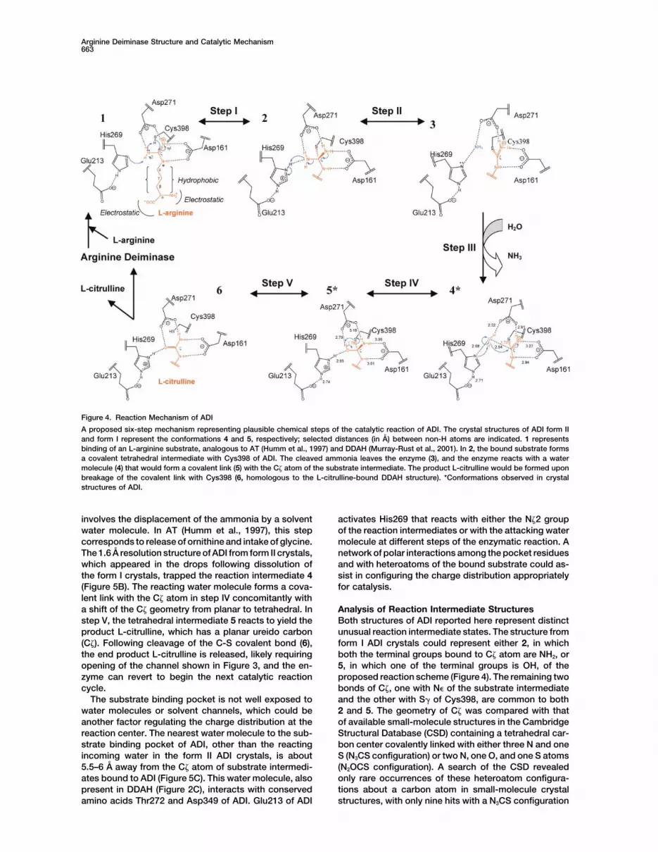
Figure 4. Reaction Mechanism of ADI
A proposed six-step mechanism representing plausible chemical steps of the catalytic reaction of ADI. The crystal structures of ADI form IIand form I represent the conformations 4 and 5, respectively; selected distances (in A) between non-H atoms are indicated. 1 representsbinding of an L-arginine substrate, analogous to AT (Humm et al., 1997) and DDAH (Murray-Rust et al., 2001). In 2, the bound substrate formsa covalent tetrahedral intermediate with Cys398 of ADI. The cleaved ammonia leaves the enzyme (3), and the enzyme reacts with a watermolecule (4) that would form a covalent link (5) with the C atom of the substrate intermediate. The product L-citrulline would be formed uponbreakage of the covalent link with Cys398 (6, homologous to the L-citrulline-bound DDAH structure). *Conformations observed in crystalstructures of ADI.
involves the displacement of the ammonia by a solvent activates His269 that reacts with either the N 2 groupof the reaction intermediates or with the attacking waterwater molecule. In AT (Humm et al., 1997), this step
corresponds to release of ornithine and intake of glycine. molecule at different steps of the enzymatic reaction. Anetwork of polar interactions among the pocket residuesThe 1.6 A resolution structure of ADI from form II crystals,
which appeared in the drops following dissolution of and with heteroatoms of the bound substrate could as-sist in configuring the charge distribution appropriatelythe form I crystals, trapped the reaction intermediate 4
(Figure 5B). The reacting water molecule forms a cova- for catalysis.lent link with the C atom in step IV concomitantly witha shift of the C geometry from planar to tetrahedral. In Analysis of Reaction Intermediate Structures
Both structures of ADI reported here represent distinctstep V, the tetrahedral intermediate 5 reacts to yield theproduct L-citrulline, which has a planar ureido carbon unusual reaction intermediate states. The structure from
form I ADI crystals could represent either 2, in which(C ). Following cleavage of the C-S covalent bond (6),the end product L-citrulline is released, likely requiring both the terminal groups bound to C atom are NH2, or
5, in which one of the terminal groups is OH, of theopening of the channel shown in Figure 3, and the en-zyme can revert to begin the next catalytic reaction proposed reaction scheme (Figure 4). The remaining two
bonds of C , one with N� of the substrate intermediatecycle.The substrate binding pocket is not well exposed to and the other with S� of Cys398, are common to both
2 and 5. The geometry of C was compared with thatwater molecules or solvent channels, which could beanother factor regulating the charge distribution at the of available small-molecule structures in the Cambridge
Structural Database (CSD) containing a tetrahedral car-reaction center. The nearest water molecule to the sub-strate binding pocket of ADI, other than the reacting bon center covalently linked with either three N and one
S (N3CS configuration) or two N, one O, and one S atomsincoming water in the form II ADI crystals, is about5.5–6 A away from the C atom of substrate intermedi- (N2OCS configuration). A search of the CSD revealed
only rare occurrences of these heteroatom configura-ates bound to ADI (Figure 5C). This water molecule, alsopresent in DDAH (Figure 2C), interacts with conserved tions about a carbon atom in small-molecule crystal
structures, with only nine hits with a N3CS configurationamino acids Thr272 and Asp349 of ADI. Glu213 of ADI


Arginine Deiminase Structure and Catalytic Mechanism665
and one hit with a N2OCS configuration at a C center Covalent Reaction Intermediates Stabilizedby Crystal Lattice Packing?(Muller et al., 2001; Klaui et al., 2002). In the above config-
urations, the ideal C-N and C-O single-bond distances Crystal structures of covalent enzyme reaction interme-diates are truly rare and have often been produced viain small molecules are 1.47 and 1.39 A, respectively. In
the form I ADI structure, the bond distances between a reaction with a “suicide substrate” inhibitor that reactsirreversibly and is unable to progress to product forma-C and the two terminal groups (Table 2) of the reaction
intermediate are almost the same, with an average dis- tion. What can explain the unusual occurrence of crys-tallizing two covalent reaction intermediates with ADI?tance of 1.47 A, suggesting that both terminal groups
could be NH2. In structure refinement, the bond dis- One possibility for the occurrence of two covalent inter-mediates in ADI crystals is that the conformations oftances and bond angles of the substrate were refined
with loose restraints compared to protein restraints. ADI with bound substrate that crystallize represent a“closed” form of the enzyme (Figure 3), with tightly boundHowever, the difference of 0.08 A between the C-N and
C-O type bonds discussed above is similar to the maxi- substrates or substrate intermediates whose configura-tions are stabilized by the ADI conformation in the crys-mal error of 0.11 A as calculated by program sfcheck
in CCP4 V4.2.2 (CCP4, 1994). Hence, the form I ADI tals. In both form I and form II ADI crystals, the enzymeconformation is somewhat compact and closed relativestructure determination alone cannot distinguish whether
the C atom of the reaction intermediate has a N3CS or to other forms of the enzyme that would presumablyhave a wider channel for binding and release of sub-N2OCS configuration.
Given the input of L-arginine substrate, it is possible strate and product. A similar situation was encounteredby Jansonius and coworkers (Malashkevich et al., 1993)that any of the species 1–6 in the reaction scheme would
be trapped in crystalline form. Based on enzymatic con- in 2.3 and 2.4 A resolution crystal structures of aspartateaminotransferase with natural substrates L-aspartatesiderations only, however, the 10-fold molar excess of
L-arginine in the crystallization solution (with 18% PEG and L-glutamate, in which the substrates were boundas ketimines (Schiff base intermediates) to the enzyme8K, 0.1 M potassium phosphate [pH 7.4] at 4�C) should
have broken down completely to product L-citrulline cofactor pyridoxal phosphate. These workers interpre-ted the higher affinity for keto substrates “in terms of awithin a few hours at most, and almost certainly within
the 1 week required for growth of form I ADI crystals. perturbation of the open/closed conformational equilib-rium by the crystal lattice, with the closed form havingAssuming that L-arginine was completely converted to
L-citrulline, ADI reaction intermediate species 4, 5, and a higher affinity for the substrate. The crystal latticecontacts provide energy required for domain closure6 would be accessible if steps IV and V of the reaction
scheme are reversible. Given that the [H2O] ��� [NH3] normally supplied by the excess binding energy of thesubstrate.” Analogously, in the present case, we pro-(about 104 higher: �40 M versus 3 mM) (assuming that
the ammonia released in step III remains in solution and pose that the “closed” forms of ADI (Figure 3) containingthe reaction intermediates bonded to the enzyme via ais not evolved as gas from the crystallization drop), it
seems implausible that the equilibrium would shift back thioether linkage are preferentially crystallized out ofsolution, with lattice contacts providing the energy thatto reform significant amounts of ADI reaction intermedi-
ate 1, 2, or 3. Hence, active ADI mixed with substrate shifts the equilibrium toward intermediates (e.g., 4 and5 versus 6).L-arginine is likely to crystallize as a complex with end
product citrulline (6), or in complexes with reaction inter-mediate 4 or 5. Possible Role of the Five-Helix Bundle Domain
The five-helix bundle domain of ADI, colored orange inWhile the stereochemical analysis and covalent ge-ometry at the reaction center carbon inclined toward Figure 1, contains an extended hydrophobic core that
has contributions from aromatic and hydrophobic sidethe assignment of the form I ADI crystal structure as 2 ofthe reaction scheme, enzymatic considerations strongly chains belonging to all of its secondary structural ele-
ments (�3, �4, and �4–�7) and from helix �10. The func-support its assignment as 5. Similarly, it is beyond thescope of our current study to unambiguously establish tionally analogous enzymes AT and DDAH lack this
structural feature. The specific role of the “clip” moietywhether the structure from form II ADI crystals repre-sents 3 or 4 of the reaction scheme, although our analy- of ADI is not clear due to large variations in its amino
acid sequence and length over different ADIs. In the ADIsis prefers the latter one. These ADI structures providethe first direct structural evidence supporting the structure, this part has some structural resemblance
with the prodomain of caspase-9, a protein belonginginvolvement of covalent reaction intermediates in cataly-sis by guanidine-group-modifying enzymes and can to the caspase requirement domain (CARD) of apoptotic
signaling motifs (Budihardjo et al., 1999; Hofmann et al.,guide further biochemical analysis of this class of en-zymes. 1997). The caspase-9 prodomain and apoptotic prote-
Figure 5. ADI Active Sites: Electron Density and Structure Comparison
(A and B) Stereo views of difference maps (using |Fo| |Fc| amplitudes and �calc phases) showing the positions and orientations of the boundsubstrate intermediates (A) in form I ADI at 2.0 A and (B) in form II ADI at 1.6 A resolution. The substrate reaction intermediates are coloredpurple, and the surrounding protein residues are colored yellow. Water molecules are labeled OW.(C) A stereo view showing superposition of the active sites of form I (light gray amino acid residues and gray substrate intermediate) andform II (cyan amino acid residues and yellow substrate intermediate) ADI structures. The water molecule OW belongs to the form II ADIstructure.

Structure666
the X-ray diffraction data used in structure determination and refine-ase-activating factor 1 (Apaf-1) complex structure (PDBment are listed in Table 1.ID: 3YGS) (Qin et al., 1999) showed that the interfacial
amino acid residues are not conserved and polar inter-Phasing, Structure Determination, and Refinementactions are predominant. The prodomain of caspase-9The ADI crystals showed low affinity toward heavy-atom reagents.
was found by a Dali search (Holm and Sander, 1993) to The only successful derivatives of the ADI were obtained by soakinghave a relatively low structural similarity (Z score of 2.2) the crystals with a few mercury- and platinum-containing com-with the five-helix bundle domain of ADI, but helices H1 pounds. Two sites each for Hg and Pt were detected by difference
Patterson and cross-verified by difference Fourier techniques. Initialand H4 of the caspase-9 prodomain (PDB ID: 3YGS),phases were obtained by the multiple isomorphous replacementwhich interacts with Apaf-1, overlay on helices �5 and(MIR) method as implemented in PHASES (Furey and Swaminathan,�6 of ADI. Also, some other proteins, including the yeast1997). The phasing powers of the derivatives were useful only up
TATA box binding protein fragment (Tan et al., 1996) to a maximum resolution of 4.0 A. The solvent-flattened electronand the cell division protein MukB N-terminal fragment density maps calculated from the 4 A MIR phases showed a clear(van den Ent et al., 1999), were found to have some solvent-protein boundary. There were two molecules per asymmet-
ric unit. Using the program DM (Cowtan and Main, 1998) imple-structural resemblance with the five-helix bundle of ADI.mented in CCP4 V 3.3, the MIR phases were extended to a maximumGiven that ADI is the first enzyme of the arginine dihy-resolution of 2.0 A with the help of solvent flattening and 2-folddrolysis pathway that generates ATP in many myco-noncrystallographic symmetry averaging protocols. However, the
plasma under anaerobic conditions, its clip portion maps calculated from the extended phases showed electron densitymight have a role in activating the series of reactions. that was neither continuous nor readily interpretable. Subsequently,Further studies on the clip moiety of the molecule will the 1.6 A data set from the form II crystal was used for successful
phase extension using DM. The extended phases were further im-be required to reveal its role(s) in cellular processesproved using the free-atom-refinement protocol of WARP V5.0 (Per-such as apoptosis.rakis et al., 1999). The electron density map calculated using WARP-refined phases was of exceptional quality, to which the atomic modelcould easily be built manually without any ambiguity. All 410 aminoExperimental Proceduresacid residues and their side chains, excluding one at its N terminus,could be traced. Iterative cycles of model building, using O (JonesIsolation, Purification, and Crystallizationet al., 1991), followed by least-squares refinement, initially usingArginine deiminase used in this crystallographic study was isolatedXplor 3.65 (Brunger, 1996) and later using CNS 1.0 (Brunger et al.,from Mycoplasma arginini using a previously described protocol1998), yielded the final form II ADI structure with an R and Rfree of(Kondo et al., 1990). The purified sample was concentrated to 300.168 and 0.202, respectively, for reflections in the resolution rangemg/ml in 0.01 M potassium phosphate (pH 7.0) and stored at 80�C.20–1.6 A (Table 1). The form II structure was used as the startingThe above sample of ADI was used for crystallization by the hangingmodel for solving the form I ADI structure by the molecular replace-drop vapor diffusion technique. The substrate L-arginine was addedment method. The final R and Rfree for the form I ADI structure refinedto the protein at an approximate molar ratio of 10:1 prior to crystalli-to 2.0 A resolution were 0.186 and 0.227, respectively.zation. Diffraction-quality crystals were obtained using 18% PEG
6000 as precipitant in 0.1 M potassium phosphate (pH 6.5) at 4�C.This crystallization condition and crystal form are different from Acknowledgmentsthose of a reported crystallization of ADI from Pseudomonas aerugi-nosa (Oudjama et al., 2002). Typical crystals grew as 0.2 � 0.15 � We thank Deena Oren, Jens Birktoft, and Stefan Sarafianos for their0.5 mm rods in 1 week. Over a period of 3–4 weeks, the crystals helpful comments, Tom Emge for helping with CSD searches, Dickin some of the drops completely dissolved into the crystallization Leidich for assistance in data collection, and the staffs at CHESSsolution and reappeared in another form, form II. The form II ADI and BNLS for helping with synchrotron data collection. G.H.B. iscrystals were considerably bigger, with nearly equal thickness along grateful to the RNR Foundation and the W.W. Smith Charitable Trustall three dimensions compared to the smaller rod-shaped form I for financial support of production and purification of ADI.ADI crystals. One or two form II crystals grew per 6 �l drop, com-pared with eight to ten of form I crystals. Form II crystals, though Received: December 8, 2003bigger, were highly sensitive to temperature as well as to other Revised: January 7, 2004physical or chemical changes; form II crystals cracked immediately Accepted: January 7, 2004upon exposure to cryoprotectant solution containing 20% glycerol, Published: April 6, 2004whereas form I crystals were stable in the same solution. The finalstructure of form II ADI revealed that an unstable enzymatic reaction
Referencesintermediate of the substrate might be responsible for the hypersen-sitivity of the crystals of this form.
Barile, M.F. (1968). Mycoplasma and cell cultures. Natl. Cancer Inst.Monogr. 29, 201–204.
Berman, H.M., Battistuz, T., Bhat, T.N., Bluhm, W.F., Bourne, P.E.,X-Ray Data CollectionBurkhardt, K., Feng, Z., Gilliland, G.L., Iype, L., Jain, S., et al. (2002).Form I crystals were used to prepare heavy-atom derivatives. Al-The Protein Data Bank. Acta Crystallogr. D Biol. Crystallogr. 58,though ADI crystals were treated with various concentrations of899–907.numerous heavy-atom reagents for various soaking times, the only
useful derivatives were obtained with tetramethyl mercury (TMM), Brunger, A.T. (1996). Recent developments for crystallographic re-methyl mercury chloride (MMCl), and K2PtCl4. All the diffraction data finement of macromolecules. Methods Mol. Biol. 56, 245–266.including that for the heavy-atom derivatives were collected from Brunger, A.T., Adams, P.D., Clore, G.M., DeLano, W.L., Gros, P.,the crystals flash cooled at about 100 K. The X-ray diffraction data Grosse-Kunstleve, R.W., Jiang, J.S., Kuszewski, J., Nilges, M.,from form I native crystals were collected at the Brookhaven National Pannu, N.S., et al. (1998). Crystallography & NMR system: a newLaboratory (BNL) X25 beamline to a maximum resolution of 2 A, and software suite for macromolecular structure determination. Actathe data from form II native crystals were collected at the Cornell Crystallogr. D Biol. Crystallogr. 54, 905–921.High Energy Synchrotron Source (CHESS) F2 beamline to a maxi-
Budihardjo, I., Oliver, H., Lutter, M., Luo, X., and Wang, X. (1999).mum resolution of 1.6 A. The data for all the heavy-atom derivativesBiochemical pathways of caspase activation during apoptosis.were collected using an R axis II mounted on a Rigaku rotatingAnnu. Rev. Cell Dev. Biol. 15, 269–290.anode X-ray generator. The data sets were processed using DENZO
and SCALEPACK (Otwinowski and Minor, 2001). The summary of CCP4 (Collaborative Computational Project 4) (1994). The CCP4
Arginine Deiminase Structure and Catalytic Mechanism667
suite: programs for protein crystallography. Acta Crystallogr. D Biol. another new scorpionate class? Angew. Chem. Int. Ed. Engl. 40,1247–1249.Crystallogr. 50, 760–763.
Murray-Rust, J., Leiper, J., McAlister, M., Phelan, J., Tilley, S., SantaCowtan, K., and Main, P. (1998). Miscellaneous algorithms for den-Maria, J., Vallance, P., and McDonald, N. (2001). Structural insightssity modification. Acta Crystallogr. D Biol. Crystallogr. 54, 487–493.into the hydrolysis of cellular nitric oxide synthase inhibitors by dimeth-Dillon, B., Holtsberg, F., Ensor, M., Bomalaski, J., and Clark, M.ylarginine dimethylaminohydrolase. Nat. Struct. Biol. 8, 679–683.(2002). Biochemical characterization of the arginine degrading en-Otwinowski, Z., and Minor, W. (2001). DENZO and SCALEPACK.zymes arginase and arginine deiminase and their effect on nitricIn Crystallography of Biological Macromolecules, Volume F, M.G.oxide production. Med. Sci. Monit. 8, BR248–BR253.Rossmann, and E. Arnold, eds. (Boston, MA: Kluwer Academic Pub-
Ensor, C.M., Holtsberg, F.W., Bomalaski, J.S., and Clark, M.A. (2002).lishers), pp. 226–235.
Pegylated arginine deiminase (ADI-SS PEG20,000 mw) inhibits hu-Oudjama, Y., Tricot, C., Stalon, V., and Wouters, J. (2002). Overex-man melanomas and hepatocellular carcinomas in vitro and in vivo.pression, purification, crystallization and preliminary X-ray crystallo-Cancer Res. 62, 5443–5450.graphic analysis of Pseudomonas aeruginosa L-arginine deiminase.
Furey, W., and Swaminathan, S. (1997). PHASES-95: a program Acta Crystallogr. D Biol. Crystallogr. 58, 2150–2152.package for the processing and analysis of diffraction data from
Perrakis, A., Morris, R., and Lamzin, V.S. (1999). Automated proteinmacromolecules. Methods Enzymol. 277, 590–620.model building combined with iterative structure refinement. Nat.
Gong, H., Zolzer, F., von Recklinghausen, G., Rossler, J., Breit, S., Struct. Biol. 6, 458–463.Havers, W., Fotsis, T., and Schweigerer, L. (1999). Arginine deimi-
Philip, R., Campbell, E., and Wheatley, D.N. (2003). Arginine depriva-nase inhibits cell proliferation by arresting cell cycle and inducingtion, growth inhibition and tumour cell death: 2. Enzymatic degrada-apoptosis. Biochem. Biophys. Res. Commun. 261, 10–14.tion of arginine in normal and malignant cell cultures. Br. J. Cancer
Gong, H., Zolzer, F., von Recklinghausen, G., Havers, W., and 88, 613–623.Schweigerer, L. (2000). Arginine deiminase inhibits proliferation of
Qin, H., Srinivasula, S.M., Wu, G., Fernandes-Alnemri, T., Alnemri,human leukemia cells more potently than asparaginase by inducing
E.S., and Shi, Y. (1999). Structural basis of procaspase-9 recruitmentcell cycle arrest and apoptosis. Leukemia 14, 826–829.
by the apoptotic protease-activating factor 1. Nature 399, 549–557.Hofmann, K., Bucher, P., and Tschopp, J. (1997). The CARD domain: Schimke, R.T., Berlin, C.M., Sweeney, E.W., and Carroll, W.R. (1966).a new apoptotic signalling motif. Trends Biochem. Sci. 22, 155–156. The generation of energy by the arginine dihydrolase pathway inHolm, L., and Sander, C. (1993). Protein structure comparison by Mycoplasma hominis 07. J. Biol. Chem. 241, 2228–2236.alignment of distance matrices. J. Mol. Biol. 233, 123–138. Shirai, H., Blundell, T.L., and Mizuguchi, K. (2001). A novel superfam-
ily of enzymes that catalyze the modification of guanidino groups.Humm, A., Fritsche, E., Steinbacher, S., and Huber, R. (1997). CrystalTrends Biochem. Sci. 26, 465–468.structure and mechanism of human L-arginine:glycine amidino-
transferase: a mitochondrial enzyme involved in creatine biosynthe- Smith, D.W., and Fahrney, D.E. (1978). Catalysis by arginine deimi-sis. EMBO J. 16, 3373–3385. nase: evidence for a covalent intermediate. Biochem. Biophys. Res.
Commun. 83, 101–106.Jones, T.A., Zou, J.Y., Cowan, S.W., and Kjeldgaard, M. (1991).Improved methods for building protein models in electron density Smith, D.W., Ganaway, R.L., and Fahrney, D.E. (1978). Arginine dei-maps and location of errors in these models. Acta Crystallogr. A 47, minase from Mycoplasma arthritidis. Structure-activity relationships110–119. among substrates and competitive inhibitors. J. Biol. Chem. 253,
6016–6020.Kang, S.W., Kang, H., Park, I.S., Choi, S.H., Shin, K.H., Chun, Y.S.,Chun, B.G., and Min, B.H. (2000). Cytoprotective effect of arginine Sugimura, K., Ohno, T., Kusuyama, T., and Azuma, I. (1992). Highdeiminase on taxol-induced apoptosis in DU145 human prostate sensitivity of human melanoma cell lines to the growth inhibitorycancer cells. Mol. Cells 10, 331–337. activity of mycoplasmal arginine deiminase in vitro. Melanoma Res.
2, 191–196.Kawauchi, Y., Muto, A., and Osawa, S. (1982). The protein composi-Sugimura, K., Ohno, T., Azuma, I., and Yamamoto, K. (1993). Poly-tion of Mycoplasma capricolum. Mol. Gen. Genet. 188, 7–11.morphism in genes for the enzyme arginine deiminase among Myco-Kidd, J.G. (1973). Asparaginase and cancer—yesterday and today—plasma species. Infect. Immun. 61, 329–331.recent results. Cancer Res. 33, 3–14.Takaku, H., Takase, M., Abe, S., Hayashi, H., and Miyazaki, K. (1992).
Klaui, W., Schramm, D., Peters, W., Rheinwald, G., and Lang, H.In vivo anti-tumor activity of arginine deiminase purified from Myco-
(2002). Tris(pyrazolyl)methanesulfonates: a novel class of water-sol-plasma arginini. Int. J. Cancer 51, 244–249.
uble ligands. Angew. Chem. Int. Ed. Engl. 39, 2464–2466.Tan, S., Hunziker, Y., Sargent, D.F., and Richmond, T.J. (1996). Crys-
Komada, Y., Zhang, X.L., Zhou, Y.W., Ido, M., and Azuma, E. (1997). tal structure of a yeast TFIIA/TBP/DNA complex. Nature 381,Apoptotic cell death of human T lymphoblastoid cells induced by 127–151.arginine deiminase. Int. J. Hematol. 65, 129–141.
Terayama, H., Koji, T., Kontani, M., and Okumoto, T. (1982). ArginaseKondo, K., Sone, H., Yoshida, H., Toida, T., Kanatani, K., Hong, Y.M., as an inhibitory principle in liver plasma membranes arresting theNishino, N., and Tanaka, J. (1990). Cloning and sequence analysis growth of various mammalian cells in vitro. Biochim. Biophys. Actaof the arginine deiminase gene from Mycoplasma arginini. Mol. Gen. 720, 188–192.Genet. 221, 81–86.
Thomas, J.B., Holtsberg, F.W., Ensor, C.M., Bomalaski, J.S., andMacAllister, R.J., Parry, H., Kimoto, M., Ogawa, T., Russell, R.J., Clark, M.A. (2002). Enzymic degradation of plasma arginine usingHodson, H., Whitley, G.S., and Vallance, P. (1996). Regulation of arginine deiminase inhibits nitric oxide production and protects micenitric oxide synthesis by dimethylarginine dimethylaminohydrolase. from the lethal effects of tumour necrosis factor alpha and endo-Br. J. Pharmacol. 119, 1533–1540. toxin. Biochem. J. 363, 581–587.Malashkevich, V.N., Toney, M.D., and Jansonius, J.N. (1993). Crystal van den Ent, F., Lockhart, A., Kendrick-Jones, J., and Lowe, J. (1999).structures of true enzymatic reaction intermediates: aspartate and Crystal structure of the N-terminal domain of MukB: a protein involvedglutamate ketimines in aspartate aminotransferase. Biochemistry in chromosome partitioning. Struct. Fold. Des. 7, 1181–1187.32, 13451–13462.
Accession NumbersMiyazaki, K., Takaku, H., Umeda, M., Fujita, T., Huang, W.D., Kimura,T., Yamashita, J., and Horio, T. (1990). Potent growth inhibition of
The coordinates and structure factors for both the form I and formhuman tumor cells in culture by arginine deiminase purified from aII ADI structures are deposited in the Protein Data Bank (Bermanculture medium of a Mycoplasma-infected cell line. Cancer Res. 50,et al., 2002) with the entry codes 1LXY and 1S9R, respectively.4522–4527.
Muller, M., Lork, E., and Mews, R. (2001). Tris(azolyl)methylthiolates: