gamma irradiation effects on stability of poly(lactide-co-glycolide) microspheres containing...
TRANSCRIPT
Journal of Controlled Release 75 (2001) 317–330www.elsevier.com/ locate / jconrel
Gamma irradiation effects on stability ofpoly(lactide-co-glycolide) microspheres
containing clonazepama , a b c c d*L. Montanari , F. Cilurzo , L. Valvo , A. Faucitano , A. Buttafava , A. Groppo ,
d dI. Genta , B. Contia `Istituto di Chimica Farmaceutica e Tossicologica, Universita di Milano, v. le Abruzzi 42, 20131 Milan, Italy
b `Laboratorio di Chimica del Farmaco, Istituto Superiore di Sanita, v. le Regina Elena 299, 00161 Rome, Italyc `Dipartimento di Chimica Generale, Universita di Pavia, v. le Taramelli 12, 27100 Pavia, Italy
d `Dipartimento di Chimica Farmaceutica, Universita di Pavia, v. le Taramelli 12, 27100 Pavia, Italy
Received 1 February 2001; accepted 22 May 2001
Abstract
This work was aimed at evaluating the effects of g irradiation on the stability of microspheres made of a poly(lactide-co-glycolide) copolymer (PLGA) and loaded with 15% w/w of clonazepam (CLO). The influence of CLO on PLGA radiolysismechanisms and the identification of possible irradiation markers were also investigated. Microspheres were prepared bymeans of a spray-drying method. g Irradiation was carried out either under vacuum or in air, at a dose of 25 kGy, by using a60Co source. The stability of CLO loaded microspheres was evaluated over a 6-month period on the basis of drug contentand dissolution profile. Radiolysis mechanisms were investigated by using electronic paramagnetic resonance (EPR)analysis. The microspheres irradiated under vacuum were stable over the considered period of time. After irradiation in air,CLO release rate increased by |10%, and did not change further in the following period of storage. The EPR analysisshowed some radicals arising from both the polymeric matrix and the active ingredient. Polymer /CLO spin transfer reactionssuggest that CLO had a radio-stabilising effect on the polymeric matrix. In the loaded microspheres, the intensity in time ofthe CLO radical signal is sufficient for its possible use as irradiation marker. 2001 Elsevier Science B.V. All rightsreserved.
Keywords: Gamma irradiation; Clonazepam; Microspheres; Poly(lactide-co-glycolide) copolymer (PLGA); Electronic paramagneticresonance (EPR)
1. Introduction preparation of biodegradable microspheres intendedfor parenteral use. As polylactide (PLA) and poly-
Aliphatic polyesters based on lactic and glycolic (lactide-co-glycolide) (PLGA) are moisture and heat-acids are the most widely used polymers for the sensitive polymers, they are good candidates for g
sterilisation. Nevertheless, ionising radiation inducesdose-dependent cross-linking and/or chain scission*Corresponding author. Tel.: 139-02-2940-3194.
E-mail address: [email protected] (L. Montanari). and concomitant molecular weight loss of these
0168-3659/01/$ – see front matter 2001 Elsevier Science B.V. All rights reserved.PI I : S0168-3659( 01 )00401-1

318 L. Montanari et al. / Journal of Controlled Release 75 (2001) 317 –330
polymers [1–3]. The radiolytic degradation of PLA cation of possible irradiation markers were investi-or PLGA increases in the presence of oxygen. gated by using electronic paramagnetic resonanceDuring the g irradiation process, the oxidation of the (EPR) analysis. The identification of possible irradia-polymer can decrease cross-linking, and increase tion markers could be very useful to prove performeddegradation or lead to chain scission [4]. irradiation on the final product.
The effects of g irradiation on the microparticulate The stability of CLO loaded microspheres ir-systems made of PLA or PLGA are also influenced radiated either under vacuum or in air was evaluatedby drug loading, and are not easily predicted because over a 6-month period on the basis of their drugof the different chemico-physical characteristics of content and dissolution profile.the active ingredient and its interactions with thepolymeric matrix. Volland et al. showed a decreasedcaptopril release from g irradiated microspheres [5]. 2. Materials and methodsIn contrast, Yoshioka et al. described an increased
progesterone release from irradiated microspheres Poly(lactide-co-glycolide) 50:50, Resomer RGwith increasing irradiation dose [6]. Mohr et al. 503 (PLGA), inherent viscosity 0.39 dl /g, 34 000showed accelerated kinetics of estradiol release with MW, was from Boehringer Ingelheim (Ingelheim amincreasing irradiation doses, due to dose dependent Rheim, Germany). Clonazepam was supplied bypolymer degradation [7]. The same work also Roche Spa (Milan, Italy). L-alanine and 1,1-showed estradiol grafting to PLGA as a consequence diphenyl-2-picrylhydrazyl (DPPH) were from Flukaof degradation. (Milan, Italy). Paraffin wax was from Aldrich
The influence of drug loading on polymer degra- (Milan, Italy) and sodium lauryl sulphate was fromdation is also discussed. PLA degradation was Carlo Erba (Milan, Italy).independent of methadone loading [8], but it was Unless specified, all other compounds were ofhigher when prometazine loading increased [9]. analytical grade.Again, Bittner et al. showed that PLGA degradationrate slowed down following the incorporation of 2.1. Preparation of microspherestetracycline in the microspheres [10]. Although theeffects of ionising radiation on PLA and PLGA Microsphere preparation was performed by usingmolecular weight and drug release have been dis- the spray-dryer Lab-Plant model SD04 (Lab-Plant,cussed in a number of papers, the stability of the West Yorkshire, UK) as described in a previous workirradiated biodegradable microparticulate systems [12]. Placebo microspheres were obtained by spray-has been scantly investigated. ing 2% w/w solution of PLGA in methylene chlo-
In this work, the effects of g irradiation either ride through a standard nozzle (inner diameter: 1under vacuum or in air at a dose of 25 kGy on the mm). The process parameters were set as follows:stability of microspheres made of PLGA and loaded inlet temperature: 508C; outlet temperature: 35–with clonazepam (CLO) was evaluated. A minimum 368C; flow rate: 10 ml /min.absorbed dose of 25 kGy was regarded as adequate CLO loaded microspheres were prepared by spray-for the purpose of sterilising pharmaceutical products ing — under the same conditions as above — 2%without providing any biological validation [11]. w/w solution of PLGA in methylene chloride inCLO was selected as model drug because it is a which CLO had been previously solubilised in thebenzodiazepine compound with marked antiepileptic following ratio: PLGA:CLO 85:15 w/w. After prep-properties used in the chronic therapy of myoclonic aration, the microspheres were lyophilised by using aseizures in children. Thus, a controlled release drug Modulyo 4K Freeze Dryer (Edwards, UK), anddelivery system intended for parenteral administra- stored in an airtight container at 48C.tion appears of interest.
The CLO microparticulate system made of a 2.2. g Irradiation of placebo and CLO loadedpoly(lactide-co-glycolide) 50:50 copolymer was pre- microspherespared by the spray-drying method [12]. The in-fluence of CLO on PLGA radiolysis and the identifi- Placebo and CLO loaded microspheres were ir-

L. Montanari et al. / Journal of Controlled Release 75 (2001) 317 –330 319
60radiated by using Co as irradiation source (Applied analysed by computer simulation by using theNuclear Energy Laboratory (L.E.N.A.), University of Hamiltonian:Pavia). Irradiation was performed at room tempera- → → → | → → →|ture either under vacuum or in air; a 25-kGy dose at H 5 2 bS ?g ?H 1 S S ?A ? I 2 S I ?Hi i i i i i
1.3-kGy/h dose rate was applied. |where H is spin Hamiltonian, b is Bohr magneton, g→ →is g tensor, H is external magnetic field, S is
| →2.3. Morphologyelectron spin operator, A is hyperfine tensor, and Iis nuclear spin operator.
Microparticle size and morphology before andThe spectra of samples irradiated at 77 K, under
after irradiation were evaluated. The microspherevacuum, were recorded at 113 K and after annealing
shape and surface were analysed using SEM (JSM-Tat 298 K. Furthermore, the spectra of the samples
800, Jeol Italia, Pieve Emanuele, Italy). The sampleswere recorded also after 18 h at room temperature
were sputtered with an Au/Pd coating in an argonand after admission of air at 298 K. The annealing
atmosphere. The microsphere size was determinedprocedure was intended to ensure suitable ex-
by light blockage method and an HIAC/ROYCOperimental conditions so that the reaction of the
apparatus, model 3000, equipped with an HC60primary species trapped during the irradiation at 77
sensor. Samples of microspheres were suspended inK could take place.
filtered and bidistilled water, and analysed whileSpectra of samples irradiated at room temperature
gently stirring. The results are the average of fivewere recorded immediately after irradiation and at
determinations.different storage times in order to study radicaldecay.
The radiolytic radical yields were determined2.4. EPR analysisthrough comparison of the EPR signals areas byusing alanine standards with a known number ofEPR analysis was performed on CLO, placebospins. The alanine standards were prepared by extru-microspheres and CLO loaded microspheres.sion of alanine powder /wax mixture (|20% wax) inIrradiation was performed at the Applied Nuclearthe form of cylinders having a similar geometry toEnergy Laboratory (L.E.N.A., Pavia University,
60 that of the samples. Stable alanine radicals wereItaly) by using a Co gamma source calibratedgenerated by irradiation; their concentration wasagainst alanine and Fricke dosimeters, at the follow-determined by comparison with a standard solutioning conditions: irradiation temperature T577 Kof DPPH.(liquid nitrogen), samples sealed under high vacuum
in EPR quartz tubes, dose rate 1.3 kGy/h, total dose25 kGy; irradiation temperature T5298 K (source
2.5. Water contenttemperature), samples sealed under high vacuum inEPR quartz tubes, dose rate 1.3 kGy/h, total dose 25
The water content of placebo and loaded micro-kGy; irradiation temperature T5298 K, sample
spheres was determined by Karl Fischer volumetriccontainers were open during irradiation, dose rate 1.3
titration (Micro KF 2026, Crison, Italy). A sample ofkGy/h, total dose 25 kGy.
25 mg — precisely weighed — was suspended inAfter irradiation, part of the sample tubes was
methanol, and titrated with a Karl Fischer pyridine-flamed by using the sliding technique in order to
free solution. Each value is obtained from triplicateeliminate the radiation induced quartz paramagnetic
determinations.centers. During this operation, the sample tempera-ture was not allowed to rise above 77 K for samples
2.6. Differential scanning calorimetry (DSC)irradiated at 77 K.EPR analysis was performed by using a Varian
Thermal analysis was performed on samples ofE-109 spectrophotometer (Palo Alto, CA, US)CLO, placebo and loaded microspheres by using aequipped with a data acquisition system and aDSC 2010 TA (TA Instruments, US).The samplestemperature control apparatus. The EPR spectra were

320 L. Montanari et al. / Journal of Controlled Release 75 (2001) 317 –330
were heated in closed aluminum pans, at a heating 3. Results and discussionrate of 108C/min under a constant flow of nitrogen.
3.1. Microsphere morphology
Both placebo and drug loaded microspheres had a2.7. Drug content
spherical shape and a wrinkled surface (Fig. 1). Thesize of over 90% of placebo and CLO loaded
A 5-mg amount of CLO-loaded and accuratelymicrospheres was in the 2–10-mm range (Fig. 2).
weighed microspheres was dissolved in 25 ml ofIrradiation of placebo and loaded microspheres per-
acetone. The samples were assayed by HPLC-UVformed either in air or under vacuum caused a
method. The HPLC system was an HP 1100 Chem-change in the particle size distribution (Fig. 2). An
Station (Hewlett Packard, Germany). Chromato-increased number of particles in the range 5–10 mm
graphic conditions were: column: C ODS 2 Hyper-18 was shown among placebo microspheres (Fig. 2A).sil 20034.6 mm (Shandon HPLC, UK); wavelength:
CLO loaded microspheres (Fig. 2B) showed the240 nm; mobile phase: acetonitrile /methanol /water(30/30/40, v /v /v); flow rate: 1.5 ml /min; injectionvolume: 10 ml. The analyses were performed at roomtemperature. The drug concentrations were deter-mined from the standard curve (5–100 mg/ml) andthe method gave 99.8% recovery of theoretical value(C.V. ,0.4% on the basis of three determinations).
In the stability study of non-irradiated and ir-radiated microspheres, the drug content was deter-mined after 0, 30, 60, 180 days in triplicate.
2.8. In vitro release test
In vitro release tests were performed according tothe Ph. Eur. III Ed. paddle dissolution method, onCLO loaded microspheres (a) not irradiated, (b)irradiated at room temperature under vacuum, and (c)irradiated at room temperature in air.
A total of 500 ml of phosphate buffered salinesolution, pH 7.4 (Ph. Eur. III Ed.) containing 0.15%SDS as wetting agent, was used as release medium.The suspensions were maintained at 378C whilestirring at 100 rpm for 24 h. The amounts of CLOreleased from the microspheres were spectrophoto-metrically determined at 240-nm wavelength. Thetest was performed in triplicate. The test was per-formed soon after irradiation (time 0), and repeatedafter 15, 30, 60, 90, 120, 150 and 180 days.
The release rate constant was calculated according0.5to Higuchi’s equation as follows: M /M 5 ktt `
where M is the amount of drug released at time t,t
M is the drug loaded in the matrix and k is the Fig. 1. Photomicrographs of CLO loaded microspheres (A) before`20.5release rate constant expressed as h . irradiation, and (B) after irradiation at 25 kGy in presence of air.
L. Montanari et al. / Journal of Controlled Release 75 (2001) 317 –330 321
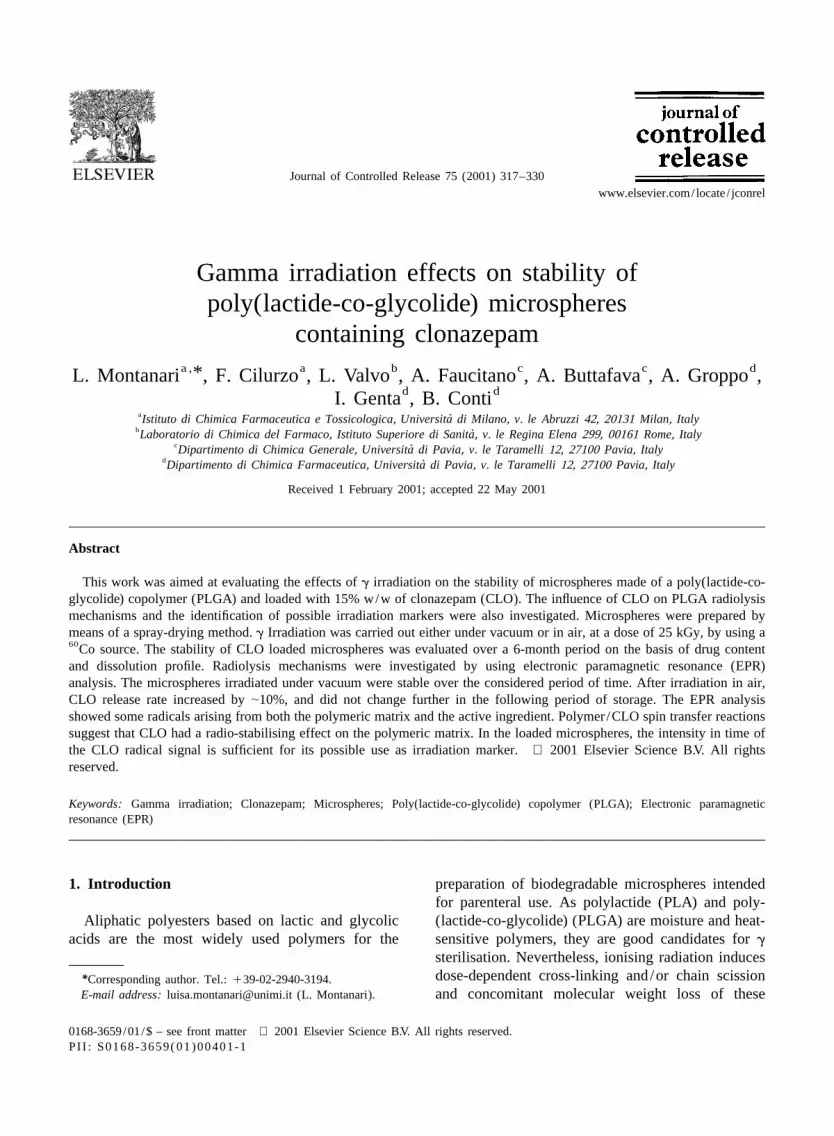
Fig. 2. Particle size distribution of placebo microspheres (A) and CLO loaded microspheres (B) before irradiation, and after irradiation at 25kGy, under vacuum and in presence of air.
opposite pattern, suggesting that CLO presence amidyl radical (Fig. 3C) formally generated by theaffected the surface properties of the irradiated N–H bond rupture; and the carbonyl radical (Fig.microparticles and, consequently, the aggregation 3D) generated after the rupture of the methylenestate of the microspheres. The scavenging of the C–H bond in the diazepinone section. Following thepolymer radicals by CLO shown by EPR analysis primary ionisations, the radiolytic formation ten-could have caused a modification of the surface dency of the three species is expected to increase; theproperties of irradiated loaded microspheres com- dissociative electron capture of the C–Cl bond andpared to the placebo microspheres. the deprotonation of the cation-radicals involving the
loss of the amido or methylene hydrogen are favoredreaction paths. Furthermore, species D can be formed
3.2. EPR analysis in hydrogen abstraction processes due to the labilityof the activated C–H bond. The EPR spectrumobtained by g irradiation of CLO in air does not
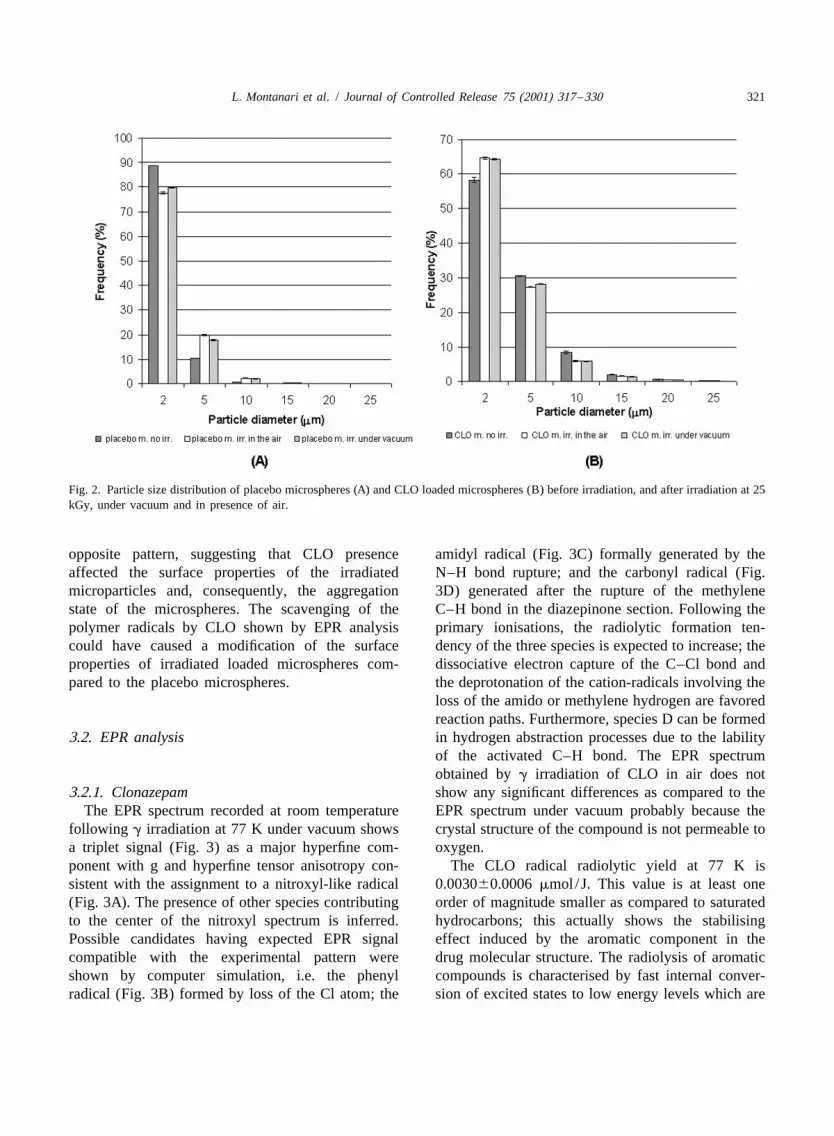
3.2.1. Clonazepam show any significant differences as compared to theThe EPR spectrum recorded at room temperature EPR spectrum under vacuum probably because the
following g irradiation at 77 K under vacuum shows crystal structure of the compound is not permeable toa triplet signal (Fig. 3) as a major hyperfine com- oxygen.ponent with g and hyperfine tensor anisotropy con- The CLO radical radiolytic yield at 77 K issistent with the assignment to a nitroxyl-like radical 0.003060.0006 mmol /J. This value is at least one(Fig. 3A). The presence of other species contributing order of magnitude smaller as compared to saturatedto the center of the nitroxyl spectrum is inferred. hydrocarbons; this actually shows the stabilisingPossible candidates having expected EPR signal effect induced by the aromatic component in thecompatible with the experimental pattern were drug molecular structure. The radiolysis of aromaticshown by computer simulation, i.e. the phenyl compounds is characterised by fast internal conver-radical (Fig. 3B) formed by loss of the Cl atom; the sion of excited states to low energy levels which are
322 L. Montanari et al. / Journal of Controlled Release 75 (2001) 317 –330
Fig. 3. EPR spectra of the neat crystalline CLO after under vacuum g irradiation at 77 K: effect of the annealing temperature. Dose rate 1.3kGy/h; dose, 25 kGy; G525 000 except where quoted. The nitroxyl radical A is the stable prominent species contributing to all the spectra.Radicals B, C, D are minor oxygen sensitive components.
often reduced due to emission of radiations or by As expected, nitroxyl radicals show a remarkabledegradation to thermal energy without leading to stability [14]; the decay rate at room temperaturemolecular decomposition [13]. leads to a 33% concentration decrease in 20 days.

L. Montanari et al. / Journal of Controlled Release 75 (2001) 317 –330 323
3.2.2. Placebo microspheres thermally activated hydrogen abstractions by theWhen considering the intermediate radicals primary radicals at the secondary and tertiary C–H
trapped at 77 K, in the g radiolysis of the polyester, bonds take place. Hydrogen abstraction radicalsprimary processes are characterised by extensive decay slowly under vacuum at room temperature. Onchain scission with a minor participation of C–H admission of oxygen, polymer radicals are convertedbond ruptures (Fig. 4). When warming above 77 K, into the corresponding peroxyl radicals at a rate
Fig. 4. EPR spectra of placebo microspheres (left column) and CLO loaded microspheres (right column) after under vacuum g irradiation at?77 K: effect of the annealing temperature. Dose rate 1.3 kGy/h; dose, 25 KGy. CLO stands for radicals A, B, C, D from clonazepam.
324 L. Montanari et al. / Journal of Controlled Release 75 (2001) 317 –330
which is almost one order of magnitude faster for the the same EPR spectra. This suggests that the poly-placebo microspheres than that of raw polymer mer morphology does not affect the radiolytic be-samples. haviors under vacuum.
Peroxyls are expected to initiate a chain hydro-peroxidative process. During this post irradiation 3.2.3. CLO loaded microspheresprocess, the peroxyl radicals decay half period was After irradiation at 77 K under vacuum, radicals|50 min at room temperature. The presence of the generated by both the polymer matrix and the active?secondary radical –CH – in relative abundance ingredient are identified in almost equal relativecompared to that of the tertiary radical seems to be at abundant amounts (Fig. 4). When warmed at roomvariance with the expected reactivity trend based on temperature, polymer radicals in CLO loaded micro-the C–H bond strengths, since the tertiary C–H bond spheres decay at a faster rate than in the case ofbreaking energy is lower. This pattern can, however, placebo microspheres; consequently, the CLO radicalbe explained in terms of stereoelectronic effects concentration increases steadily attaining 100% afterfavoring hydrogen abstraction from the secondary |45 h. The CLO EPR spectrum finally obtained isC–H bond [15]. Prolonged storage at room tempera- very similar to that of neat CLO (Fig. 3), the nitroxylture leads to the appearance of a novel species with a radical pattern being the dominant component. The?spectrum similar to that of the radical –C (CH )–,3 presence of species contributing to the central part ofbut characterised by a smaller hyperfine splitting. the nitroxyl spectrum is also observed. On admissionThis decrease is diagnostic of an ether or hydroxyl of oxygen, in contrast to the nitroxyl spectrum, thesesubstituent at the carbon radical center therefore, it is species decay rapidly. This suggests that this com-hereby suggested that the signal is assigned to the ponent may belong to oxygen sensitive species suchchain end hydrogen abstraction radical: as amidyl radicals C or carbon centered radicals B
? and D. The decay rate of nitroxyl is very slow both–O–C(=O)–C (CH )–OH3 in air and under vacuum. As a result, the nitroxylEPR signal might be used as a radiation marker
The terminal alcohol units needed for the forma-Polymer /CLO 1 g (77 K) → P 1 A 1 B 1 C 1tion of such species can be generated by the hydro-
lytic dissociation of glycolide –CH(CH )–O–3 D (298 K) → A 1 B 1 C 1 D (298 K under air) → AC(=O)– ester bonds.
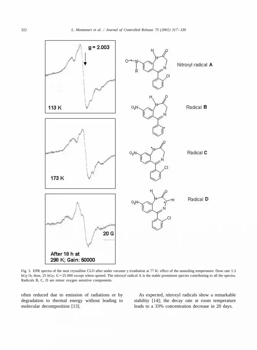
The radiolysis mechanism, which is described in where P is polymer radicals, and A, B, C and D aredetail in a previous work [3], is summarised in Fig. CLO radicals as in Fig. 3.5. The EPR spectra obtained following irradiations in
The comparative irradiations of raw polymer and air at room temperature were the same as thoseplacebo microsphere samples essentially produced produced by sample irradiation at low temperature
Fig. 5. Proposed mechanism of g radiolysis of PLGA (placebo microspheres).

L. Montanari et al. / Journal of Controlled Release 75 (2001) 317 –330 325
followed by prolonged room temperature annealing substituent is present in the para position. This extraand admission of air. stability coupled with the solid matrix trapping
efficiency explains the persistence of such species in3.2.4. Molecular interaction between CLO and the irradiated CLO and CLO loaded microspheres.polymeric matrix: evidence of radiostabilisationand spin transfer reactions
CLO in the loaded microspheres has a radios- 3.3. Differential scanning calorimetry (DSC)tabilising effect as shown by the 54.1% decrease ofoverall radical yield with respect to the computed The water content measured by Karl Fischerlinear contribution of the neat components in the titration was less than |0.4% in both irradiated andmolecular mixture (Table 1). At the same time the G non-irradiated microspheres.(radicals) value for CLO in the loaded microspheres Fig. 7 shows the DSC thermograms related tois enhanced by a factor of 40 (Table 1). Such PLGA placebo microspheres and CLO loaded micro-observations point to the existence of significant spheres before and after g irradiation, performedinteractions taking place between the polymer and under vacuum and in presence of air. The followingthe active ingredient. The radiostabilisation effect is thermal phenomena are observed: (a) an endothermiclikely to be related to the aromatic moiety of CLO peak in the region of |508C, characteristic of theacting as a channel for degradation of the radiation copolymer but present also in the drug loadedenergy to thermal energy. microspheres DSC profiles, which is presumably
The spin transfer effect leading to the enhance- reckoned with irreversible endothermic reorganisa-ment of CLO radicals is proposed to be related to the tion of the polymer matrix; (b) an exothermic peakelectron and radical scavenging properties of the in the region of 110–1308C present only in the drugnitro group according to the following reaction loaded microspheres DSC thermograms; this featurescheme (Fig. 6). Dissociative electron capture by the is likely to be related to drug recrystallisationC–Cl bond and the hydrogen abstraction at the phenomena taking place at temperatures high enoughactivated C–H bonds adjacent to the amido group in to afford the necessary molecular mobility. Thethe diazepinone section are also considered. Follow- irradiation is seen to cause a shift of the exothermicing electron scavenging or electron transfer from peak toward lower temperatures whilst the presencepolymer radicals to the nitro group, nitro anion of air is essentially without significant effects. Thisradicals are formed; these are the precursors of a low temperature shift may tentatively be imputed tonitroso derivative. The latter is likely to act as a spin changes in the polymeric matrices occurring as atrapping agent leading to the production of the consequence of radiolytic events such as chainobserved nitroxide adducts. The aromatic nitroxyl scissions. Differences in the position of the endo-radicals show generally much lower decay rates as thermic peaks in the |508C region are also observedcompared to other free radicals and their stability is as a consequence of the presence of the drug and ofgreatly enhanced when, as in the case of CLO, a the irradiation.
Table 1Radiolytic radical yields determined by EPR spectroscopy
Sample Radical yield(mmol/J)
CLO radicals 0.003060.0006Placebo microspheres: polymer radicals 0.2660.04Loaded microspheres total radicals (CLO1polymer) 0.1260.02Loaded microspheres: CLO radicals 0.06560.01Loaded microspheres: polymer radicals 0.05560.01
aLoaded microspheres: calculated total radical yield 0.2260.04a Calculated from the radiolytic yields of the neat components, polymer and CLO, weighted by the corresponding electron fractions.

326 L. Montanari et al. / Journal of Controlled Release 75 (2001) 317 –330
Fig. 6. g Radiolysis of CLO loaded microspheres: proposed mechanism for spin transfer reaction.
3.4. Drug content 3.5. In vitro release profiles
The drug contents of microspheres did not change As expected, CLO release was controlled by theafter irradiation. Moreover, they did not significantly diffusion of the drug from a monolithic matrix; itchange during storage over a 6-month period (Table followed the square root of time relationship
22). throughout the testing period (0.9920,r ,0.9958)

L. Montanari et al. / Journal of Controlled Release 75 (2001) 317 –330 327
Fig. 7. DSC thermograms of placebo and loaded microspheres irradiated and non-irradiated.
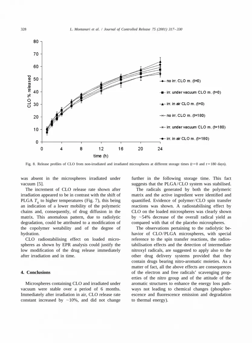
according to the Higuchi model. As an example, the Release rate constants of CLO are shown in Fig. 9.release profiles of CLO from non-irradiated and The values of the constants obtained for non-ir-irradiated microspheres at two different times of radiated and irradiated microspheres under vacuumstorage (t50 and t5180 days) are shown in Fig. 8. did not significantly change over the period of 6At the end of the experiments (6 months), the means months. After irradiation in air, the release rateof the values of CLO amounts released during the constants of CLO increased by |10%, and did notstability study were the following: change further for the rest of the storage period. The
increased rate constant of CLO release from mi-• 58.361.4% w/w non-irradiated microspheres; croparticles irradiated in air can be explained on the• 64.062.4% w/w microspheres irradiated in air; basis of the radiolytic degradation of the copolymer• 61.162.0% w/w microspheres irradiated under [3]. As a matter of fact, the presence of oxygen
vacuum. triggered a hydroperoxidative radiolysis cycle that
Table 2CLO contents determined in the non-irradiated and irradiated microspheres
Storage time Non-irradiated Irradiated under Irradiated in air(days) microspheres vacuum microspheres microspheres
(% w/w) (% w/w) (% w/w)
0 14.660.3 14.660.4 14.760.730 14.760.4 14.660.2 14.660.460 14.560.5 14.360.4 14.660.6
180 14.460.4 14.860.8 14.660.3
328 L. Montanari et al. / Journal of Controlled Release 75 (2001) 317 –330
Fig. 8. Release profiles of CLO from non-irradiated and irradiated microspheres at different storage times (t50 and t5180 days).
was absent in the microspheres irradiated under further in the following storage time. This factvacuum [5]. suggests that the PLGA/CLO system was stabilised.
The increment of CLO release rate shown after The radicals generated by both the polymericirradiation appeared to be in contrast with the shift of matrix and the active ingredient were identified andPLGA T to higher temperatures (Fig. 7), this being quantified. Evidence of polymer /CLO spin transferg
an indication of a lower mobility of the polymeric reactions was shown. A radiostabilising effect bychains and, consequently, of drug diffusion in the CLO on the loaded microspheres was clearly shownmatrix. This anomalous pattern, due to radiolytic by |54% decrease of the overall radical yield asdegradation, could be attributed to a modification of compared with that of the placebo microspheres.the copolymer wetability and of the degree of The observations pertaining to the radiolytic be-hydration. havior of CLO/PLGA microspheres, with special
CLO radiostabilising effect on loaded micro- reference to the spin transfer reactions, the radios-spheres as shown by EPR analysis could justify the tabilisation effects and the detection of intermediatelow modification of the drug release immediately nitroxyl radicals, are suggested to apply also to theafter irradiation and in time. other drug delivery systems provided that they
contain drugs bearing nitro-aromatic moieties. As amatter of fact, all the above effects are consequences
4. Conclusions of the electron and free radicals’ scavenging prop-erties of the nitro group and of the attitude of the
Microspheres containing CLO and irradiated under aromatic structures to enhance the energy loss path-vacuum were stable over a period of 6 months. ways not leading to chemical changes (phosphor-Immediately after irradiation in air, CLO release rate escence and fluorescence emission and degradationconstant increased by |10%, and did not change to thermal energy).

L. Montanari et al. / Journal of Controlled Release 75 (2001) 317 –330 329
Fig. 9. Release rate constant of CLO from irradiated and non-irradiated microspheres at different storage times.
The persistence of CLO radicals at room tempera- authors wish to thank Dr P. Riccardi, Centro Granditure in the loaded microspheres was sufficient in Strumenti, University of Pavia, for SEM analysis.order to use them as irradiation markers. On theother hand, this raises the problem of radical toxicityduring drug administration. The low concentration of
27 ReferencesCLO residual radicals (|10 mol /kg) does notseem to lead to appreciable consequences, especially
[1] A.G. Hausberger, R.A. Kenley, P.P. DeLuca, Gamma irradia-when the radical fast decay rate on water contact istion effects on molecular weight and in vitro degradation of
taken into consideration. The problem of residual poly(D,L-lactide-co-glycolide) microparticles, Pharm. Res. 12radical-induced toxicity in radiation treatment for (1995) 233–242.
[2] M.B. Sintzel, A. Merkli, C. Tabatabay, R. Gurny, Influencesterilisation or sanitisation purposes as well as theof irradiation sterilization on polymers used as drug carrierspossibility of using EPR analysis as a specific— a review, Drug Dev. Ind. Pharm. 23 (9) (1997) 857–879.technique for routine control of irradiated products
[3] L. Montanari, M. Costantini, E. Ciranni-Signoretti, L. Valvo,should be seriously taken into consideration. M. Santucci, M. Barolomei, P. Fattibene, S. Onori, A.
Faucitano, B. Conti, I. Genta, Gamma irradiation effects onpoly(D,L-lactide-co-glycolide) microspheres, J. ControlledRelease 56 (1998) 219–229.Acknowledgements
[4] A. Rothen-Weinhold, K. Besseghir, R. Gurny, Analysis ofthe influence of polymer characteristics and core loading on
This research is supported by a grant for the the in vivo release of a somatostatin analogue, Eur. J. Pharm.project ‘Sterilization processes and their influence on Sci. 5 (1997) 303–313.
`drug properties’, Istituto Superiore di Sanita. The [5] C. Volland, M. Wolff, T. Kissel, The influence of terminal

330 L. Montanari et al. / Journal of Controlled Release 75 (2001) 317 –330
gamma-sterilization on captopril containing poly(D,L-lactide- of g-irradiation on radical formation and polymer degra-co-glycolide) microspheres, J. Controlled Release 31 (1994) dation, J. Controlled Release 59 (1999) 23–32.293–305. [11] European Guideline 3AQ4a. The Use of Ionizing Radiation
[6] S. Yoshioka, Y. Aso, T. Otsuka, S. Kojima, The effect of in the Manufacture of Medicinal Products, Official Publi-g-irradiation on drug release from poly(lactide) micro- cations of the European Communities, London, July, 1992.spheres, Radiat. Phys. Chem. 46 (1995) 281–285. [12] P. Benelli, B. Conti, I. Genta, M. Costantini, L. Montanari,
[7] D. Mohr, M. Wolff, T. Kissel, Gamma irradiation for Clonazepam microencapsulation in poly-D,L-lactide-co-gly-terminal sterilization of 17b-estradiol loaded poly-(D,L-lac- colide microspheres, J. Microencapsul. 15 (4) (1998) 431–tide-co-glycolide) microparticles, J. Controlled Release 61 443.(1999) 203–217. [13] J. Woods, A.K. Pikaev, Radiolysis intermediates, in: R.J.
[8] A. Delgado, C. Evora, M. Llabres, Degradation of DL-PLA Woods, A.K. Pikaev (Ed.) Applied radiation chemistry inmethadone microspheres during in vitro release, Int. J. Radiation processing, John Wiley & Sons Inc., New York,Pharm. 140 (1996) 219–227. 1994 pp. 126-164.
[9] Y. Cha, C.G. Pitt, The acceleration of degradation-controlled [14] S.F. Nielsen, Nitrogen centred radicals in: J.K. Kochi (Ed.),drug delivery from polyester microspheres, J. Controlled Free Radicals, Vol. 2, John Wiley & Sons, New York, 1972,Release 8 (1989) 259–265. p. 543.
[10] B. Bittner, K. Mader, C. Kroll, H.H. Borchert, T. Kissel, [15] V. Malatesta, J.C. Scaiano, Absolute rate constants for theTetracycline–HCl-loaded poly(D,L-lactide-co-glycolide) reactions of tert-butoxil with ethers: importance of the stereomicrospheres prepared by a spray drying technique: influence electronic effect, J. Org. Chem. 47 (1982) 1455–1459.