release behavior and antibiofilm activity of usnic acid-loaded carboxylated poly(l-lactide)...
TRANSCRIPT
European Journal of Pharmaceutics and Biopharmaceutics xxx (2014) xxx–xxx
Contents lists available at ScienceDirect
European Journal of Pharmaceutics and Biopharmaceutics
journal homepage: www.elsevier .com/locate /e jpb
Research paper
Release behavior and antibiofilm activity of usnic acid-loadedcarboxylated poly(L-lactide) microparticles
http://dx.doi.org/10.1016/j.ejpb.2014.06.0020939-6411/� 2014 Elsevier B.V. All rights reserved.
Abbreviations: UA, usnic acid; PHA, poly(a-hydroxy acid); PLLA, poly(L-lactide);CPLLA, carboxylated poly(L-lactide); DMPA, 2,2-bis(hydroxymethyl)propionic acid;ROP, ring opening polymerization; ATR, attenuated total reflection; DSC, differentialscanning calorimetry; SEM, scanning electron microscopy; DLS, Dynamic LaserDiffraction Particle Size Analyzer; POM, polarized optical microscopy; PBS, phos-phate buffer solution; MIC, minimum inhibitory concentration; MH, Muller Hinton;RT, room temperature; UAA, usnic acid aggregates; PS, spherulite-like structures;VTF, Vogel–Tamman–Fulcher.⇑ Corresponding author. Department of Chemistry, Sapienza University of Rome,
P.le Aldo Moro 5, 00185 Rome, Italy. Tel.: +39 06 49913950; fax: +39 06 490631.E-mail address: [email protected] (A. Martinelli).
Please cite this article in press as: A. Martinelli et al., Release behavior and antibiofilm activity of usnic acid-loaded carboxylated poly(L-lactide) miticles, Eur. J. Pharm. Biopharm. (2014), http://dx.doi.org/10.1016/j.ejpb.2014.06.002
Andrea Martinelli a,⇑, Ahmed Bakry b, Lucio D’Ilario a, Iolanda Francolini a, Antonella Piozzi a,Vincenzo Taresco a
a Department of Chemistry, Sapienza University of Rome, Rome, Italyb Department of Chemistry, Faculty of Science, Helwan University, Cairo, Egypt
a r t i c l e i n f o a b s t r a c t
Article history:Received 11 April 2014Accepted in revised form 2 June 2014Available online xxxx
Keywords:PolylactideUsnic acidMicrobial biofilmMicroparticlesDrug deliveryStaphylococcus epidermidis
The use of controlled drug delivery systems could give a significant contribution to the improvement oftherapies against biofilm-based infections. The aim of this study was to develop polymer microparticles,based on carboxylated poly(L-lactide)s, to be employed as carriers for usnic acid (UA), a poorly solubledrug possessing antiviral, antiproliferative and wide spectrum antimicrobial activity. Thanks to polymersurfactant-like structure, 2.4 lm-in-size microparticles were obtained by a surfactant-free oil-in-wateremulsion/evaporation method. UA was encapsulated into these microparticles with a high loadingefficiency (80%). The drug release kinetics was found to be temperature dependent (the released doseincreasing with temperature) and showed bimodal release behavior. By polarized optical microscopyobservations and the application of kinetics models, the initial burst effect was attributed to the deliveryof the drug amorphous fraction while the slower release occurring for longer times to the crystalline one,both entrapped in the polymer amorphous phase. UA-loaded microparticles were able to promote thekilling of a 24 h-old Staphylococcus epidermidis biofilm more efficaciously than free UA.
� 2014 Elsevier B.V. All rights reserved.
1. Introduction
Microbial biofilms are known to be implicated in a large varietyof human infections, such as medical device-associated infections,chronic lung infections in cystic fibrosis patients, endocarditis, car-ies, and chronic wounds infections [1,2].
Biofilms exhibit distinct features with respect to the extremeresistance to antibiotics and antimicrobial agents as well as capac-ity to evade the host defense mechanisms. The high antibiotic tol-erance of bacteria in biofilm has been attributed to various reasons[3], such as (i) the interaction of antibiotics with biofilm matrixcomponents reducing drug diffusion through the biofilm; (ii) the
presence of slowly growing or dormant bacterial cells that arenot efficiently eradicated by antibiotics targeting cell metabolism(e.g., b-lactam); and (iii) the bacterial mutation frequency in bio-films much higher than in planktonic state due to the endogenousoxidative stress generated by bacterial cells upon exposure toantibiotics.
Therefore, there is an urgent need of either novel antifoulingsurfaces [4,5] or new antibiofilm drugs [3] as well as more effica-cious dosage forms (topical or oral) for improving activity of drugsin the biofilm.
The application of micro- and nano-technology to drug deliverycould give a significant contribution to the improvement of thera-pies against biofilm-based infections. Indeed, drug-loaded nano- ormicro-particles could protect the drug, diffuse into the exopolysac-charide (EPS) matrix surrounding bacterial colonies in biofilm, andrelease the drug in a controlled manner thus increasing drug resi-dence time and efficacy [6–8]. In addition, nano- or micro-particlescould permit the use of novel efficacious drugs having limitedtherapeutic applications due to unfavorable physicochemicalproperties, such as poor water solubility.
This is the case of usnic acid (UA), a dibenzofuran originally iso-lated from lichens, poorly soluble in water but well known for its
cropar-
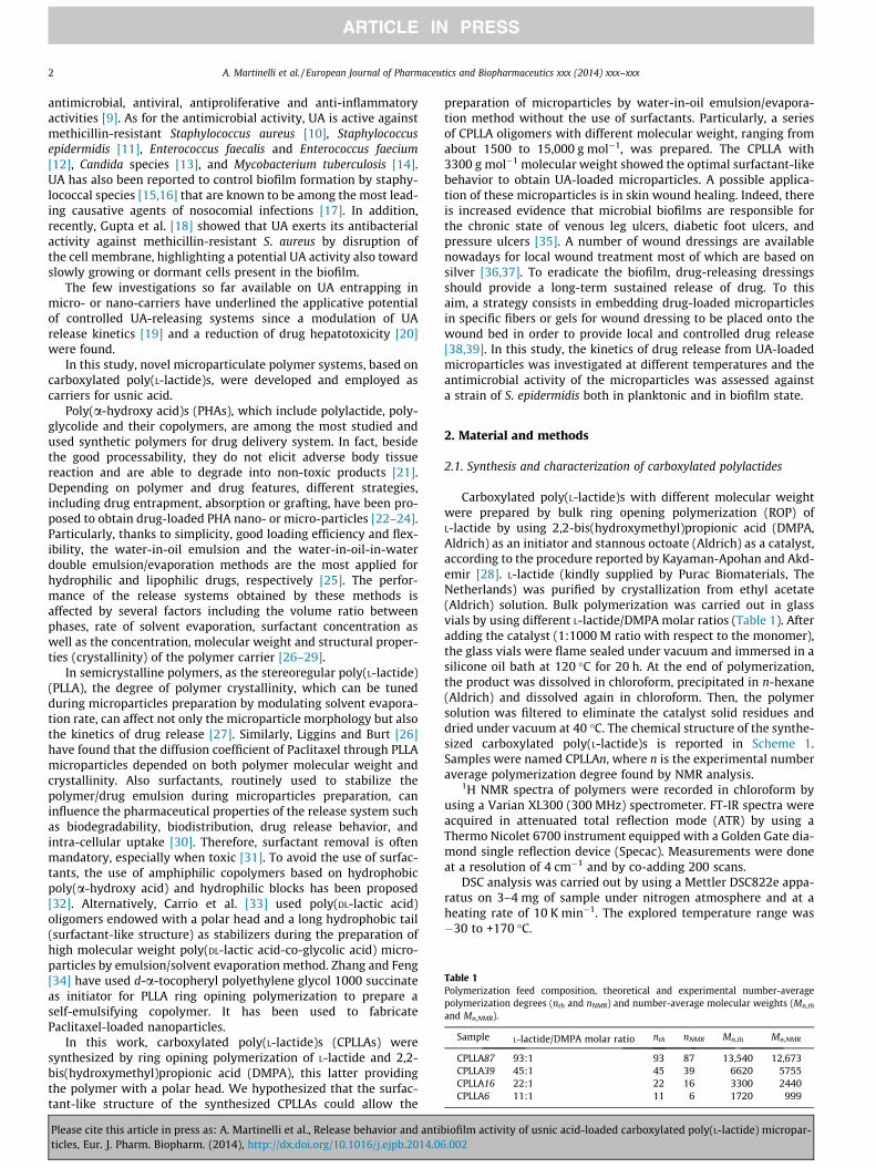
Table 1Polymerization feed composition, theoretical and experimental number-averagepolymerization degrees (nth and nNMR) and number-average molecular weights (Mn,th
and Mn,NMR).
Sample L-lactide/DMPA molar ratio nth nNMR Mn,th Mn,NMR
CPLLA87 93:1 93 87 13,540 12,673CPLLA39 45:1 45 39 6620 5755CPLLA16 22:1 22 16 3300 2440CPLLA6 11:1 11 6 1720 999
2 A. Martinelli et al. / European Journal of Pharmaceutics and Biopharmaceutics xxx (2014) xxx–xxx
antimicrobial, antiviral, antiproliferative and anti-inflammatoryactivities [9]. As for the antimicrobial activity, UA is active againstmethicillin-resistant Staphylococcus aureus [10], Staphylococcusepidermidis [11], Enterococcus faecalis and Enterococcus faecium[12], Candida species [13], and Mycobacterium tuberculosis [14].UA has also been reported to control biofilm formation by staphy-lococcal species [15,16] that are known to be among the most lead-ing causative agents of nosocomial infections [17]. In addition,recently, Gupta et al. [18] showed that UA exerts its antibacterialactivity against methicillin-resistant S. aureus by disruption ofthe cell membrane, highlighting a potential UA activity also towardslowly growing or dormant cells present in the biofilm.
The few investigations so far available on UA entrapping inmicro- or nano-carriers have underlined the applicative potentialof controlled UA-releasing systems since a modulation of UArelease kinetics [19] and a reduction of drug hepatotoxicity [20]were found.
In this study, novel microparticulate polymer systems, based oncarboxylated poly(L-lactide)s, were developed and employed ascarriers for usnic acid.
Poly(a-hydroxy acid)s (PHAs), which include polylactide, poly-glycolide and their copolymers, are among the most studied andused synthetic polymers for drug delivery system. In fact, besidethe good processability, they do not elicit adverse body tissuereaction and are able to degrade into non-toxic products [21].Depending on polymer and drug features, different strategies,including drug entrapment, absorption or grafting, have been pro-posed to obtain drug-loaded PHA nano- or micro-particles [22–24].Particularly, thanks to simplicity, good loading efficiency and flex-ibility, the water-in-oil emulsion and the water-in-oil-in-waterdouble emulsion/evaporation methods are the most applied forhydrophilic and lipophilic drugs, respectively [25]. The perfor-mance of the release systems obtained by these methods isaffected by several factors including the volume ratio betweenphases, rate of solvent evaporation, surfactant concentration aswell as the concentration, molecular weight and structural proper-ties (crystallinity) of the polymer carrier [26–29].
In semicrystalline polymers, as the stereoregular poly(L-lactide)(PLLA), the degree of polymer crystallinity, which can be tunedduring microparticles preparation by modulating solvent evapora-tion rate, can affect not only the microparticle morphology but alsothe kinetics of drug release [27]. Similarly, Liggins and Burt [26]have found that the diffusion coefficient of Paclitaxel through PLLAmicroparticles depended on both polymer molecular weight andcrystallinity. Also surfactants, routinely used to stabilize thepolymer/drug emulsion during microparticles preparation, caninfluence the pharmaceutical properties of the release system suchas biodegradability, biodistribution, drug release behavior, andintra-cellular uptake [30]. Therefore, surfactant removal is oftenmandatory, especially when toxic [31]. To avoid the use of surfac-tants, the use of amphiphilic copolymers based on hydrophobicpoly(a-hydroxy acid) and hydrophilic blocks has been proposed[32]. Alternatively, Carrio et al. [33] used poly(DL-lactic acid)oligomers endowed with a polar head and a long hydrophobic tail(surfactant-like structure) as stabilizers during the preparation ofhigh molecular weight poly(DL-lactic acid-co-glycolic acid) micro-particles by emulsion/solvent evaporation method. Zhang and Feng[34] have used d-a-tocopheryl polyethylene glycol 1000 succinateas initiator for PLLA ring opining polymerization to prepare aself-emulsifying copolymer. It has been used to fabricatePaclitaxel-loaded nanoparticles.
In this work, carboxylated poly(L-lactide)s (CPLLAs) weresynthesized by ring opining polymerization of L-lactide and 2,2-bis(hydroxymethyl)propionic acid (DMPA), this latter providingthe polymer with a polar head. We hypothesized that the surfac-tant-like structure of the synthesized CPLLAs could allow the
Please cite this article in press as: A. Martinelli et al., Release behavior and antibticles, Eur. J. Pharm. Biopharm. (2014), http://dx.doi.org/10.1016/j.ejpb.2014.06
preparation of microparticles by water-in-oil emulsion/evapora-tion method without the use of surfactants. Particularly, a seriesof CPLLA oligomers with different molecular weight, ranging fromabout 1500 to 15,000 g mol�1, was prepared. The CPLLA with3300 g mol�1 molecular weight showed the optimal surfactant-likebehavior to obtain UA-loaded microparticles. A possible applica-tion of these microparticles is in skin wound healing. Indeed, thereis increased evidence that microbial biofilms are responsible forthe chronic state of venous leg ulcers, diabetic foot ulcers, andpressure ulcers [35]. A number of wound dressings are availablenowadays for local wound treatment most of which are based onsilver [36,37]. To eradicate the biofilm, drug-releasing dressingsshould provide a long-term sustained release of drug. To thisaim, a strategy consists in embedding drug-loaded microparticlesin specific fibers or gels for wound dressing to be placed onto thewound bed in order to provide local and controlled drug release[38,39]. In this study, the kinetics of drug release from UA-loadedmicroparticles was investigated at different temperatures and theantimicrobial activity of the microparticles was assessed againsta strain of S. epidermidis both in planktonic and in biofilm state.
2. Material and methods
2.1. Synthesis and characterization of carboxylated polylactides
Carboxylated poly(L-lactide)s with different molecular weightwere prepared by bulk ring opening polymerization (ROP) ofL-lactide by using 2,2-bis(hydroxymethyl)propionic acid (DMPA,Aldrich) as an initiator and stannous octoate (Aldrich) as a catalyst,according to the procedure reported by Kayaman-Apohan and Akd-emir [28]. L-lactide (kindly supplied by Purac Biomaterials, TheNetherlands) was purified by crystallization from ethyl acetate(Aldrich) solution. Bulk polymerization was carried out in glassvials by using different L-lactide/DMPA molar ratios (Table 1). Afteradding the catalyst (1:1000 M ratio with respect to the monomer),the glass vials were flame sealed under vacuum and immersed in asilicone oil bath at 120 �C for 20 h. At the end of polymerization,the product was dissolved in chloroform, precipitated in n-hexane(Aldrich) and dissolved again in chloroform. Then, the polymersolution was filtered to eliminate the catalyst solid residues anddried under vacuum at 40 �C. The chemical structure of the synthe-sized carboxylated poly(L-lactide)s is reported in Scheme 1.Samples were named CPLLAn, where n is the experimental numberaverage polymerization degree found by NMR analysis.
1H NMR spectra of polymers were recorded in chloroform byusing a Varian XL300 (300 MHz) spectrometer. FT-IR spectra wereacquired in attenuated total reflection mode (ATR) by using aThermo Nicolet 6700 instrument equipped with a Golden Gate dia-mond single reflection device (Specac). Measurements were doneat a resolution of 4 cm�1 and by co-adding 200 scans.
DSC analysis was carried out by using a Mettler DSC822e appa-ratus on 3–4 mg of sample under nitrogen atmosphere and at aheating rate of 10 K min�1. The explored temperature range was�30 to +170 �C.
iofilm activity of usnic acid-loaded carboxylated poly(L-lactide) micropar-.002

Scheme 1. Chemical structure of the synthesized carboxylated poly(L-lactide)s.
A. Martinelli et al. / European Journal of Pharmaceutics and Biopharmaceutics xxx (2014) xxx–xxx 3
2.2. Microparticles preparation and drug loading
CPLLA microparticles were obtained by oil-in-water emulsion/evaporation method. Particularly, 1 mL of a polymer solution inchloroform (5% w/v) was added to 25 mL of deionized water at4 �C and stirred at 8000 rpm for 1 min. N2 was then flowed intothe suspension to remove chloroform. Once dried, the microparti-cles were collected by centrifugation.
To study the effect of polymer concentration on particles size,polymer concentrations of 10, 15 and 20% w/v were also tested.
CPLLA microparticles were then used as carrier for usnic acid(UA, Sigma Aldrich, M = 344 g/mol, pKa3 = 4.4, pKa9 = 8.8,pKa7 = 10.7), an antimicrobial drug poorly soluble in water(<0.1 mg/mL) [40].
To obtain usnic acid-loaded microparticles (CPLLAn-UA), UA(0.05 g) was dissolved in 10 mL of a 5% w/v polymer chloroformsolution. This solution was then employed to obtain microparticlesfollowing the procedure above described. The obtained microparti-cles were washed three times with distilled water. Finally,CPLLAn-UA microparticles were suspended in water and stored at4 �C.
To determine the UA amount loaded into the microparticles, aweighed quantity of CPLLAn-UA microparticles were dissolved inchloroform and submitted to UV–Vis analysis (Hewlett Packard8452A Diode Array single beam spectrophotometer). The drug con-centration was determined by checking the absorbance of the solu-tion at 290 nm related to drug aromatic ring. Drug loadingefficiency was defined as the ratio between the drug amount inthe microspheres and the initial drug amount.
The morphology of both pristine and UA-loaded microparticleswas observed by scanning electron microscopy (SEM, AURIGAZeiss). For SEM analysis, a drop of a microparticles water suspen-sion was placed on glass cover-slip and dried at room temperature.
Particle size distribution of the samples suspended in water wasstudied by a Dynamic Laser Diffraction Particle Size Analyzer (DLS,LS 13320 Beckman Coulter).
Polarized optical microscopy (POM) was used to study UA-loaded polymer structure. For the measurements, CPLLAn-UA filmswere analyzed being the microparticles too small for a directobservation. The films were obtained by drop-casting on glassslides a chloroform solution containing UA and the polymer atthe same concentrations used for the microparticles preparation.After drying, the films were incubated in PBS at 25 �C for either0.5 or 2 h. POM images of dry and hydrated samples were acquiredby an Optiphot2-Pol light microscopy (Nikon) connected to animage acquisition and analysis system.
2.3. Drug release kinetics
The kinetics of UA release from microparticles was studied atdifferent temperatures (Tr), 4 �C, 25 �C, 37 �C and 42 �C.
In each experiment, a fixed amount of CPLLAn-UA microparti-cles was added in glass tubes containing 10 mL of 0.02 M PBS(pH 7.4) and incubated at the chosen temperature. At determinedtime periods, a 1 mL aliquot of the release medium was withdrawnfrom the glass tube and analyzed by UV–Vis spectroscopy at290 nm to determine drug concentration. After analysis, the
Please cite this article in press as: A. Martinelli et al., Release behavior and antibticles, Eur. J. Pharm. Biopharm. (2014), http://dx.doi.org/10.1016/j.ejpb.2014.06
withdrawn medium was put back in the glass tube to continuethe release experiments. The UA loaded microspheres employedfor the experiments at 4 �C were used soon after their preparationto avoid drug release during the storage. For the experiments athigher temperature the samples were kept in the refrigerator fora maximum of 2 days.
The cumulative release fraction was expressed as M(t)/M0,where M0 is the amount of drug in the microspheres at the begin-ning and M(t) the drug released at the time t.
Each release experiment at Tr was performed by sampling twomicroparticle aliquots from each of two independent preparations(n = 4).
The cumulative release results are reported as the meanvalue ± maximum deviation.
2.4. Antimicrobial activity
The minimum inhibitory concentration (MIC) of CPLLAn-UAmicroparticles was assessed against planktonic S. epidermidis(ATCC 35984) by broth microdilution assay and compared withthat of pristine UA [11].
Several test tubes each containing a fixed amount of UA-loadedmicroparticles (1, 2, 4, 8 mg) suspended in water (1 mL) were pre-pared. Then, 1 mL of a 106 CFU/mL bacterial suspension in MullerHinton broth (MH) was added to each tube. A control tube, con-taining 1 mL of water and 1 mL of bacterial suspension in broth,was also prepared. After 18 h-incubation at 37 �C, bacterial growthin each test tube was determined by measuring the absorbance at600 nm and compared with the control tube. The MIC was the low-est concentration of CPLLAn-UA microparticles inhibiting bacterialgrowth in the test tube.
The antibiofilm activity of CPLLAn-UA microparticles was eval-uated on a 24 h-old S. epidermidis biofilm grown on glass cover-slides (10 mm in diameter). Particularly, for biofilm formation,glass coverslides were immersed in 2.0 mL of bacterial suspension(108 CFU/mL) in MH broth and incubated for 24 h at 37 �C. Then,coverslides were collected, washed twice with PBS to removeloosely adherent cells and incubated again for 18 h at 37 �C with2.0 mL of either fresh MH broth (control 1), unloaded CPLLAmicroparticles (4 mg, control 2), free UA suspension (300 lg) orCPLLAn-UA microparticles (4 mg) suspension. Following incuba-tion, glass coverslides were put in test tubes with 5 mL of PBS, son-icated for 5 min, and vortexed for 10 s to detach the biofilm. Five10-fold dilutions were then prepared, and three 10 lL aliquots ofeach dilution were plated on MH plates. CFUs were counted after18 h incubation at 37 �C.
In antimicrobial activities experiments, analysis of variancecomparisons was performed using Mini-Tab. Differences were con-sidered significant for p values of <0.05. Data are reported as means(n = 4) ± 1SD.
3. Results and discussion
3.1. Properties of the synthesized CPLLAn
Carboxylic acid functionalized poly(L-lactide)s with differentpolymerization degrees were obtained by ROP of L-lactide in pres-ence of DMPA. ATR-FTIR analysis was used to evaluate the occurredpolymerization. As an example, the spectrum of CPLLA16 isreported in Fig. 1S of the supporting information. In the spectrum,the C@O stretching peak at 1620 cm�1 related to carboxylic groupsof DMPA and the band at 1748 cm�1 related to ester C@O related toPLLA repeat unit are present.
Polymerization degree and number-average molecular weightof polymers were determined by 1H NMR analysis (Table 1) and
iofilm activity of usnic acid-loaded carboxylated poly(L-lactide) micropar-.002
4 A. Martinelli et al. / European Journal of Pharmaceutics and Biopharmaceutics xxx (2014) xxx–xxx
compared with the theoretical value (nth, Mn,th). As an example, the1H NMR spectrum of CPLLA16 is reported in Fig. 2S of the support-ing information. The polymerization degree (nNMR) of samples wasdetermined from the ratio between the integrated intensities ofthe peak at 5.20 ppm (I5.20ppm) related to methine proton of PLLArepeat unit and the peak at 4.30 ppm (I4.30ppm) of the methyleneprotons of DMPA unit (Eq. (1S) of the supporting information).
The effect of polymer molecular weight on the thermal proper-ties of CPLLAn samples was studied by DSC (Fig. 1). All polymersshowed a glass transition in the 25–55 �C temperature range(Fig. 1A). Cold-crystallization and melting during DSC heating werealso observed in all polymer thermograms, with the exception ofthe lowest molecular weight polymer (CPLLA6). This indicates thatthe polymers were in an amorphous state and, except CPPLA6,were able to crystallize during DSC heating. As can be observedin Fig. 1 B, the glass transition temperature (Tg) of polymersincreased with nNMR and MNMR. The experimental data were wellinterpolated by the empirical relation:
Tg ¼ Tg;1 �K
Mnð1Þ
where Tg,1 is the limiting glass transition temperature of infinitemolecular weight polymer and K is a constant depending on poly-mer [41]. The extrapolate value of the Tg,1 = 55 �C of an infinitemolecular weight polymer is in agreement with that found for thehigh Mn amorphous PLLA (Tg = 55–60 �C) [42]. The CPLLA6 sample,because of its low Mn, at room temperature was in the rubbery stateand resulted to be sticky.
Fig. 1. DSC thermograms of dry CPLLAn polymers (A). For the sake of clarity thethermograms were vertically shifted. Glass transition temperature (Tg) as a functionof the number average molecular weight and polymerization degree (B).
Please cite this article in press as: A. Martinelli et al., Release behavior and antibticles, Eur. J. Pharm. Biopharm. (2014), http://dx.doi.org/10.1016/j.ejpb.2014.06
The synthesized carboxylated poly(lactide)s were thenemployed to prepare microparticles by the oil-in-water emul-sion/evaporation method without the addition of specificsurfactants.
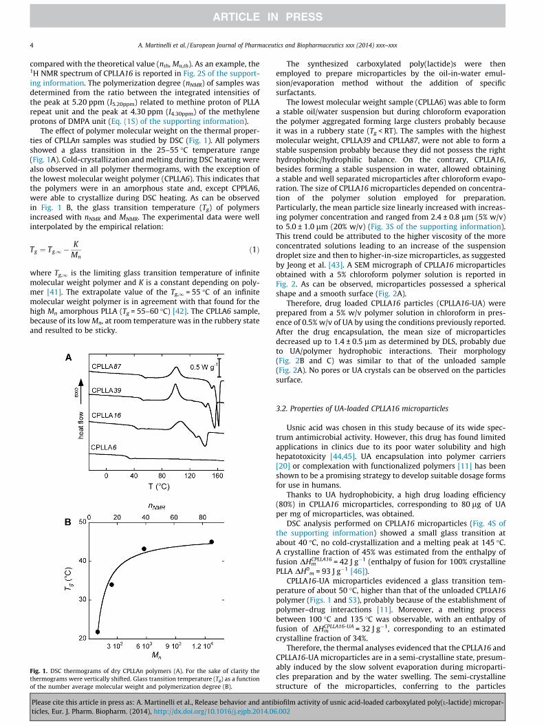
The lowest molecular weight sample (CPLLA6) was able to forma stable oil/water suspension but during chloroform evaporationthe polymer aggregated forming large clusters probably becauseit was in a rubbery state (Tg < RT). The samples with the highestmolecular weight, CPLLA39 and CPLLA87, were not able to form astable suspension probably because they did not possess the righthydrophobic/hydrophilic balance. On the contrary, CPLLA16,besides forming a stable suspension in water, allowed obtaininga stable and well separated microparticles after chloroform evapo-ration. The size of CPLLA16 microparticles depended on concentra-tion of the polymer solution employed for preparation.Particularly, the mean particle size linearly increased with increas-ing polymer concentration and ranged from 2.4 ± 0.8 lm (5% w/v)to 5.0 ± 1.0 lm (20% w/v) (Fig. 3S of the supporting information).This trend could be attributed to the higher viscosity of the moreconcentrated solutions leading to an increase of the suspensiondroplet size and then to higher-in-size microparticles, as suggestedby Jeong et al. [43]. A SEM micrograph of CPLLA16 microparticlesobtained with a 5% chloroform polymer solution is reported inFig. 2. As can be observed, microparticles possessed a sphericalshape and a smooth surface (Fig. 2A).
Therefore, drug loaded CPLLA16 particles (CPLLA16-UA) wereprepared from a 5% w/v polymer solution in chloroform in pres-ence of 0.5% w/v of UA by using the conditions previously reported.After the drug encapsulation, the mean size of microparticlesdecreased up to 1.4 ± 0.5 lm as determined by DLS, probably dueto UA/polymer hydrophobic interactions. Their morphology(Fig. 2B and C) was similar to that of the unloaded sample(Fig. 2A). No pores or UA crystals can be observed on the particlessurface.
3.2. Properties of UA-loaded CPLLA16 microparticles
Usnic acid was chosen in this study because of its wide spec-trum antimicrobial activity. However, this drug has found limitedapplications in clinics due to its poor water solubility and highhepatotoxicity [44,45]. UA encapsulation into polymer carriers[20] or complexation with functionalized polymers [11] has beenshown to be a promising strategy to develop suitable dosage formsfor use in humans.
Thanks to UA hydrophobicity, a high drug loading efficiency(80%) in CPLLA16 microparticles, corresponding to 80 lg of UAper mg of microparticles, was obtained.
DSC analysis performed on CPLLA16 microparticles (Fig. 4S ofthe supporting information) showed a small glass transition atabout 40 �C, no cold-crystallization and a melting peak at 145 �C.A crystalline fraction of 45% was estimated from the enthalpy offusion DHm
CPLLA16 = 42 J g�1 (enthalpy of fusion for 100% crystallinePLLA DH0
m = 93 J g�1 [46]).CPLLA16-UA microparticles evidenced a glass transition tem-
perature of about 50 �C, higher than that of the unloaded CPLLA16polymer (Figs. 1 and S3), probably because of the establishment ofpolymer–drug interactions [11]. Moreover, a melting processbetween 100 �C and 135 �C was observable, with an enthalpy offusion of DHm
CPLLA16-UA = 32 J g�1, corresponding to an estimatedcrystalline fraction of 34%.
Therefore, the thermal analyses evidenced that the CPLLA16 andCPLLA16-UA microparticles are in a semi-crystalline state, presum-ably induced by the slow solvent evaporation during microparti-cles preparation and by the water swelling. The semi-crystallinestructure of the microparticles, conferring to the particles
iofilm activity of usnic acid-loaded carboxylated poly(L-lactide) micropar-.002
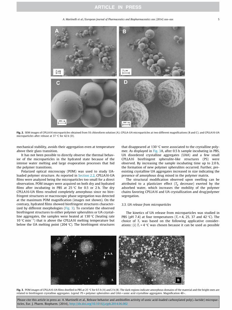
Fig. 2. SEM images of CPLLA16 microparticles obtained from 5% chloroform solution (A); CPLLA-UA microparticles at two different magnifications (B and C); and CPLLA16-UAmicroparticles after release at 37 �C for 42 h (D).
A. Martinelli et al. / European Journal of Pharmaceutics and Biopharmaceutics xxx (2014) xxx–xxx 5
mechanical stability, avoids their aggregation even at temperatureabove their glass transition.
It has not been possible to directly observe the thermal behav-ior of the microparticles in the hydrated state because of theintense water melting and large evaporation processes that hidthe polymer transitions.
Polarized optical microscopy (POM) was used to study UA-loaded polymer structure. As reported in Section 2.2, CPLLA16-UAfilms were analyzed being the microparticles too small for a directobservation. POM images were acquired on both dry and hydratedfilms after incubating in PBS at 25 �C for 0.5 or 2 h. The dryCPLLA16-UA films resulted completely amorphous since no bire-fringent structures or macroscopic phase segregation was detectedat the maximum POM magnification (images not shown). On thecontrary, hydrated films showed birefringent structures character-ized by different morphologies (Fig. 3). To correlate the observedbirefringent structures to either polymer spherulites or UA crystal-line aggregates, the samples were heated at 130 �C (heating rate10 �C min�1) that is above the CPLLA16 melting temperature butbelow the UA melting point (204 �C). The birefringent structures
Fig. 3. POM images of CPLLA16-UA films dwelled in PBS at 25 �C for 0.5 h (A) and 2 h (B). Trelated to birefringent crystalline aggregates. Legend: PS = polymer spherulites and UAA
Please cite this article in press as: A. Martinelli et al., Release behavior and antibticles, Eur. J. Pharm. Biopharm. (2014), http://dx.doi.org/10.1016/j.ejpb.2014.06
that disappeared at 130 �C were associated to the crystalline poly-mer. As displayed in Fig. 3A, after 0.5 h sample incubating in PBS,UA disordered crystalline aggregates (UAA) and a few smallCPLLA16 birefringent spherulite-like structures (PS) wereobserved. By increasing the sample incubating time up to 2.0 h,the formation of new polymer spherulites occurred. Further, pre-existing crystalline UA aggregates increased in size indicating thepresence of amorphous drug mixed in the polymer matrix.
The structural modification observed upon swelling can beattributed to a plasticizer effect (Tg decrease) exerted by theadsorbed water, which increases the mobility of the polymerchains favoring CPLLA16 and UA crystallization and drug/polymersegregation.
3.3. UA release from microparticles
The kinetics of UA release from microparticles was studied inPBS (pH 7.4) at four temperatures (Tr = 4, 25, 37 and 42 �C). Thechoice of Tr was based on the following applicative consider-ations: (i) Tr = 4 �C was chosen because it can be used as possible
he dark regions indicate amorphous domains of the material and the bright ones are= usnic acid crystalline aggregates. Magnification 40�.
iofilm activity of usnic acid-loaded carboxylated poly(L-lactide) micropar-.002
6 A. Martinelli et al. / European Journal of Pharmaceutics and Biopharmaceutics xxx (2014) xxx–xxx
storing condition of the drug loaded microspheres in refrigerator;(ii) Tr = 25 �C is the room temperature; (iii) Tr = 37 �C is the bodytemperature used in the antimicrobial activity experiments; and(iv) Tr = 42 �C is the temperature that can be potentially reachedlocally by an external stimuli or in presence of wound infection[47].
Moreover, the release kinetics dependence on temperature, asshown later, was used to relate the release mechanisms to thestructural features of the system.
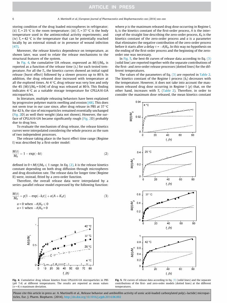
In Fig. 4, the cumulative UA release, expressed as M(t)/M0, isreported as a function of the release time (tr) for each tested tem-perature. For all the Tr, the kinetics curves showed an initial rapidrelease (burst effect) followed by a slower process up to 80 h. Inaddition, the drug released dose increased with temperature atall the explored times. At 4 �C, drug release was very low and onlythe 4% (M(t)/M0 = 0.04) of drug was released at 80 h. This findingindicates 4 �C as a suitable storage temperature for CPLLA16-UAmicroparticles.
In literature, multiple releasing behaviors have been explainedby progressive polymer matrix swelling and erosion [48]. This doesnot seem true in our case since, after drug release in PBS at 37 �Cfor 42 h, the size of microparticles remained essentially unchanged(Fig. 2D) as well their weight (data not shown). However, the sur-face of CPLLA16-UA became significantly rough (Fig. 2D) probablydue to drug loss.
To evaluate the mechanism of drug release, the release kineticscurves were interpolated considering the whole process as the sumof two independent processes.
The release taking place in the burst effect time range (RegimeI) was described by a first-order model:
MðtÞM0¼ 1� expð�ktÞ ð2Þ
defined in 0 < M(t)/M0 6 1 range. In Eq. (2), k is the release kineticsconstant depending on both drug diffusion through microspheresand drug dissolution rate. The release data for longer time (RegimeII) were, instead, fitted by a zero-order function.
Therefore, the overall release data were interpolated by aseries–parallel release model expressed by the following function:
MðtÞM0¼ p½1� expð�kItÞ� þ afAþ K IItg ð3Þ
a = 0 when �A/KII 6 0a = 1 when �A/KII > 0
Fig. 4. Cumulative drug release kinetics from CPLLA16-UA microparticles in PBS(pH 7.4) at different temperatures. The results are reported as mean values(n = 4) ± maximum deviation.
Please cite this article in press as: A. Martinelli et al., Release behavior and antibticles, Eur. J. Pharm. Biopharm. (2014), http://dx.doi.org/10.1016/j.ejpb.2014.06
where p is the maximum released drug dose occurring in Regime I,kI is the kinetics constant of the first-order process, A is the inter-cept of the straight line describing the zero-order process, KII is thekinetics constant of the zero-order process and a is a parameterthat eliminates the negative contribution of the zero-order processbefore it starts after a delay s = �A/KII. In this way no hypothesis onthe ending of the first-order process and the beginning of the zero-order one was necessary.
In Fig. 5, the best-fit curves of release data according to Eq. (3)(solid line) are reported together with the separate contributions ofthe first- and zero-order release processes (dotted lines) for the dif-ferent temperatures.
The values of the parameters of Eq. (3) are reported in Table 2.The kinetics constant of the Regime I process (kI) decreases withthe temperature. However, it does not take into account the max-imum released drug dose occurring in Regime I (p) that, on theother hand, increases with Tr (Table 2). Therefore, in order toconsider the maximum dose released, the mean kinetics constant
Fig. 5. Fit curves of release data according to Eq. (3) (solid lines) and the separatecontributions of the first- and zero-order models (dotted lines) at the differenttemperatures.
iofilm activity of usnic acid-loaded carboxylated poly(L-lactide) micropar-.002
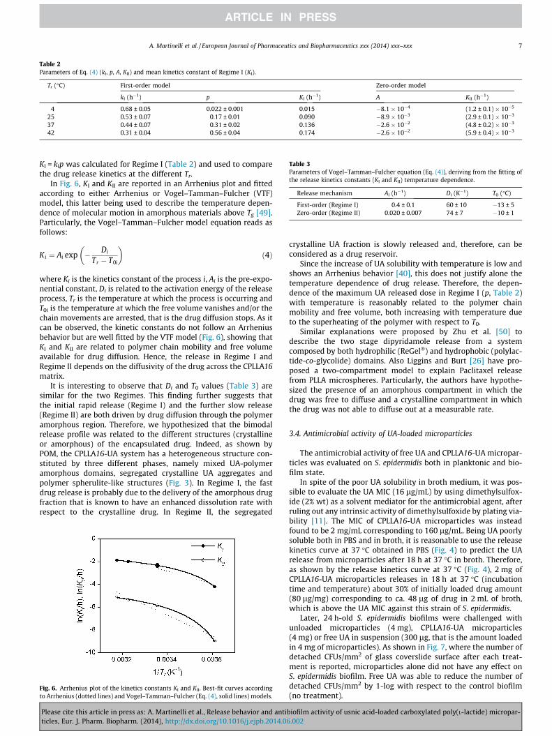
Table 2Parameters of Eq. (4) (kI, p, A, KII) and mean kinetics constant of Regime I (KI).
Tr (�C) First-order model Zero-order model
kI (h�1) p KI (h�1) A KII (h�1)
4 0.68 ± 0.05 0.022 ± 0.001 0.015 �8.1 � 10�4 (1.2 ± 0.1) � 10�5
25 0.53 ± 0.07 0.17 ± 0.01 0.090 �8.9 � 10�3 (2.9 ± 0.1) � 10�3
37 0.44 ± 0.07 0.31 ± 0.02 0.136 �2.6 � 10�2 (4.8 ± 0.2) � 10�3
42 0.31 ± 0.04 0.56 ± 0.04 0.174 �2.6 � 10�2 (5.9 ± 0.4) � 10�3
Table 3Parameters of Vogel–Tamman–Fulcher equation (Eq. (4)), deriving from the fitting ofthe release kinetics constants (KI and KII) temperature dependence.
Release mechanism Ai (h�1) Di (K�1) T0 (�C)
First-order (Regime I) 0.4 ± 0.1 60 ± 10 �13 ± 5Zero-order (Regime II) 0.020 ± 0.007 74 ± 7 �10 ± 1
A. Martinelli et al. / European Journal of Pharmaceutics and Biopharmaceutics xxx (2014) xxx–xxx 7
KI = kIp was calculated for Regime I (Table 2) and used to comparethe drug release kinetics at the different Tr.
In Fig. 6, KI and KII are reported in an Arrhenius plot and fittedaccording to either Arrhenius or Vogel–Tamman–Fulcher (VTF)model, this latter being used to describe the temperature depen-dence of molecular motion in amorphous materials above Tg [49].Particularly, the Vogel–Tamman–Fulcher model equation reads asfollows:
Ki ¼ Ai exp � Di
Tr � T0i
� �ð4Þ
where Ki is the kinetics constant of the process i, Ai is the pre-expo-nential constant, Di is related to the activation energy of the releaseprocess, Tr is the temperature at which the process is occurring andT0i is the temperature at which the free volume vanishes and/or thechain movements are arrested, that is the drug diffusion stops. As itcan be observed, the kinetic constants do not follow an Arrheniusbehavior but are well fitted by the VTF model (Fig. 6), showing thatKI and KII are related to polymer chain mobility and free volumeavailable for drug diffusion. Hence, the release in Regime I andRegime II depends on the diffusivity of the drug across the CPLLA16matrix.
It is interesting to observe that Di and T0 values (Table 3) aresimilar for the two Regimes. This finding further suggests thatthe initial rapid release (Regime I) and the further slow release(Regime II) are both driven by drug diffusion through the polymeramorphous region. Therefore, we hypothesized that the bimodalrelease profile was related to the different structures (crystallineor amorphous) of the encapsulated drug. Indeed, as shown byPOM, the CPLLA16-UA system has a heterogeneous structure con-stituted by three different phases, namely mixed UA-polymeramorphous domains, segregated crystalline UA aggregates andpolymer spherulite-like structures (Fig. 3). In Regime I, the fastdrug release is probably due to the delivery of the amorphous drugfraction that is known to have an enhanced dissolution rate withrespect to the crystalline drug. In Regime II, the segregated
Fig. 6. Arrhenius plot of the kinetics constants KI and KII. Best-fit curves accordingto Arrhenius (dotted lines) and Vogel–Tamman–Fulcher (Eq. (4), solid lines) models.
Please cite this article in press as: A. Martinelli et al., Release behavior and antibticles, Eur. J. Pharm. Biopharm. (2014), http://dx.doi.org/10.1016/j.ejpb.2014.06
crystalline UA fraction is slowly released and, therefore, can beconsidered as a drug reservoir.
Since the increase of UA solubility with temperature is low andshows an Arrhenius behavior [40], this does not justify alone thetemperature dependence of drug release. Therefore, the depen-dence of the maximum UA released dose in Regime I (p, Table 2)with temperature is reasonably related to the polymer chainmobility and free volume, both increasing with temperature dueto the superheating of the polymer with respect to T0.
Similar explanations were proposed by Zhu et al. [50] todescribe the two stage dipyridamole release from a systemcomposed by both hydrophilic (ReGel�) and hydrophobic (polylac-tide-co-glycolide) domains. Also Liggins and Burt [26] have pro-posed a two-compartment model to explain Paclitaxel releasefrom PLLA microspheres. Particularly, the authors have hypothe-sized the presence of an amorphous compartment in which thedrug was free to diffuse and a crystalline compartment in whichthe drug was not able to diffuse out at a measurable rate.
3.4. Antimicrobial activity of UA-loaded microparticles
The antimicrobial activity of free UA and CPLLA16-UA micropar-ticles was evaluated on S. epidermidis both in planktonic and bio-film state.
In spite of the poor UA solubility in broth medium, it was pos-sible to evaluate the UA MIC (16 lg/mL) by using dimethylsulfox-ide (2% wt) as a solvent mediator for the antimicrobial agent, afterruling out any intrinsic activity of dimethylsulfoxide by plating via-bility [11]. The MIC of CPLLA16-UA microparticles was insteadfound to be 2 mg/mL corresponding to 160 lg/mL. Being UA poorlysoluble both in PBS and in broth, it is reasonable to use the releasekinetics curve at 37 �C obtained in PBS (Fig. 4) to predict the UArelease from microparticles after 18 h at 37 �C in broth. Therefore,as shown by the release kinetics curve at 37 �C (Fig. 4), 2 mg ofCPLLA16-UA microparticles releases in 18 h at 37 �C (incubationtime and temperature) about 30% of initially loaded drug amount(80 lg/mg) corresponding to ca. 48 lg of drug in 2 mL of broth,which is above the UA MIC against this strain of S. epidermidis.
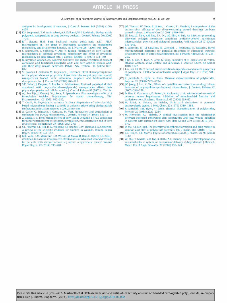
Later, 24 h-old S. epidermidis biofilms were challenged withunloaded microparticles (4 mg), CPLLA16-UA microparticles(4 mg) or free UA in suspension (300 lg, that is the amount loadedin 4 mg of microparticles). As shown in Fig. 7, where the number ofdetached CFUs/mm2 of glass coverslide surface after each treat-ment is reported, microparticles alone did not have any effect onS. epidermidis biofilm. Free UA was able to reduce the number ofdetached CFUs/mm2 by 1-log with respect to the control biofilm(no treatment).
iofilm activity of usnic acid-loaded carboxylated poly(L-lactide) micropar-.002
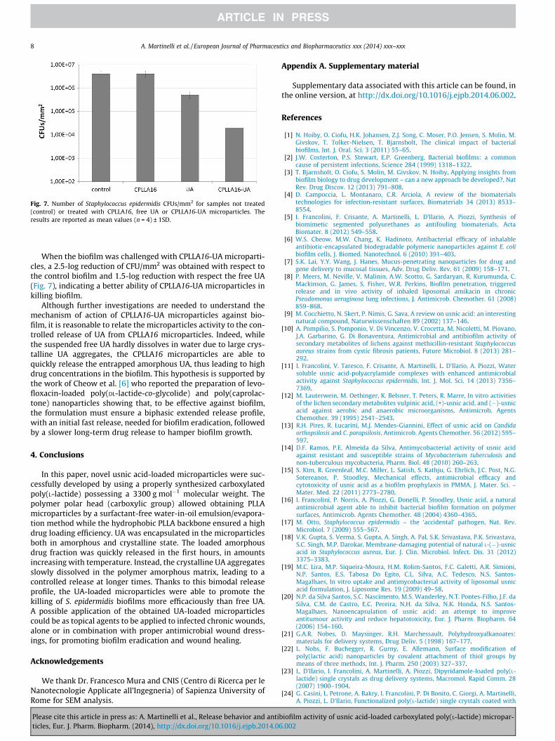
Fig. 7. Number of Staphylococcus epidermidis CFUs/mm2 for samples not treated(control) or treated with CPLLA16, free UA or CPLLA16-UA microparticles. Theresults are reported as mean values (n = 4) ± 1SD.
8 A. Martinelli et al. / European Journal of Pharmaceutics and Biopharmaceutics xxx (2014) xxx–xxx
When the biofilm was challenged with CPLLA16-UA microparti-cles, a 2.5-log reduction of CFU/mm2 was obtained with respect tothe control biofilm and 1.5-log reduction with respect the free UA(Fig. 7), indicating a better ability of CPLLA16-UA microparticles inkilling biofilm.
Although further investigations are needed to understand themechanism of action of CPLLA16-UA microparticles against bio-film, it is reasonable to relate the microparticles activity to the con-trolled release of UA from CPLLA16 microparticles. Indeed, whilethe suspended free UA hardly dissolves in water due to large crys-talline UA aggregates, the CPLLA16 microparticles are able toquickly release the entrapped amorphous UA, thus leading to highdrug concentrations in the biofilm. This hypothesis is supported bythe work of Cheow et al. [6] who reported the preparation of levo-floxacin-loaded poly(DL-lactide-co-glycolide) and poly(caprolac-tone) nanoparticles showing that, to be effective against biofilm,the formulation must ensure a biphasic extended release profile,with an initial fast release, needed for biofilm eradication, followedby a slower long-term drug release to hamper biofilm growth.
4. Conclusions
In this paper, novel usnic acid-loaded microparticles were suc-cessfully developed by using a properly synthesized carboxylatedpoly(L-lactide) possessing a 3300 g mol�1 molecular weight. Thepolymer polar head (carboxylic group) allowed obtaining PLLAmicroparticles by a surfactant-free water-in-oil emulsion/evapora-tion method while the hydrophobic PLLA backbone ensured a highdrug loading efficiency. UA was encapsulated in the microparticlesboth in amorphous and crystalline state. The loaded amorphousdrug fraction was quickly released in the first hours, in amountsincreasing with temperature. Instead, the crystalline UA aggregatesslowly dissolved in the polymer amorphous matrix, leading to acontrolled release at longer times. Thanks to this bimodal releaseprofile, the UA-loaded microparticles were able to promote thekilling of S. epidermidis biofilms more efficaciously than free UA.A possible application of the obtained UA-loaded microparticlescould be as topical agents to be applied to infected chronic wounds,alone or in combination with proper antimicrobial wound dress-ings, for promoting biofilm eradication and wound healing.
Acknowledgements
We thank Dr. Francesco Mura and CNIS (Centro di Ricerca per leNanotecnologie Applicate all’Ingegneria) of Sapienza University ofRome for SEM analysis.
Please cite this article in press as: A. Martinelli et al., Release behavior and antibticles, Eur. J. Pharm. Biopharm. (2014), http://dx.doi.org/10.1016/j.ejpb.2014.06
Appendix A. Supplementary material
Supplementary data associated with this article can be found, inthe online version, at http://dx.doi.org/10.1016/j.ejpb.2014.06.002.
References
[1] N. Hoiby, O. Ciofu, H.K. Johansen, Z.J. Song, C. Moser, P.O. Jensen, S. Molin, M.Givskov, T. Tolker-Nielsen, T. Bjarnsholt, The clinical impact of bacterialbiofilms, Int. J. Oral. Sci. 3 (2011) 55–65.
[2] J.W. Costerton, P.S. Stewart, E.P. Greenberg, Bacterial biofilms: a commoncause of persistent infections, Science 284 (1999) 1318–1322.
[3] T. Bjarnsholt, O. Ciofu, S. Molin, M. Givskov, N. Hoiby, Applying insights frombiofilm biology to drug development – can a new approach be developed?, NatRev. Drug Discov. 12 (2013) 791–808.
[4] D. Campoccia, L. Montanaro, C.R. Arciola, A review of the biomaterialstechnologies for infection-resistant surfaces, Biomaterials 34 (2013) 8533–8554.
[5] I. Francolini, F. Crisante, A. Martinelli, L. D’Ilario, A. Piozzi, Synthesis ofbiomimetic segmented polyurethanes as antifouling biomaterials, ActaBiomater. 8 (2012) 549–558.
[6] W.S. Cheow, M.W. Chang, K. Hadinoto, Antibacterial efficacy of inhalableantibiotic-encapsulated biodegradable polymeric nanoparticles against E. colibiofilm cells, J. Biomed. Nanotechnol. 6 (2010) 391–403.
[7] S.K. Lai, Y.Y. Wang, J. Hanes, Mucus-penetrating nanoparticles for drug andgene delivery to mucosal tissues, Adv. Drug Deliv. Rev. 61 (2009) 158–171.
[8] P. Meers, M. Neville, V. Malinin, A.W. Scotto, G. Sardaryan, R. Kurumunda, C.Mackinson, G. James, S. Fisher, W.R. Perkins, Biofilm penetration, triggeredrelease and in vivo activity of inhaled liposomal amikacin in chronicPseudomonas aeruginosa lung infections, J. Antimicrob. Chemother. 61 (2008)859–868.
[9] M. Cocchietto, N. Skert, P. Nimis, G. Sava, A review on usnic acid: an interestingnatural compound, Naturwissenschaften 89 (2002) 137–146.
[10] A. Pompilio, S. Pomponio, V. Di Vincenzo, V. Crocetta, M. Nicoletti, M. Piovano,J.A. Garbarino, G. Di Bonaventura, Antimicrobial and antibiofilm activity ofsecondary metabolites of lichens against methicillin-resistant Staphylococcusaureus strains from cystic fibrosis patients, Future Microbiol. 8 (2013) 281–292.
[11] I. Francolini, V. Taresco, F. Crisante, A. Martinelli, L. D’Ilario, A. Piozzi, Watersoluble usnic acid-polyacrylamide complexes with enhanced antimicrobialactivity against Staphylococcus epidermidis, Int. J. Mol. Sci. 14 (2013) 7356–7369.
[12] M. Lauterwein, M. Oethinger, K. Belsner, T. Peters, R. Marre, In vitro activitiesof the lichen secondary metabolites vulpinic acid, (+)-usnic acid, and (�)-usnicacid against aerobic and anaerobic microorganisms, Antimicrob. AgentsChemother. 39 (1995) 2541–2543.
[13] R.H. Pires, R. Lucarini, M.J. Mendes-Giannini, Effect of usnic acid on Candidaorthopsilosis and C. parapsilosis, Antimicrob. Agents Chemother. 56 (2012) 595–597.
[14] D.F. Ramos, P.E. Almeida da Silva, Antimycobacterial activity of usnic acidagainst resistant and susceptible strains of Mycobacterium tuberculosis andnon-tuberculous mycobacteria, Pharm. Biol. 48 (2010) 260–263.
[15] S. Kim, R. Greenleaf, M.C. Miller, L. Satish, S. Kathju, G. Ehrlich, J.C. Post, N.G.Sotereanos, P. Stoodley, Mechanical effects, antimicrobial efficacy andcytotoxicity of usnic acid as a biofilm prophylaxis in PMMA, J. Mater. Sci. –Mater. Med. 22 (2011) 2773–2780.
[16] I. Francolini, P. Norris, A. Piozzi, G. Donelli, P. Stoodley, Usnic acid, a naturalantimicrobial agent able to inhibit bacterial biofilm formation on polymersurfaces, Antimicrob. Agents Chemother. 48 (2004) 4360–4365.
[17] M. Otto, Staphylococcus epidermidis – the ‘accidental’ pathogen, Nat. Rev.Microbiol. 7 (2009) 555–567.
[18] V.K. Gupta, S. Verma, S. Gupta, A. Singh, A. Pal, S.K. Srivastava, P.K. Srivastava,S.C. Singh, M.P. Darokar, Membrane-damaging potential of natural L-(�)-usnicacid in Staphylococcus aureus, Eur. J. Clin. Microbiol. Infect. Dis. 31 (2012)3375–3383.
[19] M.C. Lira, M.P. Siqueira-Moura, H.M. Rolim-Santos, F.C. Galetti, A.R. Simioni,N.P. Santos, E.S. Tabosa Do Egito, C.L. Silva, A.C. Tedesco, N.S. Santos-Magalhaes, In vitro uptake and antimycobacterial activity of liposomal usnicacid formulation, J. Liposome Res. 19 (2009) 49–58.
[20] N.P. da Silva Santos, S.C. Nascimento, M.S. Wanderley, N.T. Pontes-Filho, J.F. daSilva, C.M. de Castro, E.C. Pereira, N.H. da Silva, N.K. Honda, N.S. Santos-Magalhaes, Nanoencapsulation of usnic acid: an attempt to improveantitumour activity and reduce hepatotoxicity, Eur. J. Pharm. Biopharm. 64(2006) 154–160.
[21] G.A.R. Nobes, D. Maysinger, R.H. Marchessault, Polyhydroxyalkanoates:materials for delivery systems, Drug Deliv. 5 (1998) 167–177.
[22] L. Nobs, F. Buchegger, R. Gurny, E. Allemann, Surface modification ofpoly(lactic acid) nanoparticles by covalent attachment of thiol groups bymeans of three methods, Int. J. Pharm. 250 (2003) 327–337.
[23] L. D’Ilario, I. Francolini, A. Martinelli, A. Piozzi, Dipyridamole-loaded poly(L-lactide) single crystals as drug delivery systems, Macromol. Rapid Comm. 28(2007) 1900–1904.
[24] G. Casini, L. Petrone, A. Bakry, I. Francolini, P. Di Bonito, C. Giorgi, A. Martinelli,A. Piozzi, L. D’Ilario, Functionalized poly(L-lactide) single crystals coated with
iofilm activity of usnic acid-loaded carboxylated poly(L-lactide) micropar-.002
A. Martinelli et al. / European Journal of Pharmaceutics and Biopharmaceutics xxx (2014) xxx–xxx 9
antigens in development of vaccines, J. Control. Release 148 (2010) e106–e108.
[25] K.S. Soppimath, T.M. Aminabhavi, A.R. Kulkarni, W.E. Rudzinski, Biodegradablepolymeric nanoparticles as drug delivery devices, J. Control. Release 70 (2001)1–20.
[26] R.T. Liggins, H.M. Burt, Paclitaxel loaded poly(L-lactic acid) (PLLA)microspheres. II. The effect of processing parameters on microspheremorphology and drug release kinetics, Int. J. Pharm. 281 (2004) 103–106.
[27] S. Izumikawa, S. Yoshioka, Y. Aso, Y. Takeda, Preparation of poly(l-lactide)microspheres of different crystalline morphology and effect of crystallinemorphology on drug release rate, J. Control. Release 15 (1991) 133–140.
[28] N. Kayaman-Apohan, Z.S. Akdemir, Synthesis and characterization of pendantcarboxylic acid functional poly(lactic acid) and poly(lactic-co-glycolic acid)and their drug release behaviors, Polym. Adv. Technol. 16 (2005) 807–812.
[29] S. Hyvonen, L. Peltonen, M. Karjalainen, J. Hirvonen, Effect of nanoprecipitationon the physicochemical properties of low molecular weight poly(L-lactic acid)nanoparticles loaded with salbutamol sulphate and beclomethasonedipropionate, Int. J. Pharm. 295 (2005) 269–281.
[30] S.K. Sahoo, J. Panyam, S. Prabha, V. Labhasetwar, Residual polyvinyl alcoholassociated with poly(D,L-lactide-co-glycolide) nanoparticles affects theirphysical properties and cellular uptake, J. Control. Release 82 (2002) 105–114.
[31] A.J. Ten Tije, J. Verweij, W.J. Loos, A. Sparreboom, Pharmacological effects offormulation vehicles: implications for cancer chemotherapy, Clin.Pharmacokinet. 42 (2003) 665–685.
[32] T. Ouchi, M. Toyohara, H. Arimura, Y. Ohya, Preparation of poly(L-lactide)-based microspheres having a cationic or anionic surface using biodegradablesurfactants, Biomacromolecules 3 (2002) 885–888.
[33] A. Carrio, G. Schwach, J. Coudane, M. Vert, Preparation and degradation ofsurfactant-free PLAGA microspheres, J. Control. Release 37 (1995). 133-121.
[34] Z. Zhang, S.-S. Feng, Nanoparticles of poly(lactide)/vitamin E TPGS copolymerfor cancer chemotherapy: synthesis, formulation, characterization and in vitrodrug release, Biomaterials 27 (2006) 262–270.
[35] S.L. Percival, K.E. Hill, D.W. Williams, S.J. Hooper, D.W. Thomas, J.W. Costerton,A review of the scientific evidence for biofilms in wounds, Wound RepairRegen. 20 (2012) 647–657.
[36] M.F. Valle, N.M. Maruthur, L.M. Wilson, M. Malas, U. Qazi, E. Haberl, E.B. Bass, J.Zenilman, G. Lazarus, Comparative effectiveness of advanced wound dressingsfor patients with chronic venous leg ulcers: a systematic review, WoundRepair Regen. 22 (2014) 193–204.
Please cite this article in press as: A. Martinelli et al., Release behavior and antibticles, Eur. J. Pharm. Biopharm. (2014), http://dx.doi.org/10.1016/j.ejpb.2014.06
[37] J.G. Thomas, W. Slone, S. Linton, L. Corum, S.L. Percival, A comparison of theantimicrobial efficacy of two silver-containing wound dressings on burnwound isolates, J. Wound Care 20 (2011) 580–586.
[38] J.E. Lee, J.C. Park, K.H. Lee, S.H. Oh, J.G. Kim, H. Suh, An infection-preventingbilayered collagen membrane containing antibiotic-loaded hyaluronanmicroparticles: physical and biological properties, Artif. Organs 26 (7) (2002)636–646.
[39] B. Albertini, M. Di Sabatino, N. Calonghi, L. Rodriguez, N. Passerini, Novelmultifunctional platforms for potential treatment of cutaneous wounds:development and in vitro characterization, Int. J. Pharm. 440 (2) (2013) 238–249.
[40] J. Jin, Y. Rao, X. Bian, A. Zeng, G. Yang, Solubility of (+)-usnic acid in water,ethanol, acetone, ethyl acetate and n-hexane, J. Solution Chem. 42 (2013)1018–1027.
[41] T.G. Fox, P.J. Flory, Second order transition temperatures and related propertiesof polystyrene. I. Influence of molecular weight, J. Appl. Phys. 21 (1950) 581–585.
[42] K. Jamshidi, S. Hyon, Y. Ikada, Thermal characterization of polylactides,Polymer 29 (1988) 2229–2234.
[43] J.C. Jeong, J. Lee, K. Cho, Effects of crystalline microstructure on drug releasebehavior of poly(epsilon-caprolactone) microspheres, J. Control. Release 92(2003) 249–258.
[44] D. Han, K. Matsumaru, D. Rettori, N. Kaplowitz, Usnic acid-induced necrosis ofcultured mouse hepatocytes: inhibition of mitochondrial function andoxidative stress, Biochem. Pharmacol. 67 (2004) 439–451.
[45] M. Takai, Y. Uehara, J.A. Beisler, Usnic acid derivatives as potentialantineoplastic agents, J. Med. Chem. 22 (1979) 1380–1384.
[46] K. Jamshidi, S.H. Hyon, Y. Ikada, Thermal characterization of polylactides,Polymer 29 (1988) 2229–2234.
[47] M. Fierheller, R.G. Sibbald, A clinical investigation into the relationshipbetween increased periwound skin temperature and local wound infectionin patients with chronic leg ulcers, Adv. Skin Wound Care 23 (8) (2010) 369–379.
[48] D. Ma, A.J. McHugh, The interplay of membrane formation and drug release insolution-cast films of polylactide polymers, Int. J. Pharm. 388 (2010) 1–12.
[49] L.R. Hilden, K.R. Morris, Physics of amorphous solids, J. Pharm. Sci. 93 (2004)3–12.
[50] W. Zhu, T. Masaki, Y.H. Bae, R. Rathi, A.K. Cheung, S.E. Kern, Development of asustained-release system for perivascular delivery of dipyridamole, J. Biomed.Mater. Res. B Appl. Biomater. 77 (2006) 135–143.
iofilm activity of usnic acid-loaded carboxylated poly(L-lactide) micropar-.002