integrating silicon/zinc dual elements with plga microspheres
TRANSCRIPT

3038 | J. Mater. Chem. B, 2020, 8, 3038--3049 This journal is©The Royal Society of Chemistry 2020
Cite this: J.Mater. Chem. B, 2020,
8, 3038
Integrating silicon/zinc dual elements with PLGAmicrospheres in calcium phosphate cementscaffolds synergistically enhancesbone regeneration
Weiwei Liang,†abc Min Gao,†a Jingsheng Lou,d Yunyang Bai,a Jing Zhang,e
Teliang Lu,e Xiaowen Sun,c Jiandong Ye, e Baowei Li,d Li Sun,*d
Boon Chin Heng,*f Xuehui Zhang *cg and Xuliang Dengag
Integrating multiple pro-osteogenic factors into bone graft substitutes is a practical and effective
approach to improve bone repair efficacy. Here, Si–Zn dual elements and PLGA microspheres were
incorporated into calcium phosphate cement (CPC) scaffolds (PLGA/CPC-Si/Zn) as a novel strategy to
synergistically enhance bone regeneration. The incorporation of PLGA microspheres and Si/Zn dual
elements within CPC scaffolds improved the setting time, injectability and compressive strength. The
PLGA/CPC-Si/Zn scaffolds displayed controlled sequential release of Si and Zn ions. In vitro, RAW 264.7
cells displayed the M2 phenotype with a high level of anti-inflammatory cytokines in response to PLGA/
CPC-Si/Zn. The conditioned medium of RAW 264.7 cells cultured on the PLGA/CPC-Si/Zn scaffolds
significantly enhanced the osteogenic differentiation of rat BMSCs. In a rat femur defect model, the
implanted PLGA/CPC-Si/Zn scaffolds led to obvious new bone formation after 4 weeks, apparent bone
ingrowth into the PLGA microspheres after 12 weeks, and was almost completely filled with mature new
bone upon degradation of the PLGA microspheres at 24 weeks. These findings demonstrate that the
PLGA/CPC-Si/Zn scaffolds promote osteogenesis by synergistically improving the immune microenvironment
and biodegradability. Hence, integrating multiple trace elements together with degradable components within
bone graft biomaterials can be an effective strategy for promoting bone regeneration.
1. Introduction
Biomaterial scaffolds play a critical role in bone defect repair byserving as artificial extracellular matrices that present both
chemical and physical cues for modulating osteoconductiveeffects and degradation to provide space for new bone ingrowthand to promote vascularization.1,2 However, approaches thatallow us to build devices integrating these cues in a combinatorialway are limited due to the lack of suitable component materials andappropriate manufacturing processes.
Numerous reports have confirmed that doping of biomaterialswith trace elements such as silicon (Si), zinc (Zn), magnesium(Mg), strontium (Sr) and iron (Fe) can significantly enhanceosteogenesis of calcium phosphate scaffolds3,4 i.e. calciumphosphate cement (CPC). Doping biomaterials with more thanone trace element is considered a viable approach mimickingthe natural physiological extracellular environment in whichmultiple trace elements are present to facilitate osteogenesis.5
Different elements play different roles in the bone regenerationprocess. For example, Si can promote vascularization andcollagen synthesis,6,7 while zinc can stimulate bone growth andmineralization, increase osteoblast activity and inhibit osteoclastactivity.8 The ability of multiple element incorporation to functionallycoordinate with the natural bone regeneration process providesa novel strategy for biomaterial development.9
a Department of Geriatric Dentistry, Peking University School and Hospital of Stomatology,
Beijing 100081, P. R. Chinab Department of Prosthodonitcs, Peking University School and Hospital of Stomatology,
Beijing 10081, P. R. Chinac Department of Dental Materials & Dental Medical Devices Testing Centre,
Peking University School and Hospital of Stomatology, Beijing 100081, P. R. China.
E-mail: [email protected] Anesthesia and Operation Centre, Chinese PLA General Hospital, Beijing 410078,
P. R. China. E-mail: [email protected] School of Materials Science and Engineering, South China University of
Technology, Guangzhou 510641, Chinaf Central Laboratory, Peking University School and Hospital of Stomatology,
Beijing 100081, P. R. China. E-mail: [email protected] National Engineering Laboratory for Digital and Material Technology of
Stomatology, NMPA Key Laboratory for Dental Materials & Beijing Laboratory of
Biomedical Materials, Peking University School and Hospital of Stomatology,
Beijing 100081, P. R. China
† These authors contributed equally to this work.
Received 21st December 2019,Accepted 10th March 2020
DOI: 10.1039/c9tb02901j
rsc.li/materials-b
Journal ofMaterials Chemistry B
PAPER
Publ
ishe
d on
11
Mar
ch 2
020.
Dow
nloa
ded
by P
ekin
g U
nive
rsity
on
11/3
0/20
20 2
:03:
34 A
M.
View Article OnlineView Journal | View Issue
This journal is©The Royal Society of Chemistry 2020 J. Mater. Chem. B, 2020, 8, 3038--3049 | 3039
Trace elements have also been reported to play a pivotal rolein bone biomaterial-stimulated osteogenesis through immuno-modulation.10–12 Trace element doped CPC scaffolds can effectivelyregulate the macrophage M1/M2 phenotype transition, therebypromoting bone regeneration and repair.13 Studies to date havetended to focus mainly on single trace elements, and the effectsof co-doping biomaterials with multiple trace elements on theimmune response of macrophages remain unclear.
Poor biodegradability and lack of an internal bone germinalcenter, usually compromise the bone repair efficacy of CPCscaffolds.14,15 In order to overcome these shortcomings, Ye et al.has investigated the use of poly(lactic-co-glycolic acid) (PLGA)microspheres to improve the biodegradability of CPC, whichfacilitates macropore formation conducive for cell ingrowthin situ, as well as enable the possible controlled release of bioactivefactors during the degradation process.16 The incorporated PLGAmicrospheres can provide the required high mechanical strengthto CPC scaffolds during the early stage of bone repair, andgradually degrade to create macropores for bone ingrowth.17
The synergistic effects of incorporating multiple elements andPLGA microspheres in enhancing bone regeneration is a majorconsideration in our CPC scaffold design. How the elementalrelease kinetics and PLGA microspheres degradation of the CPCscaffold is synchronized to match the developmental timescaleof bone regeneration is of great interest, and remains a majorchallenge for bone regeneration applications.
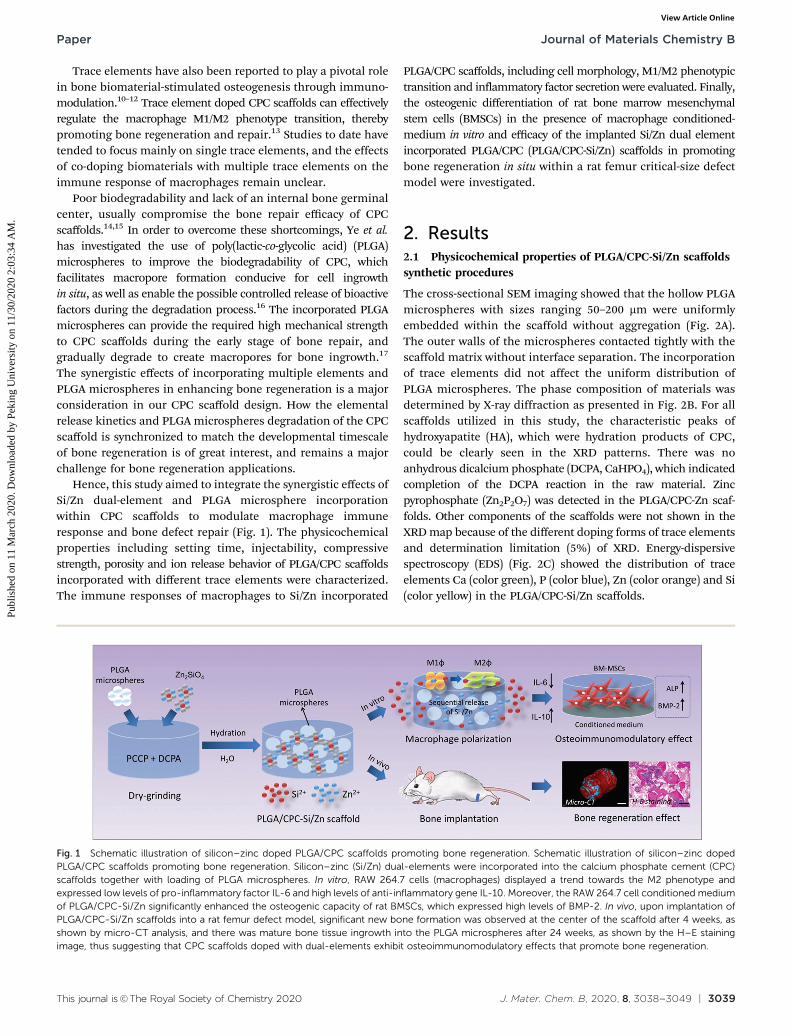
Hence, this study aimed to integrate the synergistic effects ofSi/Zn dual-element and PLGA microsphere incorporationwithin CPC scaffolds to modulate macrophage immuneresponse and bone defect repair (Fig. 1). The physicochemicalproperties including setting time, injectability, compressivestrength, porosity and ion release behavior of PLGA/CPC scaffoldsincorporated with different trace elements were characterized.The immune responses of macrophages to Si/Zn incorporated
PLGA/CPC scaffolds, including cell morphology, M1/M2 phenotypictransition and inflammatory factor secretion were evaluated. Finally,the osteogenic differentiation of rat bone marrow mesenchymalstem cells (BMSCs) in the presence of macrophage conditioned-medium in vitro and efficacy of the implanted Si/Zn dual elementincorporated PLGA/CPC (PLGA/CPC-Si/Zn) scaffolds in promotingbone regeneration in situ within a rat femur critical-size defectmodel were investigated.
2. Results2.1 Physicochemical properties of PLGA/CPC-Si/Zn scaffoldssynthetic procedures
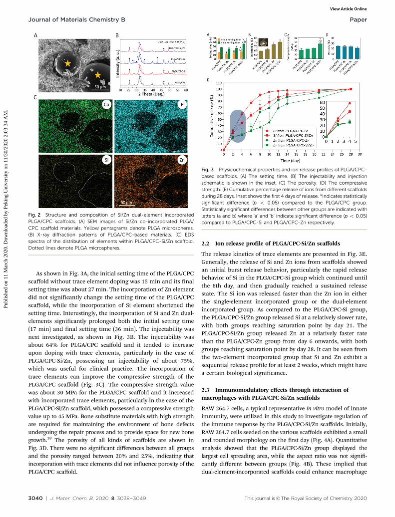
The cross-sectional SEM imaging showed that the hollow PLGAmicrospheres with sizes ranging 50–200 mm were uniformlyembedded within the scaffold without aggregation (Fig. 2A).The outer walls of the microspheres contacted tightly with thescaffold matrix without interface separation. The incorporationof trace elements did not affect the uniform distribution ofPLGA microspheres. The phase composition of materials wasdetermined by X-ray diffraction as presented in Fig. 2B. For allscaffolds utilized in this study, the characteristic peaks ofhydroxyapatite (HA), which were hydration products of CPC,could be clearly seen in the XRD patterns. There was noanhydrous dicalcium phosphate (DCPA, CaHPO4), which indicatedcompletion of the DCPA reaction in the raw material. Zincpyrophosphate (Zn2P2O7) was detected in the PLGA/CPC-Zn scaf-folds. Other components of the scaffolds were not shown in theXRD map because of the different doping forms of trace elementsand determination limitation (5%) of XRD. Energy-dispersivespectroscopy (EDS) (Fig. 2C) showed the distribution of traceelements Ca (color green), P (color blue), Zn (color orange) and Si(color yellow) in the PLGA/CPC-Si/Zn scaffolds.
Fig. 1 Schematic illustration of silicon–zinc doped PLGA/CPC scaffolds promoting bone regeneration. Schematic illustration of silicon–zinc dopedPLGA/CPC scaffolds promoting bone regeneration. Silicon–zinc (Si/Zn) dual-elements were incorporated into the calcium phosphate cement (CPC)scaffolds together with loading of PLGA microspheres. In vitro, RAW 264.7 cells (macrophages) displayed a trend towards the M2 phenotype andexpressed low levels of pro-inflammatory factor IL-6 and high levels of anti-inflammatory gene IL-10. Moreover, the RAW 264.7 cell conditioned mediumof PLGA/CPC-Si/Zn significantly enhanced the osteogenic capacity of rat BMSCs, which expressed high levels of BMP-2. In vivo, upon implantation ofPLGA/CPC-Si/Zn scaffolds into a rat femur defect model, significant new bone formation was observed at the center of the scaffold after 4 weeks, asshown by micro-CT analysis, and there was mature bone tissue ingrowth into the PLGA microspheres after 24 weeks, as shown by the H–E stainingimage, thus suggesting that CPC scaffolds doped with dual-elements exhibit osteoimmunomodulatory effects that promote bone regeneration.
Paper Journal of Materials Chemistry B
Publ
ishe
d on
11
Mar
ch 2
020.
Dow
nloa
ded
by P
ekin
g U
nive
rsity
on
11/3
0/20
20 2
:03:
34 A
M.
View Article Online
3040 | J. Mater. Chem. B, 2020, 8, 3038--3049 This journal is©The Royal Society of Chemistry 2020
As shown in Fig. 3A, the initial setting time of the PLGA/CPCscaffold without trace element doping was 15 min and its finalsetting time was about 27 min. The incorporation of Zn elementdid not significantly change the setting time of the PLGA/CPCscaffold, while the incorporation of Si element shortened thesetting time. Interestingly, the incorporation of Si and Zn dual-elements significantly prolonged both the initial setting time(17 min) and final setting time (36 min). The injectability wasnext investigated, as shown in Fig. 3B. The injectability wasabout 64% for PLGA/CPC scaffold and it tended to increaseupon doping with trace elements, particularly in the case ofPLGA/CPC-Si/Zn, possessing an injectability of about 75%,which was useful for clinical practice. The incorporation oftrace elements can improve the compressive strength of thePLGA/CPC scaffold (Fig. 3C). The compressive strength valuewas about 30 MPa for the PLGA/CPC scaffold and it increasedwith incorporated trace elements, particularly in the case of thePLGA/CPC-Si/Zn scaffold, which possessed a compressive strengthvalue up to 45 MPa. Bone substitute materials with high strengthare required for maintaining the environment of bone defectsundergoing the repair process and to provide space for new bonegrowth.18 The porosity of all kinds of scaffolds are shown inFig. 3D. There were no significant differences between all groupsand the porosity ranged between 20% and 25%, indicating thatincorporation with trace elements did not influence porosity of thePLGA/CPC scaffold.
2.2 Ion release profile of PLGA/CPC-Si/Zn scaffolds
The release kinetics of trace elements are presented in Fig. 3E.Generally, the release of Si and Zn ions from scaffolds showedan initial burst release behavior, particularly the rapid releasebehavior of Si in the PLGA/CPC-Si group which continued untilthe 8th day, and then gradually reached a sustained releasestate. The Si ion was released faster than the Zn ion in eitherthe single-element incorporated group or the dual-elementincorporated group. As compared to the PLGA/CPC-Si group,the PLGA/CPC-Si/Zn group released Si at a relatively slower rate,with both groups reaching saturation point by day 21. ThePLGA/CPC-Si/Zn group released Zn at a relatively faster ratethan the PLGA/CPC-Zn group from day 6 onwards, with bothgroups reaching saturation point by day 28. It can be seen fromthe two-element incorporated group that Si and Zn exhibit asequential release profile for at least 2 weeks, which might havea certain biological significance.
2.3 Immunomodulatory effects through interaction ofmacrophages with PLGA/CPC-Si/Zn scaffolds
RAW 264.7 cells, a typical representative in vitro model of innateimmunity, were utilized in this study to investigate regulation ofthe immune response by the PLGA/CPC-Si/Zn scaffolds. Initially,RAW 264.7 cells seeded on the various scaffolds exhibited a smalland rounded morphology on the first day (Fig. 4A). Quantitativeanalysis showed that the PLGA/CPC-Si/Zn group displayed thelargest cell spreading area, while the aspect ratio was not signifi-cantly different between groups (Fig. 4B). These implied thatdual-element-incorporated scaffolds could enhance macrophage
Fig. 2 Structure and composition of Si/Zn dual-element incorporatedPLGA/CPC scaffolds. (A) SEM images of Si/Zn co-incorporated PLGA/CPC scaffold materials. Yellow pentagrams denote PLGA microspheres.(B) X-ray diffraction patterns of PLGA/CPC-based materials. (C) EDSspectra of the distribution of elements within PLGA/CPC-Si/Zn scaffold.Dotted lines denote PLGA microspheres.
Fig. 3 Physicochemical properties and ion release profiles of PLGA/CPC-based scaffolds. (A) The setting time. (B) The injectability and injectionschematic is shown in the inset. (C) The porosity. (D) The compressivestrength. (E) Cumulative percentage release of ions from different scaffoldsduring 28 days. Inset shows the first 4 days of release. *Indicates statisticallysignificant difference (p o 0.05) compared to the PLGA/CPC group.Statistically significant differences between other groups are indicated withletters (a and b) where ‘a’ and ‘b’ indicate significant difference (p o 0.05)compared to PLGA/CPC-Si and PLGA/CPC-Zn respectively.
Journal of Materials Chemistry B Paper
Publ
ishe
d on
11
Mar
ch 2
020.
Dow
nloa
ded
by P
ekin
g U
nive
rsity
on
11/3
0/20
20 2
:03:
34 A
M.
View Article Online

This journal is©The Royal Society of Chemistry 2020 J. Mater. Chem. B, 2020, 8, 3038--3049 | 3041
adhesion and spreading compared to single- or non-incorporatedscaffolds. On the third day, macrophages cultured on the traceelement incorporated scaffolds appeared much narrower andmore elongated, as compared to cells of the non-incorporatedscaffolds, which more closely-resembled the typical morphologyof the anti-inflammatory M2 phenotype, according to previousstudies.19,20 In particular, the dual-element incorporated scaffoldgroup displayed the largest cell elongation ratio, which implied theirmost potent anti-inflammatory effects, as compared to single-element incorporated or non-incorporated scaffolds (Fig. 4C).Consistently, stimulation of RAW 264.7 cells with the PLGA/CPC-Si/Zn scaffold resulted in a shift towards the M2 phenotype,with most cells expressing the surface marker CD206, as com-pared to cells stimulated with other scaffolds (Fig. 5A). Bycontrast, the same treatment resulted in the least cells expres-sing the pro-inflammatory surface marker CCR7 in the PLGA/CPC-Si/Zn scaffold group, as compared to the single-elementincorporated or non-incorporated scaffold group (Fig. 5A).
For inflammatory gene expression, the RAW macrophagesseeded on the PLGA/CPC-Si/Zn scaffold displayed significantlylower pro-inflammatory gene TNF-a expression and higher anti-inflammatory IL-10 and TGF-1b expression levels, as compared toeither the PLGA/CPC-Si or PLGA/CPC-Zn groups on day 3 (p o 0.05)(Fig. 5B). ELISA results showed that the pro-inflammatory factor IL-6
secreted by RAW macrophages on dual-element incorporatedscaffold was at the lowest level amongst all groups from day 1 today 3 (Fig. 5C). These results demonstrated that after stimulationwith the PLGA/CPC-Si/Zn scaffold for 3 days, the RAW cells dis-played a trend towards the M2 phenotype. In addition, there weresignificantly higher expression levels of vascular genes VEGF andPDGF-BB in the PLGA/CPC-Si/Zn group, as compared to the otherthree groups on day 3 (P o 0.05) (Fig. 5D).
2.4 Osteoimmunomodulatory effects of culture mediumconditioned by PLGA/CPC-Si/Zn scaffolds in vitro
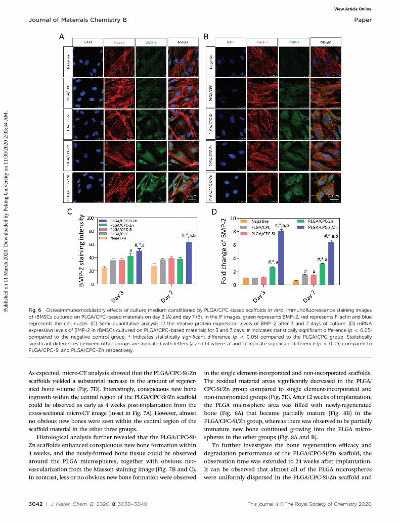
To investigate whether the immuno-modulatory function ofPLGA/CPC-Si/Zn scaffolds can exert pro-osteogenic effectsin vitro, conditioned media were collected from RAW macro-phages cultured on scaffolds to evaluate their effects on theosteogenic differentiation of rBMSCs. Immunofluorescenceanalysis showed significantly higher production of BMP-2 inthe PLGA/CPC-Si/Zn group, as compared to the other groups(Fig. 6A and B) after 3 and 7 days of culture (p o 0.05), whichwas confirmed by the quantitative analysis of fluorescenceintensity results (Fig. 6C). A similar trend was also observedat the gene expression level with the RT-qPCR result (Fig. 6D).
2.5 Bone regeneration mediated by PLGA/CPC-Si/Zn scaffoldsin vivo
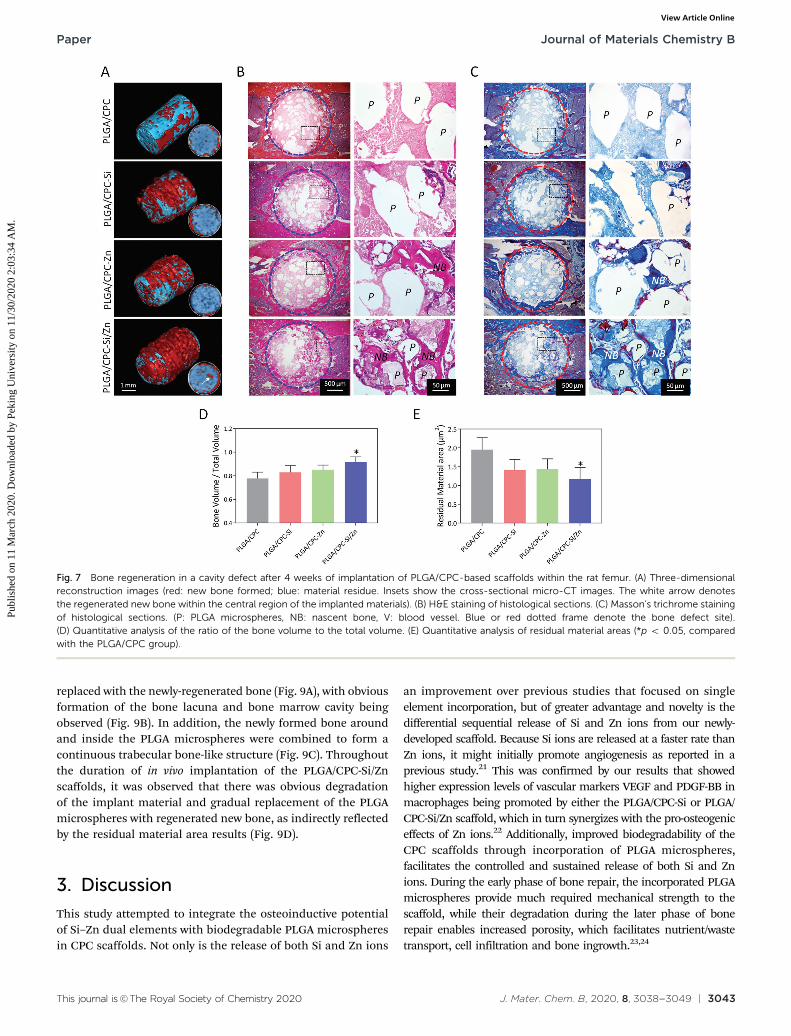
As evidenced by micro-CT detection, obvious new bone formationaround the implanted trace element incorporated scaffolds wereobserved after 4 weeks (Fig. 7A), while less new bone formationwas observed around the non-incorporated PLGA/CPC scaffold.
Fig. 4 Morphological changes of RAW 264.7 cells mediated by PLGA/CPC-based scaffolds. (A) SEM images of RAW 264.7 cells cultured onscaffolds for 1 and 3 days. (B) Cell areas and cell elongation ratio after 1 dayof co-culture. (C) Cell areas and cell elongation ratio at 3 days ofco-culture. In (B and C), symbols represent individual cells (n = 24). * Indicatesstatistically significant difference (p o 0.05) compared to the PLGA/CPCgroup. Statistically significant differences between other groups are indicatedwith letters (a and b) where ‘a’ and ‘b’ indicate significant difference (p o 0.05)compared to PLGA/CPC-Si and PLGA/CPC-Zn respectively.
Fig. 5 Immunomodulatory effects through interaction of macrophageswith PLGA/CPC-based scaffolds. (A) FACS analysis of RAW 264.7 cellsstimulated by the various scaffold materials on day 3. (B) mRNA expressionlevels of inflammatory gene markers within RAW 264.7 cells cultured ondifferent PLGA/CPC materials on day 3. (C) Cytokine expression of IL-6from day 1 to day 3 detected by ELISA. (D) mRNA expression levels ofvascular gene markers within RAW 264.7 cells cultured on different PLGA/CPC materials on day 3. * Indicates statistically significant difference(p o 0.05) compared to the PLGA/CPC group. Statistically significantdifferences between other groups are indicated with letters (a and b)where ‘a’ and ‘b’ indicate significant difference (p o 0.05) compared toPLGA/CPC-Si and PLGA/CPC-Zn respectively.
Paper Journal of Materials Chemistry B
Publ
ishe
d on
11
Mar
ch 2
020.
Dow
nloa
ded
by P
ekin
g U
nive
rsity
on
11/3
0/20
20 2
:03:
34 A
M.
View Article Online
3042 | J. Mater. Chem. B, 2020, 8, 3038--3049 This journal is©The Royal Society of Chemistry 2020
As expected, micro-CT analysis showed that the PLGA/CPC-Si/Znscaffolds yielded a substantial increase in the amount of regener-ated bone volume (Fig. 7D). Interestingly, conspicuous new boneingrowth within the central region of the PLGA/CPC-Si/Zn scaffoldcould be observed as early as 4 weeks post-implantation from thecross-sectional micro-CT image (in-set in Fig. 7A). However, almostno obvious new bones were seen within the central region of thescaffold material in the other three groups.
Histological analysis further revealed that the PLGA/CPC-Si/Zn scaffolds enhanced conspicuous new bone formation within4 weeks, and the newly-formed bone tissue could be observedaround the PLGA microspheres, together with obvious neo-vascularization from the Masson staining image (Fig. 7B and C).In contrast, less or no obvious new bone formation were observed
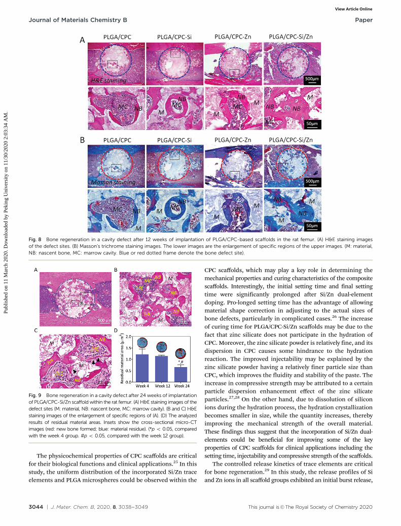
in the single element-incorporated and non-incorporated scaffolds.The residual material areas significantly decreased in the PLGA/CPC-Si/Zn group compared to single element-incorporated andnon-incorporated groups (Fig. 7E). After 12 weeks of implantation,the PLGA microsphere area was filled with newly-regeneratedbone (Fig. 8A) that became partially mature (Fig. 8B) in thePLGA/CPC-Si/Zn group, whereas there was observed to be partiallyimmature new bone continued growing into the PLGA micro-spheres in the other groups (Fig. 8A and B).
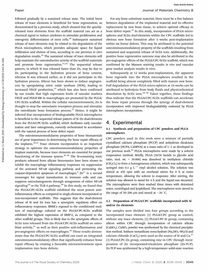
To further investigate the bone regeneration efficacy anddegradation performance of the PLGA/CPC-Si/Zn scaffold, theobservation time was extended to 24 weeks after implantation.It can be observed that almost all of the PLGA microsphereswere uniformly dispersed in the PLGA/CPC-Si/Zn scaffold and
Fig. 6 Osteoimmunomodulatory effects of culture medium conditioned by PLGA/CPC-based scaffolds in vitro. Immunofluorescence staining imagesof rBMSCs cultured on PLGA/CPC-based materials on day 3 (A) and day 7 (B). In the IF images, green represents BMP-2, red represents F-actin and bluerepresents the cell nuclei. (C) Semi-quantitative analysis of the relative protein expression levels of BMP-2 after 3 and 7 days of culture. (D) mRNAexpression levels of BMP-2 in rBMSCs cultured on PLGA/CPC-based materials for 3 and 7 days. # Indicates statistically significant difference (p o 0.05)compared to the negative control group. * Indicates statistically significant difference (p o 0.05) compared to the PLGA/CPC group. Statisticallysignificant differences between other groups are indicated with letters (a and b) where ‘a’ and ‘b’ indicate significant difference (p o 0.05) compared toPLGA/CPC-Si and PLGA/CPC-Zn respectively.
Journal of Materials Chemistry B Paper
Publ
ishe
d on
11
Mar
ch 2
020.
Dow
nloa
ded
by P
ekin
g U
nive
rsity
on
11/3
0/20
20 2
:03:
34 A
M.
View Article Online
This journal is©The Royal Society of Chemistry 2020 J. Mater. Chem. B, 2020, 8, 3038--3049 | 3043
replaced with the newly-regenerated bone (Fig. 9A), with obviousformation of the bone lacuna and bone marrow cavity beingobserved (Fig. 9B). In addition, the newly formed bone aroundand inside the PLGA microspheres were combined to form acontinuous trabecular bone-like structure (Fig. 9C). Throughoutthe duration of in vivo implantation of the PLGA/CPC-Si/Znscaffolds, it was observed that there was obvious degradationof the implant material and gradual replacement of the PLGAmicrospheres with regenerated new bone, as indirectly reflectedby the residual material area results (Fig. 9D).
3. Discussion
This study attempted to integrate the osteoinductive potentialof Si–Zn dual elements with biodegradable PLGA microspheresin CPC scaffolds. Not only is the release of both Si and Zn ions
an improvement over previous studies that focused on singleelement incorporation, but of greater advantage and novelty is thedifferential sequential release of Si and Zn ions from our newly-developed scaffold. Because Si ions are released at a faster rate thanZn ions, it might initially promote angiogenesis as reported in aprevious study.21 This was confirmed by our results that showedhigher expression levels of vascular markers VEGF and PDGF-BB inmacrophages being promoted by either the PLGA/CPC-Si or PLGA/CPC-Si/Zn scaffold, which in turn synergizes with the pro-osteogeniceffects of Zn ions.22 Additionally, improved biodegradability of theCPC scaffolds through incorporation of PLGA microspheres,facilitates the controlled and sustained release of both Si and Znions. During the early phase of bone repair, the incorporated PLGAmicrospheres provide much required mechanical strength to thescaffold, while their degradation during the later phase of bonerepair enables increased porosity, which facilitates nutrient/wastetransport, cell infiltration and bone ingrowth.23,24
Fig. 7 Bone regeneration in a cavity defect after 4 weeks of implantation of PLGA/CPC-based scaffolds within the rat femur. (A) Three-dimensionalreconstruction images (red: new bone formed; blue: material residue. Insets show the cross-sectional micro-CT images. The white arrow denotesthe regenerated new bone within the central region of the implanted materials). (B) H&E staining of histological sections. (C) Masson’s trichrome stainingof histological sections. (P: PLGA microspheres, NB: nascent bone, V: blood vessel. Blue or red dotted frame denote the bone defect site).(D) Quantitative analysis of the ratio of the bone volume to the total volume. (E) Quantitative analysis of residual material areas (*p o 0.05, comparedwith the PLGA/CPC group).
Paper Journal of Materials Chemistry B
Publ
ishe
d on
11
Mar
ch 2
020.
Dow
nloa
ded
by P
ekin
g U
nive
rsity
on
11/3
0/20
20 2
:03:
34 A
M.
View Article Online
3044 | J. Mater. Chem. B, 2020, 8, 3038--3049 This journal is©The Royal Society of Chemistry 2020
The physicochemical properties of CPC scaffolds are criticalfor their biological functions and clinical applications.25 In thisstudy, the uniform distribution of the incorporated Si/Zn traceelements and PLGA microspheres could be observed within the
CPC scaffolds, which may play a key role in determining themechanical properties and curing characteristics of the compositescaffolds. Interestingly, the initial setting time and final settingtime were significantly prolonged after Si/Zn dual-elementdoping. Pro-longed setting time has the advantage of allowingmaterial shape correction in adjusting to the actual sizes ofbone defects, particularly in complicated cases.26 The increaseof curing time for PLGA/CPC-Si/Zn scaffolds may be due to thefact that zinc silicate does not participate in the hydration ofCPC. Moreover, the zinc silicate powder is relatively fine, and itsdispersion in CPC causes some hindrance to the hydrationreaction. The improved injectability may be explained by thezinc silicate powder having a relatively finer particle size thanCPC, which improves the fluidity and stability of the paste. Theincrease in compressive strength may be attributed to a certainparticle dispersion enhancement effect of the zinc silicateparticles.27,28 On the other hand, due to dissolution of siliconions during the hydration process, the hydration crystallizationbecomes smaller in size, while the quantity increases, therebyimproving the mechanical strength of the overall material.These findings thus suggest that the incorporation of Si/Zn dual-elements could be beneficial for improving some of the keyproperties of CPC scaffolds for clinical applications including thesetting time, injectability and compressive strength of the scaffolds.
The controlled release kinetics of trace elements are criticalfor bone regeneration.29 In this study, the release profiles of Siand Zn ions in all scaffold groups exhibited an initial burst release,
Fig. 8 Bone regeneration in a cavity defect after 12 weeks of implantation of PLGA/CPC-based scaffolds in the rat femur. (A) H&E staining imagesof the defect sites. (B) Masson’s trichrome staining images. The lower images are the enlargement of specific regions of the upper images. (M: material,NB: nascent bone, MC: marrow cavity. Blue or red dotted frame denote the bone defect site).
Fig. 9 Bone regeneration in a cavity defect after 24 weeks of implantationof PLGA/CPC-Si/Zn scaffold within the rat femur. (A) H&E staining images of thedefect sites (M: material, NB: nascent bone, MC: marrow cavity). (B and C) H&Estaining images of the enlargement of specific regions of (A). (D) The analyzedresults of residual material areas. Insets show the cross-sectional micro-CTimages (red: new bone formed; blue: material residue). (*p o 0.05, comparedwith the week 4 group. #p o 0.05, compared with the week 12 group).
Journal of Materials Chemistry B Paper
Publ
ishe
d on
11
Mar
ch 2
020.
Dow
nloa
ded
by P
ekin
g U
nive
rsity
on
11/3
0/20
20 2
:03:
34 A
M.
View Article Online
This journal is©The Royal Society of Chemistry 2020 J. Mater. Chem. B, 2020, 8, 3038--3049 | 3045
followed gradually by a sustained release state. The initial burstrelease of trace elements is beneficial for bone regeneration, asdemonstrated by a previous study, which showed that the quickly-released trace elements from the scaffold material can act as achemical signal to induce cytokines to stimulate proliferation andosteogenic differentiation of stem cells.30 Subsequent sustainedrelease of trace elements may be related to the degradation of thePLGA microspheres, which provides adequate space for liquidinfiltration and elution of ions, according to our previous in vitrodegradation results.16 The sustained release of trace elements willhelp maintain the osteoinductive activity of the scaffold materialsand promote bone regeneration.31,32 The sequential releasepattern, in which Si was released faster than Zn, may be due toZn participating in the hydration process of bone cement,whereas Si was released earlier, as it did not participate in thehydration process. Silicon has been shown to induce angiogen-esis by upregulating nitric oxide synthase (NOS), leading toincreased VEGF production,33 which has also been confirmedby our results that high expression levels of vascular markersVEGF and PDGF-BB in macrophages are promoted by the PLGA/CPC-Si/Zn scaffold. Within the cellular microenvironment, Zn isthought to stop the osteoclastic resorption process and stimulatethe osteoblastic bone formation process.22 Hence, it might beinferred that incorporation of biodegradable PLGA microspheresis beneficial to the sequential release pattern of Si–Zn dual-elementsfrom the PLGA/CPC-Si/Zn scaffold, which facilitates early vascular-ization and later osteogenesis, correctly orchestrated in accordancewith the natural process of bone defect repair.
The osteoimmunomodulatory properties of bone biomaterialsare of great importance in determining the bone repair efficacy ofthe implants.34,35 Trace element incorporation is an importantstrategy to optimize the osteoimmunomodulatory properties ofimplant materials, because trace elements are essential for properfunctioning of the immune system.36–38 The Si-containing ionicproducts released from silicate bioceramics have been shown toinhibit the macrophage inflammatory responses via suppressionof the activated NF-kB signaling pathway and promoting thecaspase-dependent apoptosis of macrophages.9 Zn2+ is a secondmessenger for signal transduction in immune cells and cansuppress osteoclastogenesis through antagonism of either NF-kBsignaling39 or the TLR-4 pathway.40 In this study, we found thatthe PLGA/CPC-Si/Zn scaffold exhibited the most potent anti-inflammatory effects as compared to single-element incorporated ornon-incorporated scaffolds. This suggests that the dual-elementrelease of Si and Zn ions has a synergistic regulatory effect oninflammatory responses. BMSCs exposed to the conditioned med-ium of macrophage cultured on the PLGA/CPC-Si/Zn scaffoldexhibited the highest expression of BMP-2, as compared to theother scaffold groups. This is likely due to the synergistic effects ofSi/Zn ions released from the PLGA/CPC-Si/Zn scaffold on osteo-blast activity,41 as well as their positive anti-inflammatory andpro-angiogenic effects on macrophages.42 These results demon-strate that the PLGA/CPC-Si/Zn scaffold can exert an integratedosteoimmunomodulatory effect that significantly enhance bonerepair efficacy by creating a favorable microenvironment uponimplantation into bone defects.
For any bone substitute material, there must be a fine balancebetween degradation of the implanted material and its effectivereplacement by new bone tissue, to achieve optimal efficacy inbone defect repair.43 In this study, incorporation of PLGA micro-spheres and Si/Zn duel-elements within the CPC scaffolds led toobvious new bone formation after 4 weeks post-implantationwithin rat femur defects. This may be attributed to the potentialosteoimmunomodulatory property of the scaffolds resulting fromsustained and sequential release of Si/Zn ions. Additionally, thepositive bone regenerative outcome may also be attributed to thepro-angiogenic effects of the PLGA/CPC-Si/Zn scaffold, which wasconfirmed by the Masson staining results in vivo and vasculargene marker analysis results in vitro.
Subsequently at 12 weeks post-implantation, the apparentbone ingrowth into the PLGA microspheres resulted in thescaffold being almost completely filled with mature new bone.Full degradation of the PLGA micro-spheres at 24 weeks can beattributed to hydrolysis from body fluids and physicochemicaldissolution by Si/Zn ions.44–46 Taken together, these findingsthus indicate that the PLGA/CPC-Si/Zn scaffolds can acceleratethe bone repair process through the synergy of dual-elementincorporation with improved biodegradability endowed by PLGAmicrospheres incorporation.
4. Experimental4.1 Synthesis and preparation of CPC powders and PLGAmicrospheres
CPC powders used in this work were a mixture of partiallycrystallized calcium phosphate (PCCP) and anhydrous dicalciumphosphate (DCPA, CaHPO4) at a mass ratio of 1 : 1 as developed inour previous work.16 PLGA microspheres were made by a solventevaporation method. Briefly, PLGA (50/50 lactide to glycolic acidratio, mol. wt. = 30 000) was dissolved in methylene chloride(CH2Cl2) to form a homogeneous solution, which was subsequentlysyringed into 0.1 g L�1 vinyl alcohol solution. The solution wasstirred at 450 rpm with an overhead stirrer for 8 h at roomtemperature, allowing the solvent to evaporate. After stirring, thesolution was allowed to stand for 4 h and the liquid was decanted.The microspheres were then washed three times with deionizedwater, centrifuged and lyophilized. The microspheres were sieved inthe range of 50–200 mm and stored at �20 1C.
4.2 Preparation of PLGA/CPC scaffolds incorporated with Siand/or Zn elements
The samples were divided into four groups according to theincorporated trace element: (1) PLGA/CPC group as control,without any trace element; (2) PLGA/CPC-Si group, containingsilicon within CPC through incorporation of calcium silicate(CaSiO3). CaSiO3 powder was synthesized by the chemical precipita-tion method. Sodium metasilicate nona-hydrate (Na2SiO3�9H2O) andcalcium chloride (CaCl2) were used as the source of Si and Ca.47
(3) PLGA/CPC-Zn group, containing zinc in CPC through incor-poration of Zn incorporated-tricalcium phosphate (Zn-TCP).Zn-TCP was synthesized by a high-temperature solid-state reaction
Paper Journal of Materials Chemistry B
Publ
ishe
d on
11
Mar
ch 2
020.
Dow
nloa
ded
by P
ekin
g U
nive
rsity
on
11/3
0/20
20 2
:03:
34 A
M.
View Article Online
3046 | J. Mater. Chem. B, 2020, 8, 3038--3049 This journal is©The Royal Society of Chemistry 2020
using commercially-available calcium dihydrogen phosphate(CaHPO4�2H2O), calcium carbonate (CaCO3), and zinc acetate(Zn(CH3COO)2). Briefly, the chemical reagents were mixed inethanol with (Ca + Zn)/P molar ratio of 3 : 2 and milled into ahomogeneous slurry using a planet ball mill (250 rpm for 2 h).The slurry was oven-dried at 80 1C for 24 h and calcined in amuffle furnace at 1000 1C for 2 h. Finally, Zn-TCP powder with aparticle size of less than 106 mm were obtained by sieving withan oscillating sieving machine. (4) PLGA/CPC-Si/Zn group,containing silicon and zinc within CPC through incorporationof zinc silicate (Zn2SiO4). Zn2SiO4 was synthesized by the hydro-thermal method (170 1C, 6 h) as reported in our previous study.48
All powders of each group were mixed uniformly by dry-grinding in a ball mill for 24 h. The blended powders werehomogeneously mixed with deionized water at a powder toliquid ratio of 0.35 g ml�1. After pouring the cement into thesteel molds, the cement was pressed by a steel column with adiameter of 10.0 mm under a stress of about 700 kPa for 5s toeliminate the big air bubbles. The pastes were maintained forthree days at 37 1C and a humidity of 90–95% within a CO2
incubator, and then dried at 50 1C for 24 h. The chemicalcomposition of the various PLGA/CPC-based materials waspresented in Table 1.
4.3 Physicochemical characterization of PLGA/CPC-Si/Zncomposite scaffolds
The cross-sectional microstructure of the PLGA/CPC-Si/Zn scaffoldwas imaged with SEM (ZEISS Ultra 55, Germany). The distributionof trace elements was characterized by an energy dispersivespectrometer (EDS). The phase composition and group contentswere analyzed by X-ray diffraction analysis (XRD; D8 Advance,Japan). The compressive strength of the cements was measured bya universal material testing machine (Instron 5567, Instron, USA)and the final presented data of each group was the average value ofsix duplicate reading. The setting time of cements was measuredaccording to the Gilmore needle method.49 Injectability of thecements was tested using a syringe, which was fitted with a needleof 2.1 mm in inner diameter. The porosity of PLGA/CPC-basedmaterials was measured by the Archimedes method.23 Thesamples (3 pieces each group) were immersed within thephosphate buffer solution (PBS) and kept at 37 1C for definedtime periods (0, 2, 4, 6, 8, 10, 12, 14, 21, 28 days). The ionicconcentrations of Si and Zn released by PLGA/CPC-based materialsin the buffer solution were tested by inductively coupled plasmaatomic emission spectroscopy (ICP-AES, Varian 715 ES, California,USA). The ion concentration of every time point was tested as Cnand the cumulative release percentage (Tn) was calculated by theequation of Tn (%) = (C1 + C2 + . . .Cn)/(C1 + C2 + . . .C10) � 100.
4.4 Cell culture
The murine-derived macrophage cell line RAW 264.7 and ratbone marrow mesenchymal stem cells (BMSCs) were utilized inthis study. RAW 264.7 cells were cultured in Dulbecco’s ModifiedEagle Medium (DMEM, Life Technologies, Carlsbad, California,USA) supplemented with 10% (v/v) fetal bovine serum (FBS,Thermo Scientific, Australia) and 1% (v/v) penicillin/streptomycin(Life Technologies, Carlsbad, California, USA) within a humidifiedCO2 incubator at 37 1C. RAW 264.7 cells were passaged at approxi-mately 80% confluence by scraping. Rat BMSCs (Cyagen Bio-sciences, Inc., China) were routinely maintained in mesenchymalstem cell basal medium supplemented with 10% (v/v) FBS, 1% (v/v)penicillin/streptomycin and 2 mM glutamine (Cyagen Biosciences,Inc., China) at 37 1C within a humidified CO2 incubator. Cells wereutilized for further experiments after two passages.
4.5 Morphological observation of RAW 264.7 cells oncomposite scaffolds
RAW 264.7 cells were seeded on the various scaffolds at adensity of 2 � 105 ml�1. The cell morphology on the variousscaffolds was observed with SEM. After culture for 1 and 3 days, thesamples were washed three times with PBS, fixed in 2.5% (w/v)glutaraldehyde, incubated with 0.18 mol L�1 sucrose and dehydratedthrough a graded ethanol series (30–100%), followed by air drying.SEM images were analyzed by a semiautomatic image analysissystem (Image pro plus 6.0) and pseudo-colored by Photoshop.Additionally, image analysis was performed to quantify the spreadingarea and elongation ratio (n = 24) of RAW 264.7 cells.
4.6 M1 and M2 surface markers changes of RAW 264.7 cells
Expression of the macrophage surface markers CCR7 (M1) andCD206 (M2) were estimated by flow cytometry. RAW 264.7 cellswere seeded on different samples at a density of 1 � 106 cells perwell (6-well plate). After 1 and 3 days of culture, the cells weredetached by cell scrapers, fixed with 4% (w/v) paraformaldehyde(PFA) for 30 min, and treated with 3% (v/v) BSA to block non-specificantigen binding. The samples were incubated with CCR7 antibody(ab95669, Abcam) and CD206 antibody (ab195192, Abcam) for60 min at 4 1C. After washing with 1% (w/v) BSA, the expressionwas analyzed on a flow cytometer (BD Biosciences). The percentagesof positively-stained cells were determined and compared.
4.7 Gene expression and cytokine secretion by RAW 264.7cells cultured on composite scaffolds
RAW 264.7 cells were seeded on composite scaffolds (n = 4) at adensity of 5 � 105 cells per well (6-well plate). After 3 days ofculture, total RNA was harvested using TRIzol reagent (Invitrogen,USA). Complementary DNA was synthesized from 1000 ng of total
Table 1 Chemical composition of PLGA/CPC-based scaffold materials
Group CPC (wt%) DCPA (wt%) PLGA (wt%) CaSiO3 (wt%) Zn-TCP (wt%) Zn2SiO4 (wt%)
PLGA/CPC 42.5 42.5 15 0 0 0PLGA/CPC-Si 41.2 41.2 15 2.6 0 0PLGA/CPC-Zn 35 35 15 0 15 0PLGA/CPC-Si/Zn 40 40.5 15 0 0 5
Journal of Materials Chemistry B Paper
Publ
ishe
d on
11
Mar
ch 2
020.
Dow
nloa
ded
by P
ekin
g U
nive
rsity
on
11/3
0/20
20 2
:03:
34 A
M.
View Article Online
This journal is©The Royal Society of Chemistry 2020 J. Mater. Chem. B, 2020, 8, 3038--3049 | 3047
RNA using the reverse transcription system (Takara, Japan). Theprimer sequences used for gene expression analysis are listed inTable 2. The mRNA expression of pro-inflammatory gene (TNF-a),anti-inflammatory genes (IL-10 and TGF-1b) and vascular genes(VEGF and PDGF-BB) was assayed on the ABI Prism 8000 ThermalCycler (Applied Biosystems, USA) using SYBR Green qPCR MasterMix reagent (Roche, Switzerland). Relative gene expression wasnormalized against expression of the house-keeping GAPDH.
For ELISA analysis, RAW 264.7 cells were seeded on thescaffolds at a density of 2 � 105 cells per ml. After 1 and 3 days, themedium was collected and subjected to centrifugation at 250 g, andthe secreted IL-6 within the supernatants was detected by a mouseELISA kit (eBioscience) following the manufacturer’s instruction.
4.8 Effects of RAW 264.7 cell-conditioned medium on theosteogenic differentiation of BMSCs
4.8.1 Immunofluorescence staining. RAW 264.7 cells wereseeded on different samples at a density of 1� 106 cells per well(6-well plate). After 3 days, the medium was collected andsubjected to centrifugation at 250 g and the supernatants weremixed with normal culture medium at a ratio of 1 : 2 and storedat �80 1C for conditioned medium experiments. Rat BMSCswere plated at a density of 5 � 104 cells per well in 24-wellplates. After 24 h of incubation, the culture medium wasremoved and replaced with conditioned or control medium.After culture for 3 and 7 days, the cells were fixed with 4% (w/v)PFA for 30 min, permeabilized with 0.1% (w/v) Triton X-100 for10 min, and blocked with 3% (w/v) BSA for 1 h at roomtemperature. Samples were incubated with primary antibodiesagainst BMP-2 (ab14933, Abcam) in 1% (w/v) BSA overnight at4 1C. After washing off the primary antibody, the samples wereincubated with the secondary antibody (ab150083, Abcam) for1 h at room temperature. Then cells were treated withPhalloidin-TRITC (Sigma) for 1 h and then stained with DAPI(Roche) for 10 min at room temperature. Images were capturedunder confocal microscopy (Leica Microsystem, Germany).Additionally, image analysis was performed to quantify thefluorescence intensity of rat BMSCs. Measurements were per-formed on a minimum of 20 cells in each group.
4.8.2 Real-time PCR assay. Rat BMSCs were seeded in 6-wellplates (n = 4) at a density of 2 � 105 cells per well. After 24 h of
incubation, the culture medium was removed and replacedwith conditioned or control medium. After culturing for 3 and7 days, total RNA was extracted from each sample using TRIZOLreagent (Invitrogen, USA) following the manufacturer’s instructions.The RNA was then reverse transcribed into cDNA using the reversetranscription system (Takara, Japan). Real-time PCR was performedby using the SYBR Green detection system with an ABI PRISM8000 Real-Time PCR system (Roche, Switzerland). All reactionswere carried out in triplicates. The primer sequences of osteo-genic gene markers are listed in Table 3.
4.9 Animals and surgical procedures
A total of 38 male Sprague-Dawley rats (8-weeks old) were usedin this study. The experimental protocol was approved by theAnimal Care and Use Committee of Peking University. All therats were randomly divided into four groups corresponding toPLGA/CPC, PLGA/CPC-Si, PLGA/CPC-Zn and PLGA/CPC-Si/Zn.Firstly, the rats were anesthetized by intraperitoneal injectionof 40 mg kg�1 pentobarbital sodium. Surgical equipment wasused to make a cylindrical defect with diameter of 2 mm andheight of 5 mm. After removing the necessary bone mass, thescaffolds with diameter of 2 mm and height of 5 mm wereimplanted into the defect and the muscular fascia, subcutaneoustissue and skin were sutured in sequence. Two femurs of each ratwere implanted with different scaffolds and each scaffold grouphad at least 3 replicates at every timepoint. The rats were sacrificedafter implantation of 4, 12 and 24 weeks. The femurs were thendetached from the rats and fixed in 10% (v/v) neutral bufferedformalin, prior to further processing for histological analysis.
4.10 Micro-CT evaluation
The harvested femurs were examined using micro-CT scanning(INVEON MM GANTRY-S, SIEMENS). The Micro-CT measurementwas performed using a monochromatic beam with a voltage of80 kV and a current of 500 mA and scanning along the long axisof the specimen. The images were reconstructed by supportingsoftware (Inveon Research Workplace). The 3D reconstructedimages were further analyzed to evaluate bone regeneration effectsof implanted materials in vivo in term of bone volume-to-totalvolume ratio.
4.11 Histological analysis
Tissue processing and sectioning were performed as previouslydescribed.50 Briefly, tissue samples were fixed in 10% (w/v)neutral buffered formalin, decalcified and dehydrated, embeddedin paraffin, and sectioned at a thickness of 5 mm. H&E staining andMasson’s trichrome staining were then carried out respectively on
Table 2 Primer sequences of inflammatory gene markers utilized inRT-qPCR
Genes Primer sequences
GAPDH Forward: 50-CATCTTCCAGGAGCGAGACC-30
Reverse: 50-CTCGTGGTTCACACCCATCA-30
IL-10 Forward: 50-GCATGGCCCAGAAATCAAGG-30
Reverse: 50-GAGAAATCGATGACAGCGCC-30
PDGF-BB Forward: 50-ACTTGAACATGACCCGAGCACA-30
Reverse: 50-GCATTGCACATTGCGGTTATT-30
VEGF Forward: 50-CGATTGAGACCCTGGTGGACA-30
Reverse: 50-GTGAGGTTTGATCCGCATGATC-30
TGF-1b Forward: 50-CAGTACAGCAAGGTCCTTGC-30
Reverse: 50-ACGTAGTAGACGATGGGCAG-30
TNF-a Forward: 50-AAGAGGCACTCCCCCAAAAG-30
Reverse: 50-GCTACAGGCTTGTCACTCGAA-30
Table 3 Primer sequences of bone-related gene markers utilized inRT-qPCR
Genes Primer sequences
GAPDH Forward: 50-GGTCGGTGTGAACGGATTTGG-30
Reverse: 50-GCCGTGGGTAGAGTCATACTGGAAC-30
BMP-2 Forward: 50-TGCTCAGCTTCCATCACGAAG-30
Reverse: 50-TCTGGAGCTCTGCAGATGTGA-30
Paper Journal of Materials Chemistry B
Publ
ishe
d on
11
Mar
ch 2
020.
Dow
nloa
ded
by P
ekin
g U
nive
rsity
on
11/3
0/20
20 2
:03:
34 A
M.
View Article Online
3048 | J. Mater. Chem. B, 2020, 8, 3038--3049 This journal is©The Royal Society of Chemistry 2020
the tissue sections, according to the manufacturers’ recommendedprotocols, and images were captured under light microscopy(Olympus, Japan). Histological parameters were measured witha microscope (Leica Mtla, German). A supporting software(Bioquant Osteo Bone Biology Research System) was used tomeasure the surfaces of sections and obtain residual areas ofCPC-based materials.
4.12 Statistical analysis
All quantitative data were expressed as mean� standard deviation.Statistical analysis was performed using the SPSS 19.0 software(Chicago, IL). Differences between data sets were analyzed usingone-way analysis of variance (ANOVA). A value of p o 0.05 wasassumed to be statistically significant.
5. Conclusions
In summary, a novel PLGA microsphere-containing CPC scaffoldco-incorporated with two trace elements – silicon and zinc (PLGA/CPC-Si/Zn) was fabricated and the effects of this composite scaf-fold on mechanical properties, trace element release kinetics,immune responses and osteogenesis were evaluated. The incor-poration of Si/Zn dual-elements improved the setting time,injectability and compressive strength of the scaffolds. Thecontrollable and sequential release of Si and Zn is evident inthe PLGA/CPC-Si/Zn scaffolds. In vitro, PLGA/CPC-Si/Zn scaffoldssignificantly enhanced the anti-inflammatory gene expression andM2 phenotype transformation of macrophages. Additionally, thePLGA/CPC-Si/Zn scaffolds can also exert a systemic osteoimmuno-modulatory effect on rat BMSCs in vitro and significantly enhancebone regeneration in vivo, suggesting that the combinatorial effectsof PLGA microspheres and Si/Zn dual-elements incorporation withinthe CPC scaffolds synergistically enhanced bone regeneration.
Conflicts of interest
There are no conflicts to declare.
Acknowledgements
This work was supported by the National Key R&D Program ofChina (2018YFC1105303/04, 2017YFC1104302/03), NationalNatural Science Foundation of China (No. 51772006, 81425007,31670993, 51973004), Beijing Municipal Science & TechnologyCommission Project (Z181100002018001).
References
1 Y. Zhou, C. Wu and J. Chang, Mater. Today, 2019, 24, 41–56.2 A. Wubneh, E. K. Tsekoura, C. Ayranci and H. Uludag, Acta
Biomater., 2018, 80, 1–30.3 S. Bose, G. Fielding, S. Tarafder and A. Bandyopadhyay,
Trends Biotechnol., 2013, 31, 594–605.4 S. H. Lin, W. J. Zhang and X. Q. Jiang, Chin. J. Dent. Res.,
2019, 22, 93–104.
5 W. Gotz, E. Tobiasch, S. Witzleben and M. Schulze, Pharma-ceutics, 2019, 11, 1–27.
6 W. Zhang, D. Huang, F. Zhao, W. Gao, L. Sun, X. Li andX. Chen, Mater. Sci. Eng., C, 2018, 89, 245–255.
7 A. Ilyas, M. Velton, A. Shah, F. Monte, H. K. W. Kim, P. B.Aswath and V. G. Varanasi, J. Biomed. Nanotechnol., 2019, 15,1241–1255.
8 G. A. Fielding, W. Smoot and S. Bose, J. Biomed. Mater. Res.,Part A, 2014, 102, 2417–2426.
9 Y. Huang, C. Wu, X. Zhang, J. Chang and K. Dai, ActaBiomater., 2018, 66, 81–92.
10 A. S. Tiffany, D. L. Gray, T. J. Woods, K. Subedi and B. A. C.Harley, Acta Biomater., 2019, 93, 86–96.
11 M. Shi, Z. Chen, S. Farnaghi, T. Friis, X. Mao, Y. Xiao andC. Wu, Acta Biomater., 2016, 30, 334–344.
12 C. Wu, Z. Chen, Q. Wu, D. Yi, T. Friis, X. Zheng, J. Chang,X. Jiang and Y. Xiao, Biomaterials, 2015, 71, 35–47.
13 M. Wang, Y. Yu, K. Dai, Z. Ma, Y. Liu, J. Wang and C. Liu,Biomater. Sci., 2016, 4, 1574–1583.
14 A. J. Ambard and L. Mueninghoff, J. Prosthodontics, 2006, 15,321–328.
15 J. Zhang, W. Liu, V. Schnitzler, F. Tancret and J. M. Bouler,Acta Biomater., 2014, 10, 1035–1049.
16 X. Qi, J. Ye and Y. Wang, Acta Biomater., 2008, 4, 1837–1845.17 F. He, Y. Chen, J. Li, B. Lin, B. Yu, Y. Ouyang, Y. Xia, B. Yu
and J. Ye, J. Biomed. Mater. Res., Part A, 2015, 103, 1312–1324.18 S. Arabnejad, R. Burnett Johnston, J. A. Pura, B. Singh,
M. Tanzer and D. Pasini, Acta Biomater., 2016, 30, 345–356.19 P. J. Murray, Annu. Rev. Physiol., 2017, 79, 541–566.20 D. M. Mosser and J. P. Edwards, Nat. Rev. Immunol., 2008, 8,
958–969.21 F. Monte, T. Cebe, D. Ripperger, F. Ighani, H. V. Kojouharov,
B. M. Chen, H. K. W. Kim, P. B. Aswath and V. G. Varanasi,J. Tissue Eng. Regener. Med., 2018, 12, 2203–2220.
22 K. B. Hadley, S. M. Newman and J. R. Hunt, J. Nutr.Biochem., 2010, 21, 297–303.
23 T. Wu, H. Shi and J. Ye, RSC Adv., 2015, 5, 47749–47756.24 R. P. Felix Lanao, S. C. G. Leeuwenburgh, J. G. C. Wolke and
J. A. Jansen, Biomaterials, 2011, 32, 8839–8847.25 I. Palmer, J. Nelson, W. Schatton, N. J. Dunne, F. J. Buchanan
and S. A. Clarke, J. Biomed. Mater. Res., Part B, 2016, 104, 308–315.26 L. Ambrosio, V. Guarino, V. Sanginario, P. Torricelli, M. Fini,
M. P. Ginebra, J. A. Planell and R. Giardino, Biomed. Mater.,2012, 7, 024113.
27 M. Looney, H. O. Shea, L. Gunn, D. Crowley and D. Boyd,J. Biomater. Appl., 2013, 27, 937–947.
28 M. S. Hasan, S. Kehoe and D. Boyd, J. Biomater. Appl., 2014,29, 566–581.
29 S. Ali Akbari Ghavimi, R. R. Tata, A. J. Greenwald, B. N.Allen, D. A. Grant, S. A. Grant, M. W. Lee and B. D. Ulery,AAPS J., 2017, 19, 1029–1044.
30 T. Odatsu, T. Azimaie, M. F. Velten, M. Vu, M. B. Lyles,H. K. Kim, P. B. Aswath and V. G. Varanasi, J. Biomed. Mater.Res., Part A, 2015, 103, 2797–2806.
31 K. Zhang, S. Lin, Q. Feng, C. Dong, Y. Yang, G. Li andL. Bian, Acta Biomater., 2017, 64, 389–400.
Journal of Materials Chemistry B Paper
Publ
ishe
d on
11
Mar
ch 2
020.
Dow
nloa
ded
by P
ekin
g U
nive
rsity
on
11/3
0/20
20 2
:03:
34 A
M.
View Article Online
This journal is©The Royal Society of Chemistry 2020 J. Mater. Chem. B, 2020, 8, 3038--3049 | 3049
32 S. Zhang, Q. Liu, L. Li, Y. Bai and B. Yang, Mater. Sci. Eng., C,2018, 93, 1027–1035.
33 H. Li, K. Xue, N. Kong, K. Liu and J. Chang, Biomaterials,2014, 35, 3803–3818.
34 K. Okamoto, T. Nakashima, M. Shinohara, T. Negishi-Koga,N. Komatsu, A. Terashima, S. Sawa, T. Nitta andH. Takayanagi, Physiol. Rev., 2017, 97, 1295–1349.
35 M. C. Walsh, N. Kim, Y. Kadono, J. Rho, S. Y. Lee, J. Lorenzoand Y. Choi, Annu. Rev. Immunol., 2006, 24, 33–63.
36 S. Maggini, E. S. Wintergerst, S. Beveridge and D. H. Hornig,Br. J. Nutr., 2007, 98, 29–35.
37 P. Verma, A. K. Sharma, H. Shankar, A. Sharma andD. N. Rao, Biol. Trace Elem. Res., 2018, 184, 325–333.
38 Z. Chen, J. Yuen, R. Crawford, J. Chang, C. Wu and Y. Xiao,Biomaterials, 2015, 61, 126–138.
39 M. Yamaguchi and M. N. Weitzmann, Mol. Cell. Biochem.,2011, 355, 179–186.
40 C. Kessel, S. Fuehner, J. Zell, B. Zimmermann, S. Drewianka,S. Brockmeyer, D. Holzinger, C. Hinze, H. Wittkowski andD. Foell, J. Allergy Clin. Immunol., 2018, 142(1370–1373), e8.
41 G. Fielding and S. Bose, Acta Biomater., 2013, 9, 9137–9148.
42 T. G. Khonina, M. V. Ivanenko, O. N. Chupakhin, A. P.Safronov, E. A. Bogdanova, M. S. Karabanalov, V. V. Permikin,L. P. Larionov and L. I. Drozdova, Eur. J. Pharm. Sci., 2017, 107,197–202.
43 J. E. Dumas, E. M. Prieto, K. J. Zienkiewicz, T. Guda, J. C.Wenke, J. Bible, G. E. Holt and S. A. Guelcher, Tissue Eng.,Part A, 2014, 20, 115–129.
44 M. L. Houchin and E. M. Topp, J. Pharm. Sci., 2008, 97,2395–2404.
45 W. J. E. M. Habraken, J. G. C. Wolke, A. G. Mikos and J. A.Jansen, J. Biomater. Sci., Polym. Ed., 2008, 19, 1171–1188.
46 E. Fortunati, L. Latterini, S. Rinaldi, J. M. Kenny andI. Armentano, J. Mater. Sci.: Mater. Med., 2011, 22, 2735–2744.
47 K. Xiong, H. Shi, J. Liu, Z. Shen, H. Li and J. Ye, J. Am. Ceram.Soc., 2013, 96, 691–696.
48 K. Xiong, J. Zhang, H. Shi, J. Liu, H. Wu, H. Li and J. Ye, RSCAdv., 2015, 5, 8329–8339.
49 J. Zhang, H. S. Shi, J. Q. Liu, T. Yu, Z. H. Shen and J. D. Ye,J. Mater. Chem. B, 2015, 3, 8782–8795.
50 S. Meng, X. Zhang, M. Xu, B. C. Heng, X. Dai, X. Mo, J. Wei,Y. Wei and X. Deng, Biomed. Mater., 2015, 10, 035006.
Paper Journal of Materials Chemistry B
Publ
ishe
d on
11
Mar
ch 2
020.
Dow
nloa
ded
by P
ekin
g U
nive
rsity
on
11/3
0/20
20 2
:03:
34 A
M.
View Article Online