urease loaded alginate microspheres for blood purification
TRANSCRIPT

Journal of Microencapsulation, December 2008; 25(8): 569–576
Urease loaded alginate microspheres for blood purification
GIANNI CIOFANI1, MARIA GRAZIA CASCONE2, LORENZO PIO SERINO2,
& LUIGI LAZZERI2
1CRIM Lab, Center for Research In Microengineering, Scuola Superiore Sant’Anna, Pontedera, Italy and2Faculty of Engineering, Department of Chemical Engineering, Industrial Chemistry and Materials Science,
University of Pisa, Pisa, Italy
(Received 20 December 2007; accepted 26 March 2008)
Abstract
In this paper a device, based on urease-loaded microspheres, is presented. The first task of this work was the optimization ofa procedure for the alginate microspheres realization, having a radius as close as possible to the optimal one necessary to achievethe maximum enzyme exploitation. This optimal radius was calculated theoretically through a mathematical modelwhich describes the concentration of substrate (urea) inside the microspheres on the assumption of a diffusion-reactionmechanism. The enzyme-loaded microspheres were successfully tested in a prototypal device aimed at the depletion of urea froma circulating fluid simulating blood flow: the results showed that urea concentration in the circulating fluid drops down to lessthan 25% of the initial value after 5 h.
Keywords: Enzyme immobilization, alginate microspheres, urease, extracorporeal device
Introduction
Thanks to their special catalytic properties, enzymes are
used in a wide variety of fields, such as the food
industry, pharmaceutical industry and the biomedical
field. Unlike ordinary chemical catalysts, enzymes
can catalyse reactions in relatively simple conditions
such as water solutions, room temperature and atmo-
spheric pressure (Stryer et al. 1996). The use of
enzymes involves less damage to the temperature-
sensitive substrates and a reduced amount of energy.
Moreover, the high substrate specificity of
enzymes reduces the amount of secondary products
(Kim et al. 2006).Unfortunately, the industrial use of enzymes in water
solutions has a drawback because the enzyme can belost during the collection of reaction products.
Currently, a widespread solution consists of immobiliz-ing enzymes to make them insoluble in the reactionmedium (Mateo et al. 2007). An immobilized enzyme isdefined as ‘an enzyme that has been bound to an
insoluble organic or inorganic matrix or that has been
encapsulated’ (Kennedy 1987, pp. 347–404). Althoughimmobilization could alter the active site of the enzymeand interfere with the enzyme-substrate interaction, ithas a remarkable series of advantages: the enzyme canbe re-used; the process can be continuously monitored;the products can be easily separated; and the enzymaticactivity can be maintained for a long time.Immobilized enzymes are widely employed in biome-
dical applications (Guilbault et al. 1995, Kyrolainenet al. 1997, Chuang and Shih 2001, Prieto-Simon andFabregas 2006); among these one can find extracorpor-eal devices which eliminate undesired substances fromthe blood (Cioci et al. 1999) and biosensors (Murphy2006). The use of immobilized enzymes in extracorpor-eal devices has also the advantage that the enzyme isnot stirred into the plasmatic solution, which returns tothe body of the patient, avoiding patient reactions.Moreover, the same enzyme can be used over and overagain, thus reducing costs (Mateo et al. 2007).In this paper, a prototypal device for the
removal of urea is presented. The deviceexploits urease immobilized into an alginate matrix in
Correspondence: Gianni Ciofani, MSc, PhD, student in Bioengineering, CRIM—Center for Research In Microengineering, Scuola Superiore Sant’Anna,
Viale Rinaldo Piaggio, 34-56025 Pontedera (PI), Italy. Tel.: þ39050883026. Fax: þ39050883497. E-mail: [email protected]
ISSN 0265–2048 print/ISSN 1464–5246 online � 2008 Informa UK Ltd.
DOI: 10.1080/02652040802081227
Downloaded By: [Ciofani, Gianni] At: 05:52 11 November 2008

the form of microspheres. In the first part of this work atheoretical determination of the optimal particle sizefor the best enzyme exploitation was performed.Subsequently, urease-loaded alginate microsphereswere produced by a water-in-oil emulsification processand characterized by scanning electron microscopy anddimensional analysis. In order to verify whether theenzyme was stably entrapped into the microspheres, aspecific release assay was performed, while the activityof the entrapped enzyme was qualitatively estimated.Finally, urease-loaded microspheres were used in asimulated extracorporeal device to test their ability inremoving urea from a circulating fluid.Urea represents the end-product of aminoacids
catabolism and 90% of it is eliminated in the urine.In patients with serious renal pathologies, the level ofurea in the plasma increases and causes irreversibleconsequences: for this reason, dialysis is needed in orderto eliminate this substance artificially (Wilcken 2004).Urease is a metalloenzyme with a prosthetic group
containing nickel and catalyses the hydrolysis of ureain carbon dioxide and ammonia, enhancing by a 1014
factor the rate of the non-catalysed reaction (Krajewskaand Ciurli 2005). The complete reaction is constitutedby two steps, as reported in Figure 1. In the first step,ammonia and carbamate are formed; in the second step,carbamate is decomposed into ammonia and carbonicacid. In an aqueous environment the overall result ofthe reaction is an increase of the pH value.Alginate was chosen to produce the enzyme loaded
microspheres because of its unique properties that allowthe production of matrices for the entrapment and/ordelivery of a variety of biological agents (Clark andRoss-Murphy 1987, Chretien and Chaumeil 2005,Wang et al. 2005, Ciofani et al. 2007, 2008). Alginateis extracted from several types of brown algae andrepresents the sodium salt of alginic acid, a co-polymerof D-mannuronic acid (M) and L-guluronic acid (G).Polyvalent cations, such as Ca2þ, are responsible for
alginate cross-linking because they are able to tieguluronic acid residues. The cross-linking process ofsodium alginate by calcium salts consists of the simplesubstitution of sodium ions with calcium ions(Gombotz and Wee 1998), as in the following reaction:
2Na ðAlginateÞ þ Ca2þ ! Ca ðAlginateÞ2 þ 2Naþ
The relatively mild gelation process has enabled not
only proteins, but also cells (Murtas et al. 2005) and
DNA (Douglas and Tabrizian 2005) to be incorporated
into alginate matrices with retention of full biological
activity.The efficiency of the microparticles matrix described
in this work opens interesting perspectives in the field of
extracorporeal devices for blood purification. The
device presented in this paper could be seen as a
preliminary prototype for a new ‘chemical’ dialysis
system, that improves selectivity and efficiency of the
current ‘physical’ methodologies. The devise could be
not only an alternative to the present physical methods,
but also a support system to the technologies currently
in use. An interesting application, that will be the object
of future investigations, is the entrapment of the
enzyme heparinase, that can eliminate heparin, a
strong anti-coagulant infused to dialysed patients in
order to prevent coagula formation.If such a system will be included in the circuit of a
traditional dialisator, it could allow the elimination
of the anti-coagulant, often the cause of serious
haemorrhages, and would enable a strong improvement
of patient quality of life.
Theoretical microspheres dimensioning
It is known that enzyme-substrate interactions inside a
polymer matrix are strongly influenced by transport
phenomena near the surface (Brahim et al. 2002).The optimal microspheres size, in order to
reach maximum enzyme exploitation, can be identified
considering both the kinetics of the enzymatic reaction
and the diffusion of the substrate. Typically, an
enzymatic reaction is well described by the Michaelis
Menten equation, that shows the reaction velocity as a
function of the substrate concentration c:
vðcÞ ¼ vmaxc
km þ cð1Þ
where vmax is the limiting velocity and km is theMichaelis Menten constant. The following modelling
will consider the case km� c. This hypothesis allows a
simplified mathematical analysis without, at the same
time, affecting the reliability of the results for low
amount of substrate, that is the most important case
under a biomedical point of view. The reaction velocity
is therefore expressed as
vðcÞ ¼ vmaxc
km¼ k � c ð2Þ
Let’s consider enzyme-loaded polymeric spheres ofradius R, in contact with a water solution containing
the substrate with concentration Co. The substrate,
while diffusing inside the spheres, reacts with the
enzyme.
Figure 1. In the urease catalysed reaction, urea is convertedinto ammonia and carbamate. Subsequently, carbamateforms ammonia and carbonic acid.
570 G. Ciofani et al.
Downloaded By: [Ciofani, Gianni] At: 05:52 11 November 2008
It can be assumed, as previously described, that the
reaction inside the spheres follows a first-order kinetics
characterized by a kinetic constant k. From a massic
balance inside the sphere one obtains
NAðrÞ 4�r2
� ��NAðrþdrÞ 4�ðrþdrÞ2
� ��kcA 4�r2dr
� �¼ 0
ð3Þ
and therefore
1
r2d
drNAr
2� �
�k
DcA ¼ 0 ð4Þ
where NA is the diffusive flux and cA the substrate
concentration. Taking into account that NA¼
�DdcA/dr, integrating Equation (2) one obtains
cAðrÞ ¼ cA0R
r
SinhffiffiffiffiffiffiffiDap
r=R� �
SinhffiffiffiffiffiffiffiDap� � ð5Þ
and therefore
NðRÞ ¼ �Ddc
dr
� �r¼R
¼DcoR
ffiffiffiffiffiffiffiDap
CothffiffiffiffiffiffiffiDap
� 1h i
ð6Þ
where Da is the Damkohler number (Da¼ kR2/D)
which represents the ratio between diffusion and
reaction phenomena. The total substrate consumption
will therefore be:
Ftot ¼ 4�R2nNðRÞ ¼ 4�RnDcoffiffiffiffiffiffiffiDap
CothffiffiffiffiffiffiffiDap
� 1h i
ð7Þ
where n is the number of the employed spheres.Ftot is now maximized for a given enzyme amount,
that is the product between the spheres volume, Vtot,
and the reaction constant k (which is assumed propor-
tional to the enzyme concentration) is fixed. This means
maximizing the following adimensional equation:
� ¼Ftot
Vtotkco¼
4�RnDcoffiffiffiffiffiffiffiDap
CothffiffiffiffiffiffiffiDap
� 1� �ð4=3Þ�R3nkco
¼ 3
ffiffiffiffiffiffiffiDap
CothffiffiffiffiffiffiffiDap
� 1
Dað8Þ
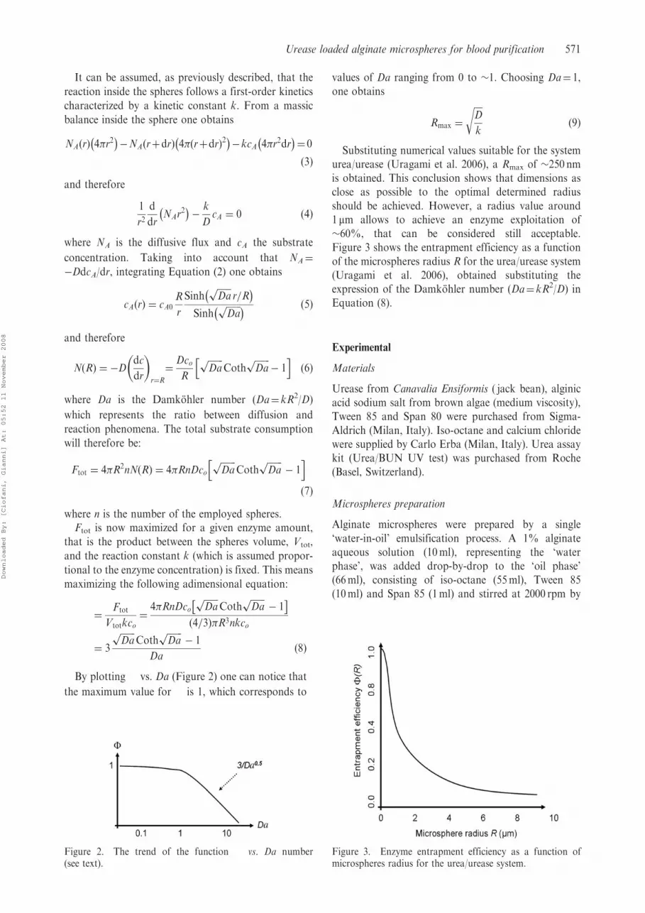
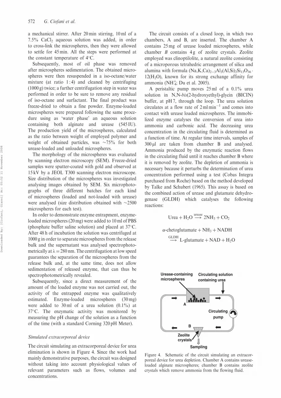
By plotting � vs. Da (Figure 2) one can notice that
the maximum value for � is 1, which corresponds to
values of Da ranging from 0 to �1. Choosing Da¼ 1,one obtains
Rmax ¼
ffiffiffiffiD
k
rð9Þ
Substituting numerical values suitable for the systemurea/urease (Uragami et al. 2006), a Rmax of �250 nmis obtained. This conclusion shows that dimensions asclose as possible to the optimal determined radiusshould be achieved. However, a radius value around1 mm allows to achieve an enzyme exploitation of�60%, that can be considered still acceptable.Figure 3 shows the entrapment efficiency as a functionof the microspheres radius R for the urea/urease system(Uragami et al. 2006), obtained substituting theexpression of the Damkohler number (Da¼ kR2/D) inEquation (8).
Experimental
Materials
Urease from Canavalia Ensiformis ( jack bean), alginicacid sodium salt from brown algae (medium viscosity),Tween 85 and Span 80 were purchased from Sigma-Aldrich (Milan, Italy). Iso-octane and calcium chloridewere supplied by Carlo Erba (Milan, Italy). Urea assaykit (Urea/BUN UV test) was purchased from Roche(Basel, Switzerland).
Microspheres preparation
Alginate microspheres were prepared by a single‘water-in-oil’ emulsification process. A 1% alginateaqueous solution (10ml), representing the ‘waterphase’, was added drop-by-drop to the ‘oil phase’(66ml), consisting of iso-octane (55ml), Tween 85(10ml) and Span 85 (1ml) and stirred at 2000 rpm by
Figure 3. Enzyme entrapment efficiency as a function ofmicrospheres radius for the urea/urease system.
Figure 2. The trend of the function � vs. Da number(see text).
Urease loaded alginate microspheres for blood purification 571
Downloaded By: [Ciofani, Gianni] At: 05:52 11 November 2008
a mechanical stirrer. After 20min stirring, 10ml of a7.5% CaCl2 aqueous solution was added, in orderto cross-link the microspheres, then they were allowedto settle for 45min. All the steps were performed atthe constant temperature of 4�C.Subsequently, most of oil phase was removed
after microspheres sedimentation. The obtained micro-spheres were then resuspended in a iso-octane/watermixture (at ratio 1 : 4) and cleaned by centrifuging(1000 g) twice; a further centrifugation step in water wasperformed in order to be sure to remove any residualof iso-octane and surfactant. The final product wasfreeze-dried to obtain a fine powder. Enzyme-loadedmicrospheres were prepared following the same proce-dure using as ‘water phase’ an aqueous solutioncontaining both alginate and urease (545 IU).The production yield of the microspheres, calculatedas the ratio between weight of employed polymer andweight of obtained particles, was �75% for bothurease-loaded and unloaded microspheres.The morphology of the microspheres was evaluated
by scanning electron microscopy (SEM). Freeze-driedsamples were sputter-coated with gold and observed at15 kV by a JEOL T300 scanning electron microscope.Size distribution of the microspheres was investigatedanalysing images obtained by SEM. Six microphoto-graphs of three different batches for each kindof microspheres (loaded and not-loaded with urease)were analysed (size distribution obtained with �2500microspheres for each test).In order to demonstrate enzyme entrapment, enzyme-
loaded microspheres (20mg) were added to 10ml of PBS(phosphate buffer saline solution) and placed at 37�C.After 48 h of incubation the solution was centrifuged at1000 g in order to separate microspheres from the releasebulk and the supernatant was analysed spectrophoto-metrically at �¼ 280 nm. The centrifugation at low speedguarantees the separation of the microspheres from therelease bulk and, at the same time, does not allowsedimentation of released enzyme, that can thus bespectrophotometrically revealed.Subsequently, since a direct measurement of the
amount of the loaded enzyme was not carried out, theactivity of the entrapped enzyme was qualitativelyestimated. Enzyme-loaded microspheres (30mg)were added to 30ml of a urea solution (0.1%) at37�C. The enzymatic activity was monitored bymeasuring the pH change of the solution as a functionof the time (with a standard Corning 320 pH Meter).
Simulated extracorporeal device
The circuit simulating an extracorporeal device for ureaelimination is shown in Figure 4. Since the work hadmainly demonstrative purposes, the circuit was designedwithout taking into account physiological values ofrelevant parameters such as flows, volumes andconcentrations.
The circuit consists of a closed loop, in which two
chambers, A and B, are inserted. The chamber A
contains 25mg of urease loaded microspheres, while
chamber B contains 4 g of zeolite crystals. Zeolite
employed was clinoptilolite, a natural zeolite consisting
of a microporous tetrahedric arrangement of silica and
alumina with formula (Na,K,Ca)2�3Al3(Al,Si)2Si13O36 �
12(H2O), known for its strong exchange affinity for
ammonia (NHþ4; Du et al. 2005).A peristaltic pump moves 25ml of a 0.1% urea
solution in N,N-bis(2-hydroxyethyl)-glycin (BICIN)
buffer, at pH7, through the loop. The urea solution
circulates at a flow rate of 2mlmin�1 and comes into
contact with urease loaded microspheres. The immobi-
lized enzyme catalyses the conversion of urea into
ammonia and carbonic acid. The decreasing urea
concentration in the circulating fluid is determined as
a function of time. At regular time intervals, samples of
300 ml are taken from chamber B and analysed.
Ammonia produced by the enzymatic reaction flows
in the circulating fluid until it reaches chamber B where
it is removed by zeolite. The depletion of ammonia is
necessary because it perturbs the determination of urea
concentration performed using a test (Cobas Integra
purchased from Roche) based on the method developed
by Talke and Schubert (1965). This assay is based on
the combined action of urease and glutamate dehydro-
genase (GLDH) which catalyses the following
reactions:
UreaþH2O �!urease
2NH3 þ CO2
�-chetoglutamateþNH3 þNADH
�!GLDH
L-glutamateþNADþH2O
Urease-containingmicrospheres
Circulating solutioncontaining urea
Sampling
Zeolite crystals
B
Circulatingpump
A
Figure 4. Schematic of the circuit simulating an extracor-poreal device for urea depletion. Chamber A contains urease-loaded alginate microspheres; chamber B contains zeolitecrystals which remove ammonia from the flowing fluid.
572 G. Ciofani et al.
Downloaded By: [Ciofani, Gianni] At: 05:52 11 November 2008
The sample is mixed to a solution containing BICINbuffer, pH7.6 50mmol l�1; GLDH� 0.80U l�1;urease� 12Uml�1; TRIS buffer, pH9.6, 100mmol l�1;2-oxoglutarate 8.3mmol l�1; NADH� 0.23mmol l�1.The decrease in absorbance at �¼ 340 nm due to theconversion of NADH into NAD is measured kineticallyat 20 s and 80 s and compared to a calibration curveobtained with samples of known urea concentrations(0, 25, 50 and 100mgdl�1).
Results and discussion
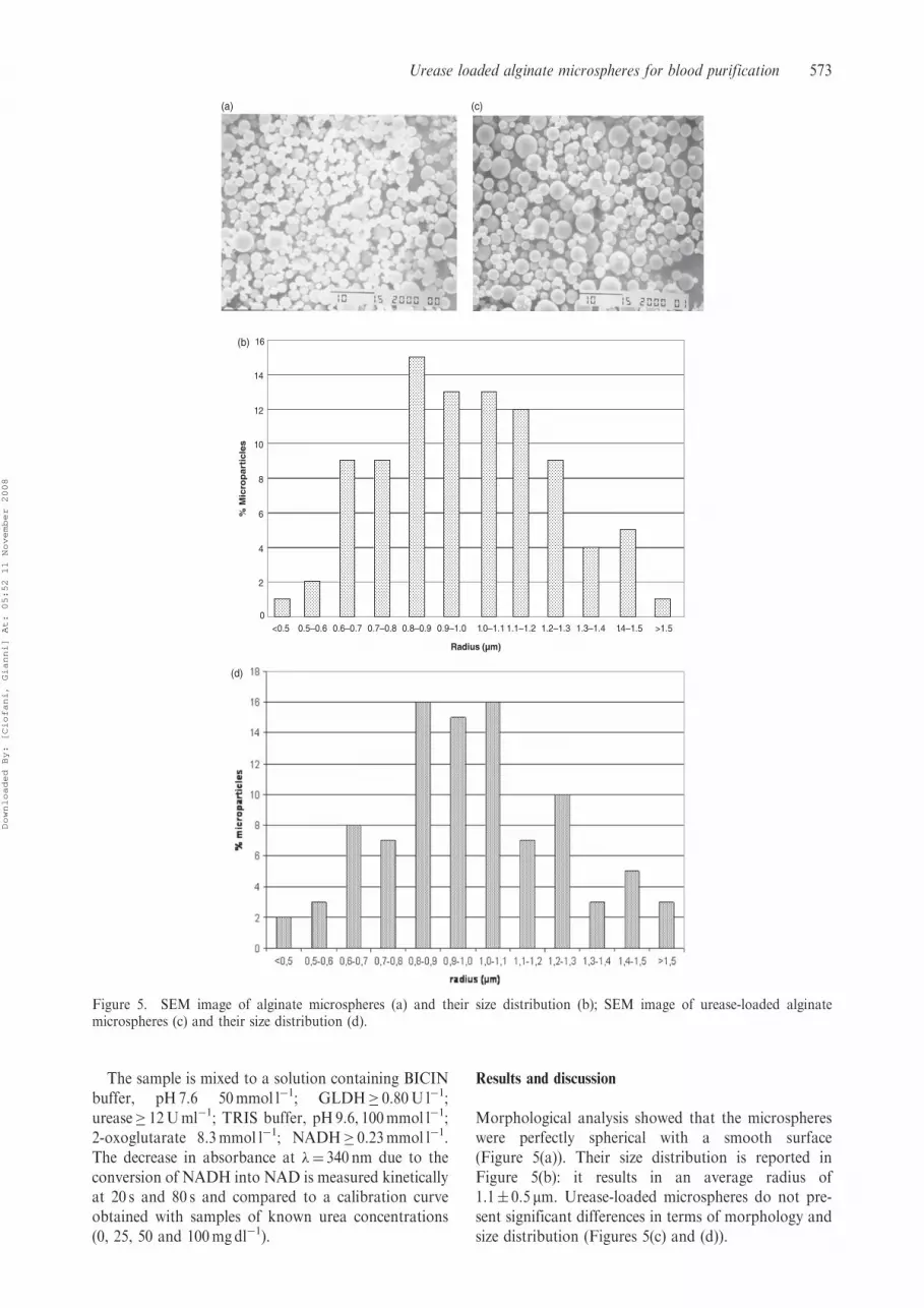
Morphological analysis showed that the microsphereswere perfectly spherical with a smooth surface(Figure 5(a)). Their size distribution is reported inFigure 5(b): it results in an average radius of1.1� 0.5 mm. Urease-loaded microspheres do not pre-sent significant differences in terms of morphology andsize distribution (Figures 5(c) and (d)).
0
2
4
6
8
10
12
14
16
<0.5 0.5–0.6 0.6–0.7 0.7–0.8 0.8–0.9 0.9–1.0 1 .0–1.1 1.1–1.2 1.2–1.3 1.3–1.4
(b)
(c)
1 .4–1.5 >1.5
Radius (µm)
% M
icro
pa
rtic
les
(a)
(d)
Figure 5. SEM image of alginate microspheres (a) and their size distribution (b); SEM image of urease-loaded alginatemicrospheres (c) and their size distribution (d).
Urease loaded alginate microspheres for blood purification 573
Downloaded By: [Ciofani, Gianni] At: 05:52 11 November 2008
The procedure described above, used for the produc-tion of the microspheres, was the result of a number ofexperimental tests performed while varying severaloperative parameters able to affect the characteristicsof the microspheres. The effect of changing differentoperative parameters, such as temperature, stirring rate,alginate concentration, surfactants concentration andCaCl2 concentration, on size and size distribution of theproduced particles was studied. The more influentialparameters, which will be subsequently discussed, arethe following.
. Agitation speed. It has been demonstrated that anincrease in the agitation speed decreases the mediumdiameter of the particles. However, a study (Pekeret al. 2001) has shown that this property is onlyverified under a well defined quantitative ratiobetween the two phases, that is not in agreementwith the procedure object of this work. Moreover,tests conducted by applying this procedure togreater speeds have shown that the average diameterof the particles was effectively lowered, but thestatistical size distribution was much worse.
. Amount and type of surfactants used. Surfactants areindispensable in lowering the superficial tension ofthe microparticles and in making the bigger particlesdivide (Cho et al. 1998). In general, an improvementin the dimensions of the particles when thesurfactant contents were increased was observed,but the cleaning procedure was very difficult withhigh surfactant concentration.
. Temperature. A decrease in temperature producesa remarkable improvement in microparticle size,maybe because the viscosity of the employedcomponents increases, as suggested by the Shinnar(1961) correlation. All experiments were thereforecarried out at 4�C.
. Alginate and CaCl2 solution concentration. A study(Lemoine et al. 1998) has shown that by decreasingthe alginate solution concentration the size of theobtained particles is improved. Although one didnot find any evidence in the literature, this studyalso varied the concentration of the reticulatingsolution in order to complete the range ofparameters.
It was noticed that by lowering the concentration ofboth solutions (alginate and CaCl2), a great improve-ment of microspheres size and size distribution can beachieved. Moreover, it was found that employing a 2%alginate solution, the lowest concentration of CaCl2 thatis needed for the microspheres formation is 5%. Underthis concentration the reticulation is not completed.After various experimental tests, it was found that
the better parameters combination that gave hugeimprovement in terms of size, size distribution andmicroparticles yields was that reported above. Thechosen parameters represent a compromise among asatisfactory yield of the process, the smallest size and
the most uniformly distributed diameter of themicrospheres.Loading the microspheres with the enzyme did not
affect their morphology, size and size distribution (see,respectively, Figures 5(c) and (d)). In order to verifywhether the enzyme was stably entrapped into themicrospheres, a specific release assay was performed.After 48 h of incubation of the microspheres in PBS, therelease bulk was analysed as described above and notraces of urease were revealed, indicating that theenzyme was firmly immobilized into the microspheres.Analysis of microspheres washing residuals did notreveal a relevant amount of enzyme, denoting thereforean almost complete urease entrapment in themicrospheres.Subsequently, the activity of the entrapped enzyme
was estimated via an indirect assay: the enzymaticactivity was monitored by measuring the pH change ofa urea solution containing the microspheres. It wasexpected that a pH increase due to the formation ofammonia as a product of the enzyme catalysed reaction:after 1 h of incubation, the pH increased from 6.05 to8.85. For comparison, a similar test was performedusing 30ml of urea solution (0.1%) added with 108.5 IUof free enzyme. In this case, an increase of pH from 6.05to 9.12 was observed after 1 h incubation.The theoretical amount of enzyme entrapped into
30mg of microspheres was calculated to be 163.5 IU,taking into account the initial enzyme-to-alginate ratio(5450 IU g�1 alginate) and the yield of the process(75%). On this basis, the final pH would have beenhigher than 9.12 (corresponding to 108.5 IU of enzyme).The lower value actually found for the pH can berelated to two main factors: first, a fraction of theenzyme loses its activity because of the preparationprocedure of the microspheres; secondly, there wouldbe a decrease of the apparent activity with respect tothat of the free enzyme because of the entrapment intothe microspheres. Nevertheless, although the pH valuewas lower than that expected, its increase from 6.05 to8.85 indicated a significant activity of the entrappedenzyme (�100 IU).With regard to the simulating circuit, in Figure 6 the
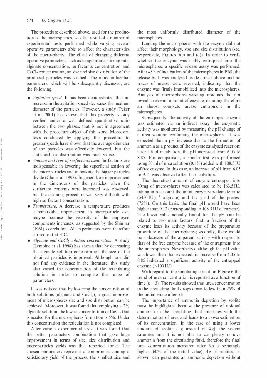
trend of urea concentration is reported as a function oftime (n¼ 3). The results showed that urea concentrationin the circulating fluid drops down to less than 25% ofthe initial value after 5 h.The importance of ammonia depletion by zeolite
must be highlighted because the presence of residualammonia in the circulating fluid interferes with thedetermination of urea and leads to an over-estimationof its concentration. In the case of using a loweramount of zeolite (1 g instead of 4 g), the systemsaturates and it is not able to completely removeammonia from the circulating fluid, therefore the finalurea concentration measured after 5 h is seeminglyhigher (60% of the initial value); 4 g of zeolites, asshown, can guarantee an ammonia depletion without
574 G. Ciofani et al.
Downloaded By: [Ciofani, Gianni] At: 05:52 11 November 2008

saturation phenomena and, therefore, without needingto substitute zeolites during the complete duration ofexperiment.
Conclusions
In this work a device for urease immobilization wasrealized based on alginate microspheres. Thanks to adiffusion/reaction model the optimum size of micro-spheres was determined: for the alginate/urease systeman optimal diameter of 0.5 mm should be achieved, inorder to obtain the maximum theoretical enzymeexploitationIn this respect, a number of experimental tests were
performed varying several operative parameters such astemperature, stirring rate, concentration of alginate,surfactants and cross-linker, able to affect the char-acteristics of the produced microspheres. The para-meters finally selected represent a compromise among asatisfactory yield of the process, the smallest size andthe most uniformly distributed diameter of the micro-spheres. The produced microspheres had an averageradius of 1.1 mm that, even if higher than the optimal,allows a fair enzyme exploitation up to 60%.Loading the microspheres with the enzyme did not
affect their morphology, size and size distribution andthe enzyme was firmly immobilized and able tomaintain most of its catalytic activity. Urease-loadedmicrospheres were successfully used in a simulatedextracorporeal device for the depletion of urea from acirculating fluid: the obtained results indicate that theinvestigated system could represent an interestingalternative to membranes currently used in dialysisprocedures for blood purification. With this respect,
work is in progress to test the performance of theurease-loaded microspheres into an extracoporealdevice mimicking physiological conditions and toevaluate the use of these microspheres for the entrap-ment of different enzymes useful for blood purification.
Acknowledgements
The authors thank Ms Aura Bonaretti for the SEMimaging and Dr Giovanni Pellegrini of CisanelloHospital (Pisa) for his helpful support in the urea tests.
References
Brahim S, Narinesingh D, Guiseppi-Elie A. 2002. Kinetics of glucose
oxidase immobilized in p(HEMA)-hydrogel microspheres in a
packed-bed bioreactor. J Molec Catalysis B Enzymatic 18:69–80.
Chretien C, Chaumeil JC. 2005. Release of a macromolecular drug
from alginate-impregnated microspheres. Int J Pharm 304:18–28.
Cho NH, Seong SY, Chun KH, Kim YH, Kwon IC, Ahn BY,
Jeong SY. 1998. Novel mucosal immunization with polysacchar-
ide–protein conjugates entrapped in alginate microspheres. J Contr
Rel 53:215–224.
Chuang CW, Shih JS. 2001. Preparation and application of
immobilized C60-glucose oxidase enzyme in fullerene C60-coated
piezoelectric quartz crystal glucose sensor. Sensors Actuators B
Chem 81:1–8.
Cioci F, Lavecchia R, Mazzocchi P. 1999. An enzymatic membrane
reactor for extracorporeal blood oxygenation. Chem Eng Sci
54:3217–3223.
Ciofani G, Raffa V, Menciassi A, Dario P. 2008. Alginate and
chitosan particles as drug delivery system for cell therapy, Biomed
Microdevices, 10:131–140.
Ciofani G, Raffa V, Menciassi A, Micera S, Dario P. 2007. A drug
delivery system based on alginate microspheres: Mass-transport
test and in vitro validation. Biomed Microdevices 9:395–403.
0
20
40
60
80
100
120
0 2000 4000 6000 8000 10000 12000 14000 16000 18000 20000
Time (s)
Ure
a c
once
ntr
atio
n (m
g/d
l)
Figure 6. Trend of urea concentration as a function of time using 4 g of zeolite.
Urease loaded alginate microspheres for blood purification 575
Downloaded By: [Ciofani, Gianni] At: 05:52 11 November 2008
Clark AH, Ross-Murphy SB. 1987. Structural and mechanical for the
preparation of biopolymer gels. Adv Polym Sci 83:57–192.
Douglas KL, Tabrizian M. 2005. Effect of experimental parameters
on the formation of alginate–chitosan nanoparticles and evaluation
of their potential application as DNA carrier. J Biomater Sci
Polym Ed 16:43–56.
Du Q, Liu S, Cao Z, Wang Y. 2005. Ammonia removal from aqueous
solution using natural Chinese clinoptilolite. Separat Purificat
Technol 44:229–234.
Gombotz WR, Wee SF. 1998. Protein release from alginate matrices.
Adv Drug Deliv Rev 31:267–285.
Guilbault GG, Palleschi G, Lubrano G. 1995. Non-invasive
biosensors in clinical analysis. Biosensors Bioelect 10:379–392.
Kennedy JF. 1987. Enzyme technology, biotechnology, Vol. 7a,
Chapter 7, pp. 347–404.
Kim J, Grate JW, Wang P. 2006. Nanostructures for enzyme
stabilization. Chem Eng Sci 61:1017–1026.
Krajewska B, Ciurli S. 2005. Jack bean (Canavalia ensiformis) urease.
Probing acid–base groups of the active site by pH variation. Plant
Physiol Biochem 43:651–658.
Kyrolainen M, Hakanson H, Mattiasson B, Vadgama P. 1997.
Minimal-Fouling enzyme electrode for continuous flow
measurement of whole blood lactate. Biosensors Bioelect
12:1073–1081.
Lemoine D, Wauters F, Bouchend’Homme S, Preat V. 1998.
Preparation and characterization of alginate microspheres contain-
ing a model antigen. Int J Pharm 176:9–19.
Mateo C, Palomo JM, Fernandez-Lorente G, Guisan JM, Fernandez-
Lafuente R. 2007. Improvement of enzyme activity, stability and
selectivity via immobilization techniques. Enzyme Microbial
Technol 40:1451–1463.
Murphy L. 2006. Biosensors and bioelectrochemistry. Curr Opin
Chem Biol 10:177–184.
Murtas S, Capuani G, Dentini M, Manetti C, Masci G, Massimi M,
Miccheli A, Crescenzi V. 2005. Alginate beads as immobilization
matrix for hepatocytes perfused in a bioreactor: A physico-
chemical characterization. J Biomater Sci Polym Ed 16:829–846.
Peker S, Bora K, Over Y. 2001. Effects of interfacial properties on the
drop size distribution of high internal phase ratio emulsion.
Colloids Surfaces A Physicochem Eng Aspects 182:43–56.
Prieto-Simon B, Fabregas E. 2006. New redox mediator-modified
polysulfone composite films for the development of dehydrogen-
ase-based biosensors. Biosensors Bioelect 22:131–137.
Shinnar R. 1961. On the behaviour of liquid dispersions in mixing
vessels. J Fluid Mech 10:259–275.
Stryer L, Gumport RI, Jonas A, Mintel R, Rhodes C. 1996.
Biochemistry. New York, USA: W.H. Freeman and Company.
Talke H, Schubert GE. 1965. Enzymatische Harnstoffbestimmung im
Blut und Serum im optischen test nach Warburg. Klin Wochschr
43:174–175.
Uragami T, Ueguchi K, Watanabe M, Miyata T. 2006. Preparation
of urease-immobilized polymeric membranes and their function.
Catalysis Today 118:158–165.
Wang MS, Childs RF, Chang PL. 2005. A novel method to enhance
the stability of alginate-poly-L-lysine-alginate microcapsules.
J Biomater Sci Polym Ed 16:89–111.
Wilcken B. 2004. Problems in the management of urea cycle
disorders. Molec Genet Metab 81:86–91.
576 G. Ciofani et al.
Downloaded By: [Ciofani, Gianni] At: 05:52 11 November 2008