alginate–chitosan–plga composite microspheres induce both innate and adaptive immune response...
TRANSCRIPT
This article appeared in a journal published by Elsevier. The attachedcopy is furnished to the author for internal non-commercial researchand education use, including for instruction at the authors institution
and sharing with colleagues.
Other uses, including reproduction and distribution, or selling orlicensing copies, or posting to personal, institutional or third party
websites are prohibited.
In most cases authors are permitted to post their version of thearticle (e.g. in Word or Tex form) to their personal website orinstitutional repository. Authors requiring further information
regarding Elsevier’s archiving and manuscript policies areencouraged to visit:
http://www.elsevier.com/authorsrights
Author's personal copy
AlginateechitosanePLGA composite microspheres induce both innateand adaptive immune response through parenteral immunization infish
Truptimayee Behera, Priyabrat Swain*
Fish Health Management Division, Central Institute of Freshwater Aquaculture, Kausalyaganga, Bhubaneswar 751 002, Orissa, India
a r t i c l e i n f o
Article history:Received 12 April 2013Received in revised form4 June 2013Accepted 10 June 2013Available online 30 June 2013
Keywords:Aeromonas hydrophilaAlginateChitosanComposite microspheresPLGA
a b s t r a c t
AlginateechitosanePLGA composite microspheres encapsulating outer membrane protein antigen ofAeromonas hydrophila as an antigen carrier was explored for the first time in a fish model. This compositemicrosphere showed distinct advantages over the conventional PLGA microparticles in aspects of thehigh encapsulation efficiency due to the protein-friendly microenvironment created by the hydrophilicalginateechitosan cores of the composite microspheres, preventing initial burst release and the elimi-nation of lyophilizing process. The antibody responses significantly increased and persist up to 9 weeksin composite microspheres unlike that PLGA microsphere, native OMP and FIA adjuvant. Moreover,several innate immune parameters as respiratory burst, lysozyme and complement activity weresignificantly increased in both composite and PLGA microspheres up to 9 weeks than other treatedgroups. It also gives protection from A. hydrophila infection and brought some hope, for its application inreplacement with conventional PLGA microparticle for antigen delivery in fish.
� 2013 Elsevier Ltd. All rights reserved.
1. Introduction
Vaccines play an outstanding role in modern medicines but theinefficiency of new generation vaccines (mostly purified proteins,which are often poorly immunogenic) is due to lack of appropriateadjuvant/delivery system. To be effective, these vaccines requiredeffective adjuvant/carrier which becomes a challenge now. Biode-gradable polymeric microspheres act as a promising vaccine carrierfor decades [1]. In recent years, poly lactic- co-glycolic acid (PLGA)microspheres gained much interest for antigen/protein deliverydue to its biocompatibility, controlled and sustained release, notoxicity and ease of administration [2]. In addition it gives signifi-cant success for inducing both innate and adaptive immunity forfish vaccination [3e6]. Despite its success, PLGA still have majordrawbacks like structural or conformational changes of entrappedproteins, protein unfolding, aggregations during preparations, in-compatibility between hydrophilic proteins and hydrophobic PLGApolymers, low loading efficiency, the low pH generated duringpolymer erosion could cause chemical degradation of entrappedproteins [7], inconsistent release profile [8] and high initial release
which is not suitable for therapeutic protein and antigen delivery[9e11].
Many attempts had been made to improve protein stability andrelease kinetics of the PLGA system by changing the physicochem-ical properties of the polymer [12]. Here, we use a novel compositemicrosphere, composed of different biodegradable polymers suchas PLGA, alginate and chitosan for antigen delivery. This microcap-sule not only gives antigen/protein a proper microenvironment bydual hydrophilic protections but also makes the initial release veryslow. Both alginate and chitosan were chosen for compositemicrosphere preparations due to its compatibility with hydrophilicproteins, muco-adhesiveness, stability in proteolytic and acid con-ditions, biodegradability, less toxicity, easily available and relativelylow cost as compared to other biodegradable materials [13e16]. Inaddition, these two polymers are a proven immunostimulant inseveral higher vertebrates, including fish [15,16]. Some earlier studysuggested the advantages of this composite microsphere forimproving stability and release profile of protein antigens and maybe a promising delivery system for hydrophilic proteins and anti-gens [17]. However, there is limited information on its in vitro andin vivo applications for antigen delivery in an animal model and incase of fish not a single study has been done until now. The use ofbiodegradable composite microspheres as an antigen delivery sys-tem in fish is a totally new area of research and fish also acts as anethical model for research on vaccine developed for other species.
* Corresponding author. Tel.: þ91 943 723 1099; fax: þ91 674 2465407.E-mail address: [email protected] (P. Swain).
Contents lists available at SciVerse ScienceDirect
Fish & Shellfish Immunology
journal homepage: www.elsevier .com/locate / fs i
1050-4648/$ e see front matter � 2013 Elsevier Ltd. All rights reserved.http://dx.doi.org/10.1016/j.fsi.2013.06.012
Fish & Shellfish Immunology 35 (2013) 785e791
Author's personal copy
Fish vaccinology also faces similar challenges to those encoun-tered in vaccine design for human and other vertebrates. Aero-monas hydrophila infection causes major devasting diseases in bothwarm and freshwater fish farming worldwide. It also causes severaldevasting diseases in both human and amphibians [18]. Lack ofefficient commercial vaccine against A. hydrophila, just exemplifiesthe problems related to its vaccine designing. Outer membraneproteins (OMP) antigen acts as a major virulence factor and itsinvolvement for inducing immune defense makes it an attractivetarget for vaccine component [19e21].
Therefore, for the first time we assess the feasibility of PLGAealginateechitosan composite microspheres on innate and adaptiveimmune response in fish Labeo rohita H. against A. hydrophila in-fections. At the same time we want to compare its efficacy withFreund’s incomplete adjuvant (FIA) a commonly used oil adjuvantsin vaccine research.
2. Materials and methods
2.1. Materials
Chitosan (>80% deacetylation, MW 80,000) was obtained fromPolysciences, Inc. Warrington, England; PLGA (copolymer ratio50:50, Inherent viscosity- 0.8 dl/g, Mw-50 kDa) was purchasedfrom Birmingham polymers, Inc. (Birmingham, AL). Alginate andPolyvinyl alcohol (PVA, average MW 30, 000 e 70,000) was pur-chased from SigmaeAldrich Co. (St Louis, MO 63195, USA). All re-agents used such as Span 80, Tween 80, iso-octane, isopropylalcohol, calcium chloride, and all other reagents were of analyticalgrade from E-merck, India and organic solvents used were of HPLCgrade. Bicinchoninic acid (BCA) protein assay kits were supplied byBangalore Genei, India.
2.2. Bacteria
Aeromonas hydrophila strain (Ahv), isolated from Channa striatusshowing clinical signs of dropsy conditions was preserved inlyophilized condition in our laboratory and was used throughoutthe study.
2.3. Isolation of outer membrane proteins
The OMP of A. hydrophila (Ahv) were prepared as described byNikaido (1997) with minor modifications [22]. The pellet obtainedfrom 1 L culture of A. hydrophila was washed twice in 40 ml of0.15 M phosphate buffered saline (PBS, pH 7.4), once in 40 ml of20 mM TriseHCl (pH 7.5) and centrifuged at 13,000 � g for 10 min.The cells were then resuspended in 20 ml TriseHCl and disruptedby sonication for 30 min at 10 W at an interval of 30 s. Unbrokencells and cellular debris were removed by centrifugation at4000 � g for 15 min. The supernatant was then further centrifugedat 10,000� g for 1 h at 4 �C. The pellet was suspended in 20ml of 2%(w/v) sarcosine and incubated at room temperature for 30 min tosolubilize the inner membrane. The solution was then centrifugedat 10,000 � g for 1 h at 4 �C. The resultant pellet of sarcosine-insoluble components was freeze-dried and stored at �20 �C un-til use. Protein concentration of OMP was determined using BCA(Bicinchoninic acid) protein estimation kit (Bangalore genei, India).
2.4. Preparation of OMP encapsulated conventional PLGAmicrospheres and PLGAealginateechitosan (composite) microspheres
OMP encapsulated conventional PLGA and PLGAealginateechitosan composite microspheres were prepared according toZheng et al. (2004) with some modifications [17]. For OMP
encapsulated PLGA microspheres, a w/o primary emulsion wasprepared bymixing 500 ml of OMP aqueous solution (250 mg/0.5ml)and 3 ml of 7.5% PLGA methylene chloride solution. The emulsifi-cation was done by sonication for 50 s to produce w/o emulsion.Then this primary w/o emulsion was added into 40 ml of 1.0% PVAand stirred at 12,000 � g with a magnetic stirrer for 3 min toproduce secondary w/o/w emulsion. The resulting w/o/w emulsionwas further stirred at 1000 � g for 3 h at 37 �C for solvent evapo-ration. Then microparticles were recovered by normal centrifuga-tion at 5000 � g for 10 min and washed three times with distilledwater. The resulting OMP loaded PLGAmicrospheres were collectedby lyophilization and stored at 4 �C for further use.
For the alginateechitosan microcapsules, 5 ml of the OMPaqueous phase solution (250 mg/0.5 ml), 2% alginate solution, anddistilled water at the volume ratio of 3:5:2 was used to prepare anemulsionwith 20ml of isooctane containing 5% Span 80 and stirredat 16,000 � g. A subsequent addition of 1 ml Tween 80 was used assecondary emulsifier and then 4 ml of 8% calcium chloride as across-linker. The resulting microspheres were further solidified byaddition of 20 ml of isopropyl alcohol. The microspheres wereincubated with chitosan solution (0.5%, pH 4) to form alginateechitosan complex membranes. The obtained alginateechitosanmicrocapsules were used for the preparation of the compositemicrospheres.
For the PLGA-alginateechitosan composite microspheres, thealginateechitosan microcapsules were suspended in 3 ml ofacetonitrile containing PLGA (50:50, 0.24 g/ml) with the ratio ofmicrospheres to PLGA in weight between is 1e9. The suspensionwas added into the soybean oil containing 6% Span 80 with a stir-ring speed of 1000 � g and then 12,000 � g. After washing withpetroleum ether and complete volatilization at 37 �C, the resultingOMP loaded composite microspheres were collected.
2.5. Emulsification with FIA (FIA-OMP)
OMP antigen (500 mg) in 1 ml PBS was drop wise added andproperly emulsified with 1 ml of Freund’s incomplete adjuvant(FIA) (Sigma, USA) at 4 �C until homogeneity and stored at 4 �Cuntill further use.
2.6. Morphology observation
The morphologies and approximate sizes of the composite mi-crospheres were studied with a scanning electron microscopy(EVOMA15, Carl Zeiss SMT, Germany). The size and distribution ofthe single and double microcapsules were determined with a laserlight scattering size analyzer (Zeta sizer 3000 HAS, Malvern In-strument, England).
2.7. Determination of encapsulation efficiency
The respective encapsulation efficiency of OMP was determinedby the following methods:
(i) OMP antigen in PLGA microspheres was analyzed using anextraction method. Briefly, 10 mg of PLGA microspheres weredissolved in 1 ml of dichloromethane, followed by addition of1 ml of PBS (pH 7.4) and place in a shaker at 30 rpm for 12 h at37 �C. The antigen concentration in the aqueous phase wasdetermined using BCA protein assay kit.
(ii) In chitosan coated alginate microcapsules an extractionmethod was used (Lemoine et al., 1998) [23]. 10 mg of OMPloaded chitosan coated alginate microspheres were taken in atube. Then 1 ml of 1N NaOH (PH 11.5) was added to it andkeeps it in shaker at 30 rpm for 12 h at 37 �C. Then samples
T. Behera, P. Swain / Fish & Shellfish Immunology 35 (2013) 785e791786
Author's personal copy
were centrifuged, collected supernatant was used to deter-mine antigen concentration using BCA protein assay kit.
(iii) In Composite microspheres, 50 mg of composite microsphereswas dissolved in 2 ml acetonitrile and the supernatant con-taining polymer was discarded after centrifugation of16,000 � g for 10 min. The pellet was vacuum dried andextracted according to the methods used in PLGA micro-spheres as described above.
From these result encapsulation efficiency in each of the mi-crospheres was calculated using the following formula: Encapsu-lation efficiency (%) ¼ (Amount of loaded OMP/Amount of totalOMP) � 100.
2.8. In vitro release study
Accurately weighted amounts of PLGA microspheres (50 mg)and composite microspheres (50 mg) were placed in eppendorftubes (3 replicates for each microspheres) containing 3 ml of PBS,pH 7.4. The tubes were then incubated at 37 �C under the rotationspeed of 70 rpm. At pre-determined time intervals, the tubes werecentrifuged at 2000 � g for 5 min. Supernatant (0.5 ml) was takenout and 0.5 ml fresh solutionwas added. The antigen concentrationwas determined by the BCA protein assay kit.
2.9. Immunization protocol
Indian major carp, Labeo rohita (rohu), juveniles of averageweight ranging from 50 to 60 g were acclimatized in the wetlaboratory of Fish Health Management Division of Central Insti-tute of Freshwater Aquaculture (CIFA), Kausalyaganga, India, 15days prior to the start of the experiment. The fish were fed withartificial carp diet with constant aeration and daily one-thirdwater exchange with water temperature 27e30 �C. Fish wereseparated into 5 groups, 20 fish in each group were intra-peritonially immunized separately with 0.1 ml of different prep-arations @ 50 mg of OMP in PLGA microspheres (Group 1), incomposite microspheres (Group 2), FIA-OMP (Group 3) and freenative OMP whereas control group (Group 4) were injected withPBS (pH, 7.4) only. All the treated group fish (10 fish in eachgroup) were bled at an interval of 3-weeks (at 21, 42 and 63days post-immunization) to study various immune parameters(Table 1).
2.10. Immunoresponse studies
2.10.1. Preparation of anti-rohu-globulin rabbit serumThe rabbit anti-rohu globulin was prepared as per a standard
method of Swain et al. (2002) [24] using sera obtained from healthyadult rohu of average weight 250e300 g. The anti-rohu globulinserawere raised in a New Zealandwhite rabbit as per themethod ofLund et al. (1991) [25].
2.10.2. Triple antibody indirect enzyme linked immunosorbentassays
The triple antibody indirect ELISA was conducted as per themethod of Swain et al. (2002) [24]. The antibody response wasexpressed in terms of O.D. value after subtracting the values ob-tained by unimmunized healthy sera.
2.10.3. Respiratory burst activityRespiratory burst activity was assayed by the reduction of
nitroblue tetrazolium (NBT) to formazan according to the methodsof Anderson and Siwicki (1994) [26].
2.10.4. Lysozyme activityA turbidometric assay utilizing lyophilized Micrococcus lyso-
deikticus cells (Sigma) was used to determine lysozyme activity inserum. A slight modification of the previously described methodwas used to measure lysozyme activity [27,28]. A suspension of150 ml of M. lysodeikticus (0.2 mg ml�1 in 0.02 M sodium acetatebuffer, pH 5.5) was added to previously dispense 15 ml of serumsamples in a 96well U-bottommicrotitre plate. Initial ODwas takenat 450 nm immediately after adding the substrate and final OD wastaken after 1 h incubation at 37 �C. Lyophilized hen egg whitelysozyme (Sigma) was used to develop a standard curve. Lysozymelevel was expressed as units ml�1 where one unit is defined as thedecrease in absorbance of 0.001 min�1.
2.10.5. Alternative complement activity (ACH50)The determination of alternative serum complement was done
according to the previous method with little modifications [29].The rabbit red blood cells (RRBCs, 2.5 � 108 cells ml�1) were takenfrom blood. One hundred microliters of diluted serum (1:25 inHBSS solution containing 10 mM Mg2þ and 10 mM ethylene glycolbis tetraacetate, pH 7.6) were put into eppendorf tubes and mixedwith 25 ml of RRBCs. The tubes were incubated for 90 min at150 rpm at 20 �C, and then they were centrifuged to spin downthe remaining RRBCs. The supernatants were transferred into 96-well plates and measured at 415 nm. The serum dilution factorwas plotted in logarithmic scale against percentage of RRBCslysed at each dilution. The dilution corresponding to 50%hemolysis ml�1 was expressed as ACH50.
2.11. Challenge study
For the challenge, another virulent strain of A. hydrophila(28 V/08, LD50: 106 CFU ml�1) was used. Two days after the lastbleeding, all the fishes (20 fish) from each group were injectedintraperitonially with 0.1 ml of 24 h culture of A. hydrophilawith the above challenge strain. The cumulative mortality andrelative percent survival (RPS) was recorded up to 20 days post-challenge [30]. The cause of death and pathological signs wereverified by re-isolation of bacteria from samples of freshly dead/infected fish.
Table 1Immunization study protocol.
Groups Formulations Single dose of antigen Route of delivery Serum sampling(days of post immunization)
1 PLGA-OMP microspheres 50 mg of OMP/0.1 ml of microsphere suspension Intraperitoneal injection 21, 42 & 632 PLGA-ALG-CHI-OMP
composite microspheres50 mg of OMP/0.1 ml of microsphere suspension Intraperitoneal injection 21, 42 & 63
3 FIA-OMP 50 mg of OMP/0.1 ml of FIA emulsion Intraperitoneal injection 21, 42 & 634 Native OMP 50 mg of OMP/0.1 ml of PBS Intraperitoneal injection 21, 42 & 63Control PBS Nil Intraperitoneal injection 21, 42 & 63
T. Behera, P. Swain / Fish & Shellfish Immunology 35 (2013) 785e791 787
Author's personal copy
2.12. Statistical analysis
The statistical analysis system (SAS) software (version 6.12) wasused to analyze the data [31].
One-way analysis of variance (ANOVA) followed by Duncan’smultiple range tests (DMRT) were done to compare the variationsin various immune parameters at significance level of difference(p < 0.05) in different injected groups. The mean standard error(�S.E.) of assayed parameters was calculated in each group of fish.
3. Results
3.1. Morphology observation
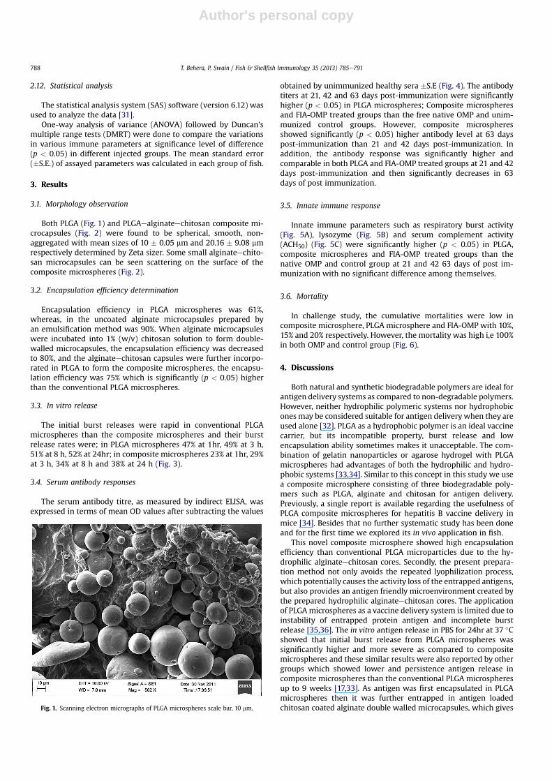
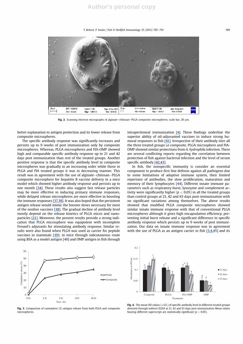
Both PLGA (Fig. 1) and PLGAealginateechitosan composite mi-crocapsules (Fig. 2) were found to be spherical, smooth, non-aggregated with mean sizes of 10 � 0.05 mm and 20.16 � 9.08 mmrespectively determined by Zeta sizer. Some small alginateechito-san microcapsules can be seen scattering on the surface of thecomposite microspheres (Fig. 2).
3.2. Encapsulation efficiency determination
Encapsulation efficiency in PLGA microspheres was 61%,whereas, in the uncoated alginate microcapsules prepared byan emulsification method was 90%. When alginate microcapsuleswere incubated into 1% (w/v) chitosan solution to form double-walled microcapsules, the encapsulation efficiency was decreasedto 80%, and the alginateechitosan capsules were further incorpo-rated in PLGA to form the composite microspheres, the encapsu-lation efficiency was 75% which is significantly (p < 0.05) higherthan the conventional PLGA microspheres.
3.3. In vitro release
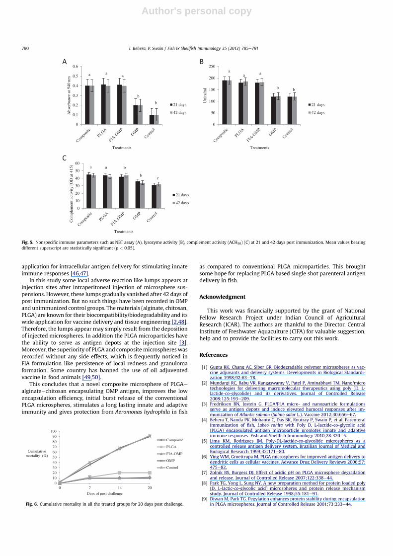
The initial burst releases were rapid in conventional PLGAmicrospheres than the composite microspheres and their burstrelease rates were; in PLGA microspheres 47% at 1hr, 49% at 3 h,51% at 8 h, 52% at 24hr; in composite microspheres 23% at 1hr, 29%at 3 h, 34% at 8 h and 38% at 24 h (Fig. 3).
3.4. Serum antibody responses
The serum antibody titre, as measured by indirect ELISA, wasexpressed in terms of mean OD values after subtracting the values
obtained by unimmunized healthy sera �S.E (Fig. 4). The antibodytiters at 21, 42 and 63 days post-immunization were significantlyhigher (p < 0.05) in PLGA microspheres; Composite microspheresand FIA-OMP treated groups than the free native OMP and unim-munized control groups. However, composite microspheresshowed significantly (p < 0.05) higher antibody level at 63 dayspost-immunization than 21 and 42 days post-immunization. Inaddition, the antibody response was significantly higher andcomparable in both PLGA and FIA-OMP treated groups at 21 and 42days post-immunization and then significantly decreases in 63days of post immunization.
3.5. Innate immune response
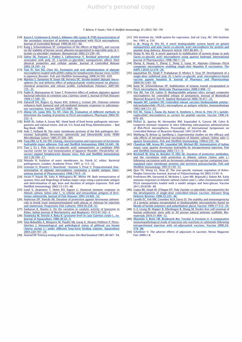
Innate immune parameters such as respiratory burst activity(Fig. 5A), lysozyme (Fig. 5B) and serum complement activity(ACH50) (Fig. 5C) were significantly higher (p < 0.05) in PLGA,composite microspheres and FIA-OMP treated groups than thenative OMP and control group at 21 and 42 63 days of post im-munization with no significant difference among themselves.
3.6. Mortality
In challenge study, the cumulative mortalities were low incomposite microsphere, PLGA microsphere and FIA-OMP with 10%,15% and 20% respectively. However, the mortality was high i,e 100%in both OMP and control group (Fig. 6).
4. Discussions
Both natural and synthetic biodegradable polymers are ideal forantigen delivery systems as compared to non-degradable polymers.However, neither hydrophilic polymeric systems nor hydrophobicones may be considered suitable for antigen delivery when they areused alone [32]. PLGA as a hydrophobic polymer is an ideal vaccinecarrier, but its incompatible property, burst release and lowencapsulation ability sometimes makes it unacceptable. The com-bination of gelatin nanoparticles or agarose hydrogel with PLGAmicrospheres had advantages of both the hydrophilic and hydro-phobic systems [33,34]. Similar to this concept in this study we usea composite microsphere consisting of three biodegradable poly-mers such as PLGA, alginate and chitosan for antigen delivery.Previously, a single report is available regarding the usefulness ofPLGA composite microspheres for hepatitis B vaccine delivery inmice [34]. Besides that no further systematic study has been doneand for the first time we explored its in vivo application in fish.
This novel composite microsphere showed high encapsulationefficiency than conventional PLGA microparticles due to the hy-drophilic alginateechitosan cores. Secondly, the present prepara-tion method not only avoids the repeated lyophilization process,which potentially causes the activity loss of the entrapped antigens,but also provides an antigen friendly microenvironment created bythe prepared hydrophilic alginateechitosan cores. The applicationof PLGA microspheres as a vaccine delivery system is limited due toinstability of entrapped protein antigen and incomplete burstrelease [35,36]. The in vitro antigen release in PBS for 24hr at 37 �Cshowed that initial burst release from PLGA microspheres wassignificantly higher and more severe as compared to compositemicrospheres and these similar results were also reported by othergroups which showed lower and persistence antigen release incomposite microspheres than the conventional PLGA microspheresup to 9 weeks [17,33]. As antigen was first encapsulated in PLGAmicrospheres then it was further entrapped in antigen loadedchitosan coated alginate double walled microcapsules, which givesFig. 1. Scanning electron micrographs of PLGA microspheres scale bar, 10 mm.
T. Behera, P. Swain / Fish & Shellfish Immunology 35 (2013) 785e791788
Author's personal copy
better explanation to antigen protection and its lower release fromcomposite microspheres.
The specific antibody response was significantly increases andpersists up to 9 weeks of post immunization only by compositemicrospheres. Whereas, PLGA microspheres and FIA-OMP showedhigh and comparable specific antibody response up to 21 and 42days post immunization than rest of the treated groups. Anotherpositive response is that the specific antibody level in compositemicrospheres was gradually in an increasing order while those inPLGA and FIA treated groups it was in decreasing manner. Thisresult was in agreement with the use of alginateechitosanePLGAcomposite microsphere for hepatitis B vaccine delivery in a micemodel which showed higher antibody response and persists up toone month [34]. These results also suggest fast release particlesmay be more effective in inducing primary immune responses,while delayed release microspheres are more effective in boostingthe immune responses [37,38]. It was also hoped that the persistentantigen release would mimic the booster doses necessary for mostof the nonlive vaccines [38]. The gradual decline of antibody levelmostly depend on the release kinetics of PLGA micro and nano-particles [21]. Moreover, the present results provide a strong indi-cation that PLGA microsphere was equipotent with incompleteFreund’s adjuvants for stimulating antibody response. Similar re-sults were also found when PLGA was used as carrier for peptidevaccines in mammals [39]; in mice through subcutaneous routeusing BSA as a model antigen [40] and OMP antigen in fish through
intraperitoneal immunization [4]. These findings underline thesuperior ability of oil-adjuvanted vaccines to induce strong hu-moral responses in fish [41]. Irrespective of their antibody titer allthe three treated groups i,e composite, PLGA microsphere and FIA-OMP showed similar protections from A. hydrophila infection. Thereare several conflicting reports regarding the correlation betweenprotection of fish against bacterial infection and the level of serumspecific antibody [42,43].
In fish, the nonspecific immunity is consider an essentialcomponent to produce first line defense against all pathogens dueto some limitations of adaptive immune system, their limitedrepertoire of antibodies, the slow proliferation, maturation andmemory of their lymphocytes [44]. Different innate immune pa-rameters such as respiratory burst, lysozyme and complement ac-tivity were significantly higher (p < 0.05) in all the treated groupsthan control groups at 21, 42 and 63 days post-immunization withno significant variations among themselves. The above resultsshowed that modified PLGA composite microspheres showedsimilar innate immune response with that of conventional PLGAmicrospheres although it gives high encapsulation efficiency, pre-venting initial burst release and a significant difference in specificantibody response which persists up to 9 weeks of post immuni-zation. Our data on innate immune response was in agreementwith the use of PLGA as an antigen carrier in fish [3,4,45] and its
Fig. 2. Scanning electron micrographs of alginateechitosanePLGA composite microspheres, scale bar, 20 mm.
Cum
ulat
ive
rele
ase
(%)
Time (hr)
Fig. 3. Comparison of cumulative (%) antigen release from both PLGA and compositemicrospheres.
a a a
d
a
a
a
d
c
b b
e
0
0.05
0.1
0.15
0.2
Composite PLGA FIA-OMP OMP
Abs
orba
nce
at 4
50 n
m
Treatments
21 days
42 days
63 days
Fig. 4. The mean OD values (�S.E.) of specific antibody level in different treated groupsdetected through indirect ELISA at 21, 42 and 63 days post-immunization Mean valuesbearing different superscript are statistically significant (p < 0.05).
T. Behera, P. Swain / Fish & Shellfish Immunology 35 (2013) 785e791 789
Author's personal copy
application for intracellular antigen delivery for stimulating innateimmune responses [46,47].
In this study some local adverse reaction like lumps appears atinjection sites after intraperitoneal injection of microsphere sus-pensions. However, these lumps gradually vanished after 42 days ofpost immunization. But no such things have been recorded in OMPand unimmunized control groups. Thematerials (alginate, chitosan,PLGA) are known for their biocompatibility/biodegradability and itswide application for vaccine delivery and tissue engineering [2,48].Therefore, the lumps appear may simply result from the depositionof injected microspheres. In addition the PLGA microparticles havethe ability to serve as antigen depots at the injection site [3].Moreover, the superiority of PLGA and compositemicrosphereswasrecorded without any side effects, which is frequently noticed inFIA formulation like persistence of local redness and granulomaformation. Some country has banned the use of oil adjuventedvaccine in food animals [49,50].
This concludes that a novel composite microsphere of PLGAealginateechitosan encapsulating OMP antigen, improves the lowencapsulation efficiency, initial burst release of the conventionalPLGA microspheres, stimulates a long lasting innate and adaptiveimmunity and gives protection from Aeromonas hydrophila in fish
as compared to conventional PLGA microparticles. This broughtsome hope for replacing PLGA based single shot parenteral antigendelivery in fish.
Acknowledgment
This work was financially supported by the grant of NationalFellow Research Project under Indian Council of AgriculturalResearch (ICAR). The authors are thankful to the Director, CentralInstitute of Freshwater Aquaculture (CIFA) for valuable suggestion,help and to provide the facilities to carry out this work.
References
[1] Gupta RK, Chang AC, Siber GR. Biodegradable polymer microspheres as vac-cine adjuvants and delivery systems. Developments in Biological Standardi-zation 1998;92:63e78.
[2] Mundargi RC, Babu VR, Rangaswamy V, Patel P, Aminabhavi TM. Nano/microtechnologies for delivering macromolecular therapeutics using poly (D, L-lactide-co-glycolide) and its derivatives. Journal of Controlled Release2008;125:193e209.
[3] Fredriksen BN, Jostein G. PLGA/PLA micro- and nanoparticle formulationsserve as antigen depots and induce elevated humoral responses after im-munization of Atlantic salmon (Salmo salar L.). Vaccine 2012;30:656e67.
[4] Behera T, Nanda PK, Mohanty C, Das BK, Routray P, Swain P, et al. Parenteralimmunization of fish, Labeo rohita with Poly D, L-lactide-co-glycolic acid(PLGA) encapsulated antigen microparticle promotes innate and adaptiveimmune responses. Fish and Shellfish Immunology 2010;28:320e5.
[5] Lima KM, Rodrigues JM. Poly-DL-lactide-co-glycolide microspheres as acontrolled release antigen delivery system. Brazilian Journal of Medical andBiological Research 1999;32:171e80.
[6] Ying WM, Groettrupa M. PLGA microspheres for improved antigen delivery todendritic cells as cellular vaccines. Advance Drug Delivery Reviews 2006;57:475e82.
[7] Zolnik BS, Burgess DJ. Effect of acidic pH on PLGA microsphere degradationand release. Journal of Controlled Release 2007;122:338e44.
[8] Park TG, Yong L, Sung NY. A new preparation method for protein loaded poly(D, L-lactic-co-glycolic acid) microspheres and protein release mechanismstudy. Journal of Controlled Release 1998;55:181e91.
[9] Diwan M, Park TG. Pegylation enhances protein stability during encapsulationin PLGA microspheres. Journal of Controlled Release 2001;73:233e44.
a aa
b
b
0
0.1
0.2
0.3
0.4
0.5
0.6A
bsor
banc
e at
540
nm
Treatments
21 days
42 days
aa a
b b
0
50
100
150
200
250
Uni
ts/m
l
Treatments
21 days
42 days
a a b
bc
0
10
20
30
40
50
60
Com
plem
ent a
ctiv
ity
(OD
at 4
15)
Treatments
21 days
42 days
Fig. 5. Nonspecific immune parameters such as NBT assay (A), lysozyme activity (B), complement activity (ACH50) (C) at 21 and 42 days post immunization. Mean values bearingdifferent superscript are statistically significant (p < 0.05).
0102030405060708090
100
0 7 14 20
Cumulative mortality (%)
Days of post challenge
Composite
PLGA
FIA-OMP
OMP
Control
Fig. 6. Cumulative mortality in all the treated groups for 20 days post challenge.
T. Behera, P. Swain / Fish & Shellfish Immunology 35 (2013) 785e791790
Author's personal copy
[10] Karen F, Griebenow K, Hsieh L, Klibanov AM, Langer R. FTIR characterization ofthe secondary structure of proteins encapsulated with PLGA microspheres.Journal of Controlled Release 1999;58:357e66.
[11] Kang J, Schwendeman SP. Comparison of the effects of Mg(OH)2 and sucroseon the stability of bovine serum albumin encapsulated in injectable poly (d, l-lactide-co-glycolide) implants. Biomaterials 2002;23:239e45.
[12] Sahoo SK, Panyam J, Prabha S, Labhasetwar V. Residual polyvinyl alcoholassociated with poly (D, L-lactide-co-glycolide) nanoparticles affects theirphysical properties and cellular uptake. Journal of Controlled Release2002;18:105e14.
[13] Tian JY, Sun XQ, Chen XG. Formation and oral administration of alginatemicrospheres loaded with pDNA coding for lymphocystis disease virus (LCDV)to Japanese flounder. Fish and Shellfish Immunology 2008;24:592e629.
[14] Martins S, Sarmento B, Souto EB, Ferreira DC. Insulin-loaded alginate micro-spheres for oral delivery effect of polysaccharide reinforcement on physico-chemical properties and release profile. Carbohydrate Polymers 2007;69:725e31.
[15] Fujiki K, Matsuyamas H, Yano T. Protective effect of sodium alginates againstbacterial infection in common carp, Cyprinus carpio L. Journal of Fish Diseases1994;17:349e55.
[16] Zaharoff DA, Rogers CJ, Hance KW, Schlom J, Greiner JW. Chitosan solutionenhances both humoral and cell-mediated immune responses to subcutane-ous vaccination. Vaccine 2007;25:2085e94.
[17] Zheng CH, Liang WQ, Yu HY, Chen HL. Evaluation of different methods todetermine the loading of proteins in PLGA microspheres. Pharmazie 2004;59:232e3.
[18] Keith AL, Sufian A, Susan MC. Hand book of food borne pathogenic microor-ganisms and natural toxins. 2nd ed. U.S. Food and Drug Administration e BadBug Book; 2006.
[19] Aoki T, Holland BI. The outer membrane proteins of the fish pathogens Aer-omonas hydrophila, Aeromonas salmonicida and Edwardsiella tarda. FEMSMicrobiology Letters 1985;27:299e305.
[20] Fang HM, Ge R, Sin YM. Cloning, characterisation and expression of Aeromonashydrophila major adhesion. Fish and Shellfish Immunology 2004;16:645e58.
[21] Tian J, Yu J. Poly (lactic-co-glycolic acid) nanoparticles as candidate DNAvaccine carrier for oral immunization of Japanese flounder (Paralichthys oli-vaceus) against lymphocystis disease virus. Fish and Shellfish Immunology2011;30:109e17.
[22] Nikaido H. Isolation of outer membranes. In: Patrik LC, editor. Bacterialpathogenesis. London: Academic Press; 1997. p. 113e22.
[23] Lemoine D, Wauters F, Bouchend ehomme S, Preat V. Preparationand char-acterization of alginate microspheres containing a model antigen. Inter-nationa Journal of Pharmaceutics 1998;176:9e19.
[24] Swain P, Nayak SK, Sahu A, Mohapatra BC, Meher PK. Bath immunization ofspawns, fries and fingerlings of Indian major carps using a particulate antigenand determination of age, dose and duration of antigen exposure. Fish andShellfish Immunology 2002;13:133e40.
[25] Lund V, Jorgensen T, Holm KO, Eggset G. Humoral immune response inAtlantic salmon, Salmo salar L., to cellular and extracellular antigens of Aero-monas salmonicida. Journal of Fish Diseases 1991;14:443e52.
[26] Anderson DP, Siwicki AK. Duration of protection against Aeromonas salmoni-cida in brook trout immunostimulated with glucan or chitosan by injectionand immersion. Progressive Fish Culturist 1994;56:258e61.
[27] Sankaran K, Shanto G. On the variation in catalytic activity of lysozyme infishes. Indian Journal of Biochemistry and Biophysics 1972;91:162e5.
[28] Studnicka M, Siwicki A, Ryka B. Lysozyme level in carp Cyprinus carpio L., Isr.Journal of Aquaculture 1986;38:22e5.
[29] Sitja-Bobadilla A, Mingarro M, Pujalte MJ, Garay E, Alvarez-Pellitero P, Pérez-Sánchez J. Immunological and pathological status of gilthead sea bream(Sparus aurata L.) under different long-term feeding regimes. Aquaculture2003;220:707e24.
[30] AmendDF. Potency testing offish vaccines. Dev Biol Standard 1981;49:447e54.
[31] SAS Institute Inc. SASR system for regression. 2nd ed. Cary, NC: SAS InstituteInc; 1991. p. 210.
[32] Li JK, Wang N, Wu XS. A novel biodegradable system based on gelatinnanoparticles and poly (lactic-co-glycolic acid) microspheres for protein andpeptide drug delivery. Research Article 1997;86:891e5.
[33] Wang N, Wu XS. A novel approach to stabilization of protein drugs in poly(lactic-co-glycolic acid) microspheres using agarose hydrogel. InternationalJournal of Pharmaceutics 1998;166:1e14.
[34] Zheng X, Huang Y, Zheng C, Dong S, Liang W. AlginateeChitosanePLGAcomposite microspheres enabling single-shot Hepatitis B vaccination. TheAAPS J 2010;12:519e24.
[35] Jaganathan KS, Singh P, Prabakaran D, Mishra V, Vyas SP. Development of asingle-dose stabilized poly (D, L-lactic-co-glycolic acid) microspheres-basedvaccine against hepatitis B. Journal of Pharmacy and Pharmacology2004;56:1243e50.
[36] Jiang W, Schwendeman SP. Stabilization of tetanus toxoid encapsulated inPLGA microspheres. Molecular Pharmaceutics 2008;5:808e17.
[37] Xue JM, Tan CH, Lukito D. Biodegradable polymer-silica xerogel compositemicrospheres for controlled release of gentamicin. Journal of BiomedicalMaterial Research Part B: Applied Biomaterial 2006;78:417e22.
[38] Aguado MT, Lambert PH. Controlled release vaccines biodegradable polylac-tide/polyglycolide (PL/GL) microspheres as antigen vehicles. Immunobiology1992;184:113e25.
[39] Ertl HC, Varga I, Xiang ZQ, Kaiser K, Stephens L, Otvos JL. Poly (D L lactide-coglycolide) microspheres as carriers for peptide vaccine. Vaccine 1996;14:879e85.
[40] Pedraz JL, Igartua M, Hernandez RM, Esquisabel A, Gascon AR, Calvo B.Longterm immune response in mice following subcutaneous administrationof BSA-PLGA microspheres. Proceedings of International Symposium onControlled Release of Bioactive Materials 1997;24:879e80.
[41] Midtlyng PJ, Reitan LJ, Speilberg L. Experimental studies on the efficacy andside effects of intraperitoneal vaccination of Atlantic salmon (Salmo salar L)against furunculosis. Fish and Shellfish Immunology 1996;6:335e50.
[42] Chandran MR, Aruna BV, Logambal SM, Michael RD. Immunisation of Indianmajor carps against Aeromonas hydrophila by intraperitoneal injection. Fishand Shellfish Immunology 2002;13:1e9.
[43] Bricknell IR, King JA, Bowden TJ, Ellis AE. Duration of protective antibodies,and the correlation with protection in Atlantic salmon (Salmo salar L.)following vaccination with an Aeromonas salmonicida vaccine containing iron-regulated outer membrane proteins and secretory polysaccharide. Fish andShellfish Immunology 1991;9:139e51.
[44] Qian YX, Wang GL, Shao JZ. The non-specific immune regulation of fishes.Ningbo University learned. Journal of Polytechnology Ed 2003;13:95e9.
[45] Fredriksen BN, Saevareid K, McAuley L, Lane ME, Bogwald J, Dalmo RA. Earlyimmune responses in Atlantic salmon (Salmo salar L.) after immunization withPLGA nanoparticles loaded with a model antigen and beta-glucan. Vaccine2011;29:8338e49.
[46] Gupta RK, Singh M, O’Hagan DT. Poly (lactide-co-glycolide) microparticles forthe development of single-dose controlled-release vaccines. Advance DrugDelivery Reviews 1998;32:225e46.
[47] Lavelle EC, Yeh MK, Coombes AGA, Davis SS. The stability and immunogenicityof a protein antigen encapsulated in biodegradable microparticles based onblends of lactide polymers and polyethylene glycol. Vaccine 1999;17:512e29.
[48] Li Z, Leung M, Hopper R, Ellenbogen R, Zhang M. Feeder-free self-renewal ofhuman embryonic stem cells in 3D porous natural polymer scaffolds. Bio-materials 2010;31:404e12.
[49] Mutoloki S, Reite OB, Brudeseth Bet, Tverdal A, Evensen O. A comparativeimmunopathological study of injection site reactions in salmonids followingintraperitoneal injection with oil-adjuvanted vaccines. Vaccine 2006;24:578e88.
[50] Scheibner V. The adverse effects of adjuvants in vaccines. Nexus MagazinePart 2000;1:8.
T. Behera, P. Swain / Fish & Shellfish Immunology 35 (2013) 785e791 791