gellan gum microspheres crosslinked with trivalent ion
TRANSCRIPT
See discussions, stats, and author profiles for this publication at: https://www.researchgate.net/publication/285202536
Gellan gum microspheres crosslinked with trivalent ion: Effect of polymer
and crosslinker concentrations on drug release and mucoadhesive properties
Article in Drug Development and Industrial Pharmacy · November 2015
DOI: 10.3109/03639045.2015.1125915
CITATIONS
8READS
109
3 authors:
Some of the authors of this publication are also working on these related projects:
Evaluation of peptides release using a natural rubber latex biomembrane as a carrier View project
pH RESPONSIVE HYDROGELS AS POTENTIAL DRUG DELIVERY SISTEM FOR BEVACIZUMAB ADMINISTRATION View project
Fernanda Isadora Boni
São Paulo State University
8 PUBLICATIONS 35 CITATIONS
SEE PROFILE
Fabíola Prezotti
São Paulo State University
11 PUBLICATIONS 103 CITATIONS
SEE PROFILE
Beatriz Stringhetti Ferreira Cury
28 PUBLICATIONS 375 CITATIONS
SEE PROFILE
All content following this page was uploaded by Fernanda Isadora Boni on 25 January 2018.
The user has requested enhancement of the downloaded file.
Full Terms & Conditions of access and use can be found athttp://www.tandfonline.com/action/journalInformation?journalCode=iddi20
Download by: [b-on: Biblioteca do conhecimento online UP], [Fernanda Isadora Boni] Date: 24 January 2017, At: 04:37
Drug Development and Industrial Pharmacy
ISSN: 0363-9045 (Print) 1520-5762 (Online) Journal homepage: http://www.tandfonline.com/loi/iddi20
Gellan gum microspheres crosslinked withtrivalent ion: effect of polymer and crosslinkerconcentrations on drug release and mucoadhesiveproperties
Fernanda Isadora Boni, Fabíola Garavello Prezotti & Beatriz StringhettiFerreira Cury
To cite this article: Fernanda Isadora Boni, Fabíola Garavello Prezotti & Beatriz StringhettiFerreira Cury (2016) Gellan gum microspheres crosslinked with trivalent ion: effect ofpolymer and crosslinker concentrations on drug release and mucoadhesive properties, DrugDevelopment and Industrial Pharmacy, 42:8, 1283-1290, DOI: 10.3109/03639045.2015.1125915
To link to this article: http://dx.doi.org/10.3109/03639045.2015.1125915
Accepted author version posted online: 29Nov 2015.Published online: 29 Jan 2016.
Submit your article to this journal
Article views: 56
View related articles
View Crossmark data
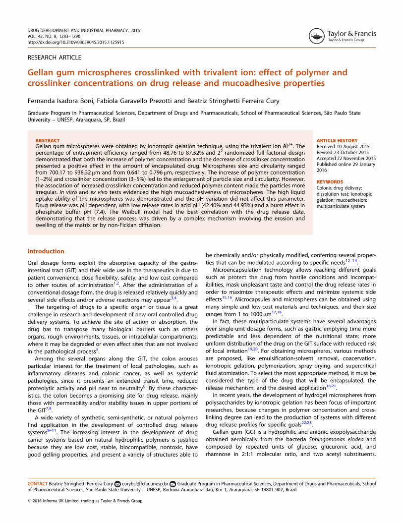
DRUG DEVELOPMENT AND INDUSTRIAL PHARMACY, 2016VOL. 42, NO. 8, 1283–1290http://dx.doi.org/10.3109/03639045.2015.1125915
RESEARCH ARTICLE
Gellan gum microspheres crosslinked with trivalent ion: effect of polymer andcrosslinker concentrations on drug release and mucoadhesive properties
Fernanda Isadora Boni, Fabıola Garavello Prezotti and Beatriz Stringhetti Ferreira Cury
Graduate Program in Pharmaceutical Sciences, Department of Drugs and Pharmaceuticals, School of Pharmaceutical Sciences, Sao Paulo StateUniversity – UNESP, Araraquara, SP, Brazil
ABSTRACTGellan gum microspheres were obtained by ionotropic gelation technique, using the trivalent ion Al3+. Thepercentage of entrapment efficiency ranged from 48.76 to 87.52% and 22 randomized full factorial designdemonstrated that both the increase of polymer concentration and the decrease of crosslinker concentrationpresented a positive effect in the amount of encapsulated drug. Microspheres size and circularity rangedfrom 700.17 to 938.32 mm and from 0.641 to 0.796 mm, respectively. The increase of polymer concentration(1–2%) and crosslinker concentration (3–5%) led to the enlargement of particle size and circularity. However,the association of increased crosslinker concentration and reduced polymer content made the particles moreirregular. In vitro and ex vivo tests evidenced the high mucoadhesiveness of microspheres. The high liquiduptake ability of the microspheres was demonstrated and the pH variation did not affect this parameter.Drug release was pH dependent, with low release rates in acid pH (42.40% and 44.93%) and a burst effect inphosphate buffer pH (7.4). The Weibull model had the best correlation with the drug release data,demonstrating that the release process was driven by a complex mechanism involving the erosion andswelling of the matrix or by non-Fickian diffusion.
ARTICLE HISTORYReceived 10 August 2015Revised 23 October 2015Accepted 22 November 2015Published online 29 January2016
KEYWORDSColonic drug delivery;dissolution test; ionotropicgelation; mucoadhesion;multiparticulate system
Introduction
Oral dosage forms exploit the absorptive capacity of the gastro-
intestinal tract (GIT) and their wide use in the therapeutics is due to
patient convenience, dose flexibility, safety, and low cost compared
to other routes of administration1,2. After the administration of a
conventional dosage form, the drug is released relatively quickly and
several side effects and/or adverse reactions may appear3,4.The targeting of drugs to a specific organ or tissue is a great
challenge in research and development of new oral controlled drug
delivery systems. To achieve the site of action or absorption, the
drug has to transpose many biological barriers such as others
organs, rough environments, tissues, or intracellular compartments,
where it may be degraded or even affect sites that are not involved
in the pathological process5.Among the several organs along the GIT, the colon arouses
particular interest for the treatment of local pathologies, such as
inflammatory diseases and colonic cancer, as well as systemic
pathologies, since it presents an extended transit time, reduced
proteolytic activity and pH near to neutrality6. By these character-
istics, the colon becomes a promising site for drug release, mainly
those with permeability and/or stability issues in upper portions of
the GIT7,8.A wide variety of synthetic, semi-synthetic, or natural polymers
find application in the development of controlled drug release
systems9–11. The increasing interest in the development of drug
carrier systems based on natural hydrophilic polymers is justified
because they are low cost, stable, biocompatible, nontoxic, have
good gelling properties, and present a variety of structures able to
be chemically and/or physically modified, conferring several proper-
ties that can be modulated according to specific needs12–14.Microencapsulation technology allows reaching different goals
such as protect the drug from hostile conditions and incompat-
ibilities, mask unpleasant taste and control the drug release rates in
order to maximize therapeutic effects and minimize systemic side
effects15,16. Microcapsules and microspheres can be obtained using
many simple and low-cost materials and techniques, and their size
ranges from 1 to 1000 mm17,18.In fact, these multiparticulate systems have several advantages
over single-unit dosage forms, such as gastric emptying time more
predictable and less dependent of the nutritional state; more
uniform distribution of the drug on the GIT surface with reduced risk
of local irritation19,20. For obtaining microspheres, various methods
are proposed, like emulsification-solvent removal, coacervation,
ionotropic gelation, polymerization, spray drying, and supercritical
fluid atomization. To select the most appropriate method, it must be
considered the type of the drug that will be encapsulated, the
release mechanism, and the desired application18,21.In recent years, the development of hydrogel microspheres from
polysaccharides by ionotropic gelation has been focus of important
researches, because changes in polymer concentration and cross-
linking degree can lead to the production of systems with different
drug release profiles for specific goals22,23.Gellan gum (GG) is a hydrophilic and anionic exopolysaccharide
obtained aerobically from the bacteria Sphingomonas elodea and
composed by repeated units of glucose, glucuronic acid, and
rhamnose in 2:1:1 molecular ratio, and two acetyl substituents,
CONTACT Beatriz Stringhetti Ferreira Cury [email protected] Graduate Program in Pharmaceutical Sciences, Department of Drugs and Pharmaceuticals, Schoolof Pharmaceutical Sciences, Sao Paulo State University – UNESP, Rodovia Araraquara–Jau, Km 1, Araraquara, SP 14801-902, Brazil
� 2016 Informa UK Limited, trading as Taylor & Francis Group
acetate, and glycerate, linked on glucose residue adjacent of theglucuronic acid24,25.
GG is widely used as an additive in food industry; at lowconcentrations, it forms gels and it is biocompatible and biodegrad-able. In aqueous solution, the gelation of GG is accompanied by thechains conformational transition from random coils to double-helical conformation and then the double-helical arrangement formordered junction zones, resulting in a three-dimensional net-work11,26. In the presence of cations, the ionotropic gelationoccurs, in which polymer negative groups interact with a divalentor multivalent counter ion, building a stronger and dense hydrogelnetwork27,28.
These characteristics make GG an interesting material forpharmaceutical applications22,29. Several drug delivery systemsbased on GG, such as in situ momethasone furoate nasal gel30, insitu gelling terbinafine hydrochloride ophtalmic nanoemulsion31,and GG tablets with metronidazole32 were developed. The devel-opment of hydrogel microspheres based on GG crosslinked withmono and divalent cations, mainly Ca2+, has been exploited toobtain different drug delivery systems22,24.
Trivalent cation (Al3+) have been evaluated for microspheresproduction because the crosslinking with trivalent ions can beadvantageous by enabling a faster crosslinking reaction due to theirextra positive charge, when compared, for example, with calciumions. Each molecule of aluminum is able to conjugate with a highernumber of sites of the polysaccharides. Therefore, the use of a lowconcentration of crosslinking solution may allow the faster forma-tion of a more rigid hydrogel, reducing the reaction time andconsequently reducing the risk of drug solubilization or degradationin the crosslinking solution, providing a more efficient drugencapsulation and drug release control33–35.
Maiti and coworkers reported the preparation of GG micro-spheres crosslinked with aluminum ions and glutaraldehyde forprolonged release of glipizide, studying the influence of the dualcrosslinking (ionic and covalent) in microspheres characteristics anddrug release profiles23. Prezotti and coworkers (2014) developedmicrospheres of GG and pectin blends, crosslinked with Al3+,demonstrating the effective reduction of release rates of ketoprofen(KP) in simulated gastric media36.
Traditional methods of performing experiments involve materialand time spending, especially when new formulations are devel-oped. The factorial design technique is an efficient method toindicate significant effects of variables and their interactions. Theuse of this technique helps optimizing the samples preparation andanalysis of the process, minimizing the amount of time andmaterials spent during the development37,38.
In this paper, GG microspheres containing KP as model drug wereprepared by ionotropic gelation using aluminum chloride ascrosslinker. The physicochemical characterization included analysesof size, shape, surface morphology and internal structure, swellingability, and entrapment efficiency (EE%). The influence of thevariables polymer and crosslinker concentrations on particle size,shape, and EE% was evaluated by a 22 randomized full factorialdesign. The mucoadhesive ability of microspheres was assessedthrough in vitro and ex vivo analysis, and the drug release profilewas evaluated in media that simulate the pH variation of the GIT.
Materials and methods
Materials
Low acyl GG (200–300 KDa) was kindly provide by CP Kelco(Kelcogel� CG-LA); KP (batch # 09072223) was obtained fromZhejiang Jiuhzou Pharmaceutical Co. (Taizhou, China); aluminum
chloride from Vetec; Mucin type II and Total Protein Kit Micro Lowry,Peterson’s Modification were purchased from Sigma Aldrich�
(St. Louis, MO). All other materials used were of analytical gradeand obtained from commercial suppliers.
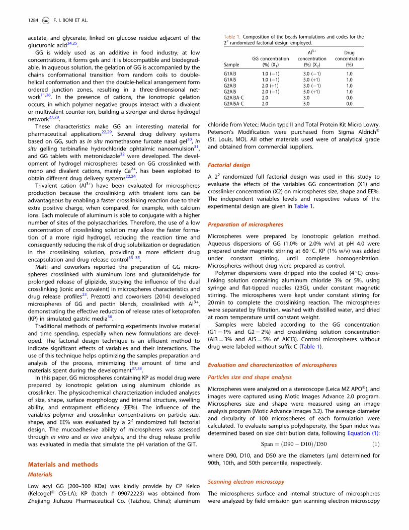
Factorial design
A 22 randomized full factorial design was used in this study toevaluate the effects of the variables GG concentration (X1) andcrosslinker concentration (X2) on microspheres size, shape and EE%.The independent variables levels and respective values of theexperimental design are given in Table 1.
Preparation of microspheres
Microspheres were prepared by ionotropic gelation method.Aqueous dispersions of GG (1.0% or 2.0% w/v) at pH 4.0 wereprepared under magnetic stirring at 60 �C. KP (1% w/v) was addedunder constant stirring, until complete homogenization.Microspheres without drug were prepared as control.
Polymer dispersions were dripped into the cooled (4 �C) cross-linking solution containing aluminum chloride 3% or 5%, usingsyringe and flat-tipped needles (23G), under constant magneticstirring. The microspheres were kept under constant stirring for20 min to complete the crosslinking reaction. The microsphereswere separated by filtration, washed with distilled water, and driedat room temperature until constant weight.
Samples were labeled according to the GG concentration(G1¼ 1% and G2¼ 2%) and crosslinking solution concentration(Al3¼ 3% and Al5¼ 5% of AlCl3). Control microspheres withoutdrug were labeled without suffix C (Table 1).
Evaluation and characterization of microspheres
Particles size and shape analysis
Microspheres were analyzed on a stereoscope (Leica MZ APO�), andimages were captured using Motic Images Advance 2.0 program.Microspheres size and shape were measured using an imageanalysis program (Motic Advance Images 3.2). The average diameterand circularity of 100 microspheres of each formulation werecalculated. To evaluate samples polydispersity, the Span index wasdetermined based on size distribution data, following Equation (1):
Span ¼ ðD90� D10Þ=D50 ð1Þ
where D90, D10, and D50 are the diameters (mm) determined for90th, 10th, and 50th percentile, respectively.
Scanning electron microscopy
The microspheres surface and internal structure of microsphereswere analyzed by field emission gun scanning electron microscopy
Table 1. Composition of the beads formulations and codes for the22 randomized factorial design employed.
SampleGG concentration
(%) (X1)
Al3+
concentration(%) (X2)
Drugconcentration
(%)
G1Al3 1.0 (�1) 3.0 (�1) 1.0G1Al5 1.0 (�1) 5.0 (+1) 1.0G2Al3 2.0 (+1) 3.0 (�1) 1.0G2Al5 2.0 (�1) 5.0 (+1) 1.0G2Al3A-C 2.0 3.0 0.0G2Al5A-C 2.0 5.0 0.0
1284 F. I. BONI ET AL.
(FEG-SEM; JEOL JSM-7500F, Japan). To evaluate the internal struc-ture, microspheres were frozen with liquid nitrogen and afterfractured. Samples were attached to the sample holder with adouble-side adhesive tape, and photomicrographs at differentmagnifications were taken.
Entrapment efficiency
To determine the EE%, a known mass of dried microspheres wasallowed to swell in 0.1 ml of phosphate buffer (pH 7.4) during 5 minand then crushed inside the centrifuge tube. The microspheres wereadded by absolute ethanol (10 ml) to dissolve the drug and keptunder stirring for 1.5 h, at room temperature. Tubes were well sealedto avoid solvent evaporation. After, samples were centrifuged at2000 rpm (5 min) to precipitate polymeric debris, and the amount ofdrug in the supernatant was quantified on a UV–Vis spectrophotom-eter (Hewlett Packard-Kayak XA), at 254 nm. Tests were performed intriplicate, and the EE% was calculated according Equation (2):
%EE ¼ AA=TAð Þ � 100 ð2Þ
where TA was the total amount of drug added, and AA was thequantified amount of drug.
Liquid uptake study
Swelling study was performed on an Enslin device with differentmedia to simulate the variation throughout the GIT: simulatedgastric fluid (0.1N HCl, pH 2.0), simulated intestinal fluid (pH 7.4),and simulated colonic fluid (pH 6.0), all of them without enzymes.The volume of media uptake by the sample was measured atpredetermined time intervals (1, 2, 5, 10, 15, 30, 60, and 90 min). Thetests were performed in triplicate and results were expressed aspercentage of swelling (%S), according to Equation (3):
%S ¼ V=m� 100 ð3Þ
where m is the initial mass of microspheres (g); V¼ volume (mL) ofmedia absorbed; %S¼percentage of swelling (%).
In vitro analysis of the mucoadhesive properties
The mucoadhesiveness was analyzed by studying the adsorptionof mucin on the microspheres3. Mucin solutions were prepared atdifferent concentrations (50, 100, 150, and 200mg/ml), and 20 mgof microspheres were dispersed in these solutions for 1 h at37 �C. Then, the dispersions were centrifuged at 3000 rpm (2 min),and the supernatant was used to quantify the free mucin contentby colorimetric method, using a Lowry protein assay modifiedby Peterson39,40. For the determination of free mucin in thesupernatant, an UV–Vis spectrophotometer at 749 nm andreagents from Total Protein Kit, Micro Lowry, Peterson’sModification (Sigma-Aldrich�, St. Louis, MO) were used36. Forthe mucoadhesive property analysis, samples containing highamount of GG (2%) were selected, since they presented highercircularity and EE%.
Ex vivo mucoadhesion evaluation
The ex vivo mucoadhesion test was performed as described byPrezotti et al. using porcine intestinal tissue. The method was basedon the procedure purposed by Rao and Bur41. Briefly, pieces of freshtissue (4 cm� 4 cm) were opened longitudinally and washedusing saline solution (0.9%) and attached to the inclined plastic
support (30 �C) of the device. The microspheres (n¼ 30) wereplaced on the tissue surface, allowing a 20-min contact time withthe mucous layer. After this, phosphate buffer pH 6.0 was used torinse the tissue for 5 min at a rate of 30 ml/min. The percentage ofmucoadhesion was calculated from the difference between thenumber of microspheres attached to the biological surface at thebeginning and at the end of the test.
Dissolution test and analysis of drug release mechanism
This analysis was performed on a Hanson Dissolution Test StationSR8-Plus (Chastworth, CA) equipped with USP apparatus 1 (basket)at 50 rpm. The experiment was conducted using media withdifferent pH values at 37 �C: simulated gastric media (900 mL of0.1N HCl pH 1.2 with sodium lauryl sulfate 0.75%) during 120 minand enteric media (900 mL of phosphate buffer pH 7.4). Atpredetermined time intervals, aliquots of 3 ml were withdrawnand immediately replaced with fresh dissolution media. The amountof drug released was quantified using an UV–Vis spectrophotometerat 258 nm and 260 nm, for gastric and enteric pH, respectively. Theexperiment was conducted with raw KP and samples G2Al3 andG2Al5, once they presented the highest values of EE%. The testswere performed in triplicate, with a mass of microspheres contain-ing 100 mg of KP.
Drug release data were fitted with different mathematical models(Korsmeyer–Peppas, Higuchi, First-order, Hixson–Crowell and Baker–Lonsdale) to determine the mechanism of drug release.
Statistical analysis
One-way analysis of variance (ANOVA) followed by Tukey’s testwas used to evaluate significant differences with a significance levelof 5%.
Results
Evaluation and characterization of microspheres
Particles size and shape analysis
The diameter of the microspheres ranged from 700.17 to 938.32 mm(Table 2) with sample G1Al3 presenting the smallest mean diameter(700.17 mm) (p50.05). GG microspheres presented circularity valuesbetween 0.641 and 0.796 (Table 2). Samples showed a mono-dispersed and unimodal size distribution, with Span index between0.08 and 0.18 (Table 2).
Scanning electron microscopy
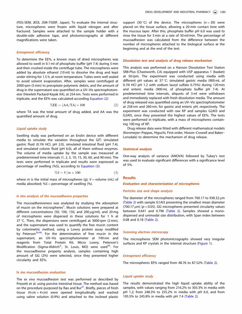
The microspheres SEM photomicrographs showed very irregularsurfaces and KP crystals in the internal structure (Figure 1).
Entrapment efficiency
The microspheres EE% ranged from 48.76 to 87.52% (Table 2).
Liquid uptake study
The results demonstrated the high liquid uptake ability of thesamples, with values ranging from 216.2% to 303.3% in media withpH 1.2; from 248.3% to 255.2% in media with pH 6.0, and from195.5% to 245.8% in media with pH 7.4 (Table 2).
DRUG DEVELOPMENT AND INDUSTRIAL PHARMACY 1285
In vitro analysis of the mucoadhesive properties
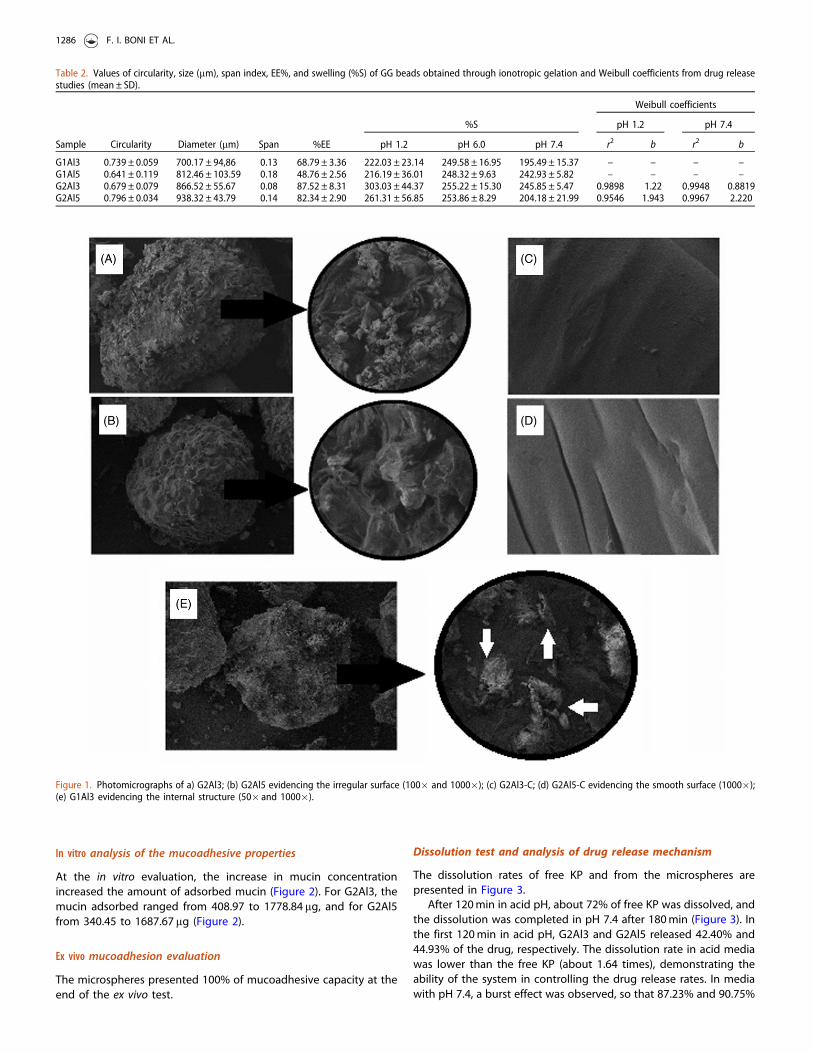
At the in vitro evaluation, the increase in mucin concentration
increased the amount of adsorbed mucin (Figure 2). For G2Al3, the
mucin adsorbed ranged from 408.97 to 1778.84 mg, and for G2Al5
from 340.45 to 1687.67 mg (Figure 2).
Ex vivo mucoadhesion evaluation
The microspheres presented 100% of mucoadhesive capacity at the
end of the ex vivo test.
Dissolution test and analysis of drug release mechanism
The dissolution rates of free KP and from the microspheres are
presented in Figure 3.After 120 min in acid pH, about 72% of free KP was dissolved, and
the dissolution was completed in pH 7.4 after 180 min (Figure 3). In
the first 120 min in acid pH, G2Al3 and G2Al5 released 42.40% and
44.93% of the drug, respectively. The dissolution rate in acid media
was lower than the free KP (about 1.64 times), demonstrating the
ability of the system in controlling the drug release rates. In media
with pH 7.4, a burst effect was observed, so that 87.23% and 90.75%
Figure 1. Photomicrographs of a) G2Al3; (b) G2Al5 evidencing the irregular surface (100� and 1000�); (c) G2Al3-C; (d) G2Al5-C evidencing the smooth surface (1000�);(e) G1Al3 evidencing the internal structure (50� and 1000�).
Table 2. Values of circularity, size (mm), span index, EE%, and swelling (%S) of GG beads obtained through ionotropic gelation and Weibull coefficients from drug releasestudies (mean ± SD).
Weibull coefficients
%S pH 1.2 pH 7.4
Sample Circularity Diameter (mm) Span %EE pH 1.2 pH 6.0 pH 7.4 r2 b r2 b
G1Al3 0.739 ± 0.059 700.17 ± 94,86 0.13 68.79 ± 3.36 222.03 ± 23.14 249.58 ± 16.95 195.49 ± 15.37 – – – –G1Al5 0.641 ± 0.119 812.46 ± 103.59 0.18 48.76 ± 2.56 216.19 ± 36.01 248.32 ± 9.63 242.93 ± 5.82 – – – –G2Al3 0.679 ± 0.079 866.52 ± 55.67 0.08 87.52 ± 8.31 303.03 ± 44.37 255.22 ± 15.30 245.85 ± 5.47 0.9898 1.22 0.9948 0.8819G2Al5 0.796 ± 0.034 938.32 ± 43.79 0.14 82.34 ± 2.90 261.31 ± 56.85 253.86 ± 8.29 204.18 ± 21.99 0.9546 1.943 0.9967 2.220
1286 F. I. BONI ET AL.
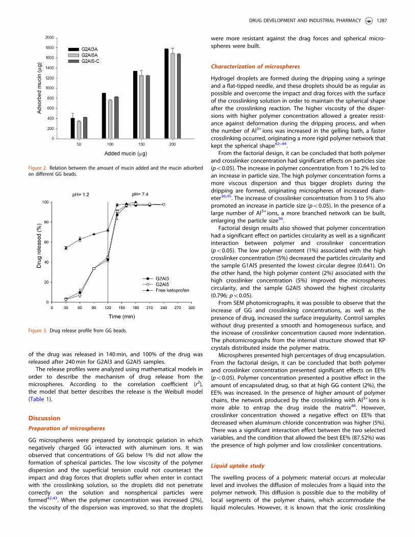
of the drug was released in 140 min, and 100% of the drug wasreleased after 240 min for G2Al3 and G2Al5 samples.
The release profiles were analyzed using mathematical models inorder to describe the mechanism of drug release from themicrospheres. According to the correlation coefficient (r2),the model that better describes the release is the Weibull model(Table 1).
Discussion
Preparation of microspheres
GG microspheres were prepared by ionotropic gelation in whichnegatively charged GG interacted with aluminum ions. It wasobserved that concentrations of GG below 1% did not allow theformation of spherical particles. The low viscosity of the polymerdispersion and the superficial tension could not counteract theimpact and drag forces that droplets suffer when enter in contactwith the crosslinking solution, so the droplets did not penetratecorrectly on the solution and nonspherical particles wereformed42,43. When the polymer concentration was increased (2%),the viscosity of the dispersion was improved, so that the droplets
were more resistant against the drag forces and spherical micro-spheres were built.
Characterization of microspheres
Hydrogel droplets are formed during the dripping using a syringeand a flat-tipped needle, and these droplets should be as regular aspossible and overcome the impact and drag forces with the surfaceof the crosslinking solution in order to maintain the spherical shapeafter the crosslinking reaction. The higher viscosity of the disper-sions with higher polymer concentration allowed a greater resist-ance against deformation during the dripping process, and whenthe number of Al3+ ions was increased in the gelling bath, a fastercrosslinking occurred, originating a more rigid polymer network thatkept the spherical shape42–44.
From the factorial design, it can be concluded that both polymerand crosslinker concentration had significant effects on particles size(p50.05). The increase in polymer concentration from 1 to 2% led toan increase in particle size. The high polymer concentration forms amore viscous dispersion and thus bigger droplets during thedripping are formed, originating microspheres of increased diam-eter36,45. The increase of crosslinker concentration from 3 to 5% alsopromoted an increase in particle size (p50.05). In the presence of alarge number of Al3+ ions, a more branched network can be built,enlarging the particle size36.
Factorial design results also showed that polymer concentrationhad a significant effect on particles circularity as well as a significantinteraction between polymer and crosslinker concentration(p50.05). The low polymer content (1%) associated with the highcrosslinker concentration (5%) decreased the particles circularity andthe sample G1Al5 presented the lowest circular degree (0.641). Onthe other hand, the high polymer content (2%) associated with thehigh crosslinker concentration (5%) improved the microspherescircularity, and the sample G2Al5 showed the highest circularity(0.796; p50.05).
From SEM photomicrographs, it was possible to observe that theincrease of GG and crosslinking concentrations, as well as thepresence of drug, increased the surface irregularity. Control sampleswithout drug presented a smooth and homogeneous surface, andthe increase of crosslinker concentration caused more indentation.The photomicrographs from the internal structure showed that KPcrystals distributed inside the polymer matrix.
Microspheres presented high percentages of drug encapsulation.From the factorial design, it can be concluded that both polymerand crosslinker concentration presented significant effects on EE%(p50.05). Polymer concentration presented a positive effect in theamount of encapsulated drug, so that at high GG content (2%), theEE% was increased. In the presence of higher amount of polymerchains, the network produced by the crosslinking with Al3+ ions ismore able to entrap the drug inside the matrix46. However,crosslinker concentration showed a negative effect on EE% thatdecreased when aluminum chloride concentration was higher (5%).There was a significant interaction effect between the two selectedvariables, and the condition that allowed the best EE% (87.52%) wasthe presence of high polymer and low crosslinker concentrations.
Liquid uptake study
The swelling process of a polymeric material occurs at molecularlevel and involves the diffusion of molecules from a liquid into thepolymer network. This diffusion is possible due to the mobility oflocal segments of the polymer chains, which accommodate theliquid molecules. However, it is known that the ionic crosslinking
Figure 3. Drug release profile from GG beads.
Figure 2. Relation between the amount of mucin added and the mucin adsorbedon different GG beads.
DRUG DEVELOPMENT AND INDUSTRIAL PHARMACY 1287
process immobilizes the polymer chains, and consequently the
swelling ability of the polymeric system can be reduced47,48.The liquid uptake ability is an important feature of drug delivery
systems since the water absorption is the first step for the drug
release process, for further matrix swelling and/or diffusion of the
drug. However, the drug dissolution and the matrix erosion can also
impel the drug release process49,50. This ability is also important
because it can ensure the access of bacterial enzymes that act in
polymer matrix degradation and the liquid uptake can assist in the
mucoadhesion process, as the absorption of liquid provides polymer
chains relaxation and makes them available to interact with mucus
layer51–53.All microspheres presented a non pH sensitive liquid uptake
behavior, since there were no significant differences in the
percentage of swelling (%S) between the different media for all
samples (p40.05).
Mucoadhesion tests
Mucoadhesion is an important property for multiparticulate sys-
tems because it may prolong the residence time in the absorp-
tion site and promote a closer contact with the epithelial
barrier, increasing drug permeability and absorption54–56.Colon has a low mucus turnover rate compared to the stomach
and the small intestine and a slow motility; these features facilitate
the contact of the polymeric system with the glycoproteins which
are the structural components of mucus and primarily responsible
for the adhesive interaction and enable the consolidation phase of
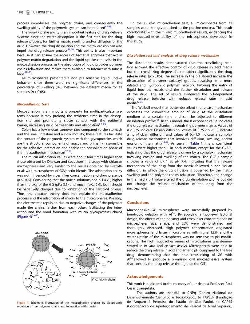
the mucoadhesion mechanism57,58.The mucin adsorption values were about four times higher than
those observed by Dhawan and coauthors in a study with chitosan
microspheres and very similar to the results obtained by Prezotti
et al. with microspheres of GG/pectin blends. The adsorption ability
was not influenced by crosslinker concentration and drug presence
(p40.05). Considering that the mucin solutions had pH 4.79, higher
than the pKa of the GG (pKa 3.5) and mucin (pKa 2.6), both should
be negatively charged due to ionization of the carboxyl groups.
Thus, the electron theory does not explain the mucoadhesion
process and the adsorption of mucin to the microspheres. Possibly,
the electrostatic repulsion due to negative charges of the polymers
made the chains farther from each other, facilitating the inter-
action and the bond formation with mucin glycoproteins chains
(Figure 4)59,60.
In the ex vivo mucoadhesion test, all microspheres from allsamples were strongly attached to the porcine mucosa. This resultcorroborates with the in vitro mucoadhesion results, evidencing thehigh mucoadhesive ability of the microspheres developed inthis study.
Dissolution test and analysis of drug release mechanism
The dissolution results demonstrated that the crosslinking reac-tion allowed the effective control of drug release in acid mediabut the crosslinking degree did not affect significantly the drugrelease rates (p40.05). The increase in the pH should increase thedissociation of polymer carboxyl groups, resulting in a moredilated and hydrophilic polymer network, favoring the entry ofliquid into the matrix and the further dissolution and releaseof the drug. The set of results evidenced the pH-dependentdrug release behavior with reduced release rates in acidmedia22,35.
The Weibull model that better described the release mechanismexpresses the cumulative amount of drug at the dissolutionmedium at a certain time and can be adjusted to differentdissolution profiles61. In this model, the b exponent value indicatesthe drug release mechanism through the polymer matrix. Values ofb50.75 indicate Fickian diffusion, values of 0.755b51.0 indicatea non-Fickian diffusion, and values of b41.0 indicate a complexdrug release mechanism that involves diffusion, swelling, and/orerosion of the matrix14,62. As seen in Table 1, the b coefficientvalues were higher than 1 in both medium, except for the G2Al3,indicating that the drug release is driven by a complex mechanisminvolving erosion and swelling of the matrix. The G2Al3 sampleshowed a value of b51 at pH 7.4, indicating that the releasemechanism of the drug from the matrix followed a non-Fickiandiffusion, in which the drug diffusion is governed by the matrixswelling and the polymer chains relaxation. Therefore, the changein the media pH value altered the drug dissolution profile but didnot change the release mechanism of the drug from themicrospheres.
Conclusions
Mucoadhesive GG microspheres were successfully prepared byionotropic gelation with Al3+. By applying a two-level factorialdesign, the effects of the polymer and crosslinker concentrations onmicrospheres size, shape, and EE% were demonstrated andthoroughly discussed. High polymer concentration originatedmore spherical and larger microspheres with higher EE%, and thewater uptake of the microspheres was no sensitive to pH modifi-cations. The high mucoadhesiveness of microspheres was demon-strated in in vitro and ex vivo assays. Microspheres were able toreduce the drug release in acid and enteric pH compared to the freedrug, demonstrating that the ionic crosslinking of GG withAl3+ allowed to produce a promising oral mucoadhesive systemthat controls the drug release throughout the GIT.
Acknowledgements
This work is dedicated to the memory of our dearest Professor RaulCesar Evangelista.
The authors are thankful to CNPq (Centro Nacional deDesenvolvimento Cientıfico e Tecnologico), to FAPESP (Fundacaode Amparo a Pesquisa do Estado de Sao Paulo), to CAPES(Coordenacao de Aperfeicoamento de Pessoal de Nıvel Superior),
Figure 4. Schematic illustration of the mucoadhesion process by electrostaticrepulsion of the polymers chains and interaction with mucin.
1288 F. I. BONI ET AL.
and to Faculdade de Ciencias Farmaceuticas de Araraquara – UNESPfor providing financial and structural support to develop this work.
Declaration of interest
The authors report no declarations of interest.
References
1. Pinto JF. Site-specific drug delivery systems within the gastro-intestinal tract: from the mouth to the colon. Int J Pharm2010;395:44–52.
2. Isha C, Nimrata S, Rana AC, Surbihi G. Oral sustained releasedrug delivery system: an overview. Int Res J Pharm 2012;3:57–
62.3. Pezzini BR, Silva MAS, Ferraz HG. Formas farmaceuticas de
liberacao prolongada: Sistemas monolıticos e multiparticula-
dos. Braz J Pharm Sci 2007;43:491–502.4. Reis AS-MJF, Pedreiro AU, Cavalcanti LN. Phosphated cross-
linked pectin as a potential excipient for specific drug delivery:
preparation and physicochemical characterization. Polymer Int2010;59:127–35.
5. Torchilin VP. Drug targeting. Eur J Pharm Sci 2000;11:S81–91.6. Philip AK, Philip B. Colon targeted drug delivery systems: a
review on primary and novel approaches. Oman Med J
2010;25:79–87.7. Friend DR. Colon-specific drug delivery. Adv Drug Deliv Rev
1991;7:149–99.8. Sinha VR, Kumria R. Polysaccharides in colon-specific drug
delivery. Int J Pharm 2001;224:19–38.9. Carbinatto FM, De Castro AD, Cury BSF, et al. Physical
properties of pectin–high amylose starch mixtures cross-linked with sodium trimetaphosphate. Int J Pharm2012;423:281–8.
10. Meneguin AB, Cury BS, Evangelista RC. Films from resistantstarch-pectin dispersions intended for colonic drug delivery.Carbohydr Polym 2014;99:140–9.
11. Osmałek T, Froelich A, Tasarek S. Application of gellan gum inpharmacy and medicine. Int J Pharm 2014;466:328–40.
12. Prezotti FG, Meneguin AB, Evangelista RC, Cury BS. Preparation
and characterization of free films of high amylose/pectinmixtures cross-linked with sodium trimetaphosphate. Drug
Dev Ind Pharm 2012;38:1354–9.13. Orlu M, Cevher E, Araman A. Design and evaluation of colon
specific drug delivery system containing flurbiprofen micro-
sponges. Int J Pharm 2006;318:103–17.14. Cury BS, Castro AD, Klein SI, Evangelista RC. Modeling a system
of phosphated cross-linked high amylose for controlled drug
release. Part 2: physical parameters, cross-linking degrees anddrug delivery relationships. Int J Pharm 2009;371:8–15.
15. Bhadouriya P, Kumar M, Pathak K. Formulation and in vitroevaluation of prolonged release floating microspheres ofatenolol using multicompartment dissolution apparatus. Drug
Dev Ind Pharm 2013;39:1663–71.16. Patil SB, Sawant KK. Chitosan microspheres as a delivery
system for nasal insufflation. Colloids Surf B Biointerfaces
2011;84:384–9.17. Vasir JK, Tambwekar K, Garg S. Bioadhesive microspheres
as a controlled drug delivery system. Int J Pharm
2003;255:13–32.18. Kumari S, Bhandari A, Sharma PK. Solvent evaporation as a
imposing method for microencapsulation – a review. J DrugDiscov Therap 2014;2:13–20.
19. Maestrelli F, Zerrouk N, Cirri M, et al. Microspheres for colonic
delivery of ketoprofen- hidroxypropyl-b-cyclodextrin complex.
Eur J Pharm Sci 2008;34:1–11.20. Schmidt C, Bodmeier R. A multiparticulate drug-delivery system
based on pellets incorporated into congealable polyethylene
glycol carrier materials. Int J Pharm 2001;216:9–16.21. Jyothi NV, Prasanna PM, Sakarkar SN, et al. Microencapsulation
techniques, factors influencing encapsulation efficiency.
J Microencapsul 2010;27:187–97.22. Agnihotri SA, Jawalkar SS, Aminabhavi TM. Controlled release
of cephalexin through gellan gum beads: effect of formulation
parameters on entrapment efficiency, size, and drug release.
Eur J Pharm Biopharm 2006;63:249–61.23. Maiti S, Ranjita S, Mandola R, et al. Al+ 3 ion cross-linked and
acetalated gellan hydrogel network beads for prolonged
release of glipizide. Carbohydr Polym 2011;85:164–72.24. Racovit a S, Vasiliu S, Popa M, Luca C. Polysaccharides based on
micro and nanoparticles obtained by ionic gelation and their
applications as drug delivery systems. Drug Deliv Syst
2009;54:709–18.25. Morris ER, Nishinari K, Rinaudo M. Gelation of gellan – a review.
Food Hydrocoll 2012;28:373–411.26. Picone CS, Cunha RL. Chitosan-gellan electrostatic complexes:
influence of preparation conditions and surfactant presence.
Carbohydr Polym 2013;94:695–703.27. Shu XZ, Zhu KJ. Controlled drug release properties of ionically
cross-linked chitosan beads: the influence of anion structure.
Int J Pharm 2002;233:217–25.28. Lopez OV, Garcıa MA, Zaritzky NE. Film forming capacity of
chemically modified corn starches. Carbohydr Polym
2008;73:573–81.29. Narkar M, Sher P, Pawar A. Stomach-specific controlled release
gellan beads of acid-soluble drug prepared by ionotropic
gelation method. AAPS PharmSciTech 2010;11:267–77.30. Cao SL, Ren XW, Zhang QZ, et al. In situ gel based on gellan
gum as new carrier for nasal administration of mometasone
furoate. Int J Pharm 2009;365:109–15.31. Tayel SA, El-Nabarawi MA, Tadros MI, Abd-Elsalam WH.
Promising ion-sensitive in situ ocular nanoemulsion gels of
terbinafine hydrochloride: design, in vitro characterization and
in vivo estimation of the ocular irritation and drug pharmaco-
kinetics in the aqueous humor of rabbits. Int J Pharm
2013;443:293–305.32. Emeje MO, Franklin-Ude PI, Ofoefule SI. Evaluation of the fluid
uptake kinetics and drug release from gellan gum tablets
containing metronidazole. Int J Biol Macromol 2010;47:158–63.33. Reddy T, Tammishetti S. Gastric resistant microbeads of metal
ion cross-linked carboxymethyl guar gum for oral drug delivery.
J Microencapsul 2002;19:311–18.34. Nokhodchi A, Tailor A. In situ cross-linking of sodium alginate
with calcium and aluminum ions to sustain the release of
theophylline from polymeric matrices. Farmaco 2004;59:999–
1004.35. Mundargi RC, Patil SA, Agnihotri SA, Aminabhavi TM.
Development of polysaccharide-based colon targeted drug
delivery systems for the treatment of amoebiasis. Drug Dev Ind
Pharm 2007;33:255–64.36. Prezotti FG, Cury BS, Evangelista RC. Mucoadhesive beads of
gellan gum/pectin intended to controlled delivery of drugs.
Carbohydr Polym 2014;113:286–95.37. Gohel MC, Amin AF. Formulation optimization of controlled
release diclofenac sodium microspheres using factorial design.
J Control Release 1998;51:115–22.
DRUG DEVELOPMENT AND INDUSTRIAL PHARMACY 1289
38. Nila MV, Sudhir MR, Cinu TA, et al. Floating microspheres ofcarvedilol as gastro retentive drug delivery system: 3(2) fullfactorial design and in vitro evaluation. Drug Deliv2014;21:110–17.
39. Dhawan S, Singla AK, Sinha VR. Evaluation of mucoadhesiveproperties of chitosan microspheres prepared by differentmethods. AAPS PharmSciTech 2004;5:e67.
40. Peterson GL. A simplification of the protein assay method ofLowry et al. which is more generally applicable. Anal Biochem1977;83:346–56.
41. Rao KVR, Buri P. A novel in situ method to test polymersand coated microparticles for bioadhesion. Int J Pharm1989;52:265–70.
42. Seifert DB, Phillips JA. Production of small, monodispersedalginate beads for cell imobilization. Biotechnol Prog1997;13:562–8.
43. Chan ES, Lee BB, Ravindra P, Poncelet D. Prediction models forshape and size Ca-alginate macrobeads produced throughextrusion-dripping method. J Colloid Interface Sci 2009;338:63–72.
44. Cellesi F, Tirelli N, Hubbell JA. Towards a fully-syntheticsubstitute of alginate: development of a new process usingthermal gelation and chemical cross-linking. Biomaterials2004;25:5115–24.
45. Kaity S, Isaac J, Ghosh A. Interpenetrating polymer network oflocust bean gum-poly (vinyl alcohol) for controlled release drugdelivery. Carbohydr Polym 2013;94:456–67.
46. Nayak AK, Das B, Maji R. Calcium alginate/gumArabic beads containing glibenclamide: developmentand in vitro characterization. Int J Biol Macromol2012;51:1070–8.
47. Mi FL, Sung HW, Shyu SS, et al. Synthesis and characterizationof biodegradable TPP/genipin co-crosslinked chitosan gelbeads. Polymer 2003;44:6521–30.
48. Colombo P, Bettini R, Santi P, Peppas NA. Swellable matrices forcontrolled drug delivery: gel-layer behaviour, mechanismsand optimal performance. Pharm Sci Technol Today2000;3:198–204.
49. Khare AR, Peppas NA. Swelling/deswelling of anionic copoly-mer gels. Biomaterials 1995;16:559–67.
50. Singh B, Bala R, Chauhan N. In vitro release dynamics of modeldrugs from psyllium and acrylic acid based hydrogels for theuse in colon specific drug delivery. J Mater Sci Mater Med2008;19:2771–80.
51. Simonoska Crcarevska M, Glavas Dodov M, Goracinova K.Chitosan coated Ca-alginate microparticles loaded with bude-sonide for delivery to the inflamed colonic mucosa. Eur J PharmBiopharm 2008;68:565–78.
52. Mulhbacher J, Ispas-Szabo P, Mateescu MA. Cross-linked highamylose starch derivatives for drug release. II. Swellingproperties and mechanistic study. Int J Pharm 2004;278:231–8.
53. Shi L, Caldwell KD. Mucin adsorption to hydrophobic surfaces.J Colloid Interface Sci 2000;224:372–81.
54. Asane GS, Nirmal SA, Rasal KB, et al. Polymers for mucoadhesivedrug delivery system: a current status. Drug Dev Ind Pharm2008;34:1246–66.
55. Gamboa JM, Leong KW. In vitro and in vivo models for thestudy of oral delivery of nanoparticles. Adv Drug Deliv Rev2013;65:800–10.
56. Peppas NA, Sahlin JJ. Hydrogels as mucoadhesive andbioadhesive materials: a review. Biomaterials 1996;17:1553–61.
57. Carvalho FC, Bruschi ML, Evangelista RC, Gremiao MPD.Mucoadhesive drug delivery systems. Braz J Pharm Sci2010;46:1–17.
58. Varum FJ, Veiga F, Sousa JS, Basit AW. An investigation into therole of mucus thickness on mucoadhesion in the gastrointes-tinal tract of pig. Eur J Pharm Sci 2010;40:335–41.
59. Sriamornsak P, Wattanakorn N, Takeuchi H. Study on themucoadhesion mechanism of pectin by atomic force micros-copy and mucin-particle method. Carbohydr Polym2010;79:54–9.
60. Joergensen L, Klosgen B, Simonsen AC, et al. New insights intothe mucoadhesion of pectins by AFM roughness parameters incombination with SPR. Int J Pharm 2011;411:162–8.
61. Adams E, De Maesschalck R, De Spiegeleer B, et al. Evaluation ofdissolution profiles using principal component analysis. Int JPharm 2001;212:41–53.
62. Papadopoulou V, Kosmidis K, Vlachou M, Macheras P. On theuse of the Weibull function for the discernment of drug releasemechanisms. Int J Pharm 2006;309:44–50.
1290 F. I. BONI ET AL.
View publication statsView publication stats