zeolites as supports for the biorecovery of hexavalent and trivalent chromium
TRANSCRIPT

Microporous and Mesoporous Materials 116 (2008) 555–560
Contents lists available at ScienceDirect
Microporous and Mesoporous Materials
journal homepage: www.elsevier .com/locate /micromeso
Zeolites as supports for the biorecovery of hexavalent and trivalent chromium
Bruna Silva a,*, Hugo Figueiredo a, Cristina Quintelas a, Isabel C. Neves b, Teresa Tavares a
a IBB – Instituto de Biotecnologia e Bioengenharia, Centro de Engenharia Biológica, Universidade do Minho, Campus de Gualtar, 4710-057 Braga, Portugalb Departamento de Química, Centro de Química, Universidade do Minho, Campus de Gualtar, 4710-057 Braga, Portugal
a r t i c l e i n f o
Article history:Received 21 January 2008Received in revised form 8 May 2008Accepted 8 May 2008Available online 15 May 2008
Keywords:NaYArthrobacter viscosusBiosorbentsCr(VI)Cr(III)
1387-1811/$ - see front matter � 2008 Elsevier Inc. Adoi:10.1016/j.micromeso.2008.05.015
* Corresponding author.E-mail address: [email protected] (B. Silva).
a b s t r a c t
The aim of this study is the preparation and characterization of new catalytic materials to be used in oxi-dation reactions through the recovery of heavy metals in wastewater. The recovery of Cr(III) and Cr(VI)from aqueous solutions by an Arthrobacter viscosus biofilm supported on NaY zeolite was investigated.Experiments were repeated without the bacteria for comparison purposes. The batch method has beenemployed, using solutions with chromium concentrations of 10 mg L�1, 25 mg L�1, 50 mg L�1 and100 mg L�1. Cr(III) was easily removed from solution due to its positive charge which allows the entrap-ment in the framework zeolite by ion exchange. However, due to its anionic form Cr(VI) was onlyremoved in the presence of the biofilm that performs its reduction to Cr(III), followed by ion exchangein the zeolite. The best uptake was achieved for initial concentration of 100 mg L�1: 14 mg g�1
zeolite forCr(III) by both systems and 3 mg g�1
zeolite for Cr(VI) by the zeolite with the bacterium biofilm. The modifiedzeolite samples have been fully characterized by surface analysis (XRD, XPS), chemical analyses (ICP-AES), spectroscopic method (FTIR) and microscopic analysis (SEM). The results show that the biofilmof A. viscosus supported on NaY zeolite is able to recover chromium from dilute solutions and the frame-work zeolite remains unchanged after chromium biosorption.
� 2008 Elsevier Inc. All rights reserved.
1. Introduction
Heavy metals released into the environment have been increas-ing continuously as a result of industrial activities and technolog-ical development, posing a significant threat to environment andpublic health because of their toxicity, accumulation in the foodchain and persistence in nature [1].
Among the different heavy metals, chromium is one of the mosttoxic and is introduced into the environment through a variety ofindustrial activities. The major sources of contamination are elec-troplating, metal finishing industries and tanneries [2]. Chromiumexists in oxidation states from +2 to +6, but only two states, +3 and+6, are of environmental significance [3]. These two oxidationstates have widely contrasting toxicity and transport characteris-tics: hexavalent chromium is more toxic, with high water solubil-ity and mobility, while trivalent chromium is less soluble in water,less mobile and less harmful [4–7]. The Cr(VI) species may be inthe form of dichromate ðCr2O2�
7 Þ, hydrochromate ðHCrO�4 Þ, or chro-mate ðCrO2�
4 Þ in a solution of different pH values. Due to the repul-sive electrostatic interactions, these Cr(VI) anion species aregenerally poorly adsorbed by the negatively charged soil particlesand, hence, they can move freely in the aqueous environments.The Cr(III) species in aqueous solutions, however, may take the
ll rights reserved.
form of trivalent chromium CrðH2OÞ3þ6 and chromium hydroxidecomplexes CrðOHÞðH2OÞ2þ5 or CrðOHÞ2ðH2OÞþ4 , depending on thesolution pH values. As these species normally carry positive elec-tric charges, they can be easily adsorbed on the negatively chargedsoil particles and thus are less mobile than the Cr(VI) species in theenvironment [8].
Numerous processes exist for removing heavy metal ions fromliquid solutions including chemical precipitation, chemical oxida-tion or reduction, ion exchange, membrane filtration and carbonadsorption [9]. However, these processes have significant disad-vantages such as incomplete metal removal, high reagent or energyrequirements, generation of toxic sludge or other waste productsand are generally very expensive when the contaminant concen-tration is in the range (10–100) mg L�1 [10]. Cost effective alterna-tive technologies or sorbents for treatment of metals contaminatedwaste streams are needed. A new system for chromium removalcombines biosorption by a bacterium with the ion exchange capac-ity of a zeolite.
In recent years, the biosorption process has been studiedextensively using microbial biomass as biosorbent for heavy me-tal removal. The major advantage of biosorption is evident partic-ularly in the treatment of large volumes of effluents with lowconcentration of pollutants [1]. It is a promising process with apotential for industrial use and this is due to the ability of micro-organisms to sorb metal ions, their suitability for natural environ-ments and low cost. Generally, biosorption processes can reduce
556 B. Silva et al. / Microporous and Mesoporous Materials 116 (2008) 555–560
capital costs by 20%, operational costs by 36% and total treatmentcosts by 28%, compared with conventional systems [11]. Biosorp-tion is generally defined as the accumulation of metals by biolog-ical materials without active uptake and can be considered as acollective term for a number of passive accumulation processeswhich may include ion exchange, coordination, complexation,chelation, adsorption and microprecipitation [12]. On the otherhand, metal uptake can also involve active metabolic passageacross the cell membrane into the cell. This is referred to as activeuptake. The combination of active and passive modes is calledbioaccumulation. Metal uptake by dead cells takes place only bythe passive mode. Living cells employ both active and passivemodes for heavy metal uptake [7].
The use of bacteria for biosorption is a fast growing field in me-tal remediation because of their ubiquity, ability to grow undercontrolled conditions and small size [7]. The bacterium used in thiswork, Arthrobacter viscosus, is a good exopolysaccharide producer,which by itself allows foreseeing good qualities for support adhe-sion and for metal ions entrapment [13].
Various treatment processes are available for heavy metals re-moval, among which ion exchange is considered to be cost effec-tive if low cost ion exchangers such as zeolites are used [14].Zeolites are hydrated aluminosilicate materials having connectedcage-like or channel structures with internal and external surfaceareas of up to 900 m2 g�1. The structures of zeolites consist ofthree-dimensional frameworks of SiO4 and AlO4 tetrahedra. Thealuminium ion is small enough to occupy the position in the centerof the tetrahedron of four oxygen atoms and the isomorphousreplacement of Si4+ by Al3+ produces a negative charge in the lat-tice. The net negative charge is balanced by the exchangeable cat-ion (sodium, potassium or calcium). The fact that zeoliteexchangeable ions are relatively innocuous makes them particu-larly suitable for removing undesirable heavy metal ions fromindustrial liquid effluents [15]. Due to their negative charge, zeo-lites have a strong affinity for transition metal cations, but onlylittle affinity for anions and non-polar organic molecules [16]. Thismay be changed by surface pre-treatment or surface coverage by aspecific biofilm [17]. Santiago et al. [18] reported unaltered zeoliteto be ineffective for Cr(VI) removal and investigated the use ofzeolites tailored with the organic cations ethylhexadecyldimethy-lammonium (EHDDMA) and cetylpyridinium (CETYL). Tailoring ofthe zeolite results in a positively charged species, allowing theanion exchange in which CrO2�
4 attaches to the tailored zeolite.Adsorption capacities of approximately 0:65 mg g�1
zeolite and0:42 mg g�1
zeolite were achieved respectively with CETYL withEHDDMA. Alternatively, recent studies have shown that certainspecies of bacteria are capable of transforming hexavalent chro-mium, Cr(VI), into the much less toxic and less mobile trivalentform, Cr(III) [19–22,4]. Bacteria may protect themselves fromtoxic substances in the environment by transforming toxic com-pounds through oxidation, reduction or methylation into morevolatile, less toxic or readily precipitating forms. In the presentwork, A. viscosus bacterium supported on the zeolite performsthe reduction of Cr(VI) to Cr(III), and then the Cr(III) is retainedin the zeolite by ion exchange. In this way, the bacteria allow me-tal loading of the zeolite, as sterical limitations and charge repul-sions would not permit the zeolite loading with the anionicspecies, Cr2O2�
7 [23].The aim of the present work was to investigate the recovery of
metallic ions from aqueous solutions of Cr(VI) and Cr(III) by an A.viscosus biofilm supported on NaY zeolite. For performancecomparison, the same study was carried out without the bacteriumbiofilm. After the biosorption process, the modified zeolites can beused as competitive and selective catalysts to be applied incatalytic oxidations of volatile organic compounds, as reported inprevious work [23].
2. Experimental
2.1. Materials and reagents
Arthrobacter viscosus was obtained from the Spanish Type Cul-ture Collection of the University of Valencia. Aqueous chromiumtrichloride solutions and aqueous potassium dichromate solutionswere prepared by diluting CrCl3�6H2O (Merck) and K2Cr2O7 (Panre-ac) in distilled water. The faujasite zeolite NaY (Si/Al = 2.83) withspecific surface area of 900 m2 g�1, was obtained from Zeolyst. Itwas calcined at 500 �C during 8 h under a dry air stream prior touse.
All glassware used for experimental purposes was washed in10% nitric acid to remove any possible interference by other met-als. Atomic absorption spectrometric standards were preparedfrom 1000 mg L�1 solution.
2.2. Methods
2.2.1. Preparation of the biofilm supported on zeolitesA medium with 10 g L�1 of glucose, 5 g L�1 of peptone, 3 g L�1
of malt extract and 3 g L�1 of yeast extract was used for themicroorganism growth. The medium was sterilized at 121 �C for20 min, cooled to room temperature, inoculated with bacteriaand kept at 28 �C for 24 h with moderate stirring in an incubator.Then, batch experiments were conducted using 1 g of the NaYzeolite with 15 mL of A. viscosus culture media and 150 mL ofthe different potassium dichromate solutions and chromium chlo-ride solutions (10, 25, 50 and 100) mg L�1 in 250 mL Erlenmeyerflasks. Experiments were repeated without the bacterium forcomparison purposes. All experimental work was conducted induplicate. The Erlenmeyer flasks were kept at 28 �C, with moder-ate stirring for about 10 days. Samples of 1 mL were taken, cen-trifuged and analyzed for metals using atomic absorptionspectrophotometry. After the experimental studies, the solid sam-ples were centrifuged and dried at 60 �C for 3 days. They havebeen identified by designation Cr(m)n–Y or *Cr(m)n–Y, where sym-bol * represents the zeolite samples without the bacterium bio-film, m the oxidation state of chromium and n the initialconcentration of chromium in the solution. For some character-ization analyses the samples were calcined using the same proce-dure described above.
2.2.2. Characterization proceduresTotal metal ions concentrations during the experiments were
measured using a Varian Spectra AA-400, an Atomic AbsorptionSpectrophotometer (AAS) operated in flame mode, with directaspiration. Room temperature FTIR spectra of zeolite samples wereobtained from powdered samples on KBr pellets, using a BomemMB104 spectrometer in the 4000–500 cm�1 range by averaging20 scans at a maximum resolution of 10 cm�1. Powder X-ray dif-fraction patterns (XRD) were recorded using a Philips AnalyticalX-ray model PW1710 BASED diffractometer system. Scans were ta-ken at room temperature, using CuKa radiation in a 2h range be-tween 5� and 70�. The morphology and particle size of zeolitesamples were evaluated by Scanning Electron Microscopy (SEM),using a Leica Cambridge S360. Solid samples were coated withAu in vacuum to avoid surface charging using a Fisons InstrumentsSC502 sputter coater. The samples with bacteria were previouslydehydrated with ethanol. Elemental chemical analyses (Si, Al, Naand Cr) were performed by Inductively Coupled Plasma AtomicEmission Spectrometry (ICP-AES) using a Philips ICP PU 7000 Spec-trometer on samples. X-ray photoelectron spectroscopy analyseswere obtained in a VG Scientific ESCALAB 250iXL spectrometerusing a monochromatic AlKa radiation at 1486.92 eV. In order tocorrect possible deviations caused by electric change of the
B. Silva et al. / Microporous and Mesoporous Materials 116 (2008) 555–560 557
samples, the C 1 s line at 285.0 eV was taken as internal standard[24,25].
3. Results and discussion
3.1. Uptake studies
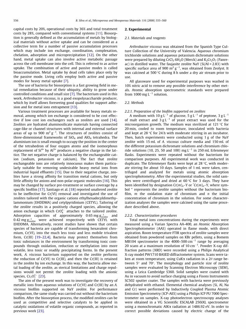
The removal of Cr(III) and Cr(VI) by NaY zeolite with and with-out the bacterium biofilm showed similar profiles for all range ofinitial concentrations. As an example, in Fig. 1 are shown the re-
C Cr 0 = 10 mg L-1
Cr (VI)
0.0
0.2
0.4
0.6
0.8
1.0
1.2
10 11t (d)
CC
r /CC
r 0
C Cr 0 = 50 mg L-1
Cr (VI)
0.0
0.2
0.4
0.6
0.8
1.0
1.2
10 11t (d)
CC
r /CC
r 0
C Cr 0 = 10 mg L-1
Cr (III)
0.0
0.2
0.4
0.6
0.8
1.0
1.2
t (d)
CC
r /CC
r 0
C Cr 0 = 50 mg L-1
Cr (III)
0.0
0.2
0.4
0.6
0.8
1.0
1.2
10 11
t (d)
CC
r /CC
r 0
0 1 2 3 4 5 6 7 8 9
10 110 1 2 3 4 5 6 7 8 9
0 1 2 3 4 5 6 7 8 9
0 1 2 3 4 5 6 7 8 9
Fig. 1. Ratio between residual and initial chromium concentration (CCr/CCr0) as afunction of contact time, for initial concentrations of Cr(VI) and Cr(III) of 10 mg L�1
and 50 mg L�1, in the presence of the zeolite with (N) and without the bacteria (s).
moval ratios of the metallic ions for initial concentrations of10 mg L�1 and 50 mg L�1.
The removal of chromium by the conjugated system (biofilm/zeolite) presented a typical and well known biosorption kinetics[13], which includes two phases: the first one, very fast andnot observable in Fig. 1, but determined by mass balance, is asso-ciated with the external cell surface, biosorption itself, and thesecond one is an intra-cellular accumulation/reaction, dependingon the cellular metabolism [26]. The results showed that Cr(III)was completely removed by both systems, due to the affinity be-tween the positive charge of the metal and the negative charge ofthe bacteria and to the easiness of entrapment in the frameworkzeolite by ion exchange. Contrarily, the zeolite was not able to re-move Cr(VI) which is related with its anionic form. The peculiaradsorptive properties of zeolites are due to the negative chargeof the framework Al atoms, which are located inside the three-dimensional pore structure of the solid. However, in the presenceof the bacterium biofilm, the removal efficiency was flagrantlyimproved by 19% for initial concentration of 10 mg L�1 and by35% for initial concentration of 50 mg L�1, as shown in Fig. 1.The role of the biofilm is the reduction of Cr(VI) to a smaller cat-ion, Cr(III), being this ion easily exchanged in the internal surfaceof the zeolite. The reduction can only occur on the outer surfaceof the zeolite as Cr2O2�
7 is not able to get inner the zeolite frame-work [23]. The lower removal ratios of Cr(VI) compared to Cr(III)seem to be connected with the lack of affinity between the globalcharge of the bacteria and the metallic ion, the charge repulsionswith the zeolite as well as with the sterical effects due to a high-er ionic radius. As it is well known, Y zeolite structure presents alarge central cavity or supercage with a diameter of 12.5 Å, shar-ing the supercages a 12-membered ring with an open diameter of7.4 Å [27]. In terms of ionic dimensions, trivalent chromium inthe form of CrðH2OÞ3þ6 has an ionic radius of 4.8 Å whereasCr2O2�
7 presents a dimension of 6.2 Å (calculations performedby using CAChe program, Oxford Molecular, 1997). In addition,there are probably limitations in the extension of the adhesionof A. viscosus to the support, as the characteristic dimension ofbacteria ranges from 1 to 10 lm. Although Y zeolite exhibitslarge specific surfaces areas, typically higher than 700 m2 g�1,most of this area is internal with an internal void volume above0.1 cm3 g�1 [27].
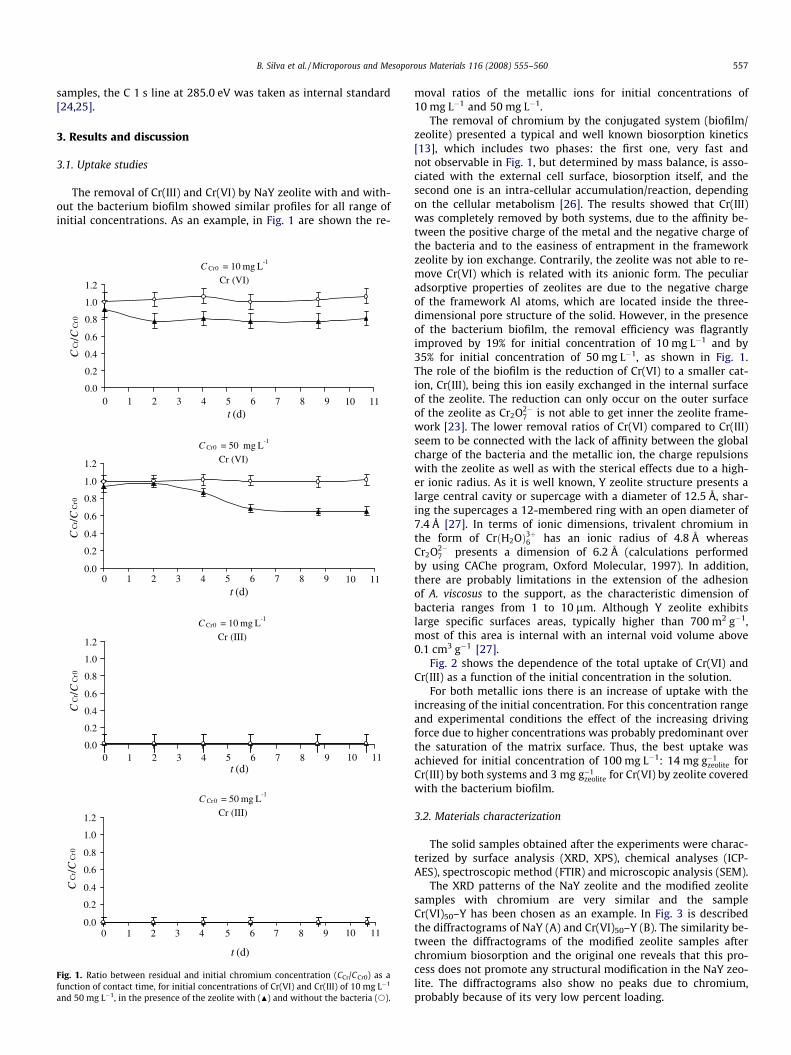
Fig. 2 shows the dependence of the total uptake of Cr(VI) andCr(III) as a function of the initial concentration in the solution.
For both metallic ions there is an increase of uptake with theincreasing of the initial concentration. For this concentration rangeand experimental conditions the effect of the increasing drivingforce due to higher concentrations was probably predominant overthe saturation of the matrix surface. Thus, the best uptake wasachieved for initial concentration of 100 mg L�1: 14 mg g�1
zeolite forCr(III) by both systems and 3 mg g�1
zeolite for Cr(VI) by zeolite coveredwith the bacterium biofilm.
3.2. Materials characterization
The solid samples obtained after the experiments were charac-terized by surface analysis (XRD, XPS), chemical analyses (ICP-AES), spectroscopic method (FTIR) and microscopic analysis (SEM).
The XRD patterns of the NaY zeolite and the modified zeolitesamples with chromium are very similar and the sampleCr(VI)50–Y has been chosen as an example. In Fig. 3 is describedthe diffractograms of NaY (A) and Cr(VI)50–Y (B). The similarity be-tween the diffractograms of the modified zeolite samples afterchromium biosorption and the original one reveals that this pro-cess does not promote any structural modification in the NaY zeo-lite. The diffractograms also show no peaks due to chromium,probably because of its very low percent loading.
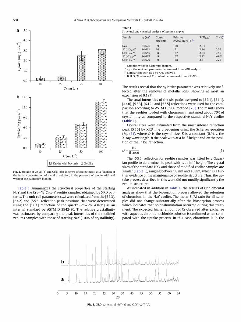
Table 1Structural and chemical analysis of zeolite samples
Sample a0 (Å)a Crystalsize (nm)
Relativecrystallinity (%)b
Si/Albulkc Cr (%)c
NaY 24.626 9 100 2.83 –*Cr(III)50–Y 24.661 10 71 2.84 0.55Cr(III)50–Y 24.656 8 67 2.84 0.52*Cr(VI)50–Y 24.667 9 67 2.82 <0.01Cr(VI)50–Y 24.670 9 68 2.81 0.25
* Samples without bacterium biofilm.a a0 is the unit cell parameter determined from XRD analysis.b Comparison with NaY by XRD analysis.c Bulk Si/Al ratio and Cr content determined from ICP-AES.
0.0
1.0
2.0
3.0
4.0
5.0
C (mg L-1)
Upt
ake
(mg
g ze
olite
-1)
0.0
3.0
6.0
9.0
12.0
15.0
C (mg L-1)
Upt
ake
(mg
g ze
olite
-1)
Zeolite with bacteria Zeolite
10 25 50 100
10 25 50 100
a
b
Fig. 2. Uptake of Cr(VI) (a) and Cr(III) (b), in terms of zeolite mass, as a function ofthe initial concentration of metal in solution, in the presence of zeolite with andwithout the bacterium biofilm.
558 B. Silva et al. / Microporous and Mesoporous Materials 116 (2008) 555–560
Table 1 summarizes the structural properties of the startingNaY and the Cr50–Y/*Cr50–Y zeolite samples, obtained by XRD pat-terns. The unit cell parameters (a0) were calculated from the [533],[642] and [555] reflection peak positions that were determinedusing the [101] reflection of the quartz (2h = 26.64187�) as aninternal standard by ASTM D 3942-80. The relative crystallinitywas estimated by comparing the peak intensities of the modifiedzeolites samples with those of starting NaY (100% of crystallinity).
0 5 10 15 20 25 30 3
b
a
2θ
Fig. 3. XRD patterns of NaY
The results reveal that the a0 lattice parameter was relatively unaf-fected after the removal of metallic ions, showing at most anexpansion of 0.18%.
The total intensities of the six peaks assigned to [331], [511],[440], [533], [642], and [555] reflections were used for the com-parison according to ASTM D3906 method [28]. The results showthat the zeolites loaded with chromium maintained about 70% ofcrystallinity as compared to the respective standard NaY zeolite(Table 1).
Crystal sizes were estimated from the most intense reflectionpeak [555] by XRD line broadening using the Scherrer equation(Eq. (1)), where D is the crystal size, K is a constant (0.9), k theX-ray wavelength, B the peak with at a half-height and 2h the posi-tion of the [hk l] reflection.
D ¼ KkB cos h
ð1Þ
The [555] reflection for zeolite samples was fitted by a Gauss-ian profile to determine the peak widths at half-height. The crystalsizes of the standard NaY and those of modified zeolite samples aresimilar (Table 1), ranging between 8 nm and 10 nm, which is a fur-ther evidence of the maintenance of zeolite structure. Thus, the up-take process described in this work did not modify significantly thezeolite structure.
As indicated in addition in Table 1, the results of Cr elementalanalysis show that the biosorption process allowed the retentionof chromium in the NaY zeolite. The molar Si/Al ratio for all sam-ples did not change substantially after the biosorption processwhich indicates that no dealumination occurred during this treat-ment. The expected higher amount of Cr observed after exchangewith aqueous chromium chloride solution is confirmed when com-pared with the uptake process. In this case, chromium is in the
5 40 45 50 55 60 65
(a) and Cr(VI)50–Y (b).
4000 3500 3000 2500 2000 1500 1000 500
b
d
c
a
Wavenumber (cm-1)
Fig. 5. FTIR spectra in the range (4000–500) cm�1 for: NaY (a), Cr(VI)50–Y (b),*Cr(VI)50–Y (c) and A. viscosus bacterium (d).
B. Silva et al. / Microporous and Mesoporous Materials 116 (2008) 555–560 559
Cr(III) form, a smaller cation and this ion is easily exchanged withinthe zeolite.
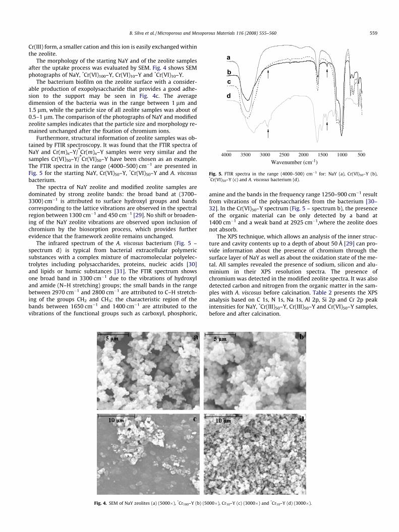
The morphology of the starting NaY and of the zeolite samplesafter the uptake process was evaluated by SEM. Fig. 4 shows SEMphotographs of NaY, *Cr(VI)100–Y, Cr(VI)10–Y and *Cr(VI)10–Y.
The bacterium biofilm on the zeolite surface with a consider-able production of exopolysaccharide that provides a good adhe-sion to the support may be seen in Fig. 4c. The averagedimension of the bacteria was in the range between 1 lm and1.5 lm, while the particle size of all zeolite samples was about of0.5–1 lm. The comparison of the photographs of NaY and modifiedzeolite samples indicates that the particle size and morphology re-mained unchanged after the fixation of chromium ions.
Furthermore, structural information of zeolite samples was ob-tained by FTIR spectroscopy. It was found that the FTIR spectra ofNaY and Cr(m)n–Y/*Cr(m)n–Y samples were very similar and thesamples Cr(VI)50–Y/*Cr(VI)50–Y have been chosen as an example.The FTIR spectra in the range (4000–500) cm�1 are presented inFig. 5 for the starting NaY, Cr(VI)50–Y, *Cr(VI)50–Y and A. viscosusbacterium.
The spectra of NaY zeolite and modified zeolite samples aredominated by strong zeolite bands: the broad band at (3700–3300) cm�1 is attributed to surface hydroxyl groups and bandscorresponding to the lattice vibrations are observed in the spectralregion between 1300 cm�1 and 450 cm�1 [29]. No shift or broaden-ing of the NaY zeolite vibrations are observed upon inclusion ofchromium by the biosorption process, which provides furtherevidence that the framework zeolite remains unchanged.
The infrared spectrum of the A. viscosus bacterium (Fig. 5 –spectrum d) is typical from bacterial extracellular polymericsubstances with a complex mixture of macromolecular polyelec-trolytes including polysaccharides, proteins, nucleic acids [30]and lipids or humic substances [31]. The FTIR spectrum showsone broad band in 3300 cm�1 due to the vibrations of hydroxyland amide (N–H stretching) groups; the small bands in the rangebetween 2970 cm�1 and 2800 cm�1 are attributed to C–H stretch-ing of the groups CH2 and CH3; the characteristic region of thebands between 1650 cm�1 and 1400 cm�1 are attributed to thevibrations of the functional groups such as carboxyl, phosphoric,
Fig. 4. SEM of NaY zeolites (a) (5000�), *Cr100–Y (b) (50
amine and the bands in the frequency range 1250–900 cm�1 resultfrom vibrations of the polysaccharides from the bacterium [30–32]. In the Cr(VI)50–Y spectrum (Fig. 5 – spectrum b), the presenceof the organic material can be only detected by a band at1400 cm�1 and a weak band at 2925 cm�1,where the zeolite doesnot absorb.
The XPS technique, which allows an analysis of the inner struc-ture and cavity contents up to a depth of about 50 Å [29] can pro-vide information about the presence of chromium through thesurface layer of NaY as well as about the oxidation state of the me-tal. All samples revealed the presence of sodium, silicon and alu-minium in their XPS resolution spectra. The presence ofchromium was detected in the modified zeolite spectra. It was alsodetected carbon and nitrogen from the organic matter in the sam-ples with A. viscosus before calcination. Table 2 presents the XPSanalysis based on C 1s, N 1s, Na 1s, Al 2p, Si 2p and Cr 2p peakintensities for NaY, *Cr(III)50–Y, Cr(III)50–Y and Cr(VI)50–Y samples,before and after calcination.
00�), Cr10–Y (c) (3000�) and *Cr10–Y (d) (3000�).
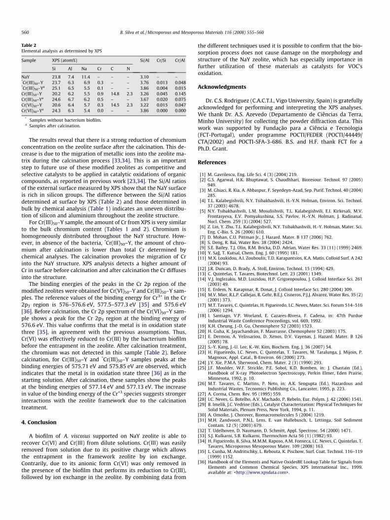
Table 2Elemental analysis as determined by XPS
Sample XPS (atom%) Si/Al Cr/Si Cr/Al
Si Al Na Cr C N
NaY 23.8 7.4 11.4 – – – 3.10 – –*Cr(III)50–Y 23.7 6.3 6.9 0.3 – – 3.76 0.013 0.048*Cr(III)50–Ya 25.1 6.5 5.5 0.1 – – 3.86 0.004 0.015Cr(III)50–Y 20.2 6.2 5.5 0.9 14.8 2.3 3.26 0.045 0.145Cr(III)50–Ya 24.6 6.7 6.2 0.5 – – 3.67 0.020 0.075Cr(VI)50–Y 20.6 6.4 5.7 0.3 14.5 2.3 3.22 0.015 0.047Cr(VI)50–Ya 24.3 6.3 5.4 0.0 – – 3.86 0.000 0.000
* Samples without bacterium biofilm.a Samples after calcination.
560 B. Silva et al. / Microporous and Mesoporous Materials 116 (2008) 555–560
The results reveal that there is a strong reduction of chromiumconcentration on the zeolite surface after the calcination. This de-crease is due to the migration of metallic ions into the zeolite ma-trix during the calcination process [33,34]. This is an importantstep to future use of these modified zeolites as competitive andselective catalysts to be applied in catalytic oxidations of organiccompounds, as reported in previous work [23,34]. The Si/Al ratiosof the external surface measured by XPS show that the NaY surfaceis rich in silicon groups. The difference between the Si/Al ratiosdetermined at surface by XPS (Table 2) and those determined inbulk by chemical analysis (Table 1) indicates an uneven distribu-tion of silicon and aluminium throughout the zeolite structure.
For Cr(III)50–Y sample, the amount of Cr from XPS is very similarto the bulk chromium content (Tables 1 and 2). Chromium ishomogeneously distributed throughout the NaY structure. How-ever, in absence of the bacteria, *Cr(III)50–Y, the amount of chro-mium after calcination is lower than total Cr determined bychemical analyses. The calcination provokes the migration of Crinto the NaY structure. XPS analysis detects a higher amount ofCr in surface before calcination and after calcination the Cr diffusesinto the structure.
The binding energies of the peaks in the Cr 2p region of themodified zeolites were obtained for Cr(VI)50–Y and Cr(III)50–Y sam-ples. The reference values of the binding energy for Cr3+ in the Cr2p3 region is 576–576.6 eV, 577.5–577.3 eV [35] and 575.6 eV[36]. Before calcination, the Cr 2p spectrum of the Cr(VI)50–Y sam-ple shows a peak for the Cr 2p3 region at the binding energy of576.6 eV. This value confirms that the metal is in oxidation statethree [35], in agreement with the previous assumptions. Thus,Cr(VI) was effectively reduced to Cr(III) by the bacterium biofilmbefore the entrapment in the zeolite. After calcination treatment,the chromium was not detected in this sample (Table 2). Beforecalcination, for Cr(III)50–Y and *Cr(III)50–Y samples peaks at thebinding energies of 575.71 eV and 575.85 eV are observed, whichindicates that the metal is in oxidation state three [36] as in thestarting solution. After calcination, these samples show the peaksat the binding energies of 577.14 eV and 577.13 eV. The increasein value of the binding energy of the Cr+3 species suggests strongerinteractions with the zeolite framework due to the calcinationtreatment.
4. Conclusion
A biofilm of A. viscosus supported on NaY zeolite is able torecover Cr(VI) and Cr(III) from dilute solutions. Cr(III) was easilyremoved from solution due to its positive charge which allowsthe entrapment in the framework zeolite by ion exchange.Contrarily, due to its anionic form Cr(VI) was only removed inthe presence of the biofilm that performs its reduction to Cr(III),followed by ion exchange in the zeolite. By combining data from
the different techniques used it is possible to confirm that the bio-sorption process does not cause damage on the morphology andstructure of the NaY zeolite, which has especially importance infurther utilization of these materials as catalysts for VOC’soxidation.
Acknowledgments
Dr. C.S. Rodriguez (C.A.C.T.I., Vigo University, Spain) is gratefullyacknowledged for performing and interpreting the XPS analyses.We thank Dr. A.S. Azevedo (Departamento de Ciências da Terra,Minho University) for collecting the powder diffraction data. Thiswork was supported by Fundação para a Ciência e Tecnologia(FCT-Portugal), under programme POCTI/FEDER (POCTI/44449/CTA/2002) and POCTI-SFA-3-686. B.S. and H.F. thank FCT for aPh.D. Grant.
References
[1] M. Gavrilescu, Eng. Life Sci. 4 (3) (2004) 219.[2] G.S. Agarwal, H.K. Bhuptawat, S. Chaudhhari, Bioresour. Technol. 97 (2005)
949.[3] M. Ghiaci, R. Kia, A. Abbaspur, F. Seyedeyn-Azad, Sep. Purif. Technol. 40 (2004)
285.[4] T.L. Kalabegishvili, N.Y. Tsibakhashvili, H.-Y.N. Holman, Environ. Sci. Technol.
37 (2003) 4678.[5] N.Y. Tsibakhashvili, L.M. Mosulishvili, T.L. Kalabegishvili, E.I. Kirkesali, M.V.
Frontasyeva, E.V. Pomyakushina, S.S. Pavlov, H.-Y.N. Holman, J. Radioanal.Nucl. Chem. 259 (3) (2004) 527.
[6] Z. Lin, Y. Zhu, T.L. Kalabegishvili, N.Y. Tsibakhashvili, H.-Y. Holman, Mater. Sci.Eng. C-Bio. S. 26 (2006) 610.
[7] D. Mohan, C.U. Pittman Jr., J. Hazard. Mater. B 137 (2006) 762.[8] S. Deng, R. Bai, Water Res. 38 (2004) 2424.[9] S.E. Bailey, T.J. Olin, R.M. Bricka, D.D. Adrian, Water Res. 33 (11) (1999) 2469.
[10] Y. Sag, T. Kutsal, Chem. Eng. J. 60 (1995) 181.[11] M.X. Loukidou, A.I. Zouboulis, T.D. Karapantsios, K.A. Matis, Colloid Surf. A 242
(2004) 93.[12] J.R. Duncan, D. Brady, A. Stoll, Environ. Technol. 15 (1994) 429.[13] C. Quintelas, T. Tavares, Biotechnol. Lett. 23 (2001) 1349.[14] V.J. Inglezakis, M.D. Loizidou, H.P. Grigoropoulou, J. Colloid Interface Sci. 261
(2003) 49.[15] E. Erdem, N. Karapinar, R. Donat, J. Colloid Interface Sci. 280 (2004) 309.[16] M.V. Mier, R.L.P. Callejas, R. Gehr, B.E.J. Cisneros, P.J.J. Alvarez, Water Res. 35 (2)
(2001) 373.[17] M.T. Tavares, C. Quintelas, H. Figueiredo, I.C. Neves, Mater. Sci. Forum 514–516
(2006) 1294.[18] I. Santiago, V.P. Worland, E. Cazares-Rivera, F. Cadena, in: 47th Purdue
Industrial Waste Conference Proceedings, vol. 669, 1992.[19] K.H. Cheung, J.-D. Gu, Chemosphere 52 (2003) 1523.[20] H. Guha, K. Jayachandran, F. Maurrasse, Chemosphere 52 (2003) 175.[21] E. Dermou, A. Velissariou, D. Xenos, D.V. Vayenas, J. Hazard. Mater. B 126
(2005) 78.[22] S.-Y. Kang, J.-U. Lee, K.-W. Kim, Biochem. Eng. J. 36 (2007) 54.[23] H. Figueiredo, I.C. Neves, C. Quintelas, T. Tavares, M. Taralunga, J. Mijoin, P.
Magnoux, Appl. Catal., B-Environ. 66 (2006) 273.[24] J.Y. Xie, P.M.A. Sherwood, Chem. Mater. 2 (3) (1990) 293.[25] J.F. Moulder, W.F. Strickle, P.E. Sobol, K.D. Bomben, in: J. Chastain (Ed.),
Handbook of X-ray Photoelectron Spectroscopy, Perkin Elmer, Eden Prairie,Minnesota, 1992, p. 10.
[26] M.T. Tavares, C. Martins, P. Neto, in: A.K. Sengupta (Ed.), Hazardous andIndustrial Wastes, Tecnomics Publishing Co., Lancaster, 1995, p. 223.
[27] A. Corma, Chem. Rev. 95 (1995) 559.[28] I.C. Neves, G. Botelho, A.V. Machado, P. Rebelo, Eur. Polym. J. 42 (2006) 1541.[29] B. Imelik, J.C. Vedrine (Eds.), Catalyst Characterization: Physical Techniques for
Solid Materials, Plenum Press, New York, 1994, p. 11.[30] A. Omoike, J. Chorover, Biomacromolecules 5 (2004) 1219.[31] M.H. Zandvoort, P.N.L. Lens, E. van Hullebusch, L. Lettinga, Soil Sediment
Contam. 12 (5) (2003) 679.[32] T. Udelhoven, D. Naumann, D. Schmitt, Appl. Spectrosc. 54 (2000) 1471.[33] S.J. Kulkarni, S.B. Kulkarni, Thermochim Acta 56 (1) (1982) 93.[34] H. Figueiredo, B. Silva, M.M.M. Raposo, A.M. Fonseca, I.C. Neves, C. Quintelas, T.
Tavares, Microporous Mesoporous Mater. 109 (2008) 163.[35] L. Cunha, M. Andritschky, L. Rebouta, K. Pischow, Surf. Coat. Technol. 116–119
(1999) 1152.[36] Handbook of the Elements and Native OxidesBE Lookup Table for Signals from
Elements and Common Chemical Species, XPS International Inc., 1999.available at: <http://www.xpsdata.com>.