properties of boron-substituted zsm-5 and zsm-11 zeolites
TRANSCRIPT
JOURNAL OF CATALYSIS 108, 1-14 (1987)
Properties of Boron-Substituted ZSM-5 and ZSM-11 Zeolites
G. COUDURIER,* A. AUROUX,* J. C. VEDRINE,*" R. D. FARLEE,? L. ABRAMS,? AND R. D. SHANNON?
*Institut de Recherches sar la Catalyse (CNRS). 2 ALvenue Alberr Einstein, 69626 Villearbanne Cede*, France; and Kentral Research and Development Department, Experimental Station, E. I. da Pont de
Nemoars & Company. Wilmington, Delaware 19898’
Received August 5, 1986; revised March 3 I. 1987
Using NMR and IR spectroscopy boron-substituted pentasil zeolites are found to contain four-coordinated B in the hydrated state and three-coordinated B in the dehydrated state. In addition a new tetrahedral B site assigned to (H0j2B(H20)(0Si) was characterized by its NMR
quadrupole coupling constant and asymmetry parameter. Ammonia adsorption microcalorimetry gave heats of adsorption of -65 kJ/mol for H-B-ZSM-I I and showed that B-substituted pentasils have only very weak acidity. Calcination at 800°C increased the heat of NH3 adsorption to - 170
kJ/mol by creation of strong Lewis acid sites. The lack of strong Bronsted acid sites in H-B-ZSM-II was confirmed by poor catalytic activity in methanol conversion and in toluene
alkylation with methanol. C lY87 Academic Prerr. Inc.
INTRODUCTION
The zeolites ZSM-5 and ZSM-I 1 belong to the high silica “pentasil” family with a network of channels having approximate diameters of 0.51-0.55 nm. The channels are straight in ZSM-1 I and straight and sinusoidal in ZSM-5 (1, 2). Strong acid sites in the interior of the channels of ZSM-5 and ZSM-11 allow these zeolites to exhibit cata- lytic shape selectivity (3) in the conversion of methanol or ethanol to hydrocarbons (4) or in the alkylation of toluene by methanol (5, 6).
The substitution of Al by B during the synthesis of pentasil zeolites has been dem- onstrated by a variety of investigators (7- 16). This substitution might be expected to modify the acidic properties of the zeolite and thereby the catalytic properties (7, 9, 10, 17). In this paper, in order to evaluate the differences between B and Al pentasil- type zeolites we have compared sorption capacities, IR spectra, acidities, and metha- nol alkylation behavior of H-B,AI-ZSMJ, and H-B-ZSM-I1 with the corresponding
’ To whom correspondence should be addressed.
L Contribution #4366.
Al forms of ZSM-5 and -11. Character- ization was accomplished by chemical anal- ysis, infrared spectroscopy, electron mi- croscopy, X-ray diffraction, sorption, NH3 microcalorimetry, and “B MAS NMR spectroscopy.
EXPERIMENTAL Starting materials for ZSM-5 were Ludox
LS-30, sodium aluminate, NaOH, and tet- rapropylammonium (TPA) bromide. Prepa- ration of ZSM-5 was made following the method described by Rollman and Valyoc- sik (18). Typical samples were prepared by combining solutions containing Ludox and TPA Br and adding to this a solution of NaOH and Na aluminate. This was placed in a Hastelloy or titanium autoclave and heated at 160°C for 24 h.
Starting materials for ZSM-11 were NaOH, sodium aluminate, Ludox LS-30, and tetrabutylphosphonium chloride or tet- rabutylammonium iodide as the template. Procedures described by Chu (19) were used to prepare ZSM- 1 I.
B-ZSM-I I was prepared according to example 20 of Ref. (20) using tetrabutylam- monium hydroxide, boric acid, tetraethyl-
I 002 I-95 17/87 $3 .OO
Copyright IS’ 1487 hy Academic Press. Inc
All right\ of reproduction in any form rcerved.
2 COUDURIER ET AL.
orthosilicate (TEOS), and KOH. B,Al- ZSM-5 was prepared according to a modi- fication of example I5 of Ref. (20) using 37.5 g HJBOj, 133.1 g TPA Br (50% solu- tion), 124.8 g TEOS, and 18.2 cm3 NH40H. The sample was heated at 165°C for 5 days. After reaction, the products were filtered, washed with water, and heated to 500°C in air to decompose the alkylammonium ion. The H+ forms were prepared by exchange with 10% NH4N03 four times at 80°C and deammoniation at 540°C in flowing N?. This procedure resulted in a lo- to 20-fold reduc- tion in the Na content, giving typically 200 ppm to 0.2 wt% Na.
X-ray diffraction patterns were obtained on samples equilibrated over saturated NH&I solutions using a Guinier-Hagg fo- cusing camera (r = 40 mm). The radiation was monochromatic CuKal (A = 0.154051 nm). KC1 (a = 0.62931 nm) was used as an internal standard. Line positions on the film were determined to ?5 ,um with a David Mann film reader. Cell dimensions were obtained by least-squares refinement.
Analysis of Na and Al content was made using atomic absorption, whereas B analy- sis was performed using the inductive coupled plasma technique. Hz0 content was determined from weight loss at 1000°C and SiO? was determined by difference.
Adsorption was measured gravimetri- tally using an apparatus similar to that described by Landolt (21). After the samples were weighed, they were mounted onto the sorption manifold and exposed to vapor at 0.1-0.5 of the vapor pressure of the sorbate at the temperature of the samples. The amount sorbed at 20 h was used for comparison purposes. The preci- sion of this technique is 20.5 mg/g of sample. Ail sorbates were vacuum distilled, subjected to several freeze-pump-thaw cy- cles, and stored over a dry zeolite 4A sieve in a bulb with a vacuum stopcock.
A sample of Cab-0-Sil fumed silica, grade MS-7, with an external surface area of 200 m2/g was used as a reference. Under the sorption conditions, the amount of sor-
bate pickup by the Cab-0-Sil was used to determine k, the surface coverage of sor- bate expressed as a sorbate per square nanometer.
IR spectra were recorded at 25°C with a Perkin-Elmer 580 spectrophotometer with a resolution of 2.5 to 3.7 cm-‘. For lattice vibrational mode studies. KBr pellets were used with a 0.25% dilution. For studies of the hydroxyl groups and of NH, ad- sorption, the samples were compressed into 2 to 5 mg . cm-’ self-supported wafers using 2 x IO6 Pa pressure. The samples were progressively evacuated up to 400 or 800°C and then small increments of NH3 (0.15 NHJu.c.) were adsorbed up to satu- ration. Desorption of NH3 was performed by progressively heating in stages of 2 h at increasing temperatures.
Both toluene alkylation with methanol at 400°C and methanol conversion reactions at 370°C were studied using a flow microreac- tor on line with two gas chromatographs. The samples (-50 mg) were activated un- der N2 at atmospheric pressure at 400°C for 14 h. In the case of toluene alkylation, a mixed feed with N? as a gas carrier and with a molar methanol : toluene ratio of ca. 1 : 4 and WHSV equal to 5 h-’ was admitted to the catalyst. For the methanol conversion reaction, the space velocity was WHSV = 10 h-’ and the reaction temperature was 370°C.
“B spectra we re obtained at 96.3 MHz (7-T magnetic field) using a Bruker CXP- 300 spectrometer. Spectra were obtained using 30” (1.5~ps) pulses and a 0.5-s recycle delay, without proton decoupling. The small pulse angle is necessary if quantita- tive spectra are to be obtained for sites with different quadrupole coupling constants. Typically, 1000 scans were averaged. Spec- tra were recorded with magic-angle spin- ning (MAS) at ca. 4.5 kHz and of stationary samples to fully separate and identify the boron sites. Chemical shifts are referenced to external BF1 * Et,0 using 1 M H3B03 in Hz0 as a secondary standard (+ 19.2 ppm).
Relative areas of the observed boron
BORON-SUBSTITUTED ZSM-5 AND ZSM-1 I ZEOLITES 3
TABLE I
Chemical Compositions of B and Al Forms of ZSM-5 and ZSM-I I
SUIlple Compositmn”lu.c. Siiilf
H-B.AI-ZSM-5 HI ZBI 8ti.4%3.8ot9?27~ W0 42.6 H-AI-ZSM-S Ht.?Naa.,Alz.oSiw.oOtsr 47.0 H-B-ZSM-I I HUBI 3% 70~125, 6H10 72.x H-AI-ZSM-I I Hz.aNw &2.&z 1019222, 4H20 32.1 H-AI-ZSM-II HI IN% IAII &s70wl8, 4H:O 72.X
” Si + Al was taken to be %: proton content was calculatrd as the ddference between the B + Al and the Na contents.
resonances were obtained using the cut- and-weigh method, or by nonlinear least- squares fitting to the theoretical powder pattern lineshapes. Although the quadru- pole coupling constant of the tetrahedral sites is small (co.5 MHz), the slight asym- metry of the lineshape and narrowing by a factor of 4 produced by MAS suggest that it is still the dominant source of line broaden- ing. Therefore, the central transition ob- served with MAS should contain negligible intensity from the satellite transitions. and integrated areas of the resonances are taken to be directly proportional to their relative concentrations.
Heats of adsorption of ammonia were measured in a Setaram Tian-Calvet high- temperature microcalorimeter maintained at 148°C. The volumetric system previously described (22) was modified to allow con- tinuous pressure measurement using a Datametrics Barocel gauge.
Samples approximately 100 mg in weight were evacuated at a temperature of 400 or
800°C for 16 h. Adsorption experiments were carried out using NH3 dried and de- oxygenated over Na spaghetti and further purified by freeze-pump-thaw cycles over dehydrated zeolite Y.
RESULTS AND DISCUSSION
A. Chemical Analysis, Electron Microscopy, X-Ray Diffraction, and Sorption
Tables 1 and 2 show the chemical compo- sitions, crystallite sizes, and sorption char- acteristics of the B- and Al-ZSM-5 sam- ples, and their ZSM-11 counterparts.
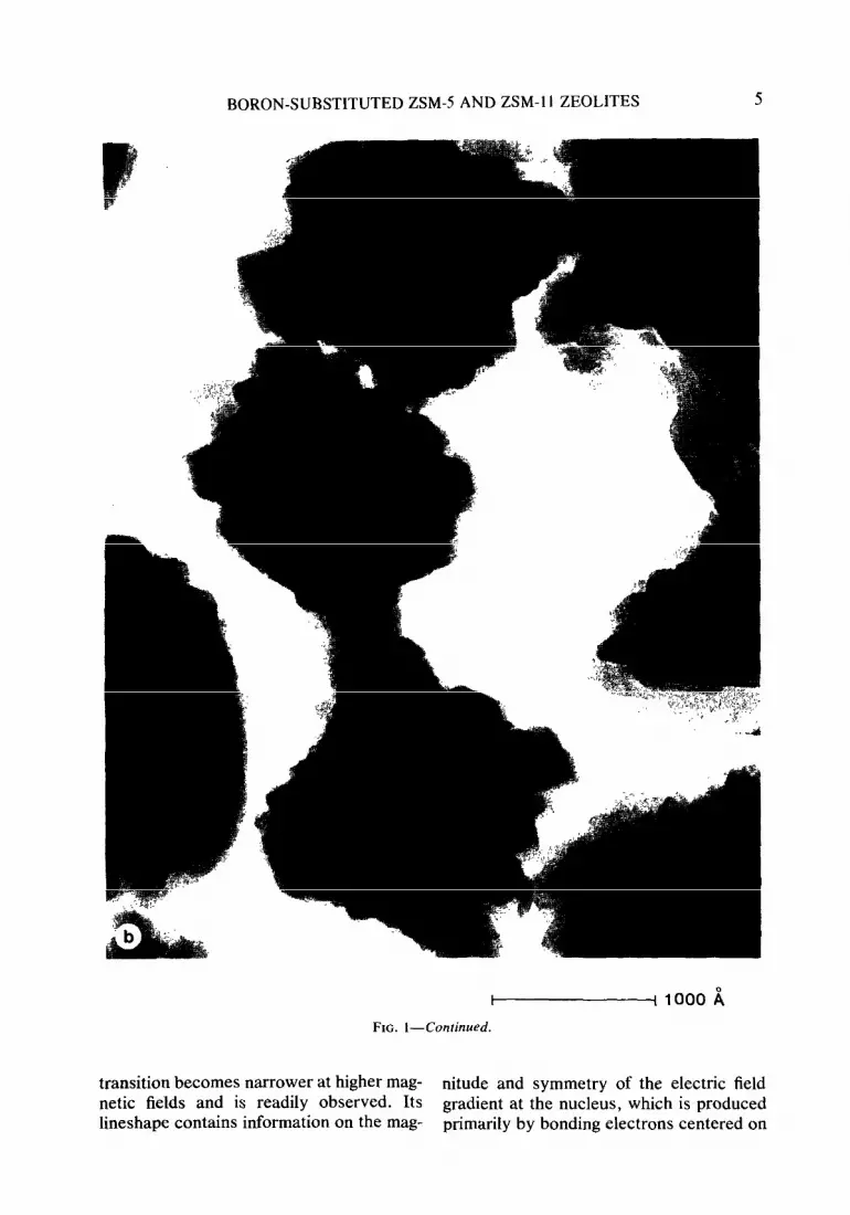
Transmission and scanning electron mi- crographs of the two boralite samples showed the crystals to have different morphologies. The B.Al-ZSM-5 crystal- lites took the form of flat platelets of the Z- 1 type bounded by (100) and (0 10) planes as already described (23-27). The B- ZSM-11 crystallites had the appearance of bundles of 90” intergrown plates (see Fig. 1).
The X-ray diffraction patterns of the ZSM-11 sample contained lines at 0.381 and 0.365 nm and the l-00-nm line appeared to be split. Neither the 0.381- and 0.361-nm lines nor the splitting can be accounted for by the tetragonal cell reported by Kokotailo et al. (2).
Reduction of unit cell dimensions be- cause of the smaller size of B’+ (0.011 nm) relative to A13+ (0.039 nm) (28) is one of the strongest arguments for the belief that the B is located substitutionally in the lattice rather than in the channels. The cell dimen-
TABLE 2
Physical Properties of B and Al Forms of ZSM-S and ZSM-I I
S2Nllpk Crystallite Cell dimensions (A) \’ c A’, Percentage sorbed in 10 h size (pm)
a b c n-hex 3MeP Ccl, 2.2DMB
H-B,AI-ZSM-5 0.2-1.5 19.884 (3 19.780 f.5) 13.300 (3) 513 I I I.5 8.2 12.4 I .4
H-AI-ZSM-5 2-4 20.135 (4) 19.947 (8) 13.458 15) 5405 13.1 10.0 13.5 1.7
H-B-ZSM-I I 0.2-0.3 19.906 (6) 13.3.59 (3) 5293 11.9 10.0 16.3 3.9
H-AI-ZSM-I I 0.3-I 20.03 (2)” 13.44 (2J” 5392 13.2 IO.5 16. I 6. I H-AI-ZSM- I I 3-6 20.08 f I) 13.46 (II 5427
o Approximate values only because of reflections which violate tetragonal symmetry.

4 COUDURIER ET AL.
sions of B-substituted samples are signifi- cantly smaller than those of the Al-substi- tuted samples. This is in agreement with the results of other investigators. Although our unit cell dimensions were determined in a manner similar to that of Meyers ef al. (29), they agree only approximately on an abso- lute or relative scale. Our Al-ZSM-5 with 2.0 Al1u.c. has a unit cell volume of 5.405 nm3 compared to an interpolated value of 5.37 nm3 found by Meyers er al. (29). Similarly a B content of 1.3/u.c. was found to reduce our unit cell volume of ZSM-I 1 by 2.5% whereas 1.7 B/u.c. reduced the unit cell volume of ZSM-5 of Meyers ef al. by 0.7%.
In the last columns of Table 2 sorption capacities of the boralites are compared with those of ZSM-5 and ZSM-11. Al- though H-Al-ZSM-1 I and H-AI-ZSM5 sorb similar quantities of smaller molecules like n-hexane and 3-methylpentane, H-AI- ZSM- 11 adsorbs significantly greater quan- tities of the larger molecules such as cy- clohexane, o-xylene, and 2,2-dimethyl- butane. Note that H-B,Al-ZSM-5 and H-B-ZSM-11 sorbed only slightly less of
the smaller molecules than the Al forms. This shows that differences in void volume due to boron incorporation in the lattice are very small; this agrees with the slight changes noted in unit cell volumes. In con- trast, differences between ZSM-5 and ZSM-11 are more significant; this is per- haps due to larger channel intersections in ZSM-I 1.
B. NMR Data
NMR spectra of quadrupolar nuclei such as “B (spin 8) consist of the superposition of central and satellite transitions. The sat- ellite transitions (e.g., +i to -+%) are shifted to first order in the quadrupole interaction. In powders, this results in a powder pattern lineshape (30) whose width is of the order of the quadrupole coupling constant (ca. 2.5 MHz for trigonal boron sites). The large width of these transitions makes them dif- ficult to observe or to quantify. However, the central transition (4 to -4) is shifted only to second order in the quadrupole interaction and has a width of the order of the quadrupole coupling constant squared divided by the Larmor frequency. This
FIG. 1. Scanning electron (a) and transmission electron micrographs (b) of B-ZSM- 11.
BORON-SUBSTITUTED ZSM-5 AND ZSM-1 I ZEOLITES 5
tF 1000 ii
FIG. I-Continued.
transition becomes narrower at higher mag- nitude and symmetry of the electric field netic fields and is readily observed. Its gradient at the nucleus, which is produced lineshape contains information on the mag- primarily by bonding electrons centered on

6 COUDURIER ET AL.
the nucleus observed. The lineshape is thus a very sensitive probe of the local structure and electronic environment.
Magic-angle spinning (MAS) removes broadening arising from dipolar couplings, first-order quadrupole interactions (3Z), and chemical-shift anisotropy. The second- order quadrupole interaction is modified, but not removed, by spinning (32). Vega (33) and Kundla et al. (34) have indepen- dently derived the second-order quadru- pole lineshape of the central transition pro- duced by MAS, and it has been observed experimentally for a number of nuclei (35). Although. MAS narrows the quadrupole splitting of the MAS lineshape by a factor of ca. 4, the singularities are typically easier to observe accurately because of the im- proved resolution. Variable-angle spinning (35, 36) sacrifices resolution and offers no discernible advantages when the number of inequivalent sites to be resolved from one another is not large.
Bray and co-workers have used nonspin- ning “B NMR to study the structure of BzOj (37) and of various borate glasses (38, 39). They distinguish trigonal and tetrahe- dral boron by a large difference in their quadrupole coupling constants, observed to be 2.6 to 2.9 MHz and <0.5 MHz, respec- tively. Further, they are able to identify neutral B03 sites (with three bridging oxy- gens) from negatively charged B03 sites (two bridging, one nonbridging oxygen) by their asymmetry parameters, ca. 0.1 and 0.6, respectively. Because of the low fre- quency at which Bray worked (8- 16 MHz), chemical-shift differences were not ob- served.
“B spectra of several borosilicate min- erals have also been obtained with MAS (40).
Scholle and Veeman (16) have reported “B spectra of one sample of H-boralite (Si/B = 75) in which trigonal boron was produced upon dehydration and could be completely and reversibly converted back to tetrahedral boron by rehydration. This was ascribed to the breaking of an Si-O-B bridge to form a planar, trigonal B03 site
adjacent to an acidic SiOH in the frame work.
Several other workers have taken the observation of tetrahedral “B NMR signal as evidence for the incorporation of boron into the ZSM5 framework (15, 41, 42).
In our samples of B-ZSM-11 and B,AI-ZSMJ with various degrees of hy- dration, three different boron sites are ob- served by “B NMR. Because of their differences in quadrupole coupling con- stants, these sites are more readily sepa- rated in nonspinning than in MAS spectra. Their NMR parameters are summarized in Table 3.
In fully or partially hydrated samples, boron is predominantly in site 1, assigned as tetrahedral B(OSik. This is readily seen as a narrow, symmetrical line with no resolvable second-order quadrupole struc- ture.
Site 2 is also observed in fully and par- tially hydrated samples. The areas of the resonances associated with the various sites are listed in Table 4. The assignment of site 2 is discussed below.
In dehydrated samples, essentially all of the boron is converted to trigonal site 3 as observed by Scholle and Veeman (16). The central transition has a width of 3.2 kHz, so fast MAS (5 kHz) can separate and mini- mize the intensity of the sidebands. As reported in Table 4, a very small amount of tetrahedral site 1 remains, even in fully dehydrated samples.
While sites 1 and 3 have been observed in a variety of materials as discussed above, site 2 has not been previously reported (see Fig. 2). The quadrupole coupling constant of site 2 is very different from that reported
TABLE 3
“B NMR Parameters of the B Sites
Site Isotropic QUad~pole Quadrupole chemical shift coupling asymmetry
@pm) constant (MHz) parameter
I -4 <0.5 2 c22 -1.9 1.0 3 +25 +2.4 0.0

BORON-SUBSTITUTED ZSMJ AND ZSM-11 ZEOLITES 7
TABLE 4
Areas of the “B Resonances
Sample Site I Site 2 Site 3
Hydrated” H-B-ZSM-I 1 0.72 0.28 0.00 H-B.AI-ZSM-5 0.69 0.31 0.00
Partially hydratedb H-B-ZSM-I I 0.65 H-B-ZSM-I 1, 800°C 0.47 H-B,AI-ZSM-5 0.44
Dehydrated’
0.35 0.00 0.53 0.00 0.56 0.00
H-B-ZSM-I 1 0.03 0.00 0.97 H-B,AI-ZSM-I I 0.02 0.00 0.98
” Exposed to saturated NH,CI solution, 25”C, 24 h. h Exposed to atmospheric air, 25”C, >I00 h. c 28o”C, vacuum, 40 h.
in the literature for any trigonal site, so site 2 must be four-coordinate. The asymmetry parameter, 1 .O, indicates substantial bond- angle distortion (local CZ”, not C3”, symme- try about boron).
There are two sites in known structures which have quadrupole asymmetry param- eters of 1.0 and coupling constants com- parable in magnitude to that of site 2. The distorted tetrahedral B(OB)j(OH) site in Na2B407 * lOHzO (borax) has a quadrupole coupling constant of + 1.63 MHz (43). Jelli- son and Bray (44) observed a site in sodium borate glasses with a quadrupole coupling constant of 0.72 MHz and assigned it to a distorted tetrahedral site bonded to both B03 and B04 groups, probably in a diborate unit.
This leads to the conclusion that site 2 is a highly distorted four-coordinate site, probably a framework defect such as HOB(OSi)3, (H0)2B(OSi)2, or (H0)2B (H20)(OSi). As it is seen at comparable levels in samples with and without Al, it is unrelated to the presence of Al in the framework. The high Ievels of site 2 ob- served in the partially hydrated samples rule out the possibility of site 2 being asso- ciated with an amorphous or other second phase. The amount of site 2 is increased after calcination at 800°C and rehydration, consistent with its assignment as a frame-
work defect. However, the NMR data alone do not allow a firm conclusion about the detailed structure of this site.
C. Microcalorimetry
Figure 3 shows plots of Q vs the quantity of NH, adsorbed (0) for H-AI-ZSM- 11 and H-B-ZSM-11. The heats of adsorption at low NH3 coverage for H-ZSM-11 are higher than those published earlier by Auroux et al. (4.5). Strong acid sites adsorb the first doses of NH3 up to 1.8 NH,/u.c. which is 65% of the total OH concentration found from chemical analysis. Heating the sample to 800°C under vacuum to remove OH increases the strength of the strongest sites from 150-160 to 175-185 kJ molK’ but in contrast to other samples of ZSM5 (46) does not seem to reduce the total number of acid sites.
It has been observed that when the NH3 increments introduced in the samples are very small and/or when adsorption time is long enough, no maximum is obtained in the Q-0 plot. This clearly shows that diffu- sion of NH3 molecules is limited within the channels due to chemical interaction with acid sites.
-10 -5 0 5 kHz
10 15
FIG. 2. “B NMR spectrum of partially hydrated H-B,AI-ZSM-5 obtained without magic-angle spin- ning (stationary sample): (a) experimental spectrum (11,500 scans using conditions specified in the text): (b) deconvolution showing the separate resonances arising from site 1 (I.1 kHz FWHH Gaussian), site 2 (quadrupole powder pattern corresponding to the pa- rameters listed in Table 3, and with 1.25 kHz FWHH Gaussian broadening), and their sum. All parameters are determined by nonlinear least-squares fitting to the experimental spectrum.

8 COUDURIER ET AL.
H-AI-EM-11
Si/AI = 32.1
OH cp. = 2.8/w 150 OH Mns= l.B/uc
H-B-ZSM-11
Si/B = 72.8
%A = 1.3hc
OHNH3= 0.2hc
Tact 0 4OO'C
Tact 6 400%
moles NH3/uc moles NH,/uc 1 2 3 1 2 0 ' . ,'a ' . %', . ' ,I8 . ,
5 IO 0 5 IO
V cm3 NH3
OdS’ g
i
FIG. 3. Differential heats of ammonia adsorption on H-AI-ZSM-I 1 at 148°C.
H-B-ZSM-1 I has only very weak acid sites (-65 kJ/mol) and only very few of these, viz. 0.2 NH3/u.c. compared to 1.3 OH/u.c. by chemical analysis. The heats of adsorption rapidly diminish to 25 kJ mol-’ then increase to about 60 kJ mol-’ before decreasing again. This maximum in the Q vs 8 plot seems to be related to the presence of boron. Such an additional maximum might be attributed to the formation of a NH4(NH&complex formed by reacting B-OH-NH3 with NH3 ligands, similar to the gas phase M(NH3)n+ clusters found by Castleman et al. (48).
Dehydroxylating H-B-ZSM-11 at 800°C dramatically increases the strength of the acid sites to 170 kJ mol-’ and sharpens the maximum at 0.8 NH3/u.c. We propose that a few strong Lewis acid sites are formed upon dehydroxylation. This is consistent with the relative Lewis acid strengths of B and Al found by Zhang (47).
In Fig. 4 H-Al-ZSM-5 is seen to be- have somewhat differently from other samples of H-ZSM-5 previously investi- gated. Instead of showing a maximum in
the Q-0 plot, there is a continuous and sharp drop in acid strengths from 178 to 110 kJ mol-’ as NH3 is added from 0 = 0 to 1.2 NH&.c. As in H-ZSM-11 there is a pla- teau at -70-80 kJ mol-‘. Strong acid sites are available up to 1.2 NH3/u.c. compared to 1.6 OH/u.c. by chemical analysis.
H-B,Al-ZSM-5 behaves similarly to a H-Al-ZSM-5 sample with a low Al con- centration. The effect of B in this sample is masked by the Al but a maximum in the Q vs 0 plot was observed as for the H-B- ZSM-11 sample.
D. IR Data
a. Mid-infkm-ed spectra. The mid-infra- red spectra of all samples (Fig. 5) are char- acteristic of the pentasil family (49,50) with absorption bands near 1230, 1100,800,560, and 455 cm-’ and optical density ratios of the 560- to 455-cm-’ bands equal to ca. 0.6 as usually observed for pentasil zeolites (49). The frequency of the llOO-cm-l band decreased from 1102 cm-’ for H-B-ZSM- 11 to 1098 cm-’ for H-Al-ZSM-11 with the SiIM ratio decreasing from 72.8 to 15.6.

BORON-SUBSTITUTED ZSMJ AND ZSM- I 1 ZEOLITES 9
0 kJ
izi
151
H-Al-ZSM-5
t
H-B, AI-ZSM-5 H-B, AI-ZSM-5
Si/(AL+B)= 42.6 Si/(AL+B)= 42.6 . .
: : ! : OH,, = 1.1/w OH,, = 1.1/w
: I OHNHa= 0.5/uc OH,+= 0.5/uc
Tact
moles NH,/uc 1 2 1
FIG. 4. Differential heats of ammonia adsorption on H-Al-ZSM-5 and H-B,AI-ZSM-5 at 148°C.
Similar shifts noted for other zeolites have been related to an increase in framework Al content (51).
Differences between B and Al pentasil zeolite spectra are best observed for self- supported wafers with respect to KBr pel- lets. New bands exist for the B samples at
*
FIG. 5. Mid-infrared spectra of H-B-ZSM- II evac- uated at 400°C. Cross-hatched areas represent typical boralite bands.
1380, 970, 920, 700, and 670 cm-‘. The strongest band at 1380 cm-’ is 70 times weaker than that at 1100 cm-’ and is barely detectable in KBr pellets (Fig. 5).
These new bands, with the exception of the 970-cm-’ band, are related to the pres- ence of boron and are observed in all boron pentasils (52) (Fig. 5). These assignments are based on comparison with IR spectra observed for binary borosilicate glasses (53-57) or films (58-60). In borates, a strong absorption in the region 1 IOO- 1400 cm-’ characterizes the B-O asymmetric stretching vibration of boron in three-fold coordination alone or in complex polymeric anions. In general higher frequencies reflect higher degrees of anion condensation (60. 61). A high-frequency shift is also observed in Bz03 frameworks, when the B next- nearest neighbors are progressively re- placed by less polarizing Si atoms. This results in an increase in the B-O bond order and consequently in a shift of the B-O asymmetric stretching frequency from 1265 to 1380 cm-’ (53-58).
The other absorptions at 920, 700, and
10 COUDURIER ET AL.
670 cm-‘, observed in all borosilicate glasses, are generally attributed to B-0-Si linkages (.5.3,54). We assign these bands to the symmetric stretching vibration (920 cm-‘) and to the asymmetric and symmetric bending vibrations (700 and 670 cm-‘) of the B03 group in a symmetry lowered from D3h to c3,.
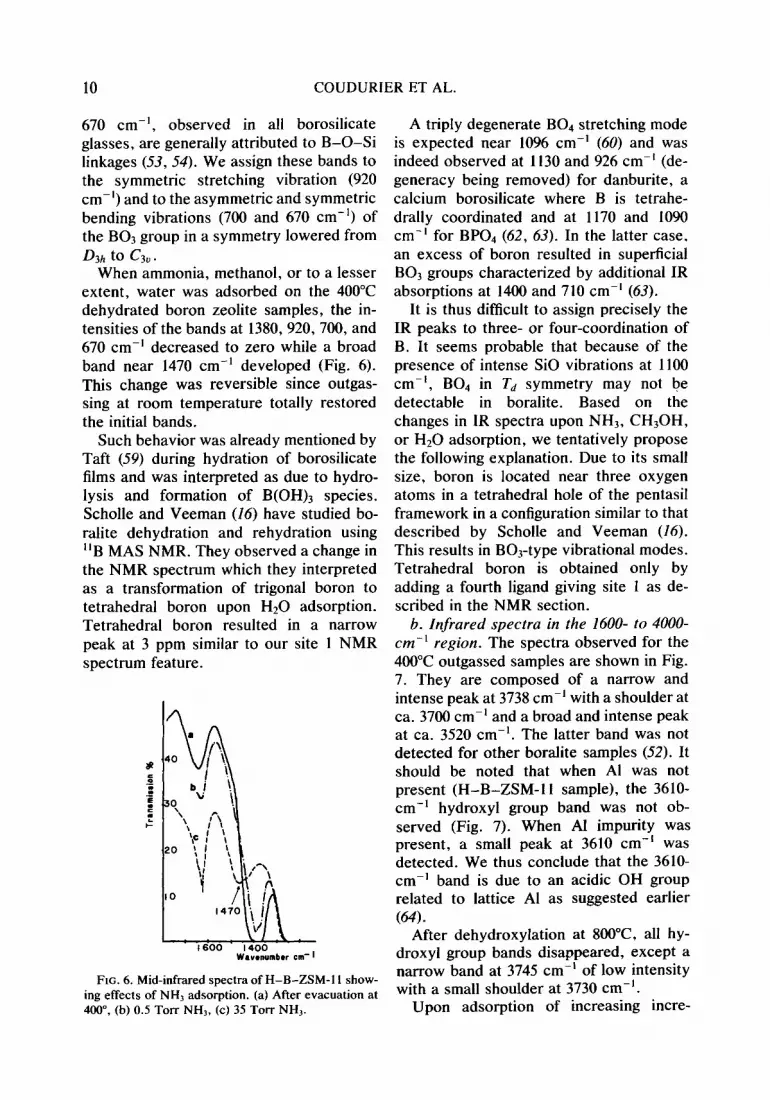
When ammonia, methanol, or to a lesser extent, water was adsorbed on the 400°C dehydrated boron zeolite samples, the in- tensities of the bands at 1380,920, 700, and 670 cm-’ decreased to zero while a broad band near 1470 cm-’ developed (Fig. 6). This change was reversible since outgas- sing at room temperature totally restored the initial bands.
Such behavior was already mentioned by Taft (59) during hydration of borosilicate films and was interpreted as due to hydro- lysis and formation of B(OH)3 species. Scholle and Veeman (16) have studied bo- ralite dehydration and rehydration using “B MAS NMR. They observed a change in the NMR spectrum which they interpreted as a transformation of trigonal boron to tetrahedral boron upon HI0 adsorption. Tetrahedral boron resulted in a narrow peak at 3 ppm similar to our site 1 NMR spectrum feature.
1600 1400 Wwwumbrr cm- 1
FIG. 6. Mid-infrared spectra of H-B-ZSM-I I show- ing effects of NH, adsorption. (a) After evacuation at 400”. (b) 0.5 Torr NH,, (c) 35 Tom NH,.
A triply degenerate B04 stretching mode is expected near 1096 cm-’ (60) and was indeed observed at 1130 and 926 cm-’ (de- generacy being removed) for danburite, a calcium borosilicate where B is tetrahe- drally coordinated and at 1170 and 1090 cm-’ for BP04 (62, 63). In the latter case. an excess of boron resulted in superficial B03 groups characterized by additional IR absorptions at 1400 and 710 cm-’ (63).
It is thus difficult to assign precisely the IR peaks to three- or four-coordination of B. It seems probable that because of the presence of intense SiO vibrations at 1100 cm-‘, B04 in Td symmetry may not be detectable in boralite. Based on the changes in IR spectra upon NH3, CH30H, or Hz0 adsorption, we tentatively propose the following explanation. Due to its small size, boron is located near three oxygen atoms in a tetrahedral hole of the pentasil framework in a configuration similar to that described by Scholle and Veeman (16). This results in B03-type vibrational modes. Tetrahedral boron is obtained only by adding a fourth ligand giving site 1 as de- scribed in the NMR section.
b. Infrared spectra in the 1600- to 4000- cm-’ region. The spectra observed for the 400°C outgassed samples are shown in Fig. 7. They are composed of a narrow and intense peak at 3738 cm-’ with a shoulder at ca. 3700 cm-’ and a broad and intense peak at ca. 3520 cm-‘. The latter band was not detected for other borahte samples (52). It should be noted that when Al was not present (H-B-ZSM-11 sample), the 3610- cm-’ hydroxyl group band was not ob- served (Fig. 7). When AI impurity was present, a small peak at 3610 cm-’ was detected. We thus conclude that the 3610- cm-’ band is due to an acidic OH group related to lattice Al as suggested earlier W).
After dehydroxylation at 8OO”C, all hy- droxyl group bands disappeared, except a narrow band at 3745 cm-’ of low intensity with a small shoulder at 3730 cm-‘.
Upon adsorption of increasing incre-

BORON-SUBSTITUTED ZSM-5 AND ZSM-I I ZEOLITES 11
FIG. 7. Hydroxyl region of (I) H-B-ZSM-I I after evacuation at 400°C. (2) H-B,AI-ZSM-5 after evacu- ation at 400°C. (3) H-B-AI-ZSM-5 after evacuation at 800°C.
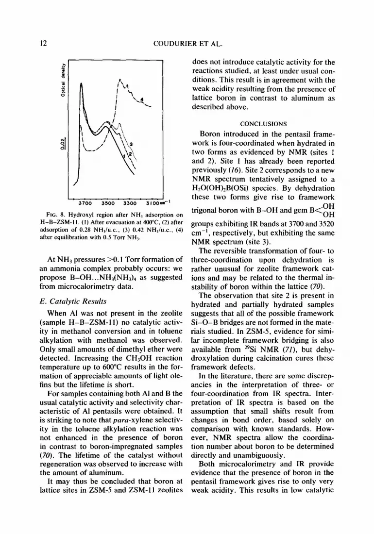
ments of NH3, two bands at ca. 3385 and 3280 cm-‘, due to N-H stretching vibra- tions, developed progressively (Fig. 8). The intensities of the 3740- and 3520-cm-’ bands were observed to decrease only when the sample was equilibrated with a residual NH3 pressure of the order of 0.1 Torr. This was totally reversible in the sense that both
bands reappeared with their starting inten- sity upon outgassing at room temperature.
By analogy with IR spectra observed for borosilicate glasses, boron phosphate, and ZSM-5 zeolite, we suggest that the 3740- and 3700-cm-’ bands are due to terminal silanols (64) and to isolated B-OH groups (63,65-68). The 3520-cm-’ band which was perturbed only at appreciable NH3 pres- sures may be assigned to gem OH groups
(B<E$ or to internal OH groups as de-
fined by Moffat and Neeleman (67). The two absorption bands at ca. 3385 and
3290 cm-’ observed upon NH, adsorption may be assigned to B t NH, species as in the case of Vycor glass (66), Bz03 on aerosil (65), and BP04 rich in B (68). They disap- peared only after desorption at 150°C. The absence of other NH3 or ammonium spe- cies stable under vacuum at room tempera- ture allows us to conclude that the material has little or no Bronsted acidity in the usual meaning but weak Lewis acidity as shown by TPD and NMR (69). It is worth noting that ammonium borate does not exist since it is immediately decomposed into NH3 and boric acid. In short, we tentatively suggest the following assignments.
Species A
Si\o/H Si,O,H+ (H20)n
I . I
o'b05ite3 o/;-\o
site 3 1 sits
Species B H H H H
\ 0 40
\
l/O _ “,B,o’ SPECIES A. A 3700-cm-’ band of weak acidity (52),
detectable in IR for the sample outgassed at 400°C. /B
LSi/O ‘Si’
o/ o/H
I SPECIES B. The 3520-cm-’ groups which interact
/\ /\ weakly and reversibly with NH, with H bonding of the
H type B-0-H...NHj and disappear irreversibly by cal- 3 site site 2 cining at 800°C.
COUDURIER ET AL.
t: : :. 3700 3500 3300 31004
FIG. 8. Hydroxyl region after NH2 adsorption on H-B-ZSM- 1 I. (I) After evacuation at 4OO”C, (2) after adsorption of 0.28 NHl/u.c., (3) 0.42 NH,/u.c., (4) after equilibration with 0.5 Torr NH,.
At NH3 pressures >O. I Torr formation of an ammonia complex probably occurs: we propose B-OH...NH3(NH& as suggested from microcalorimetry data.
E. Catalytic Results
When Al was not present in the zeolite (sample H-B-ZSM-11) no catalytic activ- ity in methanol conversion and in toluene alkylation with methanol was observed. Only small amounts of dimethyl ether were detected. Increasing the CH30H reaction temperature up to 600°C results in the for- mation of appreciable amounts of light ole- fins but the lifetime is short.
For samples containing both Al and B the usual catalytic activity and selectivity char- acteristic of Al pentasils were obtained. It is striking to note that para-xylene selectiv- ity in the toluene alkylation reaction was not enhanced in the presence of boron in contrast to boron-impregnated samples (70). The lifetime of the catalyst without regeneration was observed to increase with the amount of aluminum.
It may thus be concluded that boron at lattice sites in ZSM-5 and ZSM-I 1 zeolites
does not introduce catalytic activity for the reactions studied, at least under usual con- ditions. This result is in agreement with the weak acidity resulting from the presence of lattice boron in contrast to aluminum as described above.
CONCLUSIONS
Boron introduced in the pentasil frame- work is four-coordinated when hydrated in two forms as evidenced by NMR (sites 1 and 2). Site 1 has already been reported previously (16). Site 2 corresponds to a new NMR spectrum tentatively assigned to a H20(OH),B(OSi) species. By dehydration these two forms give rise to framework
OH trigonal boron with B-OH and gem B<oH
groups exhibiting IR bands at 3700 and 3520 cm-‘, respectively, but exhibiting the same NMR spectrum (site 3).
The reversible transformation of four- to three-coordination upon dehydration is rather unusual for zeolite framework cat- ions and may be related to the thermal in- stability of boron within the lattice (70).
The observation that site 2 is present in hydrated and partially hydrated samples suggests that all of the possible framework Si-O-B bridges are not formed in the mate- rials studied. In ZSM5, evidence for simi- lar incomplete framework bridging is also available from 29Si NMR (7Z), but dehy- droxylation during calcination cures these framework defects.
In the literature, there are some discrep- ancies in the interpretation of three- or four-coordination from IR spectra. Inter- pretation of IR spectra is based on the assumption that small shifts result from changes in bond order, based solely on comparison with known standards. How- ever, NMR spectra allow the coordina- tion number about boron to be determined directly and unambiguously.
Both microcalorimetry and IR provide evidence that the presence of boron in the pentasil framework gives rise to only very weak acidity. This results in low catalytic

BORON-SUBSTITUTED ZSM-5 AND ZSM-11 ZEOLITES
activity for acid-type reactions studied in methanol conversion and toluene alkylation with methanol.
ACKNOWLEDGMENTS
We thank C. M. Foris and R. L. Harlow for assistance in obtaining the X-ray data, H. Urbain and A. Chambosse for the Na and Al analyses, the Service Centrale d’ Analyse du CNRS-Solaize for the B anal- yses, M. van Kavelaar for the scanning electron micrographs, and E. E. Carroll for the BET measure- ments. We are especially indebted to E. F. Moran for providing the samples of Na-ZSM-5 and Na-ZSM- I I. and P. A. Jacobs of the Katholieke Universiteit of Leuven for providing a sample of ZSM- I I. A. J. Vega derived expressions for the central transition lineshape during variable angle spinning. Computer programs written by M. K. Hanafey and L. C. Takiff were used in the analysis of the NMR spectra. Valuable discus- sions with D. R. Corbin are acknowledged. The spec- tra reported here were obtained with the assistance of R. F. Carver.
REFERENCES
1. Kokotailo, G. T., Lawton. S. L., Olson. D. H.. and Meier, W. M.. Nature (Lundon) 275, 437 (1978).
2. Kokotailo, G. T., Chu, P., Lawton, S. L., and Meier, W. M., Nature (London) 275, I I9 (1978).
3. (a) Weisz. P. B., Pare Appl. Chem. 52, 2091 (1980); (b) Derouane, E. G., and Vedrine, J. C.. J. Mo1. Cafal. 8,479 (1980); (c) Derouane. E. G., in “Studies in Surf. Sci. and Catal. Series” (B. Imelik et al., Eds.) Vol. 5, p. 5. Elsevier, Amster- dam. 1980; (d) Csicsery. S. M.. ACS Monogr. 171, 202 (1984).
4. Derouane, E. Cl.. Dejaifve, P.. B.Nagy. J. van Hooff, J. H. C., Spekman. B. P.. Naccache, C.. and Vedrine. J. C.. C.R. Acad. Sci. Paris C 285, 94s (1977); J. Cutal. 53,40 (1978).
5. Chen. N. Y.. Kaeding, W. W.. and Dwyer, F. G.. J. Amer. Chem. Sot. 101, 6783 (1979).
6. Kaeding. W. W., Chu. C., Young, L. B., and Butter, S. A., J. Caral. 69, 392 (1981).
7. Klotz, M. R.. Belgian Patent 859,656 (1978). 8. (a) Taramasso, M.. Manara, G.. Fattore. V., and
Notari, B., French Patent 2.429,182 (1979); (b) Taramasso, M.. Peredgo. G., and Notari. B.. in “Proc. Vth Intern. Conf. on Molecular Sieves, Naples, 1980” (L. V. C. Rees, Ed.), p. 40. Hey- den, London, 1980.
9. Holderich, W., Eichhorn, H., Lehnert, R., Ma- rosi, L., Mross, W., Runke, R., Ruppel, W., and Schlimper, H., in “Proc. VIth Intern. Cong. on Molecular Sieves, Reno, 1983” (D. H. Olson and A Bisio, Eds.), p. 545. Butterworths, London, 1984.
IO.
II.
12.
13.
14.
15.
16.
17.
18.
19. 20.
21. 22.
23.
24.
25.
26.
27.
28.
lone, K. G., Vostrikova. L. A.. Paukshtis. E. A.. Yurchenko. E. N., and Stepanov, V. G., Dokl. Akad. Nauk, SSSR 261, 1160 (1981). Vostrikova. L. A., tone. K. G., Mastikhin, V. M.. and Petrova. A. V., React. Kinet. Catul. Lett. 26, 291 (1984). Perego, G., Cesari, M., and Allegra, G., J. Appl. Cryst. 17, 403 (1984). Wengin, P., Guowen, L., Wangrong. L., and Bingxiong, L., “Intl. Symp. on Zeolites. Porto- rose, Yugoslavia, 1984.” Gabelica, Z., B.Nagy. J., Bodart. P., and Debras, G., Chem. Lett. 1984, 1060 (1984). Kutz, N. A., in “Proc. 2nd Symp. of Industry- University Coop. Chem. Prog., April l-4, 1984,” p. 120. Texas A&M Univ. Press, 1984. Scholle, K. F. M. G. J.. and Veeman. W. S.. Zeolites 5, I I8 (1985). Klotz, M. R.. U.S. Patents 4.268.420; 4.269.813; 4.285.919 t 1981). Roliman, R. D., and Valyocsik, E. W.. Inorg. Syzrh. 22, 67 (1983). Chu. P., U.S. Patent 3.709.979 (1973). Taramasso. M.. Manara, G.. Fattore. V., and Notari, B., Ger. Patent 2,924,915 (1980). L.andolt, G. R., Anal. Chem. 42, 613 (1971). Gravelle. P. C., “Advances in Catalysis” (D. D. Eley, P. W. Selwood. and P. B. Weisz, Eds.). Vol. 22. p.191. Academic Press. New York, 1972. Dominquez, J. M., Acosta, D. R., and Schifter. I.. J. Catal. 83, 480 (1983). Haag, W. 0.. Lago. R. M.. and Weisz, P. B.. Faraday Discuss. Chem. Sot. 72, 317 (1981). von Ballmoos, R., “The O-Exchange Method in Zeolite Chemistry.” Salle Sauerlander Verlag, Frankfurt, I98 I. Foger, K., Sanders, J. V.. and Seddon, D., Zeo- lifes 4, 337 (1984). Anderson, J. R.. Foger, K., Mole. T.. Rajad- hyaksha. R. A., and Sanders, J. V., J. Catal. 58, I I4 (1979). Shannon. R. D.. Acta. Ctystallogr. A 32, 751 (1976).
29. Meyers, B. L., Ely. S. R.. Kutz, N. A., Kaduk. J. A.. and van den Bossche. E., J. Catal, 91, 352 (1985).
30. Taylor, P. C., Baugher, J. F.. and Kriz. H. M.. Chem. Rev. 75, 203 (1975).
31. Tzalmona, A., and Andrew, E. R., “Proc. 18th Ampere Conference, Nottingham,” p. 241. North- Holland, Amsterdam. 1972.
32. Nolle, A., Z. Phys. A 280, 231 (1977). 33. Vega, A. J.. personal communication, 1981. 34. Kundla. E., Samosan. A., and Lippmaa, E..
Chem. Phys. Lett. 83, 229 (1981). 35. Fyfe, C. A., Gobbi, G. C., Hartman. J. S.. Len-
kinski, R. E., and O’Brien, J. H., J. Magn. Reson. 47, 168 (1982); Meadows, M. D., Smith, K. A., Kinsey, R. A.. Rothgeb. T. M.. Skarjune, R. P..
14 COUDURIERETAL.
and Oldfield, E., Proc. Natl. Acad. Sci. USA 79, 1351 (1982) and references therein.
36. Schramm, S., and Oldfield, E., .I. C&n. Sot. Chem. Commun. 1982,980(1982).
37. Jellison, G. E., Panek, L. W., Bray, P. J., and Rouse, G. B., J. C/rem. Phys. 66, 802 (1977).
38. Dell, W. J., Bray. P. J., and Xiao, S. Z., J. Non-Cryst. Solids 58, I (1983); Bray, P. J., Geiss- berger, A. E., Bucholtz, F., and Harris, I. A., J. Non-Cryst. Solids 52, 45 (1982).
39. Bray, P. J., Bucholtz, F., Geissberger, A. E., and Harris, I. A., Nucl. Instrum. Methods 199, I (1982).
40. Turner, G. L., Smith, K. A., Kirkpatrick, R. James, and Oldfield, E., J. Magn. Reson. 67,544 (1986).
41. Gabelica, Z., Debras, G., and B.Nagy, J., in “Studies in Surf Sci. and Catal. Series” (S. Kalia- guine and A. Mahey, Eds.). Vol. 19, p. 113, 1984.
42. Vostrikova, L. A., Ione, K. G., Mastikhin, V. M., and Petrova, A. V., React. Kinet. Catal. Lett. 26, 291 (1984).
43. Farlee, R. D., unpublished results. 44. Jellison. G. E., and Bray, P. J., J. Non-Cyst.
Solids 29, 187 (1978). (Site 2 in this paper has a B-10 quadrupole coupling constant of 1.5 MHz, corresponding to 0.72 MHz for B-l 1.)
45. Auroux. A., Vedrine. J. C., and Gravelle, P. C., in “Studies in Surface Sci. and Catal. Series” (J. Rouquerol and K. S. W. Sing, Eds.), Vol. 10, p. 305. Elsevier, Amsterdam, 1982.
46. Auroux, A., Bolis, V., Wierzchowski. P., Gravelle. P. C., and Vedrine, J. C., J. Chem. Sot. Faraday Trans. 2 75, 2544 (1979).
47. Zhang, Y., Inorg. Chem. 21, 3889 (1982). 48. Castleman, A. W., Holland. P. M., Lindsay, D.
M., and Peterson, K. I., J. Amer. Chem. Sot. 100, 6039 (1978).
49. Coudurier. G., Naccache, C.. and Vedrine, J. C., J. Chem. Sot. Chem. Commun.. 1413 (1982).
50. Jansen, J. C., van der Gaag, F. J., and van Bekkum. H., Zeolites 4, 369 (1984).
51. Flanigen, E., ACS Monogr. 171, 80 (1976). 52. Coudurier, G., and Vedrine. J. C., Pure Appl.
Chem. 58, 1389 (1986). 53. Tenney, P. G., and Wong, J., .I. Chem. Phys. 56,
5516 (1972). 54. Jelliman, P. E., and Proctor, J. P., J. Sot. G/ass
Technol. 39, 173T (1955). 55. Kumar, B., Mater. Res. Bull. 19, 331 (1984). 56. Nogami, N., and Moriya, Y.. J. Non-Cryst. Solids
48, 359 (1982). 57. Kern, W., and Heim, R. C., J. Electrochem. Sot.
117,568 (1970). 58. Wong, J., J. Electrochem Sot. 127, 62 (1980). 59. Taft. E. A., J. Electrochem. Sot. 118, 1985 (1971). 60. Weir, C. E., and Schroeder, R. A., J. Res. Nat.
Bur. Stand. USA, Sect. A 68 (1964). 61. Anderson, S., Bohrn, R. L., and Kimpton, D. D.,
J. Amer. Ceram. Sot. 38, 370 (1955). 62. Osaka, A., Takahashi. K., and Ikeda, M., J.
Mater. Ski. Lett. 3, 36 (1984). 63. Haber, J., and Szybalska, U.. Faraday Discuss.
Chem. Sot. 72, 263 (1981). 64. Vedrine, J. C., Auroux, A., Bolis, V., Dejaifve,
P., Naccache, C., Wierzchowski, P., Derouane, E. G., B.Nagy, J., Gilson, J. P., van Hooff, J. H. C., van den Berg, J. P., and Wolthuizen, J., J. Catal. 58, 248 (1979).
65. Cant, N. W., and Little, L. H., Canad. J. Chem. 46, 1373 (1968).
66. Low, M. J. D., and Subramanian, N. R.. J. Phys. Chem. 70, 2740 (1966).
67. Moffat, J. B., and Neeleman, J. F.. J. Catal. 34, 376 (1974).
68. Moffat, J. B., and Neeleman, J. F., J. Catal. 39, 419 (1975).
69. Scholle, K. F. M. G. J., Kentgens, A. P. M., Veeman, W. S., Frenken, P., and van der Velden, G. P. M., J. Phys. Chem. 88, 5 (1984).
70. Sayed, M., and Vedrine. J. C., J. Catal. 101, 43 (1986).
71. B.Nagy, J.. Gabelica, Z., and Derouane, E. G., Chem. Lett. 1982, 1105 (1982).