chitosan-modified plga polymeric nanocarriers ... - permegear
TRANSCRIPT

Cp
CBa
b
c
d
a
ARRAA
KNNDBCCC
1
dlleiaatp
aR
(
h0
International Journal of Biological Macromolecules 93 (2016) 381–389
Contents lists available at ScienceDirect
International Journal of Biological Macromolecules
j ourna l h o mepa ge: www.elsev ier .com/ locate / i jb iomac
hitosan-modified PLGA polymeric nanocarriers with better deliveryotential for tamoxifen
hanchal Kiran Thakura, Nagarani Thotakuraa, Rajendra Kumarb, Pramod Kumara,hupinder Singhb,c, Deepak Chitkarad, Kaisar Razaa,∗
Department of Pharmacy, School of Chemical Sciences and Pharmacy, Central University of Rajasthan, Bandar Sindri, Dist. Ajmer 305 817, Rajasthan, IndiaUGC-Centre of Excellence in Applications of Nanomaterials, Nanoparticles and Nanocomposites, Panjab University, 160 014 Chandigarh, IndiaDivision of Pharmaceutics, University Institute of Pharmaceutical Sciences, Panjab University, 140 604 Chandigarh, IndiaDepartment of Pharmacy, Birla Institute of Technology and Science (BITS)—Pilani, Vidya Vihar Campus, Pilani 333031, Rajasthan, India
r t i c l e i n f o
rticle history:eceived 27 May 2016eceived in revised form 20 August 2016ccepted 28 August 2016vailable online 29 August 2016
eywords:anocarriersanotechnologyermatokineticsioavailability
a b s t r a c t
Breast cancer is believed as the second most common cause of cancer-related deaths in women forwhich tamoxifen is frequently prescribed. Despite many promises, tamoxifen is associated with variouschallenges like low hydrophilicity, poor bioavailability and dose-dependent toxicity. Therefore, it wasenvisioned to develop tamoxifen- loaded chitosan-PLGA micelles for potential safe and better deliveryof this promising agent. The chitosan-PLGA copolymer was synthesised and characterised by FourierTransform-Infrared, Ultraviolet-visible and Nuclear Magnetic Resonance spectroscopic techniques. Thedrug-loaded nanocarrier was characterised for drug-pay load, micrometrics, surface charge and mor-phological attributes. The developed system was evaluated for in-vitro drug release, haemolytic profile,cellular-uptake, anticancer activity by cytotoxicity assay and dermatokinetic studies. The developednano-system was able to substantially load the drug and control the drug release. The in-vitro cytotoxi-
ytotoxicityonfocal laser scanning microscopyellular uptake
city offered by the system was significantly enhanced vis-a-vis plain drug, and there was no substantialhaemolysis. The IC50 values were significantly decreased and the nanocarriers were uptaken by MCF-7 cells, noticeably. The carrier was able to locate the drug in the interiors of rat skin in considerableamounts to that of the conventional product. This approach is promising as it provides a biocompatibleand effective option for better delivery of tamoxifen.
© 2016 Elsevier B.V. All rights reserved.
. Introduction
Nanotechnology is associated with designing useful materials,evices and systems through control of matter on the micromeritic
evel, and exploitation of novel phenomena and properties at theength scale [1,2]. In the present era, nanotechnology is a rapidlymerging area, especially in medical research, which is focus-ng on development of nanoparticles/nanocarriers for diagnosticnd therapeutic applications [3]. Nanotechnology-based carriers
re being employed for the targeted drug delivery. The impor-ant examples of such carriers are liposomes, ethosomes, niosomes,olymeric micelles, solid-lipid nanoparticles, nanospheres, carbon∗ Corresponding author at: Department of Pharmacy, School of Chemical Sciencesnd Pharmacy, Central University of Rajasthan, Bandar Sindri, Distt. Ajmer 305 817,ajasthan, India.
E-mail addresses: [email protected], razakaisar [email protected]. Raza).
ttp://dx.doi.org/10.1016/j.ijbiomac.2016.08.080141-8130/© 2016 Elsevier B.V. All rights reserved.
nanotubes, C60-fullerenes and nanocapsules [4–6]. Among all thesenanocarriers, polymeric micelles are established for prolongedblood circulation, enhanced permeation and retention (EPR) effectvia tumor vessels, better delivery to tumor cells, increase thera-peutic efficiency and significant tumor accumulation [7]. Despiteall these, preparations of polymeric micelles emerge to be rela-tively simple as compared to other novel drug delivery systems [8].These carriers can easily load various poorly soluble drugs and canenhance the bioavailability of such drugs [9].
Tamoxifen (TAM) is a cytostatic drug and is known for its anti-estrogenic nature. It is most widely used for the treatment ofall stages of breast cancers, especially for metastatic breast can-cers [10]. Despite its pharmacological significance, TAM is taggedwith challenges like lower aqueous solubility and lower tissueselectivity. Apart from this, long-term usage of TAM puts patients
at increased risk of thrombo-embolic events and uterine malig-nancies. The other major adverse effects of TAM include higherincidences of endometrial cancer, liver cancer and developmentof drug resistance [11]. In order to circumvent the systemic side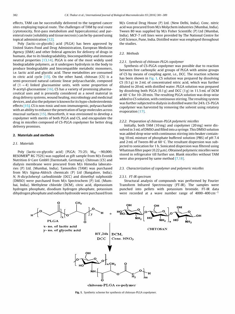
3 f Biolo
es(et
UAhnbpiisoNcddewmcdd
2
2
RNdrfN(bhd
82 C.K. Thakur et al. / International Journal o
ffects, TAM can be successfully delivered to the targeted cancerites employing topical route. The challenges of TAM by oral routecytotoxicity, first-pass metabolism and hypercalcemia) and par-nteral route (solubility and tissue necrosis) can be by-passed usingopical administration [12].
Poly (lactic-co-glycolic) acid (PLGA) has been approved bynited States-Food and Drug Administration, European Medicinegency (EMA) and other federal agencies for delivery of drugs inumans, due to its biodegradability, biocompatibility and immuneeutral properties [13,14]. PLGA is one of the most widely usediodegradable polymers, as it undergoes hydrolysis in the body toroduce biodegradable and biocompatible metabolic monomers,
.e. lactic acid and glycolic acid. These metabolites are consumedn citric acid cycle [15]. On the other hand, chitosan (CS) is aemi-processed natural cationic linear polysaccharide, composedf (1 → 4) linked glucosamine units, with some proportion of-acetyl-glucosamine [16]. CS has a variety of promising pharma-eutical uses and is presently considered as a novel material inrug delivery systems, wound healing, microbiology and medicinalevices, and also the polymer is known for its hypo-cholesterolemicffects [15]. CS is non-toxic and non-immunogenic, polysaccharideith an ability to enhance the penetration of large molecules acrossucosal surfaces [15]. Henceforth, it was envisioned to develop a
opolymer with merits of both PLGA and CS, and encapsulate therug in micelles composed of CS-PLGA copolymer for better drugelivery promises.
. Materials and methods
.1. Materials
Poly (lactic-co-glycolic acid) (PLGA; 75:25; MW ∼90,000;ESOMER® RG 752S) was supplied as gift sample from M/s Evonikutrition & Care GmbH (Darmstadt, Germany). Chitosan (CS) andialysis membrane were procured from M/s Himedia laborato-ies (P) Ltd. (Mumbai, India). Tamoxifen (TAM) was purchasedrom M/s Sigma-Aldrich chemicals (P) Ltd (Bangalore, India)., N-dicyclohexyl carbodiimide (DCC) and dimethyl sulphoxide
DMSO) were purchased from M/s Spectrochem (P) Ltd. (Mum-ai, India). Methylene chloride (DCM), citric acid, dipotassiumydrogen phosphate, disodium hydrogen phosphate, potassiumihydrogen phosphate and sodium hydroxide were purchased from
Fig. 1. Synthetic scheme for synthesi
gical Macromolecules 93 (2016) 381–389
M/s Central Drug House (P) Ltd. (New Delhi, India). Conc. nitricacid was procured from M/s Molychem industries (Mumbai, India).Tween 80 was supplied by M/s Fisher Scientific (P) Ltd (Mumbai,India). MCF-7 cell lines were provided by The National Centre forCell Science, Pune, India. Distilled water was employed throughoutthe studies.
2.2. Methods
2.2.1. Synthesis of chitosan-PLGA copolymerSynthesis of CS-PLGA copolymer was possible due to reaction
between free carboxylic acid groups of PLGA with amino groupsof CS by means of coupling agent, i.e., DCC. The reaction schemehas been shown in Fig. 1. CS solution was prepared by dissolvingCS (0.1 g) in 2 mL of concentrated nitric acid, which was furtherdiluted to 20 mL with distilled water. PLGA solution was preparedby dissolving both PLGA (0.1 g) and DCC (1 g) in 11.5 mL of DCMat 60 ◦C for 10–20 min. The resulting PLGA solution was drop-wiseadded to CS solution, with continuous stirring. The resulting systemwas further subjected to dialysis in distilled water for 24 h. CS-PLGAcopolymer was harvested by removing the solvent using rotatoryevaporation [17].
2.2.2. Preparation of chitosan-PLGA polymeric micellesInitially, both TAM (10 mg) and copolymer (20 mg) were dis-
solved in 3 mL of DMSO and filled into a syringe. This DMSO solutionwas added drop wise with continuous stirring into beaker contain-ing 10 mL mixture of phosphate buffered solution (PBS) of pH 7.4and 2 mL of Tween-80 at 60 ◦C. The resultant dispersion was sub-jected to sonication for 1 h. Sonicated dispersion was filtered usingWhatman filter paper (0.22 �m). Obtained polymeric micelles werestored in refrigerator till further use. Blank micelles without TAMwere also prepared by same method [7,18].
2.3. Characterization of copolymer and polymeric micelles
2.3.1. FT-IR spectrum
Structural analysis of compounds was performed by FourierTransform Infrared Spectroscopy (FT-IR). The samples werepunched into pellets with potassium bromide. FT-IR datawere recorded at a wave number range of 4000–400 cm−1
s of chitosan-PLGA copolymer.
f Biolo
uW
2
nSPs
2
mHfispptsaa[
2
SDowoti
2
baWmAt
2
eLoog
2
dwap[
2
2
(yo
C.K. Thakur et al. / International Journal o
sing FT-IR Spectrometer (Spectrum two, M/s Perkin Elmer Co.,altham, Massachusetts, USA).
.3.2. NMR spectrumThe 1H NMR spectrum of CS-PLGA conjugate was recorded using
uclear magnetic resonance spectrometer (ASCEND 500WB NMRpectrometer, M/s Bruker Bio Spin Corporation., Indiana, USA). CS-LGA saturated solution was prepared in deuterated DMSO andcanned for the NMR spectrum.
.3.3. Molar mass determinationGel permeation chromatography (GPC) was employed to deter-
ine molecular weight of the polymer. Tetrahydrofuran (THF;PLC grade) was used as the mobile phase, filtered through 0.22 �mlter paper and deaerated in ultrasonic bath for 30 min. Polystyrenetandards and test samples were prepared by dissolving 1 mg of theolymer in 1 mL of THF (HPLC grade), filtered through 0.22 �m filteraper and analyzed using Waters GPC system coupled with refrac-rometer 2414. Chromatographic separation was done on Water’styragel column in an isocratic mode with the stated mobile phaset a flow rate of 1 mL/min. The sample injection volume was keptt 50 �L. The data was analyzed using “Breeze 2- Add on” software19].
.3.4. CMC (Critical micellar concentration) determinationCMC was determined by using iodine/potassium iodide method.
olution of 250 mg of I2 and 500 mg of KI were dissolved in 25 mL ofCM. All the dilutions (1–10 �g/mL) prepared were incubated forvernight by adding 25 �L of iodine solution. Incubated solutionsere analyzed using UV–vis spectrophotometer at a wavelength
f 366 nm. A graph was plotted between concentration of polymeraken and absorbance observed. The point at which there is a sharpncrease in absorbance was CMC value [17].
.3.5. Particle size distribution and zeta potential studiesParticle size and poly-dispersity index values were determined
y dispersing the prepared polymeric micelles in PBS 7.4 (1 mg/mL)nd analysing the same on Malvern Zetasizer (M/s Malvern,orcestershire, UK) installed at BITS, Pilani, India. The same equip-ent was employed for the determination of zeta-potential values.verage value from three repeated observations was reported as
he final result.
.3.6. MorphologyMorphological evaluation was conducted using transmission
lectron microscopy (TEM) installed at Central Instrumentationaboratory, Panjab University, Chandigarh, India. In this process,ne drop of polymeric micellar dispersion was added with 1% aque-us solution of phosphotungstic acid on carbon film coated copperrid and the image was clicked at suitable magnification.
.3.7. Entrapment and drug loading studiesEntrapment efficacy (EE) and drug loading (DL) values were
etermined using filtration method. The residue of Section 2.2.2as used for the study. Filter paper was extracted in methanol
nd analyzed spectrophotometrically for unentrapped drug. Sam-le from blank micelles, treated analogously, served as the blank20].
.4. Evaluation studies
.4.1. In-vitro drug release studies
Pure TAM and CS-PLGA based TAM-loaded polymeric micellesequivalent to 1 mg of TAM) were weighed and packed into dial-sis bags. These dialysis bags were suspended in 30 mL solutionf methanol and PBS of pH 5.6/pH 6.8/pH 7.4 (1:9 v/v ratio) [21].
gical Macromolecules 93 (2016) 381–389 383
Temperature was maintained at 37 ± 1 ◦C with continuous stirring.Samples of 2 mL each were collected at regular intervals. Equal vol-ume of fresh diffusion medium was replaced to maintain the sinkconditions, after each sample. The samples were analysed by UVspectrophotometer for the drug content.
2.4.2. Ex-vivo hemolysis studiesThis protocol was executed on whole human blood samples.
The study was duly approved by the Institutional Ethics Committee,Central University of Rajasthan, Bandar Sindri, Kishangarh, Ajmer,India. In brief, 2 mL of blood sample was collected from healthymale human volunteer in a vial containing 124 mmol/L of sodiumcitrate solution. With the help of centrifugation, erythrocytes wereseparated and washed with normal saline. These washed erythro-cytes were re-suspended in normal saline (2 mL). Test formulations,i.e., TAM plain and TAM-loaded polymeric micelles (equivalent of1 mg of TAM) were added to 1 mL of RBC dispersion. On the otherhand, the reference dispersion was prepared by dispersing erythro-cytes in distilled water. All these test tubes were incubated at 37 ◦Cfor 1 h. After incubation, centrifugation (2000 × g) was performedfor 5 min to harvest the transparent supernatant. The supernatantwas analyzed using UV–vis spectroscopy at �max of 415 nm and thehemolysis induced by distilled water was taken as 100% [22].
2.4.3. Protein binding studiesA 2% w/w bovine serum albumin (BSA) solution was prepared
in PBS 7.4. TAM encapsulated CS-PLGA micelles (drug equivalentto 1 mg) and pure TAM (1 mg) were weighed and dispersed in1 mL of BSA solution. The dispersions were packed in dialysis bags,separately. The dialysis bags were suspended in 20 mL of PBS 7.4.Samples were withdrawn after 1 h, 2 h, 4 h and 6 h, were analyzedby UV–vis spectrophotometer [23].
2.4.4. In-vitro cytotoxicity assayHuman breast cancer MCF-7 cells were fed in 96-welled plate
with 5% CO2. The samples of pure TAM and TAM-loaded micellesin various concentrations were added to these wells and incubatedfor 24 h at 37 ◦C. Solution of MTT (5 mg/mL), 10 �L was added toeach well and incubation was done for 4 h. The developed formazancrystals were dissolved in 200 �L of DMSO. The absorbance wasrecorded at 570 nm using ELISA reader [24].
2.4.5. Confocal laser scanning microscopyMCF-7cells with a density of approx. 15,000 cells/cm2 were
seeded on petri-plates. The drug-loaded micelles were tagged withcoumarin-6. The dye-tagged nanocarriers were added to the cellculture medium. The incubation was done at 37 ◦C for 24 h. Cellswere washed using PBS 7.4. Cell fixing was done using ice coldmethanol, followed by washing with PBS 7.4. Nucleus stainingwas done with 300 nM solution of DAPI for 5 min. The cells werescanned under confocal laser scanning microscope (Nikon C2 Plus,with NIS Elements Version 4.3 Software). For DAPI, the wavelengthsused were 405 nm and 417–477 nm, whereas for coumarin-6 laserwavelengths used were 488 nm and 500–550 nm, respectively forexcitation and emission. Cells with fluorescent nuclei (blue stained)and drug-invaded regions (green-stained) were captured at 60×magnification. Experiments were performed in triplicate.
2.4.6. Dermatokinetic studiesSkins of Wistar rats were used for this study. The animal proto-
cols were duly approved by Institutional Animal Ethics Committee,Panjab University, Chandigarh, India. After sacrificing the animals,
the hair on the dorsal side of animals were removed. The skinwas harvested, discharged of adhering fat layers, and mountedon Franz diffusion cells (M/s Permegear, Inc., PA, USA), having across-sectional area of 3.142 cm2 and receptor volume of 30.0 mL.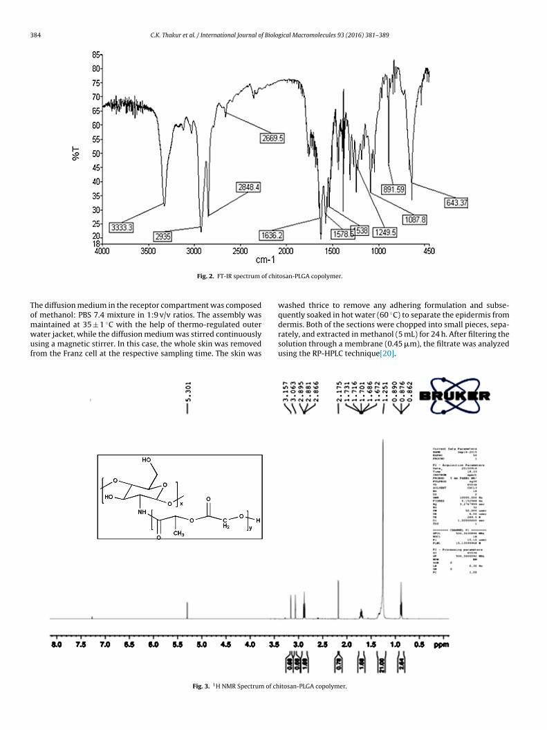
384 C.K. Thakur et al. / International Journal of Biological Macromolecules 93 (2016) 381–389
of chit
Tomwuf
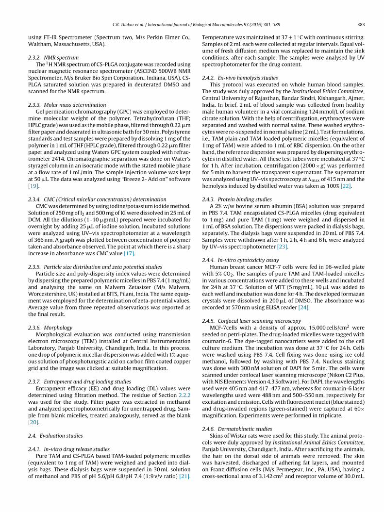
Fig. 2. FT-IR spectrum
he diffusion medium in the receptor compartment was composedf methanol: PBS 7.4 mixture in 1:9 v/v ratios. The assembly was
aintained at 35 ± 1 ◦C with the help of thermo-regulated outerater jacket, while the diffusion medium was stirred continuouslysing a magnetic stirrer. In this case, the whole skin was removedrom the Franz cell at the respective sampling time. The skin wasFig. 3. 1H NMR Spectrum of ch
osan-PLGA copolymer.
washed thrice to remove any adhering formulation and subse-quently soaked in hot water (60 ◦C) to separate the epidermis from
dermis. Both of the sections were chopped into small pieces, sepa-rately, and extracted in methanol (5 mL) for 24 h. After filtering thesolution through a membrane (0.45 �m), the filtrate was analyzedusing the RP-HPLC technique[20].itosan-PLGA copolymer.
C.K. Thakur et al. / International Journal of Biological Macromolecules 93 (2016) 381–389 385
(B) Ze
3
3
3
c(Pw
Fig. 4. Graphs of (A) Particle size and
. Results and discussion
.1. Characterization studies
.1.1. FT-IR of chitosan-PLGA copolymerFig. 2 represents FT-IR spectrum of CS-PLGA copolymer, which
onfirmed the formation of amide ( CO NH) bond between amine NH2) group of CS and free carboxylic acid ( COOH) group ofLGA. The peak at 3333.3 cm−1 represented N H stretching,here as the peak at 1636.2 cm−1 represented the presence of C O
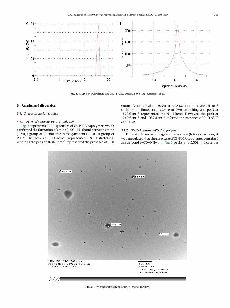
Fig. 5. TEM microphotograph
ta potential of drug-loaded micelles.
group of amide. Peaks at 2935 cm−1, 2848.4 cm−1 and 2669.5 cm−1
could be attributed to presence of C H stretching and peak at1578.6 cm−1 represented the N H bend. However, the peak at1249.5 cm−1 and 1087.8 cm−1 inferred the presence of C O of CSand PLGA.
3.1.2. NMR of chitosan-PLGA copolymerThrough 1H nuclear magnetic resonance (NMR) spectrum, it
was speculated that the structure of CS-PLGA copolymer containedamide bond ( CO NH ). In Fig. 3 peaks at ı 5.301, indicate the
of drug-loaded micelles.
386 C.K. Thakur et al. / International Journal of Biological Macromolecules 93 (2016) 381–389
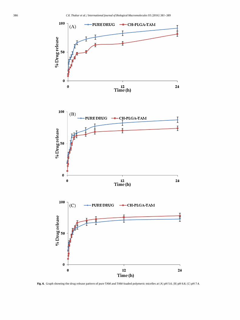
Fig. 6. Graph showing the drug release pattern of pure TAM and TAM-loaded polymeric micelles at (A) pH 5.6, (B) pH 6.8, (C) pH 7.4.
C.K. Thakur et al. / International Journal of Biological Macromolecules 93 (2016) 381–389 387
pıPois
3
wooc3mfii
3
ttiaPiwmdi
F
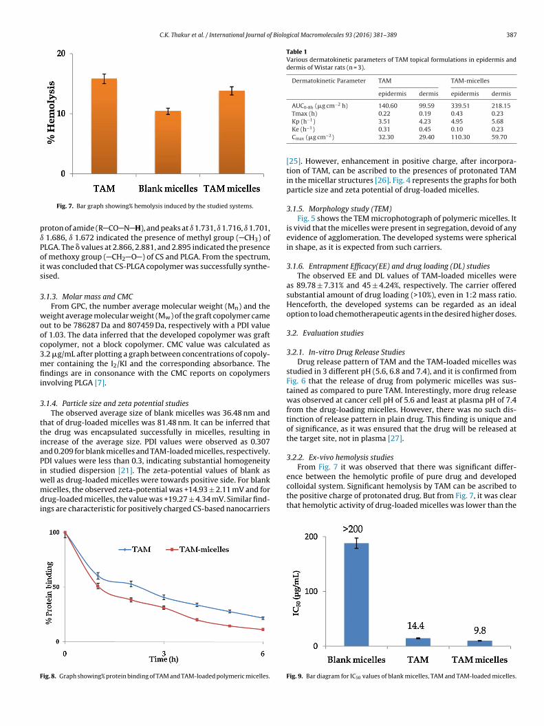
Table 1Various dermatokinetic parameters of TAM topical formulations in epidermis anddermis of Wistar rats (n = 3).
Dermatokinetic Parameter TAM TAM-micelles
epidermis dermis epidermis dermis
AUC0-8h (�g cm−2 h) 140.60 99.59 339.51 218.15Tmax (h) 0.22 0.19 0.43 0.23
−1
ence between the hemolytic profile of pure drug and developed
Fig. 7. Bar graph showing% hemolysis induced by the studied systems.
roton of amide (R CO N H), and peaks at ı 1.731, ı 1.716, ı 1.701, 1.686, ı 1.672 indicated the presence of methyl group ( CH3) ofLGA. The � values at 2.866, 2.881, and 2.895 indicated the presencef methoxy group ( CH2 O ) of CS and PLGA. From the spectrum,t was concluded that CS-PLGA copolymer was successfully synthe-ised.
.1.3. Molar mass and CMCFrom GPC, the number average molecular weight (Mn) and the
eight average molecular weight (Mw) of the graft copolymer cameut to be 786287 Da and 807459 Da, respectively with a PDI valuef 1.03. The data inferred that the developed copolymer was graftopolymer, not a block copolymer. CMC value was calculated as.2 �g/mL after plotting a graph between concentrations of copoly-er containing the I2/KI and the corresponding absorbance. The
ndings are in consonance with the CMC reports on copolymersnvolving PLGA [7].
.1.4. Particle size and zeta potential studiesThe observed average size of blank micelles was 36.48 nm and
hat of drug-loaded micelles was 81.48 nm. It can be inferred thathe drug was encapsulated successfully in micelles, resulting inncrease of the average size. PDI values were observed as 0.307nd 0.209 for blank micelles and TAM-loaded micelles, respectively.DI values were less than 0.3, indicating substantial homogeneityn studied dispersion [21]. The zeta-potential values of blank as
ell as drug-loaded micelles were towards positive side. For blankicelles, the observed zeta-potential was +14.93 ± 2.11 mV and for
rug-loaded micelles, the value was +19.27 ± 4.34 mV. Similar find-ngs are characteristic for positively charged CS-based nanocarriers
ig. 8. Graph showing% protein binding of TAM and TAM-loaded polymeric micelles.
Kp (h ) 3.51 4.23 4.95 5.68Ke (h−1) 0.31 0.45 0.10 0.23Cmax (�g cm−2) 32.30 29.40 110.30 59.70
[25]. However, enhancement in positive charge, after incorpora-tion of TAM, can be ascribed to the presences of protonated TAMin the micellar structures [26]. Fig. 4 represents the graphs for bothparticle size and zeta potential of drug-loaded micelles.
3.1.5. Morphology study (TEM)Fig. 5 shows the TEM microphotograph of polymeric micelles. It
is vivid that the micelles were present in segregation, devoid of anyevidence of agglomeration. The developed systems were sphericalin shape, as it is expected from such carriers.
3.1.6. Entrapment Efficacy(EE) and drug loading (DL) studiesThe observed EE and DL values of TAM-loaded micelles were
as 89.78 ± 7.31% and 45 ± 4.24%, respectively. The carrier offeredsubstantial amount of drug loading (>10%), even in 1:2 mass ratio.Henceforth, the developed systems can be regarded as an idealoption to load chemotherapeutic agents in the desired higher doses.
3.2. Evaluation studies
3.2.1. In-vitro Drug Release StudiesDrug release pattern of TAM and the TAM-loaded micelles was
studied in 3 different pH (5.6, 6.8 and 7.4), and it is confirmed fromFig. 6 that the release of drug from polymeric micelles was sus-tained as compared to pure TAM. Interestingly, more drug releasewas observed at cancer cell pH of 5.6 and least at plasma pH of 7.4from the drug-loading micelles. However, there was no such dis-tinction of release pattern in plain drug. This finding is unique andof significance, as it was ensured that the drug will be released atthe target site, not in plasma [27].
3.2.2. Ex-vivo hemolysis studiesFrom Fig. 7 it was observed that there was significant differ-
colloidal system. Significant hemolysis by TAM can be ascribed tothe positive charge of protonated drug. But from Fig. 7, it was clearthat hemolytic activity of drug-loaded micelles was lower than the
Fig. 9. Bar diagram for IC50 values of blank micelles, TAM and TAM-loaded micelles.
388 C.K. Thakur et al. / International Journal of Biological Macromolecules 93 (2016) 381–389
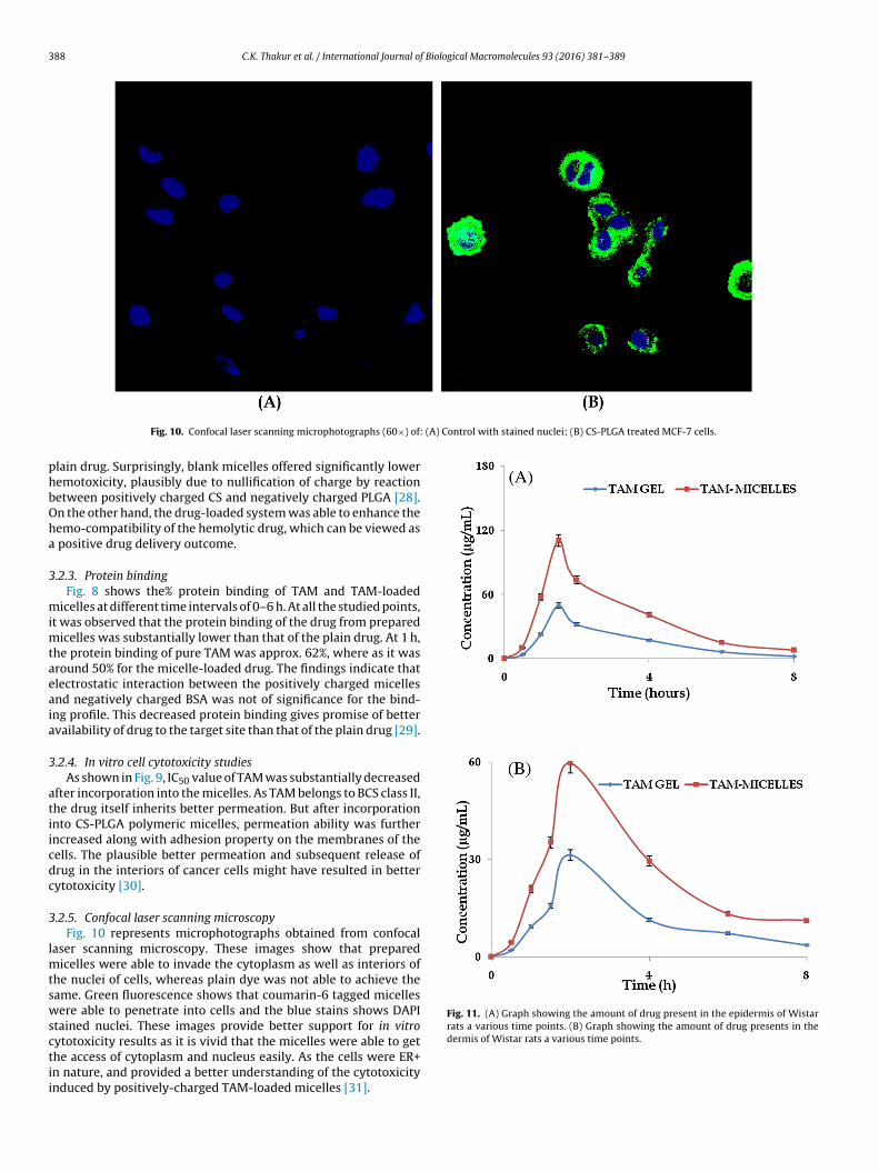
(A) Control with stained nuclei; (B) CS-PLGA treated MCF-7 cells.
phbOha
3
mimtaeaia
3
atiicdc
3
lmtswsctii
Fig. 11. (A) Graph showing the amount of drug present in the epidermis of Wistarrats a various time points. (B) Graph showing the amount of drug presents in the
Fig. 10. Confocal laser scanning microphotographs (60×) of:
lain drug. Surprisingly, blank micelles offered significantly loweremotoxicity, plausibly due to nullification of charge by reactionetween positively charged CS and negatively charged PLGA [28].n the other hand, the drug-loaded system was able to enhance theemo-compatibility of the hemolytic drug, which can be viewed as
positive drug delivery outcome.
.2.3. Protein bindingFig. 8 shows the% protein binding of TAM and TAM-loaded
icelles at different time intervals of 0–6 h. At all the studied points,t was observed that the protein binding of the drug from prepared
icelles was substantially lower than that of the plain drug. At 1 h,he protein binding of pure TAM was approx. 62%, where as it wasround 50% for the micelle-loaded drug. The findings indicate thatlectrostatic interaction between the positively charged micellesnd negatively charged BSA was not of significance for the bind-ng profile. This decreased protein binding gives promise of bettervailability of drug to the target site than that of the plain drug [29].
.2.4. In vitro cell cytotoxicity studiesAs shown in Fig. 9, IC50 value of TAM was substantially decreased
fter incorporation into the micelles. As TAM belongs to BCS class II,he drug itself inherits better permeation. But after incorporationnto CS-PLGA polymeric micelles, permeation ability was furtherncreased along with adhesion property on the membranes of theells. The plausible better permeation and subsequent release ofrug in the interiors of cancer cells might have resulted in betterytotoxicity [30].
.2.5. Confocal laser scanning microscopyFig. 10 represents microphotographs obtained from confocal
aser scanning microscopy. These images show that preparedicelles were able to invade the cytoplasm as well as interiors of
he nuclei of cells, whereas plain dye was not able to achieve theame. Green fluorescence shows that coumarin-6 tagged micellesere able to penetrate into cells and the blue stains shows DAPI
tained nuclei. These images provide better support for in vitro
ytotoxicity results as it is vivid that the micelles were able to gethe access of cytoplasm and nucleus easily. As the cells were ER+n nature, and provided a better understanding of the cytotoxicitynduced by positively-charged TAM-loaded micelles [31].dermis of Wistar rats a various time points.
f Biolo
3
dTpdmhmtiismoawte
4
damcsbctbotu
C
A
f1a[(I
R
[
[
[
[
[
[
[
[
[
[
[
[
[
[
[
[
[
[
[
[
[
encapsulating doxorubicin, Eur. J. Pharm. Biopharm. 69 (2008) 117–125.[31] C.C. Benz, G.K. Scott, J.C. Sarup, R.M. Johnson, D. Tripathy, E. Coronado, H.M.
C.K. Thakur et al. / International Journal o
.2.6. Dermatokinetic studiesFig. 11 represents the graph showing the sojourn of drug in epi-
ermis and dermis as a function of time by both pure TAM andAM-loaded polymeric micelles. It was clear that the developedolymeric micelles were able to deliver more amount of drug toermis and epidermis in comparison to pure drug. The various der-atokinetic parameters calculated as per 1 CBM per oral model
ave been tabulated in Table 1. The bioavailability of drug in epider-is was enhanced by approx. 3.5 folds vis-à-vis plain drug, whereas
he enhancement in dermis was approx. 2.2 times. The absorptionn both dermis and epidermis was enhanced, whereas the elim-nation of the drug was retarded in both the skin layers by thetrategically-designed micelles. The Cmax values were also of higheragnitude in both the layer for the TAM-loaded micelles to that
f the plain drug. The findings are encouraging as the evidence isvailable that the drug can be delivered to deeper layers of skinith desired rate, in substantial amounts and in higher concentra-
ions. The results pave a path for the development of a safer andffective topical product for TAM.
. Conclusions
In an endeavour to explore the applications of strategically-esigned nanocarriers for TAM, the results are encouragingdvocating enhanced efficacy, tolerability, cellular uptake and der-al/epidermal bioavailability. Such polymers offer a promise to
ontrol the drug release at near to neutral pH and enhance theame at the cancer cell pH. This unique attribute along withioadhesion and better endocytosis to the cancer cells make sucharriers as the front-runners in cancer chemotherapy, though athe pre-clinical/in-vitro levels. Further research employing suchiocompatible and promising systems should be extrapolated onther similar chemotherapeutic agents, and the evidence genera-ion in apt preclinical models will provide a platform for the bettertilization of such systems.
onflict of interest
The authors report no conflict of interest.
cknowledgements
University Grant Commission, New Delhi, India is acknowledgedor financial support in the form of UGC-Start-Up Grant (F.30-8/2014BSR) to the corresponding author. Dr Deepak Chitkara alsocknowledges the financial support (scheme for young scientistYSS/2014/000521]) from Science and Engineering Research BoardSERB), Department of Science and Technology, Government ofndia.
eferences
[1] I. Kaur, R. Agrawal, Nanotechnology: a new paradigm in cosmeceuticals,Recent Pat. Drug Deliv. Formul. 1 (2007) 171–182.
[2] P. Viana Baptista, Cancer nanotechnology − prospects for cancer diagnosticsand therapy, Curr. Cancer Ther. Rev. 5 (2016) 80–88.
[3] A.H. Faraji, P. Wipf, Nanoparticles in cellular drug delivery, Bioorg. Med.Chem. 17 (2009) 2950–2962.
[4] K. Raza, D. Kumar, C. Kiran, M. Kumar, S.K. Guru, P. Kumar, S. Arora, G. Sharma,S. Bhushan, O.P. Katare, Conjugation of docetaxel with multiwalled carbonnanotubes and co-delivery with piperine: implications on pharmacokineticprofile and anti-cancer activity, Mol. Pharm. 13 (2016) 2423–2432.
[5] K. Raza, M. Kumar, P. Kumar, R. Malik, G. Sharma, M. Kaur, O.P. Katare, Topical
delivery of aceclofenac: challenges and promises of novel drug deliverysystems, Biomed Res. Int. 2014 (2014) 1 (Article ID 406731,ages).[6] P. Kumar, K. Raza, L. Kaushik, R. Malik, S. Arora, O.P. Katare, Role of colloidaldrug delivery carriers in taxane-mediated chemotherapy: a review, Curr.Pharm. Des. (2016), http://dx.doi.org/10.2174/
gical Macromolecules 93 (2016) 381–389 389
1381612822666160524144926.[7] K. Raza, N. Kumar, C. Misra, L. Kaushik, S.K. Guru, P. Kumar, R. Malik, S.
Bhushan, O.P. Katare, Dextran-PLGA-loaded docetaxel micelles withenhanced cytotoxicity and better pharmacokinetic profile, Int. J. Biol.Macromol. 88 (2016) 206–212.
[8] N. Nishiyama, K. Kataoka, Current state achievements, and future prospects ofpolymeric micelles as nanocarriers for drug and gene delivery, Pharmacol.Ther. 112 (2006) 630–648.
[9] V. Mourya, N. Inamdar, Polymeric micelles: general considerations and theirapplications, Indian J. Pharm. Educ. Res. 45 (2011) 128–138.
10] B. Sahana, K. Santra, S. Basu, B. Mukherjee, Development of biodegradablepolymer based tamoxifen citrate loaded nanoparticles and effect of somemanufacturing process parameters on them: a physicochemical and in-vitroevaluation, Int. J. Nanomed. 5 (2010) 621–630.
11] D. Gal, S. Kopel, M. Bashevkin, J. Lebowicz, Oncogenic potential of tamoxifenon endometria of postmenopausal women with breast cancer—preliminaryreport, Gynecol. Oncol. 42 (1991) 120–123.
12] B. Furr, V. Jordan, The pharmacology and clinical uses of tamoxifen,Pharmacol. Ther. 25 (1984) 127–205.
13] I. Amjadi, M. Rabiee, M.S. Hosseini, M. Mozafari, Synthesis andcharacterization of doxorubicin-loaded poly(lactide-co-glycolide)nanoparticles as a sustained-release anticancer drug delivery system, Appl.Biochem. Biotechnol. 168 (2012) 1434–1447.
14] I. Amjadi, M. Rabiee, M.S. Hosseini, F. Sefidkon, M. Mozafari,Nanoencapsulation of Hypericum perforatum and doxorubicin anticanceragents in PLGA nanoparticles through double emulsion technique, IET MicroNano Lett. 8 (2013) 243–247.
15] N. Jalali, F. Moztarzadeh, M. Mozafari, S. Asgari, S. Shokri, S.N. Alhosseini,Chitosan-surface modified poly(lactide-co-glycolide) nanoparticles as aneffective drug delivery system, biomedical engineering (ICBME), 18th IranianConference of Tehran (2011) 109–114.
16] N. Jalali, G. Trujillo-de Santiago, M. Motevalian, M.Y. Karimi, N.P.S. Chauhan, Y.Habibi, M. Mozafari, Chitosan-functionalized poly(lactide-co-glycolide)nanoparticles: Breaking through the brain’s tight security gateway,bioinspired, Biomim. Nanobiomater. 5 (2016) 74–84.
17] N. Thotakura, M. Dadarwal, P. Kumar, G. Sharma, S.K. Guru, S. Bhushan, K.Raza, O.P. Katare, Chitosan-Stearic acid based polymeric micelles for theeffective delivery of tamoxifen: cytotoxic and pharmacokinetic evaluation,AAPS PharmSciTech (2016), http://dx.doi.org/10.1208/s12249-016-0563-6.
18] R. Yang, W. Shim, F. Cui, G. Cheng, Enhanced electrostatic interaction betweenchitosan-modified PLGA nanoparticle and tumor, Int. J. Pharm. 371 (2009)142–147.
19] J.C. Moore, Gel permeation chromatography. I. a new method for molecularweight distribution of high polymers, J. Polym. Sci. Part A: Ploym. Chem. 2(1964) 835–843.
20] A. Bhatia, B. Singh, K. Raza, Tamoxifen-loaded lecithin organogel (LO) fortopical application: development, optimization and characterization, Int. J.Pharm. 444 (2013) 47–59.
21] H. Yadav, P. Kumar, V. Sharma, G. Sharma, K. Raza, O.P. Katare, Enhancedefficacy and better pharmacokinetic profile of tamoxifen employingpolymeric micelles, RSC Adv. 6 (2016) 53351–53357.
22] K. Raza, N. Thotakura, P. Kumar, M. Joshi, S. Bhushan, A. Bhatia, V. Kumar, R.Malik, G. Sharma, S.K. Guru, O.P. Katare, C60-fullerenes for delivery ofdocetaxel to breast cancer cells: a promising approach for enhanced efficacyand better pharmacokinetic profile, Int. J. Pharm. 495 (2015) 551–559.
23] J. Koch-Weser, E.M. Sellers, Binding of drugs to serum albumin (first of twoparts), N. Engl. J. Med. 294 (1976) 311–316.
24] M.A. Rizvi, S. Guru, T. Naqvi, M. Kumar, N. Kumbhar, S. Akhoon, S. Banday, S.K.Singh, S. Bhushan, G. Mustafa Peerzada, B.A. Shah, An investigation of in vitrocytotoxicity and apoptotic potential of aromatic diselenides, Bioorg. Med.Chem. Lett. 24 (2014) 3440–3446.
25] Y.-T. Xie, Y.-Z. Du, H. Yuan, F.-Q. Hu, Brain-targeting study of stearicacid-Grafted chitosan micelle drug-Delivery system, Int. J. Nanomed. 7 (2012)3235–3244.
26] Y. Chen, M. Schindler, S.M. Simon, A mechanism for tamoxifen-mediatedinhibition of acidification, J. Biol. Chem. 274 (1999) 18364–18373.
27] F.-Q. Hu, G.-F. Ren, H. Yuan, Y.-Z. Du, S. Zeng, Shell cross-linked stearic acidgrafted chitosan oligosaccharide self-aggregated micelles for controlledrelease of paclitaxel, Colloids Surf. B Biointerfaces 50 (2006) 97–103.
28] S. Chakravarthi, D. Robinson, Enhanced cellular association of paclitaxeldelivered in chitosan-PLGA particles, Int. J. Pharm. 409 (2011) 111–120.
29] L.-S. Wang, L.-C. Wu, S.-Y. Lu, L.-L. Chang, I.-T. Teng, C.-M. Yang, J.-A.A. Ho,Biofunctionalized phospholipid-capped mesoporous silica nanoshuttles fortargeted drug delivery: improved water suspensibility and decreasednonspecific protein binding, ACS Nano 4 (2010) 4371–4379.
30] F.-Q. Hu, X.-L. Wu, Y.-Z. Du, J. You, H. Yuan, Cellular uptake and cytotoxicity ofshell crosslinked stearic acid-grafted chitosan oligosaccharide micelles
Shepard, C.K. Osborne, Estrogen-dependent, tamoxifen-resistant tumorigenicgrowth of MCF-7Cells transfected with HER2/neu, Breast Cancer Res. Treat. 24(1992) 85–95.