size matters: effects of plga-microsphere size in injectable cpc/plga on bone formation
TRANSCRIPT

Size matters: effects of PLGA-microsphere size ininjectable CPC/PLGA on bone formationHongbing Liao1,2†, Rosa P. Félix Lanao1†, Jeroen J. J. P. van den Beucken1, Nuo Zhou2, Sanne K. Both1,Joop G. C. Wolke1 and John A. Jansen1*1Department of Biomaterials, Radboud University Nijmegen Medical Centre, Nijmegen, The Netherlands2College of Stomatology, Guangxi Medical University, Nanning, People’s Republic of China
Abstract
The aim of this study was to evaluate the effect of PLGA microsphere dimensions on bone formationafter injection of calcium phosphate cement (CPC)/PLGA in a guinea pig tibial intramedullarly model.To this end, injectable CPC/PLGA formulations were prepared using PLGA microspheres with either asmall (~25 μm) or large (~100 μm) diameter, which were incorporated at a 20:80 ratio (wt%) withinapatite CPC. Both CPC/PLGA formulations were injected into a marrow-ablated tibial intramedullarycavity and, after an implantation period of 12 weeks, histology and histomorphometry were used toaddress bone formation. The results demonstrated bone ingrowth throughout the entire scaffoldmaterial for both CPC/PLGA formulations upon PLGA microsphere degradation. More importantly,bone formation within the CPC matrix was> two-fold higher for CPC-PLGA with 25 μm PLGAmicrospheres. Additionally, the pattern of bone and marrow formation showed distinct differencesrelated to PLGA microsphere dimension. In general, this study demonstrates that PLGA microspheredimensions of ~25 μm, leading to pores of ~25 μm within CPC, are sufficient for bone ingrowth andallow substantial bone formation. Further, the results demonstrate that PLGA microsphere dimensionsprovide a tool to control bone formation for injectable CPC/PLGA bone substitutes. Copyright © 2013John Wiley & Sons, Ltd.
Received 9 October 2012; Revised 13 February 2013; Accepted 13 September 2013
Keywords calcium phosphate cement; microspheres; size; PLGA; biodegradation; animal model
1. IntroductionWith an estimated annual number of ~2.5 million proce-dures, bone grafting has become a substantial surgicalintervention in medical and dental healthcare (Kolket al., 2012). Although autografts represent the primarychoice for a grafting material, issues related to autograftavailability and quality urge for alternatives.
From a practical point of view, the use of synthetic bonesubstitutes for bone-regenerative treatments has severaladvantages over autografts, including off-the-shelf availabil-ity and minimal patient discomfort, as no additional surgeryis necessary to harvest autologous bone. Additionally, theapplication of the bone substitute material should be simple
and effective, meaning that material preparations within theoperating theatre do not involve cumbersome steps and thatthe handling properties of the bone substitute material allowthe bone defect to be perfectly filled. In this respect,injectable bone substitute materials are appealing for rea-sons of their syringe-based application viaminimally invasivesurgery and their capacity to optimally fill irregularly-shapeddefects (Kretlow et al., 2009; Bongio et al., 2010).
In view of the composition of such synthetic bone substi-tutes, calcium phosphate (CaP)-based ceramics are wellknown for their superior biological performance in bone-regenerative approaches (Dorozhkin 2010). In an injectableform, CaP cements (CPCs) were introduced by Brown andChow (1986). Such CPCs are currently available with eitherbrushite (Tamimi et al., 2012) or apatite (Bohner, 2010) asthe main end-product. Although brushite CPCs are readilydegradable, their low mechanical properties limit theirwide application. On the other hand, apatite CPCs havehigher mechanical properties, but their slow degradationlimits timely full replacement by newly formed bone tissue.
*Correspondence to: John A. Jansen, Department of Biomate-rials, 309 Radboud University Nijmegen Medical Centre, POBox 9101, 6500 HB Nijmegen, The Netherlands. E-mail:[email protected]†These authors contributed equally to this study.
Copyright © 2013 John Wiley & Sons, Ltd.
JOURNAL OF TISSUE ENGINEERING AND REGENERATIVE MEDICINE RESEARCH ARTICLEJ Tissue Eng Regen Med (2013)Published online in Wiley Online Library (wileyonlinelibrary.com) DOI: 10.1002/term.1840

Research efforts in the past decade have focused onenhancing the degradation of apatite CPC by introducingporosity within the ceramic matrix. Initial efforts utilizedfoaming agents (e.g. CO2) that induce the formation ofgas bubbles within the CPC during setting (del Realet al., 2002). For reasons of lack of control on pore sizeand pore distribution, the inclusion of rapidly degradingmicrospheres was explored. From multiple polymeric ma-terials, poly(lactic-co-glycolic acid) (PLGA) was demon-strated to have most suitable properties regarding bothcontrol on degradation (Habraken et al., 2006) and tissueresponse (Liao et al., 2011). More specifically, the amountof PLGA microspheres (Lopez-Heredia et al., 2012a),PLGA molecular weight and end-group functionalizationwere demonstrated to have significant effects on thedegradation of PLGA microspheres, leading to fastdegradation and substantial bone formation for CPCformulations containing PLGA microspheres with a lowmolecular weight (< 20 kDa) and acid-terminated poly-mer chains (Félix Lanao et al., 2011a, 2011b).
To date, the effects of pore dimensions obtained afterhydrolytic degradation of PLGA microspheres withinCPC have not been addressed. Several discrepancies existwith respect to adequate pore size of ceramic materials toallow bone ingrowth and substantial bone formation. Anumber of reports have concluded that pore dimensionsof at least 50 μm are required to allow cell ingrowth andblood vessel ingrowth to aid in scaffold degradation(Bohner and Baumgart, 2004; Link et al., 2008). Incontrast, other studies have concluded that humanosteoblasts can penetrate into interconnective porosity ifthe pores are> 20 μm in size (Lu et al., 1999), or eventhat pore size does not significantly affect tissue ingowthand the resorption of calcium phosphate scaffolds (vonDoernberg et al., 2006).
In view of these controversial results, the aim of thecurrent study was to elucidate the true effect of poredimensions within CPC on bone formation after injectionof CPC/PLGA in a guinea pig tibial intramedullary model.To this end, injectable CPC/PLGA formulations wereprepared using PLGA microspheres with either a small(~25 μm) or a large (~100 μm) diameter, which wereincorporated at a 20:80 ratio (wt%) within apatite CPC.Both CPC/PLGA formulations were injected into a marrow-ablated tibial intramedullary cavity and, after an implanta-tion period of 12 weeks, histology and histomorphometrywere used to address bone formation.
2. Materials and methods
2.1. Materials
The chemical composition of the cement consisted ofcommercially available calcium phosphate powders: 85%w/w α-tricalcium phosphate (α-TCP; CAM Implants BV,Leiden, The Netherlands), 10% w/w dicalcium phosphateanhydrous (DCPA; J. T. Baker Chemical Co., Phillipsburg,
USA) and 5% w/w precipitated hydroxyapatite (pHA;Merck, Darmstadt, Germany). An aqueous solution of 2%Na2HPO4 (Merck) was used as the liquid component. Poly(lactic-co-glycolic acid) (PLGA; Purasorb® PDLG 5002A(MW 17 kDa, acid-terminated, L:G=50:50) was providedby Purac Biomaterials (Gorinchem, The Netherlands). Forthe preparation of the PLGA microspheres, polyvinylalcohol (PVA; 88% hydrolysed, MW 22.000, Acros, Geel,Belgium), isopropanol (IPN; analytic grade, Merck) anddichloromethane (DCM; analytic grade, Merck) were used.
2.2. Preparation of PLGA microspheres
PLGAmicrospheres were prepared using a water/oil/water(w/o/w) double emulsion solvent evaporation technique. Atotal of 1.0 g PLGA was dissolved in 4 ml DCM in a 20 mlglass tube, 0.5 mlmilliQwater was added and the emulsionwas emulsified (small diameter microspheres, 8000 rpm for90 s; large diameter microspheres, 3200 rpm for 90 s) withan emulsifier (IKA
®
T25 digital ultra-Turrax®
, IKA®
WerkeGMBH and Co. KG, Germany). Subsequently, 6 ml 0.3%PVA solution was added and emulsified again at 8000 rpmfor another 90 s. The content of the 50 ml tube wastransferred to a stirred 1000 ml beaker containing 394 ml0.3% PVA. Following addition of 400 ml 2% IPN solution,the suspension was stirred for 1 h. After the microsphereshad been allowed to settle for 15 min, the suspensionwas centrifuged and the supernatant solution wasdecanted. This washing procedure was repeated twiceand the microspheres were collected, lyophilized for 24 hand stored at �20°C.
2.3. Characterization of PLGA microspheres
The morphology and size distribution of the PLGA micro-spheres was determined by light microscopy. Microsphereswere suspended in water and images were acquired usingan optical microscope equipped with a digital camera(Leica/Leitz DM RBE Microscope system, Leica Micro-systems AG, Wetzlar, Germany). Thereafter, digital imagesoftware (Leica Qwin®, Leica Microsystems) was used todetermine microsphere size distribution, using a samplesize of> 300 microspheres. In addition, microsphere mor-phology was assessed using scanning electron microscopy(SEM; JEOL 6310) at an accelerating voltage of 10 kV,which was performed at the Microscope Imaging Centre(MIC) of the Nijmegen Centre for Molecular LifeSciences (NCMLS).
2.4. Preparation of CPC/PLGA formulations
CPC/PLGA formulations were generated by adding 0.2 gPLGA microspheres to 0.8 g CPC powder inside a 2 mlplastic syringe. The two different CPC/PLGA formulationsso created contained either smallmicrospheres (CPC/PLGA-S)or large microspheres (CPC/PLGA-L). All syringes were
H. Liao et al.
Copyright © 2013 John Wiley & Sons, Ltd. J Tissue Eng Regen Med (2013)DOI: 10.1002/term

sealed with a closed tip and sterilized using γ-irradiation(25–50 kGy; Isotron BV, Ede, The Netherlands).
2.5. Characterization of CPC/PLGA composites
Physicochemical characterization of the CPC/PLGA formu-lations was conducted as described previously (Habrakenet al., 2006; Félix Lanao et al., 2011a). Briefly, the porosityof these formulations was assessed by preparing preset,cylindrical scaffolds (n=3) using Teflonmoulds, after whichthe diameter, height andweight of these scaffolds weremea-sured before and after heating the scaffolds at 650°C for 2 h.The total porosity and macroporosity were derived usingequations described previously (Zuo et al., 2010).
Since the effect of PLGA particles on setting (Habrakenet al., 2006), strength (Habraken et al., 2006, Lopez-Herediaet al., 2012a) and degradation (Félix Lanao et al., 2011a) ofCPC has been previously reported, these types of character-ization study have not been included in this paper.
2.6. Animal model and implantation procedure
Twenty female guinea pigs (Harlan, Horst, TheNetherlands),aged 7 months, were used as experimental animals, eachreceiving unilateral surgery of only the right tibia. The studywas approved by the Animal Ethics Committee of Nijmegen,The Netherlands (Approval No. DEC 2009–127) and na-tional guidelines for the care and use of laboratory animalswere respected. General anaesthesia was maintained by0.5–2% isoflurane administered through inhalation, andthe depth of anaesthesia was monitored by a lack ofresponse to toe pinch and by monitoring the depth ofrespiration. To reduce perioperative infection risk andminimize postoperative discomfort, antibiotic prophylaxis(Baytril
®
, 2.5%; Enrofloxacin, 10 mg/kg) was administeredprior to surgery and daily on days 1–3 postsurgery.
For injection of the CPC/PLGA formulations, the animalwas immobilized on its back and the right hindlimb wasshaved, washed and disinfected with povidone–iodine.Two small longitudinal incisions were made along thedistal and proximal tibial diaphysis. After exposure of thetibia, a full-thickness cortical defect (2.0 mm in diameter)was made in both distal and proximal sites, using lowrational drill speeds (max. 450 rpm) and constant salineirrigation. The content of the marrow space was evacuatedby curettage using a dental file and repeated irrigation withsaline; a sterile cotton gauze was applied to stop bleeding.Then, the CPC/PLGA formulation was prepared within theoperating theatre by adding 0.38 ml sterile 2% Na2HPO4
solution to a syringe containing either CPC/PLGA-S orCPC/PLGA-L and mixed vigorously for 30 s (Silamat
®
mixing apparatus, Vivadent, Schaan, Liechtenstein). Afterremoving the tip of the syringe, the CPC/PLGA formulationwas immediately injected into the bone marrow-ablatedtibia medullar cavity. The distal hole served as the entranceand the proximal hole as the evacuation site for CPC/PLGA(Figure 1a). After injection, the CPC/PLGA formulationwasallowed to set for 10 min, after which the excess materialwas removed with a spatula. Subsequently, the soft tissueswere closed in two layers, using resorbable sutures(monocryl 4–0). After surgery, the animals were housedin groups and givenwater and chow ad libitum. The animalswere physically examined on a daily basis during the first10 days postsurgery, with focus on body weight, infectionand adverse reactions. At 12 weeks postimplantation, theanimals were euthanized by an injection of concentratedsodium pentobarbital.
2.7. Histological and histomorphometricalevaluation
Immediately after euthanasia, the tibias were retrievedand fixed in 4% formaldehyde for 1 week and dehydrated
Figure 1. (A) Injection of CPC/PLGA into the bone marrow-ablated tibia medullar cavity, where the distal hole served as entrance andthe proximal one as evacuation site. (B) Three different regions of the tibia defined for histological analysis; P, proximally and D,distally drilled holes. (C) Regions of interest (ROI); ROI-1 demarcates the intramedullary cavity and ROI-2 refers to the CPC–PLGAarea within ROI-1
Influence of scaffold pore size in new bone formation
Copyright © 2013 John Wiley & Sons, Ltd. J Tissue Eng Regen Med (2013)DOI: 10.1002/term

in a graded series of alcohols. After dehydration, the tibiaswere cut into two parts, proximal and distal. These partswere alternately allocated to two groups: the first groupwas composed of proximal halves of even tibias and distalhalves of odd tibias, the second group was composed ofdistal halves of even and proximal halves of odd tibias.The first group was left non-decalcified and embeddedin polymethyl methacrylate (PMMA). After polymerization,these blocks were used to prepare thin (10 μm) sections(n=3 for each half tibia), using a Leica SP1600 saw micro-tome. These sections were made perpendicular to the longaxis of the tibia and stained with methylene blue/basicfuchsin. The second group of specimens was decalcifiedwith a specific-purpose apparatus (TDE30, Sakura) anddehydrated in a graded series of ethanols. Finally, thespecimens were embedded in paraffin and histological sec-tions were prepared using a standard microtome (RM 2165,Leica) in three different regions of the tibia (Figure 1b),close to the distal entry site, in the central part of the diaph-ysis and close to the proximal exit site. For each region, a to-tal of six sections were stained with haematoxylin and eosin(H&E) and another six sections were stained with Elasticavan Gieson (EVG).
Histological evaluation was done using an optical micro-scope (Axio ImagerMicroscope Z1, Carl Zeiss MicroimagingGmbH, Göttingen, Germany) and consisted of a concisedescription of the observed specimens.
Histomorphometrical evaluation was performed usingEVG-stained paraffin sections in order to have the highestresolution for detecting bone formation inside the medullarcavity. For quantification, three sections of each region pertibia (Figure 1b) were used. Two regions of interest (ROIs)were set: ROI-1 was set to demarcate the intramedullarycavity; ROI-2 was determined based on morphological
and colour discrimination between tissue and ceramicmatrix within ROI-1 (Figure 1c). The bone areawithin boththese ROIs was measured using Leica Qwin software (LeicaMicrosystems Imaging Solution, Cambridge, UK).
2.8. Statistical analysis
Statistical analyses were performed using GraphPad Instat3.05 software (GraphPad Software Inc., San Diego, CA,USA). The statistical comparison between the two CPC/PLGA formulations was performed using an unpairedStudent’s t-test. Alternatively, a one-way ANOVAwas usedto analyse bone formation at different regions. Differenceswere considered significant at p< 0.05.
3. Results
3.1. Characterization of PLGA microspheres andCPC/PLGA composites
SEM images of the prepared PLGA microspheres andobtained CPC/PLGA composites are depicted in Figure 1.Both small and large PLGA microspheres showed aspherical morphology. The size distribution measurementsrevealed an average microsphere diameter of 27±7 μmfor small and 100±35 μm for large PLGA microspheres(p< 0.05).
When incorporated within CPC, the morphology ofpreset CPC/PLGA scaffolds showed a tight packing of thePLGA microspheres. Furthermore, the PLGA microsphereswere distributed homogeneously throughout the CPC
Figure 2. SEM images: (A) large PLGA microspheres; (B) small PLGA microspheres; (C) CPC/PLGA-L and (D) CPC/PLGA-S
H. Liao et al.
Copyright © 2013 John Wiley & Sons, Ltd. J Tissue Eng Regen Med (2013)DOI: 10.1002/term

matrix. An apparent increase in interconnection of PLGAmicrospheres was observed for CPC/PLGA-S compared toCPC/PLGA-L (Figure 2b, d). The total porosities of CPC/PLGA-S and CPC/PLGA-L were similar, with values of.67.2±1.5% and 66.9±0.6%, respectively (p> 0.05), towhich the PLGA microspheres contribute in the form ofmacroporosity of ~45% (Table 1).
3.2. Clinical observations
The surgical procedure was uneventful for all animals. Inthe course of the implantation period, three animals (twoCPC/PLGA-L and one CPC/PLGA-S) were sacrificed due toa tibial fracture. The other 17 animals remained in goodhealth and did not show any wound complications (i.e.signs of inflammation, such as swelling and redness).After the 12-week implantation period, a total of 17 tibiaswith injected CPC/PLGA formulations (i.e. nine CPC/PLGA-S and eight CPC/PLGA-L) were retrieved. An over-view of the number of implants placed and retrieved isgiven in Table 2.
3.3. Histology and histomorphometry
3.3.1. Descriptive histology
PMMA sections (Figure 3) demonstrated the presence ofCPC/PLGA within the intramedullary cavity (dark areas)at the distal side, with full contact with the cortical bone.In contrast, the proximal side showed less contactbetween CPC/PLGA and the cortical bone, leaving voids.These voids were partially filled with newly-formed bone(Figure 3C). Since the PMMA sections did not allowaccurate discrimination between CPC and tissue, paraffin-embedded sections of decalcified specimens were used.EVG-stained sections clearly showed abundant bone forma-tion within the intramedullary cavity (Figure 4). Morespecifically, H&E-stained sections (Figure 5) allowedidentification of the CPC matrix and showed two distinctmorphologies, with trabecular-like bone structures in the
areas outside the CPC matrix (either not present or de-graded) and round bone structures inside the CPC matrix(Figure 5). For CPC/PLGA-L, these round bone structures
Table 2. Experimental groups, number of implants placed and retrieved
Experimental group Composition Implants placed Implants retrieved
CPC/PLGA-L CPC with 20 wt% large PLGA microspheres 10 8*CPC/PLGA-S CPC with 20 wt% small PLGA microspheres 10 9*
*Deviation from number of implants placed due to fracture of the tibia during implantation period.
Table 1. Characteristics of PLGA microspheres and CPC/PLGAformulations
Name CPC/PLGA -S CPC/PLGA-L
Average size microspheres (μm) 27 ±7 100±35Microspheres (wt%) 20 20Porosity (%) 67.2±1.5 66.9±0.6Macroporosity (%) 44.5±2.5 44.1±1.0
Figure 3. Representative histological images of PMMA sections:(A) proximal, (B) central and (C) distal regions of the tibiacontaining CPC/PLGA-L composites. *Voids between the CPC/PLGA and cortical bone in the distal region
Influence of scaffold pore size in new bone formation
Copyright © 2013 John Wiley & Sons, Ltd. J Tissue Eng Regen Med (2013)DOI: 10.1002/term
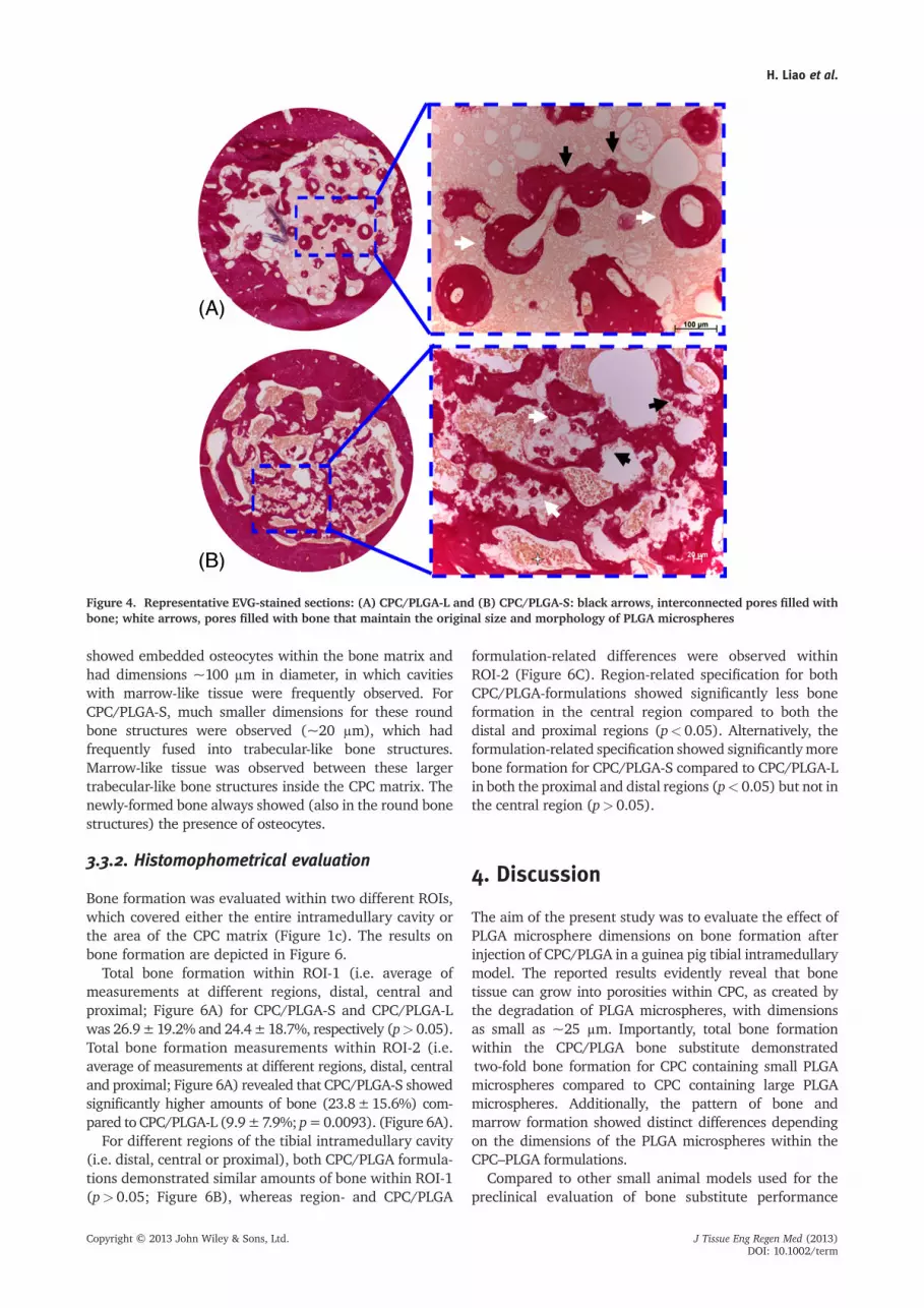
showed embedded osteocytes within the bone matrix andhad dimensions ~100 μm in diameter, in which cavitieswith marrow-like tissue were frequently observed. ForCPC/PLGA-S, much smaller dimensions for these roundbone structures were observed (~20 μm), which hadfrequently fused into trabecular-like bone structures.Marrow-like tissue was observed between these largertrabecular-like bone structures inside the CPC matrix. Thenewly-formed bone always showed (also in the round bonestructures) the presence of osteocytes.
3.3.2. Histomophometrical evaluation
Bone formation was evaluated within two different ROIs,which covered either the entire intramedullary cavity orthe area of the CPC matrix (Figure 1c). The results onbone formation are depicted in Figure 6.
Total bone formation within ROI-1 (i.e. average ofmeasurements at different regions, distal, central andproximal; Figure 6A) for CPC/PLGA-S and CPC/PLGA-Lwas 26.9±19.2% and 24.4±18.7%, respectively (p> 0.05).Total bone formation measurements within ROI-2 (i.e.average of measurements at different regions, distal, centraland proximal; Figure 6A) revealed that CPC/PLGA-S showedsignificantly higher amounts of bone (23.8±15.6%) com-pared to CPC/PLGA-L (9.9±7.9%; p=0.0093). (Figure 6A).
For different regions of the tibial intramedullary cavity(i.e. distal, central or proximal), both CPC/PLGA formula-tions demonstrated similar amounts of bone within ROI-1(p> 0.05; Figure 6B), whereas region- and CPC/PLGA
formulation-related differences were observed withinROI-2 (Figure 6C). Region-related specification for bothCPC/PLGA-formulations showed significantly less boneformation in the central region compared to both thedistal and proximal regions (p< 0.05). Alternatively, theformulation-related specification showed significantly morebone formation for CPC/PLGA-S compared to CPC/PLGA-Lin both the proximal and distal regions (p< 0.05) but not inthe central region (p> 0.05).
4. Discussion
The aim of the present study was to evaluate the effect ofPLGA microsphere dimensions on bone formation afterinjection of CPC/PLGA in a guinea pig tibial intramedullarymodel. The reported results evidently reveal that bonetissue can grow into porosities within CPC, as created bythe degradation of PLGA microspheres, with dimensionsas small as ~25 μm. Importantly, total bone formationwithin the CPC/PLGA bone substitute demonstratedtwo-fold bone formation for CPC containing small PLGAmicrospheres compared to CPC containing large PLGAmicrospheres. Additionally, the pattern of bone andmarrow formation showed distinct differences dependingon the dimensions of the PLGA microspheres within theCPC–PLGA formulations.
Compared to other small animal models used for thepreclinical evaluation of bone substitute performance
Figure 4. Representative EVG-stained sections: (A) CPC/PLGA-L and (B) CPC/PLGA-S: black arrows, interconnected pores filled withbone; white arrows, pores filled with bone that maintain the original size and morphology of PLGA microspheres
H. Liao et al.
Copyright © 2013 John Wiley & Sons, Ltd. J Tissue Eng Regen Med (2013)DOI: 10.1002/term

(e.g. murine calvaria and femoral epiphysis) (van deWatering et al., 2012; Plachokova et al., 2008), thepresently used model comprises several differences.First, an intramedullary cavity is not a bony site in thesense that the native tissue in that area consists of bone.As such, it cannot be considered as a bone defect, butrather as a bone augmentation model, in which nativebone tissue is activated from the endosteal site followingbone marrow ablation (Amsel et al., 1969; Bab, 1995).Second, the tissue surrounding the site of injection is
different from that in normal bone defect and boneaugmentation models. Whereas in these latter models thesite is surrounded by either bone tissue or soft tissue, theintramedullary cavity is composed of bone marrow and fattissue, while being circumferentially surrounded by corticalbone. Despite this difference with regular bone defectand bone augmentation models, the guinea pig tibialintramedullary model was chosen because it allows thatthe cement can indeed be tested as an injectable materialand also provides large-sized histological specimens, which
Figure 5. Representative H&E-stained histological sections of the proximal region with (A) CPC/PLGA-L and (B) CPC/PLGA-S. Lowerpanels are magnifications of the boxes indicated in the image above. Yellow and black asterisks indicate the presence of marrow-liketissue and trabecular-like bone structures containing osteocytes, respectively; black arrows, remnants of CPC
Influence of scaffold pore size in new bone formation
Copyright © 2013 John Wiley & Sons, Ltd. J Tissue Eng Regen Med (2013)DOI: 10.1002/term

can be subjected to multiple approaches for analysis (i.e.embedding in both PMMA and paraffin of alternatetibial halves).
Regarding the quantification of CPC/PLGA degrada-tion, the intramedullary model has several limitations.For a reliable assessment, it is mandatory to achievecomplete filling and setting of the injectable CPC/PLGAbone substitute. Within a secluded cavity connected toliquid areas (i.e. bone marrow) proximally and distally,it remains speculative if that can be achieved. It needs tobe emphasized that, for regular bone defect models, thedefect is maximally dried to avoid interference of bodilyfluids with filling and setting (Félix Lanao et al., 2012;Oortgiesen et al., 2013).
Examination of the PMMA sections revealed that bothCPC/PLGA formulations were still present within theintramedullary cavity at the end of the 12 weeks implan-tation time. Due to the high density of the CPC/PLGAcomposite material, the relative thickness of PMMAsections and artifacts caused by staining, analysis ofbone formation using PMMA embedding is inaccurate(Lopez-Heredia et al., 2012b). In contrast, H&E and EVGstaining of decalcified paraffin sections indicated thepresence of abundant bone formation within the CPCmatrix. As such, these results demonstrate the importanceof combined analysis (calcified/decalcified) for a completeunderstanding of the two processes of material degrada-tion and bone formation.
The quantitative assessment of bone formation showedno differences in total bone formation within the tibialintramedullary cavity related to PLGAmicrosphere dimen-sions. However, within the CPC matrix, bone formation ofCPC/PLGA with small PLGA microspheres was> two-foldhigher compared to CPC/PLGA with large PLGA micro-spheres. Further specification of this difference showedthat especially the proximal and distal regions of the tibialintramedullary cavity contributed to this result. This effectis likely to be caused by bone marrow left at the proximaland distal ends of the intramedullary cavity after theablation procedure. This remaining bone marrow couldhave acted as an initiator for new bone formation.
Review of previously performed studies indicate that noconclusive statement can be made about the scaffoldrequirements (porosity, composition, etc.) to allow boneingrowth. For example, in vivo studies with ectopic im-plantation of preset CPC/PLGA scaffolds have shown thatsoft tissue ingrowth throughout the scaffold is feasiblewith PLGA microsphere sizes> 50 μm incorporated at20 wt% (Link et al., 2008). Alternatively, the assessmentof bone ingrowth into biomaterials with different composi-tions (i.e. sintered hydroxyapatite with ‘cellular’ or ‘strut-like’ designs) and porosity characteristics (i.e. porosity32–56%, pore radius 71–167 μm) showed that pore sizewas strongly correlated to bone ingrowth, with a strongenhancement of bone ingrowth when pore diametersexceeded 100 μm (Jones et al., 2007). Also, one of ourrecent studies indicated that a CPC/PLGA formulationwith 30 wt% PLGA microspheres of 40 μm would beoptimal regarding mechanical properties, porosity andinterconnectivity (Lopez-Heredia et al., 2012a). This in-consistency in observations is confirmed by the presentstudy, which shows that bone ingrowth throughout thematerial was obtained for an injectable CPC/PLGA formu-lation with 20 wt% PLGA microspheres of ~25 μm. Thisformulation even showed significantly more bone forma-tion compared to the CPC/PLGA formulation with 20 wt%PLGA microspheres of ~100 μm, while formulationshad a similar total porosity (~67%) to which thePLGA equally contributed (i.e. macroporosity of ~44%).In contrast with previous studies, which reported that smallpore sizes (40–80 μm) led to less bone formation thanlarger pore sizes (200–400 μm) (Bobyn et al., 1980; Galoisand Mainard 2004), it seems straightforward to conclude
Figure 6. Quantification of bone formation. (A) total boneformation in ROI-1 (i.e. average of all different levels at whichsections were made, distal, central and proximal) and total boneformation in ROI-2 (i.e. average of all the different levels atwhich sections were made, distal, central and proximal). (B) Boneformation in ROI-1 at the different levels. (C) Bone formation inROI-2 at the different levels
H. Liao et al.
Copyright © 2013 John Wiley & Sons, Ltd. J Tissue Eng Regen Med (2013)DOI: 10.1002/term

that a higher pore interconnectivity of the CPC/PLGA-Smaterial caused the currently observed favourable effecton bone ingrowth. However, it has to be emphasized that,in addition to the created macroporosity due to PLGAdegradation, CPC/PLGA possesses an intrinsic nanoporosity.Therefore, the majority of CPC/PLGA pores are additionallyinterconnected via this nanoporosity, which allows forenhanced fluid flow and nutrient circulation within thematerial. At the same time, the fluid flow circulation canaccelerate the transformation as well as degradation of theCPC, resulting in more space for tissue ingrowth.
Within the tibial intramedullary cavity. the response toseveral ceramic bone substitutes has previously beendescribed to follow a pattern from endosteal bone healingvia marrow cavity remodelling to marrow restoration(Schwartz et al., 2008). The results of the present studynot only show an effect of remaining bone marrow on boneformation, but also demonstrate that marrow regenerationoccurred within the CPC matrix. Interestingly, the locationof regenerated bone marrow was dependent on the sizeof the PLGA microparticles: whereas large PLGA micro-spheres evoked bone formation in round patterns andmarrow in the centre, small PLGA microparticles appearedto give rise to trabecular-like bone formation with bonemarrow present between such trabeculae. As describedin the literature, anatomical restrictions necessitatespecific dimensions (i.e. porosity) to allow bone marrowand blood vessel formation (Mastrogiacomo et al., 2006;Sicchieri et al., 2012). However, this study provides suffi-cient evidence that new bone formation occurs throughouta CPC matrix with pores of 25 μm in diameter. Therefore, itcan be hypothesized that CPC nanoporosity, in combinationwith the porosity generated after PLGA degradation,provides sufficient space for nutrient diffusion to allowbone cells to survive, with the presence of bone marrowbut without the formation of new blood vessels, untilfurther degradation of the CPC matrix. This observationclosely resembles the situation that occurs in naturewithin the cortical bone structures, where the Haversiansystem contains blood vessels only in the Volkmann’scanal. Nutrients diffuse out from the vessel within these
canals to the osteocytes in the Haversian system vianano-sized canaliculi.
5. Conclusion
The results of the present study demonstrate that thedimensions of PLGA microspheres within injectableCPC/PLGA have a substantial effect on bone formation.Whereas both small (~25 μm) and large (~100 μm)microspheres allowed bone ingrowth throughout theCPC matrix, a> two-fold higher amount of newly formedbone was observed for CPC/PLGA with small PLGAmicrospheres within the CPC matrix. Additionally, thepattern of bone and marrow formation showed distinctdifferences related to PLGA microsphere size. In general,this study demonstrates that PLGA microsphere dimen-sions of ~25 μm, leading to pores of ~25 μm withinCPC, are sufficient for bone ingrowth and allow substan-tial bone formation. Further, the results demonstratethat PLGA microsphere dimensions provide a tool tocontrol bone formation for injectable CPC/PLGA bonesubstitutes.
Conflict of interest
The authors have declared that there is no conflict ofinterest.
Acknowledgements
The authors would like to thank Natasja van Dijk for histolog-ical assistance. Scanning electron microscopy was performedat the Microscope Imaging Centre (MIC) of the NijmegenCentre for Molecular Life Sciences (NCMLS). The authorsgratefully acknowledge the support of the SmartMixProgramme of The Netherlands Ministry of Economic Affairsand The Netherlands Ministry of Education, Culture andScience.
References
Amsel S, Maniatis A, Tavassoli M, et al.1969; The significance of intramedullarycancellous bone formation in the repairof bone marrow tissue. Anat Rec 164(1):101–111.
Bab IA. 1995; Postablation bone marrowregeneration: an in vivo model tostudy differential regulation of boneformation and resorption. Bone 17(4):437–441.
Bobyn JD, Pilliar RM, Cameron HU, et al.1980; The optimum pore size for thefixation of porous-surfaced metal implantsby the ingrowth of bone. Clin Orthop RelRes 150: 263–270.
Bongio M, van den Beucken JJJP,Leeuwenburgh SCG, et al. 2010; Develop-ment of bone substitute materials: from
’biocompatible’ to ’instructive’. J MaterChem 20(40): 8747–8759.
Bohner M 2010; Design of ceramic-basedcements and putties for bone graft substi-tution. Eur Cells Mater 20: 1–12.
Bohner M, Baumgart F. 2004; Theoreticalmodel to determine the effects of geomet-rical factors on the resorption of calciumphosphate bone substitutes. Biomaterials25(17): 3569–3582.
Brown WE, Chow LC. 1986; A new calciumphosphate water-setting cement. In CementsResearch Progress, Brown PW (ed.). AmericanCeramic Society: Westerville, OH: USA.
del Real RP, Wolke JG, Vallet-Regi M, et al.2002; A newmethod to producemacroporesin calcium phosphate cements. Biomaterials23(17): 3673–3680.
Dorozhkin SV. 2010; Bioceramics ofcalcium orthophosphates. Biomaterials 31(7): 1465–1485.
Félix Lanao RP, Leeuwenburgh SCG, WolkeJGC, et al. 2011a; In vitro degradation rateof apatitic calcium phosphate cement withincorporated PLGA microspheres. ActaBiomater 7(9): 3459–3468.
Félix Lanao RP, Leeuwenburgh SCG, WolkeJGC, et al. 2011b; In vivo bone response tofast-degrading, injectable calcium phosphatecements with enhanced in situ formingporosity. Biomaterials 32(34): 8839–8847.
Félix Lanao RP, Hoekstra JW, Wolke JG, et al.2012; Porous calcium phosphate cementfor alveolar bone regeneration. J TissueEng Regen Med. doi: 10.1002/term.1546.[Epub ahead of print]
Influence of scaffold pore size in new bone formation
Copyright © 2013 John Wiley & Sons, Ltd. J Tissue Eng Regen Med (2013)DOI: 10.1002/term

Galois L, Mainard D. 2004; Bone ingrowthinto two porous ceramics with differentpore sizes: an experimental study. ActaOrthop Belg 70(6): 598–603.
Habraken WJ, Wolke JG, Mikos AG, et al.2006; Injectable PLGAmicrosphere/calciumphosphate cements: physical properties anddegradation characteristics. J Biomater SciPolym Ed 17(9): 1057–1074.
Jones AC, Arns CH, Sheppard AP, et al. 2007;Assessment of bone ingrowth into porousbiomaterials using micro-CT. Biomaterials28(15): 2491–2504.
Kolk A, Handschel J, Drescher W, et al. 2012;Current trends and future perspectivesof bone substitute materials – fromspace holders to innovative biomaterials.J Craniomaxillofac Surg 40(8): 706–718.
Kretlow JD, Young S, Klouda L, et al. 2009;Injectable biomaterials for regeneratingcomplex craniofacial tissues. Adv Mater21(32–33): 3368–3393.
Liao H, Walboomers XF, Habraken WJ, et al.2011; Injectable calcium phosphate cementwith PLGA, gelatin and PTMCmicrospheresin a rabbit femoral defect. Acta Biomater 7(4): 1752–1759.
Link DP, van den Dolder J, van den BeuckenJJJP, et al. 2008; Evaluation of the biocom-patibility of calcium phosphate cement/PLGA microparticle composites. J BiomedMater Res A 87(3): 760–769.
Lopez-Heredia MA, Sariibrahimoglu K, YangW, et al. 2012a; Influence of the poregenerator on the evolution of the mechan-ical properties and the porosity and inter-connectivity of a calcium phosphatecement. Acta Biomater 8(1): 404–414.
Lopez-Heredia MA, Bongio M, Cuijpers VM,et al. 2012b; Bone formation analysis: ef-fect of quantification procedures on thestudy outcome. Tissue Eng C Methods 18(5): 369–373.
Lu JX, Flautre B, Anselme K, et al. 1999; Roleof interconnections in porous bioceramicson bone recolonization in vitro andin vivo. J Mater Sci Mater Med 10(2):111–120.
Mastrogiacomo M, Scaglione S, Martinetti R,et al. 2006; Role of scaffold internal structureon in vivo bone formation in macroporouscalcium phosphate bioceramics. Biomaterials27(17): 3230–3237.
Oortgiesen DA, Meijer GJ, Bronckers AL,et al. 2013; Regeneration of theperiodontium using enamel matrix deriva-tive in combination with an injectable bonecement. Clin Oral Invest 17(2): 411–421.
Plachokova AS, van den Dolder J, Jansen JA.2008; The bone-regenerative properties ofEmdogain adsorbed onto poly(D,L-lactic-coglycolic acid)/calcium phosphate com-posites in an ectopic and an orthotopicrat model. J Periodont Res 43(1): 55–63.
Schwartz Z, Doukarsky-Marx T, Nasatzky E,et al. 2008; Differential effects of bonegraft substitutes on regeneration of bonemarrow. Clin Oral Implant Res 19(12):1233–1245.
Sicchieri LG, Crippa GE, de Oliveira PT, et al.2012; Pore size regulates cell and tissue in-teractions with PLGA–CaP scaffolds usedfor bone engineering. J Tissue Eng RegenMed 6(2): 155–162.
Tamimi F, Sheikh Z, Barralet J. 2012;Dicalcium phosphate cements: brushiteand monetite. Acta Biomater 8(2):474–487.
van de Watering FC, van den Beucken JJ,Walboomers XF, et al. 2012; Calciumphosphate/poly(D,L-lactic-co-glycolic acid)composite bone substitute materials:evaluation of temporal degradation andbone ingrowth in a rat critical-sized cra-nial defect. Clin Oral Implant Res 23(2):151–159.
von Doernberg MC, von Rechenberg B,Bohner M, et al. 2006; In vivo behavior ofcalcium phosphate scaffolds with fourdifferent pore sizes. Biomaterials 27(30):5186–5198.
Zuo Y, Yang F, Wolke JG, Li Y, et al. 2010;Incorporation of biodegradable electrospunfibers into calcium phosphate cement forbone regeneration. Acta Biomater 6(4):1238–1247.
H. Liao et al.
Copyright © 2013 John Wiley & Sons, Ltd. J Tissue Eng Regen Med (2013)DOI: 10.1002/term