bacteria microencapsulation in plga microdevices by supercritical emulsion extraction
TRANSCRIPT

(This is a sample cover image for this issue. The actual cover is not yet available at this time.)
This article appeared in a journal published by Elsevier. The attachedcopy is furnished to the author for internal non-commercial researchand education use, including for instruction at the authors institution
and sharing with colleagues.
Other uses, including reproduction and distribution, or selling orlicensing copies, or posting to personal, institutional or third party
websites are prohibited.
In most cases authors are permitted to post their version of thearticle (e.g. in Word or Tex form) to their personal website orinstitutional repository. Authors requiring further information
regarding Elsevier’s archiving and manuscript policies areencouraged to visit:
http://www.elsevier.com/copyright

Author's personal copy
J. of Supercritical Fluids 63 (2012) 1– 7
Contents lists available at SciVerse ScienceDirect
The Journal of Supercritical Fluids
jou rn al h om epage: www.elsev ier .com/ locate /supf lu
Bacteria microencapsulation in PLGA microdevices by supercriticalemulsion extraction
G. Della Portaa,b,∗, F. Castaldoa, M. Scognamiglioa, L. Pacielloa, P. Parascandolaa, E. Reverchona
a Dipartimento di Ingegneria Industriale, Università di Salerno, Via Ponte don Melillo, Fisciano (SA), Italyb Laboratorio di Ingegneria Cellulare e Molecolare, DEIS (sede di Cesena), Università di Bologna, Via Venezia 52, Cesena (FC), Italy
a r t i c l e i n f o
Article history:Received 13 October 2011Received in revised form19 December 2011Accepted 20 December 2011
Keywords:Supercritical fluidEmulsionMicrodevicesPLGABacteria encapsulation
a b s t r a c t
Cell microencapsulation continues to hold significant promise for biotechnology and medicine and isconsidered an important tool for tissue engineering. Encapsulated cells would also provide a source ofsustained continuous release of therapeutic products for longer durations at the site of implantation.The present work investigates the possibility of prokaryotic cells microencapsulation by SupercriticalEmulsion Extraction (SEE) technology; Lactobacillus acidophilus was selected as a model bacterium andpoly-lactic-co-glycolic acid (PLGA, 75:25) was chosen as biopolymer because FDA approved in devices forbiomedical applications.
A double emulsion (w1-o-w2 ratio 2:18:80) was used with an internal water phase (w1) compositionof L. acidophilus suspended in MRS broth plus the 0.4% of poly-vinyl alcohol (PVA), as surfactant; the bestoverall cell mass content was found to be not higher than 10 mg/mL (that correspond to 7.5 × 10⁶ UFC/mL).Other emulsion phases were: o-phase containing ethyl acetate (EA) and PLGA at 10% (w/w) and w2-phaseof water plus 0.6% of PVA (w/w). This emulsion treated by SEE at 90 bar and 37 ◦C for 30 min allowed theformation of PLGA microcapsules with a mean size of 20 �m (±10 �m) loaded with the 0.6% (w/w) ofmicroorganism with an excellent encapsulation efficiency (80%).
Size and morphology of the produced microdevices were monitored by laser scattering and by SEM-EDX analyses and confirmed SEE as an innovative and efficient encapsulation technology. Particularly, themicrospheres were constituted by a PLGA wall containing the cells entrapped into the polymeric matrix.Cell viability less than 5% (w/w) with respect to the loaded microorganism was also evaluated; neverthe-less the biodegradable microdevice produced may be particularly interesting for several biotechnologicalapplication in which mainly the killed vectors is used as bioactive signal delivery.
© 2011 Elsevier B.V. All rights reserved.
1. Introduction
Cell microencapsulation in biodegradable devices is consideredan important challenge for tissue engineering and regenerativemedicine [1]. Indeed, cells targeting problems (live or death) canbe overcame by their delivering using biopolymer microcapsuleswhich can protect the encapsulated materials from the harsh exter-nal environments [2,3] or address it to a more specific target[4–6]. Microspheres loaded with cells have been also proposed asactivate biopolymer scaffolds for tissue engineering applications[7,8] or as immunoprotected implants for cell-based therapy [9].Encapsulated cells may also provide a source of sustained contin-uous release of therapeutic products for longer durations at thesite of implantation [10]. For example, bacteria can be genetically
∗ Corresponding author at: Dept. of Industrial Engineering, University of Salerno,Via Ponte Don Melillo, 84084 Fisciano (Sa), Italy. Tel.: +39 089 964104;fax: +39 089 964057; mobile: +39 320 7979003.
E-mail address: [email protected] (G. Della Porta).
engineered to synthesize products with high therapeutic poten-tial and used as a potential delivery vector for various antigensor therapeutic and immunomodulatory proteins [11]. In vitro orwith animal models studies demonstrated that Lactococcus lactishas emerged as potential delivery vector of different antigens and,in some specific cases, also the killed bacterium can be an inter-esting vector capable to produce comparable responses to thoseelicited by live bacteria [12]. Although native L. lactis is a gener-ally recognized as safe (GRAS), the genetically modified versionshave to be assessed for safety and biological containment. Fromthis perspective, the use of dead L. lactis vectors seems to be asafer option if the efficacy is retained, because there is no risk ofspreading recombinant DNA in the environment.
Several technologies have been tested for the production ofbacteria loaded microdevices, such as spray-drying, extrusion,phase separation and solvent extraction/evaporation of emulsions.Almost all of these technologies were tested on probiotics bacte-ria mainly to develop probiotic health based product. However,none of these reported methods has resulted in a large num-ber of shelf-stable cells necessary for use in industry [13]. Mainly
0896-8446/$ – see front matter © 2011 Elsevier B.V. All rights reserved.doi:10.1016/j.supflu.2011.12.020

Author's personal copy
2 G. Della Porta et al. / J. of Supercritical Fluids 63 (2012) 1– 7
problems of low encapsulation efficiency and reduction of cell via-bility were reported [14,15]. For example, the use of spray dryingfor Lactobacilli and Bifidobacteria microencapsulation was reportedfor a number of different strains [16,17] including Lactobacillusacidophilus [18]; however, most of the bacteria do not surviveat temperature or osmotic extremes to which they are exposedduring the spray drying or emulsification process and, in severalcases, reduced encapsulation efficiency was also reported. A dif-ferent cell coating technique was proposed by Picot and Lacroix[19,20] using an emulsion containing milk fat droplets and wheyprotein polymers with freeze-dried bacteria, then spray dried.The direct dispersion of fresh cells in a heat-treated whey pro-tein suspension followed by spray drying was suggested as theless destructive microencapsulation method, with survival ratesof 26% for Bifidobacter breve and 1.4% for the more heat-sensitiveBifidobacter longum [21]. Several biodegradable polymer matrixeshave been also tested for bacteria encapsulation such as: alginatesystems [22], cellulose acetate phthalate [23], proteins and polysac-charide mixtures [24], chitosan [25]. Poly-lactic-co-glycolic acid(PLGA) was never used until now because almost all biopolymermatrix reported above were selected for food industry applications;whereas, the encapsulation into PLGA microdevices may be muchmore interesting for biotechnological and biomedical applications[26].
The most common method of PLGA microdevices preparationis the solvent evaporation/extraction of emulsions; this technol-ogy may require elevate temperatures or reduced pressures andlong processing times (several hours), that may produce parti-cles aggregation, as well as, cells damage [27]. Supercritical fluidtechnologies were also proposed to produce PLGA microdevices.Particularly, RESS technology showed problems of low solubility ofalmost all PLGA co-polymers in SC-CO2 that will prevent affordableprocess yields [28]; whereas, the even low solubility of SC-CO2 inthe PLGA is again the main problem when using the SAS technologybecause it causes the precipitation of large polymer aggregates [29].More recently, Bifidobacteria encapsulation into PVA/PVA complexmatrix by supercritical fluid was also reported followed by a millingof the obtained complex [30].
Supercritical Emulsion Extraction (SEE) technology was recentlyproposed for the production of biopolymer microspheres by severalauthors from oil-in-water emulsions [31,32]. The authors reporteda better control of microspheres sizes and distributions, as well as,excellent drug loading achieved by SEE with respect to the conven-tional extraction/evaporation technology; the innovative processwas also characterized by mild temperature conditions. The verygood process performances were justified considering supercrit-ical fluid properties such as lower viscosity and higher diffusivitythat will improve the mass transfer and reduce the processing timesduring the extraction of the oily dispersed phase [33,34].
The present work aims to investigate the possible use of SEEtechnology, as a low impact method, for bacterial cells encap-sulation into PLGA microdevices intended for biomedical andpharmaceutical use. L. acidophilus was selected as a model bac-terium because of its high sensitivity to temperature or osmoticvariations. Our aim was also to explore SEE process conditionsthat may allow high encapsulation efficiency of intact bacteriaeven with low cell viability, considering that genetically modi-fied bacteria may eventually be also used as antigen vectors, alsowhen dead. In this sense, double water–oil–water emulsions withdifferent internal water phase compositions and biopolymer con-centrations in the oily phase were tested to monitor the effectsof these parameters on droplets stability, microspheres morphol-ogy and cells loading. The SEE process parameters like operatingpressure and temperature, flow rate and contacting time betweenthe emulsion and supercritical carbon dioxide (SC-CO2) were alsostudied with respect to microsphere size distribution and to cells
encapsulation efficiency. A comparative study between the charac-teristics of the microspheres obtained by SEE and those producedby conventional solvent evaporation (SE) was also proposed. Cellsdispersion and viability into microspheres were also monitored.
2. Experimental methods
2.1. Materials
CO2 (99.9% SON Naples, Italy), polyvinyl alcohol (PVA, mol. wt.:30,000–55,000, Aldrich Chemical Co.), ethyl acetate (EA, purity99.9%, Aldrich Chemical Co.), poly (lactic/glycolic) acid (PLGA,85:15 mol. wt.; 20,000–60,000 Aldrich Chemical Co.) were used asreceived. L. acidophilus ATCC 43121 was grown at 37 ◦C for 24 h inErlenmayer flasks containing 200 ml MRS Broth (BD 288130), usingaerobic conditions.
2.2. Biomass production
To obtain viable cells of L. acidophilus, two-step cell propaga-tion was set up in 500 mL Erlenmeyer flasks, incubated at 37 ◦Cand 150 rpm in an Orbital Incubator (Stuart Scientific S150). Inthe first step, 1 mL of a frozen culture (stored at −80 ◦C in 20.0%,v/v glycerol) was propagated in 200 mL MRS broth. During expo-nential growth, an aliquot of broth culture was withdrawn to beemployed as inoculum in the second step propagation flasks. Inthe latter case, L. acidophilus cells were allowed to grow for 24 hin 200 mL MRS broth to achieve a viable biomass suspension hav-ing an optical density of 1.8 at 590 nm. Beyond this OD value, cellconglomeration phenomena begin to occur, which reduce cell via-bility. The obtained biomass was then collected by centrifugationat 6500 rpm for 20 min and re-suspended in fresh MRS broth to beprocessed and entrapped in PLGA matrix.
2.3. Emulsions preparation
Different double emulsions were prepared to optimize thecomposition of each phase: 5 or 10 mg/mL of live cells were pre-suspended into MRS broth/PVA solution or aqueous PVA solution;a fixed amount of these suspensions was then added into EA/PLGAsolutions and sonicated for 2 min (Digital Sonifer Branson mod.450). This primary emulsion was added into a known amount ofaqueous PVA solution to form the secondary emulsion using a high-speed stirrer (mod. L4RT Silverson Machines Ltd., United Kingdom)operating at 800 rpm for 3 min at 10 ◦C, controlled using an ice bath.
2.4. Microspheres preparation by conventional solventevaporation
EA was evaporated from the emulsions for 60 min at 30 ◦Cunder controlled and mild vacuum (170 mmHg, rotating evapora-tor) under moderate stirring. During the evaporation, the emulsionswere swept by a continuous nitrogen flow at constant flow rate(70 L/h).
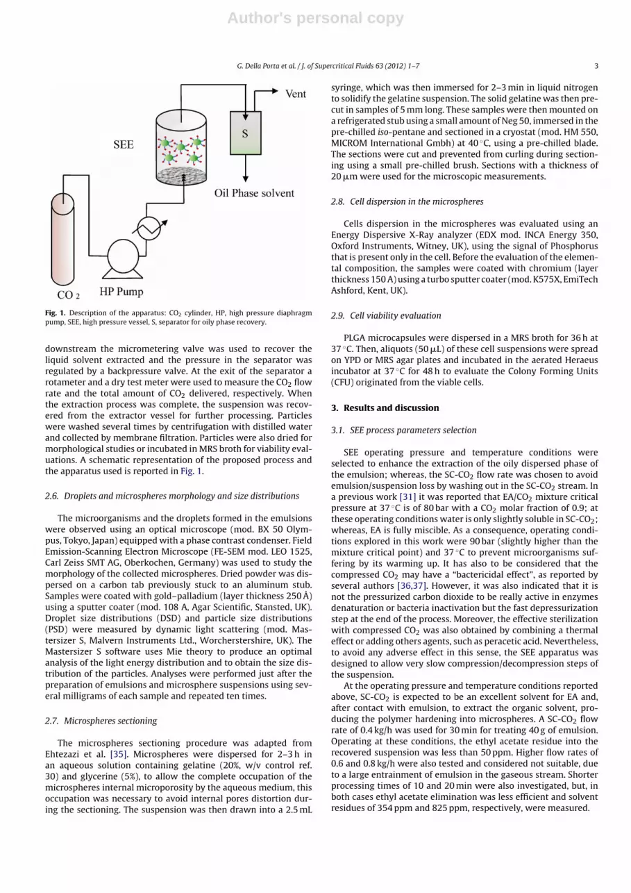
2.5. SEE apparatus: description and procedure
In a typical experiment, 40 g of emulsion were placed into the0.25 dm3 cylindrical stainless steel vessel. SC-CO2 was deliveredusing a high pressure diaphragm pump (Milton Roy, model MilroyalB, Point Saint Pierre, France) and was bubbled into the extractionvessel at a constant flow rate (0.1–0.5 kg/h), through a cylindri-cal stainless steel dispenser located at the bottom of the extractor.The cylindrical dispenser maximizes the contact between the twophases during the extraction. Temperature was maintained con-stant using an air-heated thermostated oven. A separator located
Author's personal copy
G. Della Porta et al. / J. of Supercritical Fluids 63 (2012) 1– 7 3
Fig. 1. Description of the apparatus: CO2 cylinder, HP, high pressure diaphragmpump, SEE, high pressure vessel, S, separator for oily phase recovery.
downstream the micrometering valve was used to recover theliquid solvent extracted and the pressure in the separator wasregulated by a backpressure valve. At the exit of the separator arotameter and a dry test meter were used to measure the CO2 flowrate and the total amount of CO2 delivered, respectively. Whenthe extraction process was complete, the suspension was recov-ered from the extractor vessel for further processing. Particleswere washed several times by centrifugation with distilled waterand collected by membrane filtration. Particles were also dried formorphological studies or incubated in MRS broth for viability eval-uations. A schematic representation of the proposed process andthe apparatus used is reported in Fig. 1.
2.6. Droplets and microspheres morphology and size distributions
The microorganisms and the droplets formed in the emulsionswere observed using an optical microscope (mod. BX 50 Olym-pus, Tokyo, Japan) equipped with a phase contrast condenser. FieldEmission-Scanning Electron Microscope (FE-SEM mod. LEO 1525,Carl Zeiss SMT AG, Oberkochen, Germany) was used to study themorphology of the collected microspheres. Dried powder was dis-persed on a carbon tab previously stuck to an aluminum stub.Samples were coated with gold–palladium (layer thickness 250 A)using a sputter coater (mod. 108 A, Agar Scientific, Stansted, UK).Droplet size distributions (DSD) and particle size distributions(PSD) were measured by dynamic light scattering (mod. Mas-tersizer S, Malvern Instruments Ltd., Worcherstershire, UK). TheMastersizer S software uses Mie theory to produce an optimalanalysis of the light energy distribution and to obtain the size dis-tribution of the particles. Analyses were performed just after thepreparation of emulsions and microsphere suspensions using sev-eral milligrams of each sample and repeated ten times.
2.7. Microspheres sectioning
The microspheres sectioning procedure was adapted fromEhtezazi et al. [35]. Microspheres were dispersed for 2–3 h inan aqueous solution containing gelatine (20%, w/v control ref.30) and glycerine (5%), to allow the complete occupation of themicrospheres internal microporosity by the aqueous medium, thisoccupation was necessary to avoid internal pores distortion dur-ing the sectioning. The suspension was then drawn into a 2.5 mL
syringe, which was then immersed for 2–3 min in liquid nitrogento solidify the gelatine suspension. The solid gelatine was then pre-cut in samples of 5 mm long. These samples were then mounted ona refrigerated stub using a small amount of Neg 50, immersed in thepre-chilled iso-pentane and sectioned in a cryostat (mod. HM 550,MICROM International Gmbh) at 40 ◦C, using a pre-chilled blade.The sections were cut and prevented from curling during section-ing using a small pre-chilled brush. Sections with a thickness of20 �m were used for the microscopic measurements.
2.8. Cell dispersion in the microspheres
Cells dispersion in the microspheres was evaluated using anEnergy Dispersive X-Ray analyzer (EDX mod. INCA Energy 350,Oxford Instruments, Witney, UK), using the signal of Phosphorusthat is present only in the cell. Before the evaluation of the elemen-tal composition, the samples were coated with chromium (layerthickness 150 A) using a turbo sputter coater (mod. K575X, EmiTechAshford, Kent, UK).
2.9. Cell viability evaluation
PLGA microcapsules were dispersed in a MRS broth for 36 h at37 ◦C. Then, aliquots (50 �L) of these cell suspensions were spreadon YPD or MRS agar plates and incubated in the aerated Heraeusincubator at 37 ◦C for 48 h to evaluate the Colony Forming Units(CFU) originated from the viable cells.
3. Results and discussion
3.1. SEE process parameters selection
SEE operating pressure and temperature conditions wereselected to enhance the extraction of the oily dispersed phase ofthe emulsion; whereas, the SC-CO2 flow rate was chosen to avoidemulsion/suspension loss by washing out in the SC-CO2 stream. Ina previous work [31] it was reported that EA/CO2 mixture criticalpressure at 37 ◦C is of 80 bar with a CO2 molar fraction of 0.9; atthese operating conditions water is only slightly soluble in SC-CO2;whereas, EA is fully miscible. As a consequence, operating condi-tions explored in this work were 90 bar (slightly higher than themixture critical point) and 37 ◦C to prevent microorganisms suf-fering by its warming up. It has also to be considered that thecompressed CO2 may have a “bactericidal effect”, as reported byseveral authors [36,37]. However, it was also indicated that it isnot the pressurized carbon dioxide to be really active in enzymesdenaturation or bacteria inactivation but the fast depressurizationstep at the end of the process. Moreover, the effective sterilizationwith compressed CO2 was also obtained by combining a thermaleffect or adding others agents, such as peracetic acid. Nevertheless,to avoid any adverse effect in this sense, the SEE apparatus wasdesigned to allow very slow compression/decompression steps ofthe suspension.
At the operating pressure and temperature conditions reportedabove, SC-CO2 is expected to be an excellent solvent for EA and,after contact with emulsion, to extract the organic solvent, pro-ducing the polymer hardening into microspheres. A SC-CO2 flowrate of 0.4 kg/h was used for 30 min for treating 40 g of emulsion.Operating at these conditions, the ethyl acetate residue into therecovered suspension was less than 50 ppm. Higher flow rates of0.6 and 0.8 kg/h were also tested and considered not suitable, dueto a large entrainment of emulsion in the gaseous stream. Shorterprocessing times of 10 and 20 min were also investigated, but, inboth cases ethyl acetate elimination was less efficient and solventresidues of 354 ppm and 825 ppm, respectively, were measured.

Author's personal copy
4 G. Della Porta et al. / J. of Supercritical Fluids 63 (2012) 1– 7
Fig. 2. (a) OM image of w1-o-w2 emulsion with ratio of 4:16:80 (microorganismconcentration in w1 of 5 mg/mL). (b) SEM image of PLGA microspheres after SEEprocess operating at 90 bar and 37 ◦C, with a SC-CO2 flow rate of 0.4 kg/h for 20 min.
3.2. Emulsions optimization for L. acidophilus encapsulation
The droplets in the emulsion were produced in the range of10–40 �m to be large enough to encapsulate bacterial cells thatare of about 1–2 �m in size. Different emulsion compositions weretested to find the optimal phase ratios and the optimal concentra-tion of each component. In some preliminary experiments, distilledwater with a PVA concentration of 0.4% was used as the water inter-nal phase, but this emulsion failed in entrapping bacteria inside thedroplets. In addition, monitoring the cell viability, it was observedthat cell survival in distilled water was much reduced (vitalitydecreased to 50% in 24 h). In the subsequent experiments, a MRSbroth was used as the internal water phase, with the same PVAconcentration, as surfactant. The cell mass content charged in theinternal water phase was reduced to 5 mg/mL (that correspond to3.7 × 106 CFU/mL). The oily phase contained 10% (w/w) of PLGAin EA and the external water phase was formed by distilled andsterilized water with the 0.8% of PVA, as surfactant.
Emulsions with different internal water phase ratios, namelyof 4:16:80 and 2:18:20 (w1-o-w2) were also compared. When the4:16:80 ratio was used, the emulsion was unstable as is also illus-trated in the OM image reported in Fig. 2a, in which it is wellvisible the migration of the internal water phase to the externalone. As a consequence, open microcapsules were produced afterthe SEE process and almost all bacteria were recovered outsidethe particles. An example of the produced material is illustratedin the SEM image reported in Fig. 2b. When the w1-o-w2 ratio of2:16:80 was tested, stable droplets were produced, as illustratedin the OM image reported in Fig. 3a, in which spherical droplets
Fig. 3. (a) OM image of w1-o-w2 emulsion with ratio of 2:18:80 (microorganismconcentration in w1 of 5 mg/mL). (b) SEM image of PLGA microspheres after SEEprocess operating at 90 bar and 37 ◦C, with a SC-CO2 flow rate of 0.4 kg/h for 20 min.
that contain cells inside are evident. Drying this emulsion by SEE,spherical microcapsules were obtained, as illustrated in the SEMimage reported in Fig. 3b.
An emulsion with a w1-o-w2 ratio of 2:16:80 was also tested,increasing the L. acidophilus concentration in the internal waterphase to 10 mg/mL (that correspond to 7.5 × 10⁶ CFU/mL); in thiscase, probably due to the higher cells concentration, the bacteriawere extruded from the droplets and confined on the microspheressurface, as can be observed in Fig. 4 where a SEM image of the PLGAmicroparticles obtained is illustrated.
All the emulsions described above were processed also byconventional solvent evaporation (SE) and for all the emulsioncomposition explored the microspheres obtained by SE were char-acterized by an open and irregular morphology, with a largeamount of cells outside the particles. Particularly, when the opti-mized emulsion composition of w1-o-w2 ratio 2:16:80 (with cellcontent in w1 of 5 mg/mL) was treated by SE, mainly empty andconcave particles were produced. An example of these concave andcollapsed microparticles is illustrated in the SEM image reported inFig. 5. The reason of the observed morphologies was well describedby Rosca et al. [38] and it was mainly due to the low stability ofthe double emulsion during the SE treatment. Indeed, the authorsreported that when double emulsions are processed by conven-tional solvent evaporation, since the process takes several hour,the loss of the water internal phase can easily occur, generatingconcave and collapsed shaped particles that are also expected tobe empty.

Author's personal copy
G. Della Porta et al. / J. of Supercritical Fluids 63 (2012) 1– 7 5
Fig. 4. SEM image of PLGA microspheres obtained operating at 90 bar and 37 ◦C, witha SC-CO2 flow rate of 0.4 kg/h for 20 min. They were produced using an emulsion withw1-o-w2 ratio of 2:18:80 and with a microorganism concentration in the internalw1 phase of 10 mg/mL.
3.3. Microcapsule size and bacteria loading
DSD and PSD curves, obtained by laser scattering analysis foreach emulsion and microparticle suspension studied were alsoused, as an indirect method, for encapsulation efficiency evalua-tion. Indeed, a simple bacteria suspension showed a Mean Diameter(MS) of 1.2 �m with a Standard Deviation (SD) of ±0.5 �m, whereas,oily droplets and derived PLGA microspheres where produced withmean sizes between 20 and 50 �m; therefore, when bacteria encap-sulation was not good, bimodal curves were expected. Examples ofparticle size distributions obtained are illustrated in Fig. 6a and b.Particularly, in Fig. 6a is reported the histogram representing thePSD of the microspheres obtained from the not stable emulsionwith w1-o-w2 ratio of 4:16:80 (microorganism concentration in w1of 5 mg/mL) also illustrated in Fig. 2a and, in this case, the presenceof a bacteria suspension not well entrapped into the microspherescan be easily recognized by the typical bimodal evolution of thediameter populations. In Fig. 6b is reported the histogram rep-resenting the PSD of the microspheres obtained from the stableemulsion with w1-o-w2 ratio of 2:18:80 (microorganism concen-tration in w1 of 5 mg/mL) also illustrated in Fig. 3a and, in thiscase, only one peak is present confirming the absence of the freebacteria in suspension. The microspheres produced in this lastcase showed a MD of 20 �m and SD of ±10 �m. This emulsion
Fig. 5. SEM image of PLGA microspheres obtained by the conventional solvent evap-oration process.
1001010.10
5
10
15
20
25
30
35
40a
b
Vol
ume,
%
Diameter, μm
Diameter, μm1001010.1
0
10
20
30
40
50
Vol
ume,
%
Fig. 6. (a) PSD of the microspheres obtained from not stable emulsion (w1-o-w2
ratio: 4:16:80 and cell concentration of 5 mg/mL); (b) PSD of the microspheresobtained from stable emulsion (w1-o-w2 ratio: 2:18:80 and cell concentration ofw1 of 5 mg/mL).
composition was confirmed to be the best for bacteria entrapmentafter SEE processing.
To further evaluate the bacteria spatial distribution inside theparticles, the elemental composition of microspheres was stud-ied by Energy Dispersive X-Ray (EDX) analyzer integrated in theSEM apparatus. Cells contain in a relatively high amount the Phos-phorus (P) that is not present in PLGA; therefore, it can be usedto indicate the location of the microorganism into the microde-vices. In Fig. 7 SEM image of the microspheres with elemental mapsof some constitutive elements (carbonium, oxygen and phospho-rous) are reported. It is evident that phosphorus (mapped in red) isuniformly spread over all the microspheres; i.e., cells should be uni-formly distributed into the microspheres. The EDX relative amountof the phosphorus with respect to the other elements indicates thepresence of almost 0.8% of phosphorus with respect to the overallcarbon atoms identified in the sample confirming the efficiency ofcell loading into microspheres.
Microcapsule internal structure was also investigated by sec-tioning them using a stereological method; then, selected sections

Author's personal copy
6 G. Della Porta et al. / J. of Supercritical Fluids 63 (2012) 1– 7
Fig. 7. EDX image of PLGA microspheres charged with 0.6% (w/w) of cells obtained operating at 90 bar and 37 ◦C, with a SC-CO2 flow rate of 0.4 kg/h for 20 min. Phosphoruswas highlighted in red, carbon in green and oxygen in blue. (For interpretation of the references to color in this figure legend, the reader is referred to the web version of thisarticle.)
were observed by SEM. An example of the SEM image of the sec-tion obtained is illustrated in Fig. 8; in the figure it is possible toobserve the bacteria cells inside the microcapsule. Bacteria viabil-ity was also monitored for these microcapsules by their incubationinto a MRS broth for 36 h; it resulted to be less than 5% (w/w) withrespect to the loaded microorganism.
Fig. 8. SEM images of PLGA microcapsule section obtained using SEE technology.
4. Conclusions and perspectives
The production of spherical microdevices formed by PLGA, con-taining the bacteria entrapped into the polymeric matrix wasdemonstrated to be a possible application of the SEE technologyprocessing a double water–oil–water emulsion with supercriticalcarbon dioxide at 90 bar and 37 ◦C for 30 min. A particle size tar-get of 20 �m as mean size with an optimum percentage of 0.6%(w/w) cell loaded were successfully obtained (encapsulation effi-ciency 80%). Nevertheless very low cell viability was monitoredsuggesting that the described technology can be applied, for exam-ple, in the encapsulation of genetically modified bacteria especiallywhen dead cells must be used in order to retain almost all the ther-apeutically efficacy as biological signals and to avoid any othersadverse effects. However, future perspectives of this work can bethe improvement of bacteria survival through the addition of pro-tectants to the liquid media in which they are retained in emulsionprior to SEE treatment and/or growth promoting factors includingvarious probiotic/prebiotic combinations.
References
[1] G. Orive, R.M. Hernández, A.R. Gascón, R. Calafiore, T.M.S. Chang, P. De Vos,G. Hortelano, D.D. Hunkeler, I. Lacík, J.L. Pedraz, History, challenges and per-spectives of cell microencapsulation, Trends in Biotechnology 22 (2) (2004)88–92.
[2] S. Prakash, M.L. Jones, Artificial cell therapy: new strategies for the therapeuticdelivery of live bacteria, J. Biomedicine and Biotechnology 1 (2005) 44–56.
[3] M.V. Sefton, M.H. May, S. Lahooti, J.E. Babensee, Making microencapsulationwork: conformal coating, immobilization gels and in vivo performance, J. Con-trolled Release 65 (2000) 173–186.

Author's personal copy
G. Della Porta et al. / J. of Supercritical Fluids 63 (2012) 1– 7 7
[4] F. Lim, A.M. Sun, Microencapsulated islets as bioartificial endocrine pancreas,Science 210 (4472) (1980) 908–910.
[5] S. Prakash, T.M. Chang, In vitro and in vivo uric acid lowering by artificialcells containing microencapsulated genetically engineered E. coli DH5 cells,International J. of Artificial Organs 23 (7) (2000) 429–435.
[6] T. Visted, R. Bjerkvig, P.O. Enger, Cell encapsulation technology as a therapeuticstrategy for CNS malignancies, J. Neuro-Oncology 3 (2001) 201–210.
[7] O. Smidsrod, G. Skjakbraek, Alginate as immobilization matrix for cells, Trendsin Biotechnology 8 (1990) 71–78.
[8] M.Singh, B. Sandhu, A. Scurto, C. Berkland, S. Michael, M.S. Detamore,Microsphere-based scaffolds for cartilage tissue engineering: using subcriticalCO2 as a sintering agent, Acta Biomaterialia 6 (1) (2010) 137–143.
[9] W.M. Kuhtreiber, R.P. Lanza, W.L. Chick, Cell Encapsulation Technology andTherapeutics, Birkhauser, Boston, 1999.
[10] A. Murua, A. Portero, G. Orive, R.M. Hernández, M. de Castro, J.L. Pedraz,Cell microencapsulation technology: toward clinical application, J. ControlledRelease 132 (2008) 76–83.
[11] M. Bahey-El-Din, C.G. Gahan, Lactococcus lactis: from the dairy industry toantigen and therapeutic protein delivery, Discovery Medicine 9 (48) (2010)455–461.
[12] M. Bahey-El-Din, C.G. Gahan, B.T. Griffin, Lactococcus lactis as a cell factory fordelivery of therapeutic proteins, Current Gene Therapy 10 (1) (2010) 34–45.
[13] K. Kailasapathy, Microencapsulation of probiotic bacteria: technology andpotential application, Current Issues of Intestinal Microbiology 3 (2002) 39–48.
[14] A.K. Anal, H. Singh, Recent advances in microencapsulation of probiotics forindustrial applications and targeted delivery, Trends in Food Science and Tech-nology 18 (2007) 240–251.
[15] S.J. Kim, S.J. Cho, S.H. Kim, O.J. Song, S. Shin, D.S. Cha, H.J. Park, Effect of microen-capsulation on viability and other characteristics in Lactobacillus acidophilusATCC 43121, LWT-Food Science and Technology 41 (2008) 493–500.
[16] B.M. Corcoran, R.P. Ross, G.F. Fitzgerald, C. Stanton, Comparative survival of pro-biotic lactobacilli spray-dried in the presence of prebiotic substances, J. AppliedMicrobiology 96 (2004) 1024–1039.
[17] K. O’Riordan, D. Andrews, K. Buckle, P. Conway, Evaluation of microencapsu-lation of a Bifidobacterium strain with starch as an approach to prolongingviability during storage, J. Applied Microbiology 91 (2001) 1059–1066.
[18] Z. Ruixiang, S. Junliang, T. Peter, W. Dahong, N. Shengyang, Measurement ofparticle diameter of Lactobacillus acidophilus microcapsule by spray drying andanalysis on its microstructure, World J. of Microbiology and Biotechnology 24(2008) 1349–1354.
[19] A. Picot, C. Lacroix, Production of multiphase water insoluble microcapsulesfor cell microencapsulation using an emulsification/spray-drying technology,J. Food Science 68 (2003) 2693–2700.
[20] A. Picot, C. Lacroix, Effects of micronization on viability and thermotoleranceof probiotic freeze-dried cultures, International Dairy J. 13 (2003) 455–462.
[21] A. Picot, C. Lacroix, Encapsulation of bifidobacteria in whey protein-basedmicrocapsules and survival in simulated gastrointestinal conditions and inyoghurt, International Dairy J. 14 (2004) 505–515.
[22] K.N. Chen, M.J. Chen, J.R. Liu, C.W. Lin, H.Y. Chiu, Optimization of incorporatedprebiotics as coating materials for probiotic microencapsulation, J. Food Science70 (2005) 260–266.
[23] C.S. Favaro-Trindale, C.R. Grosso, Microencapsulation of L. acidophilus (La-05)and B. lactis (Bb-12) and evaluation of their survival at the pH values of thestomach and in bile, J. Microencapsulation 19 (2002) 485–494.
[24] D. Guerin, J.C. Vuillemard, M. Subirade, Protection of Bifidobacteria encapsu-lated in polysaccharide-protein gel beads against gastric juice and bile, J. FoodProtections 66 (2003) 2076–2084.
[25] S. Lee, D.S. Cha, H.J. Park, Survival of freeze-dried Lactobacillus bulgaricus KFRI673 in chitosan-coated calcium alginate microparticles, J. Agricultural and FoodChemistry 52 (2004) 7300–7305.
[26] D.M. Yoon, J.P. Fisher, Polymeric scaffolds for tissue engineering application,in: Fisher Mikos Bronzino (Ed.), Tissue Engineering, CRC Press Taylor & FrancisGroup, FL, USA, 2007.
[27] M. Li, O. Rouaud, D. Poncelet, Microencapsulation by solvent evaporation. Stateof the art for the process engineering approaches, International J. of Pharma-ceutics 363 (2008) 26–39.
[28] B. Kongsombut, A. Tsutsumi, N. Suankaew, T. Charinpanitkul, Encapsulation ofSiO2 and TiO2 fine powders with poly-lactic-co-glycolic acid by rapid expansionof supercritical CO2 incorporated with ethanol co-solvent, Industrial Engineer-ing & Chemical Research 48 (2009) 11230–11235.
[29] T.J. Young, K.P. Johnston, Encapsulation of lysozyme by precipitation with avapor-over-liquid antisolvent, J. Pharmaceutical Sciences 88 (1999) 640–650.
[30] M.S. Thantsha, J. Guest, I. Mputle, Comparison of different methods for release ofBifidobacterium longum Bb46 from poly(vinylpyrrolidone)-poly(vinylacetate-co-crotonic acid) interpolymer complex matrix and the effect of grinding onthe microparticles, World J. of Microbiology and Biotechnology 27 (10) (2011)2443–2448.
[31] G. Della Porta, E. Reverchon, Nanostructured microspheres produced by super-critical fluid extraction of emulsion, J. Biotechnology and Bioengineering 100(5) (2007) 1033–1929.
[32] P. Chattopadhyay, B.Y. Shekunov, D. Yim, D. Cipolla, B. Boyd, S. Farr, Productionof solid lipid nanoparticle suspensions using supercritical fluid extraction ofemulsions (SFEE) for pulmonary delivery using the AERx system, AdvancedDrug Delivery Reviews 59 (2007) 444–453.
[33] G. Della Porta, N. Falco, E. Reverchon, NSAID drugs release from injectablemicrospheres produced by supercritical fluid emulsion extraction, J. Pharma-ceutical Sciences 99 (2009) 1484–1499.
[34] G. Della Porta, R. Campardelli, N. Falco, E. Reverchon, PLGA microdevices forretinoids sustained release produced by supercritical emulsion extraction:Continuous versus batch operation layouts, J. Pharmaceutical Sciences 100 (10)(2011) 4357–4367.
[35] T. Ehtezazi, C. Washington, C.D. Melia, Determination of the internal morphol-ogy of poly (d,l-lactide) microspheres using stereological methods, J. ControlledRelease 57 (1999) 301–314.
[36] S. Spilimbergo, A. Bertucco, F.M. Lauro, G. Bertoloni, Inactivation of Bacillussubtilis spores by supercritical CO2 treatment, Innovative Food Science andEmerging Technologies 4 (2003) 161–165.
[37] A. White, D. Burns, T.W. Christensen, Effective terminal sterilization usingsupercritical carbon dioxide, J. Biotechnology 123 (2006) 504–515.
[38] I.D. Rosca, F. Watari, M. Uo, Microparticles formation and its mechanism insingle and double emulsion by solvent evaporation, J. Controlled Release 99(2004) 271–280.